Fructooligosacharides reduce Pseudomonas aeruginosa PAO1 pathogenicity through distinct mechanisms
Upload
independentCategory
view
0download
0
GBA2-Encoded b-Glucosidase Activity Is Involved in theInflammatory Response to Pseudomonas aeruginosaNicoletta Loberto2., Maela Tebon1., Ilaria Lampronti3, Nicola Marchetti4, Massimo Aureli2,
Rosaria Bassi2, Maria Grazia Giri5, Valentino Bezzerri1, Valentina Lovato1, Cinzia Cantu1, Silvia Munari1,
Seng H. Cheng6, Alberto Cavazzini4, Roberto Gambari3, Sandro Sonnino2, Giulio Cabrini1,
Maria Cristina Dechecchi1*
1 Laboratory of Molecular Pathology, Department of Pathology and Diagnostics, University Hospital of Verona, Verona, Italy, 2 Department of Medical Biotechnology and
Translational Medicine, University of Milano, Milano, Italy, 3 Department of Life Sciences and Biotechnology, Section of Biochemistry and Molecular Biology, University of
Ferrara, Ferrara, Italy, 4 Department of Chemistry and Pharmaceutical Sciences, University of Ferrara, Ferrara, Italy, 5 Medical Physics Unit, Department of Pathology and
Diagnostics, University Hospital of Verona, Verona, Italy, 6 Genzyme, a Sanofi Company, Framingham, Massachusetts, United States of America
Abstract
Current anti-inflammatory strategies for the treatment of pulmonary disease in cystic fibrosis (CF) are limited; thus, there iscontinued interest in identifying additional molecular targets for therapeutic intervention. Given the emerging role ofsphingolipids (SLs) in various respiratory disorders, including CF, drugs that selectively target the enzymes associated withSL metabolism are under development. Miglustat, a well-characterized iminosugar-based inhibitor of b-glucosidase 2(GBA2), has shown promise in CF treatment because it reduces the inflammatory response to infection by P. aeruginosa andrestores F508del-CFTR chloride channel activity. This study aimed to probe the molecular basis for the anti-inflammatoryactivity of miglustat by examining specifically the role of GBA2 following the infection of CF bronchial epithelial cells by P.aeruginosa. We also report the anti-inflammatory activity of another potent inhibitor of GBA2 activity, namely N-(5-adamantane-1-yl-methoxy)pentyl)-deoxynojirimycin (Genz-529648). In CF bronchial cells, inhibition of GBA2 by miglustat orGenz-529648 significantly reduced the induction of IL-8 mRNA levels and protein release following infection by P.aeruginosa. Hence, the present data demonstrate that the anti-inflammatory effects of miglustat and Genz-529648 are likelyexerted through inhibition of GBA2.
Citation: Loberto N, Tebon M, Lampronti I, Marchetti N, Aureli M, et al. (2014) GBA2-Encoded b-Glucosidase Activity Is Involved in the Inflammatory Response toPseudomonas aeruginosa. PLoS ONE 9(8): e104763. doi:10.1371/journal.pone.0104763
Editor: Dominik Hartl, University of Tubingen, Germany
Received February 14, 2014; Accepted July 16, 2014; Published August 20, 2014
Copyright: � 2014 Loberto et al. This is an open-access article distributed under the terms of the Creative Commons Attribution License, which permitsunrestricted use, distribution, and reproduction in any medium, provided the original author and source are credited.
Funding: This research was supported by Italian Cystic Fibrosis Research Foundation (grant FFC # 14/2012) with the contribution of ‘‘Picasso. Capolavori dalMuseo Nazionale Picasso di Parigi’’, Festa d’Estate Villa Sigurta Verona, Delegazione FFC Lago di Garda e Arezzo. The funders had no role in study design, datacollection and analysis, decision to publish, or preparation of the manuscript.
Competing Interests: Seng H. Cheng is an employee and shareholder of Genzyme, a Sanofi Company, when the work was performed. This work is also part of apatent application titled ‘‘Amorphous and a crystalline form of Genz 112638 hemitartrate as inhibitor of glucosylceramide synthase’’ (International Application No:PCT/US20130137743A1). However, this does not alter the authors’ adherence to all the PLOS ONE policies on sharing data and materials.
* Email: [email protected]
. These authors contributed equally to this work.
Introduction
Cystic fibrosis (CF) lung disease is characterized by progressive
chronic infection and inflammation of the airways. The prolonged
airway inflammation is an important aspect of the obstructive lung
disease noted in CF patients. Resultant progressive remodeling
leads to irreversible damage and fibrosis, which is a major cause of
mortality in patients [1]. Significant efforts have been invested into
developing therapies that address the underlying basis of CF. For
example, recent efforts to identify small-molecule drugs that target
a mutant CF transmembrane conductance regulator (CFTR) led
to the successful development of a potentiator (Kalydeco) for
patients who harbor the mutant G551D-CFTR [2]. Moreover,
phase 3 clinical trials of Kalydeco in combination with the
corrector lumacaftor for people with two copies of the F508del-
CFTR mutation showed significant improvements in lung function
and other key measures of the disease (http://www.cff.org/about
CFFoundation/NewsEvents/2014NEWSArchive/6-24-Vertex
Phase-3-Results_Lumacaftor_Ibvacaftor.cfm). However, despite
these very encouranging results, adjuvant therapies that abate
the decline in pulmonary function in other patients are still
needed. Examples include the potential deployment of new
antibiotics, anti-mucolytic and anti-inflammatory drugs [3]. To
date, the only non-steroidal anti-inflammatory agent that has been
shown to be beneficial in CF patients is ibuprofen; however, its use
can be associated with severe adverse effects, such as gastrointes-
tinal bleeding [4]. Hence, the identification and development of
novel and more potent anti-inflammatory drugs for CF airway
disease remains a priority. The chemokine IL-8 is abundantly
expressed at sites of chronic inflammation and appears to play a
major role in driving the formation of neutrophil (PMN)-rich
exudates in the lungs of CF patients [5–8]; thus its reduction is a
key therapeutic goal in CF.
Sphingolipids (SLs) are a large group of lipids that are thought
to modulate the pathophysiology of several respiratory disorders,
including CF [9–11]. Ceramide, the central hub of SL metabo-
PLOS ONE | www.plosone.org 1 August 2014 | Volume 9 | Issue 8 | e104763

lism, is generated by de novo synthesis or hydrolysis of complex
SLs, such as sphingomyelin (SM) by acid sphingomyelinase (ASM)
and glucosylceramide (GlcCer) by glucocerebrosidases [12].
Ceramide plays an important role in the infection by P.aeruginosa by reorganizing lipid rafts on cellular membranes into
larger signaling platforms, which is a feature conducive to
internalizing bacteria, inducing apoptosis and regulating the
cytokine response [13]. Controversial findings on the association
between abnormalities in SL metabolism and inflammation in CF
have been reported. For example, ceramide has been identified as
a key regulator of inflammation in CF airways in different
CFTR-/- mouse models [14]. In contrast, decreased ceramide
levels have been demonstrated in CFTR KO mice [15], and no
significant difference has been found in basal ceramide levels in
CFTR KO lung homogenates compared to wild type mice [16].
The possible explanation for this discrepancy appears to be the
special diet required for the survival of CFTR KO mice, which
severely affects the concentration of SLs [14]. Interestingly, an
accumulation of ceramide, which has been directly correlated with
neutrophilic lung inflammation, has been demonstrated in the
lower airway of CF patients [17]. These findings suggest that the
CF pathophysiology associated with infection by P. aeruginosacan be corrected, in part, by modulating ceramide levels to their
normal physiological range, independent of the conflicting results
obtained in different CF models. To date, there is some evidence
that supports pharmacological interventions in SL metabolism as
therapeutic agents for CF lung disease [14–21].
Given the emerging importance of SLs in respiratory disorders,
novel drugs that selectively target different enzymes involved in SL
metabolism are under development. Recently developed iminosu-
gar-based inhibitors of GBA2 are of particular interest because of
their good oral bioavailability and specific immune modulatory
and chaperoning activities [22]. A well-characterized inhibitor is
miglustat (N-butyldeoxynojirimycin, NB-DNJ), which is FDA-
approved and EMA-designated for use in Europe and the USA for
the treatment of type I Gaucher and other SL storage diseases. We
previously demonstrated that miglustat exhibits an anti-inflamma-
tory effect in vitro and in vivo by reducing P. aeruginosa induced
immunoreactive ceramide levels [20,23]. Moreover, miglustat can
restore F508del-CFTR chloride channel activity in respiratory and
pancreatic cells in vitro [24,25] and in CF mice [26]. However, a
recent clinical trial in CF patients did not provide evidence of
efficacy, which may be related to the intra-individual variability of
nasal potential difference (NPD) measurements or the short
duration of exposure [27]. Nevertheless, a drug that is able to
correct both the CF channel defect and reduce the inflammatory
response is of interest and warrants further attention. Miglustat
inhibits the enzyme ceramide glucosyl-transferase (GlcCerT),
which catalyzes the first step in the glycosphingolipid biosynthetic
pathway [28] with IC50 values in the low micromolar range. It
also inhibits the activities of two different GlcCer degrading
enzymes, glucocerebrosidase (GBA1) and the non-lysosomal b-
glucosidase 2 (GBA2), with IC50 values in the high micromolar
and nanomolar range, respectively [29,30]. In addition, it is also a
potent inhibitor of a-glucosidase [31]. Therefore, miglustat could
affect the host response to P. aeruginosa through one or more of
these SL metabolism pathways. The galactose analog of miglustat,
N-butyldeoxygalactonojirimycin (NB-DGJ), also inhibits GlcCerT
and GBA2, whereas its effect on GBA1 is less clear [30,32,33].
Similar to miglustat, NB-DGJ produces an anti-inflammatory
effect in bronchial epithelial cells [25], which suggests a potential
involvement of GlcCerT and/or GBA2 in the response of
bronchial cells to P. aeruginosa. The non-lysosomal b-glucosidase
GBA2, which is extremely sensitive to deoxynojirimycin-type
inhibitors [34], is a membrane-associated enzyme located in the
plasma membrane and ER of cells [29,35]. GBA2 has been
described as a single pass transmembrane protein with its catalytic
domain facing the extracellular environment [29]. Because this
enzyme can hydrolyze GlcCer directly at the cell surface, it might
be involved in affecting transient local changes in bioactive SL
concentrations.
To gain greater insights into the molecular basis of the anti-
inflammatory activity of miglustat, we explored the potential
involvement of GBA2 in the ceramide-mediated signaling
processes following P. aeruginosa infection of CF bronchial
epithelial cells. The effects of a potent inhibitor of GBA2, N-(5-
adamantane-1-yl-methoxy)pentyl)- deoxynojirimycin or Genz-
529648 as it is referred to in this report [4,36], on the
inflammatory response to P. aeruginosa were investigated and
compared to miglustat and NB-DGJ. We also examined the
impact of lowering the expression of GBA2 in human CF
bronchial epithelial cells exposed to P. aeruginosa using siRNA
oligonucleotides. The results obtained here demonstrate that
GBA2 is a target of the anti-inflammatory effects of miglustat and
Genz-529648. Thus, these compounds provide novel insights into
the role of GBA2 in the signaling cascade activated by P.aeruginosa in CF bronchial epithelial cells.
Methods
Cell modelsIB3-1 (LGC Promochem GmbH, Teddington, Middlesex,
United Kingdom)[37] and CuFi-1 (a generous gift of A.
Klingelhutz, P. Karp and J. Zabner, University of Iowa, Iowa
City)[38] are human bronchial epithelial cells grown as previously
described [24]. Primary airway epithelial cells, i.e., mainstem
human bronchi, derived from CF individuals were obtained from
‘‘Servizio Colture Primarie’’ of the Italian Cystic Fibrosis Research
Foundation and cultured as previously described [39].
Bacterial strainsThe reference P. aeruginosa strain, PAO1, was kindly provided
by A. Prince (Columbia University, New York) and grown in
trypticase soy broth (TSB) or agar (TSA) (Difco) as previously
described [25]. Some experiments were conducted with organisms
killed by heating to 65uC for 30 minutes.
Inhibitors of SL metabolismMiglustat and NB-DGJ were obtained from Toronto Research
Chemicals, North York, ON, Canada. Genz-529648 was obtained
from Genzyme, a Sanofi Company; amitriptyline was obtained
from Sigma.
Inflammatory response in vitroCells were treated with different inhibitors or solvent alone and
then infected with PAO1 for 4 hours at 37uC as previously
described [25]. The inflammatory response to PAO1 infection was
studied at the transcriptional level by measuring IL-8 chemokine
expression as previously described [20]. An enzyme-linked
immunosorbent assay for the quantitative measurement of IL-8
protein release was performed using the Human IL-8 Instant
ELISA kit (Bender MedSystems, Vienna, Austria).
Cell toxicityThe effects of Genz-529648 on cell proliferation, viability and
apoptosis were studied to evaluate the potential toxicity as detailed
in the Supplement S3.
Role of GBA2 in P. aeruginosa Infected CF Cells
PLOS ONE | www.plosone.org 2 August 2014 | Volume 9 | Issue 8 | e104763

GBA2 silencingTo perform silencing experiments of the GBA2 gene, a
TriFECTa RNAi Kit (Integrated DNA technologies, Coralville,
Iowa, IA) was used. Cells were transiently transfected with specific
siRNA for GBA2 (sequence GGAUCAUGUUUGGAGCUA) or
scrambled (CGUUAAUCGCGUAUAAUACGCGUAT) duplex-
es complexed with cationic liposomes Lipofectamine 2000
(Invitrogen, Carlsbad, CA) diluted in 1 ml serum-free cell culture
medium. GBA2 siRNA or scrambled duplexes (10 nM) were
added and incubated for 10 minutes. Liposome:duplex complexes
(500 mL) were added to the cells grown in 2 cm2 wells and
incubated at 37uC for 6 hours. The cells were washed twice with
culture medium and maintained at 37uC for an additional 18 or
42 hours.
Analysis of cell ceramide contentThe analysis of cell ceramide content was performed via two
different approaches: by the LC-MS and LC-MS/MS method
[40] and by the metabolic labeling of cell SLs using (3H)sphin-
gosine as a precursor. Both methods are detailed in the
Supplement S1 and S2.
Enzymatic activityIB3-1 or CuFi-1 cells were treated with 2 mM miglustat, 10 nM
Genz-529648 or solvent alone for 1 hour and the infected with
heat-killed PAO1 for 4 hours. The cells were then scraped and
centrifuged; the cellular pellets were resuspended in water
containing protease inhibitors and sonicated. After protein
determination, the b-glucosidase activities were assayed in the
total cell lysates using the fluorigenic substrate 4-methylumbelli-
feryl-b-D-glucopyranoside (MUB-Glc) as previously described
[41]. To discriminate between GBA1 and GBA2 b-glucosidase
activity, the enzymatic assays were performed in the presence of
5 nM of Genz-529648 or 500 mM of Conduritol B Epoxide (CBE),
respectively.
StatisticsResults are expressed as the mean 6 standard error of the
mean. Comparisons between groups were made using Student’s t
Figure 1. Genz-529648 reduces P. aeruginosa stimulated IL-8 mRNA expression and protein release. Panels A and B. Genz-529648 reducesP. aeruginosa stimulated IL-8 mRNA expression. IB3-1 (A) and CuFi-1 (B) cells were treated with a range of doses of Genz-529648 (1–100 nM) or solventalone for 1 hour and then infected with PAO1 for 4 hours at 37uC. The inflammatory response was evaluated by studying the expression of IL-8mRNA, which was measured by Real-time qPCR and obtained by comparing the ratio IL-8 and the housekeeping gene GAPDH between non-infectedand infected cells. The results are expressed as the % of untreated cells and represent the mean 6 standard error of the mean of 4 independentexperiments in duplicate. Comparisons between groups were made by using Student’s t tests. Panels C and D. Genz-529648 reduces the P. aeruginosainduced IL-8 secretion. IB3-1 (C) and CuFi-1(D) cells were treated with Genz-529648 (100 nM) for 1 hour prior to infection with heat killed PAO1 for 24hours. Data reported are the mean 6 standard error of the mean of 4 independent experiments in duplicate. Comparisons between groups weremade by using Student’s t tests.doi:10.1371/journal.pone.0104763.g001
Role of GBA2 in P. aeruginosa Infected CF Cells
PLOS ONE | www.plosone.org 3 August 2014 | Volume 9 | Issue 8 | e104763

tests. Statistical significance was defined by p,0.05. In order to
calculate the IC50 values, experimental data were fitted by
nonlinear regression using the software ‘‘R Core Team, 2013,
‘‘R: A language and environment for statistical Computing’’, R
Foundation for Statistical Computing, Vienna, Austria, URL
http://www.R-project.org.
Results
Genz-529648 reduces the expression of IL-8 in CFbronchial epithelial cells
Several hydrophobic deoxynojirimycin derivatives have been
generated that can be used as research tools to probe the
physiological relevance of GBA2. Complete inhibition of GBA2
can be realized in cells treated with low nanomolar concentrations
of N-(5-adamantane-1-yl-methoxy)pentyl)- deoxynojirimycin
(Genz-529648). GlcCerT and oligosaccharide chain-trimming
glucosidases, which are sensitive to other hydrophobic deoxynojir-
imycin derivatives, are unaffected by Genz-529648 [34]. To
determine a possible involvement of GBA2 in the inflammatory
response to P. aeruginosa in bronchial epithelial cells, the effect of
Genz-529648 was investigated and compared to miglustat and
NB-DGJ. IB3-1 and CuFi-1 cells were treated with increasing
amounts (1–100 nM) of the inhibitors for 1 hour prior to infection
with P. aeruginosa (strain PAO1), and the IL-8 expression was
then analyzed 4 hours post-infection. As shown in panels A and B
in figure 1, Genz-529648 reduced the PAO1 induced increase in
IL-8 mRNA levels by approximately 40% in both cell lines. These
experiments were extended by measuring IL-8 chemokine
secretion in the supernatants of IB3-1 and CuFi-1 cells. Thus,
the cells were treated with Genz-529648 (100 nM) for 1 hour prior
to infection with heat killed PAO1, and the supernatants were
collected 24 hours later. Heat killed organisms were used to
prevent bronchial cell death during the 24 hours of bacterial
challenge. Figure 1, panels C and D, shows that Genz-529648
significantly decreased the amount of IL-8 released from the CF
bronchial cells infected by PAO1 by approximately 30%, which is
consistent with the results obtained at the transcriptional level
(figure 1, panels A and B).
The effects of Genz-529648 in bronchial cells were then
compared to miglustat and NB-DGJ, which also exhibit anti-
inflammatory effects [25]. IB3-1 and CuFi-1 cells were treated
with different concentrations of miglustat, NB-DGJ or Genz-
529648 and infected with PAO1 as previously described; the IL-8
mRNA levels were then measured. As summarized in table 1, a
similar maximal inhibition of approximately 50% was observed in
both cell lines treated with miglustat, NB-DGJ or Genz-529648.
However, the IC50 values of Genz-529648 in IB3-1 and CuFi-1
cells were considerably lower compared to miglustat or NB-DGJ,
which indicated that it is a more potent inhibitor of the
inflammatory response in CF bronchial cells. Moreover, the
IC50 values of Genz-529648 at inhibiting IL-8 expression were of
the same order of magnitude compared to that reported at
inhibiting GBA2 [35], which suggests that the reduction in the
inflammatory response to P. aeruginosa may have been mediated
through its action on GBA2.
Although Genz-529648 is active at nanomolar concentrations,
its potential toxicity on bronchial epithelial cells was investigated.
To determine the impact on cell proliferation, IB3-1 cells were
treated with increasing concentrations of Genz-529648 (from
0.001 to 1 mM), and the cell number/ml was analyzed after 4, 24,
48 and 72 hours. The results, which were derived from three
independent experiments, indicate that the IC50 values calculated
at these time points were always greater than 1 mM, which
Ta
ble
1.
Inh
ibit
ion
of
P.
aer
ug
ino
sast
imu
late
dIL
-8m
RN
Ae
xpre
ssio
nb
yal
kyla
ted
imin
osu
gar
sin
IB3
-1an
dC
uFi
-1ce
lls.
IB3
-1ce
lls
Cu
Fi-
1ce
lls
Inh
ibit
or
IC5
0C
IM
axim
alIn
hib
itio
nC
IIC
50
CI
Max
imal
Inh
ibit
ion
CI
(mM
)(m
M)
(%)
(%)
(mM
)(m
M)
(%)
(%)
Mig
lust
at
2.2
1.4
–3
.45
1.6
51
.1–
53
.31
.98
1.4
–2
.75
1.5
51
.0–
57
.3
NB
-DG
J0
.27
0.1
6–
0.4
44
5.0
39
.0-5
2-0
0.3
90
.00
4–
3.8
53
.04
1.0
–5
5.0
Ge
nz
-52
96
48
0.0
09
0.0
04
–0
.01
85
1.4
46
.0–
57
.00
.00
20
.00
2–
0.0
03
46
.03
8.0
–5
3.0
IB3
-1an
dC
uFi
-1ce
llsw
ere
tre
ate
dw
ith
ara
ng
eo
fd
ose
so
fth
eal
kyla
ted
imin
osu
gar
sm
iglu
stat
or
NB
-DG
J(0
.5–
10
0mM
)o
rG
en
z-5
29
64
8(1
–1
00
nM
)fo
r1
ho
ur
pri
or
toin
fect
ion
wit
hP
AO
1(1
0–
50
CFU
/ce
ll)fo
r4
hrs
,an
dIL
-8m
RN
Ae
xpre
ssio
nw
asm
eas
ure
d.
IC5
0va
lue
sw
ere
calc
ula
ted
by
fitt
ing
wit
ha
no
n-l
ine
arre
gre
ssio
ne
xpe
rim
en
tal
dat
ao
bta
ine
din
4d
iffe
ren
tin
de
pe
nd
en
te
xpe
rim
en
tsp
erf
orm
ed
ine
ach
cell
line
tre
ate
dw
ith
eac
hin
hib
ito
r.IC
50
valu
es
(i.e
.,in
hib
ito
rco
nce
ntr
atio
nth
atre
sult
sin
50
%in
hib
itio
n)
we
reca
lcu
late
db
yfi
ttin
ge
xpe
rim
en
tal
dat
aw
ith
an
on
-lin
ear
reg
ress
ion
acco
rdin
gto
the
follo
win
gfo
rmu
la:
-lo
g(I
)=p
Ki
+lo
g(V
2v)
/v.
I=in
hib
ito
rco
nce
ntr
atio
n;
v=
%in
hib
itio
n;
pK
i=IC
50
;V
=m
axim
alin
hib
itio
n;
CI=
con
fid
en
cein
terv
al9
5%
.d
oi:1
0.1
37
1/j
ou
rnal
.po
ne
.01
04
76
3.t
00
1
Role of GBA2 in P. aeruginosa Infected CF Cells
PLOS ONE | www.plosone.org 4 August 2014 | Volume 9 | Issue 8 | e104763

supports the concept that this compound is not cytotoxic at
nanomolar concentrations and does not display inhibitory activity
on CF bronchial cells. Cell viability, which was measured after 4
and 24 hours of treatment (figure S1), was always similar to the
untreated cells and between 91.3 and 97.6%. At the same time
points, treatment with Genz-529648 did not cause apoptotic
effects, even when used at the 1 mM concentration (figures S2 and
S3).
Miglustat and Genz-529648 inhibit GBA2 activity in P.aeruginosa infected CF bronchial epithelial cells
To ascertain the possible involvement of GBA2 in the signaling
processes associated with P. aeruginosa infection, total b-
glucosidase, GBA1 and GBA2 activities in the lysates of both
IB3-1 and CuFi-1 cells infected by heat killed PAO1 were
measured. To prevent potential interference because of bacterial
glucosidase activities, the infected cells were subjected to washes
with PBS that removed most bacteria; moreover, heat killed
instead of living organisms were used. In addition, the residual
GBA1 and GBA2 activities associated with heat killed bacteria
were measured by enzymatic assays on the amounts of heat killed
PAO1 from 20 to 30-fold higher compared to those used for the
cell infection. The fluorescence associated with the PAO1 samples,
which was the result of hydrolysis of the artificial substrate MUB-
Glc, was less or the same extent of that identified in the negative
controls, which indicates that heat killed PAO1 does not have
detectable b-glucosidase activity. As shown in figure 2, a
significant increase in total b-glucosidase (figure 2, panel A),
GBA1 (figure 2, panel B) and GBA2 (figure 2, panel C) activities
were observed in response to infection. The effects of pre-
treatment with miglustat or Genz-529648 on b-glucosidase activity
were then studied in both IB3-1 and CuFi-1 cells infected with
PAO1. Total b-glucosidase was slightly reduced in both cell lines
treated with the two inhibitors (figure 3, panel A), whereas GBA1
activity remained unchanged (figure 3, panel B). Importantly, both
miglustat and Genz-529648 significantly decreased GBA2 activity
in bronchial cells infected with P. aeruginosa (figure 3, panel C).
These results demonstrate that miglustat and Genz-529648
inhibited the activity of GBA2 and support the hypothesis that
GBA2 could be a target of the anti-inflammatory effects of
deoxynojirimycin-type inhibitors.
Figure 2. Infection with PAO1 increases b-glucosidase activity in IB3-1 and CuFi-1 cells. IB3-1 and CuFi-1 cells were infected with heat-killed PAO1 for 4 hours. The cells were then scraped and centrifuged; the cellular pellets were resuspended in water containing protease inhibitorsand sonicated. Similar amounts of cellular proteins were used to perform the enzymatic assays to detect the activities of total b-glucosidase (A), GBA1(B) and GBA2 (C), as reported in the Methods section. The data reported are the mean 6 standard error of the mean of 4 (IB3-1) or 3 (CuFi-1)independent experiments in triplicate. Comparisons between groups were made by using Student’s t tests.doi:10.1371/journal.pone.0104763.g002
Figure 3. Miglustat and Genz-529648 inhibit GBA2 activity in IB3-1 and CuFi-1 cells infected by P. aeruginosa. IB3-1 and CuFi-1 cellswere treated with [2 mM] miglustat, [10 nM] Genz-529648 or solvent alone for 1 hour prior to infection with heat-killed PAO1 for 4 hours. Total b–glucosidase (A), GBA1 (B) and GBA2 (C) activities were measured as indicated in figure 2. The data reported are the mean 6 standard error of themean of 3 (IB3-1) or 2 (CuFi-1) independent experiments in triplicate. Comparisons between groups were made by using Student’s t tests.doi:10.1371/journal.pone.0104763.g003
Role of GBA2 in P. aeruginosa Infected CF Cells
PLOS ONE | www.plosone.org 5 August 2014 | Volume 9 | Issue 8 | e104763

siRNA-mediated silencing of GBA2 in CF bronchial cellsdecreases IL-8 expression
To confirm if GBA2 is involved in the signaling cascade
activated by P. aeruginosa infection of CF bronchial cells, the
levels of IL-8 were measured following GBA2 silencing with
siRNA oligonucleotides. The cells were transiently transfected with
a siRNA that specifically targeted the degradation of human
GBA2 mRNA or a control duplex scrambled siRNA. As shown in
figure 4, panel A, transfection of IB3-1 cells with the GBA2-
specific siRNA significantly reduced (30%) the level of expression
of GBA2 mRNA. Transfection of CuFi-1 cells with the GBA2
siRNA produced a greater decrease (60%) in GBA2 mRNA levels
(figure 4, panel B). The experiments were then repeated in
primary CF bronchial cells, a cell model that closely resembles the
native epithelium, where a decrease of GBA2 expression (,60%)
was also identified after transfection with the GBA2 specific siRNA
(figure 4, panel C). As shown in figure 5, the silencing of GBA2
expression decreased IL-8 transcription in both uninfected cells
(figure 5, panels A, B and C) and cells infected by P. aeruginosa(figure 5, panels D, E and F); however, in IB3-1 cells, the IL-8
reduction was not significant (figure 5, panels A and D). These
findings were confirmed by measuring IL-8 protein levels in the
supernatants of CuFi-1 cells at 4 hours post-infection with PAO1.
As expected, the decrease in IL-8 mRNA expression was
accompanied by a significant reduction in the secretion of IL-8
into the supernatant (figure 6). To provide evidence that the
reduction of IL-8 levels is related to a decrease in GBA2 function,
GBA2 activity was measured in GBA2 silenced CuFi-1 cells.
Therefore, the cells were transiently transfected as previously
described, and the total b-glucosidase and GBA2 activities in cell
lysates were measured 18 and 42 hours after transfection. As
shown in figure 7, panel A, GBA2 activity was significantly
decreased at 18 hours after transfection. A further reduction of
GBA2 activity resulted from measurements performed 42 hours
after silencing. In these experimental conditions, transfection with
siRNA that targeted GBA2 had an impact only on GBA2 activity,
as demonstrated by the slight decrease of total b-glucosidase
activity identified in GBA2 silenced cells (figure 7, panel B). These
data, which demonstrate that lowering the expression and activity
of GBA2 leads to a concomitant reduction in IL-8 levels, suggest a
role for GBA2 in the inflammatory response induced by P.aeruginosa infection of CF bronchial cells.
Increase of cell ceramide content induced byP. aeruginosa in CF bronchial cells
Inhibiting the catabolism of GlcCer by GBA2 could also lower
ceramide levels and thereby reduce pulmonary inflammation in
CF patients. We have previously shown that miglustat reduced the
expression of immunoreactive ceramides (measured by immuno-
fluorescence) induced by P. aeruginosa [20]. To assess the effect of
P. aeruginosa infection on the total cell ceramide content, LC-MS
and LC-MS/MS analyses were performed as detailed in the
Supplement S1. In the PAO1 infected IB3-1 and CuFi-1 cells, a
significant increase in ceramides was identified (figure 8, panels A
and B), which indicated that the infection up-modulated whole cell
ceramide levels. Treatment with miglustat or Genz-529648
significantly reduced whole cell ceramide in IB3-1 cells by
approximately 50% (figure 8, panel A), whereas in CuFi-1 cells,
a small, albeit not significant decrease in ceramide levels was
identified (figure 8, panel B).
To better evaluate the contribution of GSL catabolism to the
ceramide increase following PAO1 infection, cell SL metabolic
labeling was performed with the radioactive precursor sphingo-
sine, which enables labeling of SLs at the steady-state. Thus, the
effects of drug treatment on the radioactive ceramide content are
only because of the modulation of the SL catabolism, thereby
excluding the de novo pathway. To discriminate between the
ceramide derived from SM catabolism and GSLs, we treated cells
with amitriptyline alone, which is an inhibitor of ASM activity, or
in combination with Genz-529648. IB3-1 and CuFi-1 cells were
also subjected to the SL metabolic labeling with (1-3H) sphingosine
and were then differently treated with 10 mM amitriptyline alone
or in combination with 10 nM Genz-529648 and infected with
PAO1 as detailed in the Supplement S2. The total lipid extracts
(ELT) obtained from cells were subjected to HPTLC separation to
distinguish ceramide from the other SLs. The radioactive
ceramide was quantified by digital autoradiography (figure 9,
panel A). In the CuFi-1 cells, the ceramide levels were under the
Figure 4. Transfection with GBA2 siRNA reduces the expression of GBA2 in CF bronchial cells. IB3-1 (A), CuFi-1 (B) or CF primarybronchial cells (C) were transfected with GBA2 siRNA or scrambled oligonucleotides for 24 h. GBA2 mRNA expression was measured by Real-timeqPCR and obtained by comparing the ratio GBA2 and the housekeeping gene GAPDH between scrambled or siRNA treated cells. The data reportedon the y-axis are relative to scrambled-treated cells and represent the mean 6 SE of five (IB3-1, panel A), eight (CuFi-1, panel B) and four (CF primarybronchial, panel C) independent experiments performed in duplicate. Comparisons between groups were made by using Student’s t tests.doi:10.1371/journal.pone.0104763.g004
Role of GBA2 in P. aeruginosa Infected CF Cells
PLOS ONE | www.plosone.org 6 August 2014 | Volume 9 | Issue 8 | e104763

sensitivity threshold of the digital autoradiograph used. By
contrast, a significant increase in the ceramide content after
PAO1 infection was observed in the IB3-1 cells. The treatment of
cells with amitriptyline caused a slight reduction of ceramide,
whereas a significant decrease of the ceramide content was
observed when infected cells were treated with both amitriptyline
and Genz-529648; these findings support a direct involvement of
GBA2 in ceramide production (figure 9, panel B).
Discussion
Recent advances in glycobiology have encouraged a search for
novel drug molecules that address new biochemical targets.
Iminosugars, which are carbohydrate-mimetics with a nitrogen
atom replacing oxygen, have many attributes that make them
suitable as small-molecule drug candidates. Pharmaceutical
interest in these compounds is related to their ability to modulate
carbohydrate processing, control cancer cell glycosylation, reduce
viral and bacterial infectivity, alter immune responses and bind
carbohydrate receptors [22]. The iminosugar miglustat, which was
approved to treat type I Gaucher disease and Niemann-Pick type
C disease, exerts an anti-inflammatory effect in CF human
bronchial epithelial cells infected by P. aeruginosa and down
modulates the neutrophil chemotaxis in murine lungs in vivo[20,23]. Here, we report that the non-lysosomal b-glucosidase 2, is
a target of the anti-inflammatory effects of miglustat and other
deoxynojirimycin-type inhibitors used in this study. This conten-
tion is supported by the findings that: i) treatment of P. aeruginosainfected CF bronchial cells with Genz-529648, a potent inhibitor
of GBA2, reduced the extent of inflammation; ii) the IC50 value of
the anti-inflammatory effect of Genz-529648 was similar com-
pared to the effect reported toward inhibiting GBA2 activity (33);
iii) treatment of CF bronchial cells with miglustat or Genz-529648
inhibited GBA2; and iv) inhibition of GBA2 by siRNA lowered
the expression of IL-8.
The alkylated iminosugars miglustat, NB-DGJ and Genz-
529648 employed in this study inhibit GlcCerT, GBA1 and
GBA2. However, the impact of these compounds on GlcCerT and
GBA activities depends greatly on their dosage [30]. Lower
concentrations of iminosugars primarily affect GBA2, whereas
higher doses inhibit all enzymes. Notably, we obtained a reduction
of P. aeruginosa stimulated IL-8 mRNA expression in CF
bronchial cells treated with Genz-529648 at very low nanomolar
Figure 5. Reduction of IL-8 is associated with a relevant decrease of GBA2 expression in CF bronchial cells. IB3-1 (A), CuFi-1 (B) or CFprimary bronchial cells (C) were transfected with GBA2 siRNA or scrambled oligonucleotides for 24 h and then infected with PAO1 (10–50 CFU/cell).IL-8 mRNA expression was measured as indicated in figure 1. The data reported on the y-axis are relative to scrambled-treated cells (A, B and C) orscrambled-treated uninfected cells (D, E and F) and represent the mean 6 SE of five (IB3-1, panels A and D), eight (CuFi-1, panels B and E) and four (CFprimary bronchial, panels C and F) independent experiments performed in duplicate. Comparisons between groups were made by using Student’s ttests.doi:10.1371/journal.pone.0104763.g005
Role of GBA2 in P. aeruginosa Infected CF Cells
PLOS ONE | www.plosone.org 7 August 2014 | Volume 9 | Issue 8 | e104763

concentrations, which completely inhibited GBA2 activity, but not
GBA1 or GlcCerT [34]. Measurements of the sensitivity of
GlcCerT, GBA1 and GBA2 to the inhibition by miglustat in
different mammalian cells/tissues revealed IC50 values in the low
mM, high mM and nM range, respectively. This finding indicates
that GBA2 is more sensitive to miglustat compared to GlcCerT
and GBA1 [30]. Although the IC50 values of miglustat at
inhibiting P. aeruginosa stimulated IL-8 mRNA expression in
CF bronchial cells (table 1) are higher compared to the IC50 values
for inhibiting GBA2, they are substantially lower compared to
GlcCerT and GBA1. Hence, GlcCerT and GBA1 are unlikely to
have been the targets of the anti-inflammatory effects of miglustat.
Furthermore, we previously reported that miglustat down mod-
ulates neutrophil chemotaxis in vivo at doses that are lower
(100 mg/Kg) [20] compared to the doses necessary to affect
GlcCerT (1800–2400 mg/Kg) [42], which further supports the
notion that the primary effect of miglustat is on GBA2 activity.
GBA2 plays a role in extra-lysosomal GlcCer catabolism,
producing ceramide that can then be rapidly converted into
sphingomyelin [29]. Although the mechanism and function of
extra-lysosomal GlcCer degradation are not well understood,
GBA2 has recently been implicated in various pathologic
conditions, such as neuronal diseases [43] or cancer [44], which
supports a role of GlcCer in cell growth, proliferation and
immunity. The present novel findings suggest that GBA2 may also
be involved in modulating the inflammatory response to P.aeruginosa infection in CF bronchial epithelial cells. Indeed, total
b-glucosidase, GBA1 and GBA2 activities were elevated in CF
bronchial cells infected by P. aeruginosa (figure 2). As for the
effects of infection on SLs, it should be noted that infection of host
epithelial cells with P. aeruginosa activates host ASM levels, which
leads to the generation of plasma membrane ceramide-enriched
platforms that promote the internalization of bacteria, induce
apoptosis and regulate the cytokine storm [13]. Based on the
observations noted in our studies, it is possible that in addition to
ASM, an overall activation of b-glucosidase activity may also be
involved in the host cell response to infection. However, additional
studies are needed to validate this assumption. Importantly,
miglustat or Genz-529648, at the concentrations used in this study,
strongly inhibited only GBA2 activity (figure 3); in parallel, we
demonstrated a reduction of P. aeruginosa stimulated IL-8 mRNA
expression and protein release in CF bronchial cells when GBA2
expression (figure 5) and function (figures 1 and 7) were decreased.
These findings support the contention that GBA2 is involved in
the inflammatory response to P. aeruginosa. GBA2 is typically
associated with plasma- and/or ER-membranes in close proximity
to the sites of GlcCer synthesis and ceramide conversion to SM
[45]. Therefore, as GBA2 is in a key position for GlcCer-mediated
signaling, it could be activated following interactions between P.aeruginosa and the host cell. It has been shown that GBA2
activation causes the phosphorylation of eukaryotic initiation
factor 2a (eIF2a), and this event is associated with an increased
expression of the ATF4 family of transcription factors [44].
Interestingly, phosphorylation of eIF2a has been observed in
models of acute infection with Clostridium difficile, as part of the
mucosal inflammatory response [46]. It can be speculated that
GBA2 activation by P. aeruginosa leads to increased expression of
the transcription factors that regulate the pro-inflammatory genes
in CF bronchial cells.
The airway epithelium is known to play a key role in the
initiation and regulation of inflammatory processes in response to
pathogens. In addition to the classical cytokines and chemokines
that are released by the respiratory epithelium, ceramide is
another important factor in pulmonary host defense [11]. Here,
we report an increase in whole cell ceramides in response to
infection by P. aeruginosa (figures 8 and 9) in CF bronchial
Figure 6. GBA2 silencing reduces the IL-8 protein release in CuFi-1 cells. CuFi-1 cells were transfected with GBA2 siRNA or scrambledoligonucleotides and then infected with PAO1 as indicated in figure 5. The supernatants were collected at the end of infection, and IL-8 proteinrelease was measured as detailed in the ‘‘Methods’’ section. The data reported are the mean 6 SE of eight independent experiments performed induplicate. Comparisons between groups were made by using Student’s t tests.doi:10.1371/journal.pone.0104763.g006
Role of GBA2 in P. aeruginosa Infected CF Cells
PLOS ONE | www.plosone.org 8 August 2014 | Volume 9 | Issue 8 | e104763
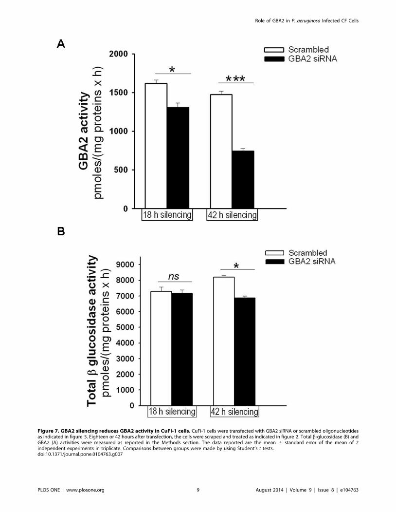
Figure 7. GBA2 silencing reduces GBA2 activity in CuFi-1 cells. CuFi-1 cells were transfected with GBA2 siRNA or scrambled oligonucleotidesas indicated in figure 5. Eighteen or 42 hours after transfection, the cells were scraped and treated as indicated in figure 2. Total b-glucosidase (B) andGBA2 (A) activities were measured as reported in the Methods section. The data reported are the mean 6 standard error of the mean of 2independent experiments in triplicate. Comparisons between groups were made by using Student’s t tests.doi:10.1371/journal.pone.0104763.g007
Role of GBA2 in P. aeruginosa Infected CF Cells
PLOS ONE | www.plosone.org 9 August 2014 | Volume 9 | Issue 8 | e104763

epithelial cells, which is consistent with the rise of ceramide levels
at the plasma membrane previously described [20]. In CuFi-1
cells, which have a lower ceramide content compared to IB3-1
cells, we observed a slight decrease in ceramide levels by miglustat
or Genz-529648 (figure 8, panel B). By contrast, IB3-1 cells
infected with PAO1 and treated with both miglustat and Genz-
Figure 8. Infection with PAO1 increases whole cell ceramides in CF bronchial epithelial cells. IB3-1 (A) and CuFi-1 (B) cells were treatedwith [2 mM] miglustat, [10 nM] Genz-529648 or solvent alone and infected with PAO1 as indicated in figure 3. After infection, whole cell ceramideswere analyzed by LC-MS and LC-MS/MS methods as described in the online supplement. The data reported are the mean 6 SE of three independentexperiments performed with both cell lines. Comparisons between groups were made by using Student’s t tests.doi:10.1371/journal.pone.0104763.g008
Role of GBA2 in P. aeruginosa Infected CF Cells
PLOS ONE | www.plosone.org 10 August 2014 | Volume 9 | Issue 8 | e104763

Figure 9. Treatment with Genz-529648 reduces the ceramide content in IB3-1 cells infected with PAO1. IB3-1 cells subjected to the SLmetabolic labeling with (1-3H)sphingosine were treated with [10 mM] amitriptyline alone, in combination with [10 nM] Genz-529648, or with solventalone and infected with PAO1 as indicated in figure 3. After lipid extraction, (3H)ceramide was separated from the other radioactive SLs by HPTLC, asdetailed in the online supplement, and detected by digital autoradiography (total lipid extracts amounts corresponding to 4 mg of cellular proteinswere applied on a 4-mm line. Time of acquisition: 48 hours). The digital autoradiography represents data obtained in three different experiments (A).The ceramide content was quantified by specific b-Vision software, and the data reported are the mean 6 SE of three independent experiments.Comparisons between groups were made by using Student’s t tests (B).doi:10.1371/journal.pone.0104763.g009
Role of GBA2 in P. aeruginosa Infected CF Cells
PLOS ONE | www.plosone.org 11 August 2014 | Volume 9 | Issue 8 | e104763

529648 showed a marked decrease of ceramide content (figure 8,
panel A). The increase in ceramide content following infection by
PAO1 could be a result of different pathways, including the denovo biosynthesis [21], SM catabolism [13] and GSL degradation.
When we evaluated the effects of drug treatment on the
radioactive ceramide content that resulted only because of the
modulation of SL catabolism, thus excluding the de novo pathway,
we identified a marked increase in the cell ceramide content after
PAO1 infection (figure 9). After ASM inhibition, we observed a
slight decrease and a further, more significant ceramide reduction
after the addition of the specific inhibitor of GBA2, Genz-529648
(figure 9), which strongly supports the direct involvement of GBA2
in the ceramide production after PAO1 infection of human
bronchial epithelial cells. Thus, the information derived from the
literature and the data presented here provide evidence that
different inhibitors, such as miglustat and Genz-529648, amitrip-
tyline [14] and myriocin [21], that target GBA2, ASM and
ceramide de novo synthesis, respectively, could represent thera-
peutic tools to reduce ceramide levels and limit excessive lung
inflammation in CF patients (figure 10). Nevertheless, drugs that
target SL metabolism must be carefully titrated to normalize
ceramide levels in CF airways, but not reduce ceramide
concentrations below a critical level that would impair normal
biological functions. Notably, systemic inhibition of ASM could
negatively affect the host defense, which has been demonstrated by
studies in mice that completely lack ASM and are unable to
control infections [13]. Interestingly, no increased susceptibility to
bacterial infections has been identified in patients affected by
Gaucher disease, treated with miglustat [47] or in a mouse model
of Sandhoff disease treated with Genz-529648 [48].
In summary, our study proposes GBA2 as a novel target to
reduce the inflammatory response to P. aeruginosa in CF
bronchial cells. These results further support the use of modulators
of SL metabolism for CF lung inflammation. In addition, as GBA2
is sensitive to very low doses of miglustat, other alkylated
iminosugars (NB-DGJ) and Genz-529648, our findings provide
evidence to develop therapeutic options for CF lung inflammation
using iminosugars, which can be effective at even low doses, thus
limiting potential adverse effects.
Supporting Information
Figure S1 Viability profile of IB3-1 cells treated for 24hours with the indicated concentrations of Genz-529648.
(TIF)
Figure S2 Apoptosis profile of IB3-1 cells treated for 24hours with the indicated concentration of Genz-529648.
(TIF)
Figure S3 Apoptotic IB3-1 cells after 4 and 24 hours oftreatment with the indicated concentrations of Genz-529648.
(TIF)
Supplement S1 Analysis of cell ceramide levels usingLC-MS and LC-MS/MS.
(DOC)
Figure 10. Metabolic pathways involved in ceramide formation. Schematic representation of the primary metabolic pathways involved inceramide production. Ceramide can be produced by the de novo biosynthesis, the hydrolysis of sphingomyelin (SM) by the action ofsphingomyelinases and the catabolism of glycosphingolipids (GSL). In particular, it has been observed that in CF bronchial epithelial cells, the use ofinhibitors of these pathways resulted in a reduction of ceramide. Myriocin acts on the first step of the de novo biosynthesis through the inhibition ofthe Serine-palmitoyl transferase (SPT); amitriptyline inhibits the acid SMase (ASM) responsible for SM catabolism; and miglustat, NB-DGJ and Genz-529648 are inhibitors of the b-glucosidases GBA1 and GBA2, which are involved in the hydrolysis of the glucosylceramide (GlcCer).doi:10.1371/journal.pone.0104763.g010
Role of GBA2 in P. aeruginosa Infected CF Cells
PLOS ONE | www.plosone.org 12 August 2014 | Volume 9 | Issue 8 | e104763

Supplement S2 Analysis of cell ceramide levels by cellSLs labeling with (3H)sphingosine.(DOC)
Supplement S3 Cellular toxicity of Genz-529648.(DOC)
Acknowledgments
We are grateful to: A. Tamanini for helpful discussions, A. Prince for the P.aeruginosa laboratory strain PAO1, ‘‘In Vitro Model and Cell Culture
Care’’ of the University of Iowa, Iowa, U.S.A. for providing CuFi-1 cells
and to ‘‘Servizio Colture Primarie’’ of the Italian Cystic Fibrosis Research
Foundation at the Laboratory of Molecular Genetics, G. Gaslini Institute,
Genova, Italy, for CF primary cells. This research was supported by the
Italian Cystic Fibrosis Research Foundation (grant FFC # 14/2012) with
the contribution of ‘‘Picasso. Capolavori dal Museo Nazionale Picasso diParigi’’, Festa d’Estate Villa Sigurta Verona, Delegazione FFC Lago diGarda e Arezzo.
Author Contributions
Conceived and designed the experiments: MCD MA SS GC. Performed
the experiments: NL MT IL NM RB VB VL CC SM. Analyzed the data:
MCD MA NL NM MGG AC RG. Contributed reagents/materials/
analysis tools: SHC MGG. Wrote the paper: MCD SHC MA RB NL .
References
1. Welsh JM, Ramsey BW, Accurso F, Cutting GR (2001) Cystic Fibrosis in Scriver
CR, Beaudet AL, Sly WS, Valle D (Eds). The Metabolic and Molecular Bases ofInherited Diseases. McGraw-Hill, New York.
2. Ramsey BW, Davies J, McElvaney NG, Tullis E, Bell SC, et al. (2011) VX08-770-102 Study Group A CFTR Potentiator in Patients with Cystic Fibrosis and
the G551D Mutation. New England J of Medicine 365: 1663–1672.
3. Hoffman LR, Ramsey BW (2013) Cystic Fibrosis Therapeutics. The road ahead.
Chest 143(1): 207–213.
4. Konstan MW, Schluchter MD, Xue W, Davis PB (2007) Clinical use of
Ibuprofen is associated with slower Fev1 decline in children with cystic fibrosis.
Am J Respir Crit Care Med 176 (11): 1084–1089.
5. Khan TZ, Wagener JS, Bost T, Martinez J, Accurso FJ, et al. (1995) Earlypulmonary inflammation in infants with cystic fibrosis. Am J Respir Crit Care
Med 151: 1075–1082.
6. Noah TL, Black HR, Cheng PW, Wood RE, Leigh MW (1997) Nasal and
bronchoalveolar lavage fluid cytokines in early cystic fibrosis. J Infect Dis 175:
638–647.
7. Tirouvanziam R, de Bentzmann S, Hubeau C, Hinnrasky J, Jacquot J, et al.(2000) Inflammation and infection in naive human cystic fibrosis airway grafts.
Am J Respir Cell Mol Biol 23(2): 121–127.
8. Chmiel JF, Berger M, Konstan MW (2002) The role of inflammation in the
pathophysiology of CF lung disease. Clin Rev Allergy Immunol 23: 5–27.
9. Lahiri S, Futerman AH (2007) The metabolism and function of shingolipids and
glycoshingolipids. Cell Mol Life Sci 64: 2270–2284.
10. Uhlig S, Gulbins E (2008) Sphingolipids in the lungs. Am J Respir Crit Care
Med 178(11): 1100–1114.
11. Yang Y, Uhlig S (2001) The role of sphingolipids in respiratory disease. Ther
Adv Respir Dis 5: 325–344.
12. Hannun YA, Obeid LM (2008) Principles of bioactive lipid signalling: lessons
from sphingolipids. Nat Rew Mol Cell Biol 9: 139–150.
13. Grassme’ H, Jendrossek V, Riehle A, von Kurthy G, Berger J, et al. (2003) Host
defense against Pseudomonas aeruginosa requires ceramide-rich membranerafts. Nature Medicine 9: 322–330.
14. Teichgraber V, Ulrich M, Endlich N, Riethmuller J, Wilker B, et al. (2008)Ceramide accumulation mediates inflammation, cell death and infection
susceptibility in cystic fibrosis. Nature Med 14: 382–391.
15. Guilbault C, De Sanctis JB, Wojewodka G, Saeed Z, Lachance C, et al. (2008)
Fenretinide corrects newly found ceramide deficiency in cystic fibrosis.Am J Respir Cell Mol Biol 38(1): 47–56.
16. Yu H, Zeidan YH, Wu BX, Jenkins RW, Flotte TR, et al. (2009) Defective acidsphingomyelinase pathway with Pseudomonas aeruginosa infection in cystic
fibrosis. American Journal of Respiratory Cell and Molecular Biology 41: 367–375.
17. Brodlie M, McKean MC, Johnson GE, Gray J, Fisher AJ, et al. (2010) Ceramideis Increased in the Lower Airway Epithelium of People with Advanced Cystic
Fibrosis Lung Disease. Am J Respir Crit Care Med 182 (3): 369–375.
18. Bodas M, Min T, Mazur S, Vij N (2011) Critical modifier role of membrane-
cystic fibrosis transmembrane conductance regulator-dependent ceramidesignaling in lung injury and emphysema. J Immunol 186: 602–613.
19. Nahrlich L, Mainz JG, Adams C, Engel C, Herrmann G, et al. (2013) Therapyof CF-patients with amitriptyline and placebo-a randomised, double-blind,
placebo-controlled phase IIb multicenter, cohort-study. Cell Physiol Biochem31(4–5): 505–312.
20. Dechecchi MC, Nicolis E, Mazzi P, Cioffi F, Bezzerri V, et al. (2011)Modulators of sphingolipid metabolism reduce lung inflammation. Am J Respir
Cell Mol Biol 45(4): 825–833.
21. Caretti A, Bragonzi A, Facchini M, De Fino I, Riva C, et al. (2014) Anti-
inflammatory action of lipid nanocarrier-delivered myriocin: therapeutic
potential in cystic fibrosis. Biochim Biophys Acta 1840(1): 586–594.
22. Nash RJ, Kato A, Yu C-Y, Fleet GWJ (2011) Iminosugars as therapeuthicagents:recent advances and promising trends. Future Med Chem 3(12): 1–9.
23. Dechecchi MC, Nicolis E, Mazzi P, Paroni M, Cioffi F, et al. (2012)Pharmacological modulators of sphingolipid metabolism for the treatment of
cystic fibrosis lung inflammation. In Dinesh D and Sriramulu D editors. Cystic
Fibrosis - the Human Agony, ISBN 979-953-307-059-8, Germany.
24. Norez C, Noel S, Wilke M, Bijvelds M, Jorna H, et al. (2006) Rescue of
functional delF508-CFTR channels in cystic fibrosis epithelial cells by the a-
glucosidase inhibitor miglustat. FEBS Lett 580: 2081–2086.
25. Dechecchi MC, Nicolis E, Norez C, Bezzerri V, Borgatti M, et al. (2008) Anti-
inflammatory effect of miglustat in bronchial epithelial cells. J Cyst Fibros 7(6):555–565.
26. Lubamba B, Lebacq J, Lebecque P, Vanbever R, Leonard A (2009) Airway
delivery of low-dose miglustat normalizes nasal potential difference in F508delcystic fibrosis mice. Am J Respir Crit Care Med 179: 1022–1028.
27. Leonard A, Lebecque P, Dingemanse J, Leal T (2012) A randomized placebo-controlled trial of miglustat in cystic fibrosi based on nasal potential difference.
J Cystic Fibrosis 11(3): 231–236.
28. Inokuchi J, Mason I, Radin NS (1987) Antitumor activity via inhibition of
glycosphingolipid biosynthesis. Cancer Lett 38: 23–30.
29. Boot RG, Verhoek M, Donker-Koopman W, Strijland A, van Marle J, et al.(2007) Identification of the non-lysosomal glucosylceramidase as beta-glucosi-
dase 2. J Biol Chem 282: 1305–1312.
30. Ridley CM, Thur KE, Shanahan J, Thillaiappan NB, Shen A, et al. (2013) b-
Glucosidase 2 (GBA2) activity and imino sugar pharmacology. J Biol Chem288(36): 26052–26066.
31. Dwek RA, Butters TD, Platt FM, Zitzmann N (2002) Targeting glycosylation as
a therapeutic approach. Nat Rev Drug Discov 1: 65–75.
32. Platt FM, Neises GR, Karlsson GB, Dwek RA, Butters TD (1994) N-
butyldeoxygalactonojirimycin inhibits glycolipid biosynthesis but does not affectN-linked oligosaccharide processing. J Biol Chem 269 (43): 27108–27114.
33. Weenekes T, Meijer AJ, Groen AK, Boot RG, Groener JE, et al. (2010) Large-
Scale synthesis of the glucosylceramide synthase inhibitor N-[5-(Adamantan-1-yl-methoxy)-pentyl]-1-deoxynojirimycin. J Med Chem 53: 689–698.
34. Overkleeft HS, Renkema Hg, Neele J, Vianello P, Hung IO, et al. (1998)Generation of specific deoxynojirimycin-type inhibitors of the non-lysosomal
glucosylceramidase. J Biol Chem 273 (41): 26522–26527.
35. Yildiz Y, Matern H, Thompson B, Allergood JC, Warren RL, et al. (2006)
Mutation of beta-glucosidase 2 causes glycolipid storage disease and impaired
male fertility. J Clin Invest 116: 2985–2994.
36. Aerts JM, Ottenhoff R, Powlson AS, Grefhorst A, van Eijk M, et al. (2007)
Pharmacological inhibition of glucosylceramide synthase enhances insulinsensitivity. Diabetes 56(5): 1341–1349.
37. Zeitlin PL, Lu L, Rhim J, Cutting G, Stetten G, et al. (1991) A cystic fibrosisbronchial epithelial cell line: immortalization by adeno-12-SV40 infection.
Am J Respir Cell Mol Biol 4: 313–319.
38. Zabner J, Karp P, Seiler M, Phillips Sl, Mitchell CJ, et al. (2003) Development ofcystic fibrosis and non cystic fibrosis airway cell lines. Am J Physiol Lung Cell
Mol Physiol 284: L844–L854.
39. Scudieri P, Caci E, Bruno S, Ferrera L, Schiavon M, et al. (2012) Association of
TMEM16A chloride channel overexpression with airway goblet cell metaplasia.
J Physiol 590 (23): 6141–6155.
40. Sullards MC, Allegood JC, Kelly S, Wang E, Haynes CA, et al. (2007) Structure-
specific, quantitative methods for analysis of sphingolipids by LC-tandem MS:‘‘inside-out’’ sphingolipids. Methods in Enzymology 432: 83–115.
41. Aureli M, Loberto N, Bassi R, Ferraretto A, Perego S, et al. (2012) Plasmamembrane-associated glycohydrolases activation by extracellular acidification
due to proton exchangers. Neurochem Res 37(6): 1296–1307.
42. Platt FM, Reinkensmeier G, Dwek RA, Butters TD (1996) Extensiveglycosphingolipid depletion in the liver and lymphoid organs of mice treated
with N- butyldeoxynojirimycin. J Biol Chem 272: 19365–19372.
43. Martin E, Schule R, Smets K, Rastetter A, Boukhris A, et al. (2013) Loss of
function of glucocerebrosidase GBA2 is responsible for motor neuron defects inhereditary spastic paraplegia. Am J Hum Genet 92(2): 238–244.
44. Sorli SC, Colie S, Albinet V, Dubrac A, Touriol C, et al. (2013) The
nonlysosomal b-glucosidase GBA2 promotes endoplasmic reticulum stress andimpairs tumorigenicity of human melanoma cells. FASEB J 27(2): 489–498.
45. Korschen HG, Yildiz Y, Raju DN, Schonauer S, Bonigk W, et al. (2013) Thenon-lysosomal b-glucosidase GBA2 is a non-integral membrane-associated
protein at the endoplasmic reticulum (ER) and Golgi. J Biol Chem 288(5): 3381–
3393.
Role of GBA2 in P. aeruginosa Infected CF Cells
PLOS ONE | www.plosone.org 13 August 2014 | Volume 9 | Issue 8 | e104763

46. Sadighi Akha AA, Theriot CM, Erb-Downward JR, McDermott AJ, Falkowski
NR (2013) Acute infection of mice with Clostridium difficile leads to eIF2aphosphorylation and pro-survival signalling as part of the mucosal inflammatory
response. Immunology 140(1): 111–122.
47. Hollak CE, Hughes D, van Schaik IN, Schwierin B, Bembi B (2009) Miglustat(Zavesca) in type 1 Gaucher disease: 5-year results of a post-authorisation safety
surveillance programme. Pharmacoepidemiol Drug Saf 18(9): 770–777.
48. Ashe KM, Bangari D, Li L, Cabrera-Salazar MA, Bercury SD, et al. (2011)
Iminosugar-based inhibitors of glucosylceramide synthase increase brain
glycosphingolipids and survival in a mouse model of Sandhoff disease. PLoS
One 6(6): 1–11.
Role of GBA2 in P. aeruginosa Infected CF Cells
PLOS ONE | www.plosone.org 14 August 2014 | Volume 9 | Issue 8 | e104763
Copyright © 2022 FDOKUMEN