Global analysis of protein aggregation in yeast during physiological conditions and arsenite stress
13
RESEARCH ARTICLE Global analysis of protein aggregation in yeast during physiological conditions and arsenite stress Sebastian Ibstedt 1 , Theodora C. Sideri 2,3 , Chris M. Grant 2 and Markus J. Tama ´s 1, * ABSTRACT Protein aggregation is a widespread phenomenon in cells and associated with pathological conditions. Yet, little is known about the rules that govern protein aggregation in living cells. In this study, we biochemically isolated aggregation-prone proteins and used computational analyses to identify characteristics that are linked to physiological and arsenite-induced aggregation in living yeast cells. High protein abundance, extensive physical interactions, and certain structural properties are positively correlated with an increased aggregation propensity. The aggregated proteins have high translation rates and are substrates of ribosome-associated Hsp70 chaperones, indicating that they are susceptible for aggregation primarily during translation/folding. The aggregation- prone proteins are enriched for multiple chaperone interactions, thus high protein abundance is probably counterbalanced by molecular chaperones to allow soluble expression in vivo. Our data support the notion that arsenite interferes with chaperone activity and indicate that arsenite-aggregated proteins might engage in extensive aberrant protein–protein interactions. Expression of aggregation-prone proteins is down-regulated during arsenite stress, possibly to prevent their toxic accumulation. Several aggregation-prone yeast proteins have human homologues that are implicated in misfolding diseases, suggesting that similar mechanisms may apply in disease- and non-disease settings. KEY WORDS: protein aggregation, protein folding, translation, arsenite, chaperone INTRODUCTION Proteins participate in virtually every biological process. To function, most proteins fold into a strictly defined three- dimensional structure, their native conformation. Proteins in a non-native conformation may aggregate and/or engage in aberrant interactions with other cellular components. Misfolded proteins are cytotoxic, and numerous neurodegenerative and age- related disorders are associated with protein misfolding and aggregation. Evolutionary conserved protein quality-control (PQC) systems protect cells against the harmful accumulation of protein aggregates. These PQC systems encompass molecular chaperones that assist folding of polypeptides into their functional conformation and degradation pathways that clear the cells from misfolded and aggregated proteins (Hartl et al., 2011; Stefani and Dobson, 2003; Tyedmers et al., 2010). The inclination of a given protein to aggregate is correlated with solvent-exposed stretches of high hydrophobicity, high b- sheet propensity, and a low net charge (Hartl et al., 2011; Stefani and Dobson, 2003). Moreover, structurally flexible proteins and proteins with intrinsically disordered regions may be more prone to aberrant interactions and aggregation (Breydo and Uversky, 2011). Aggregation-prone segments tend to be buried in the native (folded) protein. However, conditions that promote protein unfolding may lead to exposure of such segments and facilitate aggregation. Such conditions include mutations that affect the PQC systems, misprocessing phenomena such as mistranslation or defective assembly of protein complexes, changes in the intracellular environment or chemical modifications, and progressive decline in efficiency of the PQC systems during ageing (Hartl et al., 2011; Stefani and Dobson, 2003; Tyedmers et al., 2010). Much of our knowledge on protein folding and aggregation comes from in vitro studies using model peptides, and misfolding- prone or disease-associated (model) proteins (Alies et al., 2013; Breydo and Uversky, 2011; Hartl et al., 2011; Vendruscolo, 2012). In addition, computational approaches are commonly used to predict the intrinsic aggregation-propensities of proteins (Conchillo- Sole ´ et al., 2007; Fernandez-Escamilla et al., 2004; Tartaglia and Vendruscolo, 2008). In general, the algorithms used are based on certain physico-chemical characteristics of the amino acid sequence previously shown to contribute to protein aggregation (using in vitro measurements). However, the rules that govern protein aggregation in living cells are likely to be more complex than those defined from individual proteins or from in vitro studies (Vendruscolo, 2012). Recently, proteome-wide studies on aggregation in living cells have been reported. For example, it was estimated that hundreds of proteins aggregate upon mild heat stress in Escherichia coli (Winkler et al., 2010). Likewise, about 200 aggregated proteins were identified in stationary phase yeast (Saccharomyces cerevisiae) cells. For a subset of those proteins, the process of aggregation was reversible upon nutrient re-addition (Narayanaswamy et al., 2009). Similarly, numerous proteins turned insoluble with age in Caenorhabditis elegans (David et al., 2010) or co-aggregated with amyloid-forming polypeptides in mammalian cells (Olzscha et al., 2011). Widespread protein aggregation also occurs in cells defective in PQC systems (Chapman et al., 2006; Koplin et al., 2010; Rand and Grant, 2006), in response to environmental stress conditions (Jacobson et al., 2012), and in disease processes (Basso et al., 2009; Liao et al., 2004; Wang et al., 2005; Xia et al., 2008). There is accumulating evidence that certain metals influence the aggregation propensity of disease-associated proteins and 1 Department of Chemistry and Molecular Biology, University of Gothenburg, S-405 30 Gothenburg, Sweden. 2 Faculty of Life Sciences, University of Manchester, Manchester M13 9PT, UK. This is an Open Access article distributed under the terms of the Creative Commons Attribution License (http://creativecommons.org/licenses/by/3.0), which permits unrestricted use, distribution and reproduction in any medium provided that the original work is properly attributed. Received 22 May 2014; Accepted 20 August 2014 *Author for correspondence ([email protected]). 3 Current address: Department of Genetics, Evolution and Environment and UCL Cancer Institute, University College London, WC1E 6BT, London, UK. ß 2014. Published by The Company of Biologists Ltd | Biology Open (2014) 000, 1–11 doi:10.1242/bio.20148938 1 Biology Open
-
Upload
independent -
Category
Documents
-
view
3 -
download
0
Transcript of Global analysis of protein aggregation in yeast during physiological conditions and arsenite stress
RESEARCH ARTICLE
Global analysis of protein aggregation in yeast during physiologicalconditions and arsenite stress
Sebastian Ibstedt1 Theodora C Sideri23 Chris M Grant2 and Markus J Tamas1
ABSTRACT
Protein aggregation is a widespread phenomenon in cells and
associated with pathological conditions Yet little is known about the
rules that govern protein aggregation in living cells In this study we
biochemically isolated aggregation-prone proteins and used
computational analyses to identify characteristics that are linked
to physiological and arsenite-induced aggregation in living yeast
cells High protein abundance extensive physical interactions and
certain structural properties are positively correlated with an
increased aggregation propensity The aggregated proteins have
high translation rates and are substrates of ribosome-associated
Hsp70 chaperones indicating that they are susceptible for
aggregation primarily during translationfolding The aggregation-
prone proteins are enriched for multiple chaperone interactions
thus high protein abundance is probably counterbalanced by
molecular chaperones to allow soluble expression in vivo Our
data support the notion that arsenite interferes with chaperone
activity and indicate that arsenite-aggregated proteins might engage
in extensive aberrant proteinndashprotein interactions Expression of
aggregation-prone proteins is down-regulated during arsenite
stress possibly to prevent their toxic accumulation Several
aggregation-prone yeast proteins have human homologues that
are implicated in misfolding diseases suggesting that similar
mechanisms may apply in disease- and non-disease settings
KEY WORDS protein aggregation protein folding translation
arsenite chaperone
INTRODUCTIONProteins participate in virtually every biological process Tofunction most proteins fold into a strictly defined three-
dimensional structure their native conformation Proteins in a
non-native conformation may aggregate andor engage inaberrant interactions with other cellular components Misfolded
proteins are cytotoxic and numerous neurodegenerative and age-related disorders are associated with protein misfolding and
aggregation Evolutionary conserved protein quality-control
(PQC) systems protect cells against the harmful accumulation
of protein aggregates These PQC systems encompass molecularchaperones that assist folding of polypeptides into their functionalconformation and degradation pathways that clear the cells from
misfolded and aggregated proteins (Hartl et al 2011 Stefani andDobson 2003 Tyedmers et al 2010)
The inclination of a given protein to aggregate is correlated
with solvent-exposed stretches of high hydrophobicity high b-sheet propensity and a low net charge (Hartl et al 2011 Stefaniand Dobson 2003) Moreover structurally flexible proteins andproteins with intrinsically disordered regions may be more prone
to aberrant interactions and aggregation (Breydo and Uversky2011) Aggregation-prone segments tend to be buried in thenative (folded) protein However conditions that promote protein
unfolding may lead to exposure of such segments and facilitateaggregation Such conditions include mutations that affect thePQC systems misprocessing phenomena such as mistranslation
or defective assembly of protein complexes changes in theintracellular environment or chemical modifications and progressivedecline in efficiency of the PQC systems during ageing (Hartlet al 2011 Stefani and Dobson 2003 Tyedmers et al 2010)
Much of our knowledge on protein folding and aggregationcomes from in vitro studies using model peptides and misfolding-prone or disease-associated (model) proteins (Alies et al 2013
Breydo and Uversky 2011 Hartl et al 2011 Vendruscolo 2012)In addition computational approaches are commonly used topredict the intrinsic aggregation-propensities of proteins (Conchillo-
Sole et al 2007 Fernandez-Escamilla et al 2004 Tartaglia andVendruscolo 2008) In general the algorithms used are based oncertain physico-chemical characteristics of the amino acid sequence
previously shown to contribute to protein aggregation (using in vitro
measurements) However the rules that govern protein aggregationin living cells are likely to be more complex than those defined fromindividual proteins or from in vitro studies (Vendruscolo 2012)
Recently proteome-wide studies on aggregation in living cells havebeen reported For example it was estimated that hundreds ofproteins aggregate upon mild heat stress in Escherichia coli
(Winkler et al 2010) Likewise about 200 aggregated proteinswere identified in stationary phase yeast (Saccharomyces cerevisiae)cells For a subset of those proteins the process of aggregation was
reversible upon nutrient re-addition (Narayanaswamy et al 2009)Similarly numerous proteins turned insoluble with age inCaenorhabditis elegans (David et al 2010) or co-aggregated withamyloid-forming polypeptides in mammalian cells (Olzscha et al
2011) Widespread protein aggregation also occurs in cellsdefective in PQC systems (Chapman et al 2006 Koplin et al2010 Rand and Grant 2006) in response to environmental stress
conditions (Jacobson et al 2012) and in disease processes (Bassoet al 2009 Liao et al 2004 Wang et al 2005 Xia et al 2008)
There is accumulating evidence that certain metals influence
the aggregation propensity of disease-associated proteins and
1Department of Chemistry and Molecular Biology University of GothenburgS-405 30 Gothenburg Sweden 2Faculty of Life Sciences University ofManchester Manchester M13 9PT UK
This is an Open Access article distributed under the terms of the Creative Commons AttributionLicense (httpcreativecommonsorglicensesby30) which permits unrestricted use distributionand reproduction in any medium provided that the original work is properly attributed
Received 22 May 2014 Accepted 20 August 2014
Author for correspondence (markustamascmbguse)
3Current address Department of Genetics Evolution and Environment and UCLCancer Institute University College London WC1E 6BT London UK
2014 Published by The Company of Biologists Ltd | Biology Open (2014) 000 1ndash11 doi101242bio20148938
1
BiologyOpen
affect the progression of certain neurodegenerative diseases vialargely unknown mechanisms (Alies et al 2013 Bourassa and
Miller 2012 Breydo and Uversky 2011 Caudle et al 2012Savelieff et al 2013) Recent studies showed that various metalsand the metalloid arsenite inhibit protein folding in vitro (Jacobsonet al 2012 Ramadan et al 2009 Sharma et al 2008 Tamas et al
2014) Moreover we demonstrated that arsenite interferes withprotein folding in vivo by acting on unfolded or nascent polypeptidesand by directly interfering with chaperone activity (Jacobson et al
2012) Folding inhibition contributed to arsenite toxicity in twoways by aggregate formation and by chaperone inhibitionInterestingly in vitro data indicated that arsenite-induced protein
aggregates can act as seeds committing other labile proteins tomisfold and aggregate (Jacobson et al 2012) This mode of actionmay explain the suggested role of this metalloid in the etiology of
certain neurodegenerative and age-related disorders associated witharsenic poisoning However much remains to be learned about themolecular events leading to protein aggregation and aggregatetoxicity in living cells In this study we addressed the following
questions (1) What proteins are at risk for aggregation in vivo (2)What physico-chemical properties and biological functions areassociated with protein aggregation (3) How do aggregates
contribute to arsenite toxicity (4) Do cells regulate aggregation-prone proteins during environmental stress For this webiochemically isolated aggregated proteins from S cerevisiae
during physiological conditions and arsenite exposure and usedcomputational analyses to identify characteristics that are linked toprotein aggregation In this way we provide novel and extended
insights into the rules that govern protein aggregation in living cells
MATERIALS AND METHODSIdentification of aggregated proteinsYeast cells (BY4742 strain background) were grown to exponential phase
(A600 06) in YPD medium without or with arsenite (15 mM sodium
arsenite 1 hour) and equivalent cell numbers (10 A600 units) were used to
isolate aggregated proteins as described previously (Jacobson et al 2012
Rand and Grant 2006) Briefly cells were disrupted in lysis buffer
(50 mM potassium phosphate buffer pH 7 1 mM EDTA 5 glycerol
1 mM phenylmethylsulfonyl fluoride and Complete Mini protease
inhibitor cocktail (Roche)) and membrane proteins and aggregated
proteins were isolated by centrifugation (15000 g 20 minutes)
Membrane proteins were removed by washing twice with 320 ml lysis
buffer and 80 ml of 10 Igepal CA 630 (NP-40) (SigmandashAldrich)
centrifuging at 15000 g for 20 minutes each time and the final aggregated
protein extract was resuspended in 100 ml of lysis buffer Aggregated
proteins were separated on 12 reducing SDS-PAGE gels and stained
using colloidal Coomassie blue (SigmandashAldrich) Proteins were excised
trypsin-digested and identified using liquid chromatography-mass
spectrometry (LC-MS) in the Biomolecular Analysis Facility (Faculty of
Life Sciences University of Manchester) Proteins were identified using
the Mascot mass fingerprinting programme (httpwwwmatrixscience
com) to search the NCBInr and Swissprot databases
Statistical methodsStatistical analyses were performed on physiological aggregates (P-set)
and on arsenite-induced aggregates (As-set) using a largely unbiased set
of 1475 proteins (MS proteome) detected by large-scale proteome
analysis by multidimensional LC-MS (Washburn et al 2001) as
background
Analyses of physical propertiesAnalysis of functional enrichment was performed on gene ontology data
from the Saccharomyces Genome Database (SGD) (Cherry et al 2012)
and p-values were calculated with a hypergeometric test using 6607
genomic genes as background p-values were filtered with FDR 5
Physical protein properties were obtained from SGD and analysed with
MannndashWhitney U-tests
Analyses of protein abundance translation expression and half-livesProtein abundance (Ghaemmaghami et al 2003) translation rate per
protein species (Arava et al 2003) and protein half-life (Belle et al
2006) was analysed based on data collected during non-stress conditions
MannndashWhiney U-tests were used to assess the observed differences
Genome-wide expression data was obtained from (Thorsen et al 2007)
Overlap between proteins showing at least a 2-fold change in gene
expression and aggregated proteins in the As- and P-sets was evaluated
with a hypergeometric test using the MS proteome as background The
representation factor was calculated as observed overlapexpected
overlap
Analyses of structural propertiesSecondary structures were predicted with the GarnierndashOsguthorpendash
Robson algorithm (Garnier et al 1978) For each protein the relative
proportion of amino acid residues partaking in a particular secondary
structure (a-helix b-sheet) was predicted using a sliding 17 residue
window Results were confirmed by an alternative approach based on
homologies with known structures (Frishman and Argos 1995 Frishman
and Argos 1997) Proportions of residues in secondary structures were
compared with MannndashWhitney U-tests Intrinsic disorder was predicted
by calculating the fold-index defined as a function of mean
hydrophobicity and mean net charge (Prilusky et al 2005) The
genomic proportion of surface-exposed cysteines was identified by
(Marino et al 2010) and used to compare the As- and P-sets to the MS
proteome with Fisherrsquos exact test Cysteine density was calculated by
counting the number of CC CxC CxxC or CxxxC in a sliding window
across each protein p-values were computed with Fisherrsquos exact test
Differences in amino acid composition were compared with
heteroscedastic Studentrsquos t-test and adjusted for multiple testing with
HolmndashSıdak correction
Analyses of interactivityGenetic and physical interaction data were obtained from the BioGRID
database (Stark et al 2006) and subset on group-specific and global
interactions for proteins in both aggregate sets Data for calculation of
synthetic sickness was obtained from the Drygin database (Koh et al
2010) and filtered for maximum 5 false positives A difference of at
least 008 between the fitness of the double mutant and the two single
mutants |fab2fa6fb|008 was considered to display synthetic sickness
a definition which has proven to give reproducible and functionally
informative results (Koh et al 2010) Differences between the data-sets
with regard to physical genetic and synthetic sick interactions were
assessed by empirical p-values by comparing the median difference
between observed groups with 1000000 random permutations (without
replacement) of the pooled data Interactions with chaperones were based
on data from (Gong et al 2009) and results were analyzed with Studentrsquos
t-test Overrepresentation of co-translational Ssb2p substrates and
aggregation in SSBD cells (Willmund et al 2013) was analyzed with
Fisherrsquos exact test
Identification of orthologuesOrthologues between human disease aggregates in Alzheimerrsquos disease
(Liao et al 2004 Wang et al 2005) familial amyotrophic lateral
sclerosis (Basso et al 2009) or Parkinsonrsquos disease (Xia et al 2008) and
yeast were identified with the OMA browser (Schneider et al 2007) The
level of orthology was evaluated by counting the number of orthologous
cases between disease-associated aggregates and the yeast aggregates and
the genome respectively and assessed with Fisherrsquos exact test
RESULTSIdentification of aggregation-prone proteins in S cerevisiaeTo identify aggregation-prone proteins we collected exponentiallygrowing yeast cells that were either untreated (physiological
RESEARCH ARTICLE Biology Open (2014) 000 1ndash11 doi101242bio20148938
2
BiologyOpen
condition) or exposed to sodium arsenite Aggregated proteinswere isolated using a well-established method based on density
centrifugation and then identified using LC-MS (see Materials andMethods) In this way a total of 257 aggregated proteins wereunambiguously identified (supplementary material Table S3) Ofthese 114 proteins were found to aggregate both under
physiological conditions and during arsenite exposure theseproteins are likely to be generally aggregation-prone and willhereafter be referred to as the physiological-set (P-set) Since the
MS method used is qualitative our data-sets do not containinformation whether a greater percentage of a given protein in theP-set would aggregate during arsenite exposure The remaining
143 proteins were unique to arsenite-exposed cells (hereafter calledthe As-set) Thus an expanded set of proteins aggregate inresponse to arsenite exposure We previously reported the identity
of the proteins in the As-set (Jacobson et al 2012) however thatreport did not include systems-level analysis of the aggregatedproteins In this current study we performed a comprehensiveanalysis of aggregation-prone proteins during physiological
conditions and arsenite stress
Physical structural and functional characteristics ofaggregation-prone proteinsGene ontology (GO) analysis indicated that aggregated proteinsare enriched in certain functional categories (Fig 1) Functions
related to ribosome biogenesisassembly and translation arehighly overrepresented in the P-set compared to the S
cerevisiae genome In fact about 60 of the proteins in the P-
set are ribosomal proteins or have a role during translation TheAs-set is enriched for functions related to translation proteinfolding and various metabolic processes (Fig 1)
To examine whether these proteins possess particular
properties that make them aggregation-prone we comparedthem to a list of yeast proteins that are detected by MS inlogarithmically growing cells (Washburn et al 2001) By using
these proteins (hereafter called the MS proteome) as backgroundwe avoid potential bias by including proteins that are normally
not detected by MS Aggregated proteins in the P-set are clearlymore abundant (ie present in more moleculescell) highlyexpressed (indicated by a high codon adaptation index (CAI))smaller in size (ie lower molecular weight (MW)) and have a
higher isoelectric point (pI) than proteins in the MS proteome(Fig 2) These features are consistent with the high proportion ofribosomal proteins in the P-set Although less pronounced than
for the P-set the As-set is also enriched for abundant and highlyexpressed proteins (Fig 2AB) Arsenite-aggregated proteinshave slightly lower pI but do not differ in mean protein size
compared to the MS proteome (Fig 2CD)The relative amino acid composition of the aggregated proteins
differed from that in the MS proteome (Fig 3) both As- and P-
sets are enriched in the aliphatic amino acids glycine alanine andvaline whilst asparagine and serine having polar uncharged side-chains and methionine are significantly underrepresented Inaddition the P-set is enriched in the basic amino acids lysine and
arginine whilst the acidic residues aspartate and glutamate areunderrepresented (Fig 3) in agreement with the higher pI of P-set proteins (Fig 2D) Consistent with the observed enrichment
for aliphatic amino acids (Fig 3) proteins in the As- and P-setsshow a somewhat higher average hydrophobicity (GRAVY score)than those in the MS proteome (Fig 2E) Proteins in both sets are
predicted to have significantly higher a-helix content than theaverage protein in the MS proteome and the P-set is additionallyenriched for proteins with enhanced b-sheet content (Fig 2F)
Computational predictions were recently used to group 1822 S
cerevisiae proteins (representing 27 of the predicted proteome)into four categories highly structured proteins without aggregation-prone elements (SNA) highly structured proteins with aggregation-
prone elements (SA) highly unstructured proteins with non-aggregatinglysineglutamic acid-rich stretches (UNA) and highly unstructuredproteins with aggregation-prone glutamineasparagine-rich stretches
Fig 1 Functional characteristics of aggregation-prone proteins Biological processes that aresignificantly enriched in the As- and P-sets compared tothe S cerevisiae genome Circle diagrams indicate thedistribution of aggregated proteins into functionalcategories where lsquolsquoTranslationrsquorsquo excludes ribosomalproteins Bar diagrams indicate the fold-enrichment offunctional categories compared to the genome using GOdata from SGD All shown categories are significant with5 FDR
RESEARCH ARTICLE Biology Open (2014) 000 1ndash11 doi101242bio20148938
3
BiologyOpen
(UA) (Gsponer and Babu 2012) 82 of the aggregation-proneproteins that we identified (representing 32 of the proteins inour data-sets) were also present in the Gsponer and Babu data-set
Of those 69 proteins were predicted to be structured (present inSNA+SA categories) whilst 13 proteins were predicted to beunstructured (present in UNA+UA categories) (supplementary
material Table S1) Moreover the arsenite-aggregated proteinswere significantly enriched in the SNA category (supplementarymaterial Table S1) and showed less intrinsic protein disorder thanproteins in the P-set and MS proteome (Fig 2G) The proteins in
both P- and As-sets have on the average a longer half-life thanproteins in the MS proteome (Fig 2H) suggesting that theseproteins are stable in their folded states since half-lives were
determined by measuring protein abundance over time afterinhibition of protein biosynthesis (Belle et al 2006)
We conclude that high protein expression and abundance a
higher average hydrophobicity and certain structural propertiespositively correlate with physiological and arsenite-inducedprotein aggregation in vivo Moreover many of these proteinsare predicted to be structured and stable in their native folded
states
Proteins are susceptible for aggregation during translationfoldingThe observations that aggregation-prone proteins are abundantand associated with processes related to translation (Figs 1 2)
prompted us to explore this further Using data from large-scale
translation-rate estimations (Arava et al 2003) we found thataggregating proteins are translated at a significantly higher ratethan proteins in the MS proteome (Fig 4A) Proteins in the P-set
are particularly highly translated with a median translation rateabout 8-fold higher than the MS proteome whilst the As-setproteins have an about 2-fold higher translation rate than those in
the MS proteome (Fig 4A) Many proteins fold during translationand co-translational folding may be assisted by ribosome-boundchaperones of which Hsp70 is the most prominent In S cerevisiae
the closely related ribosome-associated Hsp70 proteins Ssb1p andSsb2p bind cotranslationally to nascent chains and co-translationalsubstrates of Ssb2p were recently identified (Willmund et al2013) Both the As-set (68) and the P-set (78) are enriched in
proteins that are co-translational Ssb2p substrates compared to theMS proteome (55) (Fig 4B) This enrichment is even morepronounced when compared to the genome (12 Ssb2p
interactors) (supplementary material Fig S1) Loss of Ssb1pSsb2p results in aggregation of newly synthesized proteins (Koplinet al 2010 Willmund et al 2013) The overlap between proteins
that aggregate in cells lacking Ssb1pSsb2p (SSBD) and those thataggregate in the As- and P-sets is significantly higher than the
Fig 2 Physico-chemical characteristics of aggregation-prone proteins(A) Molecules per cell The abundance of proteins in each set during non-stress conditions is plotted Proteins in the P-set are significantly moreabundant than proteins in the As-set (p 5 0017 U 5 39075) whilst proteinsin the MS proteome are less abundant than those in the As-set (p 10215U 5 100021) and P-set (p 10215 U 5 69411) (B) Expression levels Thecodon adaptation index is an indication of gene expression levels (Sharpand Li 1987) and the CAI for proteins in each data-set are displayedThe CAI for the P-set is significantly higher than for the As-set (p 5 10214U 5 34975) and the MS proteome (p 10215 U 5 1351955) whilst the As-set has a higher CAI than the MS proteome (p 10215 U 5 1469645)(C) Molecular weight The sizes of the proteins in each set are displayedProteins in the P-set are significantly smaller than those in the MSproteome (median sizes 28 and 53 kDa respectively p 5 10210 U 5 4685)and the As-set (median 58 kDa p 5 1029 U 5 11649) The slight differencein medians between the As-set and the MS proteome is not consideredsignificant (D) Isoelectric point The pI values for each data-set are shownBoth the As-set (median pI 62) and the P-set (median pI 103) are distinctfrom the MS proteome (median pI 68) but in different directions The As-and P-sets are significantly different from each other (p 5 10211 U 5
40575) The MS proteome has a slightly higher pI value than the As-set (p 5
0001 U 5 85761) and a significantly lower pI value than the P-set (p 5
10212 U 5 1137445) (E) Hydrophobicity The GRAVY scores of each data-set are shown The As-set has a median GRAVY score of 20338 whichis more hydrophobic than the MS proteome (20385 p 5 0006 U 5
108718) but similar to the P-set (20334) (F) Secondary structure Proteinsin the As- and P-sets do not show any significant differences in predicted a-helix content while they have significantly higher a-helix content than the MSproteome (p 5 00017 U 5 134098 for proteome vs As-set p 5 00011U 5 109179 for proteome vs P-set) Proteins in the P-set have asignificantly higher b-sheet content than proteins in the As-set (p 5 00015U 5 10036) and the MS proteome (p 5 461024 U 5 1108095)(G) Intrinsic disorder A fold-index was calculated where positive valuesrepresent proteins likely to be folded and negative values represent proteinslikely to be intrinsically disordered The As-set (median fold-index 012) has asignificantly lower proportion of intrinsically disordered proteins than theMS proteome (median fold-index 009 p 5 0004 U 5 111594) whereas theP-set has an insignificant increase in disordered proteins (H) Protein half-lives The half-lives for proteins in each data-set under non-stress conditionsare shown Proteins in the As- and P-sets have similar half-lives withmedians of 675 and 840 minutes respectively These are significantlyhigher than for proteins in the MS proteome with a median of 530 minutes(p 5 00021 U 5 51996 and p 5 00012 U 5 328055 respectively) Stableproteins without a measurable half-life were removed from the analysisOutliers ( 3rd quartile + 156 IQR or 1st quartile 2 156 IQR) areexcluded from all boxplots
RESEARCH ARTICLE Biology Open (2014) 000 1ndash11 doi101242bio20148938
4
BiologyOpen
overlap between proteins that aggregate in SSBD cells and the
proteins in the MS proteome (Fig 4C) Thus the majority of theaggregation-prone proteins identified here are substrates ofribosome-associated Hsp70 chaperones Together with thefinding that the proteins in the As- and P-sets are stable in their
native state (Fig 2) these data strongly suggest that proteins areparticularly susceptible for aggregation during translationfoldingboth during physiological conditions and arsenite exposure
Arsenite may inhibit chaperone activityWe next sought to gain insights into arsenite-induced protein
aggregation by identifying features that distinguish the As-setfrom the P-set Arsenite has high reactivity with sulphydrylgroups and readily forms metalndashthiol bonds with vicinal cysteinesin proteins (Delnomdedieu et al 1993) We reasoned that
cysteine residues may be exposed in nascent polypeptides andtargeted by arsenite for aggregation before folding isaccomplished To test this prediction we explored whether the
As-set is enriched for cysteine-rich proteins Unexpectedly therelative amount of cysteine was similar in the As-set and the MSproteome whereas cysteine was underrepresented in the P-set
(Fig 3) Moreover 8 of the proteins in the As-set and 9 in theMS proteome lack cysteines while 31 of proteins in the P-sethave no cysteines Likewise the As-set was not enriched for
proteins with vicinal cysteines 32 of the arsenite-aggregatedproteins contained at least one CC CxC CxxC or CxxxC motifcompared to 30 in the MS proteome and 15 in the P-set thelatter being significantly lower than the MS proteome (Fig 5A)
Proteins having surface-exposed cysteines in their native foldwere overrepresented to a similar extent in both the As-set and P-set (Fig 5B) indicating that proteins with surface-exposed
cysteines are generally susceptible for aggregation Hencearsenite-induced protein aggregation cannot be explained by a
simple model where this metalloid targets cysteine-rich nascent
proteins for aggregationOur previous data indicated that arsenite affects chaperone-
mediated protein folding in vivo (Jacobson et al 2012) and ourcurrent study suggested that a large fraction of arsenite-
aggregated proteins are co-translational substrates of Ssb2p(Fig 4B) To further explore the impact of arsenite onchaperones we scored the number of interactions between
proteins in our data-sets and the 63 chaperones present in S
cerevisiae (Gong et al 2009) Interestingly proteins in the As-setare engaged in significantly more chaperone interactions per
protein than the MS proteome the protein-coding genome or theproteins in the P-set (Fig 5C) Moreover a larger fraction ofarsenite-aggregated proteins (88) interact with chaperones thanthe proteins in the P-set (77) the MS proteome (77) or the
protein-coding genome (57) (supplementary material Fig S2)The finding that arsenite-aggregated proteins are enriched formultiple chaperone interactions supports the notion that
chaperone inhibition by this metalloid can lead to extensiveprotein aggregation in vivo (Jacobson et al 2012)
Protein aggregation and arsenite toxicityWe recently showed that arsenite-induced protein aggregation iscorrelated with toxicity of this metalloid (Jacobson et al 2012)
but how these aggregates affected cell viability was not fullyelucidated Here we examined three mutually non-exclusivemodels of how aggregation may contribute to arsenite toxicity(1) by inactivatingdepleting individual proteins with protective
or detoxification functions (2) by inactivatingdepleting proteinsacting in parallel pathways thereby producing a more severephenotype than expected from inactivation of the corresponding
individual proteins (ie by synergistic or synthetic effects) andor(3) by a seeding effect caused by aberrant interactions of the
Fig 3 Amino acid composition of aggregation-proneproteins The relative amino acid composition ofaggregated proteins is shown Relative content is shownfor each data-set and the p-values for significantdifferences between the sets are indicated with colouredbars No bar indicates p 005
Fig 4 Proteins are susceptible for aggregation during translationfolding (A) Translation rate Estimated translation rates per protein species areshown Proteins in the P-set have a significantly higher translation rate (median 16 sec21 per protein species) than proteins in the As-set (036 sec21 perspecies p 5 9610211 U 5 3649) and the MS proteome (019 sec21 per species p 5 10230 U 5 19451) while proteins in the As-set have highertranslation rates than the MS proteome (p 5 10213 U 5 49020) Outliers are not shown (B) Co-translational folding Bars indicate the proportion of proteins inthe sets that are co-translational substrates of Ssb2p Both the As-set (68 of proteins p 5 0005 Fisherrsquos exact test) and the P-set (78 p 161026)have significantly more interactions with Ssb2p than the MS proteome (55) (C) Aggregation in SSBD cells Bars show the proportion of proteins that aggregatein cells lacking Ssb1p and Ssb2p (SSBD) 56 of the As-set 83 of the P-set and 40 of the MS proteome aggregate in SSBD cells and all differences aresignificant (As-set vs MS proteome p 5 461024 P-set vs MS proteome p 10215 As-set vs P-set p 5 261026 Fisherrsquos exact test)
RESEARCH ARTICLE Biology Open (2014) 000 1ndash11 doi101242bio20148938
5
BiologyOpen
aggregated protein with many other proteins The overlapbetween arsenite-aggregated proteins and a set of gene
deletions that cause arsenite sensitivity (Thorsen et al 2009)
was poor (p 5 08 106 permutations Fig 5D) suggesting thatinactivationdepletion of individual protective proteins may not
be a major toxicity mechanism Accordingly the As- and P-setsare not substantially different from the MS proteome regardingthe proportion of essential proteins (supplementary materialTable S2)
To assess the importance of synthetic effects we evaluated thenumber of genetic interactions for each protein in the data-sets Thetotal number of genetic interactions between the As-set or the P-set
and the genome was not significantly different from the number ofgenetic interactions between the MS proteome and the genome(supplementary material Fig S3A) Likewise the number of
negative genetic or synthetic sick interactions (SSI) between theAs-set and the genome was not significantly different from thatbetween the MS proteome and the genome (supplementary
material Fig S3BC) In contrast the P-set was enriched for SSIwith the genome (median of 8 SSIprotein) compared to thatbetween the MS proteome and the genome (median 3 SSIprotein)(supplementary material Fig S3B) We next analysed whether
deletions that cause arsenite sensitivity could be candidate genesfor negative genetic interactions with arsenite-aggregatingproteins However both As- and P-sets had fewer SSI with
genes whose deletion causes arsenite-sensitivity compared to thenumber of SSI between the MS proteome and the arsenite-sensitivegene deletion mutants (supplementary material Fig S3B) The
genes that show SSI with the As- or P-sets and the arsenite-sensitive gene deletion mutants are enriched for similar functionsincluding protein fate (folding modification destination) cell
cycle cellular differentiation and cell fate (Fig 5E) Takentogether synthetic effects do not appear to be a majorcontributor to arsenite toxicity
We next scored the number of physical interactions for each
protein in the data-sets Interestingly members of both the As-and P-sets are engaged in a significantly higher number ofproteinndashprotein interactions (PPI) per protein than the proteins in
the MS proteome Comparison of the number of PPI between theAs-set and the genome with that between the MS proteome andthe genome revealed a strong overrepresentation for the As-set
(median of 43 PPIprotein for the As-set vs 28 PPIprotein for theMS proteome) The overrepresentation for the P-set is even moreextreme (median of 805 PPIprotein) (Fig 5F) The observationthat aggregation-prone proteins are enriched for multiple PPI
supports model 3 misfolded forms of these proteins might engagein extensive aberrant proteinndashprotein interactions during arseniteexposure thereby committing other proteins to misfold and
aggregate and affecting cell viability
Expression of aggregation-prone proteins is decreasedduring arsenite stressGiven that misfoldedaggregated proteins may be cytotoxic we
asked whether cells regulate expression of aggregation-proneproteins during conditions that cause widespread misfolding andaggregation For this we compared the set of proteins thataggregated in response to arsenite (the As-set) to a set of proteins
that show differential gene expression during arsenite exposure(Thorsen et al 2007) In response to arsenite 1080 genes showeda 2-fold differential (induced and decreased) expression
corresponding to 17 of the genome (Fig 6) The equivalentnumber for the MS proteome is 521 genes corresponding to 27of the proteins with sect 2-fold decreased expression and 8 with
sect 2-fold increased expression Compared to the MS proteomeproteins in the As-set showed a significantly decreased gene
Fig 5 Arsenite toxicity mechanisms (A) Proportion of proteins containingcysteine clusters (CC CxC CxxC or CxxxC) Proteins in the As-set havea similar amount of clusters as the MS proteome whilst the P-set stands out assignificantly lower in cysteine clusters (p 5 461024 Fisherrsquos exact test)(B) Occurrence of surface-exposed cysteine residues Proteins in the As-sethave a significantly higher proportion of surface-exposed redox reactivecysteines than the genome 16 of the proteins in the As-set have at least onesurface-exposed cysteine in the native fold compared to 2 of the genome (p5 18610213 binomial test) 13 of the proteins in the P-set have at leastone surface-exposed cysteine (p 5 4561028) Proteins in the As-set and P-setdo not differ significantly from each other (p 5 05 G 5 040 G-test ofindependence without continuity correction) (C) Number of chaperoneinteractions per protein All differences are significant As-set vs P-set p 5
005 As-set vs MS proteome p 5 10220 P-set vs MS proteome p 5 361026Studentrsquos t-test Error bars represent SD (D) Overlap between aggregatedproteins and arsenite-sensitive mutants 90 deletion mutants were shown to bearsenite-sensitive in three independent studies (Thorsen et al 2009) Theseprotective proteins are not significantly overrepresented in the As-set orP-set (p 5 08 106 permutations) (E) Synthetic sick interactions (SSI) betweenAs- or P-set and As-sensitive deletion mutants Genes that have SSI with theAs-set and with As-sensitive deletion mutants as well as genes that have SSIwith the P-set and with As-sensitive deletion mutants were extracted Mostsignificant functional enrichments compared to genome content are shown(genome 5 1) (F) Physical interactions Proteins in the As-set have onaverage 43 physical interactions compared to proteins in the MS proteome thathave 28 physical interactions (p 5 000034) Proteins in the P-set have onaverage 805 physical interactions (p 1026)
RESEARCH ARTICLE Biology Open (2014) 000 1ndash11 doi101242bio20148938
6
BiologyOpen
expression (40 with sect 2-fold decrease) whereas there was nocorrelation between gene induction and protein aggregation
Decreased gene expression following arsenite exposure was evenmore pronounced for the P-set proteins (69 with sect 2-folddecrease) This down-regulation would make sense since the P-set proteins also aggregate during arsenite exposure We
expanded the analysis by including a set of 114 yeast proteinsthat aggregated in stationary phase and reverted to a soluble formupon nutrient re-addition (Narayanaswamy et al 2009) These
reversible assemblies appear to represent storage depots offunctional proteins (Narayanaswamy et al 2009 Petrovska et al2014) The stationary phase set had 22 proteins in common with
the As-set and 16 in common with the P-set (supplementarymaterial Fig S4) In the stationary phase set there was no strongcorrelation between gene induction or repression and protein
aggregation during arsenite exposure (Fig 6) Taken togetherexpression of aggregation-prone proteins is decreased followingarsenite exposure Moreover cells may regulate gene expressiondifferently during acute proteotoxic stress caused by arsenite and
during a slow(er) progression into stationary phase
Human orthologues of aggregated proteinsProtein misfolding and aggregation is associated with several
neurodegenerative and age-related disorders including Alzheimerrsquosdisease (AD) amyotrophic lateral sclerosis (ALS) and Parkinsonrsquosdisease (PD) In these diseases specific proteins adopt non-nativeconformations and aggregate In addition aberrant interactions
between disease-associated and other cellular proteins might lead toextensive co-aggregation and loss of function of non-diseaseproteins (Hartl et al 2011 Stefani and Dobson 2003) Several of
the aggregation-prone yeast proteins identified in this current studyhave human or mouse orthologues that are implicated in proteinfolding disorders andor co-aggregate with specific folding disease-
associated proteins in AD (Liao et al 2004 Wang et al 2005)familial ALS (Basso et al 2009) or PD (Xia et al 2008)(supplementary material Table S4) Interestingly yeast orthologues
of disease-associated proteins are overrepresented among theaggregated proteins identified in this study Considering that theAs-set and P-set together constitute 39 of the total yeast genomeit is noteworthy that this set contains 50 more orthologues to bamyloid-associated aggregates (AD) than the genome (p 5
Fig 6 Expression of aggregation-prone proteins is decreased during arsenite exposure The histograms show the relative change in expression forproteins in the As-set P-set and stationary phase aggregates in response to 10 mM arsenite (Thorsen et al 2007) The As-set and P-set are notably shiftedtoward decreased expression The Venn diagrams show the overlap between the data-sets and proteins with at least a 2-fold change in gene expressionfollowing arsenite exposure Relative numbers give the proportion of proteins that are differentially expressed or that show a less than 2-fold change in geneexpression Numbers in parentheses give the absolute number of proteins in the intersection and the representation factor ie observedexpected isshown in red p-values were calculated with hypergeometric tests using the MS proteome as background The As-set is shifted toward lower expression having50 more proteins with sect 2-fold lower expression than expected (p 5 00003 hypergeometric test) Proteins in the P-set show a decreased expressionwith 69 having sect 2-fold decreased expression which is 26 times more than expected as compared to the MS proteome (p 5 10223) and 3 having asect 2-fold increased expression which is only one third of what is expected Proteins that aggregate in stationary phase show no consistent trend with proteinsbeing both up- and down-regulated during arsenite exposure
RESEARCH ARTICLE Biology Open (2014) 000 1ndash11 doi101242bio20148938
7
BiologyOpen
261029) whereas the corresponding number is 34 fororthologues to proteins that are present in human neurofibrillary
tangles (AD) (p 1610215) Likewise the set of aggregated yeastproteins contains 13 more orthologues to proteins that co-aggregate with a-synuclein in PD (p 1610215) than the genomeand 19 more orthologues to aggregating proteins in a familial
ALS mouse model (p 1610215) than the genome These findingssuggest that the basic mechanisms that govern protein aggregationin yeast may be relevant also during human disease processes
DISCUSSIONIn this study we addressed fundamental questions related to
protein aggregation under physiological conditions and arseniteexposure Our analyses suggest that highly expressed proteins areparticularly susceptible for aggregation and that cells invest
significant resources to ensure their solubility Our results alsosuggest that arsenite specifically interferes with cotranslationalprotein folding and that arsenite-aggregated proteins engage inmany proteinndashprotein interactions which may contribute to the
toxicity of this metalloid
Characteristics of the aggregation-prone yeast proteomeThe yeast proteins identified in this current study are abundanthave extensive physical interactions and possess certainstructural properties that may increase their susceptibility for
aggregation in vivo Some of these properties such as highhydrophobicity and b-sheet content were previously associatedwith protein aggregation (Hartl et al 2011 Stefani and Dobson
2003) For example aliphatic amino acids like glycine alanineand valine were overrepresented in our data-sets (Fig 3) as wellas in sequences with high aggregation propensity in sequencesthat promote fibril formation of disease-aggregating proteins and
in proteins that aggregate in C elegans during ageing (Davidet al 2010 Du et al 2003 Goldschmidt et al 2010 Lansburyet al 1995 Teng and Eisenberg 2009) Consistent with this
enrichment the proteins in the As- and P-sets were somewhatmore hydrophobic than those in the MS proteome (Fig 2E)Unlike yeast prion proteins and human Huntingtin the aggregated
proteins in our data-sets were neither rich in glutamine andasparagine (Fig 3) nor did they have expanded glutaminerepeats (data not shown) Our data-sets were enriched forproteins with high a-helix (As- and P-sets) and b-sheet (P-set)
content (Fig 2F) Likewise proteins that aggregate during ageingin C elegans have a high propensity to form b-sheets (Davidet al 2010) and numerous disease-related aggregates contain
b-rich amyloid structures (Stefani and Dobson 2003) It remainsto be determined whether the aggregates identified here arestructured or amorphous
Highly expressed proteins are predicted to be more soluble andless aggregation-prone than other proteins based on the findingthat in vivo expression levels of human genes are anti-correlated
with the in vitro aggregation rates of the corresponding proteins(Tartaglia et al 2007) Here we found a correlation betweenhigh protein abundance and high aggregation propensity in vivoAssuming a constant error rate during translationfolding
(Drummond and Wilke 2009) highly expressed and abundantproteins are more likely to encounter errors per protein speciesresulting in misfolding and aggregation than weakly expressed
proteins At the same time aggregating proteins in both As- andP-sets were enriched for multiple chaperone interactions(Fig 5C) indicating that high expression is counterbalanced
by molecular chaperones to allow soluble expression A large
fraction of the aggregated proteins identified here are lysine- andarginine-rich ribosomal proteins (Figs 1 3) that are known to
easily aggregate if the highly basic patches are not appropriatelyshielded by chaperones (Jakel et al 2002) Indeed the generalchaperone network as well as specific factors protects ribosomalproteins from aggregation during synthesis nuclear import and
ribosome assembly (Albanese et al 2010 Jakel et al 2002Koch et al 2012 Koplin et al 2010)
We provide evidence that proteins are susceptible for
aggregation primarily during translationfolding (1) functionsrelated to protein biosynthesis and translation were enrichedamong aggregated proteins (2) high translation rates were
associated with increased aggregation propensity and (3) alarge proportion of the aggregated proteins are co-translationalsubstrates of ribosome-associated Hsp70 Ssb2p and aggregate in
the absence of Ssb1pSsb2p Consistently loss of ribosome-associated chaperones (yeast) or the chaperonine GroEL (E coli)has been shown to cause extensive aggregation of nascentproteins (Chapman et al 2006 Koplin et al 2010 Willmund
et al 2013) Folding of nascent chains cannot be completed untilall protein domains have been synthesized (Hartl et al 2011Stefani and Dobson 2003) Our data-sets were enriched for
proteins with high a-helix and b-sheet content (Fig 2F)suggesting that these multi-domain proteins may need longertime to reach their native fold and supports the notion that
proteins are particularly susceptible for aggregation while beingtranslated or folded in vivo Biophysical studies indicated thatfolded proteins need to (partially) unfold and expose aggregation-
prone sequences to facilitate aggregation (Stefani and Dobson2003) Specific in vivo conditions may induce extensiveunfolding and aggregation of native proteins such as hightemperature The proteins in our data-sets appear relatively stable
in their native (folded) state (Fig 2 supplementary materialTable S1) Thus large-scale protein unfolding as a general causeof aggregation in vivo appears unlikely at least under
physiological growth and arsenite exposure Taken together ouranalyses indicate that in living cells newly translated proteinspresumably in a non-native form that exposes aggregation-prone
sequences are at a high risk of aggregation before they reach astable native conformation
Protein aggregation and toxicity during arsenite stressProteins in the As- and P-sets have several characteristics incommon However the features that distinguish the As-set fromthe MS proteome were often less pronounced than for the P-set
(eg protein expression and abundance translation ratesecondary structure Figs 1ndash4) and an extended set of proteinsaggregated following arsenite exposure (supplementary material
Table S3) These data suggest that arsenite may lower the overalllsquothresholdrsquo for protein aggregation and that the inclination of agiven protein to aggregate increases during exposure
Unexpectedly arsenite-aggregated proteins were not enrichedfor cysteine-rich proteins or for proteins with vicinal cysteinepairs (Figs 3 5) Hence our analysis does not support a simplemodel in which arsenite targets exposed cysteine residues in
nascent cysteine-rich polypeptides Nevertheless given thataggregation-prone proteins are abundant we cannot excludethat this mechanism contributes to the toxic action by this
metalloid Importantly the As- and P-sets are enriched formultiple chaperone interactions (Fig 5C) indicating a highdemand of chaperone assistance for proper folding of these
proteins Together with our previous findings (Jacobson et al
RESEARCH ARTICLE Biology Open (2014) 000 1ndash11 doi101242bio20148938
8
BiologyOpen
2012) these data are consistent with a model in which arsenitecauses widespread protein aggregation by interfering with chaperone
activity The As- and P-sets were enriched for proteins with surface-exposed cysteines (Fig 5B) Interestingly the ribosome-associatedSsb1p and Ssb2p as well as the cytosolic Ssa1p chaperones containsurface-exposed cysteines (Marino et al 2010) and were present in
the arsenite-aggregated protein fraction (Jacobson et al 2012)(supplementary material Table S3) Thus arsenite might target thesechaperones for inactivation andor aggregation thereby diminishing
the overall folding capacity of the cell and eliciting accumulation ofmisfolded and aggregated proteins It will be important to identifyarsenite-targeted chaperones to fully understand how this metalloid
causes aggregationHow does protein aggregation contribute to arsenite toxicity
There was no correlation between aggregation of a given protein
and arsenite-sensitivity of the corresponding gene deletion mutant(Fig 5D) Likewise synthetic interactions do not appear to be amajor contributor to arsenite toxicity (Fig 5E supplementarymaterial Fig S3) Instead proteins in the As- and P-sets were
enriched for multiple proteinndashprotein interactions (Fig 5F)Hence misfoldedaggregated forms of these proteins mightengage in extensive aberrant proteinndashprotein interactions during
arsenite exposure thereby affecting cell viability Such aberrantinteractions could be numerous given that aggregation-proneproteins are highly expressed and translated at high rates in cells
This model is in agreement with our previous in vitro datashowing that arsenite-aggregated proteins can act as seedscommitting other proteins to misfold and aggregate (Jacobson
et al 2012) Alternatively arsenite may interfere withchaperones specifically because it selectively affects proteinswith high chaperone demands In this model arsenite-inducedaggregates would be toxic because they cause a rapid depletion of
chaperone pools
Regulation of aggregation-prone proteins during stressconditionsWe show that expression of the majority of the aggregation-proneproteins in the As- and P-sets is decreased in response to arseniteexposure (Fig 6) This down-regulation could be a result of
inhibition of global protein synthesis by arsenite (Brostrom andBrostrom 1997 Liu et al 2013 Simpson and Ashe 2012) sincethe As- and P-sets are enriched for highly expressed genes
However how cells sense and signal disturbed proteinhomeostasis to the translational and transcriptional machineriesto avoid excessive aggregation is poorly understood Yeast cellsrespond to many stress conditions including arsenite by strongly
decreasing expression of ribosomal protein-encoding genes(Gasch et al 2000 Thorsen et al 2007) This response is vitalas a large part of the cellular resources are devoted to ribosomal
protein synthesis (Warner 1999) In addition to save resourcesour results suggest that this response may be important to avoidexcessive protein aggregation during arsenite exposure The
following observations support this notion (1) inhibitingtranslation with cycloheximide prevents formation of aggregatesduring arsenite exposure and improves arsenite tolerance
(Jacobson et al 2012) (2) many aggregation-prone proteins areribosomal proteins (Fig 1) and expression of ribosomal genes isdown-regulated at arsenite concentrations that induce proteinaggregation but does not affect growth to any large extent
(Jacobson et al 2012 Thorsen et al 2007) It is possible thatother misfolding-promoting conditions elicit a similar responseInterestingly our data also suggest that cells regulate gene
expression differently during acute proteotoxic stress caused byarsenite and during a slow(er) progression into stationary phase
(Fig 6) The cellular sensing and signalling mechanisms thatcontrol these responses remain to be understood
In vivo aggregation vs computational predictionsComputational predictions suggested that yeast proteins with highintrinsic potential to aggregate have low synthesis low abundance
and high turnover compared to non-aggregating proteins (Gsponerand Babu 2012) This is in contrast to the properties associatedwith in vivo protein aggregation presented here aggregation-proneproteins were abundant highly translated and have a longer half-
life than the MS proteome Thus computational tools that arebased on a limited set of rules cannot capture the complex andcrowded intracellular environment in which proteins need to fold
and assemble in order to carry out their biological functions oftenin interaction with other proteins various macromolecules ormetabolites (Vendruscolo 2012) Our current study suggests that
high protein abundance and failure rates during translationfoldingare critical factors that contribute to protein aggregation in livingsystems Moreover our analyses indicate that high expression is
counterbalanced by molecular chaperones to allow soluble proteinexpression These factors act in addition to well-described intrinsicaggregation parameters and distinguish aggregation-prone proteinsfrom the average proteome While preparing this manuscript for
submission Vendruscolo and co-workers proposed that abundantproteins are at a higher risk of aggregation and that their solubilitymust be maintained by the PQC system These lsquosupersaturatedrsquo
proteins represent a substantial fraction of the proteome and areoverrepresented in processes associated with neurodegenerativedisorders (Ciryam et al 2013) Our analyses support these
predictions abundant proteins are at high risk to aggregate areenriched for multiple chaperone interactions and are stable in theirnative folded states
ConclusionsThis study provided novel and extended insights into the rules that
govern protein aggregation in living cells and a framework toelucidate the underlying mechanisms Protein aggregation is amolecular hallmark of a number of pathological conditions including
neurodegenerative and age-related disorders Remarkably we foundseveral homologues of aggregation-prone yeast proteins to bepresent in human disease-associated aggregates in AD ALS and PD(supplementary material Table S4) Likewise an overlap between
ageing-dependent aggregation in C elegans and disease-dependentaggregation in mammals has been reported (David et al 2010)Finally protein abundance and solubility underlies physiological and
arsenite-induced protein aggregation in living yeast cells and isassociated with neurodegenerative disorders (Ciryam et al 2013)Thus the underlying mechanisms of protein aggregation appear to
be evolutionarily conserved and similar rules may apply in diseaseand non-disease settings
AcknowledgementsWe thank Philipp Christen (Zurich) Jeremy OrsquoConnell (Boston) and members ofthe Tamas lab for critical reading of the manuscript
Competing interestsThe authors have no competing interests to declare
Author contributionsSI TCS CMG and MJT designed the research and analysed the data SIand TCS performed the experiments SI and MJT wrote the paper
RESEARCH ARTICLE Biology Open (2014) 000 1ndash11 doi101242bio20148938
9
BiologyOpen
FundingWe gratefully acknowledge the foundation Ahlen-stiftelsen for funding this work(to MJT)
ReferencesAlbanese V Reissmann S and Frydman J (2010) A ribosome-anchoredchaperone network that facilitates eukaryotic ribosome biogenesis J Cell Biol189 69-81
Alies B Hureau C and Faller P (2013) The role of metal ions in amyloidformation general principles from model peptides Metallomics 5 183-192
Arava Y Wang Y Storey J D Liu C L Brown P O and Herschlag D(2003) Genome-wide analysis of mRNA translation profiles in Saccharomycescerevisiae Proc Natl Acad Sci USA 100 3889-3894
Basso M Samengo G Nardo G Massignan T DrsquoAlessandro G Tartari SCantoni L Marino M Cheroni C De Biasi S et al (2009) Characterizationof detergent-insoluble proteins in ALS indicates a causal link between nitrativestress and aggregation in pathogenesis PLoS ONE 4 e8130
Belle A Tanay A Bitincka L Shamir R and OrsquoShea E K (2006)Quantification of protein half-lives in the budding yeast proteome Proc NatlAcad Sci USA 103 13004-13009
Bourassa M W and Miller L M (2012) Metal imaging in neurodegenerativediseases Metallomics 4 721-738
Breydo L and Uversky V N (2011) Role of metal ions in aggregation ofintrinsically disordered proteins in neurodegenerative diseases Metallomics 31163-1180
Brostrom C O and Brostrom M A (1997) Regulation of translational initiationduring cellular responses to stress Prog Nucleic Acid Res Mol Biol 58 79-125
Caudle W M Guillot T S Lazo C R and Miller G W (2012) Industrialtoxicants and Parkinsonrsquos disease Neurotoxicology 33 178-188
Chapman E Farr G W Usaite R Furtak K Fenton W A Chaudhuri T KHondorp E R Matthews R G Wolf S G Yates J R et al (2006) Globalaggregation of newly translated proteins in an Escherichia coli strain deficient ofthe chaperonin GroEL Proc Natl Acad Sci USA 103 15800-15805
Cherry J M Hong E L Amundsen C Balakrishnan R Binkley G ChanE T Christie K R Costanzo M C Dwight S S Engel S R et al (2012)Saccharomyces Genome Database the genomics resource of budding yeastNucleic Acids Res 40 D700-D705
Ciryam P Tartaglia G G Morimoto R I Dobson C M and VendruscoloM (2013) Widespread aggregation and neurodegenerative diseases areassociated with supersaturated proteins Cell Reports 5 781-790
Conchillo-Sole O de Groot N S Aviles F X Vendrell J Daura X andVentura S (2007) AGGRESCAN a server for the prediction and evaluation oflsquolsquohot spotsrsquorsquo of aggregation in polypeptides BMC Bioinformatics 8 65
David D C Ollikainen N Trinidad J C Cary M P Burlingame A L andKenyon C (2010) Widespread protein aggregation as an inherent part of agingin C elegans PLoS Biol 8 e1000450
Delnomdedieu M Basti M M Otvos J D and Thomas D J (1993)Transfer of arsenite from glutathione to dithiols a model of interaction ChemRes Toxicol 6 598-602
Drummond D A and Wilke C O (2009) The evolutionary consequences oferroneous protein synthesis Nat Rev Genet 10 715-724
Du H N Tang L Luo X Y Li H T Hu J Zhou J W and Hu H Y (2003)A peptide motif consisting of glycine alanine and valine is required for thefibrillization and cytotoxicity of human alpha-synuclein Biochemistry 42 8870-8878
Fernandez-Escamilla A M Rousseau F Schymkowitz J and Serrano L(2004) Prediction of sequence-dependent and mutational effects on theaggregation of peptides and proteins Nat Biotechnol 22 1302-1306
Frishman D and Argos P (1995) Knowledge-based protein secondarystructure assignment Proteins 23 566-579
Frishman D and Argos P (1997) Seventy-five percent accuracy in proteinsecondary structure prediction Proteins 27 329-335
Garnier J Osguthorpe D J and Robson B (1978) Analysis of the accuracyand implications of simple methods for predicting the secondary structure ofglobular proteins J Mol Biol 120 97-120
Gasch A P Spellman P T Kao C M Carmel-Harel O Eisen M B StorzG Botstein D and Brown P O (2000) Genomic expression programs in theresponse of yeast cells to environmental changes Mol Biol Cell 11 4241-4257
Ghaemmaghami S Huh W K Bower K Howson R W Belle ADephoure N OrsquoShea E K and Weissman J S (2003) Global analysis ofprotein expression in yeast Nature 425 737-741
Goldschmidt L Teng P K Riek R and Eisenberg D (2010) Identifying theamylome proteins capable of forming amyloid-like fibrils Proc Natl Acad SciUSA 107 3487-3492
Gong Y Kakihara Y Krogan N Greenblatt J Emili A Zhang Z andHoury W A (2009) An atlas of chaperone-protein interactions inSaccharomyces cerevisiae implications to protein folding pathways in thecell Mol Syst Biol 5 275
Gsponer J and Babu M M (2012) Cellular strategies for regulating functionaland nonfunctional protein aggregation Cell Reports 2 1425-1437
Hartl F U Bracher A and Hayer-Hartl M (2011) Molecular chaperones inprotein folding and proteostasis Nature 475 324-332
Jacobson T Navarrete C Sharma S K Sideri T C Ibstedt S Priya SGrant C M Christen P Goloubinoff P and Tamas M J (2012) Arseniteinterferes with protein folding and triggers formation of protein aggregates inyeast J Cell Sci 125 5073-5083
Jakel S Mingot J M Schwarzmaier P Hartmann E and Gorlich D (2002)Importins fulfil a dual function as nuclear import receptors and cytoplasmicchaperones for exposed basic domains EMBO J 21 377-386
Koch B Mitterer V Niederhauser J Stanborough T Murat G RechbergerG Bergler H Kressler D and Pertschy B (2012) Yar1 protects theribosomal protein Rps3 from aggregation J Biol Chem 287 21806-21815
Koh J L Ding H Costanzo M Baryshnikova A Toufighi K Bader G DMyers C L Andrews B J and Boone C (2010) DRYGIN a database ofquantitative genetic interaction networks in yeast Nucleic Acids Res 38 D502-D507
Koplin A Preissler S Ilina Y Koch M Scior A Erhardt M andDeuerling E (2010) A dual function for chaperones SSB-RAC and the NACnascent polypeptide-associated complex on ribosomes J Cell Biol 189 57-68
Lansbury P T Jr Costa P R Griffiths J M Simon E J Auger MHalverson K J Kocisko D A Hendsch Z S Ashburn T T Spencer R Get al (1995) Structural model for the beta-amyloid fibril based on interstrandalignment of an antiparallel-sheet comprising a C-terminal peptide Nat StructBiol 2 990-998
Liao L Cheng DWang J Duong D M Losik T G Gearing M Rees H DLah J J Levey A I and Peng J (2004) Proteomic characterization ofpostmortem amyloid plaques isolated by laser capture microdissection J BiolChem 279 37061-37068
Liu B Han Y and Qian S B (2013) Cotranslational response to proteotoxicstress by elongation pausing of ribosomes Mol Cell 49 453-463
Marino S M Li Y Fomenko D E Agisheva N Cerny R L andGladyshev V N (2010) Characterization of surface-exposed reactive cysteineresidues in Saccharomyces cerevisiae Biochemistry 49 7709-7721
Narayanaswamy R Levy M Tsechansky M Stovall G M OrsquoConnell J DMirrielees J Ellington A D and Marcotte E M (2009) Widespreadreorganization of metabolic enzymes into reversible assemblies upon nutrientstarvation Proc Natl Acad Sci USA 106 10147-10152
Olzscha H Schermann S M Woerner A C Pinkert S Hecht M HTartaglia G G Vendruscolo M Hayer-Hartl M Hartl F U and VabulasR M (2011) Amyloid-like aggregates sequester numerous metastable proteinswith essential cellular functions Cell 144 67-78
Petrovska I Nuske E Munder M C Kulasegaran G Malinovska LKroschwald S Richter D Fahmy K Gibson K Verbavatz J M et al(2014) Filament formation by metabolic enzymes is a specific adaptation to anadvanced state of cellular starvation eLife 3 e02409
Prilusky J Felder C E Zeev-Ben-Mordehai T Rydberg E H Man OBeckmann J S Silman I and Sussman J L (2005) FoldIndex a simpletool to predict whether a given protein sequence is intrinsically unfoldedBioinformatics 21 3435-3438
Ramadan D Rancy P C Nagarkar R P Schneider J P and Thorpe C(2009) Arsenic(III) species inhibit oxidative protein folding in vitro Biochemistry48 424-432
Rand J D and Grant C M (2006) The thioredoxin system protects ribosomesagainst stress-induced aggregation Mol Biol Cell 17 387-401
Savelieff M G Lee S Liu Y and Lim M H (2013) Untangling amyloid-b tauand metals in Alzheimerrsquos disease ACS Chem Biol 8 856-865
Schneider A Dessimoz C and Gonnet G H (2007) OMA Browser ndashexploring orthologous relations across 352 complete genomes Bioinformatics23 2180-2182
Sharma S K Goloubinoff P and Christen P (2008) Heavy metal ions arepotent inhibitors of protein folding Biochem Biophys Res Commun 372 341-345
Sharp P M and Li W H (1987) The codon Adaptation Index ndash a measure ofdirectional synonymous codon usage bias and its potential applicationsNucleic Acids Res 15 1281-1295
Simpson C E and Ashe M P (2012) Adaptation to stress in yeast to translateor not Biochem Soc Trans 40 794-799
Stark C Breitkreutz B J Reguly T Boucher L Breitkreutz A and TyersM (2006) BioGRID a general repository for interaction datasets Nucleic AcidsRes 34 D535-D539
Stefani M and Dobson C M (2003) Protein aggregation and aggregatetoxicity new insights into protein folding misfolding diseases and biologicalevolution J Mol Med (Berl) 81 678-699
Tamas M J Sharma S K Ibstedt S Jacobson T and Christen P (2014)Heavy metals and metalloids as a cause for protein misfolding and aggregationBiomolecules 4 252-267
Tartaglia G G and Vendruscolo M (2008) The Zyggregator method forpredicting protein aggregation propensities Chem Soc Rev 37 1395-1401
Tartaglia G G Pechmann S Dobson C M and Vendruscolo M (2007) Lifeon the edge a link between gene expression levels and aggregation rates ofhuman proteins Trends Biochem Sci 32 204-206
Teng P K and Eisenberg D (2009) Short protein segments can drive anon-fibrillizing protein into the amyloid state Protein Eng Des Sel 22 531-536
Thorsen M Lagniel G Kristiansson E Junot C Nerman O Labarre Jand Tamas M J (2007) Quantitative transcriptome proteome and sulfur
RESEARCH ARTICLE Biology Open (2014) 000 1ndash11 doi101242bio20148938
10
BiologyOpen
metabolite profiling of the Saccharomyces cerevisiae response to arsenitePhysiol Genomics 30 35-43
Thorsen M Perrone G G Kristiansson E Traini M Ye T Dawes I WNerman O and Tamas M J (2009) Genetic basis of arsenite and cadmiumtolerance in Saccharomyces cerevisiae BMC Genomics 10 105
Tyedmers J Mogk A and Bukau B (2010) Cellular strategies for controllingprotein aggregation Nat Rev Mol Cell Biol 11 777-788
Vendruscolo M (2012) Proteome folding and aggregation Curr Opin StructBiol 22 138-143
Wang Q Woltjer R L Cimino P J Pan C Montine K S Zhang J andMontine T J (2005) Proteomic analysis of neurofibrillary tangles in Alzheimerdisease identifies GAPDH as a detergent-insoluble paired helical filament taubinding protein FASEB J 19 869-871
Warner J R (1999) The economics of ribosome biosynthesis in yeast TrendsBiochem Sci 24 437-440
Washburn M P Wolters D and Yates J R III (2001) Large-scale analysis ofthe yeast proteome by multidimensional protein identification technology NatBiotechnol 19 242-247
Willmund F del Alamo M Pechmann S Chen T Albanese V DammerE B Peng J and Frydman J (2013) The cotranslational function ofribosome-associated Hsp70 in eukaryotic protein homeostasis Cell 152 196-209
Winkler J Seybert A Konig L Pruggnaller S Haselmann U Sourjik VWeiss M Frangakis A S Mogk A and Bukau B (2010) Quantitative andspatio-temporal features of protein aggregation in Escherichia coli andconsequences on protein quality control and cellular ageing EMBO J 29910-923
Xia Q Liao L Cheng D Duong D M Gearing M Lah J J Levey A Iand Peng J (2008) Proteomic identification of novel proteins associated withLewy bodies Front Biosci 13 3850-3856
RESEARCH ARTICLE Biology Open (2014) 000 1ndash11 doi101242bio20148938
11
BiologyOpen
Supplementary MaterialSebastian Ibstedt et al doi 101242bio20148938
Fig S2 Chaperone interactions Histograms show the proportions ofproteins in the data-sets that interact with chaperones 88 of the proteins inthe As-set interact with at least one chaperone This is more than the P-set(77 p 5 0028) the MS proteome (77 p 5 00015) and the genome(57 p 5 1610215) Comparisons between groups were made with Fisherrsquosexact test
Fig S1 Co-translational substrates of ribosome-associated Hsp70(Ssb2p) Bars indicate the proportion of aggregated proteins that are co-translational substrates of Ssb2p Both the As-set (68 of proteins p
1610215) and the P-set (78 p 1610215) have more interactions withSsb2p than the genome (12)
Fig S3 Genetic interactions (A) Geneticinteractions for the As-set P-set and MSproteome No significant difference ingenetic interactions per protein is seenbetween the data-sets (B) Synthetic sickinteractions (SSI) for the As-set P-set andMS proteome Synthetic sickness wasdefined as |fab2fa6fb|0088 where fab isthe fitness of the double mutant and fa andfb is the fitness of the single mutants Thefirst three boxplots show total SSI per genewhile the last three boxplots show thenumber of SSI between genes in each data-set and genes whose deletion result inarsenite-induced fitness defects The P-sethas a significantly higher median amount oftotal SSI (8 per gene) than the MSproteome (3 per gene p 5 0012) butgenes in both the As-set and P-set havefewer SSI with genes that contribute to As-tolerance than genes in the MS proteomehave (C) SSI as in panel B) but excludingproteins that lack interactions The As-setand P-set still have fewer SSI with genesthat contribute to As-tolerance than genesin the MS proteome have
RESEARCH ARTICLE Biology Open (2014) 000 1ndash11 doi101242bio20148938
S1
BiologyOpen
Fig S4 Overlap between protein aggregates formed duringphysiological conditions arsenite exposure and in stationary phaseStationary phase aggregates are largely distinct from aggregates formedduring physiological conditions and arsenite exposure
Table S1 Aggregation-prone proteins are predicted to bestructurally stable
SNA SA UNA UA tot
AbsoluteAs 35 8 11 1 143P 20 6 1 0 114MS 241 103 83 57 1475tot 716 711 198 197RelativeAs 245 56 77 07P 175 53 09 00MS 163 70 56 39
The majority of the aggregation-prone proteins that we identified arepredicted to be structured according to the classification by (Gsponer andBabu 2012) and the arsenite-aggregated proteins (As-set) are significantlyenriched in the SNA category SNA highly structured proteins without non-aggregation prone elements SA highly structured proteins with hydrophobicdomains that are likely to form b-sheet aggregates UNA unstructuredproteins with KE-rich stretches that decrease aggregation propensity UAunstructured aggregation-prone proteins with QN-rich stretches
Table S2 Essential proteins
As-set P-set MS-proteome
Essential 51 35 375Non-essential 92 79 990Total 143 114 1365Essential () 357 307 275
The As-set contains relatively more essential proteins than the MS proteome(p 5 004 Fisherrsquos exact test) whereas the P-set is not different from the MSproteome
RESEARCH ARTICLE Biology Open (2014) 000 1ndash11 doi101242bio20148938
S2
BiologyOpen
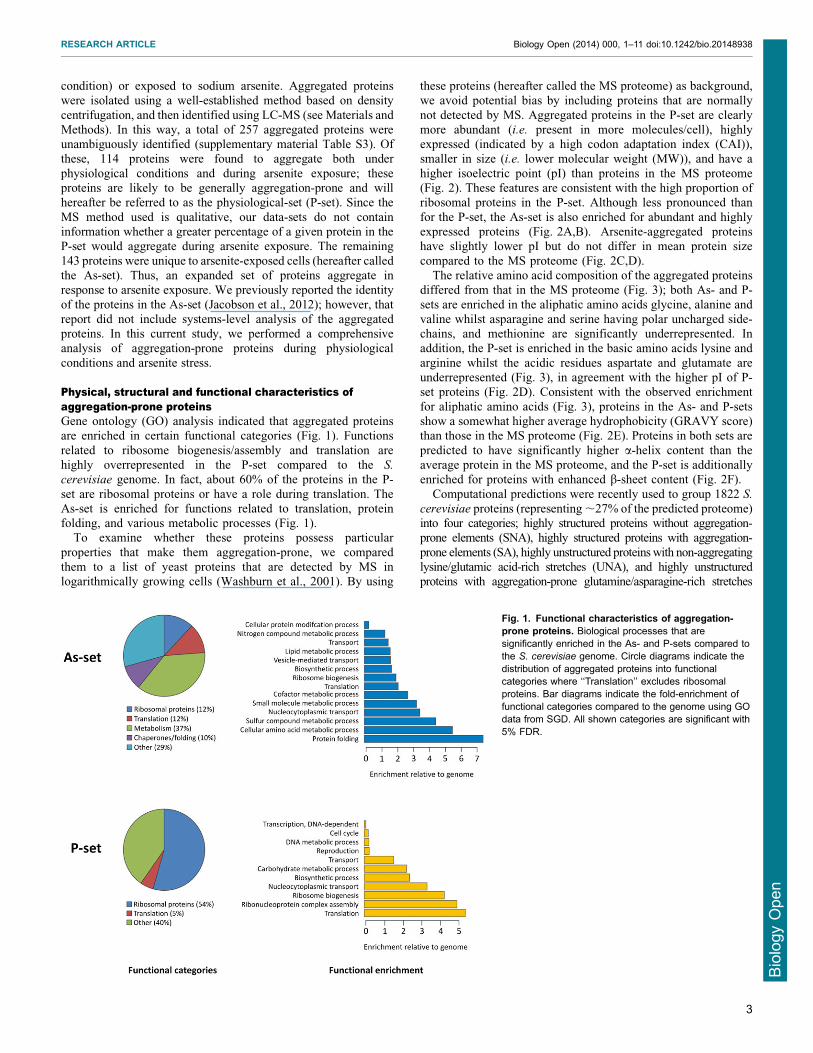
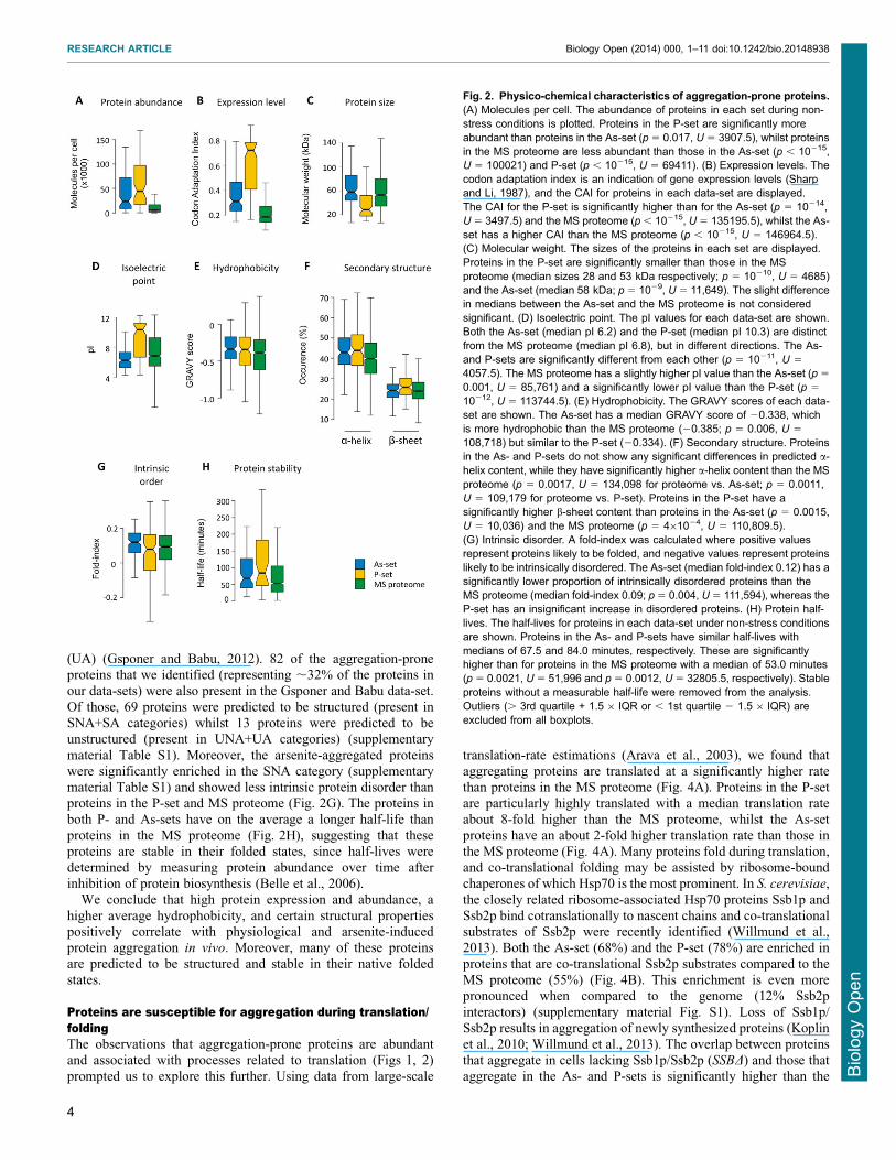
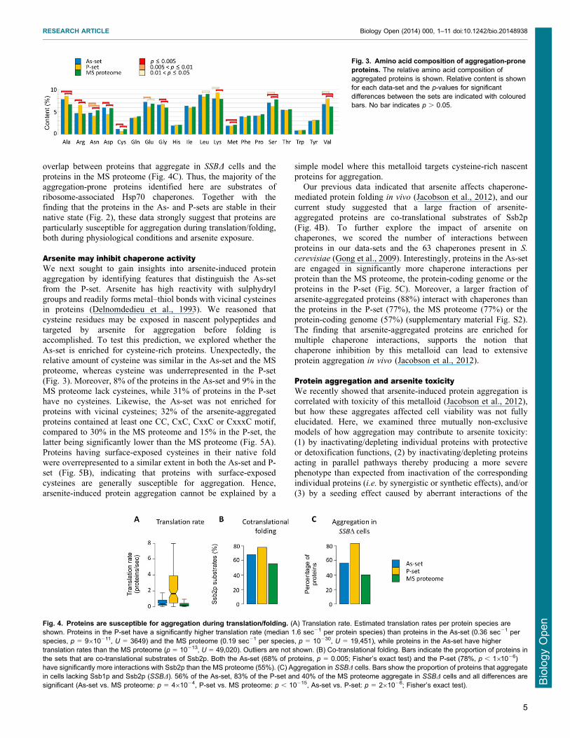
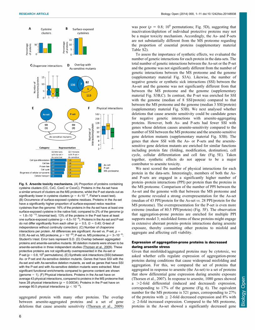
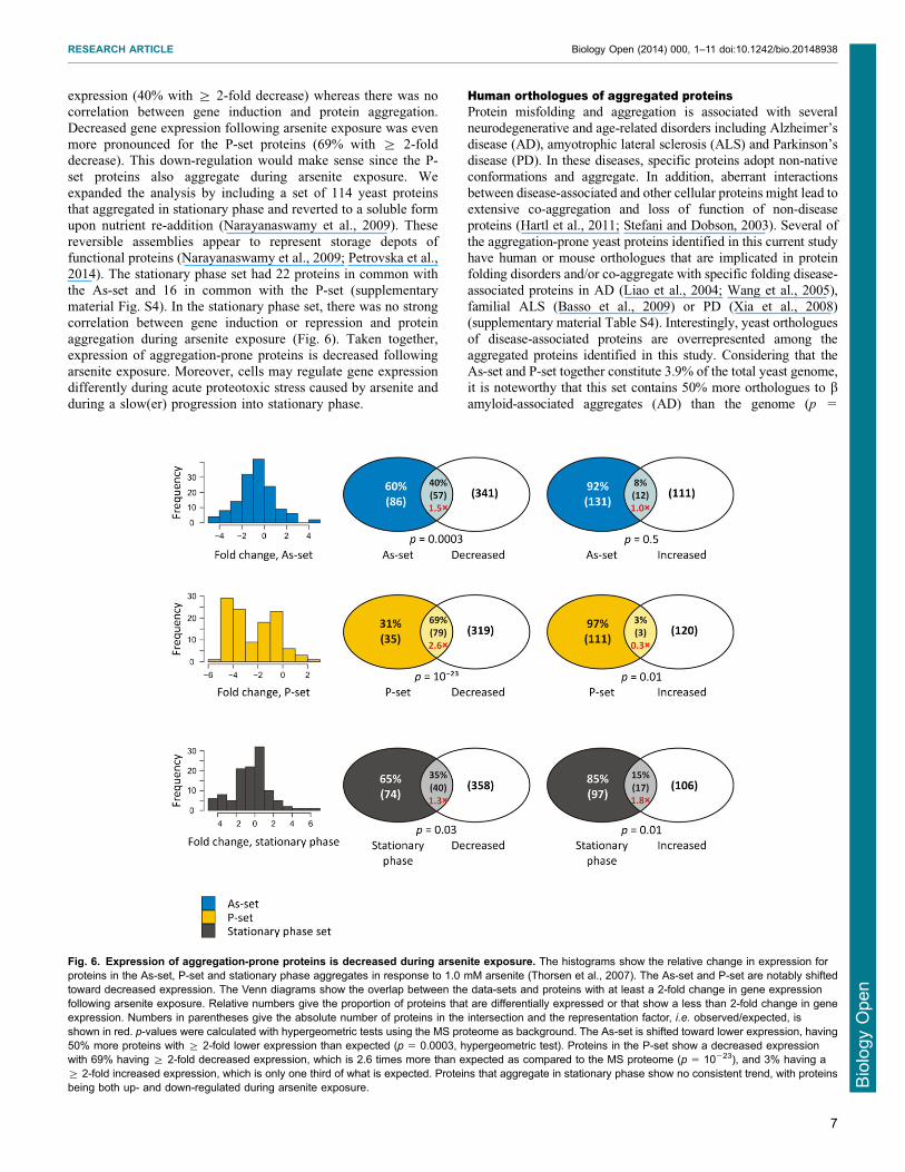
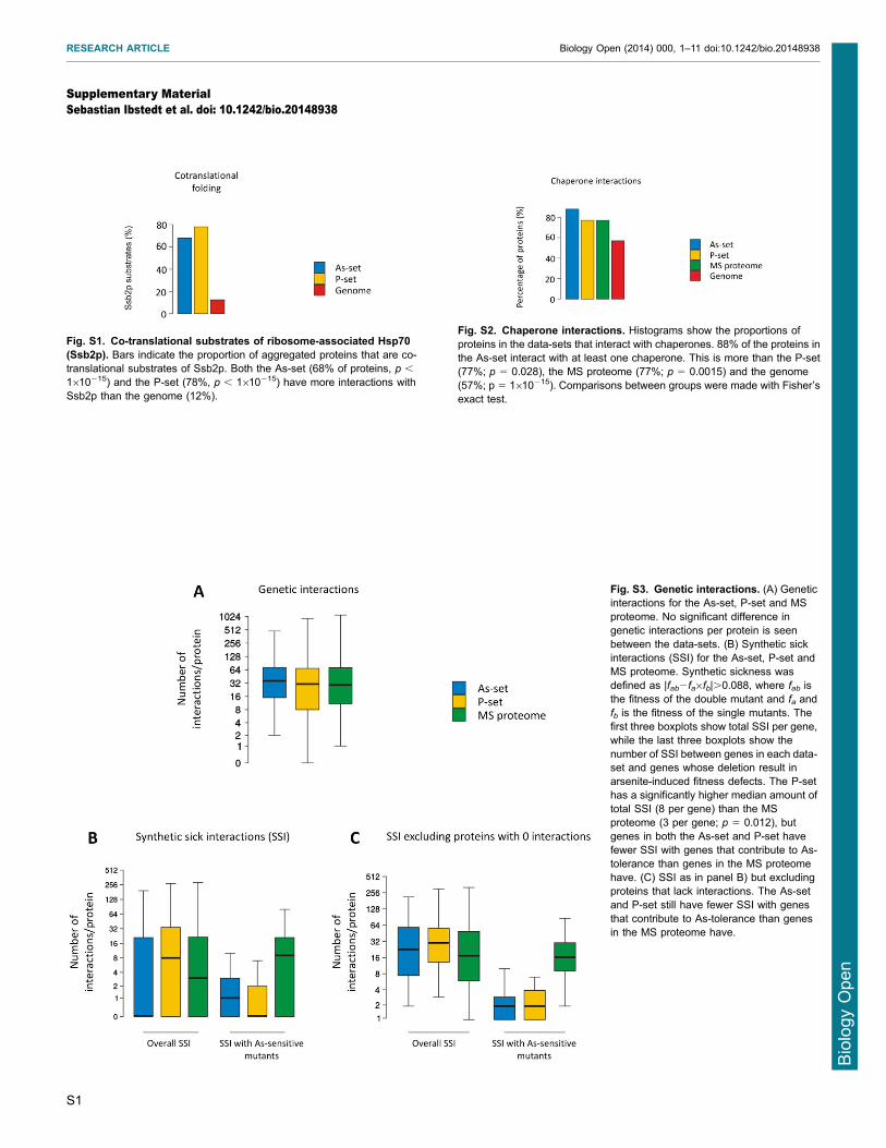
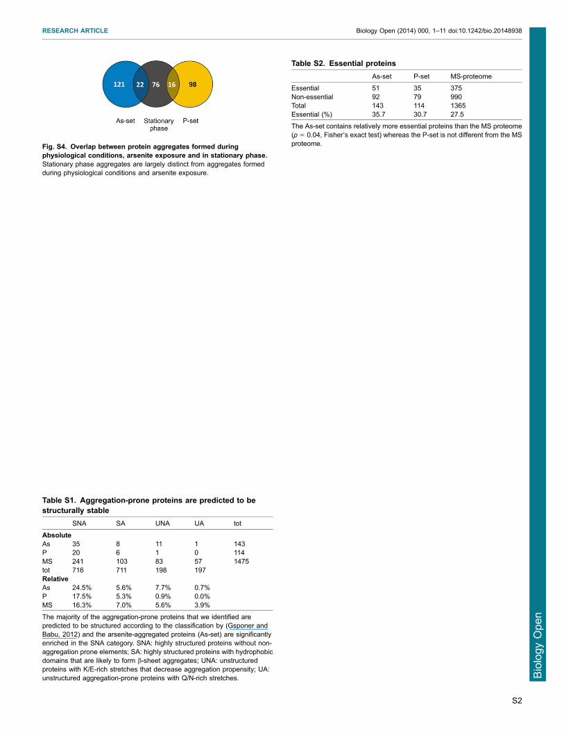