Hepatitis C virus leaves an epigenetic signature post cure of ...
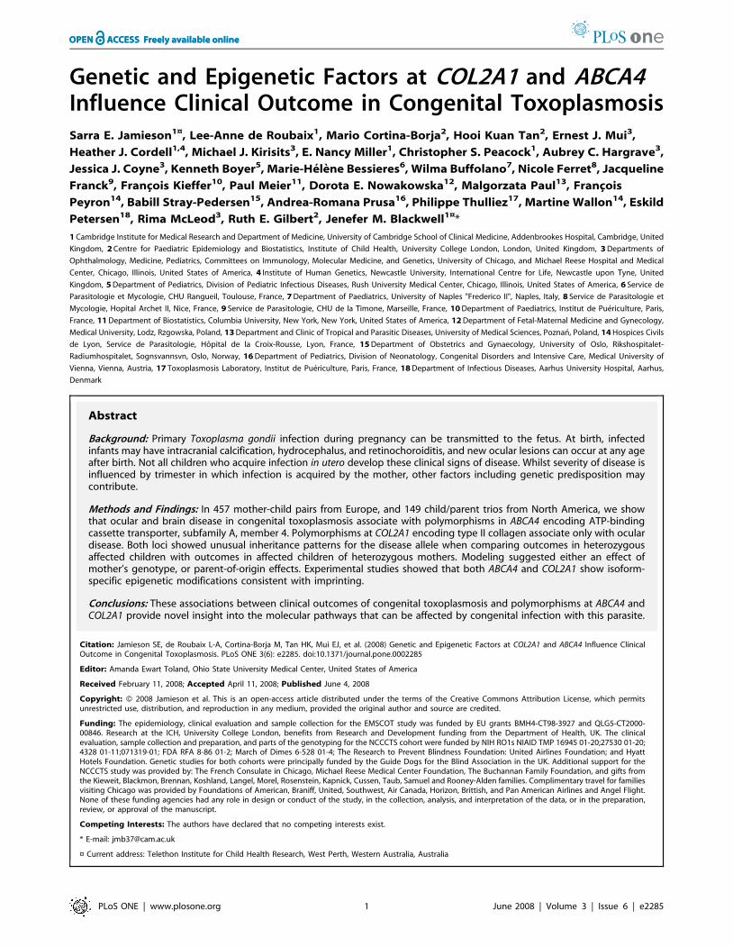
Genetic and Epigenetic Factors at COL2A1 and ABCA4Influence Clinical Outcome in Congenital ToxoplasmosisSarra E. Jamieson1¤, Lee-Anne de Roubaix1, Mario Cortina-Borja2, Hooi Kuan Tan2, Ernest J. Mui3,
Heather J. Cordell1,4, Michael J. Kirisits3, E. Nancy Miller1, Christopher S. Peacock1, Aubrey C. Hargrave3,
Jessica J. Coyne3, Kenneth Boyer5, Marie-Helene Bessieres6, Wilma Buffolano7, Nicole Ferret8, Jacqueline
Franck9, Francois Kieffer10, Paul Meier11, Dorota E. Nowakowska12, Malgorzata Paul13, Francois
Peyron14, Babill Stray-Pedersen15, Andrea-Romana Prusa16, Philippe Thulliez17, Martine Wallon14, Eskild
Petersen18, Rima McLeod3, Ruth E. Gilbert2, Jenefer M. Blackwell1¤*
1 Cambridge Institute for Medical Research and Department of Medicine, University of Cambridge School of Clinical Medicine, Addenbrookes Hospital, Cambridge, United
Kingdom, 2 Centre for Paediatric Epidemiology and Biostatistics, Institute of Child Health, University College London, London, United Kingdom, 3 Departments of
Ophthalmology, Medicine, Pediatrics, Committees on Immunology, Molecular Medicine, and Genetics, University of Chicago, and Michael Reese Hospital and Medical
Center, Chicago, Illinois, United States of America, 4 Institute of Human Genetics, Newcastle University, International Centre for Life, Newcastle upon Tyne, United
Kingdom, 5 Department of Pediatrics, Division of Pediatric Infectious Diseases, Rush University Medical Center, Chicago, Illinois, United States of America, 6 Service de
Parasitologie et Mycologie, CHU Rangueil, Toulouse, France, 7 Department of Paediatrics, University of Naples "Frederico II", Naples, Italy, 8 Service de Parasitologie et
Mycologie, Hopital Archet II, Nice, France, 9 Service de Parasitologie, CHU de la Timone, Marseille, France, 10 Department of Paediatrics, Institut de Puericulture, Paris,
France, 11 Department of Biostatistics, Columbia University, New York, New York, United States of America, 12 Department of Fetal-Maternal Medicine and Gynecology,
Medical University, Lodz, Rzgowska, Poland, 13 Department and Clinic of Tropical and Parasitic Diseases, University of Medical Sciences, Poznan, Poland, 14 Hospices Civils
de Lyon, Service de Parasitologie, Hopital de la Croix-Rousse, Lyon, France, 15 Department of Obstetrics and Gynaecology, University of Oslo, Rikshospitalet-
Radiumhospitalet, Sognsvannsvn, Oslo, Norway, 16 Department of Pediatrics, Division of Neonatology, Congenital Disorders and Intensive Care, Medical University of
Vienna, Vienna, Austria, 17 Toxoplasmosis Laboratory, Institut de Puericulture, Paris, France, 18 Department of Infectious Diseases, Aarhus University Hospital, Aarhus,
Denmark
Abstract
Background: Primary Toxoplasma gondii infection during pregnancy can be transmitted to the fetus. At birth, infectedinfants may have intracranial calcification, hydrocephalus, and retinochoroiditis, and new ocular lesions can occur at any ageafter birth. Not all children who acquire infection in utero develop these clinical signs of disease. Whilst severity of disease isinfluenced by trimester in which infection is acquired by the mother, other factors including genetic predisposition maycontribute.
Methods and Findings: In 457 mother-child pairs from Europe, and 149 child/parent trios from North America, we showthat ocular and brain disease in congenital toxoplasmosis associate with polymorphisms in ABCA4 encoding ATP-bindingcassette transporter, subfamily A, member 4. Polymorphisms at COL2A1 encoding type II collagen associate only with oculardisease. Both loci showed unusual inheritance patterns for the disease allele when comparing outcomes in heterozygousaffected children with outcomes in affected children of heterozygous mothers. Modeling suggested either an effect ofmother’s genotype, or parent-of-origin effects. Experimental studies showed that both ABCA4 and COL2A1 show isoform-specific epigenetic modifications consistent with imprinting.
Conclusions: These associations between clinical outcomes of congenital toxoplasmosis and polymorphisms at ABCA4 andCOL2A1 provide novel insight into the molecular pathways that can be affected by congenital infection with this parasite.
Citation: Jamieson SE, de Roubaix L-A, Cortina-Borja M, Tan HK, Mui EJ, et al. (2008) Genetic and Epigenetic Factors at COL2A1 and ABCA4 Influence ClinicalOutcome in Congenital Toxoplasmosis. PLoS ONE 3(6): e2285. doi:10.1371/journal.pone.0002285
Editor: Amanda Ewart Toland, Ohio State University Medical Center, United States of America
Received February 11, 2008; Accepted April 11, 2008; Published June 4, 2008
Copyright: � 2008 Jamieson et al. This is an open-access article distributed under the terms of the Creative Commons Attribution License, which permitsunrestricted use, distribution, and reproduction in any medium, provided the original author and source are credited.
Funding: The epidemiology, clinical evaluation and sample collection for the EMSCOT study was funded by EU grants BMH4-CT98-3927 and QLG5-CT2000-00846. Research at the ICH, University College London, benefits from Research and Development funding from the Department of Health, UK. The clinicalevaluation, sample collection and preparation, and parts of the genotyping for the NCCCTS cohort were funded by NIH RO1s NIAID TMP 16945 01-20;27530 01-20;4328 01-11;071319-01; FDA RFA 8-86 01-2; March of Dimes 6-528 01-4; The Research to Prevent Blindness Foundation: United Airlines Foundation; and HyattHotels Foundation. Genetic studies for both cohorts were principally funded by the Guide Dogs for the Blind Association in the UK. Additional support for theNCCCTS study was provided by: The French Consulate in Chicago, Michael Reese Medical Center Foundation, The Buchannan Family Foundation, and gifts fromthe Kieweit, Blackmon, Brennan, Koshland, Langel, Morel, Rosenstein, Kapnick, Cussen, Taub, Samuel and Rooney-Alden families. Complimentary travel for familiesvisiting Chicago was provided by Foundations of American, Braniff, United, Southwest, Air Canada, Horizon, Brittish, and Pan American Airlines and Angel Flight.None of these funding agencies had any role in design or conduct of the study, in the collection, analysis, and interpretation of the data, or in the preparation,review, or approval of the manuscript.
Competing Interests: The authors have declared that no competing interests exist.
* E-mail: [email protected]
¤ Current address: Telethon Institute for Child Health Research, West Perth, Western Australia, Australia
PLoS ONE | www.plosone.org 1 June 2008 | Volume 3 | Issue 6 | e2285

Introduction
Toxoplasma gondii is a ubiquitous protozoan parasitic infection
that, if acquired for the first time during pregnancy, can be
transmitted to the fetus. At birth, infants infected in utero may have
intracranial calcification, hydrocephalus, and ocular disease
broadly defined as retinochoroiditis or inflammation of the retina
and choroid with associated vitritis [1–3]. New ocular lesions can
occur at any age after birth, in untreated and some treated
children. Whilst severity of disease is influenced by trimester in
which infection is acquired by the mother [4,5], other factors
including genetic predisposition may contribute. For example,
previous studies suggest that genes affecting immune response,
including HLA [6], influence clinical outcome in the child.
However, since infants who have the most severe clinical signs in
the brain and eye are those infected early in pregnancy [4,5] when
fetal immunity is least well developed, we considered whether
genes that encode molecules that play a role in developmental
processes could contribute to clinical phenotype observed in the
child. This could provide unique insight into events in utero and
post-natally that determine the clinical outcome of infection. In
particular, we hypothesized that propensity for T. gondii to cause
eye disease may be associated with genes previously implicated in
congenital or juvenile onset ocular disease. Two genes were of
specific interest, ABCA4 encoding ATP-binding cassette transport-
er subfamily A member 4 associated with juvenile onset retinal
dystrophies including Stargardt’s disease [7,8], and COL2A1
encoding type II collagen associated with Stickler syndrome [9]
in which there is congenital abnormal vitreous and lattice retinal
degeneration. Although risk of transmission of the parasite to the
fetus in pregnant women with primary infection ranges from ,1–
100% depending on the time in gestation when infection is
acquired [5], incidence rates of clinical congenital toxoplasmosis,
together with the fact that it is not a reportable disease, have
precluded accumulation of large cohorts for genetic studies.
Nevertheless, two unique cohorts have recently become available
to test this specific genetic hypothesis. These cohorts are from the
European Multicentre Cohort Study on Congenital Toxoplasmo-
sis (EMSCOT) which recruited prospectively for mothers with
primary T. gondii in pregnancy [1,2], and from the National
Collaborative Chicago-based Congenital Toxoplasmosis Study
(NCCCTS) [3] in North America to which infants and children
with congenital infection with T. gondii are referred. Using these
cohorts we show that ocular and brain disease in congenital
toxoplasmosis associate with polymorphisms in ABCA4, while
polymorphisms at COL2A1 encoding type II collagen associate
only with ocular disease. Both loci show unusual inheritance
patterns for the disease allele when comparing outcomes in
heterozygous affected children with outcomes in affected children
of heterozygous mothers, and modeling suggests either an effect of
mother’s genotype or parent-of-origin effects. The latter is
supported by experimental data showing that both ABCA4 and
COL2A1 show isoform-specific epigenetic modifications consistent
with imprinting.
Methods
European study populationThe European study was undertaken as an adjunct to European
Multicentre Study of Congenital Toxoplasmosis (EMSCOT),
referred to as GENET-EMSCOT. Ethical approvals for GEN-
ET-EMSCOT were obtained through the local ethical review
boards of participating centres across Europe, and for the study as
a whole from the Research Ethics Committee for Great Ormond
Street Hospital and the Institute for Child Health in London.
Screening, treatment and follow up schedules have been described
in detail elsewhere [1]. For this study, mother-child pairs were
eligible for inclusion if the child had congenital toxoplasmosis or, if
the child was uninfected, the mother had evidence of seroconver-
sion (i.e. change from IgG negative to IgG positive for specific
antibodies to T. gondii) during pregnancy. Infected children were
identified in centres that utilized universal prenatal or neonatal
screening (Copenhagen, Stockholm, Lodz and Poznan). Uninfect-
ed children were identified only in those centres that utilized
prenatal screening (Lyon, Paris, Marseille, Toulouse, Nice,
Grenoble, Vienna, Naples, Oslo). Families were invited to
participate by the clinician responsible for follow up after diagnosis
of toxoplasmosis. The clinician recorded details reflecting the
timing of maternal seroconversion, and in the child, confirmation
of congenital infection status, detection of intracranial lesions
based on cranial ultrasound after birth, and the age at detection of
any retinochoroidal lesions or the age at the last negative
ophthalmoscopy examination. We estimate that 85% of the
mothers in the EMSCOT study had been treated prenatally [2].
Recent analyses of EMSCOT and other cohort studies provides
no evidence for a statistically significant effect of prenatal
treatment on brain or eye lesions [10–12]. Hence, although there
is variability in prenatal treatment across the EMSCOT cohort, it
is unlikely that this will confound the genetic statistical analysis
performed here. To increase the power of the study, clinicians
were asked to selectively recruit infected and affected children. For
some centres (Lyon, Toulouse, Marseille, Paris, Lodz) this included
prospective sampling and retrospective samples of stored sera or
plasma from mother-child pairs with confirmed clinical congenital
toxoplasmosis, and for whom comparable data were available to
accurately complete questionnaires developed for the GENET-
EMSCOT study. Within these groups, the study population was
assumed to be representative of mothers and children identified by
universal screening for toxoplasmosis. However, this could not be
checked as families not enrolled were not recorded. The child’s
grandparents’ countries of birth were recorded to provide an
acceptable proxy for ethnicity. Buccal swabs from the mother and
child were taken into transport/lysis buffer (10mmol/L TRIS
base, 10mmol/L EDTA, 0.5% Na sarkosyl) and kept at ambient
temperatures during postage to the laboratory in Cambridge.
Plasma or serum samples stored at 220uC were shipped frozen to
Cambridge. A total of 457 mother-child pairs met the inclusion
criteria to participate in the study, which included successful
preparation of DNA.
North American Study PopulationCase-parent trios for the North American cohort were from the
National Collaborative Chicago-based Congenital Toxoplasmosis
Study (NCCCTS) [3]. Ethical approval for the study was obtained
from the local Institutional Review Boards of the University of
Chicago and Michael Reese Hospital and Medical Center, and
oversight was provided by an Internal Data Safety Monitoring
Committee, the Data Safety Monitoring Board, and NIH. The
diagnosis of congenital toxoplasmosis was confirmed on the basis
of clinical findings and testing in the Toxoplasmosis Serology
Laboratory (Palo Alto Medical Research Institute) as described
[3,5]. At birth or time of diagnosis, each child was examined in the
same center in Chicago with standardized ophthalmologic
examination and review of all medical records and a brain CT
scan [3]. Samples for 176 clinically confirmed children were
available for the genetic study, 138 from an ongoing treatment
Genetics and Toxoplasmosis
PLoS ONE | www.plosone.org 2 June 2008 | Volume 3 | Issue 6 | e2285

trial [3,5,13]. Inclusion criteria for these 138 children were as
follows: (1) age less than 2.5 months at diagnosis, (2) diagnosis of
congenital toxoplasmosis highly likely as previously described [13],
(3) willingness to be periodically evaluated in Chicago, and (4) no
concomitant immunosuppressive conditions. The additional 38
children presented after the first year of life and were therefore not
treated during this time. However, their clinical evaluation was as
described before [5]. Peripheral blood cells were isolated and
cryopreserved from all children and their mothers and some
fathers. A small sample (10 ml) of these cells in cryopreservation
mix was placed in 100 ml transport/lysis buffer (as above), and
shipped to Cambridge at ambient temperature. A total of 149
children and available parents met the inclusion criteria to
participate in the study, which included successful preparation of
DNA.
GenotypingDNA was obtained from all samples by whole genome
amplification of DNA extracted from the buccal swab buffer, or
by direct amplification from the buccal buffer, serum, plasma, or
lysis buffer from cells, using multiple displacement amplification
(MDA, Molecular Staging, USA; now supplied by Qiagen). Over a
number of large family-based studies undertaken in our laboratory
we have demonstrated a rate of 1.1% to 2.6% allele drop out using
DNA amplified in this way. Seven single nucleotide polymor-
phisms (SNPs) at ABCA4 (rs1801574, rs2275033, rs2297671,
rs2297633, rs176375, rs3112831, rs952499) and 7 SNPs at
COL2A1 (rs6823, rs2070739, rs2276455, rs2276454, rs1635544,
rs1793958, rs3803183) were genotyped in both cohorts. We also
examined 2 SNPs at VMD2 encoding bestrophin 1 or vitelliform
macular dystrophy protein 2, mutations in which are associated
with an autosomal dominant juvenile-onset macular dystrophy
[14] but the clinical phenotype differs from congenital toxoplas-
mosis, and 3 SNPs at TIMP3 which encodes tissue inhibitor of
metalloproteinases-3 that is mutated in Sorsby fundus dystrophy,
an autosomal dominant retinopathy of late onset associated with
macular degenerative disease [15]. VMD2 and TIMP3 SNPs were
genotyped in the primary EMSCOT cohort only. To avoid
extensive amounts of multiple testing in examining the specific
hypothesis that ABCA4 and COL2A1 are candidate susceptibility
genes for congenital toxoplasmosis we did not look at other eye
disorder genes. Table S1 provides details on bp location in the
genome (NCBI Build 36.2), position within the gene/locus,
alternative alleles and their frequencies (EMSCOT, NCCCTS
and public domain) at each SNP. Figure 1 provides a
diagrammatic representation of the position of each SNP across
the ABCA4 and COL2A1 genes, as well as linkage disequilibrium
between markers determined and graphed using Haploview
available from the HapMap Project Site (http://www.hapmap.
org/). All SNPs were genotyped using TaqManH SNP Genotyping
Assays or Custom TaqManH SNP Genotyping Assays (COL2A1
rs1635544 only) (Applied Biosystems, CA, USA). Taqman assays
were performed in 384-well plates with all liquid handling carried
out using a BiomekFX robotics system (Beckman, High Wycombe,
UK). Taqman assays were analysed using an ABI 7900HT Fast
Real-Time PCR System (Applied Biosystems) and data scored for
genotype clustering using ABI SDS v2.1 software.
Statistical AnalysesTests for deviation from Hardy-Weinberg equilibrium (HWE)
for all markers were performed within STATA v8.0 (http://www.
stata.com/) using the GenAssoc package (available from http://
www-gene.cimr.cam.ac.uk/clayton/software/stata/) or using rou-
tines available from the population genetics R library version 1.2.0
developed by Gregory Warnes and Friedrich Leisch available from
http://cran.r-project.org/src/contrib/Descriptions/genetics.html.
All markers were in HWE for genetically independent individuals
within country-specific samples for EMSCOT, and for the parents
of trios in the NCCCTS cohort (data not shown). Linkage
disequilibrium was measured as D’ [16] and plots were generated
using Haploview available from the HapMap Project Site (http://
www.hapmap.org/). Power calculations for case-control samples
were performed in excel using a script prepared in-house at CIMR
by Dr Heather Cordell. Full details are provided in the Online
Supplementary Material (Tables S2A to S2D). As examples from
these tables, for an effect size (odds ratio or OR) of 3 (i.e. of the
order of magnitude of observed OR) at allele frequencies 0.2 and
P = 0.001, the European study sample had 100% power to detect
allelic association for the comparison of uninfected mothers or
children (N = 225) against infected mothers or children (N = 232);
98% power for the comparison of unaffected mothers/children
(N = 153) against affected mothers/children (N = 79); 91% power
for the comparison of unaffected mothers/children (N = 153)
against eye lesion mothers/children (N = 53); and 87% power for
the comparison of unaffected mothers/children (N = 153) against
brain lesion mothers/children (N = 45). TDT power approxima-
tions for trios were carried out using the method of Knapp [17]
(see Table S3). For example, the 124 affected (i.e. eye and or brain
lesions), 113 eye lesion, and 103 brain lesion full child/parent trios
had 81.5%, 76.3% and 68% power respectively to detect allelic
association at an odds ratio of 3 at P = 0.001 for markers at minor
allele frequencies = 0.2. All except two exonic SNPs in COL2A1
(rs2070739 both cohorts; rs3803183 Europe only) had variant
allele frequencies $ 0.2 in both European and North American
populations we studied (Table S1).
For the European study sample, genotype and allele frequencies
were compared using logistic regression analysis under a
multiplicative model (i.e. two alleles contribute twice the effect of
one allele), with test statistic (Chi-squared, x2), odds ratio (OR),
95% confidence intervals (CI) and P values used to evaluate
significance of an association. A likelihood ratio test was used to
determine dominance effects by comparing 1 degree of freedom
(df; allele-wise) and 2 df (genotype-wise) tests. For single marker
analysis, logistic regression analyses were carried out with and
without adjustment for country, trimester of pregnancy at
seroconversion of the mother, and country of origin of
grandparents. Since these adjustments had only minor effects on
the associations and significance levels obtained without adjust-
ment (i.e. we never observed significance in the unadjusted data
that was not also present after adjustment; data not shown),
stepwise and interaction analyses were performed without
adjusting for these parameters. A stepwise logistic-regression
procedure was used to evaluate the relative importance of variants
within and between the two candidate gene loci. Wald x2 tests
were used to compare models in which the main effects for both
loci were modeled with one in which the main effects at the
primary locus only were included. Interaction between loci was
also analysed using logistic regression analysis.
For the North American study sample, family-based allelic
association tests based on the TDT but generalised to allow
analysis under additive and dominant models of inheritance were
performed within FBAT [18]. Allelic associations, and relative risk
estimates, were obtained by creating a case/pseudocontrol study
where the cases comprise the genotypes of the affected offspring,
and the pseudo-sib controls are the one to three other genotypes
(depending on whether phase is known or inferred) which the
affected offspring might have received from the parents. The
relative risks were estimated using conditional logistic regression
Genetics and Toxoplasmosis
PLoS ONE | www.plosone.org 3 June 2008 | Volume 3 | Issue 6 | e2285

analysis, with test statistic (x2), OR, 95% CI and P values used to
evaluate significance of an association. A stepwise conditional
logistic-regression procedure was used to evaluate the relative
importance of variants within and between candidate gene loci, as
before. Case/pseudocontrol statistical tests implemented within
STATA were developed by Heather Cordell and David Clayton at
the Cambridge Institute for Medical Research and are available at
http://www-gene.cimr.cam.ac.uk/clayton/software/.
Since there was a priori clinical evidence to support candidacy of
each of the genes studied, and not all markers within genes were
independent (i.e. they were in linkage disequilibrium; Figure 1), we
did not apply a multiple testing correction. However, the stepwise
logistic regression analyses permitted us to determine whether
markers within each gene showed independent main effects.
To obtain statistical evidence for imprinting, we used a log-
linear method designed to evaluate parent-of-origin effects in case-
parent trios [19] once mother’s and child’s genotypes have been
included in the model. We used the program LEM [20] which also
allows assessment of maternal genotype effects with or without
including a parent-of-origin effect. For the European cohort,
where only mother-child pairs were available, the same param-
eterization was employed and the models were fitted using an in-
house program written by Dr Heather Cordell, under the
assumption of random mating and Hardy Weinberg equilibrium
Figure 1. Gene structure and linkage disequilibrium plots for ABCA4 (left panel) and COL2A1 (right panel). Upper diagrams showpositions of SNPs genotyped in relative to intron/exon structure of the gene. Lower diagrams show the linkage disequilibrium (D9) plots generated inHaploview (http://www.hapmap.org/) using data for each gene from the EMSCOT or NCCCTS cohorts as indicated. Linkage disequilibrium values(6100) between markers are indicated at the intercept of the two markers on the matrix. Where there is no value, D9 = 1 (i.e. 100). Haplotype blockswithin each gene are outlined within the black triangles. The black (high) through grey to white (low) shading indicates the degree of confidence inthe estimate of linkage disequilibrium between the markers. For the EMSCOT cohort, stepwise logistic regression analysis [50] of associationsobserved in the mothers (Online Supplementary Material, Text S1 plus Table S5) indicate that all of the association at ABCA4 is accounted for byrs2997633 and rs1761375, implying that a single etiological variant in strong linkage disequilibrium with these two markers may account for theassociation. At COL2A1, SNPs rs2276455, rs1635544 and rs3803183 all add significant main effects when compared to rs2070739, but rs2070739 doesnot add significant main effects to any one of these markers. SNPs rs2276455, rs1635544 and rs3803183 do not add significant main effects to eachother. Once pairs of these markers are taken into the model, the third SNP does not add significant main effects. A single etiological variant in thishaplotype block could account for the association with COL2A1. Neither SNP (rs2070739, rs3803183) that results in a non-synonymous amino acidsubstitution appears to be the primary functional variant.doi:10.1371/journal.pone.0002285.g001
Genetics and Toxoplasmosis
PLoS ONE | www.plosone.org 4 June 2008 | Volume 3 | Issue 6 | e2285

using public domain allele frequencies for European populations
(Table S1).
Experimental Evidence for ImprintingAnonymised EBV, Y79 human retinoblastoma (ECACC), and
WERI-RB1 human retinoblastoma (gift from Prof D. Trump,
Manchester) cell lines were cultured in RPMI 1640-Dutch
modification media. HEK293 human embryonic kidney cells (gift
from Dr C. Vacher, Cambridge) were cultured in DMEM media.
All cell cultures were supplemented with 10% FCS, 100 U/ml
Penicillin/100 mg/ml Streptomycin and 2 mM L-glutamine. All
reagents were obtained from Invitrogen. DNA was extracted from
cell pellets using the salting out method. Total RNA was extracted
using TRI-REAGENT (Sigma) according to manufacturer’s
instructions. Genomic DNA and cDNA from H9 and hSF6
human embryonic stem cells (gift from Prof R. Pederson,
Cambridge) were obtained as previously described [21]. Tissue
RNAs (placenta, uterus, adult and fetal brain), were part of a total
human RNA master panel II (BD Biosciences). To look for
monoallelic expression, total RNA (1–2 mg) was reverse tran-
scribed using M-MLV first strand cDNA synthesis system
(Invitrogen) using either oligo dT(18) or gene specific primers for
ABCA4 or COL2A1 (Table S4A). Resulting cDNAs and genomic
DNAs were amplified across regions containing polymorphic
exonic SNPs of interest using primers as detailed (Table S4B). All
PCRs were performed using a Touchdown program, briefly
annealing temperature is ramped down by 0.5uC per cycle from
63uC to 56uC followed by a further 19 cycles at 56uC. PCR
products were purified using SAP (1 U/ml)//Exo I (10 U/ml)
digestion and sequencing performed in both forward and reverse
directions using BigDye Terminator v3.1 (Applied Biosystems) and
run on an ABI 3100 Genetic Analyser. Sequence data was
analysed using pregap4 and gap4.
Results
Characteristics of the Two Cohorts StudiedDNA was successfully obtained from 457 mother-child pairs
with confirmed infection during pregnancy from the EMSCOT
cohort [1,2]. Transmission of infection during pregnancy was
confirmed in 232 (51%) infants; 225 (49%) infants remained
uninfected. All infants were monitored for clinical signs until at
least three years of age, and many have been followed for .10
years. Of the 232 infected infants, 79 (34%) had clinical signs
(referred to as affected) of congenital toxoplasmosis: 53 (67%) with
ocular lesions (retinochoroidal lesions), 45 (57%) with brain lesions
(hydrocephalus or intracranial calcifications detected on ultra-
sound examination of the brain), 19 (24%) of these infants had
both eye and brain lesions. The median age at the first detection of
retinochoroidal lesions was 84 months (range 0 to 237 months;
inter-quartile range 46 to 144 months) and at the last negative
ophthalmic examination in unaffected children was 31months
(range 0 to 252; inter-quartile range 12 to 66 months). 153 (66%)
infected infants had no clinical signs of disease up to a minimum
follow-up period of 3 years of age. Two-thirds of mother-child
pairs reported grandparents’ countries of birth. Of these, 95%
were Caucasian and 5% of African origin.
For the second independently ascertained cohort, the NCCCTS
[3], DNA was successfully obtained from 149 children with
confirmed congenital infection (69% Caucasian, 15% Hispanic,
8% Asian or Pacific Islander, 3% African American, 0.7% Native
American, 4.7% mixed race) plus available parents. At birth or
time of diagnosis, 92 (62%) infected children had brain
calcifications with/without hydrocephalus and retinal lesions, 21
(14%) had retinal lesions only, 11 (7%) had brain calcifications
with/without hydrocephalus only, and 25 (17%) infected children
were without these clinical findings. Only the 124 children with
confirmed clinical findings in eye and/or brain were included in
the allelic association analysis for this cohort. This provided data
on 124 trios classified as affected (i.e. eye or brain lesions or both),
113 trios classified as eye disease (with or without brain disease)
and 103 trios classified as brain disease (with or without eye
disease).
Genetic Associations Between ABCA4 or COL2A1 and Eyeor Brain Findings in Children with CongenitalToxoplasmosis
For the EMSCOT cohort, no significant associations were
observed for any SNPs at ABCA4 or COL2A1 when congenitally
infected children of mothers with primary gestational infection
were compared with children of mothers with primary gestational
infection who did not transmit infection to the fetus (Table 1 and
data not shown). This was as expected since it was unlikely that
genes normally associated with genetic eye disorders would
influence transmission of infection from mother to child.
Nominally significant (i.e. P values without correction for multiple
testing) allelic associations were observed for 2 SNPs at ABCA4
(rs2997633, rs3112831) and 1 SNP at COL2A1 (rs2276455) when
affected children (i.e. children with retinal or brain disease or both)
in the EMSCOT cohort were compared with infected but
unaffected children (Table 1). Large effect sizes were observed
for these associations, particularly in children homozygous for the
disease allele (Table 2, odds ratios 6.81, 2.86 and 3.63 respectively
for rs2997633, rs3112831 and rs2276455). ABCA4 rs2297633
retained significance, and an additional SNP at each marker
(ABCA4 rs1761375 and COL2A1 rs1793958) was found to be
associated with disease, when the analysis was enriched for
children with the eye lesion phenotype (with/without brain lesions,
i.e. leaving out children with brain disease only) compared to
unaffected children (Table 1). No associations were observed when
the analysis was similarly enriched for children with brain lesions
(with/without eye lesions, leaving out children with eye disease
only) compared to unaffected children. No associations for any
phenotype were observed for VMD2 or TIMP3 (data not shown),
which are genes associated with other ocular phenotypes that
would not have been predicted to play a role in ocular disease
associated with congenital toxoplasmosis. All analyses for the
EMSCOT study were adjusted for country of birth, trimester of
seroconversion, and country of origin of grandparents as a
surrogate for ethnicity.
In the NCCCTS cohort, significant allelic associations were
observed for 1 SNP at ABCA4 (rs952499) and 5 SNPs at COL2A1
(rs6823, rs2070739, rs2276454, rs1635544, rs3803183) under a
dominant model of inheritance (Table 3). The use of case/parent
trios and transmission disequilibrium testing (in FBAT) controlled
for ethnic admixture in this cohort. For COL2A1, SNPs rs2276455,
rs2276454 and rs1635544 significance improved when the analysis
was enriched for the eye lesion phenotype (with/without brain
lesions), consistent with the strong haplotype block formed by these
3 markers (Figure 1). Some significance was also retained in the
allelic association analysis (Table 3) when the analysis was
enriched for the brain lesion phenotype (with/without eye lesions),
which could reflect the larger proportion of children with more
severe disease involving both eye and brain lesions in this cohort.
There were insufficient children with brain disease only to carry
out a separate analysis.
Overall, the evidence from the children with eye and brain signs
associated with congenitally acquired toxoplasmosis in these two
Genetics and Toxoplasmosis
PLoS ONE | www.plosone.org 5 June 2008 | Volume 3 | Issue 6 | e2285
cohorts was for association with eye disease and these two
previously recognized eye disorder genes, ABCA4 and COL2A1.
Linkage disequilibrium patterns across both loci were similar in
the two cohorts, with all of the SNPs that contribute main effects
(as determined by stepwise logistic regression analysis; legend to
figure 1 and Online Supplementary Material, Text S1 and Tables
S5,S6,S7) in the associations in each cohort falling in the same
haplotype blocks (Figure 1) within these large genes: ABCA4
spanning 128.3 kb with 50 exons, 2,273 amino acids, 255.9 kDa
protein; COL2A1 spanning 31.5 kb with 54 exons, 1,418 amino
acids, 134.4 kDa protein. Inter-locus stepwise logistic regression
analysis showed that the two genes had independent effects (Tables
S5 and S6), and there was no statistical evidence for interaction
between them (data not shown).
Influence of Mother’s Genotype on Clinical Outcome inChildren with Congenital Toxoplasmosis
Additional genetic information available in the EMSCOT
cohort was that of mother’s genotype. As a first approach to
determining whether mother’s genotype had an effect on disease
outcome in the child, logistic regression analysis was carried out
comparing mothers of affected children (eye and/or brain disease)
with mothers of infected unaffected children. This proved
interesting in that (1) all of the allele-wise associations were more
significant in comparisons of mothers (Table 4) than in
comparisons of infants (Table 1), (2) additional ABCA4
(rs952499) and COL2A1 (rs2070739, rs1635544) were associated
with mothers of affected infants compared to mothers of
unaffected infants (Table 4), and (3) all of the ABCA4 SNPs but
none of the COL2A1 SNPs were associated with mothers of infants
with brain disease (Table 4, note also footnote on mothers of
children with brain disease only). Statistically, it appeared that
genetic effects of these two loci on disease in the child were being
diluted out in making direct comparisons of the affected versus
unaffected children relative to the evidence for association when
comparing the mothers. Unusual patterns of inheritance were also
observed when comparing genotype-wise associations in children
versus mothers (Table 2) suggesting that there may be a direct
effect of mother’s genotype, or that there may be parent-of-origin
effects (imprinting), i.e. that for the child it is the origin of, and not
just the combination of, alleles that is important in determining
disease risk. Using a log-linear method previously designed to
evaluate maternal genotype and/or parent-of-origin effects in
case-parent trios [19], evidence for imprinting given mother’s and
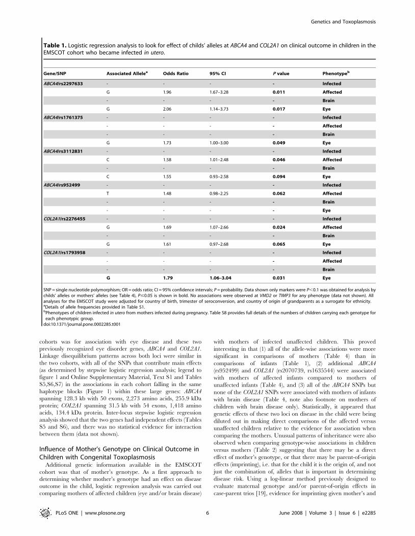
Table 1. Logistic regression analysis to look for effect of childs’ alleles at ABCA4 and COL2A1 on clinical outcome in children in theEMSCOT cohort who became infected in utero.
Gene/SNP Associated Allelea Odds Ratio 95% CI P value Phenotypeb
ABCA4/rs2297633 - - - - Infected
G 1.96 1.67–3.28 0.011 Affected
- - - - Brain
G 2.06 1.14–3.73 0.017 Eye
ABCA4/rs1761375 - - - - Infected
- - - - Affected
- - - - Brain
G 1.73 1.00–3.00 0.049 Eye
ABCA4/rs3112831 - - - - Infected
C 1.58 1.01–2.48 0.046 Affected
- - - - Brain
C 1.55 0.93–2.58 0.094 Eye
ABCA4/rs952499 - - - - Infected
T 1.48 0.98–2.25 0.062 Affected
- - - - Brain
- - - - Eye
COL2A1/rs2276455 - - - - Infected
G 1.69 1.07–2.66 0.024 Affected
- - - - Brain
G 1.61 0.97–2.68 0.065 Eye
COL2A1/rs1793958 - - - - Infected
- - - - Affected
- - - - Brain
G 1.79 1.06–3.04 0.031 Eye
SNP = single nucleotide polymorphism; OR = odds ratio; CI = 95% confidence intervals; P = probability. Data shown only markers were P,0.1 was obtained for analysis bychilds’ alleles or mothers’ alleles (see Table 4), P#0.05 is shown in bold. No associations were observed at VMD2 or TIMP3 for any phenotype (data not shown). Allanalyses for the EMSCOT study were adjusted for country of birth, trimester of seroconversion, and country of origin of grandparents as a surrogate for ethnicity.aDetails of allele frequencies provided in Table S1.bPhenotypes of children infected in utero from mothers infected during pregnancy. Table S8 provides full details of the numbers of children carrying each genotype for
each phenotypic group.doi:10.1371/journal.pone.0002285.t001
Genetics and Toxoplasmosis
PLoS ONE | www.plosone.org 6 June 2008 | Volume 3 | Issue 6 | e2285
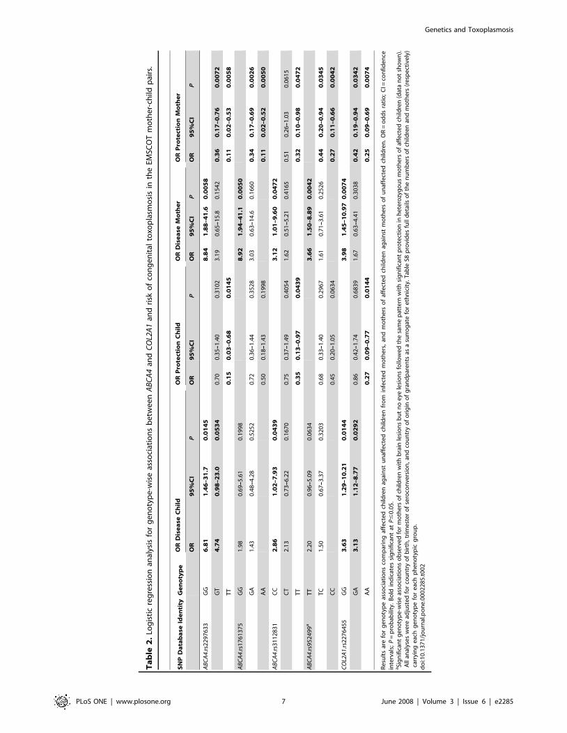
Ta
ble
2.
Log
isti
cre
gre
ssio
nan
alys
isfo
rg
en
oty
pe
-wis
eas
soci
atio
ns
be
twe
en
AB
CA
4an
dC
OL2
A1
and
risk
of
con
ge
nit
alto
xop
lasm
osi
sin
the
EMSC
OT
mo
the
r-ch
ildp
airs
.
SN
PD
ata
ba
seId
en
tity
Ge
no
typ
eO
RD
ise
ase
Ch
ild
OR
Pro
tect
ion
Ch
ild
OR
Dis
ea
seM
oth
er
OR
Pro
tect
ion
Mo
the
r
OR
95
%C
IP
OR
95
%C
IP
OR
95
%C
IP
OR
95
%C
IP
AB
CA
4.r
s22
97
63
3G
G6
.81
1.4
6–
31
.70
.01
45
8.8
41
.88
–4
1.6
0.0
05
8
GT
4.7
40
.98
–2
3.0
0.0
53
40
.70
0.3
5–
1.4
00
.31
02
3.1
90
.65
–1
5.8
0.1
54
20
.36
0.1
7–
0.7
60
.00
72
TT
0.1
50
.03
–0
.68
0.0
14
50
.11
0.0
2–
0.5
30
.00
58
AB
CA
4.r
s17
61
37
5G
G1
.98
0.6
9–
5.6
10
.19
98
8.9
21
.94
–4
1.1
0.0
05
0
GA
1.4
30
.48
–4
.28
0.5
25
20
.72
0.3
6–
1.4
40
.35
28
3.0
30
.63
–1
4.6
0.1
66
00
.34
0.1
7–
0.6
90
.00
26
AA
0.5
00
.18
–1
.43
0.1
99
80
.11
0.0
2–
0.5
20
.00
50
AB
CA
4.r
s31
12
83
1C
C2
.86
1.0
2–
7.9
30
.04
39
3.1
21
.01
–9
.60
0.0
47
2
CT
2.1
30
.73
–6
.22
0.1
67
00
.75
0.3
7–
1.4
90
.40
54
1.6
20
.51
–5
.21
0.4
16
50
.51
0.2
6–
1.0
30
.06
15
TT
0.3
50
.13
–0
.97
0.0
43
90
.32
0.1
0–
0.9
80
.04
72
AB
CA
4.r
s95
24
99
aT
T2
.20
0.9
6–
5.0
90
.06
34
3.6
61
.50
–8
.89
0.0
04
2
TC
1.5
00
.67
–3
.37
0.3
20
30
.68
0.3
3–
1.4
00
.29
67
1.6
10
.71
–3
.61
0.2
52
60
.44
0.2
0–
0.9
40
.03
45
CC
0.4
50
.20
–1
.05
0.0
63
40
.27
0.1
1–
0.6
60
.00
42
CO
L2A
1.r
s22
76
45
5G
G3
.63
1.2
9–
10
.21
0.0
14
43
.98
1.4
5–
10
.97
0.0
07
4
GA
3.1
31
.12
–8
.77
0.0
29
20
.86
0.4
2–
1.7
40
.68
39
1.6
70
.63
–4
.41
0.3
03
80
.42
0.1
9–
0.9
40
.03
42
AA
0.2
70
.09
–0
.77
0.0
14
40
.25
0.0
9–
0.6
90
.00
74
Re
sult
sar
efo
rg
en
oty
pe
asso
ciat
ion
sco
mp
arin
gaf
fect
ed
child
ren
agai
nst
un
affe
cte
dch
ildre
nfr
om
infe
cte
dm
oth
ers
,an
dm
oth
ers
of
affe
cte
dch
ildre
nag
ain
stm
oth
ers
of
un
affe
cte
dch
ildre
n.
OR
=o
dd
sra
tio
;C
I=co
nfi
de
nce
inte
rval
s;P
=p
rob
abili
ty.
Bo
ldin
dic
ate
ssi
gn
ific
ant
atP
#0
.05
.aSi
gn
ific
ant
ge
no
typ
e-w
ise
asso
ciat
ion
so
bse
rve
dfo
rm
oth
ers
of
child
ren
wit
hb
rain
lesi
on
sb
ut
no
eye
lesi
on
sfo
llow
ed
the
sam
ep
atte
rnw
ith
sig
nif
ican
tp
rote
ctio
nin
he
tero
zyg
ou
sm
oth
ers
of
affe
cte
dch
ildre
n(d
ata
no
tsh
ow
n).
All
anal
yse
sw
ere
adju
ste
dfo
rco
un
try
of
bir
th,
trim
est
er
of
sero
con
vers
ion
,an
dco
un
try
of
ori
gin
of
gra
nd
par
en
tsas
asu
rro
gat
efo
re
thn
icit
y.T
able
S8p
rovi
de
sfu
lld
eta
ilso
fth
en
um
be
rso
fch
ildre
nan
dm
oth
ers
(re
spe
ctiv
ely
)ca
rryi
ng
eac
hg
en
oty
pe
for
eac
hp
he
no
typ
icg
rou
p.
do
i:10
.13
71
/jo
urn
al.p
on
e.0
00
22
85
.t0
02
Genetics and Toxoplasmosis
PLoS ONE | www.plosone.org 7 June 2008 | Volume 3 | Issue 6 | e2285

child’s genotypes was obtained at ABCA4 rs952499 (P = 0.033) and
COL2A1 rs2070739 (P = 0.05) in the NCCCTS trios. For
EMSCOT we adapted the method for use with mother-child
pairs and found evidence for effects of maternal genotype given
child’s genotype and imprinting for both COL2A1 rs1635544
(P = 0.0088) and rs3803183 (P = 6.8761025), and evidence for
imprinting taking account of both mother’s and child’s genotype
for COL2A1 rs3803183 (P = 0.0025). Given the statistical limita-
tions of small sample size and power in the modeling analysis, and
the fact that a direct effect of mother’s genotype seemed unlikely
biologically, we looked for experimental evidence of epigenetic
effects or imprinting for ABCA4 and COL2A1.
Experimental evidence for imprintingTo obtain experimental evidence for imprinting, we screened an
anonymised EBV B cell bank (a) to find individuals heterozygous
for exonic SNPs at both loci; and (b) to determine mono-allelic
expression in elicit transcripts obtained from RNA from these
EBV cell lines. EBV lines were identified that were heterozygous
for SNP rs3112831 in exon 10 of ABCA4 and for SNP rs3737548
Table 3. FBAT analysis under dominant model of inheritancefor associations between ABCA4 and COL2A1 and congenitaltoxoplasmosis in the NCCCTS child-parent trios.
Gene/SNP Allelea Z score P value Phenotype
ABCA4/rs952499 C 22.255 0.024 Affectedb
C 21.750 0.080 Brain
C 21.750 0.080 Eye
COL2A1/rs6823 G +1.949 0.051 Affected
G +2.236 0.025 Brain
G +1.706 0.088 Eye
COL2A1/rs2070739 T +2.556 0.011 Affected
T +2.224 0.026 Brain
T +2.224 0.026 Eye
COL2A1/rs2276455 - - - Affected
- - - Brain
A 22.213 0.027 Eye
COL2A1/rs2276454 A 22.682 0.007 Affected
A 22.813 0.005 Brain
A 22.964 0.003 Eyec
COL2A1/rs1635544 C 22.269 0.023 Affected
C 22.236 0.025 Brain
C 22.683 0.007 Eyec
COL2A1/rs3803183 T +2.449 0.014 Affected
T +2.524 0.012 Brain
T +2.226 0.026 Eye
A positive Z score indicates association with disease; a negative Z scoreindicates the non-associated allele; only Z scores for P,0.1 are shown, P#0.05 isshown in bold. Significant associations in the NCCCTS cohort for COL2A1 withinfants with brain lesions likely reflect the larger proportion of children withboth brain and eye lesions. There were too few children with brain only or eyeonly lesions to analyze these as separate groups. The use of trios in the NCCCTScohort was robust to ethnic admixture.aDetails of allele frequencies provided in Table S1.bOnly significant under dominant model, disease allele recessive; using case/
pseudo-control analysis, OR for heterozygote compared to homozygousdisease allele is 0.37 (95% confidence intervals: 0.14–0.92; P = 0.032).
cAlso significant under additive model; OR for carrying disease allele is 2.45(1.22–4.94; P = 0.012) for rs2276454 and 2.6 (1.25–5.39; P = 0.010) for rs1635544.Table S9 provides full details of the genotypes of child/parent trios, includingphenotype of the child.
doi:10.1371/journal.pone.0002285.t003
Table 4. Logistic regression analysis to look for effect ofmothers’ alleles at ABCA4 and COL2A1 on clinical outcome inchildren in the EMSCOT cohort who became infected in utero.
Gene/SNPAssociatedAllelea
OddsRatio 95% CI
Pvalue Phenotype
ABCA4/rs2297633 - - - - Infected
G 2.87 1.61–5.09 0.0003 Affected
G 2.54 1.23–5.28 0.012 Brain
G 3.09 1.56–6.16 0.001 Eye
ABCA4/rs1761375 c - - - - Infected
G 2.96 1.70–5.17 0.0001 Affected
G 3.95 1.80–8.67 0.0006 Brain
G 2.50 1.34–4.660 0.004 Eye
ABCA4/rs3112831 - - - - Infected
C 1.82 1.12–2.97 0.015 Affected
C 2.05 1.08–3.86 0.027 Brain
C 1.86 1.06–3.27 0.029 Eye
ABCA4/rs952499 c - - - - Infected
T 1.93 1.23–3.02 0.004 Affected
T 2.24 1.26–3.99 0.006 Brain
T 1.60 0.98–2.63 0.061 Eye
COL2A1/rs2070739 - - - - Infected
T 2.06 1.0–4.28 0.051 Affected
- - - - Brain
- 2.92 1.34–6.34 0.007 Eye
COL2A1/rs2276455 - - - - Infected
G 2.05 1.24–3.39 0.005 Affected
- - - - Brain
G 2.06 1.16–3.66 0.014 Eye
COL2A1/rs1635544 - - - - Infected
- - - - Affected
- - - - Brain
T 2.57 1.26–5.26 0.010 Eye
COL2A1/rs3803183 - - - - Infected
T 2.83 1.42–5.61 0.003 Affected
- - - - Brain
T 2.09 1.04–4.25 0.039 Eye
SNP = single nucleotide polymorphism; OR = odds ratio; CI = 95% confidenceintervals; P = probability. Data shown only for markers were P,0.1 was obtainedfor analysis by childs’ alleles (see Table 1) or mothers’ alleles, P#0.05 is shown inbold. All analyses for the EMSCOT study were adjusted for country of birth,trimester of seroconversion, and country of origin of grandparents as asurrogate for ethnicity.aDetails of allele frequencies provided in Table S1.bPhenotypes of children infected in utero from mothers infected during
pregnancy.cSignificant allele-wise associations were also observed at ABCA4 rs1761375 andrs952499 for mothers of children with brain lesions but not eye lesions (datanot shown). None of the COL2A1 associations in EMSCOT were significantwhen the analysis was stratified for mothers of children with brain lesions (datanot shown). Table S8 provides full details of the numbers of mothers carryingeach genotype for each phenotypic group.
doi:10.1371/journal.pone.0002285.t004
Genetics and Toxoplasmosis
PLoS ONE | www.plosone.org 8 June 2008 | Volume 3 | Issue 6 | e2285

in exon 7 of COL2A1. RT/PCR analysis demonstrated that
ABCA4 and COL2A1 were expressed in human embryonic stem
cell lines before and after commitment to extra-embryonic or
ectoderm/ neural lineages, and in human placenta but not uterus
(Figure 2A and 2B). Two isoforms, with or without exon 10, were
observed for ABCA4. The exon 10-containing isoform was strongly
expressed in Y79 and WERI RB1 eye cell lines. This isoform was
also expressed in adult and fetal brain, and in EBV cell lines used
for sequence analysis, but not in HEK293 cells. The isoform
without exon 10 has not been previously reported. Since there is
no expressed sequence tag cDNA or RNA evidence for this spliced
variant reported in the ENSEMBL genome database, we cannot
be certain that this transcript is translated into a functional protein.
For the known functional exon 10-containing isoform at ABCA4
we found that 4 of the 5 EBV lines that were heterozygous for
genomic DNA showed monoallelic expression in cDNA for the
exon 10 rs3112831 SNP (Figure 2C). This mono-allelic expression
was observed independently in multiple RNA extractions from
each of which multiple cDNA preparations were made from these
EBV cell lines, demonstrating that this was not due to chance
Figure 2. Experimental evidence for monoallelic expression at ABCA4 and COL2A1. (A) and (B) show transcripts of ABCA4 andCOL2A1expressed in human embryonic stem cell lines (HSF6 shown here; H9, data not shown) before (lane 1) and after commitment to extra-embryonic (lane 2) or ectoderm/neural (lane 3) lineages, in human placenta (lane 4) but not uterus (lane 5). ABCA4 with exon 10 is expressed in Y79(lane 6) and WERI RB1 (lane 8) eye cell lines, adult (lane 10) and fetal (lane 11) brain, and EBV lines (e.g. lane 13) used for sequencing (C), but not inHEK293 (lane 12). The isoform without exon 10 is seen in eye lines (lanes 6, 8). COL2A1 isoform IIA is expressed in Y79 (lane 6) and WERI RB1 (lane 8)cells. Neither isoform is expressed in adult (lane 10) or fetal (lane 11) brain. Both are expressed in HEK293 (lane 12), and in EBV lines (e.g. lane 13) usedfor sequencing (D). Water and –RT lanes are indicated. (C) and (D) show sequence analysis of genomic DNA (gDNA) and cDNA in EBV linesheterozygous for exonic SNPs. (C) EBV lines heterozygous for ABCA4 rs3112831 in gDNA; lines EBV1 to EBV4 homozygous (i.e. monoallelic) in cDNAspecific for the exon 10-containing isoform. Line EBV5 is heterozygous, indicating that mono-allelic silencing is polymorphic. (D) EBV lines showmonoallelic expression for COL2A1 SNP rs3737548 in PCR products specific for isoform IIB, but not IIA. Positions of SNPs (*) indicated by N whereheterozygous, with the bp underlined for mono-allelic expression in cDNA.doi:10.1371/journal.pone.0002285.g002
Genetics and Toxoplasmosis
PLoS ONE | www.plosone.org 9 June 2008 | Volume 3 | Issue 6 | e2285

events in amplification of the cDNA. Examining parental
genotypes in the EBV cell bank, we determined that the
paternally-derived allele is silenced for the exon10-containing
isoform of ABCA4. However, given the small sample of polyclonal
EBV cell lines examined we cannot state definitively that it is
always the paternally-derived allele that is silenced. Hence, this
could represent random choice autosomal monoallelic expression,
which has recently been shown to be more common in the genome
than was previously supposed [22]. This could also account for the
apparent polymorphic nature of the silencing, since a majority of
genes showing random choice autosomal monoallelic expression
display biallelic expression in some clonal cell lines. Since all the
EBV cell lines employed here were polyclonal, imprinting
currently provides the more likely explanation for the monoallelic
expression we observed.
We also demonstrated (Figure 2D) monoallelic expression for a
SNP in exon 7 in cDNA specifically amplified for the IIB short
form of COL2A1, but not for cDNA amplified for the IIA long
form, consistent with isoform-specific imprinting in this case with
the maternally-derived allele silenced. This fits with the observa-
tion that COL2A1 lies within a cluster of genes on human
chromosome 12q13.11 syntenic with mouse chromosome 15 band
F2 that are known to be maternally imprinted [23], although again
we cannot definitively exclude autosomal random mono-allelic
expression.
Discussion
Herein we examined the specific genetic hypothesis that
polymorphisms in two genes known to be associated with ocular
disease, ABCA4 and COL2A1, are associated with ocular disease
caused by congenital toxoplasmosis. Evidence for genetic associ-
ations observed initially in a European cohort was replicated in an
independently ascertained cohort from North America. One value
of genetic association studies is that they provide concrete insight
into processes that determine clinical outcome of disease, in this
case events that may occur in embryonic development when it is
not easy to determine what is happening when the fetus is infected
with a parasite such as T. gondii, or what the parasite is doing
during early post-natal development. We chose to look specifically
for associations with ABCA4 and COL2A1 (a) because both had
defined single gene disorders that result in congenital or early
onset ocular disease; and (b) some parallels in clinical pathology or
putative pathogenic mechanisms could be drawn between the
genetic disorders and ocular disease induced by T. gondii infection
in utero. Although broadly defined as retinochoroiditis, ocular
disease caused by toxoplasmosis is associated with a wide range
[24–28] of ophthalmological vitreoretinal pathologies including
retinal necrosis with adjacent choroiditis, and less frequently,
vasculitis, hemorrhage, choroidal neovascularization, vitritis,
posterior vitreous detachment, thinning of the retina, retinal
detachment, optic nerve changes, cataracts, glaucoma and
myopia. Ocular features of Stickler’s syndrome and other COL2A1
genetic disorders (reviewed [29]) commonly include myopia,
vitreoretinal degeneration, retinal thinning, retinal detachment,
cataract, and glaucoma. Genetic disorders of ABCA4 are
associated with juvenile onset retinal dystrophies including
Stargardt’s disease. Population frequencies of known coding
region mutations causing the clinical genetic disorders of
Stargadt’s disease or Stickler’s syndrome are not sufficient to
account for the association with clinical outcome of T. gondii
infection we observe here. More likely, the genetic associations
that we have observed will reflect subtle regulatory polymorphisms
that lie outside the coding region and influence gene expression
and possibly imprinting. The benefit of finding a genetic
association with specific genes is that we can begin to understand
more about how the parasite triggers the development of ocular
disease during embryogenesis and fetal development by examining
what is known about the chronological expression and localization
of COL2A1 and ABCA4 during development and after birth.
COL2A1 encodes type II collagen which is found in the vitreous
humor, cornea, sclera, lens, ciliary body, retinal pigment
epithelium, and retina of the eye [29]. During embryogenesis,
type IIA procollagen is localized around cells of the developing
ganglion cell layer [30], playing a role in guiding the axonal
processes of the developing retinal ganglion cells as they traverse
the retinal surface and converge to form the optic nerve bundle
[31]. Both IIA and IIB transcripts occur in retinal pigment
epithelial cells, with the protein localized around these cells during
embryogenesis, acting to maintain the structural strength of the
attachment area of retina and pigment epitheliuim [32,33]. In the
mouse eye, transcripts for types IIA and IIB mRNA have been
detected during embryogenesis starting at least from day 10.5
(equivalent to Carnegie Stage 11; ,23–26 days in human
embryogenesis), with the relative levels of both isoforms remaining
fairly constant during normal embryonic development, and with
the type IIB isoform slightly predominating [33]. The critical
period for the development of major congenital eye disorders is
during weeks 4 to 8 of gestation (http://www.bioscience.org/
atlases/fert/embrper.htm). The genetic association between ocu-
lar disease in congenital toxoplasmosis and COL2A1 might reflect
differences in collagen expression in the retina and vitreous
influencing migration or dissemination of the parasite within the
eye, or stability of the eye structures when there are multiplying
parasites in the choroid, retina or optic nerve. This would be likely
to have its most profound effect on pathology during early
embryogenesis when the eye is forming and COL2A1 is first
expressed. Since toxoplasmosis is a complex disease, multiple
genes and modifiers will contribute to end stage pathology. Hence,
we do not expect that end stage pathology seen in congenital
toxoplasmosis will be the same as that observed in the case of a
very specific genetic disorder like Stickler’s disease. Nevertheless,
differences in collagen expression could contribute to similar end
stage consequences of disease (e.g. retinal detachment) that occur
rarely in toxoplasmosis (,10% of those with the most severe
congenital toxoplasmosis) but commonly (.70%) in Sticklers
disease, despite very different morphology of the vitreous and
retinal findings, e.g. discrete chorioretinal lesions and optic neuritis
and inflammatory cells in the vitreous for toxoplasmosis and lattice
retinal degeneration with bands in vitreous for Stickler’s disease.
Differences in collagen expression might also affect angiogenesis
and fibrogenesis, which both appear to affect ophthalmologic
sequellae [34]. It is of interest in this regard that host collagen
genes are among those with increased expression when T. gondii
infects fibroblasts [35].
Levels of type II collagen mRNA decline in the eye post-natally,
and continue to exhibit a slow age-dependent reduction [32,33].
However, reactivation of type IIA collagen can occur at later
stages, possibly during tissue repair [30], and may also modulate
proliferative processes in the vitreous [36]. Genetic differences in
COL2A1 type IIA expression, triggered directly by the parasite
and/or the tissue response to the parasite, could play a role in the
continuing postnatal development of pathology associated with
congenitally-acquired toxoplasmosis. Type IIA is the predominant
isoform in non-cartilaginous and non-ocular tissues during
embryogenesis, but there is a switch from type IIA to IIB as
chondrocytes differentiate, making the shorter form IIB the
predominant form in mature cartilage. This has important
Genetics and Toxoplasmosis
PLoS ONE | www.plosone.org 10 June 2008 | Volume 3 | Issue 6 | e2285

implications in relation to the lack of skeletal features in clinical
phenotypes observed in congenital toxoplasmosis, as discussed
below in relation to the epigenetic silencing of the maternally-
derived allele that we have observed for the type IIB isoform of
COL2A1.
ABCA4 encodes a retina-specific ATP-binding cassette trans-
porter protein that is located at the rim of the photoreceptor outer
segment disc and is involved in retinoid (N-retinylidene-phospha-
tidylethanolamine) transport across the disc membrane [37]. Less
is known about the chronological pattern of expression of ABCA4
in the eye during development compared to COL2A1, although a
high frequency of cDNA clones positive for ABCA4 has been
reported in screens of cDNA libraries prepared from developing
mouse eye [38]. Of interest too is the observation is that ABCA4 is
selectively expressed in the choroid plexus throughout develop-
ment [39,40], suggesting a possible role for ABCA4 in determining
pathology in brain in addition to eye, that would be consistent with
the association observed with hydrocephalus, in particular when
examining associations between mother’s genotype and clinical
outcome in the child for the European cohort.
The unusual patterns of inheritance of disease alleles that we
observed when comparing association in the infants with outcomes
in infants born to heterozygous mothers led us to consider whether
epigenetic effects, specifically imprinting, could be influencing
these genetic associations. We found evidence of isoform-specific
monoallelic expression of alleles at both genes, which for ABCA4
was also polymorphic. Isoform-specific [41,42] and polymorphic
[43] imprinting patterns have also been reported in other genes. At
ABCA4 it was the paternally-derived allele for the normally
expressed exon 10-containing isoform that was silenced in the
polyclonal EBV cell lines that we examined. Although consistent
with imprinting, we could not formally rule out random choice
autosomal mono-allelic expression. The patterns of monoallelic
expression that we observed in EBV cells may not reflect precisely
what occurs in the tissue-specific setting in vivo. However, if the
data for EBV cell lines does parallel the in vivo situation, children
homozygous for the disease allele will always have a disease allele
expressed in the eye or brain during embryogenesis and post-
natally, consistent with the high odds ratios (6.81) for disease
(Table 2) in children homozygous at SNP ABCA4 rs2297633 that
contributes independent main effects (see Online Supplementary
Material) in the EMSCOT cohort. For heterozygous children,
expression of the disease allele will be dependent upon which
parent it was derived from. At COL2A1, only the maternally-
derived allele for isoform IIB was silenced in the polyclonal EBV
cell lines examined. Skeletal anomalies are never associated with
congenital toxoplasmosis. Possible explanations for the observed
patterns of association between COL2A1 and clinical signs in
congenital toxoplasmosis are (a) that the etiological variant only
influences expression or function of the non-silenced exon 2-
containing IIA long-form allele; or (b) the disease-causing variant
is common to both isoforms but does not manifest as skeletal
abnormalities due to the silencing of isoform IIB expressed in
cartilage. This could also explain why Stickler’s disease with ocular
but no skeletal involvement is not confined to exon 2 variants
[44,45]. Re-sequencing is in progress to identify the etiological
variant(s) in our cohorts.
Further work is required to clarify the mechanisms of epigenetic
modifications at both COL2A1 and ABCA4, especially during
development. Such research will benefit from further analysis of
imprinting patterns in animal models of congenital toxoplasmosis,
in addition to human cell lines and clinical samples. A key question
too is how the parasite influences genetically-regulated pathogen-
esis of disease, which could be via polymorphisms in NFkB sites
that regulate gene expression and developmental processes. T.
gondii is a potent trigger for, and direct regulator of, this signaling
pathway [46,47] and its presence could upset programming of
expression of these two genes, both of which have NFkB
transcription factor binding sites in their promoters (Matinspector
[48] and Alibaba [49] software; data not shown), during eye or
brain development. It is also possible that the parasite may directly
interfere with methylation and/or histone acetylation patterns of
host DNA, thereby directly affecting epigenetic regulation of gene
expression. Overall, our finding that polymorphisms at ABCA4 and
COL2A1 are associated with ocular and other manifestations of
congenital toxoplasmosis provides novel insight into the molecular
pathways that can be affected by congenital infection with this
parasite.
Supporting Information
Text S1 Supplementary Results. This file contains the descrip-
tion of results pertaining to Tables S1,S2,S3,S4,S5,S6,S7,S8,S9.
Found at: doi:10.1371/journal.pone.0002285.s001 (0.04 MB
DOC)
Table S1 Detailed information on SNPs genotyped in the
EMSCOT and NCCCTS cohorts
Found at: doi:10.1371/journal.pone.0002285.s002 (0.07 MB
DOC)
Table S2 Power calculations for case-control samples.
Found at: doi:10.1371/journal.pone.0002285.s003 (0.09 MB
DOC)
Table S3 Power calculations for the NCCCTS cohort.
Found at: doi:10.1371/journal.pone.0002285.s004 (0.07 MB
DOC)
Table S4 Gene specific primers for reverse transcription (A); and
PCR primers used to obtain products for sequencing (B).
Found at: doi:10.1371/journal.pone.0002285.s005 (0.04 MB
DOC)
Table S5 Intra-locus and inter-locus forward stepwise logistic
regression analysis for allelic associations comparing mothers of
affected children with mothers of unaffected children from the
EMSCOT cohort.
Found at: doi:10.1371/journal.pone.0002285.s006 (0.07 MB
DOC)
Table S6 Intra-locus and inter-locus forward stepwise condi-
tional logistic regression analysis in the NCCCTS cohort.
Found at: doi:10.1371/journal.pone.0002285.s007 (0.07 MB
DOC)
Table S7 Summary of haplotype associations across COL2A1
analysed using TRANSMIT for the NCCCTS cohort.
Found at: doi:10.1371/journal.pone.0002285.s008 (0.07 MB
DOC)
Table S8 Absolute numbers of individuals with each genotype at
each marker according clinical to phenotype and appropriate
control group for (A) children and (B) mothers in the EMSCOT
study.
Found at: doi:10.1371/journal.pone.0002285.s009 (0.19 MB
DOC)
Table S9 Absolute numbers of individuals with each genotype at
each marker according to clinical phenotype for the 124 (113 and
103) possible children included in the genetic study for the
NCCCTS study.
Genetics and Toxoplasmosis
PLoS ONE | www.plosone.org 11 June 2008 | Volume 3 | Issue 6 | e2285

Found at: doi:10.1371/journal.pone.0002285.s010 (0.09 MB
DOC)
Acknowledgments
We thank persons in the collaborating EMSCOT centres for assistance as
follows: Mrs Christine Bernardoux, Hospices Civils de Lyon, Service de
Parasitologie, Hopital de la Croix-Rousse, Lyon, F-69000 France, for
explanations to parents, collection of parent’s consents, mailing of samples;
Dr Patricia Garcia-Meric, Service de Neonatologie, Hopital de la
Conception, 13385 Marseille Cedex 05, France, for collecting buccal
swabs; Dr Dorte Remmer Schmidt, Laboratory of Parasitology, Statens
Seruminstitut, Copenhagen, Denmark, for sample collection from the
Danish cohort of newborns with congenital toxoplasmosis; Prof. dr hab.
med. Jan Wilczynski and staff, Department of Fetal-Maternal Medicine
and Gynecology at the Medical University, Lodz, for assistance with
diagnosis and sample collection in Poland. Physicians, biostatisticians and
key ancillary personnel who evaluated children in the NCCCTS or were
involved with the design and analysis of other aspects of the trial include:
Kristen Kasza MS. Theodore Karrison PhD, A. Gwendolyn Noble MD,
PhD, Charles N. Swisher MD, Peter Heydemann MD, Dushyant Patel M.,
Dianna Bardo, MD, Mariyn Mets, MD, Nancy Roizen MD, Michael
Msall MD, Libby Bethel PhD, Marie Weissboard PhD, Mark Stein PhD,
Andrew Suth PhD, Barbara Danis PhD, Marissa Humphries PhD, Linda
Pfiffner PhD, Jeanne Perkins MS, Audrey Cameron MS, Lazlo Stein PhD,
William Mieler MD, Paul Latkany MD, Sanford Mers MD, Douglas G
Mack PhD, Peter Rabiah MD, Michael Kipp MD, Mark Grenwald MD,
David McLone MD, Richard Penn MD, Ellen Holfels BA, Dianna
Chamot BA, Simone Cezar BA, and Mari Sautter BA. We thank Jack
Remington MD who performed all serologic testing throughout 26 years
for the NCCCTS cohort and participated in the design of and many
discussions concerning the NCCCTS study, and Michael Grassi MD for
his careful review of and helpful comments concerning this manuscript. We
gratefully acknowledge the patients, their families, and their physicians, for
their participation in EMSCOT and the NCCCTS.
Author Contributions
Conceived and designed the experiments: JB RM RG EP SJ KB.
Performed the experiments: EM CP SJ Ld. Analyzed the data: JB HC SJ
Ld MC. Wrote the paper: JB. Other: Editing of manuscript: SJ Ld MC HT
EJM HC MK ENM CP AH JC KB MB WB NF JF FK DN MP FP BS AP
PT MW EP RM RG. Organization data: HT. EMSCOT database: HT.
Collection and organization of samples and data NCCCTS: EJM MK.
Parent-of-origin analysis: HC. DNA preparation, sample management and
storage, EMSCOT: ENM CP. Organization of data and information
NCCCTS: AH JC. Background literature search: AH. Processing of
samples: JC. Design of NCCCTS: KB. Collection samples and data: KB.
Clinical examinations and collection of samples, EMSCOT: MW FP BS
AP MB WB NF JF DN. Supervision of preparation of Naples DNAs,
EMSCOT: WB. Clinical follow up of infants: FK. Samplings of buccal
swabs and data collection: FK. Responsible for written informed consent
from the parents, EMSCOT: FK. Clinical examinations, collection of
samples, preparation of DNA, EMSCOT: MP. Responsible for EMSCOT
in Paris and supply with retrospective samples of sera from mother-child
pairs: PT. Contributed to the initial planning of the study and was one of
the principal investigators in the EMSCOT study: EP. Contributed to the
initial planning of the NCCCTS study and was one of the principal
investigators in the NCCCTS study: RM. Involved in conceiving the
GENET-EMSCOT study design, responsible for coordinating the clinical
data collection, contributed to data analyses and interpretation, joint
principal investigator for Genet-EMSCOT: RG.
References
1. Gilbert R, Gras L (2003) Effect of timing and type of treatment on the risk of
mother to child transmission of Toxoplasma gondii. Bjog 110: 112–120.
2. Gras L, Wallon M, Pollak A, Cortina-Borja M, Evengard B, et al. (2005)
Association between prenatal treatment and clinical manifestations of congenital
toxoplasmosis in infancy: a cohort study in 13 European centres. Acta Paediatr
94: 1721–1731.
3. McLeod R, Boyer K, Karrison T, Kasza K, Swisher C, et al. (2006) Outcome of
treatment for congenital toxoplasmosis, 1981–2004: the National Collaborative
Chicago-Based, Congenital Toxoplasmosis Study. Clin Infect Dis 42:
1383–1394.
4. Dunn D, Wallon M, Peyron F, Petersen E, Peckham C, et al. (1999) Mother-to-
child transmission of toxoplasmosis: risk estimates for clinical counselling. Lancet
353: 1829–1833.
5. Remington JS, McLeod R, Thullie P, Desmonts G (2005) Toxoplasmosis. In:
Remington JS, Baker C, Wilson E, Klein JO, eds. Infectious diseases of the fetus
and newborn infant, 6th Ed. Philadelphia: WB Saunders. pp 947–1091.
6. Mack DG, Johnson JJ, Roberts F, Roberts CW, Estes RG, et al. (1999) HLA-
class II genes modify outcome of Toxoplasma gondii infection. Int J Parasitol 29:
1351–1358.
7. Koenekoop RK (2003) The gene for Stargardt disease, ABCA4, is a major
retinal gene: a mini-review. Ophthalmic Genet 24: 75–80.
8. Ducroq D, Rozet JM, Gerber S, Perrault I, Barbet D, et al. (2002) The ABCA4
gene in autosomal recessive cone-rod dystrophies. Am J Hum Genet 71:
1480–1482.
9. Rose PS, Levy HP, Liberfarb RM, Davis J, Szymko-Bennett Y, et al. (2005)
Stickler syndrome: clinical characteristics and diagnostic criteria. Am J Med
Genet A 138: 199–207.
10. Thiebaut R, Leproust S, Chene G, Gilbert R (2007) Effectiveness of prenatal
treatment for congenital toxoplasmosis: a meta-analysis of individual patients’
data. Lancet 369: 115–122.
11. Binquet C, Wallon M, Quantin C, Kodjikian L, Garweg J, et al. (2003)
Prognostic factors for the long-term development of ocular lesions in 327
children with congenital toxoplasmosis. Epidemiol Infect 131: 1157–1168.
12. Freeman K, Tan HK, Prusa A, Petersen E, Buffolano W, et al. (2008) Predictors
of retinochoroiditis in children with congenital toxoplasmosis: European,
prospective cohort study. Paediatrics; in press.
13. McAuley J, Boyer KM, Patel D, Mets M, Swisher C, et al. (1994) Early and
longitudinal evaluations of treated infants and children and untreated historical
patients with congenital toxoplasmosis: the Chicago Collaborative Treatment
Trial. Clin Infect Dis 18: 38–72.
14. Kramer F, White K, Pauleikhoff D, Gehrig A, Passmore L, et al. (2000)
Mutations in the VMD2 gene are associated with juvenile-onset vitelliform
macular dystrophy (Best disease) and adult vitelliform macular dystrophy but not
age-related macular degeneration. Eur J Hum Genet 8: 286–292.
15. Weber BH, Vogt G, Pruett RC, Stohr H, Felbor U (1994) Mutations in the
tissue inhibitor of metalloproteinases-3 (TIMP3) in patients with Sorsby’s fundusdystrophy. Nat Genet 8: 352–356.
16. Hedrick PW (1987) Gametic disequilibrium measures: proceed with caution.
Genetics 117: 331–341.
17. Knapp M (1999) A note on power approximations for the transmission/
disequilibrium test. Am J Hum Genet 64: 1177–1185.
18. Horvath S, Xu X, Laird NM (2001) The family based association test method:strategies for studying general genotype–phenotype associations. Eur J Hum
Genet 9: 301–306.
19. Weinberg CR, Wilcox AJ, Lie RT (1998) A log-linear approach to case-parent-
triad data: assessing effects of disease genes that act either directly or throughmaternal effects and that may be subject to parental imprinting. Am J Hum
Genet 62: 969–978.
20. van Den Oord EJ, Vermunt JK (2000) Testing for linkage disequilibrium,
maternal effects, and imprinting with (In)complete case-parent triads, by use ofthe computer program LEM. Am J Hum Genet 66: 335–338.
21. Rugg-Gunn PJ, Ferguson-Smith AC, Pedersen RA (2005) Epigenetic status of
human embryonic stem cells. Nat Genet 37: 585–587.
22. Gimelbrant A, Hutchinson JN, Thompson BR, Chess A (2007) Widespread
monoallelic expression on human autosomes. Science 318: 1136–1140.
23. Smith RJ, Dean W, Konfortova G, Kelsey G (2003) Identification of novelimprinted genes in a genome-wide screen for maternal methylation. Genome
Res 13: 558–569.
24. Kodjikian L, Wallon M, Fleury J, Denis P, Binquet C, et al. (2006) Ocular
manifestations in congenital toxoplasmosis. Graefes Arch Clin Exp Ophthalmol244: 14–21.
25. Orefice JL, Costa RA, Orefice F, Campos W, da Costa-Lima D Jr, et al. (2007)
Vitreoretinal morphology in active ocular toxoplasmosis: a prospective study by
optical coherence tomography. Br J Ophthalmol 91: 773–780.
26. Eckert GU, Melamed J, Menegaz B (2007) Optic nerve changes in ocular
toxoplasmosis. Eye 21: 746–751.
27. Bonfioli AA, Orefice F (2005) Toxoplasmosis. Semin Ophthalmol 20: 129–141.
28. Mauget-Faysse M, Mimoun G, Ruiz-Moreno JM, Quaranta-El Maftouhi M, De
Laey JJ, et al. (2006) Verteporfin photodynamic therapy for choroidal
neovascularization associated with toxoplasmic retinochoroiditis. Retina 26:396–403.
29. Ihanamaki T, Pelliniemi LJ, Vuorio E (2004) Collagens and collagen-related
matrix components in the human and mouse eye. Prog Retin Eye Res 23:
403–434.
30. Ihanamaki T, Saamanen AM, Suominen J, Pelliniemi LJ, Harley V, et al. (2002)
Expression of Sox9 and type IIA procollagen during ocular development and
aging in transgenic Del1 mice with a mutation in the type II collagen gene.
Eur J Ophthalmol 12: 450–458.
Genetics and Toxoplasmosis
PLoS ONE | www.plosone.org 12 June 2008 | Volume 3 | Issue 6 | e2285

31. Cheah KS, Lau ET, Au PK, Tam PP (1991) Expression of the mouse alpha 1(II)
collagen gene is not restricted to cartilage during development. Development
111: 945–953.
32. Ihanamaki T, Salminen H, Saamanen AM, Pelliniemi LJ, Hartmann DJ, et al.
(2001) Age-dependent changes in the expression of matrix components in the
mouse eye. Exp Eye Res 72: 423–431.
33. Savontaus M, Ihanamaki T, Metsaranta M, Vuorio E, Sandberg-Lall M (1997)
Localization of type II collagen mRNA isoforms in the developing eyes of
normal and transgenic mice with a mutation in type II collagen gene. Invest
Ophthalmol Vis Sci 38: 930–942.
34. Benevento J, Jager R, Noble AG, Latkany P, Mieler W, et al. (2008)
Toxoplasmosis associated neovascular lesions treated successfully with ranizu-
mab. Arch Opthal; in press.
35. Saeij JP, Coller S, Boyle JP, Jerome ME, White MW, et al. (2007) Toxoplasma
co-opts host gene expression by injection of a polymorphic kinase homologue.
Nature 445: 324–327.
36. Bishop PN (2000) Structural macromolecules and supramolecular organisation
of the vitreous gel. Prog Retin Eye Res 19: 323–344.
37. Beharry S, Zhong M, Molday RS (2004) N-retinylidene-phosphatidylethanol-
amine is the preferred retinoid substrate for the photoreceptor-specific ABC
transporter ABCA4 (ABCR). J Biol Chem 279: 53972–53979.
38. Allikmets R (1997) A photoreceptor cell-specific ATP-binding transporter gene
(ABCR) is mutated in recessive Stargardt macular dystrophy. Nat Genet 15:
236–246.
39. Tachikawa M, Watanabe M, Hori S, Fukaya M, Ohtsuki S, et al. (2005) Distinct
spatio-temporal expression of ABCA and ABCG transporters in the developing
and adult mouse brain. J Neurochem 95: 294–304.
40. Bhongsatiern J, Ohtsuki S, Tachikawa M, Hori S, Terasaki T (2005) Retinal-
specific ATP-binding cassette transporter (ABCR/ABCA4) is expressed at the
choroid plexus in rat brain. J Neurochem 92: 1277–1280.
41. Kamei Y, Suganami T, Kohda T, Ishino F, Yasuda K, et al. (2007) Peg1/Mest
in obese adipose tissue is expressed from the paternal allele in an isoform-specificmanner. FEBS Lett 581: 91–96.
42. Hitchins MP, Bentley L, Monk D, Beechey C, Peters J, et al. (2002) DDC and
COBL, flanking the imprinted GRB10 gene on 7p12, are biallelically expressed.Mamm Genome 13: 686–691.
43. Zhou H, Brockington M, Jungbluth H, Monk D, Stanier P, et al. (2006)Epigenetic allele silencing unveils recessive RYR1 mutations in core myopathies.
Am J Hum Genet 79: 859–868.
44. Richards AJ, Laidlaw M, Whittaker J, Treacy B, Rai H, et al. (2006) Highefficiency of mutation detection in type 1 stickler syndrome using a two-stage
approach: vitreoretinal assessment coupled with exon sequencing for screeningCOL2A1. Hum Mutat 27: 696–704.
45. Richards AJ, Meredith S, Poulson A, Bearcroft P, Crossland G, et al. (2005) Anovel mutation of COL2A1 resulting in dominantly inherited rhegmatogenous
retinal detachment. Invest Ophthalmol Vis Sci 46: 663–668.
46. Molestina RE, Sinai AP (2005) Host and parasite-derived IKK activities directdistinct temporal phases of NF-kappaB activation and target gene expression
following Toxoplasma gondii infection. J Cell Sci 118: 5785–5796.47. Molestina RE, Sinai AP (2005) Detection of a novel parasite kinase activity at the
Toxoplasma gondii parasitophorous vacuole membrane capable of phosphor-
ylating host IkappaBalpha. Cell Microbiol 7: 351–362.48. Cartharius K, Frech K, Grote K, Klocke B, Haltmeier M, et al. (2005)
MatInspector and beyond: promoter analysis based on transcription factorbinding sites. Bioinformatics 21: 2933–2942.
49. Grabe N (2002) AliBaba2: context specific identification of transcription factorbinding sites. In Silico Biol 2: S1–15.
50. Cordell HJ, Clayton DG (2002) A unified stepwise regression procedure for
evaluating the relative effects of polymorphisms within a gene using case/controlor family data: application to HLA in type 1 diabetes. Am J Hum Genet 70:
124–141.
Genetics and Toxoplasmosis
PLoS ONE | www.plosone.org 13 June 2008 | Volume 3 | Issue 6 | e2285
Copyright © 2022 FDOKUMEN