Reduced-illuminance autofluorescence imaging in ABCA4-associated retinal degenerations
11
Reduced-illuminance autofluorescence imaging in ABCA4-associated retinal degenerations Artur V. Cideciyan, Malgorzata Swider, Tomas S. Aleman, Marisa I. Roman, Alexander Sumaroka, and Sharon B. Schwartz Scheie Eye Institute, Department of Ophthalmology, University of Pennsylvania, Philadelphia, Pennsylvania 19104, USA Edwin M. Stone Howard Hughes Medical Institute and Department of Ophthalmology, University of Iowa Hospitals and Clinics, Iowa City, Iowa 52242, USA Samuel G. Jacobson Scheie Eye Institute, Department of Ophthalmology, University of Pennsylvania, Philadelphia, Pennsylvania 19104, USA Received July 31, 2006; revised September 21, 2006; accepted September 26, 2006; posted October 2, 2006 (Doc. ID 73260); published April 11, 2007 The health of the retinal pigment epithelium (RPE) can be estimated with autofluorescence (AF) imaging of lipofuscin, which accumulates as a byproduct of retinal exposure to light. Lipofuscin may be toxic to the RPE, and its toxicity may be enhanced by short-wavelength (SW) illumination. The high-intensity and SW excitation light used in conventional AF imaging could, at least in principle, increase the rate of lipofuscin accumulation and/or increase its toxicity. We considered two reduced-illuminance AF imaging (RAFI) methods as alterna- tives to conventional AF imaging. RAFI methods use either near-infrared (NIR) light or reduced-radiance SW illumination for excitation of fluorophores. We quantified the distribution of RAFI signals in relation to retinal structure and function in patients with the prototypical lipofuscin accumulation disease caused by mutations in ABCA4. There was evidence for two subclinical stages of macular ABCA4 disease involving hyperautofluo- rescence of both SW- and NIR-RAFI with and without associated loss of visual function. Use of RAFI methods and microperimetry in future clinical trials involving lipofuscinopathies should allow quantification of subclini- cal disease expression and progression without subjecting the diseased retina/RPE to undue light exposure. © 2007 Optical Society of America OCIS codes: 170.1610, 170.3880, 170.4470, 170.4500, 170.4580, 170.6280. 1. INTRODUCTION Lipofuscin refers to granules made of heterogeneous mol- ecules that accumulate in the retinal pigment epithelium (RPE) as a result of photoreceptor outer segment (OS) phagocytosis. 1 Lipofuscin contains several distinct fluorophores, 2 which include A2E. 3 Understanding of the biogenesis of A2E accumulation in the RPE has been dra- matically advanced in recent years. 4 It is currently hy- pothesized that the photoisomerized chromophore all- trans-retinal in rod photoreceptor cells forms an adduct with phosphatidylethanolamine on the inner leaflet of rod OS membranes. The ABCA4 protein located at photore- ceptor OS rims is involved in flipping this adduct from the inner to outer leaflet, where it is reduced to all-trans- retinol. Under certain light regimes a proportion of the adduct is trapped within the OS and, upon phagocytosis of the OS ends, the trapped material is transformed to A2E in RPE. Consistent with this hypothesis, abnormal ABCA4 function and exposure to light causes dramatic in- crease in A2E accumulation in mice. 5,6 Many lines of evi- dence suggest that abnormal A2E/lipofuscin accumula- tion may be toxic to the RPE 4,7 and results in degeneration of the retina and vision loss owing to what may be termed a “lipofuscinopathy.” Pharmaceutical treatment strategies based on inhibition of the visual cycle or reduction of vitamin A supply to the eyes have been proposed to slow the natural history of lipofuscino- pathies such as Stargardt disease (STGD) and age-related macular degeneration (AMD). 6,8–11 Over the past decade, autofluorescence (AF) imaging has become a convenient method for determining the dis- tribution of lipofuscin noninvasively in human subjects (see, e.g., Refs. 12–28). The method is ideal for quantita- tively delineating regions of abnormal lipofuscin accumu- lation as well as RPE disease and atrophy. AF imaging is thought to be appropriate for natural history studies and as outcome measures of therapeutic interventions in ABCA4-associated retinal degenerations and AMD. 29 Conventional AF imaging is performed with short- wavelength (SW) high-intensity excitation light which, and at least in principle, could contribute to the A2E ac- cumulation and/or photooxidation and thus confound the Cideciyan et al. Vol. 24, No. 5/May 2007/J. Opt. Soc. Am. A 1457 1084-7529/07/051457-11/$15.00 © 2007 Optical Society of America
Transcript of Reduced-illuminance autofluorescence imaging in ABCA4-associated retinal degenerations

1Le(pflbmptwOciraoAAcd
Cideciyan et al. Vol. 24, No. 5 /May 2007/J. Opt. Soc. Am. A 1457
Reduced-illuminance autofluorescence imaging inABCA4-associated retinal degenerations
Artur V. Cideciyan, Malgorzata Swider, Tomas S. Aleman, Marisa I. Roman,Alexander Sumaroka, and Sharon B. Schwartz
Scheie Eye Institute, Department of Ophthalmology, University of Pennsylvania, Philadelphia,Pennsylvania 19104, USA
Edwin M. Stone
Howard Hughes Medical Institute and Department of Ophthalmology, University of Iowa Hospitals and Clinics,Iowa City, Iowa 52242, USA
Samuel G. Jacobson
Scheie Eye Institute, Department of Ophthalmology, University of Pennsylvania, Philadelphia,Pennsylvania 19104, USA
Received July 31, 2006; revised September 21, 2006; accepted September 26, 2006;posted October 2, 2006 (Doc. ID 73260); published April 11, 2007
The health of the retinal pigment epithelium (RPE) can be estimated with autofluorescence (AF) imaging oflipofuscin, which accumulates as a byproduct of retinal exposure to light. Lipofuscin may be toxic to the RPE,and its toxicity may be enhanced by short-wavelength (SW) illumination. The high-intensity and SW excitationlight used in conventional AF imaging could, at least in principle, increase the rate of lipofuscin accumulationand/or increase its toxicity. We considered two reduced-illuminance AF imaging (RAFI) methods as alterna-tives to conventional AF imaging. RAFI methods use either near-infrared (NIR) light or reduced-radiance SWillumination for excitation of fluorophores. We quantified the distribution of RAFI signals in relation to retinalstructure and function in patients with the prototypical lipofuscin accumulation disease caused by mutationsin ABCA4. There was evidence for two subclinical stages of macular ABCA4 disease involving hyperautofluo-rescence of both SW- and NIR-RAFI with and without associated loss of visual function. Use of RAFI methodsand microperimetry in future clinical trials involving lipofuscinopathies should allow quantification of subclini-cal disease expression and progression without subjecting the diseased retina/RPE to undue light exposure.© 2007 Optical Society of America
OCIS codes: 170.1610, 170.3880, 170.4470, 170.4500, 170.4580, 170.6280.
tdmtcbpm
ht(tltaACwac
. INTRODUCTIONipofuscin refers to granules made of heterogeneous mol-cules that accumulate in the retinal pigment epitheliumRPE) as a result of photoreceptor outer segment (OS)hagocytosis.1 Lipofuscin contains several distinctuorophores,2 which include A2E.3 Understanding of theiogenesis of A2E accumulation in the RPE has been dra-atically advanced in recent years.4 It is currently hy-
othesized that the photoisomerized chromophore all-rans-retinal in rod photoreceptor cells forms an adductith phosphatidylethanolamine on the inner leaflet of rodS membranes. The ABCA4 protein located at photore-
eptor OS rims is involved in flipping this adduct from thenner to outer leaflet, where it is reduced to all-trans-etinol. Under certain light regimes a proportion of thedduct is trapped within the OS and, upon phagocytosisf the OS ends, the trapped material is transformed to2E in RPE. Consistent with this hypothesis, abnormalBCA4 function and exposure to light causes dramatic in-rease in A2E accumulation in mice.5,6 Many lines of evi-ence suggest that abnormal A2E/lipofuscin accumula-
1084-7529/07/051457-11/$15.00 © 2
ion may be toxic to the RPE4,7 and results inegeneration of the retina and vision loss owing to whatay be termed a “lipofuscinopathy.” Pharmaceutical
reatment strategies based on inhibition of the visualycle or reduction of vitamin A supply to the eyes haveeen proposed to slow the natural history of lipofuscino-athies such as Stargardt disease (STGD) and age-relatedacular degeneration (AMD).6,8–11
Over the past decade, autofluorescence (AF) imagingas become a convenient method for determining the dis-ribution of lipofuscin noninvasively in human subjectssee, e.g., Refs. 12–28). The method is ideal for quantita-ively delineating regions of abnormal lipofuscin accumu-ation as well as RPE disease and atrophy. AF imaging ishought to be appropriate for natural history studies ands outcome measures of therapeutic interventions inBCA4-associated retinal degenerations and AMD.29
onventional AF imaging is performed with short-avelength (SW) high-intensity excitation light which,nd at least in principle, could contribute to the A2E ac-umulation and/or photooxidation and thus confound the
007 Optical Society of America

ucpgp
ittmRttcdm(lfefs
2AStinAuats
BOAshM
swoa4awfelsa�
vh6tet
psutartdL
Hpstia9fppDt
N
1458 J. Opt. Soc. Am. A/Vol. 24, No. 5 /May 2007 Cideciyan et al.
nderlying natural history of disease. This may be espe-ially relevant in a clinical trial setting where imaging iserformed repeatedly at regular intervals. The same ar-ument applies to fluorescein angiography and fundushotography.30
In the current work, we considered reduced-lluminance alternatives to conventional AF imaging suchhat a quantitative estimate of the primary disease fea-ure can be obtained in lipofuscinopathies with less risk ofodifying the natural history of lipofuscin accumulation.educed illuminance can be achieved either by reducing
he retinal irradiance and/or exposure duration of excita-ion wavelengths absorbed by photoreceptors and lipofus-in, or by choosing excitation wavelengths significantlyisplaced from photoreceptor and lipofuscin absorptionaxima. Accordingly, reduced-illuminance AF imaging
RAFI) was performed with SW and near-infrared (NIR)ight.31–33 NIR-reflectance imaging was used in place ofundus photography. Microperimetry and optical coher-nce tomography (OCT) were also used to extend the in-ormation content available for interpretation of the re-ults.
. METHODS. Subjectsubjects (n=8, ages 22–47) with normal retinas and pa-ients (n=7, ages 21–53) with macular degeneration werencluded in the study. Six of seven patients had the diag-osis of STGD and known or suspected disease-causingBCA4 mutations; patient 7 had bilateral maculopathy ofncertain etiology (Table 1). Research procedures were inccordance with institutional guidelines and the Declara-ion of Helsinki. All patients gave written informed con-ent.
. En Face Imaging with Scanning Laserphthalmoscopesecond-generation confocal scanning laser ophthalmo-
cope (cSLO), HRA2 (Heidelberg Engineering, Dossen-eim, Germany) was used to perform en face imaging.uch of the methodology was similar to our previous
Table 1. Clinical and Molec
Patientumber
Age (yr)/Gender ABCA4 Mutation
1 21/M R152X/G1961E2 25/F R1129L/L1940P3 30/M T1019M/G1961E4 32/F V935A/IVS40+5G�A5a 42/F V256V/G1961E6a 46/F V256V/G1961E7 53/M b
aPatients are siblings.bMutation unknown; clinical diagnosis bilateral maculopathy.cBest corrected visual acuity; RE, right eye; LE, left eye.dSpherical equivalent. RE, right eye; LE, left eye.
tudies with HRA1.34,35 All HRA2 imaging was performedith the high-resolution mode, where a 30° �30° regionf the retina is sampled onto a 1536�1536 pixel imagend consecutive frames can be obtained at the rate of.7 Hz. HRA2 uses three solid-state lasers25 with maximat 488, 790, and 820 nm. For SW-RAFI, 488 nm excitationas used after reducing the power output (at the cornea)
rom the manufacturer’s setting of �300 to �95 �W. Westimate36 this lower corneal power to result in an equiva-ent retinal irradiance of �100 �W cm−2. Retinal expo-ure was limited to less than 60 s per retinal region im-ged, resulting in a worst-case retinal dose of6 mJ cm−2. For reference, the most light-sensitive large
ertebrate eye known to date (rhodopsin mutant dog36)as a white-light damage threshold between 6 and0 mJ cm−2 in the pigmented RPE region of the eye; theapetal (nonpigmented) retinal region of these dogs showsven greater sensitivity to light,36 with light damagehresholds below 6 mJ cm−2.
For NIR-RAFI, 790 nm excitation was used at a cornealower of 2.5 mW, unchanged from the manufacturer’setting. For NIR-reflectance imaging, 820 nm light wassed after reducing the power output (at the cornea) fromhe manufacturer’s setting of 100 to 15 �W. Lower powerllowed visualization of fundus structures in diseasedetinas that otherwise tend to saturate the detector owingo higher-than-normal reflectance presumed to be due toepigmentation of the RPE. Internal blue (peak 460 nm)ED fixation lights were used.Both SW- and NIR-RAFI were performed with an
RA2 sensitivity setting of 95%. The resulting images ap-ear dark (�40 gray-scale units) and noisy on the acqui-ition screen; for reference, conventional AF imaging withhe same HRA2 instrument using the higher-illuminationntensity and 88% sensitivity setting produces images ofverage intensity similar to those of SW-RAFI with the5% setting. Use of the same detector sensitivity settingor SW- and NIR-RAFI has the additional advantage ofroviding images with comparable black levels. Laserower levels were checked monthly for calibration drift.etector gain and offset corresponding to the 95% sensi-
ivity setting were regularly checked and recorded with
Characteristics of Patients
Visual Acuityc Refractiond
RE LE RE LE
20/100 20/100 +0.00 +0.2520/100 20/100 −0.25 −0.7520/100 20/100 −2.00 −2.0020/30 20/40 −0.75 −1.2520/30 20/20 −2.75 −2.7520/32 20/40 −0.75 −1.0020/20 20/25 −6.25 −9.00
ular

tsr
tldr“fitRTiiatcac
COHtaI
btwwamuesr�sgaBLcrs
dAIsafctcsRltwA
lawctactvltda
DIOacblvw3wacda
EPwMmcwmAsl
ttmpdwolsn
oOtaus
Nt
Cideciyan et al. Vol. 24, No. 5 /May 2007/J. Opt. Soc. Am. A 1459
he use of the service module of the software, and overallystem throughput was confirmed by imaging solid fluo-escence standards.
The fully dilated pupil of the subject was aligned withhe optical axis of the instrument (head position stabi-ized with chin and forehead rests) in a room withimmed lights. The focus setting was optimized, and NIR-eflectance images were acquired at nine overlappingstandard” regions corresponding to the nine internalxation light choices of the HRA2. A 10th field nasal tohe optic nerve was imaged without fixation. Next, NIR-AFI imaging was performed at the same 10 locations.he focus setting was optimized to produce the brightest
mage. At each retinal location, a “stack” of 25 consecutivemages was obtained. Next, room lights were turned onnd SW-RAFI imaging was performed after optimizinghe focus setting. At each retinal location, a “stack” of 25onsecutive images was obtained; total illumination timet each retinal region did not exceed 60 s and was typi-ally 10–15 s.
. Processing of Confocal Scanning Laserphthalmoscope ImagesRA2 image frames or image stacks were exported using
he “BMP Export” facility of the manufacturer’s software,nd all further processing was performed either with
MAGEJ (version 1.37c; http://rsb.info.nih.gov/ij; developedy Wayne Rasband and provided in the public domain byhe National Institutes of Health, Bethesda, Maryland) orith custom written programs in MATLAB (The Math-orks, Natick, Massachusetts). Image stacks were visu-lly examined, and frames with blinks and midframe eyeovements were discarded. In some cases, a filter was
sed to deinterlace (replace odd lines with the average ofven lines) images in which alternating lines appearedhifted owing to low signal-to-noise ratio. Resolution waseduced from 1536�1536 to 512�512 by averaging 33 blocks of pixels. Ten frames from each stack were cho-
en and then automatically registered using a pair of pro-rams (TURBOREG and STACKREG; http://bigwww.epfl.ch/lgorithms.html/; provided in the public domain by theioengineering Group; Ecole Polytechnique Federale deausanne, Switzerland), and the mean intensity was cal-ulated. Mosaicing was performed by manually specifyingetinal landmark pairs corresponding to pairs of frameshowing overlapped retinal regions.
Previous studies in patients with retinal degenerativeiseases have shown that the local heterogeneity of theF intensity correlates with local disease severity.24,34,35
n the current work, we used a run-length-based37 mea-ure of local AF intensity heterogeneity to quantify SW-nd NIR-RAFI; the method used was slightly modifiedrom our previous description.35 In brief, a primitivealled a gray-level run, is defined as the set of consecu-ive, collinear pixels in a predefined direction having ariterion similarity of gray level; for the current work,imilarity was defined as having an intensity within 10%.un length is the number of pixels belonging to a gray-
evel run such that image regions with heterogeneous in-ensity have a shorter run length compared with regionsith more homogeneous intensity.38 To quantify the localF intensity heterogeneity, we calculated the mean run
ength at each pixel along the eight principle directionsfter spatially filtering the SW- or NIR-RAFI intensityith a Gaussian filter (radius 5 pixels) to minimize the
ontribution of the noise produced by the avalanche pho-odiode detector. A direction was not included in the aver-ge if a pixel corresponding to the edge of the frame wasontained with the gray-level run. The result of this tex-ure analysis method is a calculated image where thealue of each pixel represents the mean radius of a circu-ar region over which the AF intensity would be expectedo be nearly homogeneous. Another Gaussian filter (ra-ius 5 pixels) was applied to the resulting run-length im-ge.
. Imaging with Optical Coherence Tomographyn vivo microstructure of the retina was quantified withCT3 (Zeiss Humphrey Instruments, Dublin, California)nd protocols previously described.34,39,40 In patients withentral vision loss, the anatomical fovea was determinedy moving the scan location with respect to the fixationocation until the telltale signs of a foveal depression wereisualized. Overlapping linear scans of 4.5 mm lengthere acquired along the horizontal meridian extending0° from the anatomical fovea. At least three OCT imagesere obtained at each retinal location. A video fundus im-ge was acquired and saved with each OCT scan by theommercial software. In addition, the fundus video visibleuring the complete session was recorded continuously onvideo cassette recorder.
. Processing of Optical Coherence Tomography Dataostacquisition processing of OCT data was performedith custom programs (MATLAB 6.5, MathWorks, Natick,assachusetts). Longitudinal reflectivity profiles (LRPs)aking up the OCT scans were aligned using a dynamic
ross-correlation algorithm41 with a manual overridehen crossing structures (for example, intraretinal pig-ent), which interrupted local lateral isotropy of signals.t times, repeated scans were averaged to increase theignal-to-noise ratio and allow better definition of retinalaminae.36,39
Retinal thickness was defined as the distance betweenhe signal transition at the vitreo–retinal interface andhe major signal peak corresponding to the RPE.40 In nor-al subjects, the RPE peak was assumed to be the last
eak within the 2- or 3-peaked scattering signal complexeep in the retina. In patients, the presumed RPE peakas sometimes the only signal peak deep in the retina;ther times, it was apposed by other major peaks. In theatter case, the RPE peak was specified manually by con-idering the properties of the backscattering signal origi-ating from layers vitread and sclerad to it.Wide-field OCT scans were produced by mosaicing
verlapping shorter OCT scans. The lateral extent of theCT scans was calibrated against HRA2 images by regis-
ering a central OCT video image for each subject with anppropriate HRA2 image. The scale factor, when non-nity, was used to scale the lateral dimension of the OCTcans.
Using OCT scans, the axial extent of propagation of theIR light was estimated from the normalized partial in-
egral of the backscattering signal over retinal depth from

tpRtssamaemid
FVeNatcmfipwhcTw
sstvslww
3ATsRloc1ietrAtfan
FNistaCg
1460 J. Opt. Soc. Am. A/Vol. 24, No. 5 /May 2007 Cideciyan et al.
he RPE signal peak toward the scleral direction.40 Theartial integral was divided by the signal intensity at thePE peak in order to normalize for the pre-RPE attenua-
ion of the light intensity. We refer to the resulting mea-ure as sub-RPE backscattering index (sRBI). Under theimplifying assumption of dominant backscattering andbsorption of the NIR light occurring at or near the nor-al RPE, sRBI may be expected to be proportional to the
mount of NIR light reaching the choroidal layers. Thisxpectation is consistent with experimental results in ani-als with varying RPE and choroidal pigmentation41 and
n patients with molecularly defined choroideremia andepigmentation of the RPE.40
. Microperimetryisual function testing was performed with a microperim-ter (MP1, Nidek Technologies America Inc, Greensboro,orth Carolina). MP1 uses NIR light to image the retinand track retinal landmarks specified at the start of theest. Tracking is performed in real time, and stimulus lo-ations are adjusted according to the real-time gaze infor-ation to be presented on a prespecified test pattern de-ned on the retina. In all subjects, we used a “foveo–apillary profile” as our custom designed test pattern,hich consisted of stimulus locations extending along theorizontal meridian starting at 0.5° nasal to the anatomi-al fovea and extending to 16° nasal retina on a 0.5° grid.esting was performed in fully dark-adapted subjectsith a dim red mesopic background and Goldmann III-
ig. 1. Illustration of different imaging modalities in a 43-year-oIR-RAFI (c) mosaics of the central retina are shown spatially re
n some retinal regions with NIR imaging (arrows). Images are iity of pixels is not comparable between images. Actual recordedrace) and NIR-RAFI (black trace) modalities along the horizontalso shown (e) coregistered to the profiles and the images. Retinentral macular region (single line) shows less backscatter signaions (double lines) where NIR light appears to penetrate deeper
ized (200 ms duration) stimuli. Both red and whitetimuli were used to extend the effective dynamic range ofhis instrument from 2 log units (for each stimulus indi-idually) to �3 log units (using thresholds from bothtimuli); the mesopic effectiveness of the two stimuli over-ap by �1 log unit. The lateral extent of the MP1 profilesere calibrated against HRA2 images, and scale factorsere applied to bring them into registration.
. RESULTS. Normal RAFIhe NIR-reflectance image in a representative normalubject [Fig. 1(a), same subject as that shown in Fig. 1A ofef. 35] shows a spatially homogeneous appearing macu-
ar region surrounded by increasing heterogeneity. Somef this perimacular heterogeneity is due to the visibility ofhoroidal features.42 SW-RAFI in the same subject [Fig.(b)] has familiar features expected from conventional AFmaging (e.g., Refs. 12–28). There is a small (�2° diam-ter) dark region at the fovea dominated by absorption ofhe SW illumination by macular pigment.43 An annularegion (extending to �5° from the fovea) of relatively lowF intensity surrounds the dark center. Highest AF in-
ensities correspond to retinal regions 10–15° eccentricrom the fovea.19 Darker blood vessels are contrastedgainst a brighter background, and there is a dark opticerve head.
esentative normal subject. NIR-reflectance (a), SW-RAFI (b), andd with respect to one another. Choroidal vasculature is apparentally contrast stretched for visibility of features, and thus inten-intensities are shown (d) in gray-level units for SW-RAFI (graydian. An extended OCT mosaic along the horizontal meridian isth of zero corresponds to the approximate location of the RPE.nating from sub-RPE layers compared with more peripheral re-
ld reprgisterendividuRAFIl merial depl origi.

stgonsrRipiTRttRStf
d1Of1colscb
BaDcgs[sraacOlRtRaldad[alIltc
Fimwtri(Sltt
Cideciyan et al. Vol. 24, No. 5 /May 2007/J. Opt. Soc. Am. A 1461
NIR-RAFI in the same normal subject [Fig. 1(c)] showsimilarities and differences as compared with NIR reflec-ance and SW-RAFI. There is a broad (�10° diameter) re-ion of higher intensity on NIR-RAFI near the fovea,33 aspposed to the lower intensity seen in SW-RAFI; there iso major local contrast in NIR reflectance in this region. Aubset of normal subjects shows a small (�1° diameter)egion of low intensity apparent at the fovea both in NIR-AFI and in NIR reflectance (data not shown). NIR-RAFI
n the macular region appears more uniform than in theerimacular region where choroidal structures are vis-ble; this is similar to NIR-reflectance images [Fig. 1(c)].he optic nerve head has lower intensity than in NIR-AFI, similar to SW-RAFI but dissimilar to NIR reflec-
ance. The boundary between the optic nerve head andhe retinal background appears much sharper in NIR-AFI compared with the gradient apparent onW-RAFI;35 a greater loss of SW excitation light throughhe thicker nerve fiber layer44 could contribute to this ef-ect.
Quantitative features of these reduced illuminance mo-alities can be appreciated along horizontal profiles [Fig.(d)] and be correlated with the retinal cross section byCT, which is based on backscattering and absorption dif-
erences among retinal laminae under NIR light [Fig.(e)]. The highest-intensity central region on NIR-RAFIorresponds to the central region of least backscatterriginating from sub-RPE layers on OCT [Fig. 1(e), singleine]. Similarly, the temporal region of NIR-RAFI thathows the appearance of choroidal vasculature [Fig. 1(c)]orresponds to the OCT region of the greater sub-RPEackscatter signal [Fig. 1(e), double line].
. RAFI in Localized Abnormalities of Photoreceptorsnd Retinal Pigment Epitheliumistinct boundaries between retinal disease and health
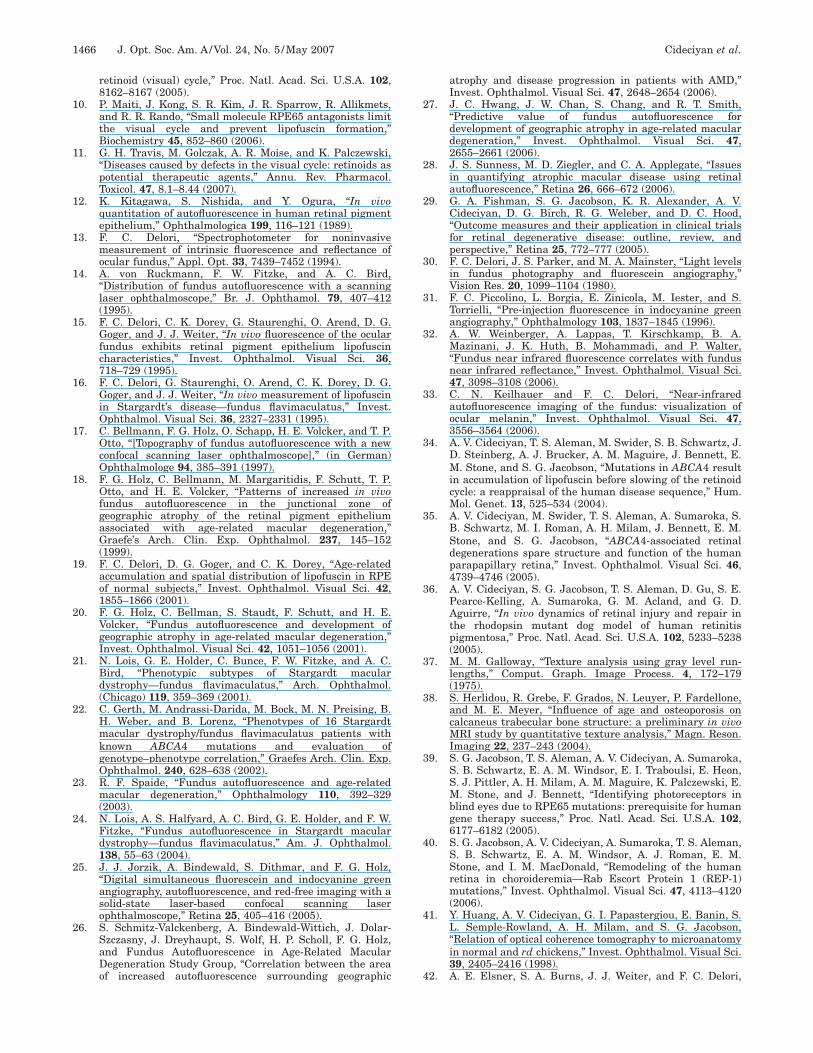
an help evaluate alternative hypotheses about tissue ori-ins of imaging signals.40 The left eye of patient 7 pre-ents such an opportunity. On NIR-reflectance imagingFig. 2(a)] there is a central lesion of �12° diameter withharp boundaries; a thin vertical peninsula of healthieretina extends downward from the superior lesion bound-ry and ends near the preserved fovea (with 20/25 visualcuity). Visibility of choroidal blood vessels within theentral lesion suggests loss of normal RPE pigmentation.n SW-RAFI the lesion appears dark, corresponding to
oss of lipofuscin secondary to diseased and/or atrophicPE [Fig. 2(b)]. There is an irregular annulus of high au-
ofluorescence surrounding the lesion [Fig. 2(b)]. NIR-AFI shows a pattern generally similar to SW-RAFI withlower intensity central lesion and a surrounding annu-
us of high intensity [Fig. 2(c)]. Within the central lesion,arker choroidal vessels appear to be blocking a diffusend low-intensity NIR-RAFI signal originating fromeeper layers [Fig. 2(c)]. An OCT scan across the lesionFig. 2(c), inset] shows thinning of the parafoveal retinand a dramatic increase in backscatter originating fromayers deep to the RPE [Fig. 2(c), inset, triple white lines].n normal OCT, backscatter from choroidal layers is muchess detectable owing to presumed absorption and/or scat-ering of this wavelength at/near the RPE–horiocapillaris region [Fig. 1(e)]. The results in the nor-
tig. 2. Interpretation of RAFI intensities in severe RPE diseasen patient 7. (a) NIR-reflectance image showing a well-delineated
acular lesion. The approximately two-fold greater reflectanceithin the lesion is likely due to depigmentation or atrophy of
he RPE. (b) SW-RAFI of the central retina showing dramaticallyeduced lipofuscin autofluorescence within the lesion. Surround-ng the lesion is an irregular annulus of hyperautofluorescence.c) NIR-RAFI shows both similarities to and differences fromW-RAFI. Choroidal vessels are distinctly apparent within the
esion. Inset: OCT image obtained at an angle (dashed white line)hrough the fovea shows the transition from a high (triple lines)o a relatively low (single line) sub-RPE backscatter region at the
emporal boundary of the lesion.
m(flNtcmi
CRHcssndtmtO
mR�gflalcctoprt
ifignafpntSpavnssttotottNt
iRld
sistdarscsntbtPccoesfwtoawbfiaap
4MlttOsfadscffssstdmritd
1462 J. Opt. Soc. Am. A/Vol. 24, No. 5 /May 2007 Cideciyan et al.
al subjects (Fig. 1) and in the patient with maculopathyFig. 2) confirm the previously proposed hypothesis thatuorophores in the RPE and choroid can be activated byIR excitation.33 Our results extend previous work by de-
ecting retinal locations suspect for increased choroidalontribution to NIR-RAFI with the use of an OCT-derivedeasure of sub-RPE backscattering amplitude as a local
ndex of the depth of penetration of the NIR light.
. Macular ABCA4 Disease Sequence: Correlation ofAFI with Visual Function and Retinal Structureuman lipofuscinopathies caused by ABCA4 mutations
an show dramatic variation in spatial distribution andeverity of retinal disease.34,35,45–50 Our previous workuggested that the region between the fovea and the opticerve may be informative of the entire gamut of ABCA4isease expression and that tests of structure and func-ion densely sampling this region may form sensitiveeasures of natural history or outcome of therapy.35 In
he current work, we used RAFI, microperimetry, andCT to sample this region.Patient 6 illustrates mild/early ABCA4 disease of theacula (Fig. 3). NIR-reflectance imaging, SW-, and NIR-AFI show a central region of abnormality extending to5° eccentricity [Figs. 3(a), 3(c), and 3(d)]. Within this re-
ion there is a small (�2° diameter) area of increased re-ectance [Fig. 3(a)] and reduced fluorescence [Figs. 3(c)nd 3(d)] visible immediately nasal to the fovea. This areaikely corresponds to severe RPE disease or atrophy, and aolocalized scotoma on microperimetry [Fig. 3(b)] and in-reased backscatter on OCT [Fig. 3(e)] are consistent withhis interpretation. Foveal sparing is visible as an islandf relative homogeneity of fluorescence, whereas thearafoveal region shows spatial heterogeneity. The retinalegion surrounding the central �10° diameter maculopa-hy appears qualitatively normal.
Visual function, retinal structure, and RAFI were stud-ed quantitatively along horizontal foveo–papillary pro-les with special emphasis on the perifoveal retinal re-ion with normal appearance. The only area showingormal parameters in all measures was about 1.5° widend located between 10.5° and 12° eccentric from theovea immediately temporal to the optic nerve. Based onrevious work34 we defined this as stage I disease. Aeighboring �4° wide area between 6.5° and 10.5° eccen-ric from the fovea showed supernormal intensity withW- and NIR-RAFI [Figs. 3(c) and 3(d), insets]; all otherarameters were within normal limits. The next definablerea was �2° wide between 4.5° and 6.5° eccentric whereisual function measured by microperimetry became ab-ormal. These two subclinical stages were defined astages IIa and IIb, respectively, based on the subclinicaltage II disease previously defined.34 Closer to the foveahere were increasing abnormalities in different modali-ies. Between �2.5° and 4.5° eccentric, there were lossesf visual sensitivity, supernormal SW- and NIR-RAFI in-ensities, and variable abnormalities in local homogeneityf autofluorescence. Between �0.5° and 2.5° eccentric,here was severe loss of visual sensitivity, counterintui-ive return of SW-RAFI to normal, and indeterminableIR-RAFI associated with dramatic increase in backscat-
er intensity originating from sub-RPE layers [Fig. 3(e),
nset]. At the fovea, NIR-RAFI was normal, whereas SW-AFI was dramatically supernormal, consistent with the
oss of macular pigment optical density in ABCA4isease.51
A summary of the quantitative multiparameter analy-is in the foveo–papillary region of six patients is shownn Fig. 4. Areas of subclinical stage II disease were alwaysandwiched between the clinically overt maculopathy andhe optic nerve or the area of stage I disease. Stage IIaisease (hyperautofluorescence with normal function) waslways more eccentric to stage IIb disease (hyperautofluo-escence with visual dysfunction). The transition fromtage II to more severe disease corresponded to an in-rease in spatial heterogeneity of autofluorescence. Inome cases there was evidence for the abnormal heteroge-eity boundary in NIR-RAFI to be more peripheral thanhat in SW-RAFI (P1, P2, P6) or vice versa (P3, P4, P5),ut in most cases, peripheral boundaries of the onset ofhese abnormalities were within about 1° of one another.arafoveal regions of abnormally increased sRBI couldorrespond to normal (P2, P5, P6) or hyperautofluores-ence (P1, P3, P4) on SW-RAFI, suggesting the existencef diseased RPE (as opposed to RPE atrophy) in thesearly/mild disease stages. Indeed, none of the patientstudied had abnormally reduced autofluorescence in theoveo–papillary region analyzed. In two of the patientsith the largest extents of maculopathies (P2 and P4),
here were perifoveal regions of abnormally reduced sRBIn OCT. In five of six patients, there was foveal hyper-utofluorescence with SW-RAFI but normal fluorescenceith NIR-RAFI; P4 showed hyperautofluorescence withoth SW and NIR modalities. Interpretation of the fovealndings will have to take into account not only RPEbnormalities33 but also consequences of retinalbnormalities51 and differences in choroidaligmentation.33
. DISCUSSIONolecular understanding of the biogenesis and toxicity of
ipofuscin over the past two decades1–7 has led to poten-ial treatment strategies designed to slow its accumula-ion in human diseases such as STGD and AMD.6,8–11
utcome of therapeutic strategies can be objectively mea-ured with AF imaging—the only direct measure of lipo-uscin accumulation.29 Conventional AF imaging has beenpplied to various patient populations for more than aecade,12–28,34,35 and recently there has been a call fortandardization and quantification.52 It is an unfortunateoincidence that the high-intensity SW illumination usedor conventional AF imaging of lipofuscin is also a riskactor contributing to the accumulation and toxicity of theame substance. Of course, all ophthalmic imaging in-truments, including those used for AF imaging, are de-igned to operate safely below retinal light damagehresholds, and there have been no reports of acute lightamage due to AF imaging. Yet it is important to keep inind that damage thresholds are based mostly on normal
etinas, whereas ophthalmic imaging is performed mostlyn diseased retinas,53 and there is increasing evidencehat some retinal degenerative diseases have lower lightamage thresholds.36,54 Therefore it would be prudent to

F(aosistpmv�
Cideciyan et al. Vol. 24, No. 5 /May 2007/J. Opt. Soc. Am. A 1463
ig. 3. Qualitative and quantitative analyses of central disease between the fovea and the optic nerve head in a representative patientpatient 6) with early/mild ABCA4 disease. (a) NIR-reflectance image showing the clinically overt maculopathy surrounded by normalppearing retina. (b) Microperimetry thresholds (in decibels, dB) with red-on-red stimulus. Stimuli not seen with red-on-red and white-n-red conditions are shown as x’s at the equivalent red-on-red threshold of −12.5 dB. (c) SW-RAFI image showing a parafoveal region ofevere RPE disease. Intensity and run-length values quantified along the horizontal profile are shown on the right panels. (d) NIR-RAFImage shows similarities to and differences from SW-RAFI. Intensity and run-length values quantified along the horizontal profile arehown on the right panels. Regions of the retina with enrichment of choroidal component to the NIR-RAFI signal are marked as unin-erpretable (x). All images (a,c,d) are shown individually contrast stretched for visibility of features. (e) OCT scan along the horizontalrofile. Pair of overlaid white lines represents the normal limits of retinal thickness from the RPE peak (retinal depth of 0 �m). Esti-ated sub-RPE backscatter index (sRBI) is shown to the right. Boundaries of subclinical disease stages IIa and IIb are shown with
ertical dashed lines coregistered in both columns across all panels. Pairs of gray lines on right panels represent normal limits
mean±2 SD� for each measure.
rsuttctd
mtSiiac
WsrtSwswcatn
elt
FiSO(as
1464 J. Opt. Soc. Am. A/Vol. 24, No. 5 /May 2007 Cideciyan et al.
educe as much as is technically possible the light expo-ure used in patients with retinal degenerative conditionsntil the extent of disease-associated photosensitivity ofhe human retina is better understood. This is especiallyrue for shorter wavelength “actinic” lights and underonditions of repetitive examination, such as in a clinicalrial. The current work attempted to take a step in thisirection with the quantitative use of RAFI methods.SW-RAFI was designed to be a lower light-dose replace-ent for conventional AF imaging. The lower illumina-
ion was also more comfortable for the subjects to view.hortcomings of SW-RAFI included noisier and darker
mages apparent on the acquisition screen, making themaging task more difficult for the operator. Another dis-dvantage was the necessity to use additional image pro-essing steps not included in the manufacturer’s software.
ig. 4. NIR-RAFI images and colocalized summary of all quanmages are shown individually contrast stretched for visibility oW-RAFI intensity (SW-RAFI/I) and run-length (SW-RAFI/RL);CT-based sub-RPE backscattering index (OCT/sRBI). Paramet
graybars), “above normal range” (cross-hatched bars), and “belowble or NIR-RAFI was excluded because of above-normal sRBIubclinical stage IIa and IIb disease are shown on NIR-RAFI im
ith postacquisition processing however, SW-RAFI re-ults were both qualitatively and quantitatively compa-able to those in the rapidly growing literature on conven-ional AF imaging (e.g., Refs. 12–28, 34, and 35).pecifically, observation of hyperautofluorescent regionsith spatial homogeneity in early ABCA4 disease and
patially heterogeneous regions in later disease stagesere consistent with previous work using quantitative
onventional AF imaging.34,35 Optimization of the appear-nce of images on the acquisition screen by the manufac-urer may allow even further reduction in SW illumi-ance in the future.AF imaging with NIR excitation, NIR-RAFI, is inher-
ntly low luminance owing to separation of this wave-ength from the region of photoreceptor pigment absorp-ion. Indeed NIR-RAFI illumination is nearly
arameters along the foveo–papillary profile in six patients. Allres. The parameters include microperimetry thresholds (MP/T);AFI intensity (NIR-RAFI/I) and run-length (NIR-RAFI/RL); andeach retinal location are classified into “within normal range”al range” (hatched bars). Retinal loci where data were not avail-are specified as “not available/not used” (white). Boundaries ofith dashed lines extended from parameter results.
tified pf featuNIR-Rers at
normvaluesages w

ilrNplflvtsoltrcwdlwspuea
osmpdprcpfmsttcfiptgittatfnca
ptest(f
emcetsfAvObataiec
ATHdFREaw
r
R
Cideciyan et al. Vol. 24, No. 5 /May 2007/J. Opt. Soc. Am. A 1465
mperceptible for most patients (it appears as a dim redight to normal eyes). NIR excitation light is also sepa-ated from the SW region of lipofuscin photoreactivity.7
IR-RAFI is a new modality,31–33 however, and the inter-retation of the images is not well established. One of theeading hypotheses33 proposes that melanin (or related)uorophores in the RPE and in the choroid contributeariably to the NIR-RAFI signal, and our data are consis-ent with this hypothesis. Our use of colocalized OCTcans showed a correspondence between retinal locationsf abnormally increased choroidal penetration of NIRight and significantly increased choroidal component tohe NIR-RAFI signal; these regions of choroidal signal en-ichment were excluded from quantitative analyses in theurrent work. There were also some disease stages inhich apparent RPE or sub-RPE deposits abnormally re-uced choroidal penetration of NIR light. At such retinalocations, NIR-RAFI would be expected to be enrichedith signals from RPE fluorophores. Future experimental
tudies seeking to define the absorption and scatteringroperties of the RPE, lipofuscin, and sub-RPE depositsnder NIR light may allow quantitative use of NIR pen-tration estimates from OCT to distinguish between RPEnd sub-RPE components of the NIR-RAFI.Perifoveal hyperautofluorescence with SW-RAFI with-
ut associated overt disease may have been a predictableubclinical disease stage in patients with ABCA4utations.5,16,21,22,24,34,35 Yet the finding of colocalized hy-
erautofluorescence with both SW- and NIR-RAFI mo-alities was unexpected. Assuming the dominant fluoro-hore of the NIR-RAFI is melanin, melanolipofuscin, or aelated pigment, there are at least three hypotheses toonsider. First, newly synthesized melanin could be de-osited in the diseased RPE together with increased lipo-uscin accumulation. Although it is thought that adultammalian RPE cells lack melanogenesis, there has been
ome evidence consistent with slow melanin production inhe RPE throughout life.55 Second, the optical cross sec-ion of existing melanin granules in the RPE could be in-reased by apical displacement1,56 due to abnormal lipo-uscin accumulation or by active transport.57 Suchncrease in melanin optical cross section would be ex-ected to decrease SW excitation light reaching more dis-al lipofuscin granules and thus cause SW-RAFI torossly underestimate the lipofuscin accumulation. Third,ncreased RPE stress in ABCA4 disease could changehe properties of melanin granules. Consistent withhis hypothesis is the finding of dramatically increasedutofluorescence emanating from oxidized melanin.58 Al-ernative hypotheses involving fluorophores differentrom melanin co-accumulating with RPE lipofuscin can-ot be ruled out at this time. Many of these hypothesesan be directly tested in animal models of lipofuscinccumulation.5,6
We have previously defined disease stages in the mid-eripheral retina of human patients with retinopathy dueo ABCA4 mutations,34 but very little was known aboutquivalent stages in the macular region. Current workhowed the existence of two subclinical disease stages inhe perifoveal region. The earliest detectable diseasestage IIa) was hyperautofluorescence with normal retinalunction and structure, and this was similar to the earli-
st disease stage observed in the midperiphery. The nextacular disease stage (stage IIb) was hyperautofluores-
ence and abnormal visual function, and this was differ-nt from midperipheral retina where loss of visual func-ion occurs in late disease.34 Our results allow thepeculation that macular retina may react differentlyrom peripheral retina to the accumulation of lipofuscin.lternatively, there could be retinal fluorophores causingisual dysfunction and contributing to the AF signal.59–61
ne such moiety has been hypothesized toe A2-rhodopsin.62 Future AF studies in experimentalnimals may be required to better understand allhe contributors to SW- and NIR-RAFI signals in healthnd disease. In the meantime, RAFI should remain anmportant tool for estimating the natural history of dis-ase progression and the outcome of therapies in lipofus-inopathies.
CKNOWLEDGMENTShis research is supported by National Institutes ofealth grant EY-013203, Macula Vision Research Foun-ation, Foundation Fighting Blindness, Macular Diseaseoundation, Ruth and Milton Steinbach Fund, and Alconesearch Institute. The authors thank Elaine E. Smilko,lizabeth A. M. Windsor, Andy Cheung, Mark Ayzenberg,nd Michelle Doobrajh for their critical help with thisork.
Corresponding author Artur V. Cideciyan can beeached by e-mail at [email protected].
EFERENCES1. L. Feeney, “Lipofuscin and melanin of human retinal
pigment epithelium. Fluorescence, enzyme cytochemical,and ultrastructural studies,” Invest. Ophthalmol. VisualSci. 17, 583–600 (1978).
2. G. E. Eldred and M. L. Katz, “Fluorophores of the humanretinal pigment epithelium: separation and spectralcharacterization,” Exp. Eye Res. 47, 71–86 (1988).
3. G. E. Eldred and M. R. Lasky, “Retinal age pigmentsgenerated by self-assembling lysosomotropic detergents,”Nature 361, 724–726 (1993).
4. J. R. Sparrow and M. Boulton, “RPE lipofuscin and its rolein retinal pathobiology,” Exp. Eye Res. 80, 595–606 (2005).
5. J. Weng, N. L. Mata, S. M. Azarian, R. T. Tzekov, D. G.Birch, and G. H. Travis, “Insights into the function of Rimprotein in photoreceptors and etiology of Stargardt’sdisease from the phenotype in abcr knockout mice,” Cell 98,13–23 (1999).
6. R. A. Radu, N. L. Mata, S. Nusinowitz, X. Liu, P. A.Sieving, and G. H. Travis, “Treatment with isotretinoininhibits lipofuscin accumulation in a mouse model ofrecessive Stargardt’s macular degeneration,” Proc. Natl.Acad. Sci. U.S.A. 100, 4742–4747 (2003).
7. M. Boulton, M. Rozanowska, B. Rozanowski, and T. Wess,“The photoreactivity of ocular lipofuscin,” Photochem.Photobiol. Sci. 3, 759–764 (2004).
8. P. A. Sieving, P. Chaudhry, M. Kondo, M. Provenzano, D.Wu, T. J. Carlson, R. A. Bush, and D. A. Thompson,“Inhibition of the visual cycle in vivo by 13-cis retinoic acidprotects from light damage and provides a mechanism fornight blindness in isotretinoin therapy,” Proc. Natl. Acad.Sci. U.S.A. 98, 1835–1840 (2001).
9. M. Golczak, V. Kuksa, T. Maeda, A. R. Moise, and K.Palczewski, “Positively charged retinoids are potent andselective inhibitors of the trans–cis isomerization in the
1
1
1
1
1
1
1
1
1
1
2
2
2
2
2
2
2
2
2
2
3
3
3
3
3
3
3
3
3
3
4
4
4
1466 J. Opt. Soc. Am. A/Vol. 24, No. 5 /May 2007 Cideciyan et al.
retinoid (visual) cycle,” Proc. Natl. Acad. Sci. U.S.A. 102,8162–8167 (2005).
0. P. Maiti, J. Kong, S. R. Kim, J. R. Sparrow, R. Allikmets,and R. R. Rando, “Small molecule RPE65 antagonists limitthe visual cycle and prevent lipofuscin formation,”Biochemistry 45, 852–860 (2006).
1. G. H. Travis, M. Golczak, A. R. Moise, and K. Palczewski,“Diseases caused by defects in the visual cycle: retinoids aspotential therapeutic agents,” Annu. Rev. Pharmacol.Toxicol. 47, 8.1–8.44 (2007).
2. K. Kitagawa, S. Nishida, and Y. Ogura, “In vivoquantitation of autofluorescence in human retinal pigmentepithelium,” Ophthalmologica 199, 116–121 (1989).
3. F. C. Delori, “Spectrophotometer for noninvasivemeasurement of intrinsic fluorescence and reflectance ofocular fundus,” Appl. Opt. 33, 7439–7452 (1994).
4. A. von Ruckmann, F. W. Fitzke, and A. C. Bird,“Distribution of fundus autofluorescence with a scanninglaser ophthalmoscope,” Br. J. Ophthamol. 79, 407–412(1995).
5. F. C. Delori, C. K. Dorey, G. Staurenghi, O. Arend, D. G.Goger, and J. J. Weiter, “In vivo fluorescence of the ocularfundus exhibits retinal pigment epithelium lipofuscincharacteristics,” Invest. Ophthalmol. Visual Sci. 36,718–729 (1995).
6. F. C. Delori, G. Staurenghi, O. Arend, C. K. Dorey, D. G.Goger, and J. J. Weiter, “In vivo measurement of lipofuscinin Stargardt’s disease—fundus flavimaculatus,” Invest.Ophthalmol. Visual Sci. 36, 2327–2331 (1995).
7. C. Bellmann, F. G. Holz, O. Schapp, H. E. Volcker, and T. P.Otto, “[Topography of fundus autofluorescence with a newconfocal scanning laser ophthalmoscope],” (in German)Ophthalmologe 94, 385–391 (1997).
8. F. G. Holz, C. Bellmann, M. Margaritidis, F. Schutt, T. P.Otto, and H. E. Volcker, “Patterns of increased in vivofundus autofluorescence in the junctional zone ofgeographic atrophy of the retinal pigment epitheliumassociated with age-related macular degeneration,”Graefe’s Arch. Clin. Exp. Ophthalmol. 237, 145–152(1999).
9. F. C. Delori, D. G. Goger, and C. K. Dorey, “Age-relatedaccumulation and spatial distribution of lipofuscin in RPEof normal subjects,” Invest. Ophthalmol. Visual Sci. 42,1855–1866 (2001).
0. F. G. Holz, C. Bellman, S. Staudt, F. Schutt, and H. E.Volcker, “Fundus autofluorescence and development ofgeographic atrophy in age-related macular degeneration,”Invest. Ophthalmol. Visual Sci. 42, 1051–1056 (2001).
1. N. Lois, G. E. Holder, C. Bunce, F. W. Fitzke, and A. C.Bird, “Phenotypic subtypes of Stargardt maculardystrophy—fundus flavimaculatus,” Arch. Ophthalmol.(Chicago) 119, 359–369 (2001).
2. C. Gerth, M. Andrassi-Darida, M. Bock, M. N. Preising, B.H. Weber, and B. Lorenz, “Phenotypes of 16 Stargardtmacular dystrophy/fundus flavimaculatus patients withknown ABCA4 mutations and evaluation ofgenotype–phenotype correlation,” Graefes Arch. Clin. Exp.Ophthalmol. 240, 628–638 (2002).
3. R. F. Spaide, “Fundus autofluorescence and age-relatedmacular degeneration,” Ophthalmology 110, 392–329(2003).
4. N. Lois, A. S. Halfyard, A. C. Bird, G. E. Holder, and F. W.Fitzke, “Fundus autofluorescence in Stargardt maculardystrophy—fundus flavimaculatus,” Am. J. Ophthalmol.138, 55–63 (2004).
5. J. J. Jorzik, A. Bindewald, S. Dithmar, and F. G. Holz,“Digital simultaneous fluorescein and indocyanine greenangiography, autofluorescence, and red-free imaging with asolid-state laser-based confocal scanning laserophthalmoscope,” Retina 25, 405–416 (2005).
6. S. Schmitz-Valckenberg, A. Bindewald-Wittich, J. Dolar-Szczasny, J. Dreyhaupt, S. Wolf, H. P. Scholl, F. G. Holz,and Fundus Autofluorescence in Age-Related MacularDegeneration Study Group, “Correlation between the areaof increased autofluorescence surrounding geographic
atrophy and disease progression in patients with AMD,”Invest. Ophthalmol. Visual Sci. 47, 2648–2654 (2006).
7. J. C. Hwang, J. W. Chan, S. Chang, and R. T. Smith,“Predictive value of fundus autofluorescence fordevelopment of geographic atrophy in age-related maculardegeneration,” Invest. Ophthalmol. Visual Sci. 47,2655–2661 (2006).
8. J. S. Sunness, M. D. Ziegler, and C. A. Applegate, “Issuesin quantifying atrophic macular disease using retinalautofluorescence,” Retina 26, 666–672 (2006).
9. G. A. Fishman, S. G. Jacobson, K. R. Alexander, A. V.Cideciyan, D. G. Birch, R. G. Weleber, and D. C. Hood,“Outcome measures and their application in clinical trialsfor retinal degenerative disease: outline, review, andperspective,” Retina 25, 772–777 (2005).
0. F. C. Delori, J. S. Parker, and M. A. Mainster, “Light levelsin fundus photography and fluorescein angiography,”Vision Res. 20, 1099–1104 (1980).
1. F. C. Piccolino, L. Borgia, E. Zinicola, M. Iester, and S.Torrielli, “Pre-injection fluorescence in indocyanine greenangiography,” Ophthalmology 103, 1837–1845 (1996).
2. A. W. Weinberger, A. Lappas, T. Kirschkamp, B. A.Mazinani, J. K. Huth, B. Mohammadi, and P. Walter,“Fundus near infrared fluorescence correlates with fundusnear infrared reflectance,” Invest. Ophthalmol. Visual Sci.47, 3098–3108 (2006).
3. C. N. Keilhauer and F. C. Delori, “Near-infraredautofluorescence imaging of the fundus: visualization ofocular melanin,” Invest. Ophthalmol. Visual Sci. 47,3556–3564 (2006).
4. A. V. Cideciyan, T. S. Aleman, M. Swider, S. B. Schwartz, J.D. Steinberg, A. J. Brucker, A. M. Maguire, J. Bennett, E.M. Stone, and S. G. Jacobson, “Mutations in ABCA4 resultin accumulation of lipofuscin before slowing of the retinoidcycle: a reappraisal of the human disease sequence,” Hum.Mol. Genet. 13, 525–534 (2004).
5. A. V. Cideciyan, M. Swider, T. S. Aleman, A. Sumaroka, S.B. Schwartz, M. I. Roman, A. H. Milam, J. Bennett, E. M.Stone, and S. G. Jacobson, “ABCA4-associated retinaldegenerations spare structure and function of the humanparapapillary retina,” Invest. Ophthalmol. Visual Sci. 46,4739–4746 (2005).
6. A. V. Cideciyan, S. G. Jacobson, T. S. Aleman, D. Gu, S. E.Pearce-Kelling, A. Sumaroka, G. M. Acland, and G. D.Aguirre, “In vivo dynamics of retinal injury and repair inthe rhodopsin mutant dog model of human retinitispigmentosa,” Proc. Natl. Acad. Sci. U.S.A. 102, 5233–5238(2005).
7. M. M. Galloway, “Texture analysis using gray level run-lengths,” Comput. Graph. Image Process. 4, 172–179(1975).
8. S. Herlidou, R. Grebe, F. Grados, N. Leuyer, P. Fardellone,and M. E. Meyer, “Influence of age and osteoporosis oncalcaneus trabecular bone structure: a preliminary in vivoMRI study by quantitative texture analysis,” Magn. Reson.Imaging 22, 237–243 (2004).
9. S. G. Jacobson, T. S. Aleman, A. V. Cideciyan, A. Sumaroka,S. B. Schwartz, E. A. M. Windsor, E. I. Traboulsi, E. Heon,S. J. Pittler, A. H. Milam, A. M. Maguire, K. Palczewski, E.M. Stone, and J. Bennett, “Identifying photoreceptors inblind eyes due to RPE65 mutations: prerequisite for humangene therapy success,” Proc. Natl. Acad. Sci. U.S.A. 102,6177–6182 (2005).
0. S. G. Jacobson, A. V. Cideciyan, A. Sumaroka, T. S. Aleman,S. B. Schwartz, E. A. M. Windsor, A. J. Roman, E. M.Stone, and I. M. MacDonald, “Remodeling of the humanretina in choroideremia—Rab Escort Protein 1 (REP-1)mutations,” Invest. Ophthalmol. Visual Sci. 47, 4113–4120(2006).
1. Y. Huang, A. V. Cideciyan, G. I. Papastergiou, E. Banin, S.L. Semple-Rowland, A. H. Milam, and S. G. Jacobson,“Relation of optical coherence tomography to microanatomyin normal and rd chickens,” Invest. Ophthalmol. Visual Sci.39, 2405–2416 (1998).
2. A. E. Elsner, S. A. Burns, J. J. Weiter, and F. C. Delori,

4
4
4
4
4
4
4
5
5
5
5
5
5
5
5
5
5
6
6
6
Cideciyan et al. Vol. 24, No. 5 /May 2007/J. Opt. Soc. Am. A 1467
“Infrared imaging of sub-retinal structures in the humanocular fundus,” Vision Res. 36, 191–205 (1996).
3. F. C. Delori, D. G. Goger, C. Keilhauer, P. Salvetti, and G.Staurenghi, “Bimodal spatial distribution of macularpigment: evidence of a gender relationship,” J. Opt. Soc.Am. A 23, 521–538 (2006).
4. R. W. Knighton, S. G. Jacobson, and C. M. Kemp, “Thespectral reflectance of the nerve fiber layer of the macaqueretina,” Invest. Ophthalmol. Visual Sci. 30, 2392–2402(1989).
5. R. Allikmets, N. Singh, H. Sun, N. F. Shroyer, A.Hutchinson, A. Chidambaram, B. Gerrard, L. Baird, D.Stauffer, A. Peiffer, A. Rattner, P. Smallwood, Y. Li, K. L.Anderson, R. A. Lewis, J. Nathans, M. Leppert, M. Dean,and J. R. Lupski, “A photoreceptor cell-specific ATP-bindingtransporter gene �ABCR� is mutated in recessive Stargardtmacular dystrophy,” Nat. Genet. 15, 236–246 (1997).
6. A. Martinez-Mir, E. Paloma, R. Allikmets, C. Ayuso, T. delRio, M. Dean, L. Vilageliu, R. Gonzalez-Duarte, and S.Balcells, “Retinitis pigmentosa caused by a homozygousmutation in the Stargardt disease gene ABCR,” Nat.Genet. 18, 11–12 (1998).
7. F. P. Cremers, D. J. van de Pol, M. van Driel, A. I. denHollander, F. J. van Haren, N. V. Knoers, N. Tijmes, A. A.Bergen, K. Rohrschneider, A. Blankenagel, A. J. Pinckers,A. F. Deutman, and C. B. Hoyng, “Autosomal recessiveretinitis pigmentosa and cone–rod dystrophy caused bysplice site mutations in the Stargardt’s disease geneABCR,” Hum. Mol. Genet. 7, 355–362 (1998).
8. G. A. Fishman, E. M. Stone, S. Grover, D. J. Derlacki, H. L.Haines, and R. R. Hockey, “Variation of clinical expressionin patients with Stargardt dystrophy and sequencevariations in the ABCR gene,” Arch. Ophthalmol. (Chicago)117, 504–510 (1999).
9. A. R. Webster, E. Heon, A. J. Lotery, K. Vandenburgh, T. L.Casavant, K. T. Oh, G. Beck, G. A. Fishman, B. L. Lam, A.Levin, J. R. Heckenlively, S. G. Jacobson, R. G. Weleber, V.C. Sheffield, and E. M. Stone, “An analysis of allelicvariation in the ABCA4 gene,” Invest. Ophthalmol. VisualSci. 42, 1179–1189 (2001).
0. G. A. Fishman, E. M. Stone, D. A. Eliason, C. M. Taylor, M.Lindeman, and D. J. Derlacki, “ABCA4 gene sequencevariations in patients with autosomal recessive cone–roddystrophy,” Arch. Ophthalmol. (Chicago) 121, 851–855(2003).
1. T. S. Aleman, A. V. Cideciyan, E. A. M. Windsor, S. B.Schwartz, L. M. Gardner, J. M. Emmons, K. G. Duncan, J.D. Steinberg, E. M. Stone, and S. G. Jacobson, “Macular
pigment in ABCA4-associated retinal degenerations:response to lutein supplementation,” Invest. Ophthalmol.Visual Sci. 47, E-Abstract 5810 (2006).
2. J. Hopkins, A. Walsh, and U. Chakravarthy, “Fundusautofluorescence in age-related macular degeneration: anepiphenomenon?” Invest. Ophthalmol. Visual Sci. 47,2269–2271 (2006).
3. D. Sliney, D. Aron-Rosa, F. Delori, F. Fankhauser, R.Landry, M. Mainster, J. Marshall, B. Rassow, B. Stuck, S.Trokel, T. M. West, M. Wolffe, and InternationalCommission on Non-Ionizing Radiation Protection,“Adjustment of guidelines for exposure of the eye to opticalradiation from ocular instruments: statement from a taskgroup of the International Commission on Non-IonizingRadiation Protection (ICNIRP),” Appl. Opt. 44, 2162–2176(2005).
4. D. M. Paskowitz, M. M. Lavail, and J. L. Duncan, “Lightand inherited retinal degeneration,” Br. J. Ophthamol. 90,1060–1066 (2006).
5. U. Schraermeyer, “Does melanin turnover occur in the eyesof adult vertebrates?” Pigment Cell Res. 6, 193–204 (1993).
6. J. J. Weiter, F. C. Delori, G. L. Wing, and K. A. Fitch,“Retinal pigment epithelial lipofuscin and melanin andchoroidal melanin in human eyes,” Invest. Ophthalmol.Visual Sci. 27, 145–152 (1986).
7. D. Gibbs, S. M. Azarian, C. Lillo, J. Kitamoto, A. E. Klomp,K. P. Steel, R. T. Libby, and D. S. Williams, “Role of myosinVIIa and Rab27a in the motility and localization of RPEmelanosomes,” J. Cell. Sci. 117, 6473–6483 (2004).
8. P. Kayatz, G. Thumann, T. T. Luther, J. F. Jordan, K. U.Bartz-Schmidt, P. J. Esser, and U. Schraermeyer,“Oxidation causes melanin fluorescence,” Invest.Ophthalmol. Visual Sci. 42, 241–246 (2001).
9. G. S. Tucker, “Refractile bodies in the inner segments ofcones in the aging human retina,” Invest. Ophthalmol.Visual Sci. 27, 708–715 (1986).
0. M. Iwasaki and H. Inomata, “Lipofuscin granules inhuman photoreceptor cells,” Invest. Ophthalmol. VisualSci. 29, 671–679 (1988).
1. C. D. Birnbach, M. Jarvelainen, D. E. Possin, and A. H.Milam, “Histopathology and immunocytochemistry of theneurosensory retina in fundus flavimaculatus,”Ophthalmology 101, 1211–1219 (1994).
2. N. Fishkin, Y. P. Jang, Y. Itagaki, J. R. Sparrow, and K.Nakanishi, “A2-rhodopsin: a new fluorophore isolated fromphotoreceptor outer segments,” Org. Biomol. Chem. 1,1101–1105 (2003).