Pneumococcal capsule synthesis locus cps as evolutionary ...
Upload
independentCategory
view
1download
0
Analysis of the ABCA4 genomic locusin Stargardt disease
Jana Zernant1, Yajing (Angela) Xie1, Carmen Ayuso3,4, Rosa Riveiro-Alvarez3,4, Miguel-Angel
Lopez-Martinez3,4, Francesca Simonelli5, Francesco Testa5, Michael B. Gorin6,7, Samuel P.
Strom6,7,8, Mette Bertelsen9, Thomas Rosenberg9, Philip M. Boone10, Bo Yuan10, Radha Ayyagari11,
Peter L. Nagy2, Stephen H. Tsang1,2, Peter Gouras1, Frederick T. Collison12, James R. Lupski10,
Gerald A. Fishman12 and Rando Allikmets1,2,∗
1Department of Ophthalmology and 2Department of Pathology and Cell Biology, Columbia University, New York, NY, USA,3Department of Genetics, Instituto de Investigacion Sanitaria-University Hospital Fundacion Jimenez Diaz, UAM (IIS-FJD),
Madrid, Spain, 4Centro de Investigacion Biomedica en Red (CIBER) de Enfermedades Raras, ISCIII, Madrid, Spain, 5Eye
Clinic, Multidisciplinary Department of Medical, Surgical and Dental Sciences, Second University of Naples, Naples, Italy,6Department of Ophthalmology, 7Department of Human Genetics, Jules Stein Eye Institute and 8Department of Pathology,
DavidGeffenSchoolofMedicineatUCLA,LosAngeles,CA,USA, 9KennedyCenterEyeClinic,GlostrupHospital,Glostrup,
Denmark, 10Department of Molecular and Human Genetics, Baylor College of Medicine, Houston, TX, USA, 11Department
of Ophthalmology, University of California San Diego, La Jolla, CA, USA and 12The Pangere Center for Hereditary Retinal
Diseases, The Chicago Lighthouse for People Who are Blind or Visually Impaired, Chicago, IL, USA
Received May 29, 2014; Revised May 29, 2014; Accepted July 29, 2014
Autosomal recessive Stargardt disease (STGD1, MIM 248200) is caused by mutations in the ABCA4 gene.Complete sequencing of ABCA4 in STGD patients identifies compound heterozygous or homozygous dis-ease-associated alleles in 65–70% of patients and only one mutation in 15–20% of patients. This study wasdesigned to find the missing disease-causing ABCA4 variation by a combination of next-generation sequencing(NGS), array-Comparative Genome Hybridization (aCGH) screening, familial segregation and in silico analyses.The entire 140 kb ABCA4 genomic locus was sequenced in 114 STGD patients with one known ABCA4 exonicmutation revealing, on average, 200 intronic variants per sample. Filtering of these data resulted in 141 candi-dates for new mutations. Two variants were detected in four samples, two in three samples, and 20 variants intwo samples, the remaining 117 new variants were detected only once. Multimodal analysis suggested 12 newlikely pathogenic intronic ABCA4 variants, some of which were specific to (isolated) ethnic groups. No copynumber variation (large deletions and insertions) was detected in any patient suggesting that it is a very rareevent in the ABCA4 locus. Many variants were excluded since they were not conserved in non-human primates,were frequent in African populations and, therefore, represented ancestral, and not disease-associated, var-iants. The sequence variability in the ABCA4 locus is extensive and the non-coding sequences do not harbor fre-quent mutations in STGD patients of European-American descent. Defining disease-associated alleles in theABCA4 locus requires exceptionally well characterized large cohorts and extensive analyses by a combinationof various approaches.
INTRODUCTION
Mutations in the ABCA4 gene are responsible for a wide varietyof retinal dystrophy phenotypes from autosomal recessive
Stargardt disease (STGD1) (1) to cone–rod dystrophy (CRD)(2,3) and, in some advanced cases, retinitis pigmentosa (RP)(2,4,5). While CRD and RP phenotypes are also caused by muta-tions in many other genes, ABCA4 is the only recognized gene
∗To whom correspondence should be addressed. Email: [email protected]
# The Author 2014. Published by Oxford University Press. All rights reserved.For Permissions, please email: [email protected]
Human Molecular Genetics, 2014 1–10doi:10.1093/hmg/ddu396
HMG Advance Access published August 16, 2014 at H
AM
-TM
C L
ibrary on August 20, 2014
http://hmg.oxfordjournals.org/
Dow
nloaded from
responsible for STGD1 (MIM 248200), a predominantlyjuvenile-onset macular dystrophy frequently associated withearly-onset central visual impairment, progressive bilateralatrophy of the foveal retinal pigment epithelium, and the pres-ence of yellowish flecks, defined as lipofuscin deposits, aroundthe macula and/or in the central and near-peripheral areas ofthe retina.
Over 800 disease-associated ABCA4 variants have beenalready identified (6) and the most frequent of these have beendescribed in only �10% of STGD1 patients (7). Severalstudies have identified frequent ‘ethnic group-specific’ ABCA4alleles, such as the p.G863A/G863del founder mutation inNorthern European patients (8), the p.[L541P;A1038V] complexallele in patients of mostly German origin (3,9), the p.R1129Lfounder mutation in Spain (10), the p.N965S variant in theDanish population (11) and the p.A1773V variant in Mexico (12).
Complete sequencing of the ABCA4 coding and adjacentintronic sequences in patients with STGD1 routinely discovers�80% of mutations with the fraction of patients harboring theexpected two disease-associated alleles at 65–70%, with onemutation �15–20%, and with no mutations in the remaining�15% (13). These fractions depend on many variables, mostimportantly the quality of the clinical diagnosis and the ethniccomposition of the cohort. Most of the cases with no detectedABCA4 mutations likely represent phenocopies (13); i.e. inthose patients mutations in other gene(s) cause a STGD1-likephenotype. However, based on the known carrier frequency ofpathogenic ABCA4 variants, in most cases with one ABCA4allele the second allele is expected to reside in the ABCA4locus. It can present as a copy number variant (CNV, large dele-tion or insertion of one exon or more) which eludes detection byPCR-based sequencing techniques, a synonymous variant in thecoding region, or a (deep) intronic variant, which may affectsplicing or a regulatory region, such as a promoter or an enhancer(14). Very few of these have been identified (13,15,16).
This study was designed to find the missing ABCA4 mutationsby a combination of next-generation sequencing, array-comparative genomic hybridization (aCGH) arrays, in silicoand RNA analyses, and segregation analyses in families.
RESULTS
Discovery of new disease-associated variantsby next-generation sequencing
Sequencing of the entire ABCA4 genomic locus, at an averagedepth of coverage of 100×, in 130 patients with ABCA4-associated disease harboring one previously known ABCA4disease-associated allele, and 6 patients with no knownABCA4 mutations, resulted in detecting 1745 different variants.Eighty-three of these were previously known disease-associatedor benign variants from coding regions and pathogenic splice sitevariants. Six hundred and ninety-five (695) variants were alsodetected in 1000 Genomes Project or Exome SequencingProject, with no statistically significant differences in allele fre-quencies between the general population and the patient cohort,unless the variants were on the same allele (haplotype) with thefrequent known ABCA4 coding mutation, p.G1961E. Fivehundred and twenty-six (526) variants were incorrectly calleddeletions or insertions from single nucleotide repeat areas
(homopolymers) that have proven to be difficult for the NGS ap-proach. We also experienced a relatively high A.C/C.A/T.G/G.T false-positive calling rate with Illumina sequen-cing. The number of false positives can be reduced by more strin-gent criteria for variant calling; however, this may also excludesome real variants. After the filtering and verification steps 141new intronic ABCA4 variants remained in 114 patients. In 22patients with one previously known ABCA4 mutation, thesecond pathogenic ABCA4 allele was also found in the coding se-quence or adjacent splice sites. In 6/22 cases this was due to re-evaluation of several variants which had been classified asbenign, e.g. p.G991R and p.A1773V. The remaining 16 casesrepresented false-negative results, probably due to technicalreasons in the first, sequencing, step of the ABCA4 codingregions.
Of the 141 new possible candidates for disease-associated var-iants, two variants, c.4539+2064C.T and c.5461-1389C.A,were detected together (one the same chromosome) as acomplex allele in four patients of Spanish or Italian descent(Table 1). The c.5461-1389C.A variant is in an evolutionarilyless conserved area, the c.4539+2064C.T variant is adjacentto the recently reported c.4539+2028C.T and c.4539+2001G.A variants from a conserved area (14). According topredictive programs, none of these variants have any effect onsplicing, whether on existing cryptic splice sites or on creatingnew sites. The c.4539+2064C.T and c.5461-1389C.Ahaplotype segregated with the disease in all three STGD1 fam-ilies from Spain (Fig. 1A–C), and were absent in 100 matchedSpanish control samples, making these variants very likely can-didates for intronic ABCA4 mutations.
Two variants, c.5196+1056A.G and c.6006-609T.A,were detected in 3/114 unrelated patients each and were absentin 368 matched control samples. The c.5196+1056A.Gvariant segregated with the disease in two families; i.e. it wason a different chromosome than the proband’s other ABCA4 mu-tation (Fig. 1D). This variant is also predicted to strengthen acryptic splice donor (Supplementary Material, Fig. S1A). Thevariant was found, in addition, in 1/119 patients from our repli-cation cohort of STGD1 patients with one known ABCA4 muta-tion and has been recently reported as a disease-associatedallele (14). The aggregate evidence suggests that the c.5196+1056A.G variant is a rare, deep intronic disease-associatedallele.
The c.6006-609T.A variant, which does not have a predictedeffect on splicing, was detected in 6/119 additional unrelatedsamples from our replication cohort of STGD1 patients withone known ABCA4 mutation. Two of these samples were fromthe Columbia University patient cohort and four from Europeancohorts including two from Denmark. The variant was notpresent in 368 European-American control samples, but wasdetected in 2/182 Danish control samples. While the frequencyof this variant is 10× elevated in STGD1 patients as comparedwith all controls (1.9 versus 0.18%) and 3.5× if comparedwith Danish controls (0.55%), it is premature to unequivocallycall the variant as associated with the disease.
Three variants, c.570+1798A.G, c.2161-8G.A, andc.859–9T.C were each detected in 2/114 different patients inour primary ABCA4 locus screening cohort, but absent in 368control samples and also in 119 additional STGD1 samplesfrom the replication cohort (Table 1). The c.570+1798A.G
2 Human Molecular Genetics, 2014
at HA
M-T
MC
Library on A
ugust 20, 2014http://hm
g.oxfordjournals.org/D
ownloaded from
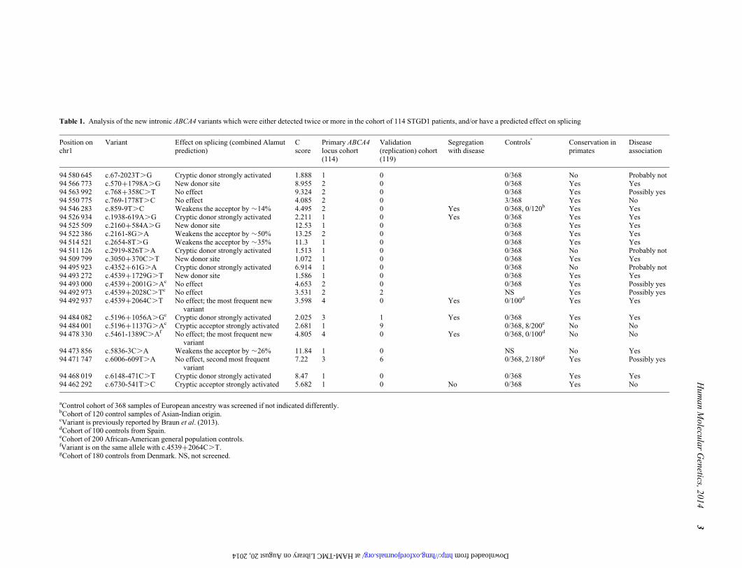
Table 1. Analysis of the new intronic ABCA4 variants which were either detected twice or more in the cohort of 114 STGD1 patients, and/or have a predicted effect on splicing
Position onchr1
Variant Effect on splicing (combined Alamutprediction)
Cscore
Primary ABCA4locus cohort(114)
Validation(replication) cohort(119)
Segregationwith disease
Controlsa
Conservation inprimates
Diseaseassociation
94 580 645 c.67-2023T.G Cryptic donor strongly activated 1.888 1 0 0/368 No Probably not94 566 773 c.570+1798A.G New donor site 8.955 2 0 0/368 Yes Yes94 563 992 c.768+358C.T No effect 9.324 2 0 0/368 Yes Possibly yes94 550 775 c.769-1778T.C No effect 4.085 2 0 3/368 Yes No94 546 283 c.859-9T.C Weakens the acceptor by �14% 4.495 2 0 Yes 0/368, 0/120b Yes Yes94 526 934 c.1938-619A.G Cryptic donor strongly activated 2.211 1 0 Yes 0/368 Yes Yes94 525 509 c.2160+584A.G New donor site 12.53 1 0 0/368 Yes Yes94 522 386 c.2161-8G.A Weakens the acceptor by �50% 13.25 2 0 0/368 Yes Yes94 514 521 c.2654-8T.G Weakens the acceptor by �35% 11.3 1 0 0/368 Yes Yes94 511 126 c.2919-826T.A Cryptic donor strongly activated 1.513 1 0 0/368 No Probably not94 509 799 c.3050+370C.T New donor site 1.072 1 0 0/368 Yes Yes94 495 923 c.4352+61G.A Cryptic donor strongly activated 6.914 1 0 0/368 No Probably not94 493 272 c.4539+1729G.T New donor site 1.586 1 0 0/368 Yes Yes94 493 000 c.4539+2001G.Ac No effect 4.653 2 0 0/368 Yes Possibly yes94 492 973 c.4539+2028C.Tc No effect 3.531 2 2 NS Yes Possibly yes94 492 937 c.4539+2064C.T No effect; the most frequent new
variant3.598 4 0 Yes 0/100d Yes Yes
94 484 082 c.5196+1056A.Gc Cryptic donor strongly activated 2.025 3 1 Yes 0/368 Yes Yes94 484 001 c.5196+1137G.Ac Cryptic acceptor strongly activated 2.681 1 9 0/368, 8/200e No No94 478 330 c.5461-1389C.Af No effect; the most frequent new
variant4.805 4 0 Yes 0/368, 0/100d No No
94 473 856 c.5836-3C.A Weakens the acceptor by �26% 11.84 1 0 NS No Yes94 471 747 c.6006-609T.A No effect, second most frequent
variant7.22 3 6 0/368, 2/180g Yes Possibly yes
94 468 019 c.6148-471C.T Cryptic donor strongly activated 8.47 1 0 0/368 Yes Yes94 462 292 c.6730-541T.C Cryptic acceptor strongly activated 5.682 1 0 No 0/368 Yes No
aControl cohort of 368 samples of European ancestry was screened if not indicated differently.bCohort of 120 control samples of Asian-Indian origin.cVariant is previously reported by Braun et al. (2013).dCohort of 100 controls from Spain.eCohort of 200 African-American general population controls.fVariant is on the same allele with c.4539+2064C.T.gCohort of 180 controls from Denmark. NS, not screened.
Hu
ma
nM
olecu
lar
Gen
etics,2
01
43
at HAM-TMC Library on August 20, 2014 http://hmg.oxfordjournals.org/ Downloaded from
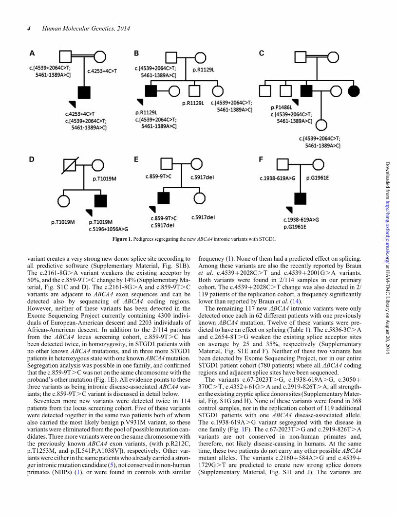
variant creates a very strong new donor splice site according toall predictive software (Supplementary Material, Fig. S1B).The c.2161-8G.A variant weakens the existing acceptor by50%, and the c.859-9T.C change by 14% (Supplementary Ma-terial, Fig. S1C and D). The c.2161-8G.A and c.859-9T.Cvariants are adjacent to ABCA4 exon sequences and can bedetected also by sequencing of ABCA4 coding regions.However, neither of these variants has been detected in theExome Sequencing Project currently containing 4300 indivi-duals of European-American descent and 2203 individuals ofAfrican-American descent. In addition to the 2/114 patientsfrom the ABCA4 locus screening cohort, c.859-9T.C hasbeen detected twice, in homozygosity, in STGD1 patients withno other known ABCA4 mutations, and in three more STGD1patients in heterozygous state with one known ABCA4 mutation.Segregation analysis was possible in one family, and confirmedthat the c.859-9T.C was not on the same chromosome with theproband’s other mutation (Fig. 1E). All evidence points to thesethree variants as being intronic disease-associated ABCA4 var-iants; the c.859-9T.C variant is discussed in detail below.
Seventeen more new variants were detected twice in 114patients from the locus screening cohort. Five of these variantswere detected together in the same two patients both of whomalso carried the most likely benign p.V931M variant, so thesevariants were eliminated from the pool of possible mutation can-didates. Three more variants were on the same chromosome withthe previously known ABCA4 exon variants, (with p.R212C,p.T1253M, and p.[L541P;A1038V]), respectively. Other var-iants were either in the same patients who already carried a stron-ger intronic mutation candidate (5), not conserved in non-humanprimates (NHPs) (1), or were found in controls with similar
frequency (1). None of them had a predicted effect on splicing.Among these variants are also the recently reported by Braunet al. c.4539+2028C.T and c.4539+2001G.A variants.Both variants were found in 2/114 samples in our primarycohort. The c.4539+2028C.T change was also detected in 2/119 patients of the replication cohort, a frequency significantlylower than reported by Braun et al. (14).
The remaining 117 new ABCA4 intronic variants were onlydetected once each in 62 different patients with one previouslyknown ABCA4 mutation. Twelve of these variants were pre-dicted to have an effect on splicing (Table 1). The c.5836-3C.Aand c.2654-8T.G weaken the existing splice acceptor siteson average by 25 and 35%, respectively (SupplementaryMaterial, Fig. S1E and F). Neither of these two variants hasbeen detected by Exome Sequencing Project, nor in our entireSTGD1 patient cohort (780 patients) where all ABCA4 codingregions and adjacent splice sites have been sequenced.
The variants c.67-2023T.G, c.1938-619A.G, c.3050+370C.T, c.4352+61G.A and c.2919-826T.A, all strength-en the existing cryptic splice donors sites (Supplementary Mater-ial, Fig. S1G and H). None of these variants were found in 368control samples, nor in the replication cohort of 119 additionalSTGD1 patients with one ABCA4 disease-associated allele.The c.1938-619A.G variant segregated with the disease inone family (Fig. 1F). The c.67-2023T.G and c.2919-826T.Avariants are not conserved in non-human primates and,therefore, not likely disease-causing in humans. At the sametime, these two patients do not carry any other possible ABCA4mutant alleles. The variants c.2160+584A.G and c.4539+1729G.T are predicted to create new strong splice donors(Supplementary Material, Fig. S1I and J). The variants are
Figure 1. Pedigrees segregating the new ABCA4 intronic variants with STGD1.
4 Human Molecular Genetics, 2014
at HA
M-T
MC
Library on A
ugust 20, 2014http://hm
g.oxfordjournals.org/D
ownloaded from
absent in 368 control samples and in the replication cohort of 119STGD1 patients (Table 1). Since these positions are highly con-served among species we suggest that the c.2160+584A.Gand c.4539+1729G.T variants are very good candidates forintronic ABCA4 mutations.
The variants c.5196+1137G.A, c.6148-471C.T andc.6730-541T.C have a predicted effect of strengthening theexisting cryptic splice acceptors (Supplementary Material,Fig. S1K–M), and were each all detected in one patient in theprimary locus screening cohort. The c.6730-541T.C variantwas on the same chromosome with the probands other knownABCA4 mutation, and can therefore be excluded from possiblenew mutations list. The c.6148-471C.T and c.6730-541T.Cvariants were not detected in 368 control samples, nor in 119additional STGD1 samples. The recently reported (14)c.5196+1137G.A variant was additionally found in nineSTGD1 samples with one previously known ABCA4 variant(Table 1). Three of these patients were from Denmark, threewere of African-American origin, and three of unknown ethni-city. Screening matched control cohorts revealed no carriersfor this allele in 180 Danish control samples, but it was detectedin 8/200 (MAF ¼ 2%) general population controls of African-American descent, strongly suggesting non-pathogenicity.Additional support for this variant not being pathogenic comesfrom evolutionary analysis (see below). The human majorallele c.5196+1137G is the minor allele in Macaca mulattaand the suggested mutant allele A is the major allele in macaques.
Analysis of regulatory sequences
To assess the effect of the new ABCA4 intronic variants onputative regulatory regions we compared their location againstthe chromosome coordinates of the DNaseI hypersensitivityand transcription factor binding regions from the ENCODEproject. Since regulatory regions, in particular promoters, tendto be DNase sensitive, variants that fall within such regionsmay affect regulatory potential. Also, variants that are locatedin transcription factor binding sites may potentially have aneffect on protein binding. The combined DNaseI hypersensitiv-ity data were derived from 125 cell types, and the dataset fortranscription factor binding regions involved 161 transcriptionfactors in 91 cell types. Unfortunately the datasets did notcontain eye-specific cell types or transcription factors. Thedefined regions were assigned normalized scores in the rangeof 0–1000, with higher scores indicating stronger signalstrength. Twenty-four of the 141 new ABCA4 intronic variantsare located in regions with both DNaseI hypersensitivity and
transcription factor binding scores of various strength (Supple-mentary Material, Table S1). Family members were availablefor segregation analyses in three cases. Variantsc.570+1499C.A and c.67-3166C.T were on the samechromosome with the probands’ other mutation, whilec.571-1707C.T and c.5018+289T.C were both detected inone patient and on the different chromosome than this patient’sother mutation, suggesting possible pathogenicity. Twenty-onevariants fall within regions with only DNaseI hypersensitivityscore, and 12 variants within regions of transcription factorbinding consensus sequences.
The new ABCA4 intronic variants were also subjected tothe Combined Annotation Dependent Depletion (CADD)algorithm (http://cadd.gs.washington.edu/score) (17). The CADDalgorithm combines a diverse array of annotations into one metric(C score) for each variant, ranking a variant relative to all possiblesubstitutions of the human genome (17). C score correlates withallelic diversity, pathogenicity and experimentally measured regu-latory effects. A C score of .10 indicates that the variant is amongthe top 10% of most deleterious substitutions in the humangenome. C scores for the new ABCA4 intronic variants rangefrom 0.004 to 15.14 (Supplementary Material, Table S1), withnine variants resulting in a C score greater than 10. Four of thosevariants were already classified as possibly disease-associateddue to their strong predicted effect on splicing (Table 1). Theother five variants with higher C scores included c.4539+1971T.C, c.67-3779T.G, c.303-906A.G, c.859-41T.C andc.1357-221T.A (Supplementary Material, Table S1).
In summary, we found a very strong or a probable intronicmutation candidate in 27/114 (23.7%) patients with one existingdefinite ABCA4 mutation (Table 1). No immediately plausiblecandidates for intronic mutations were found in 36/114(31.6%) patients. The remaining 51 (44.7%) patients possessedone or more new intronic variants that were only detected once,had no effect on splicing according to prediction programs and,therefore, are very difficult to confirm or refute as disease-associated alleles with the available methods and the impossi-bility of obtaining the patient RNA. However, it is highlylikely that a fraction of these are associated with STGD1.
Analysis of previously reported variants
Recently several ABCA4 intronic variants were suggested toaccount for a substantial fraction of pathogenic ABCA4 alleles(14). Since we have directly sequenced the ABCA4 gene andflanking intronic sequences in .780 STGD1 patients and theentire ABCA4 genomic locus in 114 STGD patients with one
Table 2. Frequency of the variants described in Braun et al. (2013), and in the current study.
Variant #(Braun et al.)
Location on chr1 Variant Braun et al. primarycohort (n¼28)
CU locus cohort(n ¼ 114)
CU exon seq cohort(n ¼ 780)
V1 94 484 001 c.5196+1137G.A 4 1 –V2 94 483 922 c.5196+1216C.A 1 0 –V3 94 484 082 c.5196+1056A.G 1 3 –V4 94 493 000 c.4539+2001G.A 1 2 –V5 94 492 973 c.4539+2028C.T 3 2 –V6 94 466 602 c.6342G.A/p.V2114V 1 0 1V7 94 487 399 c.4773+3A.G 3 0 0
Human Molecular Genetics, 2014 5
at HA
M-T
MC
Library on A
ugust 20, 2014http://hm
g.oxfordjournals.org/D
ownloaded from
mutation, we compared the Braun et al. data to ours (Table 2).The silent exonic p.V2114V variant, which we had describedin our earlier study as a possibly disease-associated mutation(13), is very rare; it was detected in 1 out of 780 STGD1 patients(Table 2). The ‘near-exonic’ c.4773+3A.G variant was notdetected in any of our 780 patients. We were also unable todetect one of the remaining five ‘deep intronic’ variants(c.5196+1216C.A) in any patient with one mutation and theother four were seen, in total, in 9/114 (7.9%) STGD1 patients,a statistically significant difference from the Braun et al. data10/28 (17.9%; P ¼ 0.001).
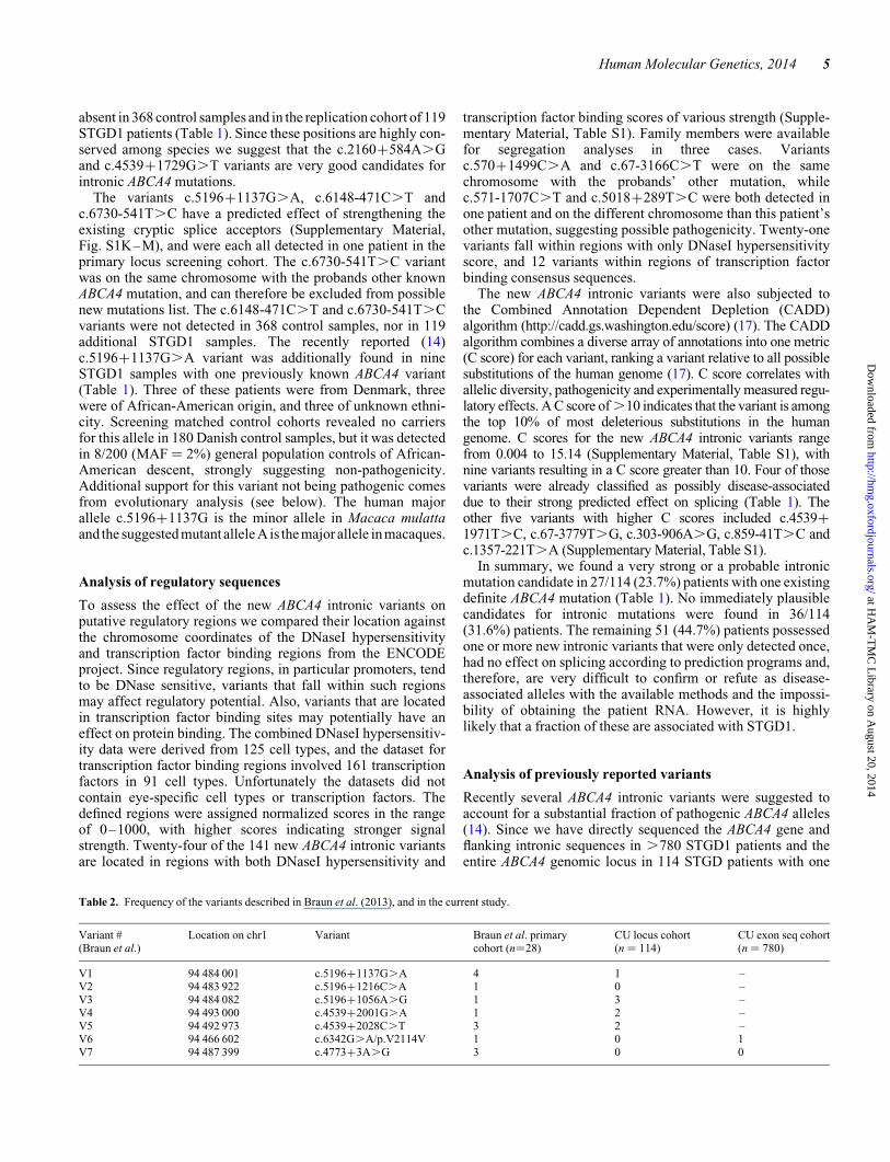
We then analyzed the evolutionary and/or ethnic origin of thevariants focusing first on the most frequent variant in the Braunet al. study, c.5196+1137G.A. Evolutionary conservation of anucleotide is one of the most important criteria for determiningthe pathogenicity of a variant, especially in a highly conservedgene such as ABCA4. The ABCA4 protein performs a very spe-cialized function in the visual cycle; therefore it is exceptionallyconserved in mammals and in all vertebrates with visual cycle.For example, the mouse and human ABCA4 proteins are 88%identical, allowing the human protein to perform the transportfunction in mouse (18). The conservation extends beyondcoding sequences and includes splice sites; in fact, the ABCA4gene has the same structure, consisting of exactly the same 50exons in non-human primates, such as Pan troglodytes andM. mulatta. The evolutionary conservation extends, in someregions, deep into the introns; for example, the 200 bp sequencessurrounding the c.5196+1137G.A variant are 96% identicalbetween human and macaque. Most importantly, M. mulattahas the adenosine nucleotide in human position c.5196+1137
(Fig. 2), as a major allele with guanosine as a minor allele; i.e.a situation exactly the reverse to that observed in humans, sug-gesting that this is an ancestral variant and not a disease-causingmutation. To further prove this assumption, we screened 200unrelated individuals from the general population of African-American descent and identified eight heterozygotes, resultingin the allele frequency of �2%. The cohort of STGD1 patientsat Columbia University contains 46 African-American patientsof whom one carried the c.5196+1137G.A variant, resultingin the allele frequency of �1%, comparable to that in thegeneral population. No other disease-associated ABCA4variant was identified in this patient. In addition, we detectedthe c.5196+1137G.A variant only once in 114 STGD1patients (allele frequency 0.4%) of European descent in the Col-umbia cohort (Tables 1 and 2). However, it was found in 3 out of24 STGD1 patients (allele frequency 6.25%) in a STGD1 cohortfrom Denmark (Table 1). Interestingly, all three patients derivedfrom the same region in Denmark, suggesting either admixture,or that this variant may be more frequent also in some (isolated)ethnic groups other than of African descent since the variant wasnot detected in 180 unaffected Danish individuals from Copen-hagen. Finally, RNA analysis from macaque homozygous for theA allele at the human position c.5196+1137 clearly showed noeffect on splicing (Fig. 2), which eliminates the c.5196+1137G.A variant as possibly pathogenic in STGD1 patients.
The three remaining deep intronic variants described byBraun et al. (14), c.5196+1056A.G, c.4539+2028C.T andc.4539+2001G.A are discussed in the previous section. Allof these are very rare and are conserved in NHPs. Thec.5196+1056A.G variant, which affects splicing, is likely a
Figure 2. Analysis of the c.5196+1137G.A variant in M. mulatta. (A) Alignment of the Homo sapiens chromosome1: 94 484 046–94 483 967, GRCh37.p13Primary Assembly, with M. mulatta respective sequences. ABCA4 intronic position c.5196 + 1137G is marked with large font in bold. Differences in macaquesequence compared with human are designated with letters. (B) Confirmation of correct splicing of ABCA4 exons 36 and 37 in a c.5196+1137A macaque retinain RT-PCR analysis. No alternate splicing products were detected.
6 Human Molecular Genetics, 2014
at HA
M-T
MC
Library on A
ugust 20, 2014http://hm
g.oxfordjournals.org/D
ownloaded from
disease-associated mutation. The other two are not predicted toaffect splicing and are too rare to investigate by other means,so the pathogenicity of these variants remains unconfirmed.
New frequent intronic variant in STGD1 patientsof Asian Indian descent
As described above, we detected the c.859-9T.C variant in 2/114 patients in our primary ABCA4 locus screening cohort.Since the c.859-9T.C variant weakens the existing spliceacceptor only by 14%, we initially did not consider the varianta strong candidate for disease association. However, subse-quently the variant was detected in homozygous state in twoSTGD1 patients who did not harbor any other exonic or intronicABCA4 mutations and, heterozygously, in three more STGD1patients with one known ABCA4 mutation. Review of theethnic origin of all these patients determined that they were allof Asian Indian descent originating from either Pakistan, Indiaor Bangladesh. Segregation analysis was possible in onefamily, and confirmed that the c.859-9T.C segregated withthe disease (Fig. 1E); i.e. it was in trans configuration with thesecond ABCA4 mutation, c.5917del, in the proband. Thec.859-9T.C variant is adjacent to ABCA4 exon sequencesand is, therefore, detected also by exome sequencing, or bydirect sequencing of ABCA4 coding regions. However, it hasnot been detected in the Exome Sequencing Project currentlycontaining 4300 individuals of European-American descentand 2200 individuals of African-American descent. It wasalso absent in 368 control samples and also in 119 additionalSTGD1 samples of European-American descent with oneknown ABCA4 mutation (Table 1). Further screening inAsian-Indian population did not detect this variant in 120 sub-jects, both from the general population (50 individuals), orfrom the patients affected with Leber congenital amaurosisor Leber hereditary optic neuropathy (70 cases). Altogether,the data suggest that the variant is not frequent in the ethnicallymatched general population and is very frequent in Indianpatients with STGD1 (7 alleles in 15 patients—23.3%) suggest-ing that the c.859-9T.C variant is a frequent disease-associatedABCA4 allele in patients of Asian-Indian origin.
Analysis of the copy number variants by aCGH arrays
Few lines of evidence have suggested that some disease-associated ABCA4 alleles can present as CNVs, mostly in theform of large deletions encompassing one or more exons (15).These reports have been rare, so we reasoned that a small fractionof missing ABCA4 alleles might present as CNVs. In total of 104STGD1 patients with one known ABCA4 allele and 5 patientswith no ABCA4 mutations were screened on the custom CGHarrays, 57 of these were also included in the locus sequencing.aCGH data of the total number of 109 samples was analyzedby using Agilent Genomic Workbench (version 7.0), whereCNVs were called by using ADM-2 algorithm with thresholdof 4.0. No large (.500 bp) CNVs were identified in theABCA4 locus, while ultra-small, seemingly true-positive CNVcalls were further validated by PCR, which did not confirmany, reflecting the decreased accuracy of aCGH for the ultra-small CNVs Since the array contained several genes in theCFH locus with known frequent CNVs and confirmed in a
positive control a heterozygous 1030 bp deletion in the ABCA4locus, we can exclude any technical issues and conclude that,despite reports of CNVs (mainly large deletions) in 1–2% ofSTGD1 patients, these events are likely much more rare andthe CNVs do not account for a reasonable fraction of missingABCA4 alleles in all populations studied.
DISCUSSION
The ABCA4 gene was described as the causal gene for STGD1 17years ago (1) and has been subjected to intensive geneticresearch; however, the complete understanding of genetic caus-ality has yet to be elucidated. Major factors challenging geneticanalysis are: (1) the size of the gene, (2) the exceptional geneticheterogeneity, (3) the expression of the gene in only photorecep-tors, rods and cones and, consequently and most importantly, (4)the impossibility of obtaining retinal tissue samples in vivo frompatients for RNA analyses. Until recently, even the analysis ofamino acid-changing variants was stymied by the lack of adirect functional assay; these analyses were limited to indirectin vitro studies of the transporters’ ability to bind and/or hydro-lyze ATP (19,20). This problem has been somewhat alleviatedby the recent studies of Molday et al. (21,22), where direct trans-port assays, albeit still in vitro, have been proposed and success-fully used for several ABCA4 missense alleles. Large-scale useof the assay for hundreds of documented ABCA4 variants isstill time- and cost-prohibitive. However, the functional ana-lyses of variants that do not affect the protein sequence directly,such as splicing defects and variants in regulatory regions likelymodulating levels of expression, remain refractory to experi-mental verification.
Some studies (9,14) have employed the ‘illegitimate tran-script’ or ‘minigene’ strategies to assess the effect of certain var-iants on splicing, but these studies have serious limitations(in part stemming from the word ‘illegitimate’) and the results,while suggestive, cannot be considered unequivocal, as alsodemonstrated in the current study. With the lack of patientRNA for direct structural and expression studies, the analysisis limited to the assessment of variant frequencies in STGD1patients and in the matched general populations, to in silico sug-gestions by predictive software programs, and to segregationanalyses in families. The latter approach is also seriously ham-pered by the fact that most variants in ABCA4 coding and non-coding regions are extremely rare, most often represented insingleton cases and the available families/pedigrees areusually nuclear, i.e. mostly very small, where the segregationanalysis has a very limited power.
In the current study of non-coding genetic variation on thelargest STGD1 cohort analyzed to date, we were able to(almost) unequivocally assign pathogenicity to only 12/141 var-iants in intronic sequences of ABCA4. Most of the variants whichoccurred only once in the ABCA4 locus cannot be called espe-cially when the predictive programs do not suggest any effecton splicing. Moreover, the assessment based on predictive pro-grams is not unequivocal, several studies have suggested thatmolecularly confirmed predictions range from 70 to 80% (23).One has to take into account the fact that assessment of theeffect on splicing addresses just one possible mechanism fornon-coding sequences. Effect on these on other regulatory
Human Molecular Genetics, 2014 7
at HA
M-T
MC
Library on A
ugust 20, 2014http://hm
g.oxfordjournals.org/D
ownloaded from
elements affecting the gene expression, such as transcriptionfactor binding sites, enhancers, promoters, etc., is still very dif-ficult to assess in most cases (24). The best known example ofan intronic variant in the ABCA4 locus is the c.5461-10T.Cvariant in intron 38, which is the second or third most frequentvariant (found in �7% of STGD1 patients of Europeandescent) after the p.G1961E and p.[L541P;A1038V] mutations(13). The c.5461-10T.C variant always segregates with thedisease phenotype in families, is very rare in the general popula-tion (,0.001) and is shown not to affect splicing in a minigeneapproach (9). Since there is no mutation in the ABCA4 codingsequence on the same chromosome with the c.5461-10T.Cvariant, the latter has to be a disease-associated mutation,although its functional consequences remain unknown.
A recent study suggested a few deep intronic variants in theABCA4 locus which were associated with STGD1 in a largefraction, up to 50%, of patients with one exonic mutation.Our detailed analysis of these data with a multi-facetedapproach on a much larger cohort of European-Americandescent could not confirm these conclusions. Specifically, thereported variants were much, 10–50 times, rarer than original-ly claimed (Table 2) and some of them were deemed not asso-ciated with STGD1 after evolutionary and RNA analysesemploying NHPs (Fig. 2). Concerns also include the ethnicorigin and relatedness of patients in cohorts described byBraun et al. (14), since the statistically significant differencein allele frequencies between two studies can occur, excludingtechnical issues, for two main reasons: (1) cohorts includerelated subjects or, (2) difference in the racial or ethnic originof the cohorts. For example, recently several missenseABCA4 variants that were originally considered pathogenicsince they were very rare in populations of European descenthave now been classified non-pathogenic since they aremajor alleles (i.e. the ‘wild-type’ variants) in non-human pri-mates (e.g. p.R1300Q) and/or frequent in some racial andethnic groups, for example, in African-Americans (e.g.p.L1201R, p.R1300Q and p.V643M) (25).
In summary, the analysis of the entire ABCA4 locus in largecohorts of STGD1 patients revealed the following:
(1) The genetic variability in the non-coding sequences in theABCA4 locus, similar to the coding sequences, is excep-tionally vast.
(2) There are no frequent pathogenic variants in the non-coding sequences of the ABCA4 locus; all definitely orlikely disease-associated variants are individually rarein the populations of European descent.
(3) Some variants are more frequent in specific racial andethnic groups.
(4) Copy number variations in the ABCA4 locus are very rare.
Analysis of the pathogenicity of specifically intronic ABCA4variants, which affect splicing and regulatory regions influen-cing the gene expression, is complicated due to the impossibletask of obtaining RNA from photoreceptor cells from affectedindividuals in vivo. Studying iPS cells obtained from individualpatients, which are then directed towards differentiating intophotoreceptors with the goal of expressing ABCA4, could be aplausible approach.
MATERIALS AND METHODS
Patients
STGD1 patients (255) were, after written informed consent,recruited and clinically examined during a 10-year period in dif-ferent centers in the USA, Italy, Spain and Denmark. Controlcohorts included samples from centers in the USA, Spain andDenmark. In total, 255 patients, and 918 control samples wereincluded in the analyses.
Our primary locus sequencing cohort of 136 samples con-sisted of 49 STGD1 patients from Columbia University, 22from the University of Illinois at Chicago and the PangereCenter at the Chicago Lighthouse, 22 from UCLA, 25 patientsfrom Italy and 18 from Spain. The second, validation/replicationcohort consisted of 119 STGD1 patients from the same centers.Age of onset was defined as the age at which symptoms were firstreported. Visual acuity was measured using the Early TreatmentDiabetic Retinopathy Study Chart 1 or a Snellen acuity chart.Clinical examination, fundus photography, fundus autofluores-cence and spectral domain-optical coherence tomography(SD-OCT) (Heidelberg Spectralis HRA + OCT) were per-formed using standard acquisition protocols following pupildilation with Tropicamide 1% and Neosynephrine 2%. Allresearch was carried out with the approval of the InstitutionalReview Boards at the respective centers and in accordancewith the Declaration of Helsinki.
M. mulatta samples
DNA samples from 86 unrelated rhesus macaques (M. mulatta)from NHP colonies at the National Institutes of Health and thePrimate Facility at the University of Oregon were genotypedfor the c.5196+1137G.A variant.
Sequencing
The first set of 48 patients were sequenced using RainDancemicrodroplet-PCR target enrichment (RainDance Technolo-gies, Billerica, MA) with subsequent sequencing on Roche 454platform (454 Life Sciences, Branford, CT). We targeted thegenomic region chr1:94 453 727–94 595 732 (GRCh37/hg19Assembly); including the ABCA4 genomic locus chr1:94 458394–94 586 705 and 9027 bp of 5′UTR and 4667 of 3′UTRsequences. The design covered 100% of the targeted area via473 amplicons of 350–500 bp.
The second set of 95 patient samples were analyzed by the Illu-mina Truseq Custom Amplicon target enrichment strategy fol-lowed by sequencing on Illumina MiSeq platform (Illumina,San Diego, CA). The Illumina design targeted the genomicregion 94 456 700–94 591 600, including the ABCA4 locus,and 4895 bp of 5′UTR and 1694 bp of 3′UTR sequences. Theregion was divided into nine targets, with seven 500 bp andone 1100 bp gap introduced into the repeating elements. The cu-mulative target via Illumina design involved 130 319 bp,covered by 94% with 421 amplicons of 425 bp. The next-generation sequencing reads were analyzed using the variant dis-covery software NextGENe (SoftGenetics, State College, PA).The reads were aligned against the targeted region in the refer-ence genome GRCh37/hg19. For a variant to be called, werequired the read containing the variant to match 85% to the
8 Human Molecular Genetics, 2014
at HA
M-T
MC
Library on A
ugust 20, 2014http://hm
g.oxfordjournals.org/D
ownloaded from
aligned position, the variant to be covered by at least 10 reads andthe variant to be present in 20% of all reads aligned to the givenposition. We also used the overall confidence score of 10 of theNextGENe software as a further filter. We used the previouslydetermined ABCA4 coding variants as controls to set thesefiltering criteria. On average, �200 variants were called perindividual patient.
Analysis of the ABCA4 variants
All variants and their allele frequencies were compared with the1000 Genomes database (26), and to the Exome Variant Server(EVS) dataset, NHLBI Exome Sequencing Project, Seattle,WA, USA (http://snp.gs.washington.edu/EVS/; accessedNovember 2013). New variants that were not recorded in thesedatabases were further analyzed by a combination of predictivein silico methods and statistical analyses. The possible effect ofall new non-coding ABCA4 variants on splicing was assessedusing five different algorithms (SpliceSiteFinder, MaxEntScan,NNSPLICE, GeneSplicer, Human Splicing Finder) via Alamutsoftware (http://www.interactive-biosoftware.com). In order toassess the regulatory potential of the new ABCA4 intronic var-iants we compared their chromosome coordinates against thepredicted regulatory regions from two ENCODE datasets:(1) Combined DNaseI hypersensitivity clusters from 125 celltypes (‘Digital DNaseI Hypersensitivity Clusters in 125 celltypes from ENCODE’ http://genome.ucsc.edu/cgi-bin/hgTrackUi?hgsid=366969521&c=chr1&g=wgEncodeRegDnaseClusteredV2, filename: wgEncodeRegDnaseClusteredV2.bed.gz, lastaccessed on 23 August 2012); and (2) ChIP-seq clusteredregions for 161 transcription factors in 91 cell types (‘Transcrip-tion Factor ChIP-seq V4 (161 factors) with Factorbook motifsfrom ENCODE’http://genome.ucsc.edu/cgi-bin/hgTrackUi?hgsid=366969521&c=chr1&g=wgEncodeRegTfbsClusteredV3,filename: wgEncodeRegTfbsClusteredV3.bed.gz, last accessedon 21 July 2013). Evolutionary conservation of the variantswas noted via UCSC Genome Browser (http://genome.ucsc.edu). The Combined Annotation Dependent Depletion(CADD) algorithm (http://cadd.gs.washington.edu/score) wasused to estimate combined predicted general deleteriousnessof every variant. The variants’ segregation with the disease inavailable families was analyzed by Sanger sequencing, andscreening of patient and control cohorts for allele frequencies invarious populations was performed using TaqMan Genotypingtechnology (Life Technologies, Carlsbad, CA). For genotypingfor the c.5196+1137G.A variant we used PCR RFLP withforward primer 5′GTGGGCCTAGCTCCTTTTAT3′, reverseprimer 5′GGAGACCAACACAAATGACC3′ (Life technolo-gies, Carlsbad, CA), and the DNA restriction endonucleaseBssSI (New England Biolabs, Ipswich, MA)
Array-comparative genome hybridization (aCGH)
Custom aCGH arrays (Agilent Technologies, CA), in a 8 × 60Kformat, was designed with high-density probes tiling the criticalgenetic loci of ABCA4-associated disease were designed toassess for CNVs involving these loci. The ABCA4 locus andeight other known genes causing macular disease (ELOVL4,PRPH2, BEST1, RS1, CNGB3), and major age-related maculardegeneration-associated loci (CFH, ARMS2, C2/CFB), were
considered as ‘primary loci’ and therefore probed with ultra-highdensity throughout the entire genomic length of the genes plusthe 5′ promoter regions and 3′ downstream regions, as well as atslightly lower density for flanking 5′ and 3′ conserved regions.Eighteen genetic loci (C3, APOE, CFI, LIPC, SYN3/TIMP3,CETP, COL8A1, BBX, PLD1, SPEF2, ADAM19, VEGFA,FRK, MEPCE, CHMP7/LOXL2, TGFBR1, NPS, PICK1) wereconsidered as ‘secondary loci’ and probed with ultra-highdensity in exons and with lower resolution throughout eachgene plus the flanking 5′ promoter regions and 3′ downstreamregions. The details of the array design are described in the Sup-plementary Material, Table S2.
Array-CGH analysis was performed on DNA from 104 indivi-duals diagnosed with STGD1, for each of whom only one muta-tion in ABCA4 had been found by sequencing. A DNA samplewith a known, previously reported 1030 bp heterozygous dele-tion of exon 18 in ABCA4, was used as positive control (15).Experimental procedures of aCGH were performed as describedpreviously (27) with minor modifications. Agilent GenomicWorkbench version 7.0 software (Agilent Technologies, CA)was used for data analysis, and PCR validations were performedfor the plausible true-positive CNVs after being filtered againstseveral criteria.
ABCA4 RNA analysis from rhesus macaque retina
Total RNA was isolated from a snap frozen rhesus macaque(M. mulatta) retina using AllPrep DNA/RNA Mini Kit(QIAGEN Cat. No 80204) with a fast spin-column procedure.cDNA synthesis was achieved using TaqMan Reverse Tran-scription Reagents (Life technologies, Carlsbad, CA) in a30 min incubation at 488C. The primer pair for further PCR amp-lification was designed to encompass ABCA4 exons 36 and 37,with the forward primer in the exon 36 of the ABCA4 gene, 5′GATTTTCTCCATGTCCTTCG3′ and the reverse primer in theexon 37, 5′CTTTCTTCTGAAACCCGATG3′, resulting in anamplicon of 205 bp, in the case of correct splicing.
SUPPLEMENTARY MATERIAL
Supplementary Material is available at HMG online.
ACKNOWLEDGEMENTS
The authors thank Dr John Fingert for the analysis of a subset ofAsian-Indian samples and Dr Dwight Stambolian for providingAfrican-American general population samples.
Conflict of Interest statement. None declared.
FUNDING
This work was supported, in part, by grants from the NationalEye Institute/NIH EY021163, EY019861, EY021237 andEY019007 (Core Support for Vision Research); FoundationFighting Blindness (Owings Mills, MD), Harold and PaulinePrice Foundation, unrestricted funds from Research to PreventBlindness (New York, NY) to the Departments of Ophthalmol-ogy, Columbia University and UCLA, the Pangere Family
Human Molecular Genetics, 2014 9
at HA
M-T
MC
Library on A
ugust 20, 2014http://hm
g.oxfordjournals.org/D
ownloaded from
Foundation, Chicago Lighthouse, FIS PI13/00226 & CIBERERfrom ISCIII, Madrid, Spain, ONCE and Fundaluce (Spain).
REFERENCES
1. Allikmets, R., Singh, N., Sun, H., Shroyer, N.F., Hutchinson, A.,Chidambaram, A., Gerrard, B., Baird, L., Stauffer, D., Peiffer, A. et al.(1997) A photoreceptor cell-specific ATP-binding transporter gene (ABCR)is mutated in recessive Stargardt macular dystrophy. Nat. Genet., 15,236–246.
2. Cremers,F.P., van de Pol,D.J., van Driel, M.,den Hollander,A.I., van Haren,F.J., Knoers, N.V., Tijmes, N., Bergen, A.A., Rohrschneider, K.,Blankenagel, A. et al. (1998) Autosomal recessive retinitis pigmentosa andcone–rod dystrophy caused by splice site mutations in the Stargardt’sdisease gene ABCR. Hum. Mol. Genet., 7, 355–362.
3. Maugeri, A., Klevering, B.J., Rohrschneider, K., Blankenagel, A., Brunner,H.G., Deutman, A.F., Hoyng,C.B. and Cremers, F.P. (2000) Mutations in theABCA4 (ABCR) gene are the major cause of autosomal recessive cone–roddystrophy. Am. J. Hum. Genet., 67, 960–966.
4. Martinez-Mir, A., Paloma, E., Allikmets, R., Ayuso, C., del Rio, T., Dean,M., Vilageliu, L., Gonzalez-Duarte, R. and Balcells, S. (1998) Retinitispigmentosa caused by a homozygous mutation in the Stargardt disease geneABCR. Nat. Genet., 18, 11–12.
5. Shroyer, N.F., Lewis, R.A., Yatsenko, A.N. and Lupski, J.R. (2001) Nullmissense ABCR (ABCA4) mutations in a family with Stargardt disease andretinitis pigmentosa. Invest. Ophthalmol. Vis. Sci., 42, 2757–2761.
6. Allikmets, R. (2007) Tombran-Tink, J. and Barnstable, C.J. (eds), In RetinalDegenerations: Biology, Diagnostics and Therapeutics, Humana Press,Totowa, NJ, in press, pp. 105–118.
7. Burke, T.R., Fishman, G.A., Zernant, J., Schubert, C., Tsang, S.H., Smith,R.T., Ayyagari, R., Koenekoop, R.K., Umfress, A., Ciccarelli, M.L. et al.(2012) Retinal phenotypes in patients homozygous for the G1961E mutationin the ABCA4 gene. Invest. Ophthalmol. Vis. Sci., 53, 4458–4467.
8. Maugeri, A., van Driel, M.A., van de Pol, D.J., Klevering, B.J., van Haren,F.J., Tijmes, N., Bergen, A.A., Rohrschneider, K., Blankenagel, A.,Pinckers, A.J. et al. (1999) The 2588G-.C mutation in the ABCR gene is amild frequent founder mutation in the Western European population andallows the classification of ABCR mutations in patients with Stargardtdisease. Am. J. Hum. Genet., 64, 1024–1035.
9. Rivera, A., White, K., Stohr, H., Steiner, K., Hemmrich, N., Grimm, T.,Jurklies, B., Lorenz, B., Scholl, H.P., Apfelstedt-Sylla, E. et al. (2000) Acomprehensive survey of sequence variation in the ABCA4 (ABCR) gene inStargardt disease and age-related macular degeneration. Am. J. Hum. Genet.,67, 800–813.
10. Valverde, D., Riveiro-Alvarez, R., Bernal, S., Jaakson, K., Baiget, M.,Navarro, R. and Ayuso, C. (2006) Microarray-based mutation analysis of theABCA4 gene in Spanish patients with Stargardt disease: evidence of aprevalent mutated allele. Mol. Vis., 12, 902–908.
11. Rosenberg, T., Klie, F., Garred, P. and Schwartz, M. (2007) N965S is acommon ABCA4 variant in Stargardt-related retinopathies in the Danishpopulation. Mol. Vis., 13, 1962–1969.
12. Chacon-Camacho, O.F., Granillo-Alvarez, M., Ayala-Ramirez, R. andZenteno, J.C. (2013) ABCA4 mutational spectrum in Mexican patients withStargardt disease: identification of 12 novel mutations and evidence of afounder effect for the common p.A1773V mutation. Exp. Eye Res., 109,77–82.
13. Zernant, J., Schubert, C., Im, K.M., Burke, T., Brown, C.M., Fishman, G.A.,Tsang, S.H., Gouras, P., Dean, M. and Allikmets, R. (2011) Analysis of theABCA4 gene by next-generation sequencing. Invest Ophthalmol. Vis. Sci.,52, 8479–8487.
14. Braun, T.A., Mullins, R.F., Wagner, A.H., Andorf, J.L., Johnston, R.M.,Bakall, B.B., Deluca, A.P., Fishman, G.A., Lam, B.L., Weleber, R.G. et al.
(2013) Non-exomic and synonymous variants in ABCA4 are an importantcause of Stargardt disease. Hum. Mol. Genet., 22, 5136–5145.
15. Yatsenko, A.N., Shroyer, N.F., Lewis, R.A. and Lupski, J.R. (2003) AnABCA4 genomic deletion in patients with Stargardt disease. Hum. Mutat.,21, 636–644.
16. Stenirri, S., Battistella, S., Fermo, I., Manitto, M.P., Martina, E., Brancato,R., Ferrari, M. and Cremonesi, L. (2006) De novo deletion removes aconserved motif in the C-terminus of ABCA4 and results in cone–roddystrophy. Clin. Chem. Lab. Med., 44, 533–537.
17. Kircher, M., Witten, D.M., Jain, P., O’Roak, B.J., Cooper, G.M. andShendure, J. (2014) A general framework for estimating the relativepathogenicity of human genetic variants. Nat. Genet., 46, 310–315.
18. Kong, J., Kim, S.R., Binley, K., Pata, I., Doi, K., Mannik, J., Zernant-Rajang,J., Kan, O., Iqball, S., Naylor, S. et al. (2008) Correction of the diseasephenotype in the mouse model of Stargardt disease by lentiviral genetherapy. Gene Ther., 15, 1311–1320.
19. Sun, H., Smallwood, P.M. and Nathans, J. (2000) Biochemical defects inABCR proteinvariants associatedwith human retinopathies. Nat. Genet., 26,242–246.
20. Shroyer, N.F., Lewis, R.A., Yatsenko, A.N., Wensel, T.G. and Lupski, J.R.(2001) Cosegregation and functional analysis of mutant ABCR (ABCA4)alleles in families that manifest both Stargardt disease and age-relatedmacular degeneration. Hum. Mol. Genet., 10, 2671–2678.
21. Quazi, F. and Molday, R.S. (2013) Differential phospholipid substrates anddirectional transport by ATP-binding cassette proteins ABCA1, ABCA7,and ABCA4 and disease-causing mutants. J. Biol. Chem., 288,34414–34426.
22. Quazi,F. and Molday,R.S. (2014)ATP-bindingcassette transporter ABCA4and chemical isomerization protect photoreceptor cells from the toxicaccumulation of excess 11-cis-retinal. Proc. Natl. Acad. Sci. U. S. A., 111,5024–5029.
23. Liu, Y.H.,Li, C.G. and Zhou,S.F. (2009) Predictionof deleterious functionaleffects of non-synonymous single nucleotide polymorphisms in humannuclear receptor genes using a bioinformatics approach. Drug Metab. Lett.,3, 242–286.
24. Hardison, R.C. and Taylor, J. (2012) Genomic approaches towards findingcis-regulatory modules in animals. Nat. Rev. Genet., 13, 469–483.
25. Utz, V.M., Chappelow, A.V., Marino, M.J., Beight, C.D., Sturgill-Short,G.M., Pauer, G.J., Crowe, S., Hagstrom, S.A. and Traboulsi, E.I. (2013)Identification of three ABCA4 sequence variations exclusive to AfricanAmerican patients in a cohort of patients with Stargardt disease. Am J
Ophthalmol, 156, 1220–1227. e1222.
26. Genomes Project, C., Abecasis, G.R., Auton, A., Brooks, L.D., DePristo,M.A., Durbin, R.M., Handsaker, R.E., Kang, H.M., Marth, G.T. andMcVean, G.A. (2012) An integrated map of genetic variation from 1,092human genomes. Nature, 491, 56–65.
27. Gonzaga-Jauregui, C., Zhang, F., Towne, C.F., Batish, S.D. and Lupski, J.R.(2010) GJB1/connexin 32 whole gene deletions in patients with X-linkedCharcot-Marie-Tooth disease. Neurogenetics, 11, 465–470.
10 Human Molecular Genetics, 2014
at HA
M-T
MC
Library on A
ugust 20, 2014http://hm
g.oxfordjournals.org/D
ownloaded from
Copyright © 2022 FDOKUMEN