Modified lipid metabolism and cytosolic phospholipase A2 ...
Upload
independentCategory
view
2download
0
Genetic Ablation of Calcium-independent Phospholipase A2γLeads to Alterations in Mitochondrial Lipid Metabolism andFunction Resulting in a Deficient Mitochondrial BioenergeticPhenotype*
David J. Mancuso‡,§, Harold F. Sims‡,§, Xianlin Han‡,§, Christopher M. Jenkins‡,§, ShaoPing Guan‡,§, Kui Yang‡,§, Sung Ho Moon‡,§, Terri Pietka¶,§, Nada A. Abumrad¶,§,||, PaulH. Schlesinger||, and Richard W. Gross‡,§,**,‡‡,1‡ Division of Bioorganic Chemistry and Molecular Pharmacology, Washington University School ofMedicine, St. Louis, Missouri 63110¶ Division of Nutritional Sciences, Washington University School of Medicine, St. Louis, Missouri63110§ Department of Medicine, Washington University School of Medicine, St. Louis, Missouri 63110|| Department of Cell Biology and Physiology, Washington University School of Medicine, St. Louis,Missouri 63110** Department of Molecular Biology and Pharmacology, Washington University School of Medicine,St. Louis, Missouri 63110‡‡ Department of Chemistry, Washington University, St. Louis, Missouri 63130
AbstractPreviously, we identified a novel calcium-independent phospholipase, designated calcium-independent phospholipase A2 γ (iPLA2γ), which possesses dual mitochondrial and peroxisomalsubcellular localization signals. To identify the roles of iPLA2γ in cellular bioenergetics, wegenerated mice null for the iPLA2γ gene by eliminating the active site of the enzyme throughhomologous recombination. Mice null for iPLA2γ display multiple bioenergetic dysfunctionalphenotypes, including 1) growth retardation, 2) cold intolerance, 3) reduced exercise endurance, 4)greatly increased mortality from cardiac stress after transverse aortic constriction, 5) abnormalmitochondrial function with a 65% decrease in ascorbate-induced Complex IV-mediated oxygenconsumption, and 6) a reduction in myocardial cardiolipin content accompanied by an alteredcardiolipin molecular species composition. We conclude that iPLA2γ is essential for maintainingefficient bioenergetic mitochondrial function through tailoring mitochondrial membrane lipidmetabolism and composition.
Mitochondria transduce chemical energy from multiple dietary substrates into ATP and heatand are important participants in cellular lipid metabolism and signaling (1,2). Through theprecisely regulated partitioning of energy derived from substrates between ATP synthesis and
*This research was supported by National Institutes of Health Grant 5PO1HL57278-10 and by Clinical Nutrition Research Unit GrantDK56351.1To whom correspondence should be addressed: Washington University School of Medicine, Division of Bioorganic Chemistry andMolecular Pharmacology, 660 S. Euclid Ave., Campus Box 8020, St. Louis, MO 63110. Tel.: 314-362-2690; Fax: 314-362-1402;[email protected].
NIH Public AccessAuthor ManuscriptJ Biol Chem. Author manuscript; available in PMC 2010 November 12.
Published in final edited form as:J Biol Chem. 2007 November 30; 282(48): 34611–34622. doi:10.1074/jbc.M707795200.
NIH
-PA Author Manuscript
NIH
-PA Author Manuscript
NIH
-PA Author Manuscript
heat production, a finely tuned dynamic balance is maintained to promote organism survival.In addition, excess energy derived from substrates, not immediately needed to fulfill chemicalor thermodynamic demands, is directed into the production of lipid synthetic intermediates forenergy storage (e.g. mitochondrial synthesis of lysophosphatidic acid, a precursor oftriglycerides). Moreover, mitochondria generate diverse chemical signals that reflect thebioenergetic status of the cell (e.g. NO and cytochrome c) and thus integrate multiple energetic,metabolic, and signaling cascades (1–5).
Mitochondrial phospholipases are important participants in the regulation of mitochondrialfunction and signaling (6–11). Mitochondrial phospholipases catalyze the production of non-esterified fatty acids that regulate UCP function, release lipid second messengers, such as 2-arachidonoyl lysophosphatidylcholine (LPC),2 and modulate membrane molecular dynamics(12). Maladaptive activation of phospholipases has deleterious metabolic effects in manysystems, and previous studies have implicated calcium-independent phospholipases A2(iPLA2s) as the enzymic mediators of membrane dysfunction in diabetic cardiomyopathy(13–16). Metabolic flexibility is dependent on the efficient coordinated transitions of substrateflux that occur during alterations in energy sources (e.g. glucose versus fatty acid) or energydemand (e.g. exercise). Obesity and type II diabetes are thought to result from disruption ofthese transitions (17). Diabetic cardiomyopathy is characterized by altered lipid metabolism(i.e. greatly increased utilization of fatty acids) that results in changes in the chemicalcomposition of cardiac membranes, modifying their biophysical properties and function (13,18–20). Many of the metabolites used in mitochondrial energy metabolism possess dual rolesin modulating signaling cascades (e.g. acyl-CoA, acylcarnitines, fatty acids, andlysophosphatidic acid), thereby integrating lipid energy metabolism and cellular signalingfunctions (21–25). Ultimately, the disproportionate utilization of fatty acids in diabeticmyocardium leads to a profound metabolic imbalance resulting in bioenergetic inefficiency,resulting, at least in part, from alterations of cardiolipin (CL) molecular species that occur indiabetic myocardium (18,20,26,27).
Previously, we cloned and characterized iPLA2γ, a major phospholipase activity in murinemyocardium based on activity assays, mRNA content, and kinetic analyses using enantiomericselective mechanism-based inhibitors (28). Subsequently, iPLA2γ was found to possess dualmitochondrial and peroxisomal localization signals, suggesting its importance in regulatingmyocardial bioenergetics and lipid metabolism. Next, we demonstrated that cardiac myocyte-specific overexpression of iPLA2γ results in dramatic reductions of cardiac phospholipid massand mitochondrial dysfunction (29). Remarkably, fasting of cardiac myocyte-restrictediPLA2γ overexpressing mice resulted in large increases in myocardial triglyceride content thatprecipitated hemodynamic dysfunction (29).
To gain further insight into the role of iPLA2γ in cardiac function, we inactivated the iPLA2γgene in mice by targeted deletion of exon 5 encoding the lipase active site (GVSTG). Geneticablation of iPLA2γ resulted in growth retardation, cold intolerance, and dramatically reducedexercise endurance. The importance of iPLA2γ in the ability of myocardium to respond to stresswas identified by the dramatically reduced survival of iPLA2γ knock-out mice followingtransverse aortic constriction. Finally, mitochondrial function assays in mice null for iPLA2γdemonstrated markedly decreased complex IV function that was associated with decreases intetra-18:2 cardiolipin content. Collectively, these studies identify mitochondrial dysfunction
2The abbreviations used are: LPC, Lysophosphatidylcholine; iPLA2, calcium-independent phospholipase A2; iPLA2γ, calcium-independent phospholipase A2γ (AF263613); KO, knock-out; BEL, (E)-6-(bromomethylene)-3-(1-naphthalenyl)-2H-tetrahydropyran-2-one; TAC, transverse aortic constriction; CL, cardiolipin; cPLA2, cytosolic phospholipase A2; WT, wild type; WAT, white adiposetissue; BAT, brown adipose tissue; UCP, uncoupling protein; nt, nucleotide; TMPD, tetramethyl p-phenylene diamine.
Mancuso et al. Page 2
J Biol Chem. Author manuscript; available in PMC 2010 November 12.
NIH
-PA Author Manuscript
NIH
-PA Author Manuscript
NIH
-PA Author Manuscript

resulting from alterations in mitochondrial lipid metabolism as the mechanistic basis for thephenotypic features of mice null for iPLA2γ.
EXPERIMENTAL PROCEDURESMaterials
Radiolabeled nucleotides and 1-palmitoyl-2-[1-14C]arachidonoyl phosphatidylcholine werepurchased from PerkinElmer Life Sciences. ECL reagents were purchased from GE LifeSciences. PCR reagents were purchased from Applied Biosystems (Foster City, CA) and usedwith an ABI GeneAmp PCR System 9700 thermocycler. Racemic BEL was purchased fromCalbiochem. A rabbit antibody to a peptide derived from the human iPLA2γ sequence was usedin these studies and prepared by affinity chromatography as previously described in detail(28). Normal rabbit serum was affinity-purified in an identical manner for use as a controlprimary antibody. Synthetic phospholipids used as internal standards in mass spectrometricanalyses were purchased from Avanti Polar Lipids (Alabaster, AL), Nu-Chek Prep, Inc.(Elysian, MN), and Cambridge Isotope Laboratories, Inc. (Cambridge, MA) and used asdescribed previously (30). Solvents for sample preparation and for mass spectrometric analysiswere purchased from Burdick and Jackson (Honeywell International Inc., Burdick and Jackson,Muskegon, MI). Most other reagents were obtained from Sigma.
Ablation of the Active Site of iPLA2γMouse genomic DNA was utilized as a template for PCR amplification with primers P1 andP2 (Table 1) to generate a 4917-nt fragment containing sequence 5′ of exon 5 and flanked byEcoRI restriction sites. This fragment was cloned into the corresponding site of vector pPNT(31) upstream of the neomycin gene sequence. Similarly, a 1055-nt PCR fragment amplifiedutilizing primers P3 and P4 (Table 1) and corresponding to sequence 3′ of exon 6 was clonedinto XhoI and NotI restriction sites downstream of the neomycin gene sequence. This generateda construct containing iPLA2γ genomic sequence, for which exon 5 was replaced with thepositive selection marker (neomycin in the opposite orientation) and leaving regions ofhomology on either side of the neomycin gene. An SfiI restriction site within the plasmidconstruct containing the iPLA2γ was used for linearization. The linearized construct waselectroporated into male SCC#10 ES cells (derived from 129X1Sv/J mice; JacksonLaboratories (Bar Harbor, ME)) for subsequent neomycin selection using G418. Homologousrecombinants were identified by Southern blot analysis and tail PCR. For genomic Southernblot analysis, PstI restriction site differences between wild type (WT) and KO mouse genomicDNA sequence were utilized for detection of 7.4-kb WT and 5.9-kb KO iPLA2γ bands byhybridization with a 459-nt genomic probe generated by PCR amplification with primers P5and P6 (Table 1). Two clones of 238 screened by Southern analysis were positive for thehomologous recombination. These clones were injected into C57BL/6 blastocysts, followedby implantation into pseudopregnant mice to generate the chimeric mouse founder line utilizingthe Neurosciences Transgenic Core facility at Washington University. Chimeras were matedto C57BL6/J mice, and germ line transmission to heterozygous offspring was confirmed bySouthern blotting of tail DNA. Interbreeding of heterozygous offspring was used to generatehomozygous KO and WT littermates used in all studies. In addition to genomic Southernanalysis, tail PCR was routinely performed for genotyping. “Neoforward” and “CommonREV” primers (Table 1) were utilized for amplification of a 354-nt product from the knock-out allele, whereas paired “iPLA2 FOR” and “Common REV” primers (Table 1) were utilizedto amplify a 181-nt product for detection of the wild type allele. All experiments wereperformed using sex- and age-matched littermate controls as noted.
Mancuso et al. Page 3
J Biol Chem. Author manuscript; available in PMC 2010 November 12.
NIH
-PA Author Manuscript
NIH
-PA Author Manuscript
NIH
-PA Author Manuscript

General Animal StudiesAnimal protocols were in strict accordance with the National Institutes of Health guidelinesfor humane treatment of animals and were reviewed and approved by the Animal CareCommittee of Washington University. Conscious animals fed ad libitum were weighed atvarious time points after weaning up to 10 months of age. Following euthanasia, tissues weredissected from male and female mice, weighed, and expressed as a fraction of total body weight.Rectal temperature was measured to the nearest 0.1°C by inserting the probe of a thermocouple(Fisher) 20 mm into the rectum until a stable reading was obtained. For these temperaturestudies, mice were singly housed at ambient temperature with food and water provided adlibitum, and the mean of temperatures taken at 8 a.m. on three consecutive days was reported.For cold tolerance studies, male and female WT and KO mice were singly housed at 4°C forup to 24 h with food and water provided ad libitum. For cold adaptation studies, core bodytemperatures were monitored utilizing a rectal probe at time 0 and hourly thereafter withconstant monitoring of mice for signs of lethargy. Mice were removed from the study whentheir core temperature dropped below 28°C. For food intake studies, ad libitum food intake ofindividually housed mice was measured at 1, 1.5, 4, and 9 months of age (n = 4) over a periodof 3–5 consecutive days.
RNA, DNA, and Protein AnalysesFor Northern blotting, total RNA was isolated utilizing an RNeasy tissue kit, and hybridizationswere performed as previously described (8), utilizing labeled ~1-kb iPLA2γ probe derived fromIMAGE clone 6591346 (ATCC, Manassas, VA). For quantitative PCR analyses, total RNAwas isolated from tissues using Trizol (Invitrogen) according to the manufacturer’s directions.RNA was quantified by spectrophotometry (NanoDrop, Wilmington, DE). cDNA wassynthesized from RNA using Superscript III Reverse Transcriptase (Invitrogen). cDNAsamples were amplified using Power SYBR Green PCR Master Mix (Applied Biosystems) onthe ABI 7500 real time PCR system (Applied Biosystems). Acidic ribosomal protein 36B4message was used for normalization, and results were analyzed using the comparative Ctmethod. Primers used for quantitative PCR were as follows (5′ –3′): UCP1,CTCACTCAGGATTGGCCTCTA (forward) and TCTGACCTTCACGACCTCTGTA(reverse); UCP2, TCCACGCAGCCTCTACAAT (forward) andGACCTTTACCACATCTGTAGGC (reverse); UCP3, CAGAGGGACTATGGATGCCTAC(forward) and AGGTGAGACTCCA-GCAACTTCT (reverse); 36B4,GCAGACAACGTGGGCTCCAAGCAGAT (forward) andGGTCCTCCTTGGTGAACACGAAGCCC (reverse). Results were analyzed by comparingthe threshold crossing (Ct) of each sample after normalization to the control genes (dCt). Thechanges in the threshold crossing (dCt) were used to calculate the relative levels of each mRNAfrom the various samples using the formula 2−dCt. Genomic Southern blot studies wereperformed by PstI digestion of 10 mg of genomic DNA, followed by electrophoreses on a 0.8%TAE gel and transfer to a Hybond-XL (GE Life Sciences) membrane for hybridization. Westernblotting was performed as previously described (28) utilizing an ECL detection system (GELife Sciences).
Treadmill StudiesTreadmill studies were performed utilizing the Mouse Cardiovascular Phenotyping Core atWashington University School of Medicine. WT and knock-out mice (ages ~2–9 months) wererun to exhaustion utilizing a motorized, speed-controlled, modular treadmill system (ColumbusInstruments, Columbus, OH) equipped with an electric shock stimulus and an adjustableinclination angle as previously described (32). Runs were performed on a level inclination, andrunning velocity was initiated at 10 m/min and then incrementally increased at a rate of 3 m/min until exhaustion, defined as the inability to remain on the treadmill for 5 s.
Mancuso et al. Page 4
J Biol Chem. Author manuscript; available in PMC 2010 November 12.
NIH
-PA Author Manuscript
NIH
-PA Author Manuscript
NIH
-PA Author Manuscript

Transverse Aortic ConstrictionTransverse Aortic Constriction studies were performed by the Mouse CardiovascularPhenotyping Core at the Washington University School of Medicine. The protocol fortransverse aortic constriction (TAC) has been previously described (33,34). In brief, surgerywas performed on mice anesthetized with a xylazine (16 mg/kg) and ketamine (80 mg/kg)mixture, and a 7–0 silk suture was placed around the transverse aorta and tied around a 25-gauge blunt needle, which was subsequently removed. Following constriction, the gradientacross the band was measured to ensure that adequate stenosis had been achieved.Subsequently, the thorax was closed, and mice were allowed to recover on a heating pad untilresponsive to stimuli. Surgeons were blinded to mouse genotypes. As controls, sham-operatedanimals underwent identical surgery but without aortic constriction.
Mitochondrial Respiration StudiesMitochondria were prepared essentially as previously described (35,36). Hearts were removedfrom WT and knock-out animals and immediately placed at 10% (w/v) into ice-cold pH 7HEPES buffer containing 1 mM EDTA and 250 mM sucrose. Hearts were homogenized witha loose fitting dounce homogenizer. Next, the homogenate was centrifuged at 500 × g for 10min to remove cellular debris, nuclei, and connective tissue. The supernatant solution wascentrifuged at 9,000 × g for 35 min to isolate mitochondria, which were resuspended inhomogenization buffer. Protein concentration was determined using the micro-BCA proteinassay kit (Pierce). Oxygen consumption was measured polarographically using a dual channelInstech dissolved oxygen measuring system. In the presence of malate and succinate, O2consumption was initiated with ADP to determine substrate-supported O2 consumption andability to generate ATP. Cytochrome oxidase activity, after the addition of rotenone, wasdetermined by the addition of ascorbate and TMPD. The addition of azide completely abolishedoxygen uptake in the rote-none-inhibited preparation.
Mass Spectrometric Analysis of CLEnhanced shotgun lipidomics analyses of CL were performed on a QqQ mass spectrometer(Thermo-Fisher Scientific, San Jose, CA) equipped with an electrospray ion source aspreviously described (26). All electrospray ionization mass spectrometric analyses of lipidswere conducted by direct infusion employing a Harvard syringe pump at a flow rate of 4 μl/min. Typically, a 1-min period of signal averaging was employed for each mass spectrum, anda 2-min period of signal averaging was employed for each tandem mass spectrometricspectrum. A mass resolution of 0.4 Thomson was employed for acquisition of mass spectrawith a QqQ instrument.
Statistical AnalysisData were analyzed using a two-tailed Student’s t test, χ test, one-way analysis of variance (fortreadmill studies), and the log-rank test (for TAC studies). Differences were regarded assignificant at p < 0.05. All data are reported as the mean ± S.E. unless otherwise noted.
RESULTSTargeted Disruption of the iPLA2γ Gene
Since iPLA2γ has multiple promoters and splice variants (8,28), we employed a knock-outstrategy targeting the ablation of exon 5, which contains the active site. A neomycin-basedgene-targeting vector was designed to undergo homologous recombination, leading to deletionof exon 5 of the murine iPLA2γ gene. The strategy for the targeting event employed to effecthomologous recombination and the multiple methods used for its identification are shown inFig. 1A. First, a linearized vector was used to electroporate SCC#10 ES cells (derived from
Mancuso et al. Page 5
J Biol Chem. Author manuscript; available in PMC 2010 November 12.
NIH
-PA Author Manuscript
NIH
-PA Author Manuscript
NIH
-PA Author Manuscript
129X1/SvJ mice) as described under “Experimental Procedures.” After neomycin selection,colonies were screened for targeted disruption of exon 5 by Southern blot analyses. Of the 238ES clones identified possessing the neomycin resistance cassette, two independent clones(clones 7 and 48) exhibited evidence of the desired insertion/recombination event. These cloneswere separately injected into C57BL/6 blastocysts and implanted into pseudopregnant femalesas described under “Experimental Procedures.” Male chimeras (10 chimerics from clone 7 and1 chimeric from clone 48) whose coat color ranged from 50 to 100% agouti were subsequentlybred with C57BL/6 mice. Heterozygotes originating from clone 7 were used for the subsequentgeneration of all mice. Breeding of C57BL/6 mice containing the insert from clone 7 led to thegeneration of viable mice null for the iPLA2γ gene as demonstrated by both tail PCR analysesand genomic Southern analyses (Fig. 1B).
Further characterization of the targeting strategy for the disruption of the iPLA2γ genedemonstrated that the ablation of exon 5 resulted in an unstable transcript, since multipledifferent regions of the gene could not be detected by reverse transcription-PCR analyses (Fig.1C) and Northern blot analyses of
mRNA purified from heart and other tissues in iPLA2γ KO mice did not demonstrate anymessage corresponding to the altered transcript (Fig. 1D). Targeted disruption of the iPLA2γactive site resulted in over a 60% reduction in the production of 2-[14C]arachidonoyl LPC from1- palmitoyl 2-[14C]arachidonoyl-PC in incubations with mitochondrial homogenates preparedfrom mice null for iPLA2γ (Fig. 2). The marked reduction in this “signature” reaction ofiPLA2γ substantiates the targeted ablation of iPLA2γ activity. The remaining measurablemitochondrial phospholipase activity was probably largely due to iPLA2β, since it wasinhibitable by BEL, and substantive amounts of iPLA2β are known to be present in murinemitochondria (7,9). In addition, iPLA2β can hydrolyze the sn-1-position of 1-palmitoyl-2-arachidonoyl choline glycerophospholipids, albeit at much slower rates and with lessregiospecificity in comparison with iPLA2γ (12). It should be noted that small amounts of acalcium-independent activity that was not inhibitable by BEL were also present in both WTand iPLA2γ−/− murine mitochondria. This activity probably results from cPLA2γ that islocalized in the mitochondrial compartment and is both calcium-independent and notinhibitable by BEL (37). Taken together, these results support the conclusion that the gene-targeting event resulted in an iPLA2γ null allele and that iPLA2γ−/− mice were viable.
General Phenotypic Features of Mice Null for iPLA2γSubsequent large scale breeding of heterozygous mice resulted in normal litter sizes with nounexpected deaths of offspring. Genotypic analyses of 751 offspring from heterozygouscrosses resulted in 201 WT, 395 heterozygous, and 155 homozygous KO offspring, indicatinga small (17%) reduction in the expected Mendelian inheritance of the homozygous KO (0.89:2.26:1.15 versus the expected 1:2:1 ratio (p < 0.02 by the χ test). Most of the reduction wasdue to the lower proportion of female KO mice (71 versus the expected 94), a 24% reductionin anticipated female offspring (p < 0.05). Although iPLA2γ knock-out mice appeared normaland healthy after birth and weaning by visual inspection, body weights of iPLA2γ KO micewere consistently 9% lower compared with sex-matched WT littermates (8.78 ± 0.4 versus10.95 ± 0.2 g, respectively, at 28 days of age (p < 0.01)) and remained lower throughapproximately day 50 (Fig. 3A). In contrast to their WT littermates, which progressivelyincreased in weight, mice null for iPLA2γ did not gain significant weight after adolescence.By day 288, KO male animals had a mean weight of 22.2 ± 0.4 g versus 34.1 ± 3.1 g for WTmales (i.e. 35% lower weight). Female KO animals weighed 21.4 ± 1.1 versus 31.2 ± 5.2 g forWT females at day 255 (31% lower weight) (Fig. 3B). Along with a reduction in body mass,by 9 months of age, KO male mice had a significantly shorter body length by ~8% (measuredfrom the tip of the nose to the anus). KO mice were 9.3 ± 0.1 cm versus 10.0 ± 0.1 cm for WT
Mancuso et al. Page 6
J Biol Chem. Author manuscript; available in PMC 2010 November 12.
NIH
-PA Author Manuscript
NIH
-PA Author Manuscript
NIH
-PA Author Manuscript
littermates (p < 0.001, n = 11 WT and 13 KO). A slightly reduced food intake (9%) wasidentified for the 2–3-month-old iPLA2γ KO animals, which was not statistically significant.In contrast, a 24% reduction in food intake was observed in 9-month-old male KO animals(3.7 g/day for KO versus 4.7 g/day for WT, p < 0.06, five separate measurements of n = 6 pergroup). This reduction was not significant after adjusting for differences in body weight. Bothmale and female KOs (between 3 and 12 months of age) had a significantly lower basal corebody temperature by nearly 0.5°C. The mean core body temperatures were 36.3 ± 0.1°C (WTmales) versus 35.7 ± 0.1°C (KO males, p < 0.02) and 36.8 ± 0.1°C (WT females) versus 36.4± 0.1°C (KO females) based on n of ~19 per genotype and an average of 50 readings pergenotype. The weights of brain, liver, kidney, skeletal muscle, brain, WAT, and BAT relativeto total body weight were not significantly different compared with wild type littermates. Othermeasured parameters, including fasting blood glucose levels (116.5 ± 11.3 mg/dl; averageblood pressure measured by five consecutive tail cuff readings (99.4 ± 8.16/86.2 ± 5.7 mm Hgfor iPLA2γ knockout mice versus 113.4 ± 1.97/96.9 ± 3.1 mm Hg for wild type littermates))and glucose tolerance (blood glucose measurements following 16 h of fasting andintraperitoneal glucose administration), were within the normal range and were notsignificantly different in 2–4-month-old KO and WT littermates. Echocardiographic analysesof iPLA2γ mice at ages of 2–3 months did not reveal any significant differences in chambersizes or ventricular function compared with WT control mice fed ad libitum, after fasting, orafter high fat feeding for 6 weeks. No differences were found between iPLA2γ knock-out miceand iPLA2γ controls in general ambulatory activity studies (data not shown).
Compromised Thermal Adaptation in iPLA2γ KO MiceTo compare the ability of 4–6-month-old WT and iPLA2γ KO animals to adapt to cold,decreases in core body temperature were examined as a function of time following exposureto 4°C with ready access to food and water as described under “Experimental Procedures.” AllWT littermates were able to maintain core body temperatures for 24 h at 4°C. In sharp contrast,cold intolerance was manifest in both male and female mice null for iPLA2γ (15.3 ± 2.7 and11.5 ± 3.4 h mean endurance times at 4°C, respectively) (Fig. 4). Similarly, aged mice (9–11months) null for iPLA2γ also demonstrated cold intolerance, whereas their WT littermates didnot. The mean endurance times for KO male and female animals were 8 ± 0.4 and 7.1 ± 0.6 h,respectively, whereas both WT male and female animals maintained their body temperaturesthroughout the entire experimental interval. Collectively, these results demonstrated that bothyoung and old iPLA2γ knock-out mice had defects in cold adaptation.
Uncoupling of electron transport and conversion of the proton-motive force across the innermembrane into heat by uncoupling proteins (UCPs) is a well described mechanism for heatproduction. In particular, mitochondrial uncoupling in brown adipose tissue mediated by UCP1has been previously shown to support body temperature after exposure to cold (reviewed inRef. 38). Accordingly, we examined levels of mRNA encoding UCP1 levels in brown adiposetissue by reverse transcription-PCR during normothermic and hypothermic conditions (Fig.5A). Although the basal UCP1 message level of the KO is slightly higher than that of WT mice(1.5-fold), the data indicate that both WT and KO mice responded to cold with similar (~2-fold) increases in brown adipose UCP1 mRNA expression after exposure to cold. We alsomeasured levels of UCP2 and UCP3 in cardiac muscle in both WT and KO animals. Althoughno alterations in UCP2 levels were detected (Fig. 5B), iPLA2γ KO mice had 8-fold lower levelsof UCP3 mRNA as determined by reverse transcription-PCR in comparison with their WTcounterparts (Fig. 5C).
Decreased Exercise Endurance of iPLA2γ KO MiceConsidering the defects in cold adaptation in the iPLA2γ KO mice, we next focused on theability of these mice to tolerate prolonged exercise on a treadmill in comparison with their WT
Mancuso et al. Page 7
J Biol Chem. Author manuscript; available in PMC 2010 November 12.
NIH
-PA Author Manuscript
NIH
-PA Author Manuscript
NIH
-PA Author Manuscript
counter-parts. Interestingly, 53-day-old mice null for iPLA2γ exhibited a dramatic 50%decrease in exercise endurance (19.1 ± 1.3 min) compared with their WT littermates (39.3 ±1.5 min, p < 0.001) when the animals were run to exhaustion on a treadmill (Fig. 6A). Thisexercise deficit was progressive, since the endurance time of 86-day-old KO mice relative toWT decreased to less than 25% relative to WT littermates (9.6 ± 0.7 versus 32 ± 3.2 min forthe KO versus WT, respectively, p < 0.001) (Fig. 6B). By day 270, the KO treadmill time hadfurther decreased to 5 min (5.3 ± 0.7 min) (Fig. 6B).
Compromised Myocardial Functional Reserve in iPLA2γ KO MiceTAC with careful monitoring of pressure gradients has been a useful model in the evaluationof myocardial metabolic reserve in response to hemodynamic stress. Mice null for iPLA2γ weredramatically more susceptible to sudden death following transverse aortic constriction. Six of8 knock-out mice (ages 2–4 months) died within 24 h following surgery (25% survival),whereas all of the sham operated mice or wild type littermates survived. No significantalterations in pressure gradients generated by banding between the two groups were present.These results demonstrate that iPLA2γ KO mice are unable to successfully compensate for theincreased myocardial energy demands generated by hemodynamic-induced increases inventricular afterload.
Decreased Mitochondrial Function in iPLA2γ KO Mice Due to Inefficient Complex IV-mediatedO2 Reduction
The markedly decreased survival of mice null for iPLA2γ in response to pressure overloadsuggested an iPLA2γ-induced defect in cardiac mitochondrial function. To determine if thiscontributed to the increased TAC mortality rate and decreased exercise endurance observed iniPLA2γ knock-out mice, mitochondria were isolated from WT and iPLA2γ KO myocardiumfor measurement of respiratory function as described under “Experimental Procedures.”Initially, the ability of WT and iPLA2γ KO mitochondria to support cytochrome oxidase(Complex IV)-mediated reduction of molecular oxygen in the presence of a surrogate electrondonor (ascorbate) was measured. Remarkably, the rate of O2 consumption by iPLA2γ KOmitochondria was decreased by 65% compared with that of WT controls (Fig. 7A). Todemonstrate the functional integrity of the isolated mitochondria, ADP, succinate, and malatewere added in separate experiments to the mitochondria, which resulted in robust O2consumption in both WT and iPLA2γ KO mitochondria with only minimal differences in therate of O2 reduction (Fig. 7B). After correction for azide inhibition, a replot of ascorbate-TMPDoxygen consumption (relative site IV Vmax) more dramatically demonstrated the reducedfunctional activity of complex IV in both young and old iPLA2γ KOs versus WT controls (Fig.7C).
Alterations in Cardiolipin Content and Molecular Species Distribution in iPLA2γ KO MiceTo determine if genetic ablation of iPLA2γ resulted in changes in myocardial lipid composition,shotgun lipidomics was performed on heart tissue from WT and KO animals aged 4–6 months.No statistically different alterations in major lipid classes (e.g. phosphatidylcholine,phosphatidylethanolamine, phosphatidylglycerol, triacylglycerol, etc.) or molecular speciesexamined were noted with the exception of a decrease in CL content and an altered CLmolecular species distribution (Fig. 8). Specifically, the total CL content was reduced by 15 ±0.2 mol % in iPLA2γ KO hearts in comparison with their wild type littermates (Fig. 9A).Moreover, a more marked decrease in the symmetric CL species tetra-18:2 CL was present inmice null for iPLA2γ in comparison with their WT littermates (33 ± 0.6 mol %, p < 0.02) (Fig.9B). Similar results were obtained from analyses of hearts from animals 9 –11 months of age(data not shown). The peak at m/z 747.5 was composed of tri-18:2–22:6 CL and was notsubstantially changed in WT versus iPLA2γ KO mice. Collectively, these results demonstrate
Mancuso et al. Page 8
J Biol Chem. Author manuscript; available in PMC 2010 November 12.
NIH
-PA Author Manuscript
NIH
-PA Author Manuscript
NIH
-PA Author Manuscript

that iPLA2γ participates in mitochondrial cardiolipin metabolism, presumably by supplyingthe lysocardiolipin acceptor substrate for acylation/transacylation remodeling reactions. Thus,although iPLA2γ is not obligatory for the production of tetra-18:2 CL (i.e. tetra-18:2 CL ispresent in the iPLA2γ KO), the absence of iPLA2 cannot be completely compensated for byother enzymes.
DISCUSSIONThrough the use of targeted genetic ablation of iPLA2γ, we have demonstrated the fundamentalrole of this enzyme in cellular bioenergetics that cannot be compensated for by alterations inother enzymic activities. Previously, we hypothesized that iPLA2γ had an important role inmodulating cellular bioenergetics based on identification of its mitochondrial localization (8,29). The findings in this study that iPLA2γ KO mice have attenuated growth, decreased exerciseendurance, cold intolerance, and the inability to compensate for increased cardiac stress afterhemodynamic overload collectively provide strong evidence of mitochondrial dysfunctioninduced by iPLA2γ deficiency. The biochemical mechanisms leading to mitochondrialdysfunction were localized to defects in Complex IV function demonstrated by the inability toincrease O2 reduction provoked by ascorbate. In addition, lipidomics analyses indicated thatthe dysfunction was probably related to alterations in the content of tetra-18:2 cardiolipinmolecular species. It has been demonstrated previously by multiple techniques, including co-crystallization and blue native gel electrophoresis, that CL is tightly associated with ComplexIV (39–42). Moreover, the CL molecular species specifically associated with cytochromeoxidase is symmetric tetra-18:2 CL, which occupies a tightly associated and highly symmetricposition in the complex (43). The absence of iPLA2γ in murine myocardium probably resultsin a paucity of CL lysocardiolipin acceptor, which limits CL remodeling and the resultantsynthesis of tetra-18:2 CL (Fig. 9) (44). The deficiency of tetra-18:2 CL was anticipated basedupon the known importance of phospholipases in the remodeling of newly synthesized CLmolecular species. This deficiency in symmetric CL molecular species probably results ininefficient Complex IV function, as demonstrated by mitochondrial function assays in vitro.The inability of mitochondria to adapt effectively to a variety of stressors underpathophysiologic conditions underscores the importance of specific CL molecular species inmitochondrial bioenergetic efficiency and the important role of iPLA2γ in generating thelysocardiolipin acceptor necessary for the generation of symmetric tetra-18:2 CL molecularspecies. We specifically point out that other alterations, in addition to the changes in CL contentand molecular species composition, could contribute to the dysfunctional mitochondrialadaptations to stress that were demonstrated in this study, including cryptic changes in lipid-protein interactions, alterations in membrane dynamics, and changes in membrane surfacecharge and distribution.
The iPLA2γ KO phenotype is consistent with an extensive literature demonstrating theassociation between mitochondrial dysfunction and deficiencies in growth as well as cold andexercise tolerance (3,45–48). Previously, it has been demonstrated in many models thatmitochondrial integrity is essential for cold adaptation (38,49–52). Thus, the ability to adaptto cold has previously been utilized to identify mitochondrial dysfunction in many differenttypes of mice with genetic alterations in energy-producing pathways (e.g. MCAD (53), UCP1(54), GPAT1 (55), orphan nuclear receptor estrogen (56), and PGC-1γ (57,58)). Mutations inmitochondrial proteins encoded by both mitochondrial DNA and nuclear DNA have beenimplicated in a wide range of degenerative diseases, which include clinical features ofmyopathy, cardiomyopathy, growth retardation, movement disorders, and diabetes. Manyanimal models have confirmed that alterations in mitochondrial energy generation, ROSproduction, and apoptosis can all contribute to the pathophysiology of mitochondrialdysfunction (59–62).
Mancuso et al. Page 9
J Biol Chem. Author manuscript; available in PMC 2010 November 12.
NIH
-PA Author Manuscript
NIH
-PA Author Manuscript
NIH
-PA Author Manuscript
Recent investigations into the role of mitochondrial membrane dynamics have uncovered thecritical role of mitochondrial membrane fusion and fission in facilitating mitochondrialfunction (63–65). Although the precise mechanisms that mediate fusion and fission are notknown with certainty, it is clear that CL, which has a propensity for HII phase formation, playsa critical role in this process (66). The large difference in the volume occupied by the fourhighly unsaturated aliphatic chains of CL compared with that of the polar head groups probablypromotes prominent curvature of the inner membrane (67). In addition, the doubly anioniccharge present in CLs has profound effects on membrane surface charge and facilitates specificlipid-protein interactions. Although the structural features of polyunsaturated CLs have yet tobe assigned specific mechanistic roles, the unique characteristics of this molecule and its highlysymmetric configuration are probably critical to mitochondrial function on many levels. Theproduction of symmetric CL species is facilitated by iPLA2γ and, as shown in the geneticknock-out, cannot be compensated for by the other phospholipases present in the mitochondrialcompartment.
The iPLA2γ KO mice had significantly reduced cold tolerance, yet there were no largedifferences in expression of UCP1 under basal conditions or after cold induction. Consideringthe cold intolerance and lower basal core body temperature of the KO (probably due tomitochondrial inefficiency in heat generation), the small increase in KO UCP1 is notunexpected and may represent a compensatory mechanism for heat generation. Prior work hasdemonstrated the obligatory role of fatty acids in facilitating the function of uncoupling proteins(68–71). Thus, by providing fatty acids locally, mitochondrial phospholipases could functionin the acute regulation of inner membrane UCP activity. The significance of the large down-regulation of UCP3 expression in the myocardium of iPLA2γ KO mice is unknown, as is theprecise functional role of UCP3 itself.
Phospholipases could also play a key role in regulating the relative proportion of Gibbs freeenergy in substrate converted into either chemical energy (e.g. ATP) or heat in themitochondria. In many systems previously studied, the role of phospholipases is to regulatesignaling functions. In this regard, we point out that iPLA2γ selectively generates 2-arachidonyl(AA) (20:4) LPC, which serves as a node in lipid second messenger signaling pathways (12,72,73). Support for the role of iPLA2γ in the generation of 2-AA LPC as well as 2-docosahexaenoyl (2-DHA) (22:6)-LPC in vivo has come from transgenic mice overexpressingiPLA2γ in a cardiac restricted manner, where dramatic increases in 2-AA LPC and 2-DHALPC have been demonstrated (29).
A fourth function of phospholipase activity in mitochondria may be to regulate the binding anddynamics of cytochrome c to the inner membrane, which is highly dependent on CL content,inner membrane surface charge, and membrane molecular dynamics (74). Thus, abnormal CLcontent in conjunction with alterations in the physical properties of the mitochondrial innermembrane could significantly change mitochondrial responses to physiologic andpathophysiologic perturbations. Some of the sequelae of altered CL content have beenuncovered in the study of the genetic disease Barth syndrome, where a defect in tafazzinsynthesis precludes the production of physiologic amounts of CL mass and molecular species(75–78). Notably, CL remodeling by tafazzin requires the generation of lysocardiolipin forsubsequent transacylation/reacylation (44). Through the genetic knock-out of iPLA2γ, itappears that physiologic CL remodeling does not occur effectively, precipitating mitochondrialdysfunction. It is intriguing to note that the phenotype of Barth syndrome includes growthretardation and cardiomyopathy (79–81), two features recapitulated in the present study.
Genetic ablation of iPLA2γ results in many readily discernable phenotypic alterations. Thesestand in sharp contrast to the normal growth and development of cPLA2α and iPLA2β KO micein which discernable alterations were either limited to the reproductive system or required
Mancuso et al. Page 10
J Biol Chem. Author manuscript; available in PMC 2010 November 12.
NIH
-PA Author Manuscript
NIH
-PA Author Manuscript
NIH
-PA Author Manuscript
careful manipulations of specialized cells to identify alterations (82–85). Recent studies havedemonstrated the close structural similarity and evolutionary relationship between the activesites of the iPLA2 and cPLA2 families of enzymes (86). The results of this study clearly identifythe role of iPLA2γ in mitochondrial bioenergetic function and underscore its importance incellular energy metabolism and homeostasis. Based on the importance of mitochondrialefficiency in energy metabolism, it is likely that the alterations described in the present workrepresent only the major and most easily identifiable abnormalities apparent after geneticablation. Future studies will probably uncover additional roles for iPLA2γ in mammalianbiology as our understanding of the roles of phospholipases and their contribution to theregulation of mitochondrial function expands. The iPLA2γ KO mouse should provide avaluable model toward this goal.
References1. Goldenthal MJ, Marin-Garcia J. Mol Cell Biochem 2004;262:1–16. [PubMed: 15532704]2. Marin-Garcia J, Goldenthal MJ. J Mol Med 2004;82:565–578. [PubMed: 15221079]3. Vercesi AE, Kowaltowski AJ, Oliveira HC, Castilho RF. Front Biosci 2006;11:2554–2564. [PubMed:
16720333]4. Garrido C, Galluzzi L, Brunet M, Puig PE, Didelot C, Kroemer G. Cell Death Differ 2006;13:1423–
1433. [PubMed: 16676004]5. Duchen MR. Diabetes 2004;53(Suppl 1):96–102.6. Kinsey GR, McHowat J, Beckett CS, Schnellmann RG. Am J Physiol 2007;292:F853–F860.7. Gadd ME, Broekemeier KM, Crouser ED, Kumar J, Graff G, Pfeiffer DR. J Biol Chem 2006;281:6931–
6939. [PubMed: 16407316]8. Mancuso DJ, Jenkins CM, Sims HF, Cohen JM, Yang J, Gross RW. Eur J Biochem 2004;271:4709–
4724. [PubMed: 15606758]9. Williams SD, Gottlieb RA. Biochem J 2002;362:23–32. [PubMed: 11829736]10. Brustovetsky T, Antonsson B, Jemmerson R, Dubinsky JM, Brustovetsky N. J Neurochem
2005;94:980–994. [PubMed: 16092941]11. Seleznev K, Zhao C, Zhang XH, Song K, Ma ZA. J Biol Chem 2006;281:22275–22288. [PubMed:
16728389]12. Yan W, Jenkins CM, Han X, Mancuso DJ, Sims HF, Yang K, Gross RW. J Biol Chem
2005;280:26669–26679. [PubMed: 15908428]13. Su X, Han X, Mancuso DJ, Abendschein DR, Gross RW. Biochemistry 2005;44:5234–5245.
[PubMed: 15794660]14. Turk J, Ramanadham S. Can J Physiol Pharmacol 2004;82:824–832. [PubMed: 15573142]15. Ramanadham S, Hsu FF, Zhang S, Jin C, Bohrer A, Song H, Bao S, Ma Z, Turk J. Biochemistry
2004;43:918–930. [PubMed: 14744135]16. Wang Z, Ramanadham S, Ma ZA, Bao S, Mancuso DJ, Gross RW, Turk J. J Biol Chem
2005;280:6840–6849. [PubMed: 15576376]17. Huss JM, Kelly DP. J Clin Invest 2005;115:547–555. [PubMed: 15765136]18. Han X, Abendschein DR, Kelley JG, Gross RW. Biochem J 2000;352:79–89. [PubMed: 11062060]19. Han X, Gross RW. Mass Spectrom Rev 2005;24:367–412. [PubMed: 15389848]20. Han X, Yang J, Yang K, Zhao Z, Abendschein DR, Gross RW. Biochemistry 2007;46:6417–6428.
[PubMed: 17487985]21. Mingrone G. Ann N Y Acad Sci 2004;1033:99–107. [PubMed: 15591007]22. Budnik LT, Mukhopadhyay AK. Biol Reprod 2002;67:935–944. [PubMed: 12193405]23. Budnik LT. Reprod Biol Endocrinol 2003;1:37–38. [PubMed: 12740030]24. Haber EP, Ximenes HM, Procopio J, Carvalho CR, Curi R, Carpinelli AR. J Cell Physiol 2003;194:1–
12. [PubMed: 12447984]25. MacDonald MJ, Fahien LA, Brown LJ, Hasan NM, Buss JD, Kendrick MA. Am J Physiol
2005;288:E1–E15.
Mancuso et al. Page 11
J Biol Chem. Author manuscript; available in PMC 2010 November 12.
NIH
-PA Author Manuscript
NIH
-PA Author Manuscript
NIH
-PA Author Manuscript
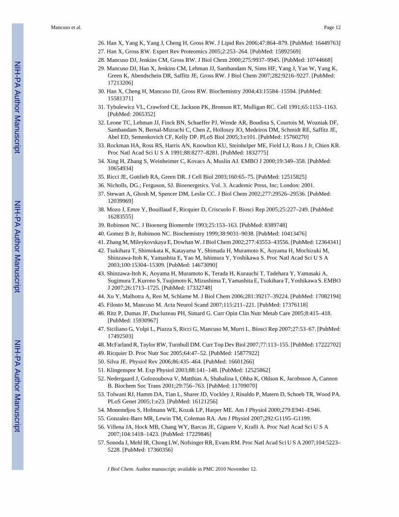
26. Han X, Yang K, Yang J, Cheng H, Gross RW. J Lipid Res 2006;47:864–879. [PubMed: 16449763]27. Han X, Gross RW. Expert Rev Proteomics 2005;2:253–264. [PubMed: 15892569]28. Mancuso DJ, Jenkins CM, Gross RW. J Biol Chem 2000;275:9937–9945. [PubMed: 10744668]29. Mancuso DJ, Han X, Jenkins CM, Lehman JJ, Sambandam N, Sims HF, Yang J, Yan W, Yang K,
Green K, Abendschein DR, Saffitz JE, Gross RW. J Biol Chem 2007;282:9216–9227. [PubMed:17213206]
30. Han X, Cheng H, Mancuso DJ, Gross RW. Biochemistry 2004;43:15584–15594. [PubMed:15581371]
31. Tybulewicz VL, Crawford CE, Jackson PK, Bronson RT, Mulligan RC. Cell 1991;65:1153–1163.[PubMed: 2065352]
32. Leone TC, Lehman JJ, Finck BN, Schaeffer PJ, Wende AR, Boudina S, Courtois M, Wozniak DF,Sambandam N, Bernal-Mizrachi C, Chen Z, Holloszy JO, Medeiros DM, Schmidt RE, Saffitz JE,Abel ED, Semenkovich CF, Kelly DP. PLoS Biol 2005;3:e101. [PubMed: 15760270]
33. Rockman HA, Ross RS, Harris AN, Knowlton KU, Steinhelper ME, Field LJ, Ross J Jr, Chien KR.Proc Natl Acad Sci U S A 1991;88:8277–8281. [PubMed: 1832775]
34. Xing H, Zhang S, Weinheimer C, Kovacs A, Muslin AJ. EMBO J 2000;19:349–358. [PubMed:10654934]
35. Ricci JE, Gottlieb RA, Green DR. J Cell Biol 2003;160:65–75. [PubMed: 12515825]36. Nicholls, DG.; Ferguson, SJ. Bioenergetics. Vol. 3. Academic Press, Inc; London: 2001.37. Stewart A, Ghosh M, Spencer DM, Leslie CC. J Biol Chem 2002;277:29526–29536. [PubMed:
12039969]38. Mozo J, Emre Y, Bouillaud F, Ricquier D, Criscuolo F. Biosci Rep 2005;25:227–249. [PubMed:
16283555]39. Robinson NC. J Bioenerg Biomembr 1993;25:153–163. [PubMed: 8389748]40. Gomez B Jr, Robinson NC. Biochemistry 1999;38:9031–9038. [PubMed: 10413476]41. Zhang M, Mileykovskaya E, Dowhan W. J Biol Chem 2002;277:43553–43556. [PubMed: 12364341]42. Tsukihara T, Shimokata K, Katayama Y, Shimada H, Muramoto K, Aoyama H, Mochizuki M,
Shinzawa-Itoh K, Yamashita E, Yao M, Ishimura Y, Yoshikawa S. Proc Natl Acad Sci U S A2003;100:15304–15309. [PubMed: 14673090]
43. Shinzawa-Itoh K, Aoyama H, Muramoto K, Terada H, Kurauchi T, Tadehara Y, Yamasaki A,Sugimura T, Kurono S, Tsujimoto K, Mizushima T, Yamashita E, Tsukihara T, Yoshikawa S. EMBOJ 2007;26:1713–1725. [PubMed: 17332748]
44. Xu Y, Malhotra A, Ren M, Schlame M. J Biol Chem 2006;281:39217–39224. [PubMed: 17082194]45. Filosto M, Mancuso M. Acta Neurol Scand 2007;115:211–221. [PubMed: 17376118]46. Ritz P, Dumas JF, Ducluzeau PH, Simard G. Curr Opin Clin Nutr Metab Care 2005;8:415–418.
[PubMed: 15930967]47. Siciliano G, Volpi L, Piazza S, Ricci G, Mancuso M, Murri L. Biosci Rep 2007;27:53–67. [PubMed:
17492503]48. McFarland R, Taylor RW, Turnbull DM. Curr Top Dev Biol 2007;77:113–155. [PubMed: 17222702]49. Ricquier D. Proc Nutr Soc 2005;64:47–52. [PubMed: 15877922]50. Silva JE. Physiol Rev 2006;86:435–464. [PubMed: 16601266]51. Klingenspor M. Exp Physiol 2003;88:141–148. [PubMed: 12525862]52. Nedergaard J, Golozoubova V, Matthias A, Shabalina I, Ohba K, Ohlson K, Jacobsson A, Cannon
B. Biochem Soc Trans 2001;29:756–763. [PubMed: 11709070]53. Tolwani RJ, Hamm DA, Tian L, Sharer JD, Vockley J, Rinaldo P, Matern D, Schoeb TR, Wood PA.
PLoS Genet 2005;1:e23. [PubMed: 16121256]54. Monemdjou S, Hofmann WE, Kozak LP, Harper ME. Am J Physiol 2000;279:E941–E946.55. Gonzalez-Baro MR, Lewin TM, Coleman RA. Am J Physiol 2007;292:G1195–G1199.56. Villena JA, Hock MB, Chang WY, Barcas JE, Giguere V, Kralli A. Proc Natl Acad Sci U S A
2007;104:1418–1423. [PubMed: 17229846]57. Sonoda J, Mehl IR, Chong LW, Nofsinger RR, Evans RM. Proc Natl Acad Sci U S A 2007;104:5223–
5228. [PubMed: 17360356]
Mancuso et al. Page 12
J Biol Chem. Author manuscript; available in PMC 2010 November 12.
NIH
-PA Author Manuscript
NIH
-PA Author Manuscript
NIH
-PA Author Manuscript
58. Lelliott CJ, Medina-Gomez G, Petrovic N, Kis A, Feldmann HM, Bjursell M, Parker N, Curtis K,Campbell M, Hu P, Zhang D, Litwin SE, Zaha VG, Fountain KT, Boudina S, Jimenez-Linan M,Blount M, Lopez M, Meirhaeghe A, Bohlooly YM, Storlien L, Stromstedt M, Snaith M, Oresic M,Abel ED, Cannon B, Vidal-Puig A. PLoS Biol 2006;4:e369. [PubMed: 17090215]
59. Wallace DC. Methods Mol Biol 2002;197:3–54. [PubMed: 12013805]60. Wallace DC. Am J Med Genet 2001;106:71–93. [PubMed: 11579427]61. Wallace DC. Annu Rev Genet 2005;39:359–407. [PubMed: 16285865]62. Wallace DC. Ment Retard Dev Disabil Res Rev 2001;7:158–166. [PubMed: 11553931]63. Chen JL, Peacock E, Samady W, Turner SM, Neese RA, Hellerstein MK, Murphy EJ. J Biol Chem
2005;280:25396–25402. [PubMed: 15888453]64. Heath-Engel HM, Shore GC. Biochim Biophys Acta 2006;1763:549–560. [PubMed: 16574258]65. McBride HM, Neuspiel M, Wasiak S. Curr Biol 2006;16:R551–560. [PubMed: 16860735]66. Siegel DP. Biophys J 1984;45:399–420. [PubMed: 6365189]67. Dahlberg M. J Phys Chem B 2007;111:7194–7200. [PubMed: 17542632]68. Skulachev VP. FEBS Lett 1991;294:158–162. [PubMed: 1756853]69. Garlid KD, Orosz DE, Modriansky M, Vassanelli S, Jezek P. J Biol Chem 1996;271:2615–2620.
[PubMed: 8576230]70. Breen EP, Gouin SG, Murphy AF, Haines LR, Jackson AM, Pearson TW, Murphy PV, Porter RK. J
Biol Chem 2006;281:2114–2119. [PubMed: 16291746]71. Porter RK. Biochim Biophys Acta 2006;1757:446–448. [PubMed: 16730638]72. Di Marzo V, De Petrocellis L, Sugiura T, Waku K. Biochem Biophys Res Commun 1996;227:281–
288. [PubMed: 8858137]73. Pete MJ, Exton JH. J Biol Chem 1996;271:18114–18121. [PubMed: 8663471]74. Pinheiro TJ, Watts A. Biochemistry 1994;33:2459–2467. [PubMed: 8117706]75. Vreken P, Valianpour F, Nijtmans LG, Grivell LA, Plecko B, Wanders RJ, Barth PG. Biochem
Biophys Res Commun 2000;279:378–382. [PubMed: 11118295]76. Schlame M, Towbin JA, Heerdt PM, Jehle R, DiMauro S, Blanck TJ. Ann Neurol 2002;51:634–637.
[PubMed: 12112112]77. Xu Y, Kelley RI, Blanck TJ, Schlame M. J Biol Chem 2003;278:51380–51385. [PubMed: 14551214]78. Schlame M, Ren M, Xu Y, Greenberg ML, Haller I. Chem Phys Lipids 2005;138:38–49. [PubMed:
16226238]79. Spencer CT, Bryant RM, Day J, Gonzalez IL, Colan SD, Thompson WR, Berthy J, Redfearn SP,
Byrne BJ. Pediatrics 2006;118:e337–346. [PubMed: 16847078]80. Bolhuis PA, Hensels GW, Hulsebos TJ, Baas F, Barth PG. Am J Hum Genet 1991;48:481–485.
[PubMed: 1998334]81. Schlame M, Ren M. FEBS Lett 2006;580:5450–5455. [PubMed: 16973164]82. Bonventre JV, Huang Z, Taheri MR, O’Leary E, Li E, Moskowitz MA, Sapirstein A. Nature
1997;390:622–625. [PubMed: 9403693]83. Fujishima H, Sanchez Mejia RO, Bingham CO III, Lam BK, Sapirstein A, Bonventre JV, Austen KF,
Arm JP. Proc Natl Acad Sci U S A 1999;96:4803–4807. [PubMed: 10220374]84. Rosenberger TA, Villacreses NE, Contreras MA, Bonventre JV, Rapoport SI. J Lipid Res
2003;44:109–117. [PubMed: 12518029]85. Bao S, Miller DJ, Ma Z, Wohltmann M, Eng G, Ramanadham S, Moley K, Turk J. J Biol Chem
2004;279:38194–38200. [PubMed: 15252026]86. Wilson PA, Gardner SD, Lambie NM, Commans SA, Crowther DJ. J Lipid Res 2006;47:1940–1949.
[PubMed: 16799181]
Mancuso et al. Page 13
J Biol Chem. Author manuscript; available in PMC 2010 November 12.
NIH
-PA Author Manuscript
NIH
-PA Author Manuscript
NIH
-PA Author Manuscript
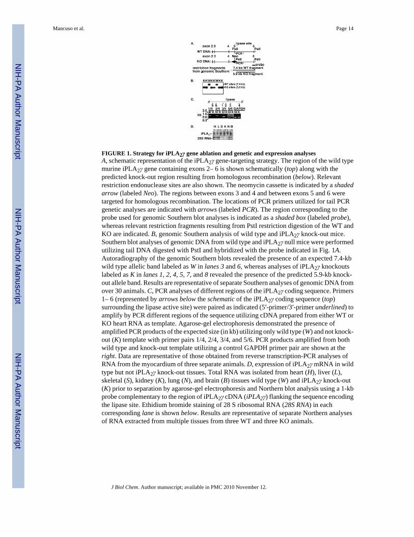
FIGURE 1. Strategy for iPLA2γ gene ablation and genetic and expression analysesA, schematic representation of the iPLA2γ gene-targeting strategy. The region of the wild typemurine iPLA2γ gene containing exons 2– 6 is shown schematically (top) along with thepredicted knock-out region resulting from homologous recombination (below). Relevantrestriction endonuclease sites are also shown. The neomycin cassette is indicated by a shadedarrow (labeled Neo). The regions between exons 3 and 4 and between exons 5 and 6 weretargeted for homologous recombination. The locations of PCR primers utilized for tail PCRgenetic analyses are indicated with arrows (labeled PCR). The region corresponding to theprobe used for genomic Southern blot analyses is indicated as a shaded box (labeled probe),whereas relevant restriction fragments resulting from PstI restriction digestion of the WT andKO are indicated. B, genomic Southern analysis of wild type and iPLA2γ knock-out mice.Southern blot analyses of genomic DNA from wild type and iPLA2γ null mice were performedutilizing tail DNA digested with PstI and hybridized with the probe indicated in Fig. 1A.Autoradiography of the genomic Southern blots revealed the presence of an expected 7.4-kbwild type allelic band labeled as W in lanes 3 and 6, whereas analyses of iPLA2γ knockoutslabeled as K in lanes 1, 2, 4, 5, 7, and 8 revealed the presence of the predicted 5.9-kb knock-out allele band. Results are representative of separate Southern analyses of genomic DNA fromover 30 animals. C, PCR analyses of different regions of the iPLA2γ coding sequence. Primers1– 6 (represented by arrows below the schematic of the iPLA2γ coding sequence (top)surrounding the lipase active site) were paired as indicated (5′-primer/3′-primer underlined) toamplify by PCR different regions of the sequence utilizing cDNA prepared from either WT orKO heart RNA as template. Agarose-gel electrophoresis demonstrated the presence ofamplified PCR products of the expected size (in kb) utilizing only wild type (W) and not knock-out (K) template with primer pairs 1/4, 2/4, 3/4, and 5/6. PCR products amplified from bothwild type and knock-out template utilizing a control GAPDH primer pair are shown at theright. Data are representative of those obtained from reverse transcription-PCR analyses ofRNA from the myocardium of three separate animals. D, expression of iPLA2γ mRNA in wildtype but not iPLA2γ knock-out tissues. Total RNA was isolated from heart (H), liver (L),skeletal (S), kidney (K), lung (N), and brain (B) tissues wild type (W) and iPLA2γ knock-out(K) prior to separation by agarose-gel electrophoresis and Northern blot analysis using a 1-kbprobe complementary to the region of iPLA2γ cDNA (iPLA2γ) flanking the sequence encodingthe lipase site. Ethidium bromide staining of 28 S ribosomal RNA (28S RNA) in eachcorresponding lane is shown below. Results are representative of separate Northern analysesof RNA extracted from multiple tissues from three WT and three KO animals.
Mancuso et al. Page 14
J Biol Chem. Author manuscript; available in PMC 2010 November 12.
NIH
-PA Author Manuscript
NIH
-PA Author Manuscript
NIH
-PA Author Manuscript
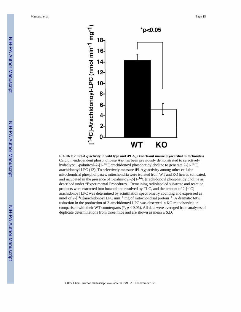
FIGURE 2. iPLA2γ activity in wild type and iPLA2γ knock-out mouse myocardial mitochondriaCalcium-independent phospholipase A2γ has been previously demonstrated to selectivelyhydrolyze 1-palmitoyl-2-[1-14C]arachidonoyl phosphatidylcholine to generate 2-[1-14C]arachidonoyl LPC (12). To selectively measure iPLA2γ activity among other cellularmitochondrial phospholipases, mitochondria were isolated from WT and KO hearts, sonicated,and incubated in the presence of 1-palmitoyl-2-[1-14C]arachidonoyl phosphatidylcholine asdescribed under “Experimental Procedures.” Remaining radiolabeled substrate and reactionproducts were extracted into butanol and resolved by TLC, and the amount of 2-[14C]arachidonoyl LPC was determined by scintillation spectrometry counting and expressed asnmol of 2-[14C]arachidonoyl LPC min−1 mg of mitochondrial protein−1. A dramatic 60%reduction in the production of 2-arachidonoyl LPC was observed in KO mitochondria incomparison with their WT counterparts (*, p < 0.05). All data were averaged from analyses ofduplicate determinations from three mice and are shown as mean ± S.D.
Mancuso et al. Page 15
J Biol Chem. Author manuscript; available in PMC 2010 November 12.
NIH
-PA Author Manuscript
NIH
-PA Author Manuscript
NIH
-PA Author Manuscript
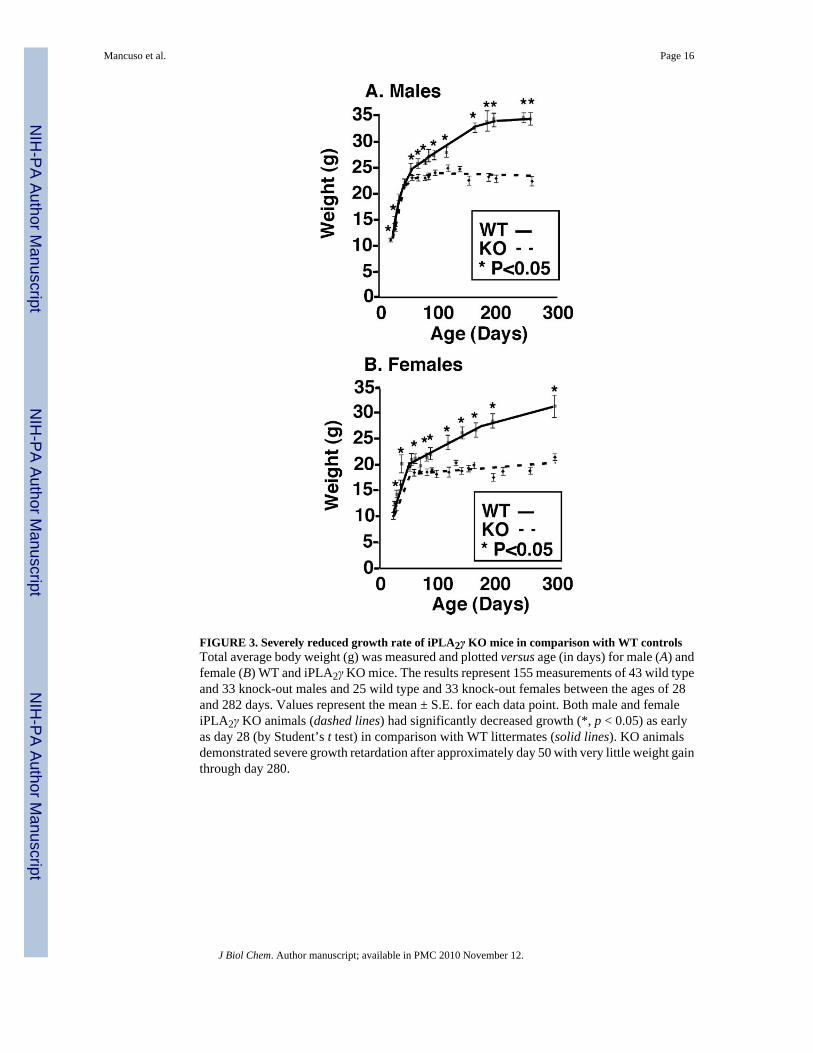
FIGURE 3. Severely reduced growth rate of iPLA2γ KO mice in comparison with WT controlsTotal average body weight (g) was measured and plotted versus age (in days) for male (A) andfemale (B) WT and iPLA2γ KO mice. The results represent 155 measurements of 43 wild typeand 33 knock-out males and 25 wild type and 33 knock-out females between the ages of 28and 282 days. Values represent the mean ± S.E. for each data point. Both male and femaleiPLA2γ KO animals (dashed lines) had significantly decreased growth (*, p < 0.05) as earlyas day 28 (by Student’s t test) in comparison with WT littermates (solid lines). KO animalsdemonstrated severe growth retardation after approximately day 50 with very little weight gainthrough day 280.
Mancuso et al. Page 16
J Biol Chem. Author manuscript; available in PMC 2010 November 12.
NIH
-PA Author Manuscript
NIH
-PA Author Manuscript
NIH
-PA Author Manuscript
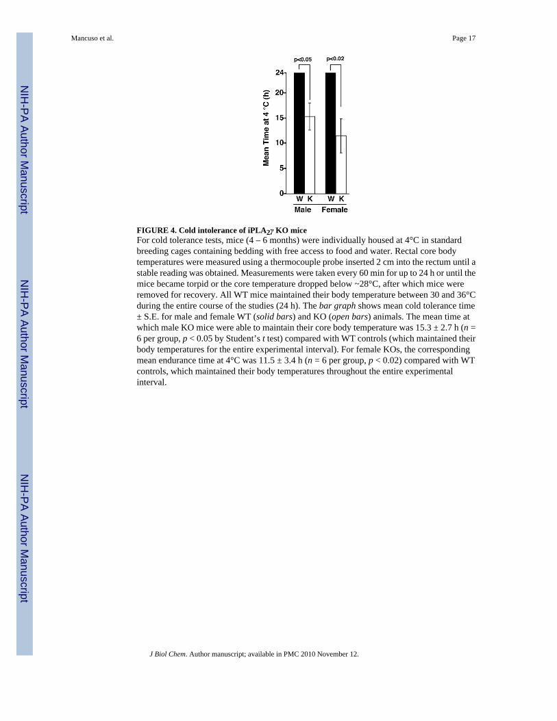
FIGURE 4. Cold intolerance of iPLA2γ KO miceFor cold tolerance tests, mice (4 – 6 months) were individually housed at 4°C in standardbreeding cages containing bedding with free access to food and water. Rectal core bodytemperatures were measured using a thermocouple probe inserted 2 cm into the rectum until astable reading was obtained. Measurements were taken every 60 min for up to 24 h or until themice became torpid or the core temperature dropped below ~28°C, after which mice wereremoved for recovery. All WT mice maintained their body temperature between 30 and 36°Cduring the entire course of the studies (24 h). The bar graph shows mean cold tolerance time± S.E. for male and female WT (solid bars) and KO (open bars) animals. The mean time atwhich male KO mice were able to maintain their core body temperature was 15.3 ± 2.7 h (n =6 per group, p < 0.05 by Student’s t test) compared with WT controls (which maintained theirbody temperatures for the entire experimental interval). For female KOs, the correspondingmean endurance time at 4°C was 11.5 ± 3.4 h (n = 6 per group, p < 0.02) compared with WTcontrols, which maintained their body temperatures throughout the entire experimentalinterval.
Mancuso et al. Page 17
J Biol Chem. Author manuscript; available in PMC 2010 November 12.
NIH
-PA Author Manuscript
NIH
-PA Author Manuscript
NIH
-PA Author Manuscript

FIGURE 5. Expression of UCP1 mRNA in brown adipose tissue and also UCP2 and UCP3 mRNAin heart tissue from WT and KO miceExpression levels of UCP1 (A), UCP2 (B), and UCP3 (C) mRNA were measured using realtime PCR as described under “Experimental Procedures.” A, UCP1 message levels in brownadipose tissue were measured under ambient conditions (control) and during exposure to cold(4°C). Animals were exposed to 4°C until their core body temperature dropped to hypothermiclevels (below ~28°C). The mean cold tolerance time for KO animals was 8 h, and WT controls(still maintaining the core body temperatures) were sacrificed at the same mean time. B, nosignificant difference was observed for UCP2 expression levels in WT and KO hearts. C, asignificant reduction in UCP3 expression was observed in the hearts of iPLA2γ KO mice (p <0.05). n = 4 per group. Values represent arbitrary units (AU) after normalization to a ribosomal36B4 internal standard. Data are presented as means ± S.E. Statistical significance wasperformed using the Student’s t test, and significant differences are indicated by brackets.
Mancuso et al. Page 18
J Biol Chem. Author manuscript; available in PMC 2010 November 12.
NIH
-PA Author Manuscript
NIH
-PA Author Manuscript
NIH
-PA Author Manuscript
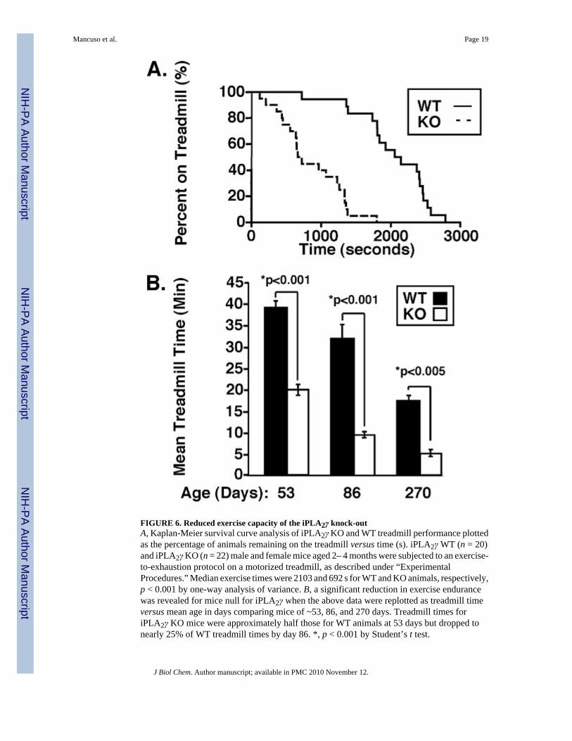
FIGURE 6. Reduced exercise capacity of the iPLA2γ knock-outA, Kaplan-Meier survival curve analysis of iPLA2γ KO and WT treadmill performance plottedas the percentage of animals remaining on the treadmill versus time (s). iPLA2γ WT (n = 20)and iPLA2γ KO (n = 22) male and female mice aged 2– 4 months were subjected to an exercise-to-exhaustion protocol on a motorized treadmill, as described under “ExperimentalProcedures.” Median exercise times were 2103 and 692 s for WT and KO animals, respectively,p < 0.001 by one-way analysis of variance. B, a significant reduction in exercise endurancewas revealed for mice null for iPLA2γ when the above data were replotted as treadmill timeversus mean age in days comparing mice of ~53, 86, and 270 days. Treadmill times foriPLA2γ KO mice were approximately half those for WT animals at 53 days but dropped tonearly 25% of WT treadmill times by day 86. *, p < 0.001 by Student’s t test.
Mancuso et al. Page 19
J Biol Chem. Author manuscript; available in PMC 2010 November 12.
NIH
-PA Author Manuscript
NIH
-PA Author Manuscript
NIH
-PA Author Manuscript
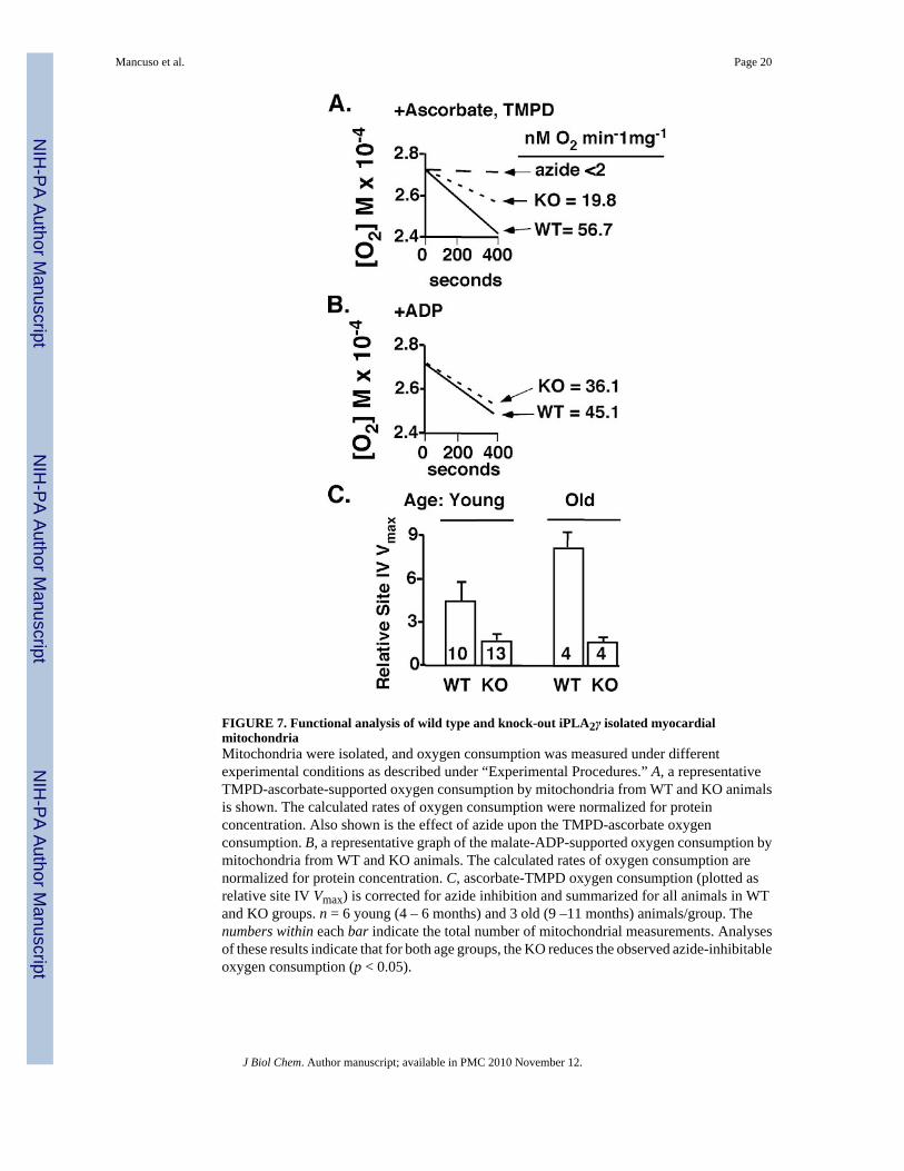
FIGURE 7. Functional analysis of wild type and knock-out iPLA2γ isolated myocardialmitochondriaMitochondria were isolated, and oxygen consumption was measured under differentexperimental conditions as described under “Experimental Procedures.” A, a representativeTMPD-ascorbate-supported oxygen consumption by mitochondria from WT and KO animalsis shown. The calculated rates of oxygen consumption were normalized for proteinconcentration. Also shown is the effect of azide upon the TMPD-ascorbate oxygenconsumption. B, a representative graph of the malate-ADP-supported oxygen consumption bymitochondria from WT and KO animals. The calculated rates of oxygen consumption arenormalized for protein concentration. C, ascorbate-TMPD oxygen consumption (plotted asrelative site IV Vmax) is corrected for azide inhibition and summarized for all animals in WTand KO groups. n = 6 young (4 – 6 months) and 3 old (9 –11 months) animals/group. Thenumbers within each bar indicate the total number of mitochondrial measurements. Analysesof these results indicate that for both age groups, the KO reduces the observed azide-inhibitableoxygen consumption (p < 0.05).
Mancuso et al. Page 20
J Biol Chem. Author manuscript; available in PMC 2010 November 12.
NIH
-PA Author Manuscript
NIH
-PA Author Manuscript
NIH
-PA Author Manuscript
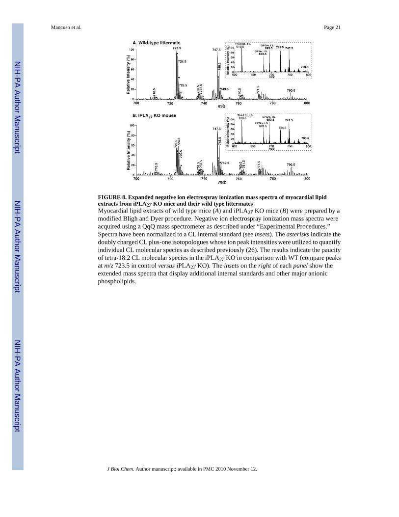
FIGURE 8. Expanded negative ion electrospray ionization mass spectra of myocardial lipidextracts from iPLA2γ KO mice and their wild type littermatesMyocardial lipid extracts of wild type mice (A) and iPLA2γ KO mice (B) were prepared by amodified Bligh and Dyer procedure. Negative ion electrospray ionization mass spectra wereacquired using a QqQ mass spectrometer as described under “Experimental Procedures.”Spectra have been normalized to a CL internal standard (see insets). The asterisks indicate thedoubly charged CL plus-one isotopologues whose ion peak intensities were utilized to quantifyindividual CL molecular species as described previously (26). The results indicate the paucityof tetra-18:2 CL molecular species in the iPLA2γ KO in comparison with WT (compare peaksat m/z 723.5 in control versus iPLA2γ KO). The insets on the right of each panel show theextended mass spectra that display additional internal standards and other major anionicphospholipids.
Mancuso et al. Page 21
J Biol Chem. Author manuscript; available in PMC 2010 November 12.
NIH
-PA Author Manuscript
NIH
-PA Author Manuscript
NIH
-PA Author Manuscript
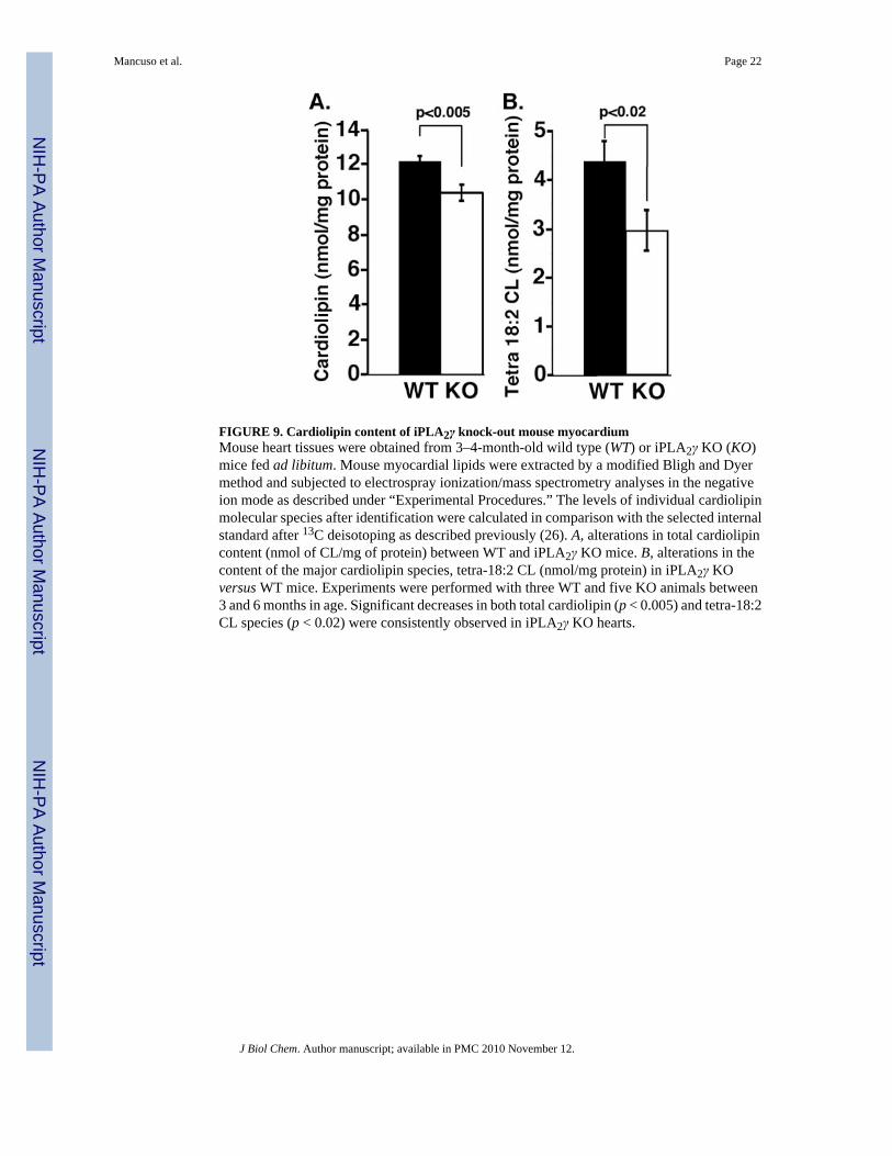
FIGURE 9. Cardiolipin content of iPLA2γ knock-out mouse myocardiumMouse heart tissues were obtained from 3–4-month-old wild type (WT) or iPLA2γ KO (KO)mice fed ad libitum. Mouse myocardial lipids were extracted by a modified Bligh and Dyermethod and subjected to electrospray ionization/mass spectrometry analyses in the negativeion mode as described under “Experimental Procedures.” The levels of individual cardiolipinmolecular species after identification were calculated in comparison with the selected internalstandard after 13C deisotoping as described previously (26). A, alterations in total cardiolipincontent (nmol of CL/mg of protein) between WT and iPLA2γ KO mice. B, alterations in thecontent of the major cardiolipin species, tetra-18:2 CL (nmol/mg protein) in iPLA2γ KOversus WT mice. Experiments were performed with three WT and five KO animals between3 and 6 months in age. Significant decreases in both total cardiolipin (p < 0.005) and tetra-18:2CL species (p < 0.02) were consistently observed in iPLA2γ KO hearts.
Mancuso et al. Page 22
J Biol Chem. Author manuscript; available in PMC 2010 November 12.
NIH
-PA Author Manuscript
NIH
-PA Author Manuscript
NIH
-PA Author Manuscript

NIH
-PA Author Manuscript
NIH
-PA Author Manuscript
NIH
-PA Author Manuscript
Mancuso et al. Page 23
TABLE 1Primers for construction and genotyping of the iPLA2γ knock-out
iPLA2γ KO primers P1–P4 were used in PCR to prepare the KO construct. PCR primers P5 and P6 were usedto generate the 459-nt genomic probe for genotyping by genomic Southern blot analyses. The tail PCR primerset was used for genotyping. “Neo forward” paired with the “Common REV” amplified a 354-nt product fromKO DNA whereas “iPLA2γ FOR” paired with “Common REV” amplified a 181-nt product from WT DNA.Reverse transcription-PCR primers 1–6 were used to demonstrate an absence of iPLA2γ message expressionfrom the KO, whereas “GAPDH F” and “GAPDH R” PCR primers were used as positive controls foramplification of GAPDH PCR product from both WT and KO cDNA.
Primers Sequence (5′–3′)
iPLA2γ KO primers
P1 AAAAGAATTCAGGCAGAGGAGAAAAAGCGTGTGCTACTTC
P2 AAAAGAATTCGCTCACAACCATCCGTAATGAGATCTGGATG
P3 AAAACTCGAGCCAAGAACATGCAGCACAGGCTTGAGGCCAT
P4 AAAAGCGGCCGCTTGTTGACATCATAAAAACCATAATT
P5 CTCTATCGAAAGTTGGGCTCAGATGT
P6 AATAGGTAAATAAATACTAAAATGGA
Tail PCR primer set
NEO forward AAAGGCCTACCCGCTTCCATTGCTCA
Common REV GCCTCAAGCCTGTGCTGCATGTTCTT
iPLA2γ FOR CTGTTCACAGAGGTGTGGTTGC
KO band = 354 base pairs
WT band = 181 base pairs
Reverse transcription-PCR primers
1 CAAGAAGGCAGAGGAGAAAAAGC
2 CCCAAAGCTCTGTATTACTAGGG
3 GATTACATCTGTGGAGTAAGCACAGGG
4 CGTGAAAGGCCAAGGCCGAAGGG
5 AGTGAGCATTCATATGTCCCG
6 CGGAGAATGACTCTCCAAATCTG
GAPDH F TGTTGCCATCAATGACCCCTTCA
GAPDH R TGATGATCTTGAGGCTGTTGTC
J Biol Chem. Author manuscript; available in PMC 2010 November 12.
Copyright © 2022 FDOKUMEN