Gadolinium-loaded polymeric nanoparticles modified with Anti-VEGF as multifunctional MRI contrast...
10
Gadolinium-loaded polymeric nanoparticles modified with Anti-VEGF as multifunctional MRI contrast agents for the diagnosis of liver cancer Yongjun Liu a,1 , Zhijin Chen a, 1 , Chunxi Liu a , Dexin Yu b , Zaijun Lu c , Na Zhang a, * a School of Pharmaceutical Science, Shandong University, Ji’nan, People’s Republic of China b Department of Radiology Medicine, Affiliated Qilu Hospital, Shandong University, Ji’nan, People’s Republic of China c School of Chemistry and Chemical Engineering, Shandong University, Ji’nan, People’s Republic of China article info Article history: Received 19 March 2011 Accepted 29 March 2011 Available online 24 April 2011 Keywords: Polymeric nanoparticle Gadolinium MRI Hepatocellular carcinoma Early diagnosis abstract Molecular imaging is essential to increase the sensitivity and selectivity of cancer diagnosis especially in the early stage of tumor. Here, we designed a novel multifunctional polymeric nanoparticle contrast agent (Anti-VEGF PLA-PEG-PLL-Gd NP) simultaneously modified with Gadolinium-diethylenetriamine pentaacetic acid (Gd-DTPA) and anti-vascular endothelial growth factor (VEGF) antibody to deliver Gd-DTPA to the tumor area and achieve the early diagnosis of hepatocellular carcinoma (HCC). The Anti-VEGF PLA-PEG-PLL-Gd NPs exhibited high T 1 relaxivity and no obvious cytotoxicity under the experimental concentrations in human hepatocellular carcinoma (HepG2) cells. The results of in vitro cell uptake experiments demonstrated that the uptake process of NPs was both concentration and time depended. Compared with non-targeted NPs, the Anti-VEGF antibody modified NPs showed much higher cell uptake in the HepG2 cells. During in vivo studies, the targeted NPs showed significantly signal intensity enhancement at the tumor site (mouse hepatocarcinoma tumor, H22) compared with non- targeted NPs and Gd-DTPA injection in tumor-bearing mice and the imaging time was significantly prolonged from less than an hour (Gd-DTPA injection group) to 12 h. These results demonstrated that this novel MRI contrast agent Anti-VEGF PLA-PEG-PLL-Gd NPs showed great potential in the early diagnosis of liver tumors. Ó 2011 Elsevier Ltd. All rights reserved. 1. Introduction Human hepatocellular carcinoma (HCC) is the most common malignancies worldwide and with over 700,000 cases each year [1]. Early diagnosis of premalignant and malignant lesions is essential for improving the current poor survival of patients with HCC. Magnetic resonance imaging (MRI), which produces high- resolution three-dimensional maps delineating morphological features of the specimen and lack of ionizing radiation, is one of the best strategies used in clinic to diagnose cancer [2]. To enhance the differentiation between tissues, contrast agents are used to shorten the relaxation parameters (T 1 and T 2 ) of water. There are a variety of approved metallic contrast agents available in clinical such as Mn (Teslascan Ò ) [3], Gd (ProHance Ò , Dotarem Ò and Magnevist Ò ) [4] and Fe (Endorem Ò , and Feridex Ò ) [5]. Gd-based contrast agents are employed in the overwhelming majority of the cases among different kinds of contrast agents. It is suggested that more than 10 million MRI studies are performed worldwide using Gd-based contrast agents each year [6]. Compared to superparamagnetic iron oxide (SPIO), Gd-based contrast agents are positive agents for both vascular imaging and tumor imaging. It could shorten the longitudinal relaxation time due to its large magnetic moment (because of seven unpaired electrons) and relatively long electron- spin-relaxation time (10 9 s) in the magnetic field [4]. To avoid the toxicity of free Gd ions, Gd-chelates such as Gd-DTPA (Magnevist Ò ) were widely used in clinic. However, Gd-chelates are easily elimi- nated by renal filtration because of the low molecular weight, thus resulted in short imaging time in vivo. Furthermore, the lack of specificity to target organs is among the greatest obstacles to improve the precision and accuracy of MRI. To overcome these drawbacks, contrast agents prepared by nanotechnology have been studied for decades and showed great potential in tumor diagnosis. These nanoparticles (NPs) include lipid-based NPs [7], polymeric NPs [8], micells [9], dendrimers [10] and silica NPs [11]. Advantages of these novel contrast agents such as passive targeting properties, the prolonged imaging time, the enhancement of the contrast and low toxicity were observed [8]. However, to achieve early diagnosis of cancer and molecular imaging, it is not efficient to deliver the NP agents just by passive * Corresponding author. Tel.: þ86 531 88382015; fax: þ86 531 88382548. E-mail address: [email protected] (N. Zhang). 1 These two authors contributed equally to this work. Contents lists available at ScienceDirect Biomaterials journal homepage: www.elsevier.com/locate/biomaterials 0142-9612/$ e see front matter Ó 2011 Elsevier Ltd. All rights reserved. doi:10.1016/j.biomaterials.2011.03.077 Biomaterials 32 (2011) 5167e5176
-
Upload
independent -
Category
Documents
-
view
4 -
download
0
Transcript of Gadolinium-loaded polymeric nanoparticles modified with Anti-VEGF as multifunctional MRI contrast...
lable at ScienceDirect
Biomaterials 32 (2011) 5167e5176
Contents lists avai
Biomaterials
journal homepage: www.elsevier .com/locate/biomater ia ls
Gadolinium-loaded polymeric nanoparticles modified with Anti-VEGF asmultifunctional MRI contrast agents for the diagnosis of liver cancer
Yongjun Liu a,1, Zhijin Chen a,1, Chunxi Liu a, Dexin Yu b, Zaijun Lu c, Na Zhang a,*
a School of Pharmaceutical Science, Shandong University, Ji’nan, People’s Republic of ChinabDepartment of Radiology Medicine, Affiliated Qilu Hospital, Shandong University, Ji’nan, People’s Republic of Chinac School of Chemistry and Chemical Engineering, Shandong University, Ji’nan, People’s Republic of China
a r t i c l e i n f o
Article history:Received 19 March 2011Accepted 29 March 2011Available online 24 April 2011
Keywords:Polymeric nanoparticleGadoliniumMRIHepatocellular carcinomaEarly diagnosis
* Corresponding author. Tel.: þ86 531 88382015; faE-mail address: [email protected] (N. Zhan
1 These two authors contributed equally to this wo
0142-9612/$ e see front matter � 2011 Elsevier Ltd.doi:10.1016/j.biomaterials.2011.03.077
a b s t r a c t
Molecular imaging is essential to increase the sensitivity and selectivity of cancer diagnosis especially inthe early stage of tumor. Here, we designed a novel multifunctional polymeric nanoparticle contrastagent (Anti-VEGF PLA-PEG-PLL-Gd NP) simultaneously modified with Gadolinium-diethylenetriaminepentaacetic acid (Gd-DTPA) and anti-vascular endothelial growth factor (VEGF) antibody to deliverGd-DTPA to the tumor area and achieve the early diagnosis of hepatocellular carcinoma (HCC). TheAnti-VEGF PLA-PEG-PLL-Gd NPs exhibited high T1 relaxivity and no obvious cytotoxicity under theexperimental concentrations in human hepatocellular carcinoma (HepG2) cells. The results of in vitro celluptake experiments demonstrated that the uptake process of NPs was both concentration and timedepended. Compared with non-targeted NPs, the Anti-VEGF antibody modified NPs showed much highercell uptake in the HepG2 cells. During in vivo studies, the targeted NPs showed significantly signalintensity enhancement at the tumor site (mouse hepatocarcinoma tumor, H22) compared with non-targeted NPs and Gd-DTPA injection in tumor-bearing mice and the imaging time was significantlyprolonged from less than an hour (Gd-DTPA injection group) to 12 h. These results demonstrated thatthis novel MRI contrast agent Anti-VEGF PLA-PEG-PLL-Gd NPs showed great potential in the earlydiagnosis of liver tumors.
� 2011 Elsevier Ltd. All rights reserved.
1. Introduction
Human hepatocellular carcinoma (HCC) is the most commonmalignancies worldwide and with over 700,000 cases each year[1]. Early diagnosis of premalignant and malignant lesions isessential for improving the current poor survival of patients withHCC. Magnetic resonance imaging (MRI), which produces high-resolution three-dimensional maps delineating morphologicalfeatures of the specimen and lack of ionizing radiation, is one of thebest strategies used in clinic to diagnose cancer [2]. To enhance thedifferentiation between tissues, contrast agents are used to shortenthe relaxation parameters (T1 and T2) of water. There are a variety ofapproved metallic contrast agents available in clinical such as Mn(Teslascan�) [3], Gd (ProHance�, Dotarem� and Magnevist�) [4]and Fe (Endorem�, and Feridex�) [5]. Gd-based contrast agentsare employed in the overwhelming majority of the cases amongdifferent kinds of contrast agents. It is suggested that more than
x: þ86 531 88382548.g).rk.
All rights reserved.
10 million MRI studies are performed worldwide using Gd-basedcontrast agents each year [6]. Compared to superparamagneticiron oxide (SPIO), Gd-based contrast agents are positive agents forboth vascular imaging and tumor imaging. It could shorten thelongitudinal relaxation time due to its large magnetic moment(because of seven unpaired electrons) and relatively long electron-spin-relaxation time (10�9 s) in the magnetic field [4]. To avoid thetoxicity of free Gd ions, Gd-chelates such as Gd-DTPA (Magnevist�)were widely used in clinic. However, Gd-chelates are easily elimi-nated by renal filtration because of the low molecular weight, thusresulted in short imaging time in vivo. Furthermore, the lack ofspecificity to target organs is among the greatest obstacles toimprove the precision and accuracy of MRI. To overcome thesedrawbacks, contrast agents prepared by nanotechnology have beenstudied for decades and showed great potential in tumor diagnosis.These nanoparticles (NPs) include lipid-based NPs [7], polymericNPs [8], micells [9], dendrimers [10] and silica NPs [11]. Advantagesof these novel contrast agents such as passive targeting properties,the prolonged imaging time, the enhancement of the contrast andlow toxicity were observed [8].
However, to achieve early diagnosis of cancer and molecularimaging, it is not efficient to deliver the NP agents just by passive
Y. Liu et al. / Biomaterials 32 (2011) 5167e51765168
targeting. In these cases, active targeting strategy was employed toachieve effective delivery. This approach provides an opportunityto accumulate contrast agents highly and specifically in malignanttumors and allows an accurate diagnosis at the early stage. Thecommonly used active targeting agents are mainly peptides, anti-bodies, small molecules, proteins and aptamers [12]. Among thesetargeting strategies, vascular endothelial growth factor (VEGF) is anendothelial cell-specific targeting site and angiogenic factor in vivo.It is suggested that VEGF is a very important marker in HCC wherethe overexpression of VEGF has been demonstrated compared withnormal tissue even in early hepatocarcinogenesis stage [13]. So,many researches had been concentrated on HCC therapy by tar-geting to VEGF and showed high target efficiency [14e16]. There-fore, the VEGF was chosen as the target site to for early targetingcontrast diagnostic of the HCC in our experiments.
Gd loaded strategy is another concern in the design of the activetargeting molecular MRI contrast agent. Gd-chelates could beencapsulated inside the core of the NPs [17], adsorbed on thesurface of NPs [18] or linked with the polymer by covalently bound[19]. Because the relaxivities of the Gd contrast agent due tothe exchange of water protons with the contrast agents, it isessential to modify Gd-chelates on the surface of the NPs [20]. Toachieve this goal, recent methods concentrated on link the chelatesto the hydrophilic block of the polymer for NP preparation bycovalently bound [21]. By this approach, the chelate moleculeswhich were covalent linked on the end of the hydrophilic block ofthe polymer were trend to distribute on the surface of the NPs andGd ions could connect with the surface chelate molecules easily toachieve the anchor of contrast agents on the surface of the NPs. Inour group, PLA-PEG-DTPA was synthesized and used to preparecontrast agents. It showed a significant enhancement in theimaging intensity and a prolonged imaging time [22]. One draw-back of this method is one polymer molecule for NP preparationcould only be link to one chelate molecule, thus the amount of thesurface Gd is limited for there is only one active end group in onelinear polymer molecule could be involved in the covalentlybound of chelate molecule. Thus, the design of novel polymer withmulti-activity end groups for Gd loading is expected. Therefore,PLA-PEG-PLL was synthesized for this purpose based on PLA-PEG.Poly (L-lysine) (PLL) is biodegradable polypeptide with amino-acid lysine as a repeat unit. So, PLA-PEG-PLL could be useful forthe further synthesis of PLA-PEG-PLL-g-DTPA for the anchor ofGd3þ.
The goal of this research was to develop Gd loaded PLA-PEG-PLLNPs as the molecular MRI contrast agents to efficiently targetHCC for the detection and treatment of HCC as early as possible.PLA-PEG-PLL was selected to prepare the NPs and the multi-aminogroups of the PLL were employed to covalently link with thefunctional groups, including the chelate molecules, the fluorescentimaging molecules, and the targeting agents to obtain the multi-functional NPs. Anti-VEGF as the targeting agent was chosenand human hepatocellular carcinoma cells (HepG2) and murinehepatocellular carcinoma cells (H22) were selected as the cellmodel for VEGF were overexpressed on the membrane these cell.The cytotoxicity, targeting efficiency and imaging capability wasevaluated both in vitro and in vivo.
2. Materials and methods
2.1. Materials
Diethylenetriamine pentaacetic acid (DTPA), avidin modified fluoresceinisothiocyanate (avidin-FITC) and Gd2O3 were purchased from SigmaeAldrich (USA).Lysine (Lys) and Biotin were purchased from Aladdin (China). 33% HBr/HAc solutionwas purchased from Acros (Belgium). Gadopentetic Acid Dimeglumine Salt Injection(Gd-DTPA, Magnevist�) was purchased from Schering (Germany). Anti-VEGF was
purchased from Santa Cruz Biotechnology (USA). PLA-PEG-NH2 (MWPLA ¼ 7500,MWPEG ¼ 2000) was provided by School of Chemistry and Chemical Engineering ofShandong University (Jinan, China). All other chemicals were of analytical reagent.
2.2. Synthesis of new materials
2.2.1. Synthesis of PLA-PEG-PLLPLA-PEG-PLL was synthesized (Scheme 1) in accordance with the method
described by Lu et al. [23]. Firstly, Lys was activated. Briefly, tri-phosgene (1.58 g) andLys (3.0 g) were dissolved in 40 mL and 20 mL tetrahydrofuran (THF) respectivelyand the two reactants were then mixed. After stirring for 1.5 h at 50 �C, excesstri-phosgene was removed and 180 mL hexane was added and the mixture wasstirred for 24 h at room temperature (RT) to precipitate benzyloxycarbonyl lysineanhydride (Zlys-NCA) completely. Then, the precipitate was collected and dried invacuum at RT to obtain Zlys-NCA.
Zlys-NCA (1.2 g) and PLA-PEG-NH2 (4 g) was dissolved in 40 mL dime-thylformamide (DMF) and the polymerization was continued under the protectionof argon at 40 �C for 48 h. The product (PLA-PEG-PZLL) was precipitated by droppingthe reacting complexes into excess anhydrous diethyl ether and the precipitationwas collected by centrifuge. Then the harvested product was re-dissolved in DMFand re-precipitated using excess anhydrous diethyl ether. The dissolve-precipitationprocess was repeated for 3 times totally to purify PZLL-b-PEG-b-PLA. Finally,PZLL-b-PEG-b-PLA was dried in vacuum at 40 �C.
PLA-PEG-PZLL (2 g) was dissolved in 20 mL trifluoroacetic acid, then 1.6 mL33% HBr/HAc solution was added into and the reaction system was stirred in an icebath (0 �C) for 1 h. The obtained PLA-PEG-PLL was precipitated by dropping thereacting complexes into excess anhydrous diethyl ether and the precipitation wascollected by centrifuge. Then the similar purification was carried out as describedabove and the purified PLA-PEG-PLL was dried in vacuum at 40 �C.
2.2.2. Synthesis of PLA-PEG-PLL-biotinThe PLA-PEG-PLL-biotin was synthesized by DCC-NHS reaction. Briefly, 0.5 g
biotin was dissolved in DMF, and 0.25 g NHS and 0.5 g DCC were added intorespectively under stirring at 50 �C. After reacting for 16 h, the biotin succinimideester was collected by precipitated into excess anhydrous diethyl ether and purifiedby re-crystallization in isopropyl alcohol. The activated biotin (250 mg) andPLA-PEG-PLL (126 mg) were dissolved in DMF respectively and the two reactantswere then mixed. Then the mixture was continued stirring at 50 �C for 16 h underthe protection of N2. The reaction was terminated by adding 10 mL 10%NaHCO3 andthe resulting product was collected by precipitated into excess anhydrous diethylether and the purified PLA-PEG-PLL-biotin was dried in vacuum at RT after washedby anhydrous diethyl ether for 3 times.
2.2.3. Synthesis of PLA-PEG-PLL-DTPAFirstly, DTPA-bis-anhydride (DTPAba) was prepared according the method
published previously by our group [22]. Then, PLA-PEG-PLL-DTPA was synthesizedby EDC (1-ethyl-3-(3-dimethylamino propyl) carbodiimide hydrochloride) reaction.Briefly, DTPAba (0.1 g) and PLA-PEG-PLL (0.26 g) were dissolved in DMF, EDCsolution (35mg EDC in 2mL DMF) and 26 mL triethylaminewere added drop by droprespectively under stirring and the protection of N2 at 60 �C. After 3 h, the productwas precipitated in distilled water, filtered, re-dissolved in dimethyl sulfoxide, andthen dialysised against deionized water for 3 days. The inner dialyzate was lyoph-ilized to obtain PLA-PEG-PLL-DTPA.
2.3. Preparation of multifunctional nanoparticles
2.3.1. The preparation of nanoparticlesThe DTPA loading NPs were prepared by solvent diffusion method (Scheme 2).
Briefly, the mixture of PLA-PEG-PLL-DTPA (40 mg) and PLA-PEG-PLL (20 mg) wasdissolved in 2 mL DMF. Then the organic polymer solution was added into 40 mLaqueous solution at 12 mL/h (Syringe pump, Kd Scientifc, MA, USA) under magneticstirring (700 rpm) at RT and stirred for 2 h. The organic solution was removed bydialyzed against deionized water for 72 h. The NPs were concentrated by centrifugecentrifugation at 15,000 rpm, 4 �C for 20 min (Cetra-MP4 centrifuge, InternationalEquipment Company, MI, USA). The pellet was resuspended in deionized water. Gdions were chelated to surface DTPA groups of the NPs to from PLA-PEG-PLL-Gd NPsaccording the method published previously by our group [22]. The obtainedPLA-PEG-PLL-Gd NPs were used in MRI evaluation.
The biotin loading NPs were prepared by the same method described above.Briefly, the mixture of PLA-PEG-PLL-biotin (40 mg) and PLA-PEG-PLL (20 mg) wasused to prepare biotin loading NPs. The avidin-FITC was linked to the surface biotingroups to form PLA-PEG-PLL-FITC NPs. Briefly, avidin-FITC (100 mL, 5 � ) wasdropped into 5 mL PLA-PEG-PLL-biotin NPs solution and incubated for 1 h. Theresulting NPs were dialysis against deionized water for 3 days remove excess avidin-FITC and obtained PLA-PEG-PLL-FITC NPs.
2.3.2. The modification of antibody on the NPsPLA-PEG-PLL-Gd NPs and PLA-PEG-PLL-FITC NPs were further modified using
vascular endothelial growth factor (VEGF) antibody to obtain targeting NPs. The
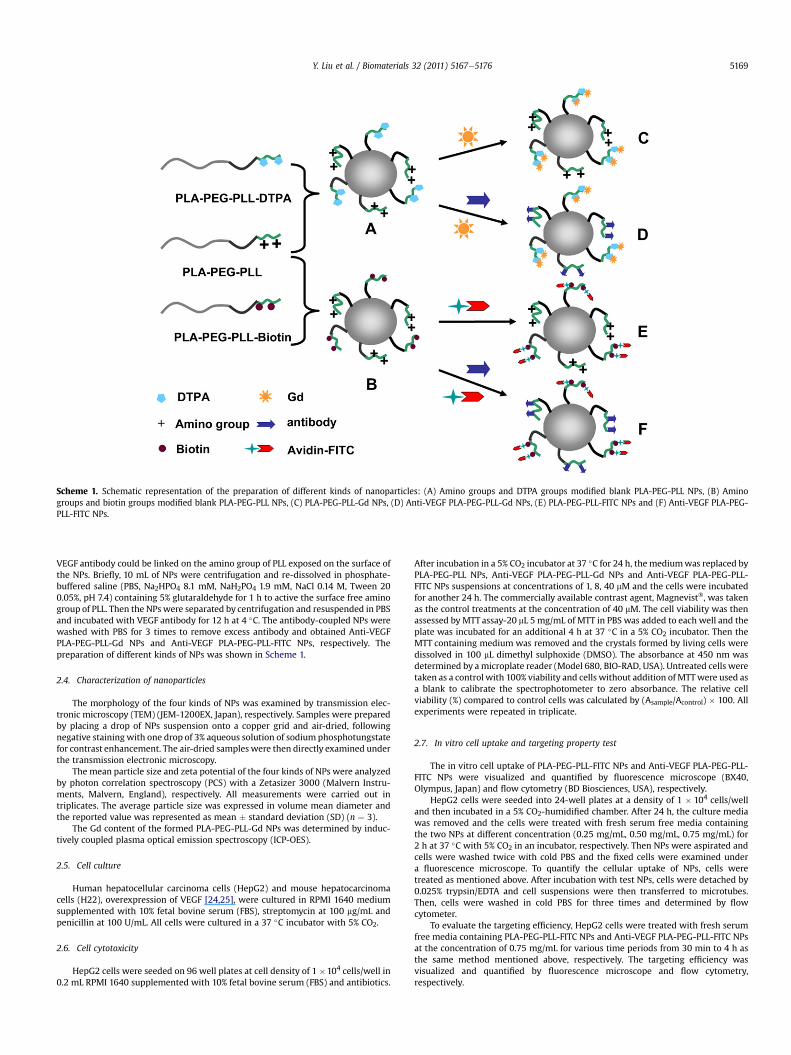
Scheme 1. Schematic representation of the preparation of different kinds of nanoparticles: (A) Amino groups and DTPA groups modified blank PLA-PEG-PLL NPs, (B) Aminogroups and biotin groups modified blank PLA-PEG-PLL NPs, (C) PLA-PEG-PLL-Gd NPs, (D) Anti-VEGF PLA-PEG-PLL-Gd NPs, (E) PLA-PEG-PLL-FITC NPs and (F) Anti-VEGF PLA-PEG-PLL-FITC NPs.
Y. Liu et al. / Biomaterials 32 (2011) 5167e5176 5169
VEGF antibody could be linked on the amino group of PLL exposed on the surface ofthe NPs. Briefly, 10 mL of NPs were centrifugation and re-dissolved in phosphate-buffered saline (PBS, Na2HPO4 8.1 mM, NaH2PO4 1.9 mM, NaCl 0.14 M, Tween 200.05%, pH 7.4) containing 5% glutaraldehyde for 1 h to active the surface free aminogroup of PLL. Then the NPswere separated by centrifugation and resuspended in PBSand incubated with VEGF antibody for 12 h at 4 �C. The antibody-coupled NPs werewashed with PBS for 3 times to remove excess antibody and obtained Anti-VEGFPLA-PEG-PLL-Gd NPs and Anti-VEGF PLA-PEG-PLL-FITC NPs, respectively. Thepreparation of different kinds of NPs was shown in Scheme 1.
2.4. Characterization of nanoparticles
The morphology of the four kinds of NPs was examined by transmission elec-tronic microscopy (TEM) (JEM-1200EX, Japan), respectively. Samples were preparedby placing a drop of NPs suspension onto a copper grid and air-dried, followingnegative staining with one drop of 3% aqueous solution of sodium phosphotungstatefor contrast enhancement. The air-dried samples were then directly examined underthe transmission electronic microscopy.
The mean particle size and zeta potential of the four kinds of NPs were analyzedby photon correlation spectroscopy (PCS) with a Zetasizer 3000 (Malvern Instru-ments, Malvern, England), respectively. All measurements were carried out intriplicates. The average particle size was expressed in volume mean diameter andthe reported value was represented as mean � standard deviation (SD) (n ¼ 3).
The Gd content of the formed PLA-PEG-PLL-Gd NPs was determined by induc-tively coupled plasma optical emission spectroscopy (ICP-OES).
2.5. Cell culture
Human hepatocellular carcinoma cells (HepG2) and mouse hepatocarcinomacells (H22), overexpression of VEGF [24,25], were cultured in RPMI 1640 mediumsupplemented with 10% fetal bovine serum (FBS), streptomycin at 100 mg/mL andpenicillin at 100 U/mL. All cells were cultured in a 37 �C incubator with 5% CO2.
2.6. Cell cytotoxicity
HepG2 cells were seeded on 96 well plates at cell density of 1 �104 cells/well in0.2 mL RPMI 1640 supplemented with 10% fetal bovine serum (FBS) and antibiotics.
After incubation in a 5% CO2 incubator at 37 �C for 24 h, themediumwas replaced byPLA-PEG-PLL NPs, Anti-VEGF PLA-PEG-PLL-Gd NPs and Anti-VEGF PLA-PEG-PLL-FITC NPs suspensions at concentrations of 1, 8, 40 mM and the cells were incubatedfor another 24 h. The commercially available contrast agent, Magnevist�, was takenas the control treatments at the concentration of 40 mM. The cell viability was thenassessed by MTT assay-20 mL 5 mg/mL of MTT in PBS was added to each well and theplate was incubated for an additional 4 h at 37 �C in a 5% CO2 incubator. Then theMTT containing medium was removed and the crystals formed by living cells weredissolved in 100 mL dimethyl sulphoxide (DMSO). The absorbance at 450 nm wasdetermined by amicroplate reader (Model 680, BIO-RAD, USA). Untreated cells weretaken as a control with 100% viability and cells without addition of MTTwere used asa blank to calibrate the spectrophotometer to zero absorbance. The relative cellviability (%) compared to control cells was calculated by (Asample/Acontrol) � 100. Allexperiments were repeated in triplicate.
2.7. In vitro cell uptake and targeting property test
The in vitro cell uptake of PLA-PEG-PLL-FITC NPs and Anti-VEGF PLA-PEG-PLL-FITC NPs were visualized and quantified by fluorescence microscope (BX40,Olympus, Japan) and flow cytometry (BD Biosciences, USA), respectively.
HepG2 cells were seeded into 24-well plates at a density of 1 � 104 cells/welland then incubated in a 5% CO2-humidified chamber. After 24 h, the culture mediawas removed and the cells were treated with fresh serum free media containingthe two NPs at different concentration (0.25 mg/mL, 0.50 mg/mL, 0.75 mg/mL) for2 h at 37 �C with 5% CO2 in an incubator, respectively. Then NPs were aspirated andcells were washed twice with cold PBS and the fixed cells were examined undera fluorescence microscope. To quantify the cellular uptake of NPs, cells weretreated as mentioned above. After incubation with test NPs, cells were detached by0.025% trypsin/EDTA and cell suspensions were then transferred to microtubes.Then, cells were washed in cold PBS for three times and determined by flowcytometer.
To evaluate the targeting efficiency, HepG2 cells were treated with fresh serumfree media containing PLA-PEG-PLL-FITC NPs and Anti-VEGF PLA-PEG-PLL-FITC NPsat the concentration of 0.75 mg/mL for various time periods from 30 min to 4 h asthe same method mentioned above, respectively. The targeting efficiency wasvisualized and quantified by fluorescence microscope and flow cytometry,respectively.
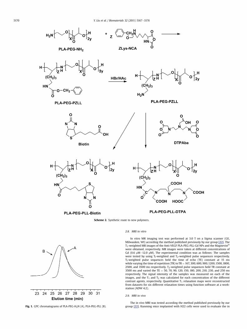
Fig. 1. GPC chromatograms of PLA-PEG-H2N (A), PLA-PEG-PLL (B).
Scheme 2. Synthetic route to new polymers.
Y. Liu et al. / Biomaterials 32 (2011) 5167e51765170
2.8. MRI in vitro
In vitro MR imaging test was performed at 3.0 T on a Sigma scanner (GE,Milwaukee, WI) according the method published previously by our group [22]. TheT1-weighted MR images of the Anti-VEGF PLA-PEG-PLL-Gd NPs and the Magnevist�
were obtained, respectively. MR images were taken at different concentrations ofGd (0.6 mMe12.0 mM). The experimental condition was as follows: The sampleswere tested by using Tl-weighted and T2-weighted pulse sequences respectively.Tl-weighted pulse sequences held the time of echo (TE) constant at 15 mswhile varying the time of repetition (TR) toTR¼ 167, 300, 600, 900,1200,1500,1800,2500, and 3500 ms respectively. T2-weighted pulse sequences held TR constant at3500 ms and varied the TE ¼ 50, 70, 90, 120, 150, 180, 200, 210, 230, and 250 msrespectively. The signal intensity of the samples was measured on each of theimages, and the T1 and T2 was calculated for each concentration of the differentcontrast agents, respectively. Quantitative T1 relaxation maps were reconstructedfrom datasets for six different relaxation times using function software at a work-station (ADW 4.2).
2.9. MRI in vivo
The in vivo MRI was tested according the method published previously by ourgroup [22]. Kunming mice implanted with H22 cells were used to evaluate the in
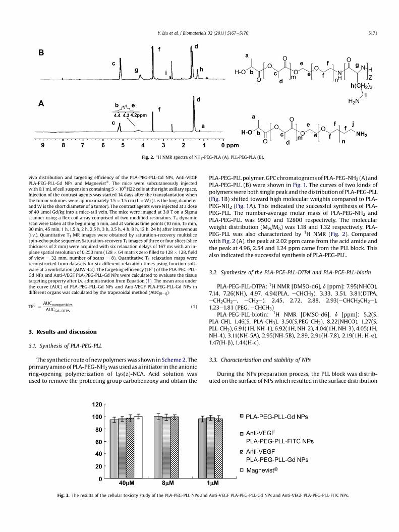
Fig. 2. 1H NMR spectra of NH2-PEG-PLA (A), PLL-PEG-PLA (B).
Y. Liu et al. / Biomaterials 32 (2011) 5167e5176 5171
vivo distribution and targeting efficiency of the PLA-PEG-PLL-Gd NPs, Anti-VEGFPLA-PEG-PLL-Gd NPs and Magnevist�. The mice were subcutaneously injectedwith 0.1mL of cell suspension containing 5�104 H22 cells at the right axillary space.Injection of the contrast agents was started 14 days after the transplantation whenthe tumor volumes were approximately 1.5 � 1.5 cm (L �W) (L is the long diameterand W is the short diameter of a tumor). The contrast agents were injected at a doseof 40 mmol Gd/kg into a mice-tail vein. The mice were imaged at 3.0 T on a Sigmascanner using a flex coil array comprised of two modified resonators. T1 dynamicscan were taken at the beginning 5 min, and at various time points (10 min, 15 min,30 min, 45 min, 1 h, 1.5 h, 2 h, 2.5 h, 3 h, 3.5 h, 4 h, 8 h, 12 h, 24 h) after intravenous(i.v.). Quantitative T1 MR images were obtained by saturation-recovery multislicespin-echo pulse sequence. Saturation-recovery T1 images of three or four slices (slicethickness of 2 mm) were acquired with six relaxation delays of 167 ms with an in-plane spatial resolution of 0.250 mm (128 � 64 matrix zero filled to 128 � 128, fieldof view ¼ 32 mm, number of scans ¼ 8). Quantitative T1 relaxation maps werereconstructed from datasets for six different relaxation times using function soft-ware at a workstation (ADW 4.2). The targeting efficiency (TEC) of the PLA-PEG-PLL-Gd NPs and Anti-VEGF PLA-PEG-PLL-Gd NPs were calculated to evaluate the tissuetargeting property after i.v. administration from Equation (1). The mean area underthe curve (AUC) of PLA-PEG-PLL-Gd NPs and Anti-VEGF PLA-PEG-PLL-Gd NPs indifferent organs was calculated by the trapezoidal method (AUC[0et])�
TEC ¼ AUCnanoparticles
AUCGd�DTPA(1)
3. Results and discussion
3.1. Synthesis of PLA-PEG-PLL
The synthetic routeof newpolymerswas shown inScheme2. Theprimaryamino of PLA-PEG-NH2was used as a initiator in the anionicring-opening polymerization of Lys(z)-NCA. Acid solution wasused to remove the protecting group carbobenzoxy and obtain the
Fig. 3. The results of the cellular toxicity study of the PLA-PEG-PLL NPs and
PLA-PEG-PLL polymer. GPC chromatogramsof PLA-PEG-NH2 (A) andPLA-PEG-PLL (B) were shown in Fig. 1. The curves of two kinds ofpolymerswereboth single peak and thedistributionof PLA-PEG-PLL(Fig. 1B) shifted toward high molecular weights compared to PLA-PEG-NH2 (Fig. 1A). This indicated the successful synthesis of PLA-PEG-PLL. The number-average molar mass of PLA-PEG-NH2 andPLA-PEG-PLL was 9500 and 12800 respectively. The molecularweight distribution (Mw/Mn) was 1.18 and 1.32 respectively. PLA-PEG-PLL was also characterized by 1H NMR (Fig. 2). Comparedwith Fig. 2 (A), the peak at 2.02 ppm came from the acid amide andthe peak at 4.96, 2.54 and 1.24 ppm came from the PLL block. Thisalso indicated the successful synthesis of PLA-PEG-PLL.
3.2. Synthesize of the PLA-PGE-PLL-DTPA and PLA-PGE-PLL-biotin
PLA-PEG-PLL-DTPA: 1H NMR [DMSO-d6], d [ppm]: 7.95(NHCO),7.14, 7.26(NH), 4.97, 4.94(PLA, eCHCH3), 3.33, 3.51, 3.81(DTPA,eCH2CH2e, eCH2e), 2.45, 2.72, 2.88, 2.93(eCHCH2CH2e),1.23e1.81 (PEG, eCHCH3)
PLA-PEG-PLL-biotin: 1H NMR [DMSO-d6], d [ppm]: 5.2(S,PLA-CH), 1.46(S, PLA-CH3), 3.50(S,PEG-CH2), 8.22(NHCO), 1.27(S,PLL-CH2), 6.91(1H, NH-1), 6.92(1H, NH-2), 4.04(1H, NH-3), 4.05(1H,NH-4), 3.11(NH-5A), 2.95(NH-5B), 2.89, 2.91(H-7,8), 2.19(1H, H-a),1.47(H-b), 1.44(H-e).
3.3. Characterization and stability of NPs
During the NPs preparation process, the PLL block was distrib-uted on the surface of NPswhich resulted in the surface distribution
Anti-VEGF PLA-PEG-PLL-Gd NPs and Anti-VEGF PLA-PEG-PLL-FITC NPs.
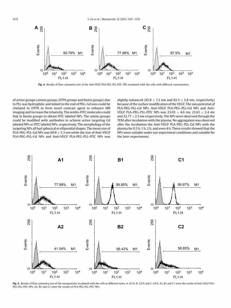
Fig. 4. Results of flow cytometry test of the Anti-VEGF PLA-PEG-PLL-FITC NPs incubated with the cells with different concentration.
Y. Liu et al. / Biomaterials 32 (2011) 5167e51765172
of active groups (amino groups, DTPA groups and biotin groups) dueto PLLwas hydrophilic and linked to the end of PEG. Gd ions could bechelated to DTPA to form novel contrast agent to enhance MRimaging and increase the relaxivity. The avidin-FITCmolecules couldlink to biotin groups to obtain FITC labeled NPs. The amino groupscould be modified with antibodies to achieve active targeting GdlabeledNPs or FITC labeledNPs, respectively. Themorphology of thetargetingNPs all had spherical or ellipsoidal shapes. Themeansize ofPLA-PEG-PLL-Gd NPs was 69.8� 5.3 nmwhile the size of Anti-VEGFPLA-PEG-PLL-Gd NPs and Anti-VEGF PLA-PEG-PLL-FITC NPs was
Fig. 5. Results of flow cytometry test of the nanoparticles incubated with the cells at differenPEG-PLL-FITC NPs. A2, B2 and C2 were the results of PLA-PEG-PLL-FITC NPs.
slightly enhanced (85.8 � 7.2 nm and 82.5 � 5.8 nm, respectively)becauseof the surfacemodificationof theVEGF. The zatapotential ofPLA-PEG-PLL-Gd NPs, Anti-VEGF PLA-PEG-PLL-Gd NPs and Anti-VEGF PLA-PEG-PLL-FITC NPs was 23.03 � 4.6 mv, 21.63 � 2.4 mvand22.77�2.5mv respectively. TheNPswere observed through theTEMafter incubationwith the plasma. No aggregationwas observedafter the incubation the Anti-VEGF PLA-PEG-PLL-Gd NPs with theplasma for 0.5 h,1 h, 2 h, and even 4 h. These results showed that theNPswere suitable under our experiment conditions and suitable forthe later experiments.
t times. A: 0.5 h, B: 2.0 h and C: 4.0 h. A1, B1 and C1 were the results of Anti-VEGF PLA-
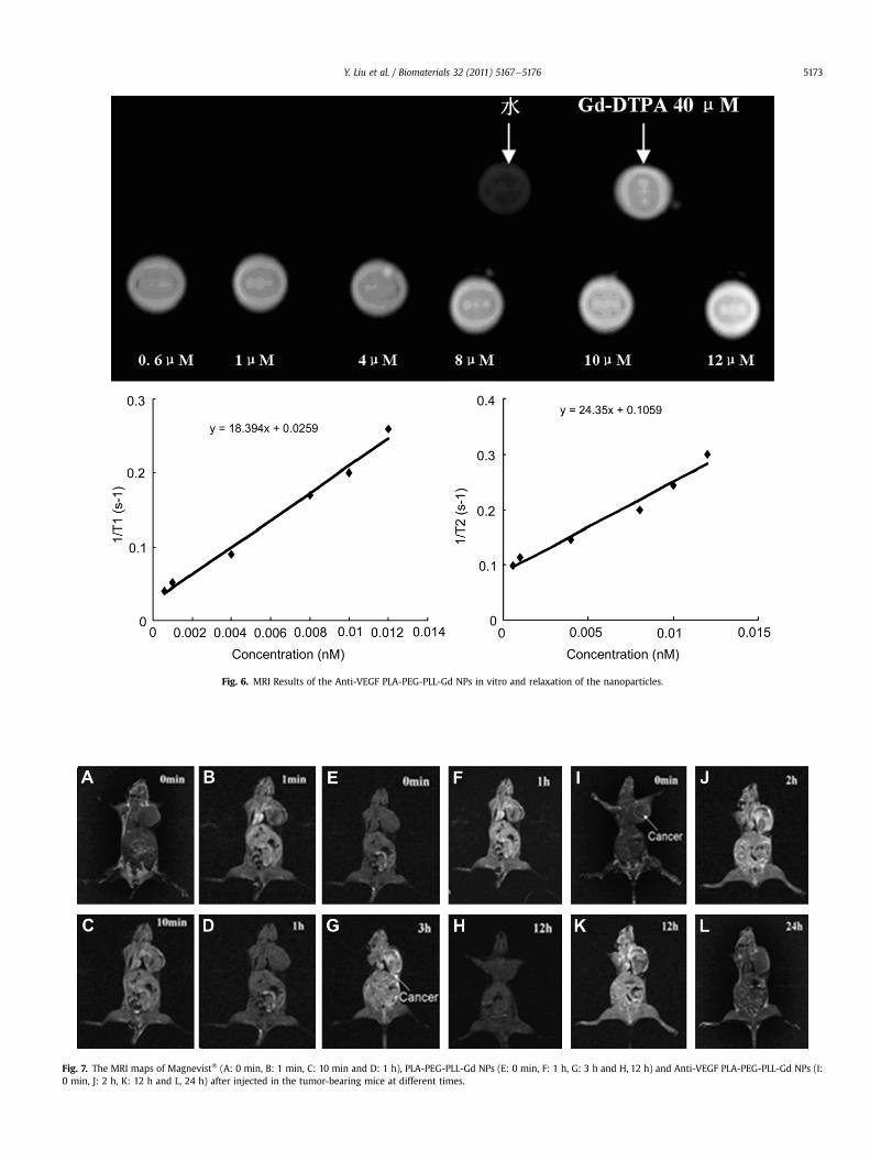
Fig. 6. MRI Results of the Anti-VEGF PLA-PEG-PLL-Gd NPs in vitro and relaxation of the nanoparticles.
Fig. 7. The MRI maps of Magnevist� (A: 0 min, B: 1 min, C: 10 min and D: 1 h), PLA-PEG-PLL-Gd NPs (E: 0 min, F: 1 h, G: 3 h and H, 12 h) and Anti-VEGF PLA-PEG-PLL-Gd NPs (I:0 min, J: 2 h, K: 12 h and L, 24 h) after injected in the tumor-bearing mice at different times.
Y. Liu et al. / Biomaterials 32 (2011) 5167e5176 5173
Y. Liu et al. / Biomaterials 32 (2011) 5167e51765174
3.4. Cell cytotoxicity
In vitro toxicity of the PLA-PEG-PLL NPs, Anti-VEGF PLA-PEG-PLL-Gd NPs and Anti-VEGF PLA-PEG-PLL-FITC NPs at different Gdconcentrations was evaluated by MTT assay in HepG2 cells (Fig. 3)[26]. Magnevist� at a high concentration (40 mM) was taken asa positive control. The cell survival rates of three NP groups wereslight lower than Magnevist� group because of the cationic shell ofNPs. However, the cell survival rates of all groups were higher than90% at the experimental dose which indicated that three kinds ofNPs were safe to the HepG2 cells at the test concentrations.Compared with the Magnevist� group, the cell survival rates of thethree NP groups did not have significant deviation (P > 0.05) and
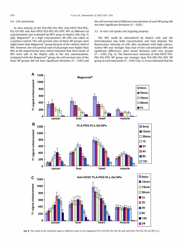
Fig. 8. The results of the enhanced signal in different tissue in vivo (Magnev
the cell survival rate of different concentration of each NP group didnot have significant deviation (P > 0.05).
3.5. In vitro cell uptake and targeting property
The NPs could be internalized by HepG2 cells and theinternalization was both concentration and time depend. Thefluorescence intensity of cells after incubated with high concen-tration NPs was stronger than that of low concentration NPs andsignificant differences were found between each two groups(P < 0.05) (Fig. 4). The fluorescence intensity of Anti-VEGF PLA-PEG-PLL-FITC NP group was stronger than PLA-PEG-PLL-FITC NPgroup an each time point (P< 0.05) (Fig. 5). It was indicated that the
ist�(A), PLA-PEG-PLL NPs (B) and Anti-VEGF PLA-PEG-PLL-Gd NPs (C)).
Y. Liu et al. / Biomaterials 32 (2011) 5167e5176 5175
VEGF PLA-PEG-PLL-FITC NPs could significantly increase the in vivouptake of HepG2 cells by active targeting.
3.6. MRI in vitro
To explore the potential of VEGF-PLA-PEG-PLL-Gd NPs as MRIcontrast agent, the ability to shorten the T1 longitudinal relaxationtime of protons of water were measured [18]. The Anti-VEGF PLA-PEG-PLL-Gd NPs ([Gd] ¼ 0.6 mMe12.0 mM) and Gd-DTPA injection([Gd]¼ 40 mM)were evaluated at 3.0 Ton a clinical MR scanner (GE,Milwaukee, WI) at 25 �C (Fig. 6). For T1 weighting, the higher Gdconcentration of the NPs showed higher signal intensity. The signalof the PLA-PEG-PLL-Gd NPs (8 mM) was similar with the Gd-DTPAinjection of 40 mM. The relaxivity was calculated as the slope ofthe curves 1/T1 (1/T2) with respect to gadolinium concentration.The r2/r1 values below 2 presented that the agent belonged topositive contrast agent [27]. The r1 of the NPs was 18.394 mM�1s�1
and the r2 was 24.863 mM�1s�1, respectively. These results of MRIin vitro showed that Anti-VEGF PLA-PEG-PLL-Gd NPs couldnoticeably increase the relaxivity of T1 and T2. The relaxation theorypredicts that higher relaxation rates are obtained upon increase ofthe rotational correlation time (sR) of contrast agents and increaseof the exchange of water protons with the contrast agents [28].Since the Gd-DTPA molecules were linked to the large size NP, thesR was increased correspondingly. Meanwhile, Gd-DTPA wasmodified on the surface of the NPs, the exchange of water protonswith Gd was also increased. So, the new NP agents could consid-erably enhance the relaxivity of the chelate unit. The results of theMRI measurements indicated that the PLA-PEG-PLL-Gd NPs werefitting for novel MRI molecular contrast agent.
3.7. MRI in vivo
PLA-PEG-PLL-Gd NPs, Anti-VEGF PLA-PEG-PLL-Gd NPs andMagnevist�were administrated to tumor-bearingmice through thevena caudalis of the 40 mmol Gd/kg, respectively. To examinewhether Anti-VEGF PLA-PEG-PLL-Gd NPs could be used to effec-tively identify VEGF-positive tumors in living subjects, subcuta-neous H22 cell xenografts were selected as the tumor model. Theresult of the enhaned signal in different tissue in vivo of the PLA-PEG-PLL-Gd NPs, Anti-VEGF PLA-PEG-PLL-Gd NPs and Magnevist�
was shown in Fig. 7.Thewhole body of themicewas gray as well as the tumor before
injection. After the administration of the Magnevist�, the contrast
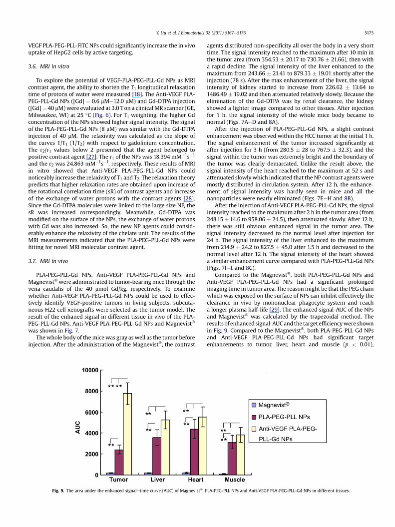
Fig. 9. The area under the enhanced signaletime curve (AUC) of Magnevist�, P
agents distributed non-specificity all over the body in a very shorttime. The signal intensity reached to the maximum after 10 min inthe tumor area (from 354.53 � 20.17 to 730.76 � 21.66), then witha rapid decline. The signal intensity of the liver enhanced to themaximum from 243.66 � 21.41 to 879.33 � 19.01 shortly after theinjection (78 s). After the max enhancement of the liver, the signalintensity of kidney started to increase from 226.62 � 13.64 to1486.49 � 19.02 and then attenuated relatively slowly. Because theelimination of the Gd-DTPA was by renal clearance, the kidneyshowed a lighter image compared to other tissues. After injectionfor 1 h, the signal intensity of the whole mice body became tonormal (Figs. 7AeD and 8A).
After the injection of PLA-PEG-PLL-Gd NPs, a slight contrastenhancement was observed within the HCC tumor at the initial 1 h.The signal enhancement of the tumor increased significantly atafter injection for 3 h (from 280.5 � 28 to 767.5 � 32.3), and thesignal within the tumor was extremely bright and the boundary ofthe tumor was clearly demarcated. Unlike the result above, thesignal intensity of the heart reached to the maximum at 52 s andattenuated slowlywhich indicated that the NP contrast agents weremostly distributed in circulation system. After 12 h, the enhance-ment of signal intensity was hardly seen in mice and all thenanoparticles were nearly eliminated (Figs. 7EeH and 8B).
After the injection of Anti-VEGF PLA-PEG-PLL-Gd NPs, the signalintensity reached to themaximum after 2 h in the tumor area (from248.15 � 14.6 to 958.06 � 24.5), then attenuated slowly. After 12 h,there was still obvious enhanced signal in the tumor area. Thesignal intensity decreased to the normal level after injection for24 h. The signal intensity of the liver enhanced to the maximumfrom 214.9 � 24.2 to 827.5 � 45.0 after 1.5 h and decreased to thenormal level after 12 h. The signal intensity of the heart showeda similar enhancement curve compared with PLA-PEG-PLL-Gd NPs(Figs. 7IeL and 8C).
Compared to the Magnevist�, both PLA-PEG-PLL-Gd NPs andAnti-VEGF PLA-PEG-PLL-Gd NPs had a significant prolongedimaging time in tumor area. The reasonmight be that the PEG chainwhich was exposed on the surface of NPs can inhibit effectively theclearance in vivo by mononuclear phagocyte system and reacha longer plasma half-life [29]. The enhanced signal-AUC of the NPsand Magnevist� was calculated by the trapezoidal method. Theresults of enhanced signal-AUCand the target efficiencywere shownin Fig. 9. Compared to the Magnevist�, both PLA-PEG-PLL-Gd NPsand Anti-VEGF PLA-PEG-PLL-Gd NPs had significant targetenhancements to tumor, liver, heart and muscle (p < 0.01),
LA-PEG-PLL NPs and Anti-VEGF PLA-PEG-PLL-Gd NPs in different tissues.
Y. Liu et al. / Biomaterials 32 (2011) 5167e51765176
respectively. The reason might be that NPs could significantlyshorten the longitudinal relaxation time compared to Magnevist�
becauseNPshad largemagneticmoment due to the covalent linkageof Gd-DTPA to NPs and fast exchange of water protons with thecontrast agents due to the surface modification of the Gd-DTPA.Meanwhile, NPs had longer circulating time and they could be tar-geted and accumulated to the tumor area by the enhanced perme-ability and retention (EPR) effect of NPs [30]. Especially, the targetefficiency of Anti-VEGF PLA-PEG-PLL-Gd NPs was 3.2 times in thetumor area compared with PLA-PEG-PLL NPs, while was 1.5, 1.3 and1.2 times in the liver, heart and muscle (do not have significantdifference), respectively. These results indicated that Anti-VEGFPLA-PEG-PLL-Gd NPs had more significant target efficiency totumor (p < 0.01). The possible reason was that VEGF was overex-pressed in mice hepatocarcinoma cells (H22) [26], the Anti-VEGFPLA-PEG-PLL-Gd NPs could linked to the tumor cells and restrictedin the tumor area by the tumor cell surface VEGF-antibody reaction.These bindings are essential in the enhancement of MRI in the earlydiagnose of hepatocarcinoma to achieve molecular imaging.
4. Conclusions
We have successfully developed multifunctional Anti-VEGFPLA-PEG-PLL-Gd NPs as the molecular MRI contrast agents toefficiently target to liver tumor cells for the early detection of HCC.Anti-VEGF PLA-PEG-PLL-Gd NPs significantly facilitated thespecific delivery of Gd to VEGF-positive liver tumor cells bothin vitro and in vivo. Also, Anti-VEGF PLA-PEG-PLL-Gd NPs showedsignificant enhancement of signal intensity and prolongedimaging time at liver tumor area in mice compared withconventional MRI contrast agent (Magnevist�) and non-targetNPs. The Anti-VEGF PLA-PEG-PLL-Gd NP contrast agent haspromising value for the early diagnostic imaging of the HCC inclinical applications.
Acknowledgments
This work was supported by the Independent InnovationFoundation of Shandong University, IIFSDU, (2010JC019).
References
[1] Skelton MR, O’Neil B. Targeted therapies for hepatocellular carcinoma. ClinAdv Hematol Oncol 2008;6(3):209e18.
[2] Weissleder R. Molecular imaging in cancer. Science 2006;312(5777):1168e71.[3] Kamaoui I, Milot L, Durieux M, Ficarelli S, Mennesson N, Pilleul F. Value of
MRCP with Mangafodipir Trisodium (Teslascan) injection in the diagnosis andmanagement of bile leaks. J Radiol 2007;88(12):1881e6.
[4] Sharma P, Brown SC, Walter G, Santra S, Scott E, Ichikawa H, et al. Gd nano-particulates: from magnetic resonance imaging to neutron capture therapy.Adv Powder Technol 2007;18(6):663e98.
[5] Thorek DL, Chen AK, Czupryna J, Tsourkas A. Superparamagnetic iron oxidenanoparticleprobes formolecular imaging. AnnBiomedEng2006;34(1):23e38.
[6] Caravan P. Strategies for increasing the sensitivity of gadolinium based MRIcontrast agents. Chem Soc Rev 2006;35(6):512e23.
[7] Ghaghada K, Hawley C, Kawaji K, Annapragada A, Mukundan Jr S. T1 relaxivityof core-encapsulated gadolinium liposomal contrast agents e effect of lipo-some size and internal gadolinium concentration. Acad Radiol 2008;15(10):1259e63.
[8] Ratzinger G, Agrawal P, Korner W, Lonkai J, Sanders HM, Terreno E, et al.Surface modification of PLGA nanospheres with Gd-DTPA and Gd-DOTA forhigh-relaxivity MRI contrast agents. Biomaterials 2010;31(33):8716e23.
[9] Grogna M, Cloots R, Luxen Ae, Jerome C, Detrembleur C. Polymer micellesdecorated by gadolinium complexes as MRI blood contrast agents: design,synthesis and properties. Polym Chem 2010;1(9):1485e90.
[10] Luo K, Liu G, He B, Wu Y, Gong Q, Song B, et al. Multifunctional gadolinium-based dendritic macromolecules as liver targeting imaging probes. Biomate-rials 2011;32(10):2575e85.
[11] van Schooneveld MM, Cormode DP, Koole R, van Wijngaarden JT, Calcagno C,Skajaa T, et al. A fluorescent, paramagnetic and PEGylated gold/silica nano-particle for MRI, CT and fluorescence imaging. Contrast Media Mol 2010;5(4):231e6.
[12] Byrne JD, Betancourt T, Brannon-Peppas L. Active targeting schemes fornanoparticle systems in cancer therapeutics. Adv Drug Deliv Rev 2008;60(15):1615e26.
[13] Finn RS, Zhu AX. Targeting angiogenesis in hepatocellular carcinoma: focus onVEGF and bevacizumab. Expert Rev Anticancer Ther 2009;9(4):503e9.
[14] Chen KJ, Wolahan SM, Wang H, Hsu CH, Chang HW, Durazo A, et al. A smallMRI contrast agent library of gadolinium(III)-encapsulated supramolecularnanoparticles for improved relaxivity and sensitivity. Biomaterials 2011;32(8):2160e5.
[15] Maeng JH, Lee DH, Jung KH, Bae YH, Park IS, Jeong S, et al. Multifunctionaldoxorubicin loaded superparamagnetic iron oxide nanoparticles for chemo-therapy and magnetic resonance imaging in liver cancer. Biomaterials 2011;31(18):4995e5006.
[16] Hu KW, Hsu KC, Yeh CS. pH-dependent biodegradable silica nanotubesderived from Gd(OH)3 nanorods and their potential for oral drug delivery andMR imaging. Biomaterials 2011;31(26):6843e8.
[17] Delli Castelli D, Dastru W, Terreno E, Cittadino E, Mainini F, Torres E, et al.In vivo MRI multicontrast kinetic analysis of the uptake and intracellulartrafficking of paramagnetically labeled liposomes. J Control Release 2010;144(3):271e9.
[18] Chen ZJ, Yu DX, Wang SJ, Zhang N, Ma CH, Lu ZJ. Biocompatible nano-complexes for molecular targeted MRI contrast agent. Nanoscale Res Lett2009;4(7):618e26.
[19] Darras V, Nelea M, Winnik FM, Buschmann MD. Chitosan modified withgadolinium diethylenetriaminepentaacetic acid for magnetic resonanceimaging of DNA/chitosan nanoparticles. Carbohydr Polym 2010;80(4):1137e46.
[20] Bellin MF. MR contrast agents, the old and the new. Eur J Radiol 2006;60(3):314e23.
[21] Kielar F, Tei L, Terreno E, Botta M. Large relaxivity enhancement of para-magnetic lipid nanoparticles by restricting the local motions of the Gd(III)chelates. J Am Chem Soc 2010;132(23):7836e7.
[22] Chen ZJ, Yu DX, Liu CX, Yang XY, Zhang N, Ma CH, et al. Gadolinium-conju-gated PLA-PEG nanoparticles as liver targeted molecular MRI contrast agent.J Drug Target; 2010 [online].
[23] Lu ZJ, Song JB, Zhang GZ, inventors. Synthesis and preparation of a blockcopolymer and its nanoparticles. China Patent No.101565497A, 2009.
[24] Tian T, Nan KJ, Wang SH, Liang X, Lu CX, Guo H, et al. PTEN regulatesangiogenesis and VEGF expression through phosphatase-dependent and-independent mechanisms in HepG2 cells. Carcinogenesis 2010;31(7):1211e9.
[25] Lei Z, Li B, Yang Z, Fang H, Zhang GM, Feng ZH, et al. Regulation of HIF-1alphaand VEGF by miR-20b tunes tumor cells to adapt to the alteration of oxygenconcentration. PLoS One 2009;4(10):e7629.
[26] Wang L, Neoh KG, Kang ET, Shuter B. Multifunctional polyglycerol-graftedFeO@SiO nanoparticles for targeting ovarian cancer cells. Biomaterials 2011;32(8):2166e73.
[27] Favier A, D’Agosto F, Charreyre M-T, Pichot C. Synthesis of N-acrylox-ysuccinimide copolymers by RAFT polymerization, as reactive building blockswith full control of composition and molecular weights. Polymer 2004;45(23):7821e30.
[28] Caravan P, Ellison JJ, McMurry TJ, Lauffer RB. Gadolinium(III) chelates as MRIcontrast agents: structure, dynamics, and applications. Chem Rev 1999;99(9):2293e352.
[29] Wan F, You J, Sun Y, Zhang XG, Cui FD, Du YZ, et al. Studies on PEG-modifiedSLNs loading vinorelbine bitartrate (I): preparation and evaluation in vitro.Int J Pharm 2008;359(1e2):104e10.
[30] Maeda H, Bharate GY, Daruwalla J. Polymeric drugs for efficient tumor-targeted drug delivery based on EPR-effect. Eur J Pharm Biopharm 2009;71(3):409e19.