Solid-Liquid Interdiffusion Bonding of Silicon ... - UC Berkeley
Upload
khangminh22Category
view
1download
0
FORMULATION AND IN-VITRO EVALUATION OF LIQUID AND SOLID SELF MICROEMULSIFYING DRUG DELIVERY SYSTEM OF PITAVASTATIN
CALCIUM
A Dissertation submitted to
THE TAMIL NADU DR. M.G.R. MEDICAL UNIVERSITY CHENNAI-600 032.
In partial fulfillment of the requirements for the award of the degree of
MASTER OF PHARMACY
IN BRANCH I- PHARAMACEUTICS
Submitted by
K.MAHALAKSHMI
(Reg.No: 261611302)
Under the guidance of
Dr. A. ABDUL HASAN SATHALI, M.Pharm., Ph.D.,
DEPARTMENT OF PHARMACEUTICS
COLLEGE OF PHARMACY, MADURAI MEDICAL COLLEGE,
MADURAI- 625 020
MAY - 2018
CERTIFICATE
This is to certify that the dissertation entitled “FORMULATION
AND IN VITRO EVALUATION OF LIQUID AND SOLID SELF
MICRO EMULSIFYING DRUG DELIVERYSYSTEM OF
PITAVASTATIN CALCIUM” is a bonafide work done by
Ms. K. MAHALAKSHMI (Reg.No:261611302), Department of
Pharmaceutics, College of Pharmacy, Madurai Medical College
in partial fulfillment of The Tamil Nadu Dr.M.G.R Medical
University rules and regulations for award of MASTER OF
PHARMACY IN PHARMACEUTICS under my guidance and
supervision during the academic year 2017–2018.
Name & Signature of the Guide
Name & Signature of the Head of Department
Name & Signature of the Dean/Principal

ACKNOWLEDGEMENT
ACKNOWLEDGEMENT
First and foremost of all I wish to thanks the ALMIGHTY who has
granted me an opportunity to do higher studies in this noble field of
pharmacy and blessed me with the strength and intellect to pursue this
research work.
It is my pleasure to express my respectful regards and
thanks to Dr.D.MARUDUPANDIAN, M.S., F.I.C.S., F.A.I.S., Dean,
Madurai Medical College, Madurai for providing all kinds of supportive
facilities required to carry out my project work.
I am thankful to Dr.V.DHANALAKSHMI, M.D., Vice Principal,
Madurai Medical College, Madurai for her support and encouragement to
carry out the work.
It is my immense pleasure and honor to express my deep sense of
gratitude and heartfelt thanks to Prof. Dr. A.ABDUL HASAN SATHALI, M.Pharm., Ph.D., Principal, College of pharmacy, Madurai Medical
College for his excellence in guidance, contribution and
encouragement which helped me in the successful completion of each
and every stage of my project work.
I thank Mr. Arun,M.Pharm., Dr. C. Pandiyan,M.Pharm.,Ph.D., Dr. R.Senthil Prabhu, M.Pharm., Ph.D., Dept of Pharmaceutics for their
support and valuable suggestion throughout my work.
I also express my sincere thanks to Mrs. D.Uma maheswari, M.Pharm., Mr. M.Prabhu, M.Pharm., for their timely co operation to carry
out my project work.
I also extend my thanks to our department staff Mrs.Sophia, Mrs. Tamilselvi, Mrs. Mumtaj, for their contribution throughout my project
work.
I express my heartiest thanks to my senior Mr. Senthil Ramesh , Ms. Anandhi., Shashun Pharmaceuticals, for providing the drug
Pitavastatin calcium as a gift sample to carry out my project work.
I express my heartiest thanks to Madras pharmaceuticals, Chennai, for providing the excipients as gift samples to carry out my
project work.
I also thank Nirmalgiri University, Kerala for their help to carry out
the evaluation (X-ray diffraction ) studies.
I also thank JSS College of Pharmacy, Ooty, for their help to carry
out the DSC studies.
I express my heartiest thanks to Universal Scientific Appliances
for providing chemicals to carry out my project work.
I also extend my thanks to the Department of Pharmaceutical Chemistry MMC, Madurai For permitting me to carry out the IR
study and UV spectrophotometric studies in connection to my
dissertation work and Mr. Lakshmanan, Department of Pharmaceutical Chemistry, to carry out UV spectrophotometric
studies.
I would like to give my sincere thanks to my classmates, Ms. T. Nithya, Ms. M. Muthumari, Mr. C.A. Muniyasamy, Mr. M. Selvakumar, Mr. R. Vignesh, Mr. S. Zameer, Ms. J. Jeyapriya, Mrs. S. Sivapriya for their timely help and co-operation.
I also extend my thanks to all the staff members and P.G. Students
of Department of Pharmaceutical Chemistry and special thanks to
Mr. A. Ponnudurai, Ms. S. Swathi for their kind cooperation to carry out
my project work.
I also express my thanks to all the staff members and P.G. Students
of Department of Pharmacognosy for their support to carry out my project.
I express my whole hearted and sincere thanks to my seniors for
their moral support to carry out my project work.
I sincerely thank to my UG friends Mr. N. Ramanathan, Mr. A. Tamilmani, for their moral support to carry out my project work.
Last but definitely not the least, I pledge my deepest sense of
gratitude towards my Father (Mr. P. Karuppiah), my Mother (Mrs. K. Ramalakshmi) and my lovable brothers (Mr. K.Karthik and Mr. K. Vignesh) and sister (Mrs. Eswari Ramachandran) who stood with
me, supporting in all my endeavors.
I am extremely thankful to the Library Madurai medical college and
staff of Chennai Xerox, Laser Point, for their kind co-operation regarding
printing and binding of this dissertation work.
Place: Madurai
Date: (K. MAHALAKSHMI)
CONTENTS
CHAPTER NO.
TITLE PAGE NO
I INTRODUCTION 1
II SELF MICRO EMULSIFYING DRUG DELIVERY-
REVIEW 12
III LITERATURE REVIEW 31
IV AIM OF THE WORK 56
V PLAN OF WORK 58
VI MATERIALS AND EQUIPMENTS 60
VII DRUG PROFILE 62
VII EXCIPIENTS PROFILE 69
IX EXPERIMENTAL PROTOCOL 80
X RESULTS AND DISCUSSION
TABLES & FIGURES 97
XI SUMMARY AND CONCLUSION 157
REFERENCES
ABBREVIATIONS
% Percentage
˚C Degree Celsius
λmax Maximum wavelength
µm Micrometer
µg Microgram
Abs. Absorbance
AR Analytical reagent
BCS Biopharmaceutical classification system
Conc. Concentration
CDR Cumulative drug release
Cm Centimeter
cps Centipoises
DSC Differential scanning colorimetri
e.g. Example
etc Excetra
FDA Food and drug administration
FTIR Fourrier transfer infrared
g Gram
GIT Gastro intestinal tract
IP Indian pharmacopoeia
Kg kilogram
L
Liter
mg Milligram
min Minute
ml Milliliter
mm Millimeter
nm Nanometer
ppm Parts per million
PXRD Powdered X-ray diffraction
RH Relative humidity
rpm Rotation per minute
SD Standard deviation
SEDDS Self Emulsifying Drug Delivery System
SMEDDS Self Micro Emulsifying Drug Delivery
System
SEM Scanning Electron Microscopy
UV Ultra Violet spectrophotometry

CHAPTER I
INTRODUCTION
CHAPTER I INTRODUCTION
DEPARTMENT OF PHARMACEUTICS, COLLEGE OF PHARMACY, MMC, MADURAI. 1
CHAPTER - I
INTRODUCTION
Most of the drugs are administered through the oral route; it has always been
preferred route for the formulators over other routes. But this route is limited to such
drug molecules which has poor aqueous solubility, which is one of the important
parameters to achieve desired concentration of drug in systemic circulation for
desired (anticipated) pharmacological response. A large number of potential drug
candidates suffer from low aqueous solubility and low dissolution rate. This leads to
low drug concentration at the absorption site and results in low oral bio availability.
In BCS classification system these poor solubility drugs are classified as BCS class
II drugs.
Biopharmaceutical Classification System (BCS):
Biopharmaceutics Classification System (BCS) was introduced in 1995 as a
basis for predicting the likelihood of In vitro-In vivo correlations for immediate release
dosage forms. BCS has been used as a regulatory Tool for the replacement of certain
bioequivalence (BE) studies with accurate in vitro dissolution tests. This step certainly
reduces timelines in the drug development process, both directly and indirectly, and
reduces unnecessary drug exposure in healthy volunteers.
The BCS also takes account of the dissolution of the drug product and hence covers
the three main factors which govern the rate and extent of drug absorption from
immediate release (IR) solid oral dosage forms.
• Dissolution rate
• Solubility
• Permeability
CHAPTER I INTRODUCTION
DEPARTMENT OF PHARMACEUTICS, COLLEGE OF PHARMACY, MMC, MADURAI. 2
Solubility:
Solubility is defined as the maximum amount of solute that can be dissolved in a
given amount of solvent. Quantitatively it is defined as “the concentration of the solute
in a saturated solution at a certain temperature. Qualitatively the solubility may be
defined as an interaction of two or more substances to form homogenous molecular
dispersion. Solubility of a drug plays a prime role in controlling its dissolution from
dosage form. Aqueous solubility of a drug is a major factor which determines its
dissolution rate.
A drug substance is considered highly soluble when the highest dose strength is
soluble in 250 ml or less of aqueous media over a pH range of 1 to 7.5 (equilibrium
solubility at 37°C).
Permeability:
In the absence of evidence suggesting instability in the gastrointestinal tract, a
drug substance is considered highly permeable when the extent of absorption in
humans is determined to be 90% or more of an administered dose based on mass
balance determination or in comparison to an intravenous reference dose (absolute
bioavailability study).
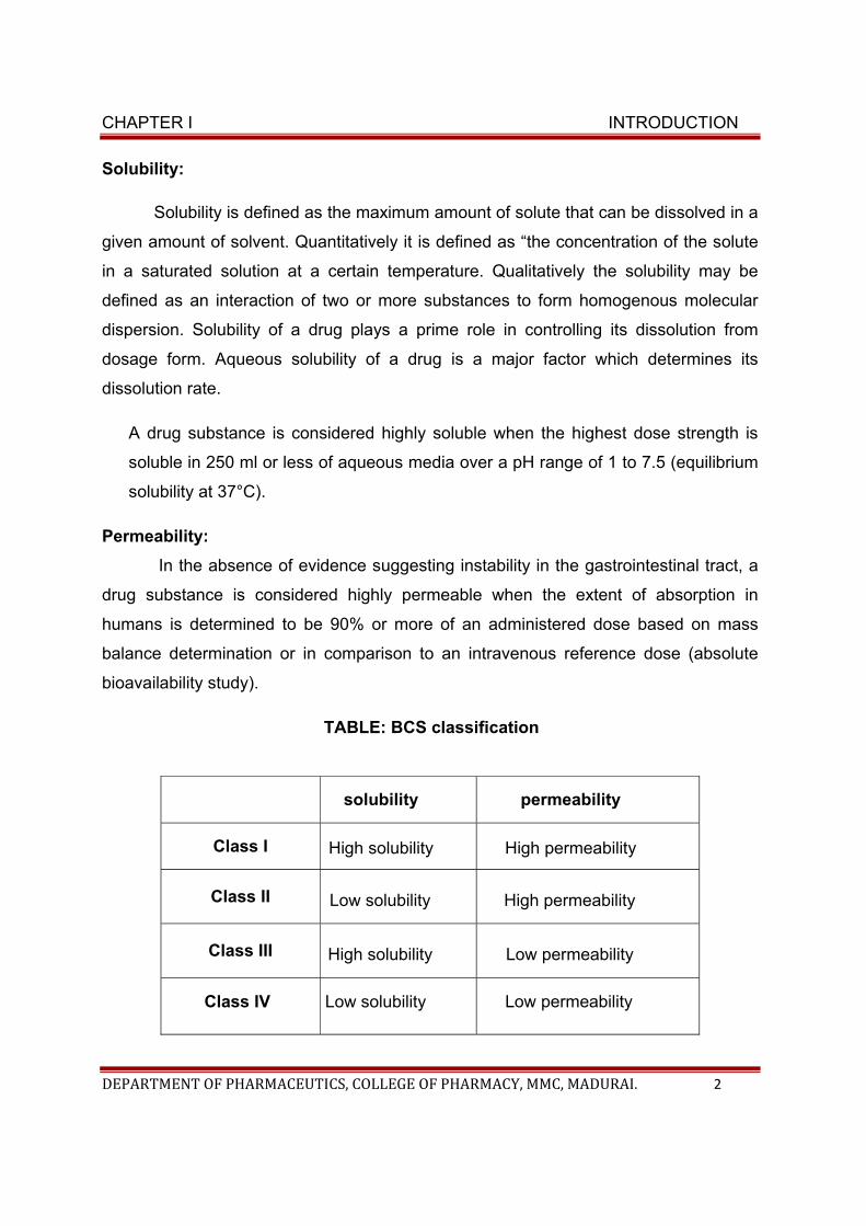
TABLE: BCS classification
solubility permeability
Class I High solubility High permeability
Class II Low solubility High permeability
Class III High solubility Low permeability
Class IV Low solubility Low permeability
CHAPTER I INTRODUCTION
DEPARTMENT OF PHARMACEUTICS, COLLEGE OF PHARMACY, MMC, MADURAI. 3
Class I Drugs
Class I drugs exhibit a high absorption and a high dissolution. The rate
limiting step is drug dissolution. Gastric emptying rate becomes the rate
determining step if dissolution is very rapid. Generally 85% drug is released
within 15 min dissolution study. According to FDA (1997) guideline,
bioavailability and bioequivalence studies are unnecessary for such products.
IVIVC would not be expected for these drugs. Examples include amitriptyline
hydrochloride, chloroquine phosphate, chlorpheniramine maleate,
chlorpromazine hydrochloride, cloxacillin sodium, phenytoin sodium,
prednisolone, promethazine, propranolol hydrochloride, quinine sulfate,
verapamil hydrochloride and warfarin sodium etc (Kasim et al. 2004).
Class II Drugs
Class II drugs have a high absorption but a low dissolution rate. In-vivo drug
dissolution is then a rate limiting step for absorption except at a very high dose
number. These drug exhibited variable bioavailability and need the enhancement in
dissolution for increasing the bioavailability. In vitro- In vivo correlation (IVIVC) is
usually expected for class II drugs.
Examples include phenytoin. danazol, ketoconazole, mefenamic acid,
nifedinpine. felodipine, nicardipine, nisoldipine, atorvastatin calcium, simvastatin,
rosuvastatin calcium etc (Kasim et al. 2004).
Class III Drugs
Permeability is rate limiting step for drug absorption of class III drugs. These
drugs exhibit a high variation in the rate and extent of drug absorption. Since the
dissolution is rapid, the variation is attributable to alteration of physiology and
membrane permeability rather than the dosage for factors. These drugs are
problematic for controlled release development. These drugs showed the low
bioavailability and need enhancement in permeability.
CHAPTER I INTRODUCTION
DEPARTMENT OF PHARMACEUTICS, COLLEGE OF PHARMACY, MMC, MADURAI. 4
Examples include cimitidine, ranitidine, acyclovir, alendronate, captopril,
enalaprilat neomycin B, atenolol etc (Kasim et al. 2004).
Class IV Drugs
Class IV drugs exhibit poor and variable bioavailability. Several factors such as
dissolution rate, permeability and gastric emptying are the rate limiting steps for the
drug absorption. These drugs are not suitable for controlled release formulation.
Examples include acetazolamide, allopurinol, dapsone, doxycycline, nalidixic acid,
sulfadiazine, sulfamethoxazole, trimethoprim etc (Kasim et al. 2004).
So oral route is not advisable for these poorly soluble drugs, which may be
inefficient to achieve desired drug concentration on the site of action due to its poor
solubility and dissolution.
METHODS TO OVER COME SOLUBILITY PROBLEMS:
Different formulation approaches like micronization, solid dispersion, and
complexation with cyclodextrins have come up for the oral bioavailability of poorly
water soluble drugs. Indeed, in some selected cases, these approaches have been
successful but they offer many other disadvantages. The main problem with
micronization is chemical / thermal stability. Many drugs may degrade and lose
bioactivity when they are micronized by conventional method. For solid dispersion the
amount of carriers used is often large, and thus if the dose of active ingredient is high,
the tablets or capsules formed will be large in volume and difficult to swallow.
Moreover, since the carriers used are usually expensive and freeze-drying or spray-
drying method requires particular facilities and processes, leading to high production
cost. Though, traditional solvent method can be adopted instead, it is difficult to deal
with co- precipitates with high viscosity. Complexation with cyclodextrins techniques is
not applicable for drug substances which are not soluble in both aqueous and organic
solvents.
CHAPTER I INTRODUCTION
DEPARTMENT OF PHARMACEUTICS, COLLEGE OF PHARMACY, MMC, MADURAI. 5
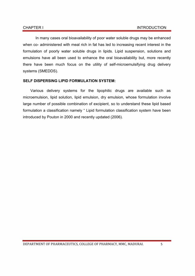
In many cases oral bioavailability of poor water soluble drugs may be enhanced
when co- administered with meal rich in fat has led to increasing recent interest in the
formulation of poorly water soluble drugs in lipids. Lipid suspension, solutions and
emulsions have all been used to enhance the oral bioavailability but, more recently
there have been much focus on the utility of self-microemulsifying drug delivery
systems (SMEDDS).
SELF DISPERSING LIPID FORMULATION SYSTEM:
Various delivery systems for the lipophilic drugs are available such as
microemulsion, lipid solution, lipid emulsion, dry emulsion, whose formulation involve
large number of possible combination of excipient, so to understand these lipid based
formulation a classification namely “ Lipid formulation classification system have been
introduced by Pouton in 2000 and recently updated (2006).
CHAPTER I INTRODUCTION
DEPARTMENT OF PHARMACEUTICS, COLLEGE OF PHARMACY, MMC, MADURAI. 6
Poorly soluble drug Remain insoluble
Figure: Diagramatic illustration of Lipid based formulation
ON ORAL ADMINISTRATION
SURFACTANT OIL CO-SURFACTANT
Lipid based
DDS
CHAPTER I INTRODUCTION
DEPARTMENT OF PHARMACEUTICS, COLLEGE OF PHARMACY, MMC, MADURAI. 7
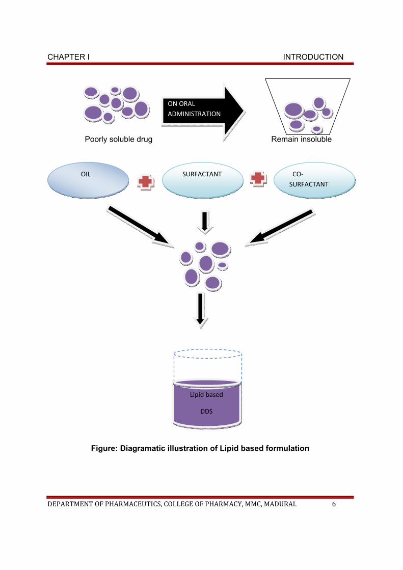
Lipid formulation mainly comprises of
• Macro emulsion
• Micro emulsion
• Self micro emulsifying drug delivery system (SMEDDS).
• Solid –lipid nano particle(SLN),
• Liposomes and
• Lipolexes.
In recent years, much attention has been focused on lipid based formulation, with
particular emphasis on SMEDDS.
Classification of lipid formulation system:
According to the composition and the effect of dilution and digestion on the
ability to prevent precipitation of drug, lipid based formulations are broadly classified
into four categories.
• Type I formulation
• Type II formulation
• Type III formulation, and
• Type IV formulation
CHAPTER I INTRODUCTION
DEPARTMENT OF PHARMACEUTICS, COLLEGE OF PHARMACY, MMC, MADURAI. 8
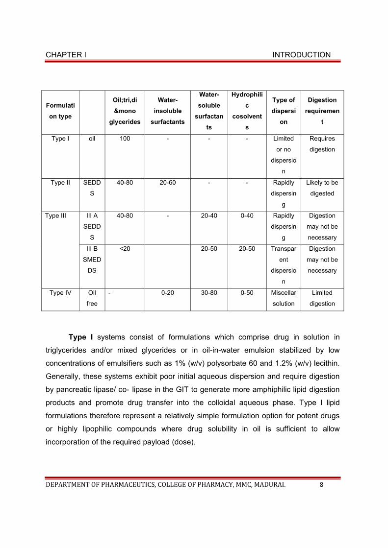
Formulati
on type
Oil;tri,di
&mono
glycerides
Water-
insoluble
surfactants
Water-
soluble
surfactan
ts
Hydrophili
c
cosolvent
s
Type of
dispersi
on
Digestion
requiremen
t
Type I oil 100 - - - Limited
or no
dispersio
n
Requires
digestion
Type II SEDD
S
40-80 20-60 - - Rapidly
dispersin
g
Likely to be
digested
Type III III A
SEDD
S
40-80 - 20-40 0-40 Rapidly
dispersin
g
Digestion
may not be
necessary
III B
SMED
DS
<20 20-50 20-50 Transpar
ent
dispersio
n
Digestion
may not be
necessary
Type IV Oil
free
- 0-20 30-80 0-50 Miscellar
solution
Limited
digestion
Type I systems consist of formulations which comprise drug in solution in
triglycerides and/or mixed glycerides or in oil-in-water emulsion stabilized by low
concentrations of emulsifiers such as 1% (w/v) polysorbate 60 and 1.2% (w/v) lecithin.
Generally, these systems exhibit poor initial aqueous dispersion and require digestion
by pancreatic lipase/ co- lipase in the GIT to generate more amphiphilic lipid digestion
products and promote drug transfer into the colloidal aqueous phase. Type I lipid
formulations therefore represent a relatively simple formulation option for potent drugs
or highly lipophilic compounds where drug solubility in oil is sufficient to allow
incorporation of the required payload (dose).
CHAPTER I INTRODUCTION
DEPARTMENT OF PHARMACEUTICS, COLLEGE OF PHARMACY, MMC, MADURAI. 9
Type II lipid formulations constitute SEDDS. Self-emulsification is generally
obtained at surfactant contents above 25% (w/w). However at higher surfactant
contents (greater than 50– 60% (w/w) depending on the materials) the progress of
emulsification may be compromised by the formation of viscous liquid crystalline gels at
the oil/water interface. Type II lipid- based formulations provide the advantage of
overcoming the slow dissolution step typically observed with solid dosage forms and as
described above generate large interfacial areas which in turn allows efficient
partitioning of drug between the oil droplets and the aqueous phase from where
absorption occurs.
Type III lipid-based formulations, commonly referred to as self-microemulsifying
drug delivery systems (SMEDDS), are defined by the inclusion of hydrophilic
surfactants (HLB>12) and co-solvents such as ethanol, propylene glycol and
polyethylene glycol. Type III formulations can be further segregated (somewhat
arbitrarily) into Type IIIA and Type IIIB formulations in order to identify more hydrophilic
systems (Type IIIB) where the content of hydrophilic surfactants and co-solvents
increases and the lipid content reduces. Type IIIB formulations typically achieve
greater dispersion rates when compared with Type IIIA although the risk of drug
precipitation on dispersion of the formulation is higher given the lower lipid content.
Type IV: In order to capture the recent trend towards formulations which contain
predominantly hydrophilic surfactants and co-solvents, this category was recently
added. Type IV formulations do not contain natural lipids and represent the most
hydrophilic formulations. These formulations commonly offer increased drug payloads
when compared to formulations containing simple glyceride lipids and also produce
very fine dispersions when introduced in aqueous media. Little is known however, as to
the solubilisation capacity of these systems In vivo and in particular whether they are
equally capable of maintaining poorly water soluble drug in solution during passage
along the GIT when compared with formulations comprising natural oils (Type II and
Type III). An example of a Type IV formulation is the current capsule formulation of the
HIV protease inhibitor amprenavir (Agenerase) which contains TPGS as a surfactant
and PEG 400 and propylene glycol as co- solvents.
CHAPTER I INTRODUCTION
DEPARTMENT OF PHARMACEUTICS, COLLEGE OF PHARMACY, MMC, MADURAI. 10
Self Micro Emulsifying Drug Delivery System (SMEDDS)
The self emulsifying drug delivery system (SEDDS), are well known for their
potential as alternative approach for delivery of hydrophobic drugs, which are
associated with poor water solubility and low oral bioavailability . SEDDSs are
isotropic and thermodynamically stable solutions consisting of oil, surfactant, co-
surfactant and drug mixtures that spontaneously form oil-in-water (o/w) emulsion
when mixed with water under gentle stirring. The motility of stomach and intestine
provides the agitation required for self- emulsification in-vivo. This spontaneous
formation of an emulsion in the gastrointestinal tract presents the drug in a
solubilized form, and the small size of the formed droplet provides a large interfacial
surface area for drug absorption . Apart from solubilization, the presence of lipid in
the formulation further helps to improve bioavailability by enhancing the drug
absorption. Selection of a suitable self-emulsifying formulation depends upon the
assessment of the solubility of the drug in various components and the droplet size
distribution of the resultant emulsion following self-emulsification. SEDDS are
mostly prepared in liquid dosage form in soft and hard gelatin capsules. Solid
SEDDS are new approach to make solid dosage form such as tablets, capsules etc.
Here it is essential to understand the differences between microemulsion
and nano emulsions SMEDDS finally forms microelmulsion after oral administration.
Micro emulsions are isotropic, thermodynamically stable systems composed of oil,
water, surfactant and co-surfactants or co-solvents. The main driving force for
microemulsion formation is the ultra low interfacial tension, which is usually
achieved by the use of two or more emulsifiers (surfactant and co-surfactant). Out
of the two emulsifiers used, one is essentially water soluble (surfactant) and the
other one is oil soluble (co-surfactant).
Now a days SMEDDS has emerged as a vital strategy to formulate poor
soluble compounds for bioavailability enhancement. However certain limitations are
associated with SMEDDS formulations which include in vivo drug precipitation,
formulation handling issues, limited lymphatic uptake, lack of predictive in vitro tests
CHAPTER I INTRODUCTION
DEPARTMENT OF PHARMACEUTICS, COLLEGE OF PHARMACY, MMC, MADURAI. 11
and oxidation of unsaturated fatty acids. These limitations restrict their potential
usage. Inclusion of polymers or precipitation inhibitors within lipid based
formulations help to maintain drug super saturation after dispersion. This thereby,
improves the bioavailability.
CHAPTER II
SELF MICRO EMULSIFYING DRUG DELIVERY- REVIEW
CHAPTER II SMEDDS -REVIEW
DEPARTMENT OF PHARMACEUTICS, COLLEGE OF PHARMACY, MMC, MADURAI. 12
CHAPTER- II
SELF MICRO EMULSIFYING DRUG DELIVERY SYSTEM- REVIEW
Self Emulsifying Drug Delivery system (SMEDDS) is a new approach
to improve the solubility of poorly soluble drugs. It can be ideally called as an
isotropic mixture. Drugs which are lipophilic in nature can be formulated in this
lipid based drug delivery system. SMEDDS improves solubility thereby
increases dissolution rate and bioavailability of drugs. Drug, oil, surfactant,
solvent and co-solvent are the components of SMEDDS. It forms small
droplets due to agitation. The size of the droplet is 10- 100nm. Absorption of
drug is improved by small droplets due to its ability to increase the interfacial
surface area. In this system, the drug is dissolved in oil, solvent or surfactant
.Co-solvents are used when required. Once it enters into the stomach it forms
micro emulsion due to mild agitation. Agitation is caused by the digestive
motility and intestine. SMEDDS is available as different dosage forms such as
capsules, tablets, suppositories and topical preparations.
ADVANTAGES OF SMEDDS :
• Enhances oral bioavailability.
• Delivers peptides, protein.
• Available in both liquid and solid dosage form.
• Poorly water soluble drugs can be used.
• Improve patient compliance.
• The drug is protected by oil droplets.
• Drugs will not be affected by presence of food.
• Has reproducible drug absorption profile.
• Gives prolonged release due to use of appropriate Polymer.
CHAPTER II SMEDDS -REVIEW
DEPARTMENT OF PHARMACEUTICS, COLLEGE OF PHARMACY, MMC, MADURAI. 13
Limitations Of SMEDDS :
• Lack of in-vitro models for evaluation.
• Dissolution test cannot be completely relied on, because this
formulation depends on digestion.
• It causes GIT irritation due to the excess amount of Surfactant.
• Use of co-solvents can destroy the soft gelatin.
FACTORS AFFECTING SMEDDS
• Dose of drug: The drugs which have low solubility at high dose are not
suitable for SMEDDS. The drugs required to administer at high dose
should possess good solubility in the components used at least in oil
phase.
• Solubility of drug: The drug should be highly soluble which influences
its bioavailability. The incorporation of surfactants and co-surfactants at
high concentration can cause risk of precipitation.
• Polarity of lipid phase: Release of drug is highly influenced by
polarity of lipid phase. High polarity value increases the rate of release.
• Droplet size and charge: Smaller the droplet size and larger the
surface area increases absorption and if the droplet is positively
charged the drugs can penetrate into the physiological barrier in deep
leads to improved bioavailability.
FORMULATION OF SMEDDS
The following should be considered in the formulation of SMEDDS.
• The solubility of the drug in different oil, surfactants and co-solvents
• The selection of oil, surfactant and co-surfactant based on the solubility
of the drug
• Preparation of the phase diagram
• The preparation of SMEDDS formulation, by dissolving the drug in a
mixture of oil, surfactant and co-surfactant.
CHAPTER II SMEDDS -REVIEW
DEPARTMENT OF PHARMACEUTICS, COLLEGE OF PHARMACY, MMC, MADURAI. 14
COMPONENTS OF SMEDDS:
Self micro emulsification has been shown to be specific to
• The nature of the oil/surfactant pair.
• The surfactant concentration and oil/surfactant ratio.
• The temperature at which self emulsification occurs.
In support of these facts, it has also been demonstrated that only very
specific pharmaceutical excipient combinations could lead to efficient self
emulsifying systems.
Poorly water soluble drugs can be delivered orally by pre-dissolving
the compounds in appropriate solvent and fill the formulation into capsules.
The initial rate limiting step of particulate dissolution in the aqueous
environment within the GI tract can be overcome by this approach. The main
problem is that the formulation may disperse in the GI tract which produces
precipitation of drugs in the solution, It occurs mostly with hydrophilic solvents
(e.g. polyethylene glycol). Occurrence of precipitation on dilution in the GIT
can be avoided by dissolving the components in lipid vehicle. Water-soluble
polymer can be used to aid solubility of the drug compound. For example,
polyvinylpyrrolidone (PVP) and polyethylene glycol (PEG 6000) have been
used for preparing solid solutions with poorly soluble drugs. Crystallisation of
the polymer matrix due to thermodynamically stable state is one of the
problems in this type of formulation that affects the physical stability of the
product which can be studied by Differential scanning calorimetry or X-ray
crystallography. SMEDDS are novel approach to enhance the solubility,
bioavailability and protect the drug from gastric environment which gives
better systemic absorption of drugs.
DRUG:
Lipophilicity and dose of the drug are the main criteria to be considered
be considered before the development of SMEDDS formulation. Drugs which
are administered at very high dose are not suitable for SMEDDS unless they
exhibit extremely good solubility in at least one of the components of
CHAPTER II SMEDDS -REVIEW
DEPARTMENT OF PHARMACEUTICS, COLLEGE OF PHARMACY, MMC, MADURAI. 15
SMEDDS. Ideally drug should have low dose, log P> 2 and should not
possess extensive first pass metabolism. High melting point drugs with log P
values about 2 are poorly suited to SMEDDS. At the other end of the
spectrum, lipophilic drugs with log P values greater than 5, are good
candidates for SMEDDS.
OILS:
The oil represents one of the most important excipients in the self
emulsifying formulations not only because it can solubilised marked amounts
of the lipophilic drug or facilitate self emulsification but also and mainly
because it can increase the fraction of lipophilic drug transported via the
intestinal lymphatic system, thereby increasing absorption from the GI tract
depending on the molecular nature of the triglyceride. Both long and medium
chain triglyceride oils with different degrees of saturation have been used for
the design of self emulsifying formulations. Furthermore edible oils which
could represent the logical and preferred lipid excipients choice for the
development of SMEDDS are not frequently selected due to their poor ability
to dissolve large amounts of lipophilic drugs. Modified or hydrolyzed vegetable
oils have been widely used since these excipients form good emulsification
system with a large number of surfactants approved for oral administration
and exhibit better drug solubility properties. They offer formulative and
physiological advantages and their degradation products resemble the natural
end products of intestinal digestion. Novel semi synthetic medium chain
derivatives, which can be defined as amphiphilic compounds with surfactant
properties, are progressively and effectively replacing the regular medium
chain triglyceride oils in the SMEDDS formulation.
SURFACTANTS:
Several compounds exhibiting surfactant properties may be employed
for the design of self emulsifying systems, but the choice is limited as very few
surfactants are orally acceptable. The most widely recommended ones being
the non-ionic surfactants with a relatively high hydrophilic –lipophilic balance
(HLB). The most widely recommended ones being the non ionic surfactants
CHAPTER II SMEDDS -REVIEW
DEPARTMENT OF PHARMACEUTICS, COLLEGE OF PHARMACY, MMC, MADURAI. 16
with a relatively high HLB value. the commonly used emulsifiers are various
solid or liquid ethoxylated polyglycolyzed glycerides and polyoxyethylene 20
oleate (Tween 80). Safety is a major determining factor in choosing a
surfactant. Emulsifiers of natural origin are preferred since they are
considered to be safer than the synthetic surfactants. However these
surfactants have a limited self emulsification capacity. Non-ionic surfactants
are less toxic than ionic surfactants but they may lead to reversible changes in
the permeability of the intestinal lumen. Usually the surfactant concentration
ranges between 30 and 60% w/w in order to form stable SMEDDS. It is very
important to determine the surfactant concentration properly as large amounts
of surfactants may cause GI irritation. Surfactants are amphiphilic in nature
and they can dissolve or solubilized relatively high amounts of hydrophobic
drug compounds. The lipid mixtures with higher surfactant and co-
surfactant/oil ratios lead to the formation of SMEDDS. (shukla Prachi et al.
IRJP 2012,3(9)
CO-SURFACTANTS:
Co-surfactants ensures flexibility of the interfacial layer, i.e. it reduces
the interfacial tension to a negative value. Co-surfactants form a flexible
interfacial film in order to acquire different curvatures required to form micro
emulsions over a wide range of compositions. Medium chain length alcohols
(C3-C8) are commonly employed as co-surfactants. Organic solvents such
as, ethanol, propylene glycol, and poly ethylene glycol are suitable for oral
delivery, and they enable the dissolution of large quantities of either the
hydrophilic surfactant or the drug in the lipid base. On the other hand alcohols
have the disadvantage of evaporating into the shells of the soft gelatin, or
hard, sealed gelatin capsules in conventional SMEDDS leading to drug
precipitation. Thus, alcohol free formulations have been designed, but their
lipophilic drug dissolution ability may be limited.
MECHANISM OF SELF-EMULSIFICATION:
Self emulsification process occurs, when the entropy changes. The
energy required to increase the surface area of the dispersion is smaller than
CHAPTER II SMEDDS -REVIEW
DEPARTMENT OF PHARMACEUTICS, COLLEGE OF PHARMACY, MMC, MADURAI. 17
the dispersion. The free energy of conventional emulsion formation is a direct
function of the energy required to create a new surface between the two
phases and can be described by the equation
δG=ΣNiπri2σ
Where,
δG- is the free energy associated with the process (ignoring the free
energy of mixing)
N- is the number of droplets of radius r
σ is the interfacial energy with time
The two phases of the emulsion will tend to separate, in order to reduce
the interfacial area and subsequently, the free energy of the system. Therefore,
the emulsions resulting from aqueous dilution are stabilized by conventional
emulsifying agents, which form a monolayer around the emulsion droplets and
hence, reduce the interfacial energy, as well as providing a barrier to
coalescence.
In case of self-emulsifying system, the free energy required to form the
emulsion is either very low or positive or negative then, the emulsion process
occurs spontaneously. Emulsification requires very little input energy, which
involves destabilization through contraction of local interfacial regions. For
emulsification to occur, it is necessary for the interfacial structure to have no
resistance to surface shearing.
In earlier work it was suggested that the case of emulsification could be
associated with the ease by which water penetrates into the various liquid
crystal or phases get formed on the surface of the droplet. The addition of a
binary mixture (oil/non-ionic surfactant) to the water results in the interface
formation between the oil and aqueous continuous phases, followed by the
solubilisation of water within the oil phase owing to aqueous penetration
through the interface, which occurs until the solubilisation limit is reached
close to the interface. Further aqueous penetration will result in the formation
CHAPTER II SMEDDS -REVIEW
DEPARTMENT OF PHARMACEUTICS, COLLEGE OF PHARMACY, MMC, MADURAI. 18
of the dispersed liquid crystalline phase.
As the aqueous penetration proceeds, eventually all materials close to
the interface will be liquid crystal, the actual amount depending on the
surfactant concentration in the binary mixture once formed, rapid penetration
of water into the aqueous cores, aided by the gentle agitation of the self
emulsification process causes interface disruption and droplet formation. A
combination of particle size analysis and low frequency dielectric
spectroscopy was used to examine self-emulsifying properties of a series of
Imwitor 742 (a mixture of mono and diglycerides of Caprylic acids/Tween 80)
systems, which provided evidence that the formation of the emulsion may be
associated with liquid crystal formation, although the relationship was clearly
complex.
The presence of the drug may alter the emulsion characteristics,
possibly by interacting with the liquid crystal phase. The droplet structure can
pass from a reversed spherical droplet to a reversed rod-shaped droplet,
hexagonal phase, lamellar phase, cubic phase or other structures until, after
appropriate dilution, a spherical droplet will be formed again.
DOSAGE FORM DEVELOPMENT OF S-SMEDDS:
Various dosage forms of S-SMEDDS are
• Dry emulsions
• Self- emulsifying capsules
• Self –emulsifying sustained/controlled release tablets
• Self-emulsifying sustained/controlled pellets
• Self-emulsifying solid dispersions
• Self-emulsifying beads
• Self-emulsifying sustained release microspheres
• Self –emulsifying nanoparticles
• Self-emulsifying suppositories
• Self-emulsifying implant.
CHAPTER II SMEDDS -REVIEW
DEPARTMENT OF PHARMACEUTICS, COLLEGE OF PHARMACY, MMC, MADURAI. 19
Self-emulsifying tablets:
Incorporation of lipid formulation into a solid dosage form combines the
advantages of lipid-based drug delivery systems with those of solid dosage
forms. Attama (2003) formulated a solid self-emulsifying formulation using
goat fat and tween for the delivery of diclofenac. Fatty material was melted
and mixed with surfactant and the drug incorporated into this mixture. This
wet mass was poured into plastic molds and cooled to form a tablet. During
the processing of this formulation it was observed that agitation during
fabrication of tablets reduced the liquification time, resulting in faster
emulsification. These results demonstrated that different formulation ratios
possess varying dissolution profiles at constant speed/agitation, and the
optimized formulation showed good release profiles with acceptable tablet
properties.
Nazzal and Khan (2006), evaluated the effect of some parameters
(colloidal silicates-X1, magnesium stearate mixing time X2, and compression
force X3) on coenzyme Q10 (CoQ10) dissolution from tablets of eutectic
based SMEFs. The optimized conditions (X1= 1.06%, X2 = 2 min, X3 = 1670
kg) were achieved by a face centred cubic design.
In order to significantly reduce the amount of solidifying excipients
required for transformation of SEFs into solid dosage forms, gelled SEFs
have been developed by Patil (2004). In this study, colloidal silicon dioxide
(Aerosil 200) was selected as a gelling agent for the oil-based systems.
Colloidal silicon dio- xide served a dual purpose: (i) – reducing the amount of
solidifying excipients required; and (ii) aiding in reducing drug release.
In a clinical study, it was found that SE tablets may be of use in reducing
adverse effects (Schwarz, 2003). The incorporation of indomethacin (or other
hydrophobic NSAIDs) in SE tablets was found to increase the penetration
efficacy of the drug through the GI mucosal membranes, potentially reducing
GI bleeding. The SEF in this study composed of glycerol monolaurate and
Tyloxapol TM (a copolymer of alkyl phenol and formaldehyde). The tablets
consistently maintained a higher active ingredient concentration in blood
CHAPTER II SMEDDS -REVIEW
DEPARTMENT OF PHARMACEUTICS, COLLEGE OF PHARMACY, MMC, MADURAI. 20
plasma over the same time period compared with a non-emulsifying tablet.
Self-emulsifying powder formulation (SE powder formulation)
Arida et al. (2007) formulated an SE powder formulation in order to
enhance the dissolution and absorption of the poorly water-soluble drug
griseofulvin. Capmul GMO-50, poloxamer and myvacet were used as
surfactants and co-surfactants. A significant enhancement in dissolution
(without ultra-micronisation) and bioavailability of griseofulvin was observed.
Balakrishnan et al. (2009) developed a novel solid SEF of dexibuprofen
using spray drying. Aerosil 200 was used as an inert solid carrier. Both in-vitro
and in-vivo studies were carried out. The optimization of the SEF
composition was carried out by assessing solubility, prepa- ration of phase
diagram, particle size analysis, drug release studies etc. The study showed
that Labrafil M 1944 CS, Labrafil M 2125, Labrasol, Capryol 90 and
Lauroglycol FCC could enhance the solubility of CoQ10 and provide the
desired drug loading.
Self-emulsifying pellets (SE pellets)
Oral pellets are known to overcome the poor and variable GIT
absorption of drugs and have shown the ability to reduce or eliminate the
influence of food on bioavailability. Thus, it appears highly appealing to
combine the advantages of pellets with those of SETs by formulating SE
pellets. Kang et al. (2004) as part of their study to develop a self-emulsifying
drug delivery system, have reported considerable differences in the solubility
of simvastatin in a range of surfactants. The authors suggest that the
properties of surfactants need to be considered when selecting them for the
formulation of SE pellets.
Franceschinis et al. (2005) developed a new method for preparing self-
emulsifying pellets by wet granulation consisting of a binder solution containing
an oil (mono and diglycerides), polysorbate 80 and nimesulide as a model
drug. The oil surfactant mixture was added to water to obtain binder solution.
The prepared binder solutions were sprayed onto the granules (prepared from
CHAPTER II SMEDDS -REVIEW
DEPARTMENT OF PHARMACEUTICS, COLLEGE OF PHARMACY, MMC, MADURAI. 21
microcrystalline cellulose and lactose) to give pellets. In vivo studies indicated
significantly higher bioavailability with the prepared pellets in comparison to
the corresponding emulsions.
Tuleu (2004) conducted a comparative bioavailability study of
progesterone from SE pellet formulation, SE solution, capsule and an
aqueous suspension in dogs. Complete drug release was seen within 30 min
of capsule administration and within 5 min of administration of the self-
emulsifying system. However, in the case of aqueous suspension the drug
release was very low (~50% of the dose in 60 min). Plasma drug
concentration was significantly higher when the drug was orally administered
from self-emulsifying pellets and self-emulsifying solution when compared to
aqueous suspension at the same dose.
Abdalla and Mader (2007) prepared three self- emulsifying pellet
formulations by melting cithrol GMS (mono and diglycerides) and solutol HS
15. To this molten blend, the drug (diazepam) and dry microcrystalline
cellulose (MCC) were added to obtain a suitable mass for extrusion. A dye
was incorporated for assessment of self-emulsification and spin probe was
added to assess the release kinetics and microenvironment of pellets. The
results from the release study, with higher load of diazepam and lower volume
of the dissolution media, indicated that the formulation was able to create and
maintain a state of supersaturation for the poorly water-soluble diazepam.
Nearly 90% of the drug was released within an hour while only 55% was
released from the GMS/MCC pellets.
Wang et al. (2010) demonstrated that the extrusion/ spheronization
technique is a large-scale production method for preparing solid SE pellets
from the liquid SEF to improve oral absorption. SE pellets of a hydrophobic
drug (nitrendipine) were prepared. Formulation stability and solubilisation
capacity were noted. The system was optimized on the basis of equilibrium
solubility, pseudo- ternary phase diagram and supersaturation studies. The
liquid SEFs were solidified using adsorbents (porous silicon dioxide), MCC
and lactose to form fine flowable powder. Crospovidone was added to the
CHAPTER II SMEDDS -REVIEW
DEPARTMENT OF PHARMACEUTICS, COLLEGE OF PHARMACY, MMC, MADURAI. 22
formulation. The AUC of nitrendipine from the SE pellets was two-fold
greater than the conventional tablets and was comparable with the liquid
SEFs.
Controlled release self-emulsifying pellets
Serratoni and Newton (2007) observed that the release of methyl
paraben (MP) and propyl parabens (PP) from pellet formulations could be
controlled by incorporating them into self-emulsifying systems containing
water soluble plasticiser and talc. Oil and surfactant were mixed and added
to the damp mass of MCC and lactose mono- hydrate. Extrusion
spheronization of the wet mass was carried out. The pellets obtained were
initially coated with ethyl cellulose and subsequently with an aqueous solution
of hydroxy propylmethyl cellulose in a fluid bed coater. Results obtained from
the in vitro study revealed that the presence of self-emulsifying system
enhanced drug release of MP and PP while the film coating considerably
reduced the drug release from pellets.
Iosio et al. (2008) prepared two types of pellets containing vinpocetine
(model insoluble drug) where Type I pellets contained a self-emulsifying
system internally and an inert matrix externally, whereas Type II contained
an inert matrix internally and a self-emulsifying system externally.
Formulations were prepared in two steps. In the first step, the oil-surfactant
mixture was added to water to form self-emulsifying systems whereas in the
next stage this mixture was loaded onto MCC and lactose to form extrusion-
spheronization mass for pellets. Results indicated that Type I pellets released
90% of vinpocetine within 30 min while the same quantity was released within
20 min from Type II pellets. The physical mixture of the excipients with drug
was able to release around 25% of the drug in 60 min. Although both types of
pellets demonstrated adequate morphological and technological
characteristics, type II pellets showed better drug solubility and in vivo
bioavailability. The above investigations suggest that a solid dosage form
containing a self-emulsifying system is a promising approach for the
formulation of drug compounds with poor aqueous solubility.
CHAPTER II SMEDDS -REVIEW
DEPARTMENT OF PHARMACEUTICS, COLLEGE OF PHARMACY, MMC, MADURAI. 23
Self-emulsifying beads (SE beads)
Self-emulsifying beads can be formulated as a solid dosage form using
smaller amounts of different excipients. Patil and Paradkar formulated an
isotropic formulation of loratadine consisting of Captex 200, Cremophore EL
and Capmul MCM. The SE mixture was loaded onto poly propylene beads
(PPB) using the solvent evaporation method. Formulations were optimized for
loading efficiency and in vitro drug release by evaluating their geometrical
features such as bead size and pore architecture. Results indicated that the
poly propylene beads are potential carriers for solidification of SE mixture,
with sufficiently high SE mixture to PPB ratios for the solid form. The results
indicated that self- emulsifying beads can be formulated as a solid dosage
form with a minimal amount of solidifying agents.
Self-emulsifying sustained-release microspheres
You et al. (2006) prepared solid SE sustained-release microspheres of
zedoary turmeric oil (oil phase) using the quasi-emulsion-solvent-diffusion
method involving spherical crystallization. The release behaviour of zedoary
turmeric oil from the formulation was found to be dependent upon the hydroxyl
propyl methylcellulose acetate succinate to Aerosil 200 ratio. The plasma
concentration time profiles after oral administration in rabbits showed a
bioavailability of 135.6% compared with the conventional liquid SEFs.
Self-emulsifying implants (SE implants)
Research into SE implants has greatly increased the use and application
of solid self-emulsifying formulation (S-SEF). Carmustine (BCNU) is a
chemotherapeutic agent used to treat malignant brain tumours. However, its
effectiveness is hindered by its short half life. In order to enhance its
stability, the SEF of carmustine was formulated using tributyrin,
Cremophor RH 40 (polyoxyl 40 hydrogenated castor oil) and Labrafil 1944
(polyglycolyzed glyceride). The self-emulsified BCNU was fabricated into
wafers with a flat and smooth sur- face by compression moulding. The

CHAPTER II SMEDDS -REVIEW
DEPARTMENT OF PHARMACEUTICS, COLLEGE OF PHARMACY, MMC, MADURAI. 24
release profile was compared with a wafer implant fabricated using poly (d,
l-lactide-co-glycolide) acetic acid. It was found that SEF increased the in vitro
half-life of BCNU to 130 min compared with 45 min with intact BCNU. The in
vitro release of BCNU from self-emulsifying PLGA wafers was prolonged up to
7 days and was found to have higher in vitro anti-tumor activity (Chae et al.,
2005).
Self-microemulsifying formulations
Self-micro emulsifying formulations (SMEFs) have attracted great
attention recently. In an attempt to combine the advantages of SMEFs with
those of solid dosage forms and overcome the shortcomings of liquid
formulations, increasing attention has been focused on solid self-(micro)
emulsifying formulations. The thermotropic stability of SMEFs and their high
drug loading efficiency make them a promising system for low aqueous soluble
drugs (Jannin et al., 2007). SMEFs are usually placed in soft gelatin
capsules, but can also be transformed into granules, pel- lets, powders for
dry filled capsules or tablet preparations (Nazzal, Khan, 2006; Serratoni,
Newton, 2007; Abdalla et al., 2008; Tan et al., 2009). The commercial
success of the SMEF, Neoral® drew greater attention to the development of
SMEFs. Many poorly water-soluble drugs such as acyclovir, atorvastatin, and
fenofibrate have been reported to offer improved oral bioavailability by SMEFs
(Wang et al., 2006; Shen, Zhong, 2006; Patel, Vavia, 2007).
Postolache et al. (2002) compared the bioavailability of two cyclosporine
capsule products with different pharmaceutical formulations. Results showed
that the test cyclosporine non-SMEFs formulation was not bioequivalent to the
cyclosporine SMEFs formulation due to a statistically significantly lower
absorption rate. These authors demonstrated that the non-self
microemulsifying capsules are not totally interchangeable with the self
microemulsifying capsules unless validated clinical and laboratory conversion
protocols for each kind of organ transplantation are enforced.
CHAPTER II SMEDDS -REVIEW
DEPARTMENT OF PHARMACEUTICS, COLLEGE OF PHARMACY, MMC, MADURAI. 25
Catarzi et al. (2008) reported the comparative impact of Transcutol and
Neusilin US2 on SMEFs. Results showed that the Neusilin- formulation
resulted in hard tablets with a low tablet weight. However, Neusilin® tablets
had similar disintegration times compare to Aeroperl (Evonik Degussa). The
dissolution profile obtained from the tablets showed improved profile when
compared to Glyburide alone. Zvonar et al. (2010) suggested that, SMEFs
possessing a composition similar to microcapsules with Ca-pectinate shell
and a drug loaded SMEFs as the core phase, would be a potential
approach for enhancing low permeability and solubility of BCS class II
drugs.
Self nanoemulsifying formulations (SNEFs)
The classical lipid nanoparticles that have been proposed for drug
delivery are composed of solid lipids. A distinct advantage of SNEFs over
polymeric nanoparticles is that the lipid matrix is made from physiologically
tolerated lipid components, which decreases potential acute and chronic
toxicity.
Nazzal et al. (2002) developed a SNEF based on the eutectic properties
of ubiquinone (CoQ10) and also studied the progress of emulsion formation
and drug release mechanisms by turbidimetry and droplet size analysis.
Results obtained from study revealed that eutectic-based semisolid SEFs
can overcome the drawbacks of the traditional emulsified systems such as
low solubility and irreversible precipitation of the active drug in the vehicle
with time.
Cyclosporine lipid nanoparticles (lipospheres) consisting of
phospholipids, Span 80, Tween 80, Tricaprin, and Cremophor RH 40 were
prepared (Bekerman et al., 2004). The CsA dispersion systems prepared had a
particle size ranging from 25 nm to 400 nm. Particles with a size of 25 nm
showed maximum oral bioavailability.
In a study by Nepal et al.(2010), the surfactant–co-surfactant blend
(Witepsol H35 and Solutol HS15) at a ratio of 1:4 led to sufficient reduction in
CHAPTER II SMEDDS -REVIEW
DEPARTMENT OF PHARMACEUTICS, COLLEGE OF PHARMACY, MMC, MADURAI. 26
free energy of the system to resist thermodynamic instability of the nano-
emulsion as well as providing a sufficient mechanical barrier to coalescence
oil droplets.
Koynova et al. (2010) suggested the use of nano sized self-emulsifying
lipid vesicles as carriers for the inclusion of lipophilic dietary supplements.
These were proposed as good alternatives to liposomal preparations which
pose problems in stability, sterilization, and non- reproducibility between
batches.
Supersaturable self-emulsifying formulation
Supersaturation represents a potent technique for enhancing absorption
by generating and maintaining a supersaturated state in the intestine. Such
formulations contain both a reduced amount of surfactant(s) and a
polymeric precipitation inhibitor (e.g., water-soluble cellulosic polymers,
such as HPMC). These maintain a supersaturated state of the drug in the body.
As the literature suggested, directly supersaturating a system with a drug
during manufacture adds to the risk of recrystallization of the product. Various
ways of inhibiting recrystallization have been identified. Thermodynamic
“freezing” inside a polymer is one such option. Under storage conditions, the
drug is mobilized by thermodynamic changes in the poly- meric structure. To
avoid risk of direct supersaturation, several strategies can be employed, for
example:
• Evaporation of a solvent from the system
• Activation of thermodynamically “frozen” drug- supersaturated islands
by hydration.
However, attaining full knowledge of these processes, especially in a
multi- component formulation, requires extensive research. Recently, Gao et al.
(2008) investigated the mechanism responsible for the enhanced intestinal
absorption of hydrophobic drugs from supersaturable SEFs containing
HPMC. This effect could be attributed to enhanced permeation of drug to the
enterocyte brush border region through the aqueous pathway by mimicking, or
equilibrating with, the bile acid /bile acid mixed micelle pathway.
CHAPTER II SMEDDS -REVIEW
DEPARTMENT OF PHARMACEUTICS, COLLEGE OF PHARMACY, MMC, MADURAI. 27
Techniques used to produce solid SMEDDS:
Various techniques have been employed to produce SMEDDS.
• Spray drying:
In this method, all the excipients are mixed together. The formulation
is atomized into small droplets. The droplets are introduced into the drying
chamber the temperature and airflow is maintained as per required. Further it
can be prepared as capsules /tablets.
• Adsorption onto solid carrier
In this method the liquids are mixed with the excipients. The powdered
mixture is filled into capsules or it can be formulated as tablets. The benefit of
this technique is it ensures content uniformity.
• Melt agglomeration:
Powder agglomeration can be obtained by melt granulation. It is
obtained by using binder which melts at low temperature.
• Melt extrusion:
It is a solvent free method. In this process, the raw material which has
plastic properties converts into a product with uniform density and shape; it is
obtained by forcing the raw material into a die. This process is carried out
under controlled pressure, temperature and proper flow of product. The
advantage of this method is high drug loading and content uniformity.

CHAPTER II SMEDDS -REVIEW
DEPARTMENT OF PHARMACEUTICS, COLLEGE OF PHARMACY, MMC, MADURAI. 28
Limitations of SMEDDS:
Although SMEDDS formulations has several advantages, there are
certain limitations associated with this system
1. Drug precipitation on dilution:
Diluted SMEDDS undergo precipitation of drug in gastro intestinal fluid.
A common requirement for the lipid formulation is that they should be able to
keep the drug in the solubilised form in the gastrointestinal tract. Precipitation
of the drug from the system nullifies the advantage offered by the lipid based
formulation system. The precipitation tendency of the drug on dilution is
higher due to the dilution effect of the hydrophilic solvent. It thereby requires
incorporation of polymers to minimize drug precipitation.
2. Encapsulation in soft gelatin capsules:
Most of the marketed SMEDDS formulations are available as soft
gelatin capsule. However gelatin capsules are associated with few
drawbacks. Manufacturing cost, transmissible spongiform encephalopathy
(TSE) and consumer preference are the few issues associated with animal
gelatin. Volatile co-solvents in self-micro emulsifying formulations are known
to migrate into the shells of soft or hard gelatin capsules, resulting in the
precipitation of the lipophilic drugs. These problem drive the market
requirement to find substitute for soft gelatin capsules. The current alternative
material of choice, to animal gelatin capsules are HPMC capsules. The HPMC
capsule shell has been explored as an alternative approach for encapsulating
supersaturable SMEDDS formulation.
3. Storage and handling:
Liquid SMEDDS exhibit problems in handling, storage and stability.
Thus, formulating solid SMEDDS seems to be a logical solution to address
these problems.

CHAPTER II SMEDDS -REVIEW
DEPARTMENT OF PHARMACEUTICS, COLLEGE OF PHARMACY, MMC, MADURAI. 29
4. Limited targeting to lymphatics:
Targeting lymphatics confers two primary advantages over
conventional absorption via the portal blood. First, transport through the
intestinal lymph avoids pre-systemic hepatic metabolism and thereby
enhances the concentration of orally administered drugs reaching the
systemic circulation. Second, site-specific hepatic metabolism and thereby
enhances the concentration of orally administered drugs reaching the
systemic circulation. Second, site-specific drug delivery to lymphatic organs
could be achieved. Normally high triglyceride solubility and high log P is
required for lymphatic transport. However, the amount of drug transported into
lymphatics is variable from drug to drug. Hence, lipophilicity and triglyceride
solubility of drug in correlation with lymphatic transport needs to be completely
understood and a more adequate predictive models is required
5. Lack of good in vitro models:
Another obstacle in the development of SMEDDS and other lipid-based
formulations is the lack of good predicative in vitro models for the assessment
of the formulations. Traditional dissolution methods do not work, as these
formulations potentially are dependent on digestion of lipid in the gut, prior to
release of the drug. Although to mimic this, an in vitro model simulating the
digestive processes of the duodenum has been developed. This in vitro model
needs further refinement and validation before its strength can be evaluated.
Further development can be based on in vitro- in vivo correlations and
therefore, different prototype lipid based formulations need to be developed
and evaluated in vivo in a suitable animal model.
6. Oxidation and polymorphism of the lipids used in formulation SMEDS:
Lipid excipients containing unsaturated fatty acids and its derivatives
are prone to lipid oxidation. This requires inclusion of lipid soluble antioxidant
in capsule formation. Polymorphism associated with thermo-softening lipid
CHAPTER II SMEDDS -REVIEW
DEPARTMENT OF PHARMACEUTICS, COLLEGE OF PHARMACY, MMC, MADURAI. 30
excipients requires specific process control in their application, in order to
minimize polymorphic changes of the excipient matrix.
Handling and storage issues with liquid SMEDDS
SMEDDS are normally viscous liquids that are administered in hard or
soft gelatin capsule. Lipid formulation may leach into and interact with the
capsule shells. This may cause brittleness or softness of the capsule shell,
leakage of the content and precipitation of the drug or excipients, especially at
lower temperatures. To address these problems, several attempts have been
made to transform liquid SMEDDS into solid SMEDDS dosage forms. These
approaches combine the advantages of SMEDDS with those of solid dosage
forms while overcoming the disadvantages of liquid SMEDDS. Formulating
solid SMEDDS combine the advantages of SMEDDS with those of solid
dosage forms, ie. Low production cost, convenience of process control, high
stability, reproducibility, better patient compliance. Thus, the presentation of
liquid SMEDDS in a solid dosage form provides more stable and robust
dosage form with lower manufacturing costs.
CHAPTER III
LITERATURE REVIEW
CHAPTER III REVIEW OF LITERATURE
DEPARTMENT OF PHARMACEUTICS, COLLEGE OF PHARMACY, MMC, MADURAI 31
CHAPTER - III
REVIEW OF LITERATURE
Tarkeshwar Prasad Shukla et al., (2017), were formulated
Acetazolamide SMEDDS and evaluated. The optimized formula was selected
to have the smallest mean particle size and the highest absolute Zeta
potential, which should yield the formation of a stable emulsion. The in vitro
dissolution indicated a significant improvement in Acetazolamide release
characteristics. The preformulation study of drug was carried out initially in
terms of physical appearance, melting point, solubility study, λ max
determination (wavelength at maximum absorbance) solubility study, and
results directed for the further course of formulation. The formulations were
prepared with different ratios of oil, surfactant and co-surfactant. The
SMEDDS were prepared and are evaluated in terms of droplet size, drug
release etc. The system consist of Arachis Oil, Lutrol F-68, Triacetin &
Propylene Glycol as oil phase, surfactant and co-surfactant respectively.NIP
C1 & NIP C8 selected as optimized batches showed promising results. Zeta
potential of NIP C8 indicates good stability. NIP C1 & NIP C8 showed Poly
dispersity index below 0.3 which shows good uniformity in the droplet size
distribution. Time of emulsification was under grade A (visual assessment
criteria), thermodynamic stability testing & accelerated stability study was
performed for optimized batches. In this study SMEDDS is a promising
approach to improve the solubility, dissolution rate and bioavailability of
Acetazolamide.
Yuxia Zhang et al., (2017), developed and evaluated colon-specific
capsule with alginate beads containing a self-micro emulsifying drug delivery
system (SMEDDS). The SMEDDS technique was used to improve the
solubility of curcumin (Cur). After encapsulating the Curcumin -loaded
SMEDDS, the alginate beads were placed inside an impermeable capsule
body. A konjac glucomannan/lactose/hydroxypropyl methylcellulose (KG/Lac/
HPMC) plug tablet was then prepared and placed in the mouth of the capsule.
The capsule demonstrated a pulsatile drug-release profile with a specific lag
CHAPTER III REVIEW OF LITERATURE
DEPARTMENT OF PHARMACEUTICS, COLLEGE OF PHARMACY, MMC, MADURAI 32
time and subsequent sustained-release phase. The lag time was modified by
changing the type of HPMC and the ratio of KG/Lac/HPMC. In addition, 0.5%
b-mannase solution and 5% rat cecal solution were used to simulate the colon
fluid, significantly decreasing the lag time of the capsule. The results show
that the capsule has potential for use in colon-specific drug delivery and
exhibits a sustained-release characteristic.
Pallavi Patharkar et al., (2016), were formulated and evaluated solid
self emulsifying drug delivery system of Olmesartan Medoxomil by adsorption
to solid carrier technique to improve the oral bioavailability of drug. The
solubility of OLM was deterimiced in various vehicles kike oils, surfactants and
co-surfactant. Pseudoternary phase diagrams were constructed to identify the
efficient self emulsifying region. The system consists of Olmesartan, Acrysol
k-150, Labrasol, Transcutol P as a drug, oil, surfactant and co-surfactant. The
optimized liquid SMEDDS was transformed into a free flowing powder using
Avicel or Aerosil 200 as the adsorbent. Prepared formulations were tested for
microemulsifying properties and the resultant micro emulsions were evaluated
for robustness to dilution, assessment of efficiency of self emulsification,
emulsification time, turbidity measurement, viscosity, drug content and in-vitro
dissolution. The physical state of the drug in solid self micro emulsifying
powder was revealed by Differential Scanning Calorimetric and X-ray powder
diffraction studies which indicated the presence of the drug was enhanced
significantly from the SMEDDS formulations as compared to pure drug.
Karunakara reddy et al., (2016), formulated self micro emulsifying drug
delivery system of atazanavir and evaluated for their properties and invitro
release studies. Solubility of atazanavir was determined in various oils,
surfactants, and co-surfactants. Pseudo ternary phase diagrams were
constructed to determine the concentration range of oils, surfactant, and co-
surfactant. The optimized formulation consists of castor oil as an oil phase,
kolliphore RH as a surfactant and PEG 400 as a co-surfactant. The liquid
SMEDDS formulations were prepared and evaluated for zeta potential,
polydispersity index, % transmittance, drug content and invitro dissolution
studies. Then the optimized liquid formulation ( drug content -97.76%; drug
CHAPTER III REVIEW OF LITERATURE
DEPARTMENT OF PHARMACEUTICS, COLLEGE OF PHARMACY, MMC, MADURAI 33
release-95.42%) was converted into free flowing powder using Neusilin US2.
Then the solid SMEDDS were evaluated for drug content (97.89%). The solid
SMEDDS showed better drug release than the pure drug of Atazanavir. So
the SMEDDS approach may be useful for the enhancement of solubility of
poorly water soluble compounds.
Xiaolin Bi et al., (2016), were developed a solid self-microemulsifying
drug delivery system (S-SMEDDS to load the various active constituents of
salvia (dried root of Salvia miltiorrhiza) which contains both lipophilic (e.g.,
tanshinone IIA, tanshinone I, cryptotanshinone and dihydrotanshinone I) and
hydrophilic (e.g., danshensu and salvianolic acid B) constituents, into a single
drug delivery system and improve their oral bioavailability. A prototype
SMEDDS was designed using solubility studies and phase diagram
construction, and characterized by self-emulsification performance, stability,
morphology, droplet size, polydispersity index and zeta potential.
Furthermore, the S-SMEDDS was prepared by dispersing liquid SMEDDS
containing liposoluble extract into a solution containing aqueous extract and
hydrophilic polymer, and then freeze-drying. In vitro release of tanshinone IIA,
salvianolic acid B, cryptotanshinone and danshensu from the S-SMEDDS was
examined; showing approximately 60%–80% of each active component was
released from the S-SMEDDS in vitro within 20 min. In vivo bioavailability of
these four constituents indicated that the S-SMEDDS showed superior in vivo
oral absorption to a drug suspension after oral administration in rats. It can be
concluded that the novel S-SMEDDS developed in this study increased the
dissolution rate and improved the oral bioavailability of both lipophilic and
hydrophilic constituents of salvia. Thus, the S-SMEDDS can be regarded as a
promising new method by which to deliver salvia extract, and potentially other
multicomponent drugs, by the oral route.
Nilesh mahadeo et al., (2016), were developed Liquid self micro
emulsifying drug delivery system (L-SMEDDS) of Cefpodoxime proxetil (CFP)
a poorly absorbable, high dose antibiotic having pH dependant solubility to
overcome the problems associated with oral delivery. Solubility of CFP in
various oils was determined to select the key components of SMEDDS.
CHAPTER III REVIEW OF LITERATURE
DEPARTMENT OF PHARMACEUTICS, COLLEGE OF PHARMACY, MMC, MADURAI 34
Surfactants and co surfactants were screened and selected on the basis of
their % transmittance. In this formulation Capmul MCM (650 mg of drug/1g of
oil), Cremophore RH 40 (% T-90.12 %), Labrafil 2125 CS (% T-99.03%) were
used as an oil, surfactant and co-surfactant respectively. Neusilin US2 was
used as an adsorbing agent to convert optimized L-SMEDDS to Solid Self
micro emulsifying drug delivery system (S-SMEDDS), which were then filled in
hard gelatin capsule and evaluated further. The optimized L-SMEDDS shows
Globule size 30-35 nm, Zeta potential -10 to -11 (negative ZETA POTENTIAL
due to the presence of fatty acids in the structure of the excipients), Drug
content 99.24±0.52 %. The S-SMEDDS shows acceptable flow properties,
and Particle size of 33.40 nm, Polydispersibility index of 0.125, Zeta potential
of -11.5 mv, Drug content of 98.32±0.14%. SEM studies showed that there is
loss of crystalline structure of drug has been converted into amorphous state
i.e completely solubilised in oil phase of L-SMEDDS and adsorbed on Neusilin
US2 surface. FTIR studies confirmed that there is no drug- excipient
interaction.
Bhalekar MR et al., (2016), formulated self micro emulsifying drug
delivery system of Darunavir, an anti retroviral protease inhibitor to increase
its solubility and bioavailability. The SMEDDS system compromising of
Imwitor 988 as oil phase. Tween 20 and span 20 as binary surfactant system
were optimized with respect to drug solubilization, particle size, zeta potential,
dispersibility, optical clarity cloud point, in-vitro release and thermodynamic
stability. SMEDDS system containing Imwitor 988 920%) and surfactant mix
(Smix) (80%) showed maximum drug solubility with least particle size. The ex-
vivo lymphatic uptake studies of Darunavir SMEDDS in presence of lymphatic
uptake blocker showed 36. 69% drug permeation which increased to 64. 245
in absences of lymphatic blocker, indicating the drug transport by lymphatic
path. Cmax of Darunavir SMEDDS was higher than that of the marketed tablet.
Pluronic f68 treated rats show lesser plasma concentration as compared to
those administered with SMEDDS. The results suggest that SMEDDS is a
promising drug delivery system to improve solubility and lymphatic transport
of anti-HIV drug Darunavir.
CHAPTER III REVIEW OF LITERATURE
DEPARTMENT OF PHARMACEUTICS, COLLEGE OF PHARMACY, MMC, MADURAI 35
Divya G. Shereker et al., (2016), were developed and evaluated Self-
micro emulsifying drug delivery system (SMEDDS) of Ketoprofen, a widely
prescribed analgesic drug which belongs to BCS class II and exhibit low and
variable oral bioavailability due to its poor aqueous solubility. Solubility study,
emulsification ability, ternary phase diagram and central composite design
(CCD) were used as primary tools to select the components of the system
and optimize the composition of liquid Ketoprofen SMEDDS. The globule size
of optimized liquid SMEDDS was 105.08±5.16nm, zeta potential -4.23mV and
polydispersity index 0.097, % Transmittance 98.45% and self emulsification
time 28 sec. Optimized formulation F3 containing Oleic acid (10%), Tween 80
(30%) and Propylene glycol (60%) was adsorbed onto inert solid carrier
Aerosil 200 in 1:1 ratio to form dry, free flowing powder. The liquid crystal
phase viscosity increased significantly with increasing amount of aerosil 200
which in turn increase average globule size of solid SMEDDS
(303.69±0.933nm) and slower the drug release. S-SMEDDS also
characterized for DSC, XRD, SEM etc. The in vitro dissolution study indicates
improved dissolution characteristics with higher percent drug release for solid
SMEDDS (89.78%) compared to marketed preparation (81.26%) and pure
drug (71.32%). In conclusion, S-SMEDDS for Ketoprofen holds promise to be
developed as potential system for improved oral delivery.
Madhavi K et al., (2016), formulate solid self emulsifying drug delivery
systems in order to improve the solubility of the highly lipophilic
antihypertensive drug ramipril. The system was formulated by homogeneously
mixing oils, surfactants and co-surfactants along with the drug component.
Based on solubility studies Capmul PG8 NF, Gelucire 44/14 and Transcutol P
were selected as oil, surfactant co-surfactant respectively in order to prepare
liquid SMEDDS. Nine different liquid SMEDDS formulations were prepared
and subjected to various evaluation tests in order to obtain optimized L-
SMEDDS. Out of 9 different formulations S9 formulation was optimized as it
formed thermodynamically stable emulsion without any drug precipitation and
phase separation on storage and also showed least globule size (22.56 nm).
The optimized formulation was loaded onto inert carrier (Sylysia FCP 350) to
obtain S-SMEDDS, which shows acceptable flow properties. they were further
CHAPTER III REVIEW OF LITERATURE
DEPARTMENT OF PHARMACEUTICS, COLLEGE OF PHARMACY, MMC, MADURAI 36
processed for solid state characterization such as XRD, DSC and SEM and
the results confirmed the transformation of native crystalline nature of drug to
an amorphous state. FTIR studies also confirmed no drug-excipient
interaction. S-SMEDDS showed improved invitro dissolution behavior of
Ramipril over that of pure drug.
Nilesh khutle et al., (2015), developed Self micro emulsifying drug
delivery system of Pravastatin sodium (BCS Class III drug with high aqueous
solubility and low permeability characteristics. The drug shows low absolute
biovailability (approximately 17%) due to decreased permeability and high first
pass extraction. So the study was designed to enhance the permeability
characteristics of hydrophilic drugs and to protect them from hostile
environment of gut. The solubility of Pravastatin sodium was determined to
identify the oil phase of SMEDDS. Various surfactants and co-surfactants
were screened for their ability to emulsify the selected oil. Pseudetenary
phase diagrams were constructed to identify the efficient self-emulsifying
region. The SMEDDS formulation was optimized by freeze thaw cycles,
robustness to dilution and droplet size and zeta potential tests. The optimized
L-SMEDDS formulation containing Pravastatin -10 mg, Capmul -100 mg,
Cremophore RH 40 -66.66mg and Labrafil M 2125-33.33 mg was further
evaluated by in-vitro and ex-vivo release studies. L-SMEDDS was further
converted into T-SMEDDS by “Liquid loading technique”. T-SMEDDS of
Pravastatin contained Neusilin, crosspovidone, magnesium stearate and L-
SMEDDS loaded onto it. The results from both L-SMEDDS and T-SMEDDS
suggest the potential use of SMEDDS to improve GI instability and intestinal
permeability of BCS class III drug pravastatin
Kanav midha et al., (2015,) were formulate dispersible self
microemulsifying (SMEDDS) tablet of atorvastatin for promoting its solubility
and its oral bioavailability. The liquid SMEDDS were prepared by water
titration method using oleic acid, Tween 80, and PEG 400 as oil, surfactant,
and co-surfactant respectively. Then the L-SMEDDS were converted into solid
SMEDDS by adsorption on solid carriers (Neusilin US2). The S-SMEDDS
were blended with sodium starch glycolate (disintegrant) and tablet excipients
CHAPTER III REVIEW OF LITERATURE
DEPARTMENT OF PHARMACEUTICS, COLLEGE OF PHARMACY, MMC, MADURAI 37
and compressed into tablets that were dispersible and self micro emulsifying
in nature. All these formulations were assessed for various physicochemical
parameters viz. weight variation, hardness, friability, disintegration test. Invitro
studies of pure drug, SMEDDS, S-SMEDDS and dispersible SMEDDS tablets
were carried out. Pure drug released only 29.84±0.16% up to 60 minutes and
all the SMEDDS formulation released 100% of drug in comparatively lesser
time. The optimized formulation containing 30% of oleic acid, 65% of tween
80 and 5% co-surfactant show the best results in in vitro studies. And FB1
(tablet) was considered to e the best since it release 100% drug in 35 mints
and also has advantages over SMEDDS and S-SMEDDS in tms of stability
and patient compliance.
Nilesh S. Kulkarni et al., (2015), developed rosuvastatin calcium-
loaded self-nanoemulsifying powder for improved oral delivery of the drug.
Solubility study was carried out in different oils, surfactants and co-
surfactants. Based on the solubility study, liquid formulations were prepared
using LAS/Capryol 90: Maisine 35-1 as oil phase and Tween 20 with Lutrol
E400 as surfactant mixture (Smix). The liquid formulations were adsorbed
onto Aerosil 200 in a ratio of 1: 0.25 % w/w to convert them into a solid form.
The formulations were evaluated for globule size, zeta potential, and emulsion
properties. Transmittance study, scanning electron microscopy, and in-vitro
dissolution studies were carried out. Biochemical studies were carried out
using a triton-induced hyperlipidemia model in Wistar rats. The developed
formulations exhibited some desirable characteristics of self-emulsifying
systems with nano-sized globules in the range 119.8 to 228.9 nm, rapid
emulsification in approximately 60 s and transmittance of close to 100 %. In-
vitro dissolution studies on the developed formulations indicate a 4-fold
increase in drug release in 10 min, compared to the pure drug (ROC) while
pharmacodynamic data showed significant improvement in oral bioavailability
compared to the pure drug. the results concluded that the developed
formulation containing the oils, LAS and combination of capryol 90 with
Maisine 35-1, has the capability to improve the solubility and bioavailability of
rosuvastatin calcium when formulated as a self-nanoemulsifying product.
CHAPTER III REVIEW OF LITERATURE
DEPARTMENT OF PHARMACEUTICS, COLLEGE OF PHARMACY, MMC, MADURAI 38
Sujit Arun Desai et al., (2015), was formulated and liquid SMEDDS of
Lovastatin and evaluated for its transparency, droplet size, zeta potential,
viscosity, conductivity, percentage assay, and phase separation study. The
micro emulsion produced by water titration method shows good particle size
(95 nm). Refractive index and % transmittance showed good isotropic
formulation. Solubilisation capacity of the system was also determined. An
accelerated stability study of optimized system was carried out to check the
stability of the formulation. The prepared micro emulsion was compared with
the pure drug solution and commercially available Lovastatin tablet for in-vitro
drug release. It revealed overall increase in release from micro emulsion
(76.72%) compared to Lovastatin marketed tablet formulation (52.3%).
Comparative oral absorption of Lovastatin from the micro emulsion and
suspension of the commercially available Lovastatin was investigated through
an in-vivo study in a rat model. FTIR studies revealed that there is no physical
interaction between drug and excipients.
Anna Czajkowska-Kosnik et al., (2015), designed and characterized
liquid and solid self-emulsifying drug delivery systems (SEDDS) for poorly
soluble drug atorvastatin. To optimize the composition of liquid atorvastatin-
SEDDS, solubility tests, pseudoternary phase diagrams, emulsification
studies and other in vitro examinations (thermodynamic stability, droplet size
and zeta potential analysis) were performed. Due to the disadvantages of
liquid SEDDS (few choices for dosage forms, low stability and portability
during the manufacturing process), attempts were also made to obtain solid
SEDDS. Solid SEDDS were successfully obtained using the spray drying
technique. Overall, the studies demonstrated the possibility of formulating
liquid and solid SEEDS as promising carriers of atorvastatin. SEDDS, with
their unique solubilization properties, provide the opportunity to deliver the
lipophilic drugs to the gastrointestinal tract in a solubilized state, thus
enhancing bioavailability of BCS class 2 drugs.
Vikas Bhandari et al., (2015), developed self emulsifying drug
delivery system in liquid and then convert it into a pellet form that would
result in improved solubility, dissolution and permeability of the poorly water
CHAPTER III REVIEW OF LITERATURE
DEPARTMENT OF PHARMACEUTICS, COLLEGE OF PHARMACY, MMC, MADURAI 39
soluble drug carvedilol. Pellets were prepared using extrusion-
spheronization technique incorporating liquid SEDDS (carvedilol, capmul
MCM EP, cremophore EL, tween 20, propylene glycol), adsorbents ( and
crospovidone), microcrystalline cellulose and binder (povidone K-30). Ternary
phase diagram was constructed to identify different oil-surfactant-
cosurfactant mixtures according to the proportion of each point in it. The
optimal CAR-SEDDS pellets showed a quicker redispersion with a droplet
size of the reconstituted microemulsion being 160.47 nm, which was almost
unchanged after solidification. SEM analysis confirmed good spherical
appearance of solid pellets; DSC and XRD analysis confirmed that there was
no crystalline carvedilol in the pellets. Pellets were then capable of
transferring lipophilic compounds into the aqueous phase and significantly
enhancing its release with respect to pure drug.
Vidyadhara. S et al., (2015), formulated and evaluated Rosuvastatin
SMEEDS. The SMEDDS were prepared by simple admixing method using
Capmul MCM and Capryol 90 as oils, Polaxomer 407 as surfactant and
Transcutol HP and Soluphor P as co surfactants. The liquid SMEDDS were
then converted into free flowing powder by adsorbing onto solid carrier
magnesium aluminium silicate. Then the formulations were evaluated for
particle size, phase separation, droplet size, drug content and for in vitro drug
release. The FTIR and DSC analysis on optimized formulations revealed that
there were no major interactions between drug and excipients. Among the
prepared SMEEDS the formulations prepared with oil to co surfactant ratios of
1:3 shows highest rates of dissolution.
Krishna mohan chinnala et al., (2015), formulated Self micro emulsion
of Furosemide which emulsify when exposed to the aqueous phase, and
evaluated their properties and invitro release studies and determined whether
they are able to improve oral bioavailability. Formulations from A to D,
nonionic surfactant Tween 20 was used, and formulations E to H nonionic
surfactant Cremophore RH 40 was used. From A-H Ethanol & oleic acid were
used as co-surfactant and oil phase respectively. The minimal emulsification
time was found with the formulations F(49sec), G (57sec), H (53sec) &D
CHAPTER III REVIEW OF LITERATURE
DEPARTMENT OF PHARMACEUTICS, COLLEGE OF PHARMACY, MMC, MADURAI 40
(58sec). The highest emulsification time was found to be in the formulations A
(1.23min). This may be due to the high viscosity of Tween 20. If the viscosity
of the oil and co-surfactant used is also high or the content of Tween 20 is
very high will lead to a low emulsification rate and the emulsification time will
be greater.
Parul jaiswal et al., (2014), developed Self Micro Emulsifying drug
delivery system of Telmisartan to overcome the problems of poor solubility
and bioavailability. The formulation strategy included selection of oil phase
based on saturated solubility studies and surfactant and co-surfactant
screening on the basis of their emulsification ability. Ternary phase diagrams
were constructed to identify the self-emulsifying region using a dilution
method. The prepared formulations of SMEDDS were evaluated for their drug
content, loading efficiency, morphology, globule size determination. Solid-
SMEDDS were prepared by adsorption technique using microcrystalline
cellulose (1% w/w) and were evaluated for micromeritic properties, scanning
electron microscopy, differential scanning calorimetry, X-ray diffraction.The
formulation containing om (20 mg), castor oil (30% w/w), tween 20 (55% w/w),
propylene glycol (15% w/w) was concluded to be optimized. The optimized
SMEDDS and solid-SMEDDS exhibited 100% in vitro drug release up to 120
min, which was significantly higher (P < 0.05, t-test) than that of the pure drug.
Solid-SMEDDS may be considered as a better solid dosage form as solidified
formulations are more ideal than liquid ones in terms of its stability. These
results suggest the potential use of SMEDDS and solid-SMEDDS to improve
the dissolution and hence oral bioavailability of poorly water-soluble drugs like
telmisartan through oral route.
Ahmed A. Hussein et al., (2014), developed solid and liquid self-
microemulsifying drug delivery system of poorly water soluble drug
mebendazole using Aerosil 200 as solid carrier. Oleic acid, tween 80 and
polypropylene glycol were selected as oil, surfactant and co-surfactant
respectively and for preparation of stable SMEDDS, micro emulsion region
was identified by using pseudo ternary phase diagram containing different
proportion of surfactant: co- surfactant (1:1, 2:1 and 3:1), oil and water. In brief
CHAPTER III REVIEW OF LITERATURE
DEPARTMENT OF PHARMACEUTICS, COLLEGE OF PHARMACY, MMC, MADURAI 41
S/ CoS mix means surfactant to co-surfactant and oil were mixed at ratio of
1:9, 2:8, 3:7, 4:6, 5:5, 6:4, 7:3, 8:2 and 9:1 manner. To the resultant mixtures,
water was added drop wise till the first sign of turbidity in order to identify the
end point and after equilibrium; if the system became clear then the water
addition was continued. Prepared optimised formula of microemulsion was
evaluated for SEM, particle size analysis, polydispersity index, phase
separation, viscosity determination, zeta potential, in- vitro dissolution study
and in- vivo studies. The optimized microemulsion was converted into solid
form by Spray Drying technique by using Aerosil 200 as solid carrier.
Prepared SMEDDS was characterized for same parameters as that of
microemulsion. Solid SMEDDS of mebendazole prepared using Aerosil 200
by spray drying technique showed good drug content uniformity. After
reconstitution it formed microemulsion with micrometric range. In-vitro drug
release and in-vivo plasma drug concentration of microemulsion and
SMEDDS was much higher than that of marketed praparation. Hence lipid
based drug delivery system may efficiently formulate microemulsion and
which enhances dissolution rate and thus concomitantly bioavailability. In
conclusion, self micro emulsifying drug delivery system has become promising
tool to overcome bioavailability problems due to poor solubility.
Jawad akram et al., (2014), developed A self micro-emulsifying drug
delivery system (SMEDDS) to enhance diffusion rate and oral bioavailability
of Flurbiprofen. The solubility of Flurbiprofen was checked in different oils,
surfactants, and co-surfactants and ternary phase diagrams were constructed
to evaluate the micro-emulsion domain. The Flurbiprofen SMEDDS was
prepared using Capmul MCM (oil), Tween 80 (surfactant), and polyethylene
glycol 400 (co-surfactant). The particle size distribution, zeta potential, and
polydispersity index were determined and found to be 12.3 nm, −0.746, and
0.138, respectively. Diffusion rate of Flurbiprofen was measured by In- Vitro
dialysis bag method using phosphate buffer pH 6.8 as diffusion media.
Developed high-performance liquid chromatography method was used to
determine drug content in diffusion media. Oral bioavailability of Flurbiprofen
SMEDDS was checked by using mice model. Results of diffusion rate and
oral bioavailability of Flurbiprofen SMEDDS were compared with those of pure
CHAPTER III REVIEW OF LITERATURE
DEPARTMENT OF PHARMACEUTICS, COLLEGE OF PHARMACY, MMC, MADURAI 42
drug solution and of marketed formulation. Diffusion of Flurbiprofen SMEDDS
showed maximum drug release when compared to pure drug solution and
marketed formulation. The area under curve and time showed significant
improvement as the values obtained were 607ng h/mL and 1 h for SMEDDS
in comparison to 445.36 and 1.36 h for market formulation suggesting
significant increase (p<0.01) in oral bioavailability of Flurbiprofen SMEDDS.
Yeole Monali et al., (2014) , developed a solid self-micro emulsifying
drug delivery system (SMEDDS) of Racecadotril. Design of SMEDDS
formulations helps to improve the oral absorption of highly lipophilic
compounds. For formulation of stable SMEDDS, micro emulsion region was
identified by constructing Pseudo ternary phase diagram for selected oil,
surfactant, co-surfactant using water titration method. Stable SMEDDS was
prepared in the ratio of 4:6 using combination of Capryol 90 and Captex 200
in the ratio of (1:1) as an oil, and Cremophore EL and Transcutol at the km
value of 2:1. And the prepared liquid SMEDDS were evaluated for all
parameters. Then the liquid SMEDDS were converted into solid SMEDDS
using Aerosil 200 by adsorption technique. Prepared S-SMEDDS were
evaluated for micromeretic properties, drug content, dispersibility test, self
micro emulsification time, globule size, transparency test, in vitro drug release
and in vivo study on male wistar albino rats. From the result it showed that
drug released from S-SMEDDS formulations were found to be significantly
higher as compared with that of pure drug and marketed formulation and from
in vivo study it was found that S-SMEDDS showed lower frequency, stool
volume and wet diarrheal drop as compared to L-SMEDDS, Plain drug and
marketed formulation. Thus study concluded with S-SMEDDS provides useful
solid dosage form to improve solubility and dissolution rate of Racecadotril
and concomitantly bioavailability.
Maria Saifee et al., (2013), developed solid self micro emulsifying drug
delivery system (S-SEDDS) with Aerosil 200 for enhancement of dissolution
rate of model drug Glibenclamide (GBM). SEDDS was prepared using
Capmul MCM C8TM, Cremophor RH 40TM, and Transcutol PTM as oil,
surfactant and co-surfactant respectively. For formulation of stable SEDDS,
CHAPTER III REVIEW OF LITERATURE
DEPARTMENT OF PHARMACEUTICS, COLLEGE OF PHARMACY, MMC, MADURAI 43
micro emulsion region was identified by constructing pseudo ternary phase
diagram containing different proportion of surfactant: co-surfactant (1:1, 2:1
and 3:1), oil and water. Prepared SEDDS was evaluated for turbidity
measurement, globule size and zeta potential, viscosity determination and %
transmittance. S-SEDDS was prepared by adsorption technique using Aerosil
200 as solid carrier. Prepared S-SEDDS was evaluated for flow properties,
drug content, and FTIR, SEM, DSC and in-vitro dissolution study. Results
showed that prepared liquid SEDDS passed all evaluation tests. Globule size
was found to be 142.8 nm with polydispersity index 0.396. S-SEDDS showed
good flow property and drug content. From the experiment, it is clear that
even after conversion of the liquid SEDDS into the solid one there was no
significant alteration in the properties of solid SEDDS. In-vitro dissolution
studies showed that there was enhancement of dissolution rate of GBM as
compared with that of plain drug and marketed formulation. From the results it
is concluded that, Aerosil 200 can be used to develop S-SEDDS by
adsorption technique to enhance dissolution rate of poorly water soluble
model drug GBM.
Pranav V. Patel et al., (2013), developed and evaluated self micro
emulsifying drug delivery system of Tacrolimus to enhance the poor solubility
and bioavailability of tacrolimus. Solubility of the tacrolimus was estimated in
various oils, surfactants, and co-surfactants. Various in vitro tests such as
percentage transmittance, emulsification time, cloud point, precipitation, and
thermodynamic stabilities were used to find out optimized formulations.
Optimized liquid self micro-emulsifying (SMEDDS) were characterized by
particle size analysis and converted in solid by using the Florite RE as an
adsorbent, which is further characterized by differential scanning calorimetry
(DSC), scanning electron microscopy (SEM), X-ray diffraction (XRD), Fourier
transform infrared spectroscopy (FTIR), and particle size analysis. The
optimized liquid SMEDDS formulation contained 10% Lauroglycol FCC as an
oil, 60% Cremophor RH, and 30% PEG (polyethylene glycol) 400 as a
surfactant and co-surfactant respectively. The optimized liquid and solid
SMEDDS showed higher drug release than the marketed capsule and pure
API (active pharmaceutical ingredient) powder. For optimized liquid SMEDDS
CHAPTER III REVIEW OF LITERATURE
DEPARTMENT OF PHARMACEUTICS, COLLEGE OF PHARMACY, MMC, MADURAI 44
and solid SMEDDS, the globule sizes were found 113 nm and 209 nm
respectively. The solid state characterization of solid-SMEDDS by SEM, DSC,
FTIR, and XRD revealed the absence of crystalline tacrolimus in the solid-
SMEDDS. Shelf-lives for liquid SMEDDS and solid SMEDDS were found to
be 1.84 and 2.25 year respectively The results indicate that liquid SMEDDS
and solid SMEDDS of tacrolimus, owing to nano-sized, have potential to
enhance the absorption of the drug..
Hyma. P et al., (2013), developed self-microemulsifying drug delivery
system (SMEDDS) of a poorly water soluble drug, pioglitazone. Phase
solubility studies were conducted for the maximum solubility of pioglitazone.
Highest was found in Tween 80 (surfactant) polyethylene glycol 400
(cosurfactant) and cottonseed oil. Ternary phase diagrams were constructed
to evaluate microemulsion regions. FTIR analysis was done for investigating
the drug–excipient interactions. The mean globule size of SMEDDS was
observed to be below 200 nm for the optimized formulations and the zeta
potential was negative. The dissolution of emulsion formulations was
compared with commercial tablets; the results indicated that the rate of
dissolution of developed formulations containing pioglitazone was 2 to 3 folds
increased compared with that of commercial tablets. SEM studies were done
for the shape and morphology of the globules. Thus, SMEDDS can be
regarded as novel and commercially feasible alternative to the current
pioglitazone formulations.
Smita S. Pimple et al., (2013), were developed Risperidone Self-micro
emulsifying drug delivery system to enhance the solubility and dissolution of
Risperidone, and evaluated for their potential. Primary tools used to select the
components and to optimize the components of liquid SMEDDS were
solubility study, emulsification ability ternary phase diagrams and central
composite design (CCD). The liquid SMEDDS were formulated using Capryol
m990 as oil, Cremophore EL and Transcutol Pas surfactant and co-surfactant.
The liquid SMEDDS were evaluated for globule size, transmittance and self-
emulsification time. Then the liquid SMEDDS converted into solid SMEDDS
using Aerosil 200 by spray drying method. The SEM analysis, DSC, XRD
CHAPTER III REVIEW OF LITERATURE
DEPARTMENT OF PHARMACEUTICS, COLLEGE OF PHARMACY, MMC, MADURAI 45
spectra reveal the presence of Risperidone in molecular state in solid
SMEDDS. The in vitro dissolution study indicated improved dissolution
characteristics with higher percent drug release for solid SMEDDS (92.30%)
compared to marketed formulation(80.48%).
Bharat bhushan subudhi et al., (2013), developed self-micro
emulsifying drug delivery system (SMEDDS) of Ibuprofen for investigating its
intestinal transport behavior using the single-pass intestinal perfusion (SPIP)
method in rat. Ibuprofen loaded SMEDDS (ISMEDDS) was developed and
was characterized. The permeability behavior of Ibuprofen over three different
concentrations (20, 30, and 40 µg/mL) was studied in each isolated region of
rat intestine by SPIP method at a flow rate of 0.2 mL/min. The human
intestinal permeability was predicted using the Lawrence compartment
absorption and transit (CAT) model since effective permeability coefficients
values for rat are highly correlated with those of human, and comparative
intestinal permeability of Ibuprofen was carried out with plain drug suspension
(PDS) and marketed formulation (MF). The developed ISMEDDS was stable,
emulsified upon mild agitation with 44.4 nm ± 2.13 and 98.86% ± 1.21 as
globule size and drug content, respectively. Higher in colon with no
significant difference in jejunum, duodenum, and ileum was observed. The
estimated human absorption of Ibuprofen for the SMEDDS was higher than
that for PDS and MF. Developed ISMEDDS would possibly be advantageous
in terms of minimized side effect, increased bioavailability, and hence the
patient compliance.
Shailesh R. Prajapati et al., (2012), developed a self-microemulsifying
drug delivery system (SMEDDS) of Olmesartan medoxomil ( OLM) to
enhance the oral absorption of OLM. The solubility of OLM in various oils,
surfactants, and co-surfactants was determined. Pseudoternary phase
diagrams were constructed using Acrysol EL 135, Tween 80, Transcutol P,
and distilled water to identify the efficient self-microemulsification region.
Prepared SMEDDS was further evaluated for its emulsification time, drug
content, optical clarity, droplet size, zeta potential, in vitro dissolution, and in
vitro and ex vivo drug diffusion study. The optimized formulation contained
CHAPTER III REVIEW OF LITERATURE
DEPARTMENT OF PHARMACEUTICS, COLLEGE OF PHARMACY, MMC, MADURAI 46
OLM (20 mg), Tween 80 (33%v/v), Transcutol P (33%v/v), and Acrysol EL 135
(34%v/v) had shown the smallest particle size, maximum solubility, less
emulsification time, good optical clarity, and in vitro release. The in vitroand ex
vivo diffusion rate of the drug from the SMEDDS was significantly higher than
that of the plain drug suspension. It was concluded that SMEDDS would be a
promising drug delivery system for poorly water-soluble drugs by the oral
route.
Arundhati Bhattacharyya et al., (2012), developed a self-emulsifying
drug delivery system sss(SEDDS) of amphotericin B and evaluated the
dissolution and permeability of amphotericin B from the formulation. The
solubility of amphotericin B in various oils, surfactants and co-surfactants was
determined. Various SMEDDS formulations were prepared with varying
amounts of oil, surfactant and co-surfactant. Evaluation parameters for
formulation optimization were drug content, self-emulsification, droplet size
analysis, and precipitation studies. In vitro dissolution was studied in
comparison to the pure drug. Permeability was studied using non-everted
intestinal sac method. The optimized formulation consisted of glycerol mono-
oleate (10%, w/w), tween 80 (36%, w/w), polyethylene glycol 400 (27%, w/w),
and propylene glycol (27%, w/w) with a drug content of about 8 mg per ml. The
self-emulsifying formulation showed 100% dissolution within 30 minutes
whereas the pure drug exhibited a very poor rate of dissolution. In vitro
intestinal permeability was studied by non-everted intestinal sac method using
rat intestine. The self-emulsifying formulation showed 100% drug permeation
within 30 minutes compared to negligible permeation from the drug suspension.
The study was demonstrated that SMEDDS approach may be useful for
enhancement of dissolution and intestinal permeation of amphotericin B
belonging to class IV of Biopharmaceutic Classification System.
Shailesh T. Prajapati et al., (2012), were formulated and evaluated
Self micro emulsifying drug delivery system (SMEDDS) of Olmesartan
medoxomil (OLM) that has absolute bioavailability of only 26% due to the poor
aqueous solubility (7.75 g/ml), to enhance the oral absorption of OLM. The
solubility of OLM in various oils, surfactants, and cosurfactants was
CHAPTER III REVIEW OF LITERATURE
DEPARTMENT OF PHARMACEUTICS, COLLEGE OF PHARMACY, MMC, MADURAI 47
determined. Pseudo ternary phase diagrams were constructed using Acrysol
EL 135, Tween 80, Transcutol P, and distilled water to identify the effcient
self-microemulsifcation region. Prepared SMEDDS was further evaluated for
its emulsifcation time, drug content, optical clarity, droplet size, zeta potential,
in vitro dissolution, and in vitro and ex vivo drug diffusion study. The optimized
formulation S2 contained OLM (20 mg), Tween 80 (33%v/v), Transcutol P
(33%v/v), and Acrysol EL 135 (34%v/v) had shown the smallest particle size,
maximum solubility, less emulsifcation time, good optical clarity, and in vitro
release. The in vitro and ex vivo diffusion rate of the drug from the SMEDDS
was signifcantly higher than that of the plain drug suspension. It was
concluded that SMEDDS would be a promising drug delivery system for
poorly water-soluble drugs by the oral route.
Urvashi goyal et al., (2012), formulated Self-micro emulsifying drug
delivery system (SMEDDS) of lovastatin to overcome the problems of poor
solubility and bioavailability. The formulation strategy included selection of oil
phase based on saturated solubility studies and surfactant and co-surfactant
screening on the basis of their emulsification ability. Ternary phase diagrams
were constructed to identify the self-emulsifying region. Capryol 90 (20 %) as
oil, Cremophore RH40 (40 %) as surfactant and Transcutol P (40 %) as co-
surfactant were concluded to be optimized components. The prepared
SMEDDS was characterized through its droplet size, zeta potential,
emulsification time, rheological determination and transmission electron
microscopy. The optimized formulation exhibited 94 % in vitro drug release,
which was significantly higher than that of the drug solution. In vivo studies
using the Triton-induced hyperlipidemia model in Wistar rats revealed
considerable reduction in lipid levels compared to pure lovastatin. The study
confirmed the potential of lovastatin SMEDDS for oral administrations.
Suryakanta Swain et al., (2012), developed and evaluated novel solid
self-nano emulsifying drug delivery systems (S-SNEDDS) of valsartan with
improved oral bioavailability, and evaluated their in vitro and in vivo
performance. Preliminary solubility studies were carried out and
pseudoternary phase diagrams were constructed using blends of oils (Capmul
CHAPTER III REVIEW OF LITERATURE
DEPARTMENT OF PHARMACEUTICS, COLLEGE OF PHARMACY, MMC, MADURAI 48
MCM), surfactant (Labrasol), and co-surfactant (Tween 20). The SNEDDS
were systemically optimized by response surface methodology employing
methodology employing 32 Box- Behnken design. The prepard SNEDDS were
characterized for viscosity refractive index, globule size, zeta potential, and
TEM. Optimized liquid SNEEDS were formulated into free flowing granules by
adsorption on the porous carriers like Aerosil 200, Sylysia (350) and Neusilin
US2, and compressed into tablets. In vitro dissolution studies of S-SNEDDS
revealed that 3 to 3.5 fold increased in dissolution rate of the drug due to
enhanced solubility. In vivo pharmacodynamic studies in wistar rats showed
significant reduction in mean systolic BP by S-SNEDDS via oral suspension
(p<0;05)owing to the drug absorption through lymphatic pathways. Solid-state
characterization of S-SNEDDS using FT- IR and powder XRD studies
confirmed lack of any significant interaction of drug with lipidic excipients and
porous carriers. Further, the accelerated stability studies for 6 month revealed
that S-SNEDDS are found to be stable without any change in physiochemical
properties. Thus the present studies demonstrated the bio availability
enhancement potential of porous carriers based S-SNEDDS for a BCS class
II drug, valsartan.
Rohit ramesh shah et al., (2012), developed self-micro emulsifying
drug delivery system (SMEDDS) to enhance the oral absorption of the poorly
water-soluble drug, Efavirenz. The influence of the oil, surfactant and co-
surfactant types on the drug solubility and their ratios on forming efficient and
stable SMEDDS were investigated in detail. The SMEDDS were characterized
by morphological observation, droplet size and zeta potential determination,
freeze thawing and in vitro release study. The optimum formulation consisted
of 45% Acconon MC-8 EP, 26.66% Cremophor EL and 13.33% Polyethylene
glycol 400. In vitro release test showed a complete release of Efavirenz from
SMEDDS in an approximately 4 h. The absorption of Efavirenz from SMEDDS
showed increase in absorption compared with that of the marketed
formulation. This study demonstrated the promising use of SMEDDS for the
delivery of Efavirenz by the oral route.
Dhivyakumar bora et al., (2012), formulated self microemulsifying
drug delivery system (SMEDDS) of Atorvastatin (BCS class II ),which is very
CHAPTER III REVIEW OF LITERATURE
DEPARTMENT OF PHARMACEUTICS, COLLEGE OF PHARMACY, MMC, MADURAI 49
slightly soluble in water. The solubility of atorvastatin in individual
microemulsion components viz. oil and surfactants was determined. The
surfactants were screened for emulsification ability. Based on the solubility
determinations and emulsification properties sunflower oil and surfactants
cremophor RH 40 and capmul MCM C8 were selected for further study. The
solubility of atorvastatin in different ratios of selected oil and surfactants was
determined. The composition of oil:surfactants with maximum solubility for
atorvastatin was used for SMEDDS formulation. Pseudoternary phase
diagrams were used to evaluate the microemulsification existence area.
Formulation development and screening was done based on results obtained
from phase diagrams and characteristics of resultant microemulsions. The
microemulsions were evaluated for emulsion droplet size, self emulsification
and phase separation, In vitro dissolution and stability. The SMEDDS
formulation showed complete release in 30 min. as compared with the plain
drug, which showed a limited dissolution rate. Thus the study demonstrated
that SMEDDS is a promising drug delivery system to enhance the
bioavailability of poorly water soluble drugs.
Solanki S et al., (2012), enhanced the dissolution rate and bioavailability
of Ampelopsin, one of the most common flavonoids by developing
microemulsion. Capmul MCM-based ME formulation with Cremophor EL as
surfactant and Transcutol as cosurfactant was developed for oral delivery of
ampelopsin. The optimised microemulsion formulation containing ampelopsin,
Capmul MCM (5.5%), Cremophor EL (25%), Transcutol P (8.5%), and distilled
water showed higher in vitro drug release, as compared to plain drug
suspension and the suspension of commercially available tablet. These
results demonstrate the potential use of ME for improving the bioavailability of
poor water soluble compounds, such as ampelopsin[1].
Gundogdu E et al., (2012), formulated imatinib (IM) loaded to oral
water-in-oil (w/o) microemulsions as an alternative formulation for cancer
therapy and evaluated the cytotoxic effect of microemulsions Caco-2 and
MCF-7. Moreover, permeability studies were also performed with Caco-2
cells. According to cytotoxicity studies, all formulations did not exert a
cytotoxic effect on Caco-2 cells. The permeability studies of IM across Caco-2
CHAPTER III REVIEW OF LITERATURE
DEPARTMENT OF PHARMACEUTICS, COLLEGE OF PHARMACY, MMC, MADURAI 50
cells showed that permeability value from apical to basolateral was higher
than permeability value of other formulations. In conclusion, the
microemulsion formulations as a drug carrier, especially M3IM formulation,
may be used as an effective alternative breast cancer therapy for oral delivery
of Imatinib.
Ramesh jakki et al., (2012), prepared a self-micro emulsifying drug
delivery system (SMEDDS) for enhan- cement of oral bioavailability of
domperidone, a poorly water soluble drug. The solubility of the drug was
determined in various vehicles. A pseudo ternary phase diagram was
constructed to identify the self-micro emulsification region. The in vitro self-
micro emulsification properties and droplet size analysis of SMEDDS were
studied following their addition to water under mild agitation. Further, the
resultant formulations were investigated for clarity, phase separation, globule
size, effect of pH and dilutions (1:100, 1:500, 1:1000) and freeze-thaw stability.
The optimized formulation of SMEDDS used for in vitro dissolution and
bioavailability assessment, contained oil (Labrafac CC, 25 %, m/m),
surfactant (Tween 80, 55 %, m/m), and co-surfactant (Transcutol, 20 %,
m/m). The preliminary oral bio- availability of domperidone from SMEDDS
was 1.92-fold higher compared to that of domperidone suspension in rats. The
results suggested a significant increase (p < 0.05) in oral bioavailability of
domperidone from SMEDDSS than that of the conventional formulation.
Du H et al., (2012), reform the dosage forms of andrographolide to
improve its aqueous solubility and oral bioavailability. A formulation of O/W
microemulsion consisting of an oil phase of isopropyl myristate, a surfactant
phase of Tween 80, a co-surfactant of alcohol, and water was found to be
ideal, with mean droplet size of 15.9 nm, a high capacity of solubilisation for
andrographolide. Such an andrographolide-loaded microemulsion was stable
by monitoring the time, temperature and gravity-dependent change, and had a
much better anti-inflammatory effect and a higher biological availability than
andrographolide tablets.
CHAPTER III REVIEW OF LITERATURE
DEPARTMENT OF PHARMACEUTICS, COLLEGE OF PHARMACY, MMC, MADURAI 51
Park JH et al., (2011), investigated the effects of silymarin, on oral
bioavailability of paclitaxel in rats, and to compare pharmacokinetic parameters
of paclitaxel between a commercial formulation of paclitaxel and a paclitaxel
microemulsion. Oral bioavailability of paclitaxel in a Taxol® formulation was
enhanced in the combination with silymarin. In particular, the mean maximum
plasma concentration and the mean area under the plasma concentration-time
curve of paclitaxel in the formulation were significantly increased 3- fold and 5-
fold compared with control, respectively, following oral co-administration with
10mg/kg of silymarin (p<0.01). When the paclitaxel microemulsion was co-
administered with silymarin orally, it caused a maximum increase in the
absolute bioavailability of paclitaxel.
Hetal thaker et al., (2011), formulated and evaluated Raloxifene self
micro emulsifying drug delivery system. Raloxifene is a second generation
selective estrogen receptor modulator used to prevent osteoporosis in
postmenopausal women. The bioavailability of drug is only 2% because of
extensive hepatic first pass metabolism. The SMEDDS were prepared by
employing dimple admixing method using Capmul MCM C8, Tween 20 and
akrysol K140 and PEG 200. The concentration range of surfactant and co-
surfactant was choose by constructing Pseudo ternary phase diagram. The
prepared formulations were characterized for drug loading, size,
transparency, zeta potential and in vitro intestinal permeability. The results
indicated that high drug loading, optimum size and desired zeta potential was
achieved. The TEM studies indicated the absence of aggregation. The in vitro
permeability results showed that the permeation of the drug from the self
micro emulsion was significantly higher than that obtained from the drug
dispersion and marketed formulation.
Sajal Kumar Jha et al., (2011), developed an oral microemulsion
formulation for enhancing the bioavailability of famotidine is a BCS class III
drugs which are known to have high solubility but low permeability. An Olive
oil based microemulsion formulation with Tween 80 as surfactant and PEG
400 as cosurfactant was developed for oral delivery of famotidine. A single
isotropic region, which was considered to be a bicontinuous microemulsion,
was found in the pseudoternary phase diagrams developed at various
CHAPTER III REVIEW OF LITERATURE
DEPARTMENT OF PHARMACEUTICS, COLLEGE OF PHARMACY, MMC, MADURAI 52
Tween80: PEG 400: oil ratios. The microemulsion system was also
investigated in terms of other characteristics, such as viscosity, pH,
conductivity, clarity, particle size, in vitro drug release, in vitro intestinal
permeability study compared to the plain drug solution (64.18%). The
developed microemulsion system improved the permeability (93.43%) by
increasing the lipophillicity due to the oil phase and also by destabilizing the
membrane stability due the surfactants and may be used as an enhanced
delivery of BCS class III drugs.
M. Nagaraju Patro et al., (2010), developed a stable self micro
emulsifying drug delivery system (SMEDDS) of valproic acid (VPA) and
evaluated its in vitro potential. The solubility of VPA was determined in various
vehicles. Pseudo ternary phase diagrams were used to evaluate the micro
emulsification existence area and the release rate of VPA was investigated
using a dissolution method. SMEDDS were characterized for clarity,
precipitation and particle size distribution. Formulation development and
screening was done based on results of solubility from phase diagram. The
optimized formulation used for in vitro dissolution was composed of castor oil
(38.4 %), Cremaphor RH 40 (42.4 %), PEG 400 (14.4 %). The SMEDDS
formulation showed complete release in 15 min. as compared with the plain
drug and conventional marketed formulation which showed a limited
dissolution rate. VPA loaded SMEDDS were subjected to various conditions
of storage as per ICH guidelines for 3 months. VPA SMEDDS successfully
withstood the stability testing. It has been found that dissolution profile of
valproic acid from SMEDDS was much improved than valproic acid.
Maulik J.Patel et al., (2010), developed Self-micro emulsifying drug
delivery system (SMEDDS) of Lovastatin to increase the solubility and oral
bioavailability of Lovastatin. To formulate the SMEDDS, solubility study was
performed in different excipients and the excipients are selected based on the
solubility of lovastatin. Microemulsion region was decided by preparing ternary
phase diagram. Drug excipients interaction was demonstrated by performing
DSC and FTIR studies. After preliminary study, SMEDDS were formulated
using sunflower oil, Acrsol K 140, Capmul MCM C8, and PEG 400 as oil,
surfactant, co-surfactant and co-solvent respectively. The SMEDDS were
CHAPTER III REVIEW OF LITERATURE
DEPARTMENT OF PHARMACEUTICS, COLLEGE OF PHARMACY, MMC, MADURAI 53
prepared by simple admixing method. The prepared SMEDDS were evaluated
for macroscopic evaluation visal assessments, self emulsification,
transmittance test, droplet size, zeta potential and stability study, in vitro
dissolution. The droplet size was found to be 18 t0 24 nm. Drug release was
found to be up to 92% in one hour. Release data was compare with marketed
product. It showed that the release from the SMEDDS was better than that of
the marketed formulation. It was concluded that the SMEDDS is a promising
drug delivery for enhancing oral bioavailability of poorly soluble drugs.
Jill B. Shukla et al., (2010), formulated Self micro emulsifying drug
delivery system (SMEDDS) of Candesartan cilexetil and evaluated it’s in vitro
and in vivo potential. The solubility of Candesartun cilexetil was determined in
various vehicles. Pseudo ternary phase diagrams were used to evaluate the
micro emulsification existence area, and the release rate of Candesartann
cilexetil was investigated by using in vitro dissolution studies. Formulation
development and screening was done by the results obtained from the phase
diagrams. The optimized formulation was composed of Transcutol P as an oil,
Capryol 90 as surfactant and plurol oleique as co-surfactant. And this was
evaluated for various parameters the SMEDDS formulation showed complete
release in 60 minutes. Thus the study confirmed that the SMEDDS
formulation can be used as a possible alternative to traditional oral
formulations of Candesartan cilexetil to improve its bioavailability.
Cui J et al., (2009), prepared a self-microemulsifying drug delivery
system to improve the solubility and oral absorption of curcumin. Suitable
compositions of SMEDDS formulation were screened via solubility studies of
curcumin and compatibility tests. The optimal formulation of SMEDDS was
comprised of 57.5% surfactant, 30.0% co- surfactant and 12.5% oil (ethyl
oleate). The solubility of curcumin (21 mg/g) significantly increased in
SMEDDS. The average particle size of SMEDDS-containing curcumin was
about 21 nm when diluted in water. No significant variations in particle size
and curcumin content in SMEDDS were observed over a period of 3 months
at 4 degrees C. The results of oral absorption experiment in mice showed that
SMEDDS could significantly increase the oral absorption of curcumin
compared with its suspension.
CHAPTER III REVIEW OF LITERATURE
DEPARTMENT OF PHARMACEUTICS, COLLEGE OF PHARMACY, MMC, MADURAI 54
Mandawgadea SD et al., (2008) has investigated SMEDDS of β-
Artemether (BAM) using a novel, indigenous natural lipophile (N-LCT) as an
oily phase. SMEDDS based on N-LCT and commercially available modified oil
(capryol90) was formulated. Comparative in vivo anti-malarial performance of
the developed SMEDDS was evaluated against the (Larither) in Swiss male
mice infected with lethal ANKA strain of Plasmodium berghei. Both the BAM–
SMEDDS showed excellent self- microemulsification efficiency and released
>98% of the drug in just 15 min whereas Larither showed only 46% drug
release at the end of 1h. The anti-malarial studies revealed that BAM–
SMEDDS resulted in significant improvement in the anti-malarial activity (P
<0.05) as compared to that of (Larither) and BAM solubilized in the oily
phases and surfactant.
Ying chen et al (2008), formulated and evaluated self-microemulsifying
drug delivery system (SMEDDS of vinpocetine (VIP), a poor water-soluble
drug ) to increase the solubility, dissolution rate and oral bioavailability. The
formulations of VIP-SMEDDS were optimized by solubility assay, compatibility
tests, and pseudo-ternary phase diagrams analysis. The optimal ratio in the
formulation of SMEDDS was found to be Labrafac : oleic acid : Cremophor EL
: Transcutol P=40 : 10 : 40 : 10 (w/w). The average particle diameter of VIP
was less than 50 nm. In vitro dissolution study indicated that the dialysis
method in reverse was better than the ultra filtration method and the dialysis
method in simulating the drug in vivo environment. Comparing with VIP crude
drug power and commercial tablets, (− ) VIP-SMEDDS caused a 3.4- and 2.9-
fold increase in the percent of accumulated dissolution at 3 h. Further study
on the absorption property of VIP-SMEDDS employing in situ intestine of rats
demonstrated that VIP in SMEDDS could be well-absorbed in general
intestinal tract without specific absorption sites. In addition, the developed
SMEDDS formulations significantly improved the oral bioavailability of VIP in
rats. Relative bioavailability of (−) VIP-SMEDDS and (+ ) VIP-SMEDDS
increased by 1.85- and 1.91-fold, respectively, in relative of VIP crude powder
suspension. The mechanisms of enhanced bioavailability of VIP might
contribute to the improved release, enhanced lymphatic transport, and
increased intestinal permeability of the drug.
CHAPTER III REVIEW OF LITERATURE
DEPARTMENT OF PHARMACEUTICS, COLLEGE OF PHARMACY, MMC, MADURAI 55
Ashok R. Patel et al., (2007), developed SMEDDS (self-
microemulsifying drug delivery system) of fenofibrate and evaluated its in vitro
and in vivo potential. The solubility of fenofibrate was determined in various
vehicles. Pseudoternary phase diagrams were used to evaluate the
microemulsification existence area, and the release rate of fenofibrate was
investi- gated using an in vitro dissolution test. SMEDDS formulations were
tested for microemulsifying properties, and the resultant microemulsions were
evaluated for clarity, precipitation, and particle size distribution. Formulation
development and screening was done based on results obtained from phase
diagrams and characteristics of resultant microemulsions. The optimized
formulation was composed of Labrafac CM10 (31.5%), Tween 80 (47.3%), and
polyethylene glycol 400 (12.7%). The SMEDDS formulation showed complete
release in 15 min-utes as compared with the plain drug, which showed a limited
dissolution rate. Comparative pharmacodynamic evaluation was investigated in
terms of lipid-lowering efficacy, using a Triton-induced hypercholesterolemia
model in rats. The SMEDDS formulation significantly reduced serum lipid levels
in phases I and II of the Triton test, as compared with plain fenofibrate. The
optimized formulation was then subjected to stability studies and was found to be
stable over 12 months. Thus, the study confirmed that the SMEDDS formulation
can be used as a possible alternative to traditional oral formulations of
fenofibrate to improve its bioavailability
Ghosh PK et al., (2006), developed microemulsion system of Acyclovir
for improvement of oral bioavailability. A labrafac-based microemulsion
formulation with labrasol as surfactant and plurol oleique as co-surfactant was
developed for oral delivery of Acyclovir. With the increase of labrasol
concentration, the microemulsion region area and the amount of water and
labrafac solubilized into the microemulsion system increased; however, the
increase of plurol oleique percentage produced opposite effects. Acyclovir, a
poorly soluble drug, displayed high solubility in a microemulsion formulation
using labrafac (10%), labrasol (32%), plurol oleique (8%), and water (50%).
The in vitro intraduodenal diffusion and in vivo study revealed an increase of
bioavailability (12.78 times) after oral administration of the microemulsion
formulation as compared with the commercially available tablets.

CHAPTER IV
AIM OF THE WORK
CHAPTER IV AIM OF THE WORK
DEPARTMENT OF PHARMACEUTICS, COLLEGE OF PHARMACY, MMC, MADURAI. 56
CHAPTER - IV
AIM OF THE WORK
Most of the New Chemical Entities (NCE) that are being discovered
are lipophillic in nature and have poor aqueous solubility, thereby posing
problems in their formulation into delivery systems. Because of their low
aqueous solubility and low permeability, dissolution and/or release rate from
the delivery system forms the rate-limiting step in their absorption and
systemic availability. More than 60% of potential drug products suffer from
poor water solubility. For the therapeutic delivery of such drugs, having poor
aqueous solubility, number of technologies had been developed like solid
dispersions, cyclodextrins, micronization, nano particles, permeation
enhancer, etc.
Although some approaches are available for enhancing the dissolution
of poorly soluble drugs, but has certain drawbacks like low drug loading and
large dose.
Pitavastatin as a synthetic lipid lowering agent is an inhibitor of 3-
hydroxy-3-methyl-glutaryl-co-enzyme A (HMGCoA) reductase which catalyzes
the conversion of HMGCoA to mevalonate an early rate -limiting step in
choloesterol biosynthesis;this then results in a compensatory incresase in the
expression of LDL receptors on hepatocyte membranies and a stimulation of
LDL catabolisam. In addition to the ability of HMGCoA reductase inhibitors to
decrease levels of high sensitivity C reactive protein (hsCRP),they also
possess pleiotropic properties including endothelilal function, reducd
inflammation at the site of the coronary plaque, inhibiton of platelet
aggregation ,and anticoagulant effects. The absolute oral bioavailability of
Pitavastatin following oral administration is 58%-60%. It is soluble in ethyl
alcohol and very slightly soluble in water. (Harish Chander et al., 2011)
The Self-microemulsifying drug delivery system is one of the
promising approaches to overcome the formulation difficulties of various
hydrophobic/lipophillic drugs and to improve the oral bioavailability of poorly
CHAPTER IV AIM OF THE WORK
DEPARTMENT OF PHARMACEUTICS, COLLEGE OF PHARMACY, MMC, MADURAI. 57
absorbed drugs and also drugs are protected from hostile environment in
gut. SMEDDS are isotropic mixture of oil, surfactant, drug and sometime
containing co-surfactant and administered orally which on mild agitation with
GI fluids forms O/W microemulsion.
The present study is carried out to develop L-SMEDDS and
S-SMEDDS of Pitavastatin in order to enhance the solubility and bioavailability
by providing a lipidic environment to the drug which ensure its better solubility.
The components to prepare the L-SMEDDS are choosed based on the
solubility studies and % transmittance value. The concentration of components
is optimized by constructing Pseudo ternary phase diagrams. The liquid
SMEDDS of pitavastatin are prepared by simple admixing method, and then
converted into S-SMEDDS on an inert carrier by adsorption technique. The
best formulation is selected based on the drug content, invitro dissolution
studies, particle size and solubility studies. The in vitro dissolution profile of S-
SMEDDS of Pitavastatn is compared with the pure drug and conventional
tablet.

CHAPTER V
PLAN OF WORK
CHAPTER V PLAN OF WORK
DEPARTMENT OF PHARMACEUTICS, COLLEGE OF PHARMACY, MMC, MADURAI. 58
CHAPTER-V
PLAN OF WORK
1. STANDARD CURVES FOR PITAVASTATIN
• Preparation of calibration medium
• Estimation of absorption maximum (λmax).
• Preparation of standard curve.
2. PREFORMULATION (COMPATABILITY) STUDIES
• Fourier Transform infra-red spectroscopic study.
• Differential scanning calorimetric studies. (DSC)
3. FORMULATION OF PITAVASTATIN LIQUID SMEDDS
The liquid SMEDDS are prepared by simple admixing method. The
preparation process involves following steps.
• Determining solubility of Pitavastatin in various oil, surfactant,
and co-surfactant.
• Screening of surfactant and co-surfactant.
• Construction of Pseudo ternary phase diagram.
• Formulation of liquid SMEDDS.
4. CHARACTERIZATION OF PITAVASTATIN LIQUID SMEDDS
• Visual assessment
• Dispersibilty test
• Determination of Self emulsification time.
• Thermodynamic stability studies.
• Cloud point measurement.
• Robustness to Dilution.
• Refractive index and Percentage Transmittance.
• Determination of viscosity.
• Determination of Drug content.
CHAPTER V PLAN OF WORK
DEPARTMENT OF PHARMACEUTICS, COLLEGE OF PHARMACY, MMC, MADURAI. 59
5. Conversion of liquid SMEDDS into Solid SMEDDS:
The liquid SMEDDS are converted into solid SMEDDS by adsorption
technique. In this method the Liquid SMEDDS are mixed with MCC at 1:1 w/w
ratio to produce S-SMEDDS.
6. Characterization of S-SMEDDS:
• Determination of micromerertic properties
• Determination of Drug content.
• Determination of In vitro drug release study.
• Comparison of release profile with pure drug and conventional
tablet.
7. Selection on Best formulation:
The best formulation is selected depending on the results obtained from
physiochemical characterization, drug content and in vitro drug release
studies.
8. Evaluation of Best formulation:
The best formulation will be evaluated for,
• Fourier transform infra-red spectroscopic (FT-IR).
• Scanning Electron Microscopy (SEM).
• X-ray Powder Diffraction (XRPD).
• Differential Scanning Colorimetry.
CHAPTER VI
MATERIALS AND EQUIPMENTS
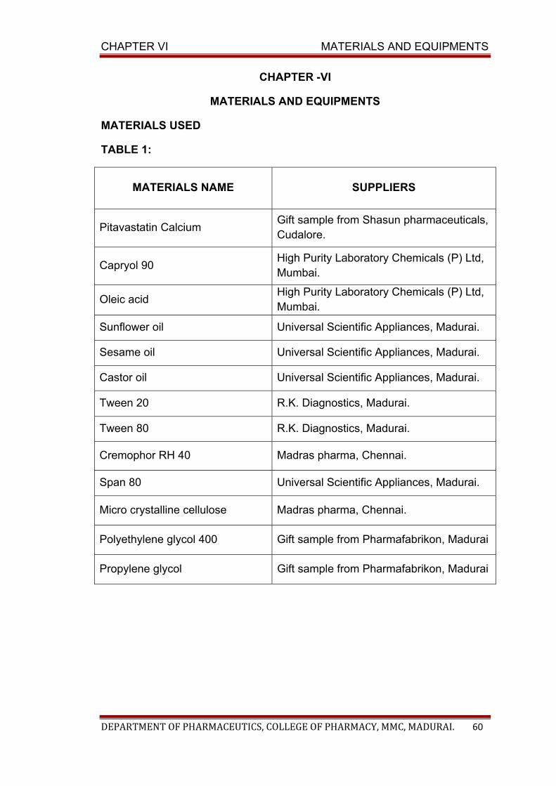
CHAPTER VI MATERIALS AND EQUIPMENTS
DEPARTMENT OF PHARMACEUTICS, COLLEGE OF PHARMACY, MMC, MADURAI. 60
CHAPTER -VI
MATERIALS AND EQUIPMENTS
MATERIALS USED
TABLE 1:
MATERIALS NAME SUPPLIERS
Pitavastatin Calcium Gift sample from Shasun pharmaceuticals, Cudalore.
Capryol 90 High Purity Laboratory Chemicals (P) Ltd, Mumbai.
Oleic acid High Purity Laboratory Chemicals (P) Ltd, Mumbai.
Sunflower oil Universal Scientific Appliances, Madurai.
Sesame oil Universal Scientific Appliances, Madurai.
Castor oil Universal Scientific Appliances, Madurai.
Tween 20 R.K. Diagnostics, Madurai.
Tween 80 R.K. Diagnostics, Madurai.
Cremophor RH 40 Madras pharma, Chennai.
Span 80 Universal Scientific Appliances, Madurai.
Micro crystalline cellulose Madras pharma, Chennai.
Polyethylene glycol 400 Gift sample from Pharmafabrikon, Madurai
Propylene glycol Gift sample from Pharmafabrikon, Madurai
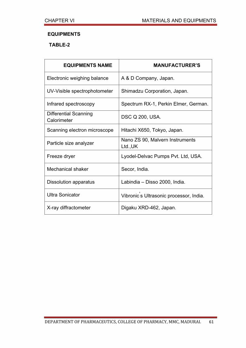
CHAPTER VI MATERIALS AND EQUIPMENTS
DEPARTMENT OF PHARMACEUTICS, COLLEGE OF PHARMACY, MMC, MADURAI. 61
EQUIPMENTS
TABLE-2
EQUIPMENTS NAME MANUFACTURER’S
Electronic weighing balance A & D Company, Japan.
UV-Visible spectrophotometer Shimadzu Corporation, Japan.
Infrared spectroscopy Spectrum RX-1, Perkin Elmer, German.
Differential Scanning Calorimeter
DSC Q 200, USA.
Scanning electron microscope Hitachi X650, Tokyo, Japan.
Particle size analyzer Nano ZS 90, Malvern Instruments Ltd.,UK
Freeze dryer Lyodel-Delvac Pumps Pvt. Ltd, USA.
Mechanical shaker Secor, India.
Dissolution apparatus Labindia – Disso 2000, India.
Ultra Sonicator Vibronic’s Ultrasonic processor, India.
X-ray diffractometer Digaku XRD-462, Japan.
CHAPTER VII
DRUG PROFILE
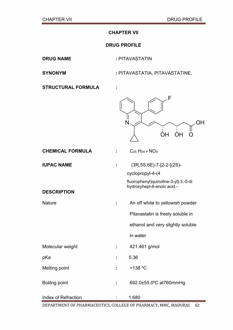
CHAPTER VII DRUG PROFILE
DEPARTMENT OF PHARMACEUTICS, COLLEGE OF PHARMACY, MMC, MADURAI. 62
CHAPTER VII
DRUG PROFILE
DRUG NAME : PITAVASTATIN
SYNONYM : PITAVASTATIA, PITAVASTATINE,
STRUCTURAL FORMULA :
CHEMICAL FORMULA : C25 H24 F NO4
IUPAC NAME : (3R,5S,6E)-7-[2-2-[(2S)-
cyclopropyl-4-(4
fluorophenyl)quinoline-3-yl]-3,-5-di hydroxyhept-6-enoic acid.-
DESCRIPTION Nature : An off white to yellowish powder Pitavastatin is freely soluble in
ethanol and very slightly soluble
in water
Molecular weight : 421.461 g/mol
pKa : 5.36
Melting point : >138 oC
Bolilng point : 692.0±55.0ºC at760mmHg
Index of Refraction : 1.680
CHAPTER VII DRUG PROFILE
DEPARTMENT OF PHARMACEUTICS, COLLEGE OF PHARMACY, MMC, MADURAI. 63
HISTORY OF PITAVASTATIN
Pitavastatin (previously known itavastatin, itabavastatin,
nisvastatin) was discovered in JAPAN by NISSAN CHEMICAL INDUSTRIES
and developed further by KOWA pharmaceuticals, TOKYO. Pitavastatin was
approved for use in the united states by the FDA on 08/03/2009 under the
trade name Livalo. Pitavastatin has been also approved by the Medicines and
Helathcare products Regulatory Agency (MHRA) in UK on August 2010.
(Reid,J; Reckless, J.P.D et al 2006)
PHARMACOLOGY
It is a calcium slat of Pitavastatin, a novel statin that induces plaque
regression and elevates HDL cholesterol level.
MECHANISM OF ACTION
Pitavastatin competitively inhibits HMG-CoA reductase, which is a
rate-determining enzyme involved with biosynthesis of cholesterol, in a
manner of competition with the substrate so that it inhibits cholesterol
synthesis in the liver. As a result, the expression of LDL-receptors followed
by the uptake of LDL from blood to liver is accelerated and then the plasma
TC decreases. Further, the sustained inhibition of cholesterol synthesis in
the liver decreases level of very low density lipoproteins.
PHARMACOKINETICS a) Absorption
Pitavastatin is rapidly absorbed from the upper gastrointestinal tract
and peak concentrations are achieved in one hour after oral administration.
CHAPTER VII DRUG PROFILE
DEPARTMENT OF PHARMACEUTICS, COLLEGE OF PHARMACY, MMC, MADURAI. 64
Cmax is decreased by 43%, if the drug is taken with a fatty meal. Unchanged
drug undergoes enterohepatic circulation and is well absorbed from the
jejunum and ileum. The absolute bioavailability of Pitavastatin is 51%.
b) Distribution Pitavastatin is more than 99% protein bound in human plasma,
mainly to albumin and alpha 1-acid glycoprotein, and the mean volume of
distribution is approximately 148 L. Pitavastatin is actively transported into
hepatocytes, the site of action and metabolism, by multiple hepatic
transporters including OATP1B1 and OATP1B3. Plasma AUC is variable
with an approximately 4-fold range between the highest and lowest values.
Studies with SLCO1B1 (the gene which encodes OATP1B1) suggests that
polymorphism of this gene could account for much of the variability in AUC.
Pitavastatin is not a substrate for p-glycoprotein.
C) Volume of distribution Mean volume of distribution at steady-state of Pitavastatin is approximately 148 liters.
d) Protein binding:
>99%,primariliy to albumin and alpha 1-acid glycoprotein. e) Metabolism
Mainly by liver through glucuronidation. The principal metabolite is
the inactive lactone which is formed via an ester-type Pitavastatin
glucuronide conjugate by UDP glucuronosyltransferase (UGT1A3 and
2B7). In vitro studies, using 13 human cytochrome P450 (CYP) isoforms,
indicate that the metabolism of Pitavastatin by CYP is minimal; CYP2C9
(and to a lesser extent CYP2C8) is responsible for the metabolism of
Pitavastatin to minor metabolite.
CHAPTER VII DRUG PROFILE
DEPARTMENT OF PHARMACEUTICS, COLLEGE OF PHARMACY, MMC, MADURAI. 65
f) Excretion Unchanged Pitavastatin is rapidly cleared from the liver in the bile,
but undergoes enterohepatic recirculation, contributing to its duration of
action. The plasma elimination half-life ranges from 5.7 hours (single dose)
to 8.9 hours (steady state) and clearance is 23.6 liter/hour.
g) Route of elimination
79% feces
15% urine
DOSAGE FORMS : film coated tablet of varying concentration are available. Tablet oral 1 mg
Tablet oral 1.045 mg
Tablet oral 2 mg
Tablet oral 2.09 mg
Tablet oral 4 mg
Tablet oral 4.18 mg
ADVERSE REACTIONS
Rhabdomyolysis with myoglobinuria and acute renal failure and
myopathy(including myosistis).
CONTRAINDICATIONS
Hypersenstivity to Pitavastatin or any component of the formulation;
active liver disease;pregnancy, lactation,co-admistered with cyclosporine.
DRUG INTERACTIONS
a) Atorvastatin
The serum concentration of Pitavastatin can be increased when it is
combined with atorvastatin

CHAPTER VII DRUG PROFILE
DEPARTMENT OF PHARMACEUTICS, COLLEGE OF PHARMACY, MMC, MADURAI. 66
b) Benzocaine
The serum concentration of Pitavastatin can be increased when it is
combined with benzocaine.
c) Caffeine
The serum concentration of Pitavastatin can be increased when it is
combined with caffeine.
d)cyclosporine
The serum concentratin of Pitavastatin can be decreased when it is
combined with cyclosporine.
e) Lopinavir/Ritonavir
The metabolisam of Pitavastatin can be decreased when combine with lopinavir.
f) Erythromycin
Erythromycin significantly increased Pitavastatin exposure. In
patients taking erythromycin, a dose of Pitavastatin 1mg once daily should not
be exceeded.
g) Rifampin
Rifampin significantly increased pitavstatin exposure.In patients taking
rifampin, a dose of Pitavastatin once daily should not be exceeded.
CHAPTER VII DRUG PROFILE
DEPARTMENT OF PHARMACEUTICS, COLLEGE OF PHARMACY, MMC, MADURAI. 67
h) Fibrates
It is known that the risk of myopathy during treatment with HMG-CoA
reductase inhibitors may be increased with concurrent administration of
fibtrates, pitavstatin should be administrated with caution when used
concomitantly with gemfibrozil or other fibrates.
i) Lovastatin
The serum concentration of pitavstatin can be increased when it is
combine with Lovastatin.
j) Niacin
The risk of skeletal muscle effects may be enhanced when Pitavastatin
is used in combination with niacin;a reduction in Pitavastatin dosage should
be considered in this setting.
k) Warfarin
Pitavastatin had no significant pharmacokinetic interaction with R-
ans S- warfarin. Pitavstatin had no significant effcet on prothrombin
time(PT) and international normalized ratio when administered to patients
receiving chronic warfarin. However, patients receiving warfarin should
have their PT and INR monitored when Pitavastatin is added to their
theraphy.
FOOD INTERACTION
Do not take with red yeast rice. It may increase the risk of myopathy of
Pitavastatin via pharmacokinetic synergism. Red yeast rice contain
monocolin k. (similar to lovastatin)
CHAPTER VII DRUG PROFILE
DEPARTMENT OF PHARMACEUTICS, COLLEGE OF PHARMACY, MMC, MADURAI. 68
OVER DOSE
There is no known specific treatment in the event of over dose of
Pitavastatin.in the event of over dose, the patient should be treated
symptomatically and supportive measures instituted as required. Hemolysis
is unlikely to be of benefit due to high protein binding ratio of Pitavastatin.
BRAND NAME
Livazo(coated tablets) Livalo (flim coatessd tablets)
CHAPTER VIII
EXCIPIENTS PROFILE

CHAPTER VIII EXCIPIENTS PROFILE
DEPARTMENT OF PHARMACEUTICS, COLLEGE OF PHARMACY, MMC, MADURAI. 69
CHAPTER –VIII
EXCIPIENTS PROFILE
1. OLEIC ACID
SYNONYM:
1. 9 Octadecenoic Acid
2. 9-Octadecenoic Acid
3. cis 9 Octadecenoic Acid
4. cis-9-Octadecenoic Acid
5. Oleate
6. Oleic Acid
STRUCTURE:
EMPIRICAL FORMULA : C18H34O2
MOLECULAR WEIGHT : 282.47g/mol
DESCRIPTION:
• Colour : colourless to pale yellowish
• Odour : Odourless
• Taste : Peculiar lard-like taste
• Solubility : Insoluble in water; soluble in ethanol, ether, acetone,
chloroform, dimethyl formamide and dimethyl sulfoxide
• Meltilng point : 13 to 14oC
• Viscosity : 25.6 cp at 30oC
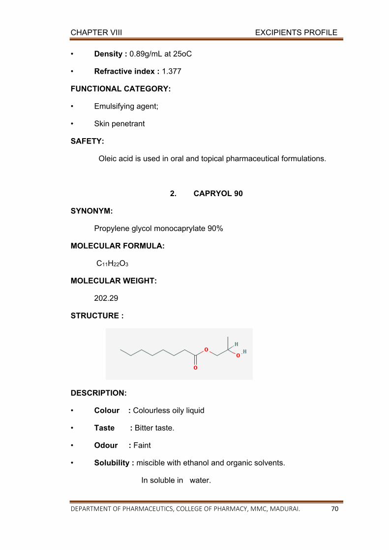
CHAPTER VIII EXCIPIENTS PROFILE
DEPARTMENT OF PHARMACEUTICS, COLLEGE OF PHARMACY, MMC, MADURAI. 70
• Density : 0.89g/mL at 25oC
• Refractive index : 1.377
FUNCTIONAL CATEGORY:
• Emulsifying agent;
• Skin penetrant
SAFETY:
Oleic acid is used in oral and topical pharmaceutical formulations.
2. CAPRYOL 90
SYNONYM:
Propylene glycol monocaprylate 90%
MOLECULAR FORMULA:
C11H22O3
MOLECULAR WEIGHT:
202.29
STRUCTURE :
DESCRIPTION:
• Colour : Colourless oily liquid
• Taste : Bitter taste.
• Odour : Faint
• Solubility : miscible with ethanol and organic solvents.
In soluble in water.
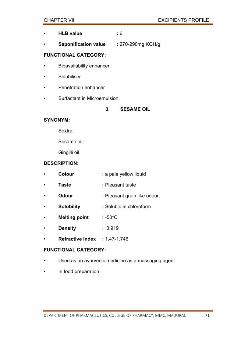
CHAPTER VIII EXCIPIENTS PROFILE
DEPARTMENT OF PHARMACEUTICS, COLLEGE OF PHARMACY, MMC, MADURAI. 71
• HLB value : 6
• Saponification value : 270-290mg KOH/g
FUNCTIONAL CATEGORY:
• Bioavailability enhancer
• Solubiliser
• Penetration enhancer
• Surfactant in Microemulsion.
3. SESAME OIL
SYNONYM:
Sextra;
Sesame oil,
Gingilli oil.
DESCRIPTION:
• Colour : a pale yellow liquid
• Taste : Pleasant taste
• Odour : Pleasant grain like odour.
• Solubility : Soluble in chloroform
• Melting point : -50oC
• Density : 0.919
• Refractive index : 1.47-1.746
FUNCTIONAL CATEGORY:
• Used as an ayurvedic medicine as a massaging agent
• In food preparation.
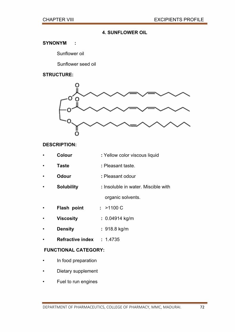
CHAPTER VIII EXCIPIENTS PROFILE
DEPARTMENT OF PHARMACEUTICS, COLLEGE OF PHARMACY, MMC, MADURAI. 72
4. SUNFLOWER OIL
SYNONYM :
Sunflower oil
Sunflower seed oil
STRUCTURE:
DESCRIPTION:
• Colour : Yellow color viscous liquid
• Taste : Pleasant taste.
• Odour : Pleasant odour
• Solubility : Insoluble in water. Miscible with
organic solvents.
• Flash point : >1100 C
• Viscosity : 0.04914 kg/m
• Density : 918.8 kg/m
• Refractive index : 1.4735
FUNCTIONAL CATEGORY:
• In food preparation
• Dietary supplement
• Fuel to run engines
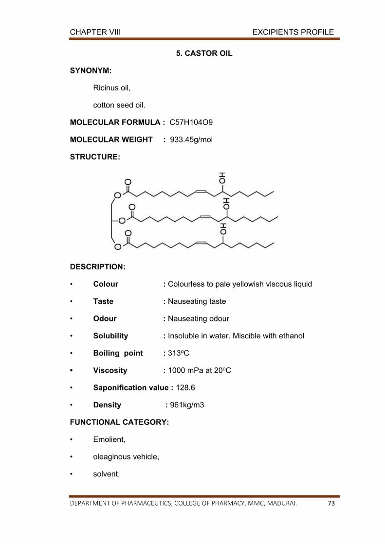
CHAPTER VIII EXCIPIENTS PROFILE
DEPARTMENT OF PHARMACEUTICS, COLLEGE OF PHARMACY, MMC, MADURAI. 73
5. CASTOR OIL
SYNONYM:
Ricinus oil,
cotton seed oil.
MOLECULAR FORMULA : C57H104O9
MOLECULAR WEIGHT : 933.45g/mol
STRUCTURE:
DESCRIPTION:
• Colour : Colourless to pale yellowish viscous liquid
• Taste : Nauseating taste
• Odour : Nauseating odour
• Solubility : Insoluble in water. Miscible with ethanol
• Boiling point : 313oC
• Viscosity : 1000 mPa at 20oC
• Saponification value : 128.6
• Density : 961kg/m3
FUNCTIONAL CATEGORY:
• Emolient,
• oleaginous vehicle,
• solvent.
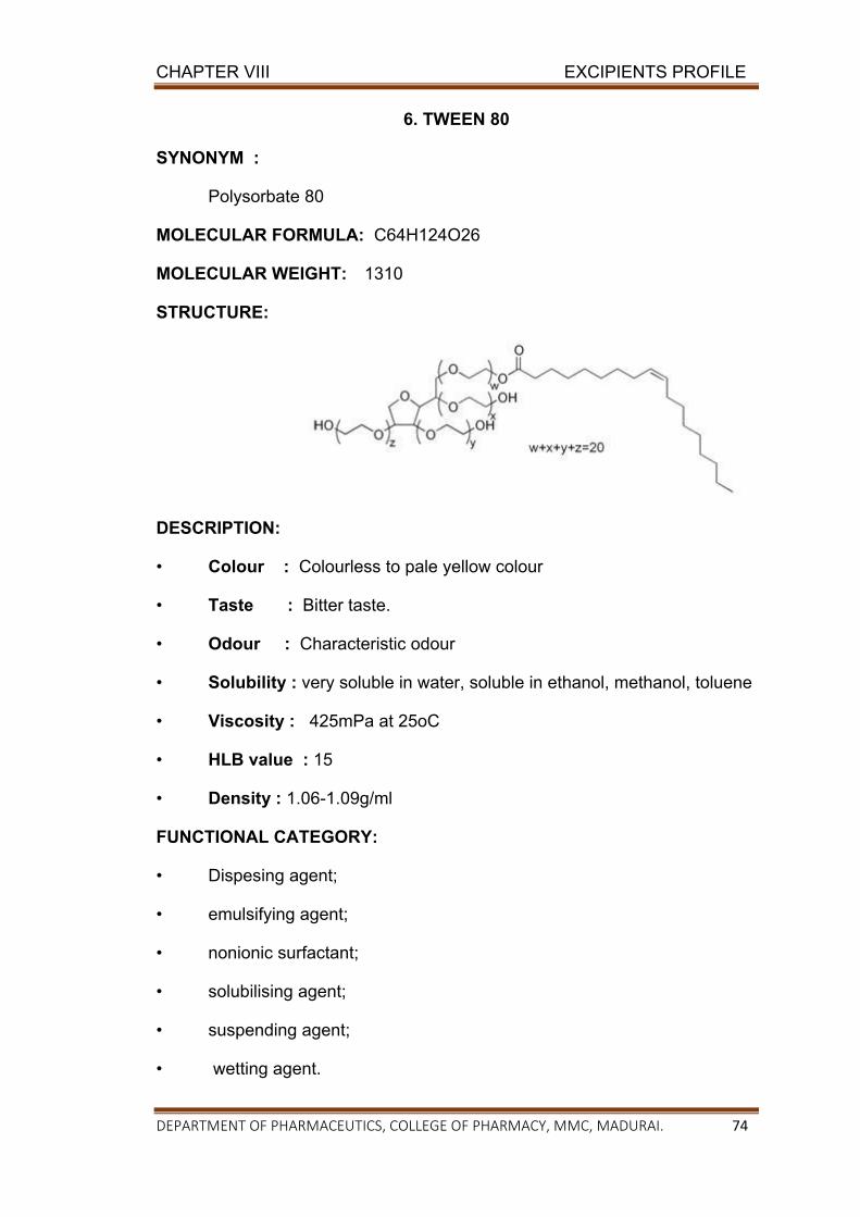
CHAPTER VIII EXCIPIENTS PROFILE
DEPARTMENT OF PHARMACEUTICS, COLLEGE OF PHARMACY, MMC, MADURAI. 74
6. TWEEN 80
SYNONYM :
Polysorbate 80
MOLECULAR FORMULA: C64H124O26
MOLECULAR WEIGHT: 1310
STRUCTURE:
DESCRIPTION:
• Colour : Colourless to pale yellow colour
• Taste : Bitter taste.
• Odour : Characteristic odour
• Solubility : very soluble in water, soluble in ethanol, methanol, toluene
• Viscosity : 425mPa at 25oC
• HLB value : 15
• Density : 1.06-1.09g/ml
FUNCTIONAL CATEGORY:
• Dispesing agent;
• emulsifying agent;
• nonionic surfactant;
• solubilising agent;
• suspending agent;
• wetting agent.
CHAPTER VIII EXCIPIENTS PROFILE
DEPARTMENT OF PHARMACEUTICS, COLLEGE OF PHARMACY, MMC, MADURAI. 75
7. CREMOPHOR RH 40
Chemical name : Polyoxyl 40 hydrogenated
• Colour : White to yellowish thin paste.
• Taste : Very faint odour
• Odour : Very faint taste
• Solubility : Soluble in water, ethanol, 2-propanol, n-propanol,
ethyl acetate chloroform, carbon tetra chloride, and
toluene.
• HLB value : 14-16
• Saponification value : 50-60
METHOD OF MANUFACTURING: Reacting hydrogenated castor oil
with ethylene oxide.
FUNCTIONAL CATEGORY:
Solubilizers,
In perfume compositions.
8. POLYETHYLENE GLYCOL 400
SYNONYM:
Carbowax;
Carbowax Sentry;
Lipoxol;
Lutrol E;
macrogola;
PEG;
Pluriol E and
polyoxyethylene glycol.
CHAPTER VIII EXCIPIENTS PROFILE
DEPARTMENT OF PHARMACEUTICS, COLLEGE OF PHARMACY, MMC, MADURAI. 76
MOLECULAR FORMULA: HOCH2 (CH2OCH2)8.7CH2OH
MOLECULAR WEIGHT: 380-420 g/mol
DESCRIPTION:
• Colour : Colorless liquid
• Taste : Bitter
• Odour : Charaticcteric
• Solubility : Soluble in water, Acetone, Alcohols, Benzene, Glycerin.
• Viscosity : 105-130 mPas
• HLB value : 16
• Density : 1.120
INCOMPATIBILITIES:
The chemical reactivity of polyethylene glycols is mainly confined to the
two terminal hydroxyl groups, which can be either esterified or etherified.
However, all grades can exhibit some oxidizing activity owing to the presence
of peroxide impurities and secondary products formed by autoxidation. Liquid
and solid polyethylene glycol grades may be incompatible with some coloring
agents.
STABILITY AND STORAGE:
Polyethylene glycols are chemically stable in air and in solution, although
grades with a molecular weight less than 2000 are hygroscopic. Polyethylene
glycols do not support microbial growth, and they do not become rancid.
Polyethylene glycols and aqueous polyethylene glycol solutions can be
sterilized by autoclaving, filtration, or gamma irradiation.
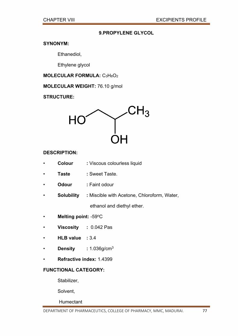
CHAPTER VIII EXCIPIENTS PROFILE
DEPARTMENT OF PHARMACEUTICS, COLLEGE OF PHARMACY, MMC, MADURAI. 77
9.PROPYLENE GLYCOL
SYNONYM:
Ethanediol,
Ethylene glycol
MOLECULAR FORMULA: C3H8O2
MOLECULAR WEIGHT: 76.10 g/mol
STRUCTURE:
DESCRIPTION:
• Colour : Viscous colourless liquid
• Taste : Sweet Taste.
• Odour : Faint odour
• Solubility : Miscible with Acetone, Chloroform, Water,
ethanol and diethyl ether.
• Melting point: -59oC
• Viscosity : 0.042 Pas
• HLB value : 3.4
• Density : 1.036g/cm3
• Refractive index: 1.4399
FUNCTIONAL CATEGORY:
Stabilizer,
Solvent,
Humectant
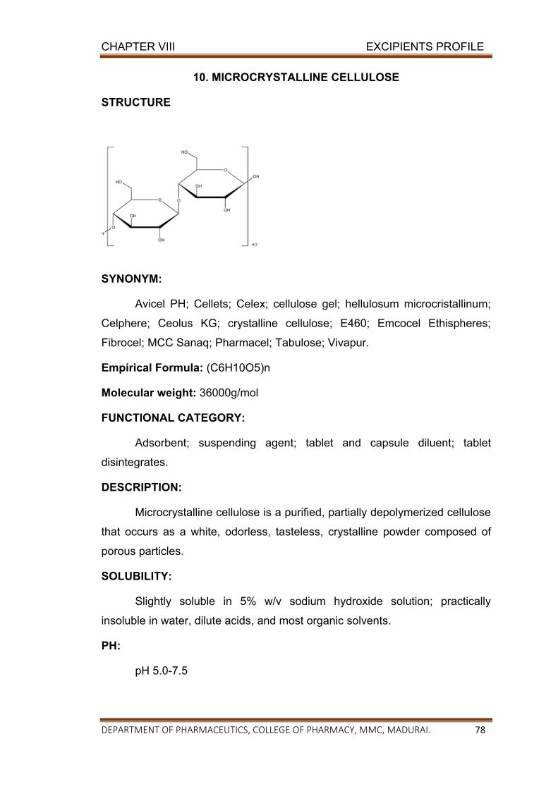
CHAPTER VIII EXCIPIENTS PROFILE
DEPARTMENT OF PHARMACEUTICS, COLLEGE OF PHARMACY, MMC, MADURAI. 78
10. MICROCRYSTALLINE CELLULOSE
STRUCTURE
SYNONYM:
Avicel PH; Cellets; Celex; cellulose gel; hellulosum microcristallinum;
Celphere; Ceolus KG; crystalline cellulose; E460; Emcocel Ethispheres;
Fibrocel; MCC Sanaq; Pharmacel; Tabulose; Vivapur.
Empirical Formula: (C6H10O5)n
Molecular weight: 36000g/mol
FUNCTIONAL CATEGORY:
Adsorbent; suspending agent; tablet and capsule diluent; tablet
disintegrates.
DESCRIPTION:
Microcrystalline cellulose is a purified, partially depolymerized cellulose
that occurs as a white, odorless, tasteless, crystalline powder composed of
porous particles.
SOLUBILITY:
Slightly soluble in 5% w/v sodium hydroxide solution; practically
insoluble in water, dilute acids, and most organic solvents.
PH:
pH 5.0-7.5
CHAPTER VIII EXCIPIENTS PROFILE
DEPARTMENT OF PHARMACEUTICS, COLLEGE OF PHARMACY, MMC, MADURAI. 79
MELTING POINT:
Chars at 260–2708C.
INCOMPATIBILITIES:
Microcrystalline cellulose is incompatible with strong oxidizing agents.
STABILITY & STORAGE CONDITION:
Microcrystalline cellulose is a stable though hygroscopic material. The
bulk material should be stored in a well-closed container in a cool, dry place.
HANDLING PRECAUTION:
Microcrystalline cellulose may be irritant to the eyes. Gloves, eye
protection, and a dust mask are recommended
APPLICATIONS:
1. Microcrystalline cellulose is widely used in pharmaceuticals, primarily as
a binder/diluent in oral tablet and capsule formulations where it is used
in both wet-granulation and direct-compression Processes.
2. In addition to its use as a binder/diluent, microcrystalline cellulose also
has some lubricant and disintegrates properties that make it useful in
tableting.
3. Microcrystalline cellulose is also used in cosmetics and food products;
CHAPTER IX
EXPERIMENTAL PROTOCOL
CHAPTER IX EXPERIMENTAL PROTOCAL
DEPARTMENT OF PHARMACEUTICS, COLLEGE OF PHARMACY, MMC, MADURAI. 80
CHAPTER IX
EXPERIMENTAL PROTOCAL
Preformulation study:
Preformulation study is the process of optimizing the delivery of drug
through determination of physicochemical properties of the new compound
that could affect drug performance and development of an efficacious, stable
and safe dosage forms, first preformulation studies were performed. Various
preformulation studies which were carried out are discussed in following
sections.
IDENTIFICATION:
1. Determination of melting point:
Melting point of Pitavastatin was determined by using open capillary tube
method in digital melting ploint apparatus.
Method:
In this method the capillary tube was sealed with gentle heating from
one end. Then the small quantity of Pitavastatin was filled into the sealed
capillary. Capillary existed tied to the tube containing the oil phase in such a
way that the sealed part of the capillary containing Pitavastatin was dipped
into the oil. Gently the oil bath was heated. As soon as the powder starts
melting, the heating was stopped and the temperature was note down.
2. Solubility of Pitavastatin:
Solubility of Pitavastatin was determined in various medias like ethanol,
methanol, Dichloromethane, Dimethylsulfoxide, 6.8 phosphate buffer etc.
3. Drug-Excipients Compatibility studies :
A proper design and formulation of dosage form require consideration
of physical, chemical and biological characteristics of both drug and excipients
CHAPTER IX EXPERIMENTAL PROTOCAL
DEPARTMENT OF PHARMACEUTICS, COLLEGE OF PHARMACY, MMC, MADURAI. 81
used in formulation of the product. Compatibility must be established between
active drug and other excipients to produce a stable, efficacious, attractive
and safe product. Hence before producing the actual formulation compatibility
of drug with different excipients was tested using the FTIR and DSC
techniques.
Fourier Transmittance Infra-Red (FTIR):
In order to check the integrity (compatibility) of the drug in the
formulations FTIR spectra of formulations along with the pure drug and other
excipients were obtained and compared using shimadzu FTIR
spectrophotometer. In the present study potassium bromide (KBr) pellet
method was employed. The samples were thoroughly blended with dry
powdered KBr crystals. The mixture was compressed to form a disc. The disc
was placed in spectrophotometer and spectrum was recorded.
Differential scanning Calorimetry (DSC):
Differential scanning calorimeter or DSC is a thermo analytical
technique in which difference in the amount of heat required to increase the
temperature of a sample and reference are measured as a function of
temperature. Both the sample and reference mentioned at nearly sample
temperature throughout the experiment.
Method:
Accurately weighed Pitavastatin was analyzed by using an automatic
thermal analyzer system (DSC60 Shimadzu Corporation, Japan0. Sealed and
perforated aluminium pans were used in this experiment for all the samples.
Temperature calibrations were performed using indium as standard. An empty
pan sealed in the same way as for the sample was used as a reference. The
entire samples were run at a scanning rate of 10oC/min from 50o-300oC.

CHAPTER IX EXPERIMENTAL PROTOCAL
DEPARTMENT OF PHARMACEUTICS, COLLEGE OF PHARMACY, MMC, MADURAI. 82
4. STANDARD CURVES FOR PITAVASTATIN
Phosphate buffer pH 6.8 ( Indian pharmacopoeia 2017)
A known volume (50 ml) of 0.2 M potassium dihydrogen phosphate is
placed in a 200 ml volumetric flask. 22.4 ml of 0.2M sodium hydroxide is
added and makeup to the volume with distilled water.
0.2M potassium di hydrogen phosphate:
A known quantity (27.218g) of potassium di hydrogen phosphate is
dissolved and diluted to 1000 ml with water.
0.2 M sodium hydroxide:
A known quantity (8 g) of sodium hydroxide is dissolved and makeup to
1000 ml with water.
Determination of λmax :
Standard stock solution containing Pitavastatin was prepared by
dissolving 100 mg of Pitavastatin in 10 ml of Dimethyl sulphoxide in 100 ml
volumetric flask to dissolve the drug. Then the volume was made up to 100 ml
using phosphate buffer of pH 6.8 to obtain a concentration of 100μg/ml. the
stock solution is further diluted using a phosphate buffer pH (6.8) to prepare
10 μg/ml concentration. The resultant solution was scanned in the range of
200-400 nm in UV spectrophotometer (UV -1700 shimadzu Corporation,
Japan) to get absorption maximum (λmax) using phosphate buffer as blank.
The wave length of maximum absorbance considered for further studies.
Preparation of standard curves:
From the above prepared stock solution, solutions containing 2 to
10 μg/ml concentrations were prepared using the phosphate buffer pH 6.8
solution. The absorbance of these solutions are measured at λmax by
UV-spectophotometer (UV-1700Sshimadzu corporation Japan). A standard
curve is plotted using concentration on x-axis and the absorbance obtained on
Y-axis.
CHAPTER IX EXPERIMENTAL PROTOCAL
DEPARTMENT OF PHARMACEUTICS, COLLEGE OF PHARMACY, MMC, MADURAI. 83
5. FORMULATION OF PITAVASTATIN SELF MICRO EMULSION:
The SMEDDS of Pitavastatin were prepared by simple admixing
method. The preparation process involves following steps.
I. Determination of solubility of Pitavastatin in various vehicles.(oil,
surfactant, and co-surfactant.
II. Selection of oil, surfactant and co-surfactant based on the
solubility and % transmittance.
III. Construction of pseudo ternary phase diagram.
IV. Formulation of SMEDDS by dissolving the drug in a mixture of
oil, surfactant and co-surfactant.
Determination of solubility of Pitavastatin:
Solubility of Pitavastatin in various oils (Capryol 90, oleic acid, castor
oil, sesame oil, castor oil), surfactants (tween 20, tween 60, tween 80,
cremophor RH, span 80), co-surfactant (Propylene glycol, poly ethylene glycol
400) was determined by dissolving an excess amount of Pitavastatin in 2 ml
of each of selected oils, surfactant and co-surfactant in stoppered vials. The
mixtures were continuously stirred using vortex mixer for 10 min and kept at
37o C±0.5o C in water bath shaker for 78 hours to attain equilibrium. The
equilibrated samples were centrifuged (3000 rpm for 15 mins) and
supernatant was filtered through 0.45 μm membrane filter and diluted with
suitable solvent. Drug content was quantified by using ultraviolet - visible
(UV_VIS) spectrophotometer.
Screening of surfactant:
In order to find appropriate surfactant with good solubilising capacity,
an emulsifying ability of different surfactants (tween 20, tween 60, tween 80,
cremophore RH, and span 80) with the screened oil was investigated. 300 mg
of oil phase and 300 mg of surfactant were weighed and vortexed for two
minutes followed by warming at 40- 45 C for 30 seconds. So we can obtain an
isotropic mixture. 50 mg of isotropic mixture was taken and diluted with double
distilled water previously filtered through 0.45µm membrane filter in a
CHAPTER IX EXPERIMENTAL PROTOCAL
DEPARTMENT OF PHARMACEUTICS, COLLEGE OF PHARMACY, MMC, MADURAI. 84
volumetric flask. Number of flask inversions was observed visually to form a
clear emulsion. The resulting emulsions allowed standing for 2 hours after that
transmittance were observed at 638 nm. The surfactant which forms a clear
emulsion with lesser number of inversions and with more transmittance was
selected.
Screening of co-surfactant:
In order to find appropriate co- surfactant with good solubilising
capacity, after screening of an oil emulsifying ability of different co- surfactants
(Propylene glycol, poly ethylene glycol 400) with the screened oil was
investigated. 300 mg of oil phase and 300 mg of surfactant were weighed and
vortexed for two minutes followed by warming at 40- 45 C for 30 seconds. So
we can obtain an isotropic mixture. 50 mg of isotropic mixture was taken and
diluted with double distilled water previously filtered through 0.45µm
membrane filter in a volumetric flask. Number of flask inversions was
observed visually to form a clear emulsion. The resulting emulsions allowed
standing for 2 hours after that transmittance were observed at 638 nm. The
co- surfactant which forms a clear emulsion with lesser number of inversions
and with more transmittance was selected.
Construction of pseudo ternary phase diagram:
Phase diagrams are constructed to obtain the proportion of
components that can result in maximum microemulsion existence area. These
diagrams were constructed with oil, surfactant/co-surfactant and water using
water titration method at room temperature. The procedure consisted of
preparing solutions of different ratio of surfactant to co-surfactant by weight
such as 1:1, 2:1, 3:1, etc, these solutions then vortexed for 5 mins and placed
at 50o C for one hour, so that an isotropic mixture was obtained. Each of these
solutions were used for preparing a mixture containing oil and Smix (mixture of
surfactant and co-surfactant) in the following ratios by weight, 1:9, 2:8, 3:7,
4:6, 5:5, 6:4, 7:3, 8:2, 9:1 and after preparation vortexed for 5 mins followed
by placing in oven at 50o C for one hour. All the mixtures were then placed at
room temperature for 24 hour. Water from 5 % to 95 % of the mixtures was
CHAPTER IX EXPERIMENTAL PROTOCAL
DEPARTMENT OF PHARMACEUTICS, COLLEGE OF PHARMACY, MMC, MADURAI. 85
observed for their appearance (turbid or clear). Turbidity of the samples would
indicate formation of a coarse emulsion, whereas clear isotropic solution
would indicate the formation of micro emulsion. Percentage of oil, Smix, water
at which clear mixture was formed was selected and the values were used to
construct ternary phase diagram.
FORMULATION OF LIQUID SMEDDS:
From the ternary phase diagram ratio of surfactant to co-surfactant
was optimized. Then by varying ratio of oil to Smix, different formulations were
prepared. Formulations were prepared by preparing optimized ratio of Smix
first, for this surfactant and co-surfactant were accurately weighed and then
vortexed for 5-10 mints. After that Smix was placed in oven at 500 C for one
hour. Oil with different ratio was added to Smix. Then these formulations were
vortexed for 5-10 mints and placed in oven at 50o C for one hour so that an
isotropic mixture was formed. Drug was loaded to these isotropic mixtures at
the end and vortexed by vortex shaker until clear solution was obtained.
CHARACTERIZATION OF LIQUID SMEDDS:
Visual assessment
Pitavastatin liquid SMEDDS was diluted with purified water (100 ml)
and gently stirred with magnetic stirrer. Temperature should be 370 C.
Dispersibility test
The dispersibility test of SMEDDS was carried out to acssess to
compatibility to disperse into emulsion and the size of resulting globules to
categorize them as SMEDDS. It was carried by using a standard USP paddle
typw dissolution test apparatus, formulation was added to 500 ml of water at
37 0.5 C and the paddle was rotated at 50 rpm. On titration with water the
SMEDDS formulation forms a mixture which was of different type. Depending
upon which the in vitro performance of formulation can be assessed.
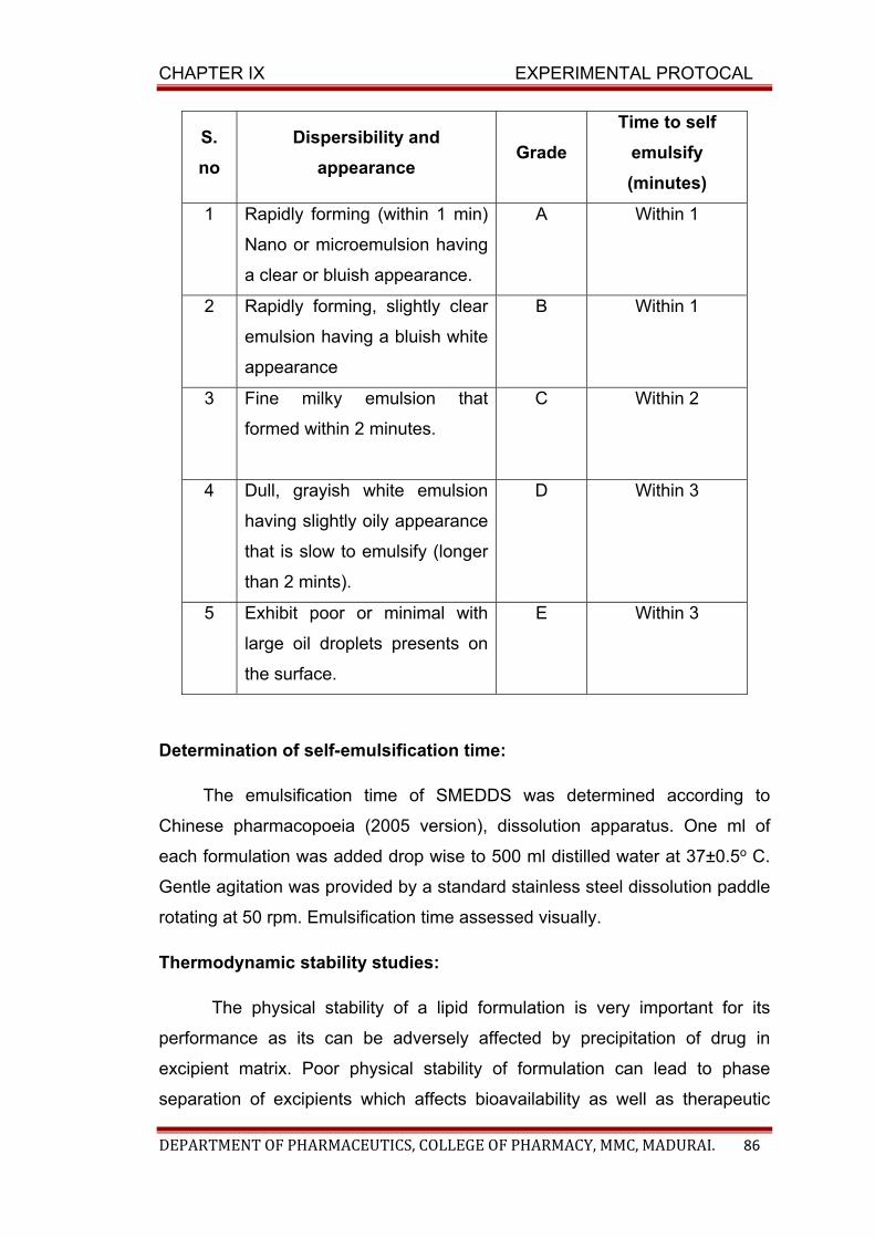
CHAPTER IX EXPERIMENTAL PROTOCAL
DEPARTMENT OF PHARMACEUTICS, COLLEGE OF PHARMACY, MMC, MADURAI. 86
S.
no
Dispersibility and
appearance Grade
Time to self
emulsify
(minutes)
1 Rapidly forming (within 1 min)
Nano or microemulsion having
a clear or bluish appearance.
A Within 1
2 Rapidly forming, slightly clear
emulsion having a bluish white
appearance
B Within 1
3 Fine milky emulsion that
formed within 2 minutes.
C Within 2
4 Dull, grayish white emulsion
having slightly oily appearance
that is slow to emulsify (longer
than 2 mints).
D Within 3
5 Exhibit poor or minimal with
large oil droplets presents on
the surface.
E Within 3
Determination of self-emulsification time:
The emulsification time of SMEDDS was determined according to
Chinese pharmacopoeia (2005 version), dissolution apparatus. One ml of
each formulation was added drop wise to 500 ml distilled water at 37±0.5o C.
Gentle agitation was provided by a standard stainless steel dissolution paddle
rotating at 50 rpm. Emulsification time assessed visually.
Thermodynamic stability studies:
The physical stability of a lipid formulation is very important for its
performance as its can be adversely affected by precipitation of drug in
excipient matrix. Poor physical stability of formulation can lead to phase
separation of excipients which affects bioavailability as well as therapeutic
CHAPTER IX EXPERIMENTAL PROTOCAL
DEPARTMENT OF PHARMACEUTICS, COLLEGE OF PHARMACY, MMC, MADURAI. 87
efficiency. Also the incompatibilities between formulation and shell of capsule
may cause brittleness, softness, and deleted disintegration or incomplete
release of drug. The following cycles was carried out for these studies.
Heating cooling cycle
The optimized SMEDDS formulations were diluted with 100 times
distilled water. Six cycles between cooling temperature (4oC) and heating
temperature (45o C) with exposure at each temperature for not less than 48
hours were carried. That formulation, which was stable, then was subjected to
centrifugation test.
Centrifugation
In order to estimate metastable systems, the optimized SMEDDS
formulations were diluted with 100 times distilled water. Which pass heating-
cooling cycles are centrifuged at 3500 rpm for 30 mints. Those formulations
that does not show any phase separation are taken for the freeze thaw stress
test.
Freeze thaw cycle
This test was performed for accelerated stability testing of Nano
emulsion formulation. In this study three freez thaw cycle of formulations were
exposed between temperatures 21o C-25o C for each temperature cycles not
more than 48 hours. For the better estimation of accelerated stability studies
six such cycles should be run for each batch of formulation. The formulations
which showed the maximum stability were selected for further study.
Cloud point measurement:
Dilute the formulation 1 ml with 1000 ml of water in beaker and placed on a
water bath with gradually increasing the temperature until the diluted
formulation turned to cloudy or turbid. It gives the information about the
stability of the micro emulsion at body temperature.
CHAPTER IX EXPERIMENTAL PROTOCAL
DEPARTMENT OF PHARMACEUTICS, COLLEGE OF PHARMACY, MMC, MADURAI. 88
Robustness to dilution:
Robustness to dilution was studied by diluting SMEDDS to 50, 100,
1000 times with water, phosphate buffer pH 6.8, phosphate buffer pH 7.4. The
diluted SMEDDS were stored for 12 hour and observed for any signs of phase
separation or drug precipitation.
Refractive index and percent transmittance:
Refractive index and percent transmittance proved the transparency of
formulation. The refractive index measured using Abbes refractometer. The
percent transmittance of the system is measured at particular wavelength
using UV-spectrophotometer keeping distilled water as blank. Stability of
micro emulsion formulation with respect to dilution is checked by diluting one
ml of formulation with 100 ml of distilled water and measuring transmittance
using U.V. Spectrophotometer. Transmittance of samples is measured at 638
nm and for each sample three replicate assays are performed.
Viscosity determination:
The rheological properties of the micro emulsion are evaluated by
Ostwald visco meter. These viscosity determinations confirm whether the
system is w\o or o\w. If system has low viscosity, then it is o/w type of the
system and if high viscosities then it are w\o type of the system.
Absolute drug content in L-SMEDDS:
Liquid SMEDDS containing Pitavastatin, equivalent to 4 mg was diluted
in suitable quantity of methanol. The sample was mixed thoroughly to dissolve
the drug in methanol by stirring. The solvent extract is filtered through 0.45 µm
membrane filter. Drug content was determined by measuring the absorbance
in UV spectro photometer against the standard solvent solution of drug.
Conversion of liquid SMEDDS into solid S-SMEDDS:
Solid SMEDDS were prepared by mixing liquid SMEDDS containing
Pitavastatin with microcrystalline cellulose (MCC) in 1:1 proportion. Liquid
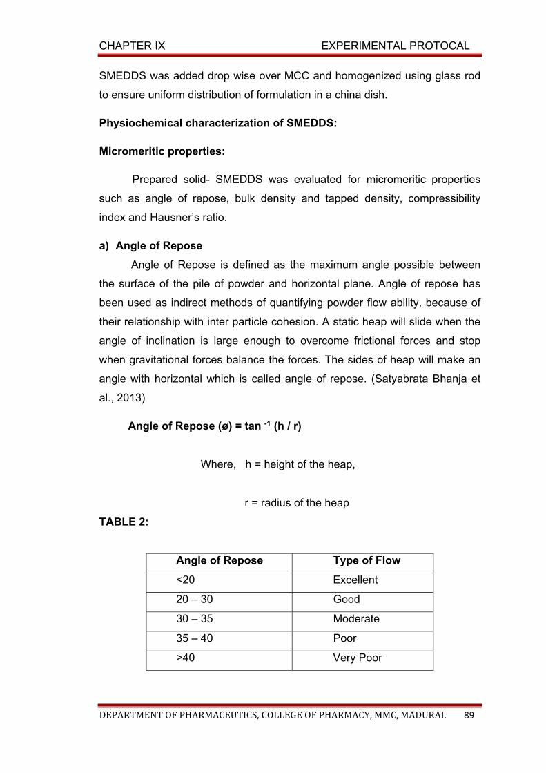
CHAPTER IX EXPERIMENTAL PROTOCAL
DEPARTMENT OF PHARMACEUTICS, COLLEGE OF PHARMACY, MMC, MADURAI. 89
SMEDDS was added drop wise over MCC and homogenized using glass rod
to ensure uniform distribution of formulation in a china dish.
Physiochemical characterization of SMEDDS:
Micromeritic properties:
Prepared solid- SMEDDS was evaluated for micromeritic properties
such as angle of repose, bulk density and tapped density, compressibility
index and Hausner’s ratio.
a) Angle of Repose
Angle of Repose is defined as the maximum angle possible between
the surface of the pile of powder and horizontal plane. Angle of repose has
been used as indirect methods of quantifying powder flow ability, because of
their relationship with inter particle cohesion. A static heap will slide when the
angle of inclination is large enough to overcome frictional forces and stop
when gravitational forces balance the forces. The sides of heap will make an
angle with horizontal which is called angle of repose. (Satyabrata Bhanja et
al., 2013)
Angle of Repose (ø) = tan -1 (h / r)
Where, h = height of the heap,
r = radius of the heap
TABLE 2:
Angle of Repose Type of Flow
<20 Excellent
20 – 30 Good
30 – 35 Moderate
35 – 40 Poor
>40 Very Poor
CHAPTER IX EXPERIMENTAL PROTOCAL
DEPARTMENT OF PHARMACEUTICS, COLLEGE OF PHARMACY, MMC, MADURAI. 90
b) Bulk Density:
Bulk density denotes the total density of the material. It includes the
true volume of interparticle spaces and intraparticle pores. The packing of
particles is mainly responsible for bulk. Bulk density is the ratio between a
given mass of powder and its bulk volume. Apparent bulk density is determined
by pouring the weighed granules into a graduated cylinder via funnel and
measuring the volume. Density is calculated by using the formula,
(SatyabrataBhanjaal.,2013 )
Bulk density = weight of the powder = W
Bulk volume of the powder Vo
c) Tapped Density:
A known quantity of sample is transferred to a graduated cylinder
and placed on tapped density apparatus and operated for a fixed number
of taps (100).It is the ratio of weight of sample to tapped
volume.(Satyabrata Bhanja et al., 2013)
Tapped density = Weight of the powder (W)
Tapped volume of powder Vf
d) Compressibility (or) Carr’s index:
Based on the apparent bulk density and the tapped density, the
percentage compressibility of the bulk drug is studied by using the
following formula.
Carr’s index (%) = Tapped density - bulk density × 100
Tapped density
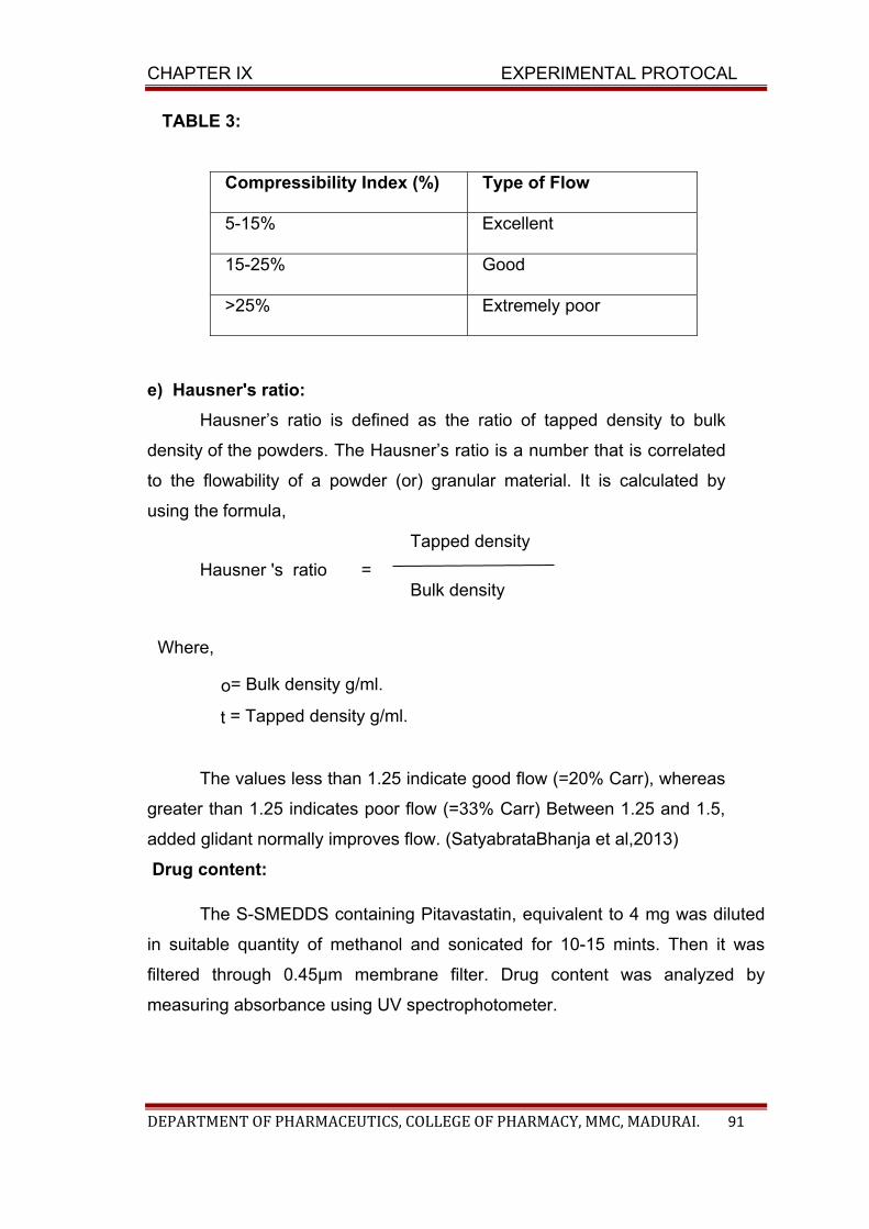
CHAPTER IX EXPERIMENTAL PROTOCAL
DEPARTMENT OF PHARMACEUTICS, COLLEGE OF PHARMACY, MMC, MADURAI. 91
TABLE 3:
Compressibility Index (%) Type of Flow
5-15% Excellent
15-25% Good
>25% Extremely poor
e) Hausner's ratio:
Hausner’s ratio is defined as the ratio of tapped density to bulk
density of the powders. The Hausner’s ratio is a number that is correlated
to the flowability of a powder (or) granular material. It is calculated by
using the formula,
Tapped density
Hausner 's ratio = Bulk density
Where,
o= Bulk density g/ml.
t = Tapped density g/ml.
The values less than 1.25 indicate good flow (=20% Carr), whereas
greater than 1.25 indicates poor flow (=33% Carr) Between 1.25 and 1.5,
added glidant normally improves flow. (SatyabrataBhanja et al,2013)
Drug content:
The S-SMEDDS containing Pitavastatin, equivalent to 4 mg was diluted
in suitable quantity of methanol and sonicated for 10-15 mints. Then it was
filtered through 0.45μm membrane filter. Drug content was analyzed by
measuring absorbance using UV spectrophotometer.
CHAPTER IX EXPERIMENTAL PROTOCAL
DEPARTMENT OF PHARMACEUTICS, COLLEGE OF PHARMACY, MMC, MADURAI. 92
Invitro drug release from S-SMEDDS:
The in vitro drug release of prepared S-SMEDDS was assessed in
triplicate using United States Pharmacopoeia (USP) Dissolution Type II
apparatus (Paddle type) at 37±0.5o C. S-SMEDDS containing 4 mg equivalent
of drug was placed in 900 ml of dissolution medium ( phosphate buffer pH
6.8with methanol in 9:1 ratio). The revolution speed of the paddle was
maintained at 100 rpm. At predetermined time intervals, 5 ml of dissolution
medium was collected, filtered and the same volume of fresh dissolution
medium was replenished to maintain the sink conditions. The samples were
analyzed for the drug concentration using UV-VIS spectrophotometer at 245
nm
f) Release Kinetics:
The results of in vitro release profiles obtained for all the formulations
were fitted into five models of data treatment as follows:
1. Cumulative percent drug released versus time (zero-order kinetic model).
2. Log cumulative percent drug remaining versus time. (First-order kinetic
model).
3. Cumulative percent drug released versus square root of time (Higuchi’s
model).
4. Log cumulative percent drug released versus log time (Korsmeyer-
Peppasequation).
5. Cumulative percent drug released versus cube root of time
(Hixson and Crowell’scubic root law of dissolution)
Zero Order Kinetics A zero-order releases would be predicted by the
following equation.
At = A0 – K0t…………………………………….(6)
Where:
At = Drug release at time‘t’
A0 = Initial drug concentration
K0 = Zero-order rate constant (hr)
CHAPTER IX EXPERIMENTAL PROTOCAL
DEPARTMENT OF PHARMACEUTICS, COLLEGE OF PHARMACY, MMC, MADURAI. 93
When the data is plotted as cumulative percent drug release versus
time, if the plot is linear then the data obeys zero-order release kinetics,
with a slope equal to K0.
1) First Order Kinetics: A first-order release would be predicted by
the following equation
Log C = Log C0 – 303.2Kt……………………….. (7)
Where:
C = Amount of drug remained at time ‘t’
C0= Initial amount of drug.
K = First-order rate constant (hr).
When the data are plotted as a log of percent cumulative drug release
remaining versus time yields a straight line, indicating that the release follows
First-order kinetics.
The constant ‘K’ can be obtained by multiplying 2.303 with slope values.
2.Higuchi’s Model
Drug released from the matrix devices by diffusion has been described
by following Higuchi’s classical diffusion equation.
1/2 ……………………. (8)
Where,
Q = Amount of drug released at time‘t’
D = Diffusion coefficient of the drug in the matrix
A = Total amount of drug in unit volume of matrix
CS = The solubility of the drug in the diffusion medium
CHAPTER IX EXPERIMENTAL PROTOCAL
DEPARTMENT OF PHARMACEUTICS, COLLEGE OF PHARMACY, MMC, MADURAI. 94
ε = Porosity of the matrix τ = Tortuosity t = Time (hrs) at which ‘Q’
amount of drug is released.
Equation-8 may be simplified if one assumes that D, CS and A are
constant. Then equation-8 becomes:
Q = Kt½ ………………………….. (9)
When the data is plotted according to equation-4 i.e., cumulative drug
released versus square root of time, yields a straight line, indicating that
the drug was released by diffusion mechanism. The slope is equal to ‘K’.
3) Korsmeyer and Peppas Model
The release rates from controlled release polymeric matrices can be
described by the equation (10) proposed by korsmeyer et al.
n Q = K1t ………………………………. (10)
Q is the percentage of drug released at time‘t’, K is a kinetic
constant incorporating structural and geometric characteristics of the
tablets and ‘n’ is the diffusional exponent indicative of the release
mechanism.
4) Hixson and Crowell’s cubic root law of dissolution
The Noyes-Whitney’s equation assumes that surface area of the
dissolving solid remains constant during dissolution, which is practically
not possible for dissolving particles. Hence, dissolution methods that
involve use of constant surface area discs are employed to determine the
rate of dissolution.
To account for the particle size decrease and change in surface
area accompanying dissolution, Hixson and Crowell’s cubic root law of
dissolution is used:
W0 1/3 – W1/3 = Kt
CHAPTER IX EXPERIMENTAL PROTOCAL
DEPARTMENT OF PHARMACEUTICS, COLLEGE OF PHARMACY, MMC, MADURAI. 95
EVALUATION OF BEST FORMULATION:
a) Infrared (IR) spectroscopic studies:
Infrared (IR) spectrum analysisare carried out for the selected
formulation of Liquid SMEDDS and S-SMEDDS to find out the interactions
between the drug and excipients by using IR spectrophotometer (Spectrum
RX-1 Perkin Elmer, German). The pellets are prepared on KBr-press under
hydraulic pressure of 150kg / cm2; the spectra is scanned over the wave
number range of 4000 to 400 cm-1 at the ambient temperature (Sinco.C et al.,
2011).
b) Morphological studies of S-SMEDDS by using Scanning Electron
Microscopy (SEM):
Morphological evaluation of the selected Pitavastatin nanocrystal
formulation is carried out in scanning electron microscope (SEM) (Hitachi
X650, Tokyo, Japan). All samples are examined on a brass stub using carbon
double-sided tape. Powder samples are glued and mounted on metal sample
plates. The samples are gold coated (thickness ≈15–20 nm) with a sputter
coater (Fison Instruments, UK) using an electrical potential of 2.0 kV at 25 mA
for 10 min. An excitation voltage of 20 kV was used in the experiments (Yuan
Gao et al., 2011).
c) X-ray Powder Diffraction (XRPD) analysis:
The crystalline state of the samples, including the drug and freeze-
dried powders are studied in X-ray diffractometer (XRD-462, Digaku, Japan).
XRPD is carried out in symmetrical reflection mode using Copper line as the
source of radiation and the wavelength is set at 1.5405A˚. Standard runs
using a 40 kV and 30 mA in this process. Samples are performed with a
scanning rate of 0.1000◦/min and the scanning range of the 2 from the initial
angle 4˚ to the final angle 90˚ (Dianrui Zhang et al., 2011).
CHAPTER IX EXPERIMENTAL PROTOCAL
DEPARTMENT OF PHARMACEUTICS, COLLEGE OF PHARMACY, MMC, MADURAI. 96
d) Determination of Particle size and Zeta potential:
The mean particle size (z-average), and zeta potential of Pitavastatin
SMEDDS formulations are determined by dynamic light scattering technique
using a zeta size analyzer (Nano ZS 90, Malvern Instruments Ltd., UK). The
freeze dried powders are redispersed with water to obtain a proper scattering
intensity before measurement (Dianrui Zhang et al., 2014).
e) Differential Scanning Calorimetry:
Physical state of Pitavastatin in S-SMEDDS was characterized using
differential scanning calorimeter, thermo grams of Pitavastatin, and S-
SMEDDS were measured in Perkin elmer pyris(Shelton, CT) equipped with an
intracooler 2P cooler accessory.Samples of 4mg are placed in standard
aluminium pans and sealed with a lid.
Heating scans by 100C/min, is applied with a nitrogen purge of
20ml/min, over a temperature range of 350C to 3800C. An empty aluminium
pan is used as reference. (Solanki. D.et al., 2013).

CHAPTER X
RESULTS AND DISCUSSION TABLES & FIGURES
CHAPTER X RESULTS AND DISCUSSION
DEPARTMENT OF PHARMACEUTICS, COLLEGE OF PHARMACY, MMC, MADURAI. 97
CHAPTER X
RESULTS AND DISCUSSION
1. STANDARD CURVES FOR PITAVASTATIN
a) Preparation of dissolution medium
The calibration medium was (phosphate buffer and pH 6.8) was
prepared by as per the I.P procedure (I.P 2014)
b) Estimation of absorption maximum (λmax)
The λmax of Pitavastatin was estimated by scanning the 10μg/ml
concentration of the drug solution in buffer solution of phosphate buffer pH
(6.8). It showed the λ max of 245nm (Narendra Chary., et al., 2012) in
phosphate buffer solution of pH (6.8). The result was shown in table 1 and
figure 1. The linear correlation coefficient was found to be 0.9993 for pH (6.8).
Pitavastatin obeys the Beer’s law within the concentration range of 2 to 10
µg/ml.
2. Infrared (IR) spectroscopic studies
Infrared (IR) spectroscopic studies were carried out to confirm the
compatibility between drug and excipients used for the preparation of
SMEDDS. The IR studies were performed for pure drug, stabilizers and
physical mixture of drug with stabilizers. The spectra studied at 4000cm-1 to
400 cm-1 were shown in (Table 15) and (Figure 2(a)-2(f). The principal peaks
for pure drug were observed at wave numbers 3066 cm-1, 2958 cm-1,
3003 cm-1, 1720 cm-1, 1490 cm-1 and 1219.01 cm-1, 678.94 cm-1. THE
results clearly indicated that there was no interaction between the drug and
excipients and the drug was present in its unchanged form.
CHAPTER X RESULTS AND DISCUSSION
DEPARTMENT OF PHARMACEUTICS, COLLEGE OF PHARMACY, MMC, MADURAI. 98
3. FORMULATION OF PITAVASTATIN SELF MICRO EMULSION:
The SMEDDS of Pitavastatin were prepared by simple admixing
method. The preparation process involves following steps.
i. Determination of solubility of pitavastatin in various vehicles.(oil,
surfactant, and co-surfactant.
ii. Selection of oil, surfactant and co-surfactant based on the solubility and
% transmittance.
iii. Construction of pseudo ternary phase diagram.
iv. Formulation of SMEDDS by dissolving the drug in a mixture of oil,
surfactant and co-surfactant.
Determination of solubility :
The components in the formulation of SMEDDS were selected to have
maximum solubility of Pitavastatin along with good miscibility with each other to
produce an isotropic and stable system. Solubility of pitavastatin in various
vehicles was screened and the results are presented in Table 2 and figure 4.
Pitavastain had significantly higher solubility in oleic acid (97.89±0.081%) and
capryol 90 (93.69±0.38%) other than castor oil, Sesame oil, sunflower oil.
Among surfactant and co-surfactants, Tween 80 (90.22±0.37%), Cremophor
RH (88.73±0.24%), Propylene glycol (93.07±1.26%) showed highest solubility.
Therefore, oleic acid and Capryol 90 were selected as oil phase based on
solubility studies.
Screening of components:
Surfactants and co-surfactants are selected based on the %
Transmittance. Out of various surfactants and co-surfactant screened Tween
80 revealed (93.52±0.22%), and with oleic acid. And Cremophor RH RH 40
revealed 97.65±0.28%. Tween 80 and Cremophor RH RH 40 showed the
highest value amongst all. Hence both are selected for the development of the
formulation.
Similarly, in case with co-surfactants Propylene glycol resulted in higher
% Transmittance value (95.29±0.10% and 97.82±0.27%) for both the oils oleic
CHAPTER X RESULTS AND DISCUSSION
DEPARTMENT OF PHARMACEUTICS, COLLEGE OF PHARMACY, MMC, MADURAI. 99
acid and Capryol 90 respectively. Hence Tween 80 and Cremophor RH RH 40
were selected as surfactants to form a stable emulsion. And Propylene glycol
was selected as a co-surfactant. And the results were shown in Table (3) and
figure 5 (a) & 5 (b).
Construction of Pseudo ternary phase diagram:
The microemulsion region was determined by plotting data in pseudo
ternary phase diagram. The selected oil, surfactants and co-surfactants were
used to formulate micro emulsion. Nine different combination of oil and Smix
were selected to construct phase diagram for two types of formulation (oleic
acid and Capryol 90). The ratio of Smix was selected as 1:1, 2:1, 3:1. The
diagrams are depicted in figure (6) and (7). From the figures for the oleic acid
formulation it showed that Smix ratio of 3:1 (figure 6 c) has more emulsification
area as compared to Smix ratio of 1:1 and 2:1. And for the Capryol 90
formulation Smix ration of 2:1 (figure 7 b) have more emulsification area as
compared to 1:1 and 3:1. Hence for the formulation of SMEDDS the ratio of
surfactant mixture was kept as 3:1 for oleic acid formulation and 2:1 for the
Capryol 90 formulation.
FORMULATION OF LIQUID SMEDDS:
Based on the pseudoternary phase diagrams, the formulation with the
best self-emulsifying properties, containing oleic acid (10-50%) with Smix of
Tween 80 and Propylene glycol (50-90%), and Capryol 90 (10-50%) with S of
Cremophor RH RH 40 and Propylene glycol (50-90%) were formulated with
varying ratios of oil, surfactatnt and co-surfactant as shown in Table 4 (a), 4
(b).
CHARACTERIZATION OF LIQUID SMEDDS:
Dispersibility test and Visual assessment
Pitavastatin liquid SMEDDS was diluted with purified water (100 ml) and
gently stirred with magnetic stirrer. Temperature should be 370 C. The results
represented in Table 5(a) & 5(b).
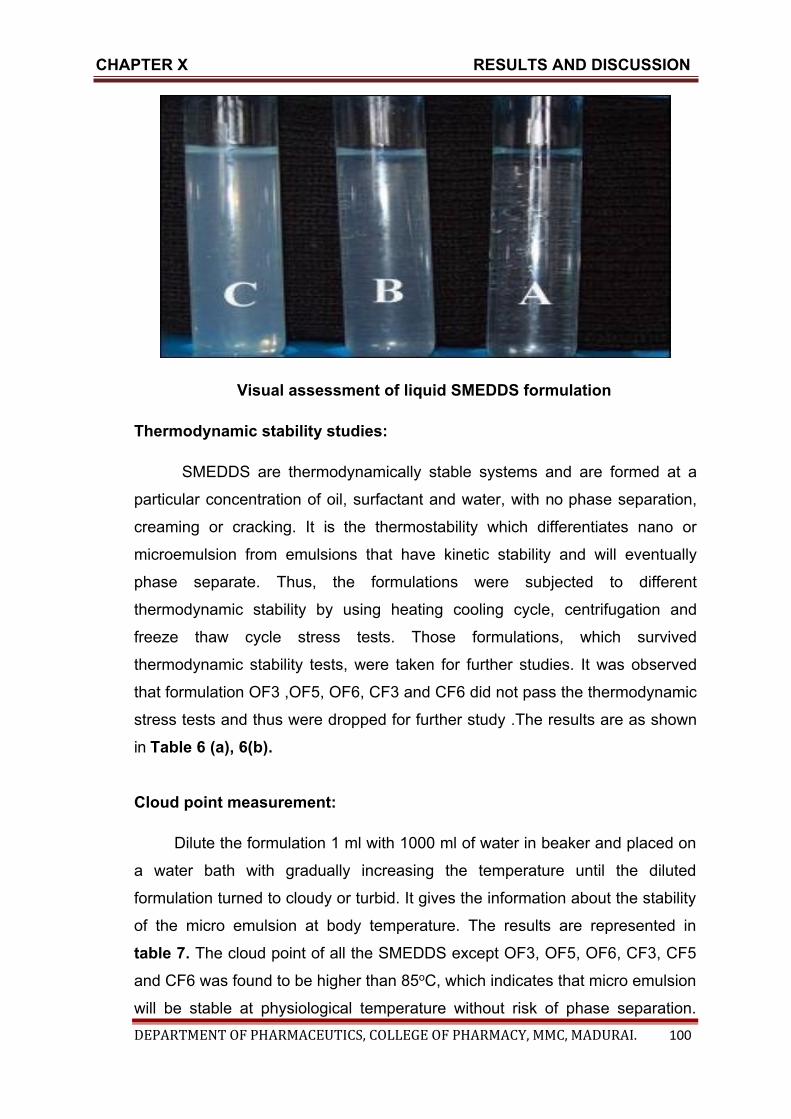
CHAPTER X RESULTS AND DISCUSSION
DEPARTMENT OF PHARMACEUTICS, COLLEGE OF PHARMACY, MMC, MADURAI. 100
Visual assessment of liquid SMEDDS formulation
Thermodynamic stability studies:
SMEDDS are thermodynamically stable systems and are formed at a
particular concentration of oil, surfactant and water, with no phase separation,
creaming or cracking. It is the thermostability which differentiates nano or
microemulsion from emulsions that have kinetic stability and will eventually
phase separate. Thus, the formulations were subjected to different
thermodynamic stability by using heating cooling cycle, centrifugation and
freeze thaw cycle stress tests. Those formulations, which survived
thermodynamic stability tests, were taken for further studies. It was observed
that formulation OF3 ,OF5, OF6, CF3 and CF6 did not pass the thermodynamic
stress tests and thus were dropped for further study .The results are as shown
in Table 6 (a), 6(b).
Cloud point measurement:
Dilute the formulation 1 ml with 1000 ml of water in beaker and placed on
a water bath with gradually increasing the temperature until the diluted
formulation turned to cloudy or turbid. It gives the information about the stability
of the micro emulsion at body temperature. The results are represented in
table 7. The cloud point of all the SMEDDS except OF3, OF5, OF6, CF3, CF5
and CF6 was found to be higher than 85oC, which indicates that micro emulsion
will be stable at physiological temperature without risk of phase separation.

CHAPTER X RESULTS AND DISCUSSION
DEPARTMENT OF PHARMACEUTICS, COLLEGE OF PHARMACY, MMC, MADURAI. 101
Formulations OF3, OF5, OF6, CF3, CF5 and CF6 shows phase separation due
to precipitation of drug. So these cannot be considered as the good
formulations due to phase separation at higher temperature
Robustness to dilution:
After diluting SMEDDS to 50, 100 and 1000 times with water, buffer pH
7.4 and pH 6.8 and storing for 12 h, it was observed that there was no sign of
phase separation or drug precipitation in formulations except OF3, OF5, OF6,
CF3, CF5 and CF, which turned hazy after standing for long hours. Hence,
formulations OF3, OF5, OF6, CF3, CF5 and CF6 were rejected as they were
also showing phase separation and became hazy on dilution. The results were
shown in Table 8 (a) and 8 (b).
Refractive index and percent transmittance:
Refractive Index and percent transmittance are determined to check the
transparency of formulation. Refractive Index of the formulation is measured by
refractometer by placing drop of solution on slide and then compare it with
water (RI=1.333). If refractive index is similar to refractive index of water and
percentage transmittance above 90%, then formulations have a transparent
nature. The results of Refractive index and percentage Transmittance are
shown in Table 9. Formulation OF6 , OF7 ,OF9, CF1, CF2, CF3, CF4, CF8,
and CF9 have transmittance value greater than 90%, suggesting their clarity.
And the refractive index of formulation OF2, OF8, and CF4, CF8, CF9 has the
values nearest to RI of water.
Viscosity determination: OF3, OF5, OF6, CF3, CF5 and CF6 were
rejected due to instability. Remaining formulations were subjected to viscosity
determination.
From the viscosity determination it was observed that as the
concentration of co-surfactant increased viscosity of the formulation also
increased. So formulation F1-F4 was highly viscous due to higher co-surfactant
concentration.

CHAPTER X RESULTS AND DISCUSSION
DEPARTMENT OF PHARMACEUTICS, COLLEGE OF PHARMACY, MMC, MADURAI. 102
The concentration of surfactant also increases the viscosity of the
formulation. It was expected that F5-F9 would show the highest viscosity due to
high concentration of oil. Out of 18 formulations, CF8 and OF8 (oil-45% and
Smixture-55%) shown optimum viscosity due to optimum concentration of oil,
surfactant and co-surfactant. The results were shown in table 10.
Absolute drug content in L-SMEDDS:
Drug content was determined by measuring the absorbance in UV
spectro photometer. The results are in the range of 92.79 %-97.43%. The
results are represented in Table 11 and figure 8 (a) and 8 (b). From the results,
formulation OF8 (97.8±0.08%) and CF8 (97.33±0.52%) show the highest drug
content amongst all the other formulations.
Conversion of liquid SMEDDS into solid S-SMEDDS:
Solid SMEDDS were prepared by mixing liquid SMEDDS containing
pitavastatin with microcrystalline cellulose (MCC) in 1:1 proportion. Liquid
SMEDDS was added drop wise over MCC and homogenized using glass rod to
ensure uniform distribution of formulation in a china dish.
Characterization of S-SMEDDS:
a) Micromeretic properties:
Angle of Repose
Angle of repose for the best formulation (OF8 and CF8) was 26°54’ and
28o38’ respectively. The results suggested that the powder blend of formulation
shows good flow properties. The result of angle of repose of best formulation
was shown in table 12 a & b.
Bulk density ( gm /ml)
The bulk density for the best formulations (CF8 & OF8) were
0.3436±0.006g/ml and 0.3423±0.006g/ml.The results indicated that the powder
blends of formulation have good flow properties. The results were summarized
in table12 a & b.
CHAPTER X RESULTS AND DISCUSSION
DEPARTMENT OF PHARMACEUTICS, COLLEGE OF PHARMACY, MMC, MADURAI. 103
Tapped density (gm/ml)
Tapped density for the best formulation (CF8 & OF8) 0.4043±0.001g/ml
and 0.404±0.001g/ml respectively. The results indicating the presence of
smaller particles occupying the voids between the bigger particles. The results
were summarized in table 12 a & b.
Carr’s index
The Carr’s index of the best formulation CF8 & OF8 was found to be
15.01±0.001% and 15.27±0.001%, which indicated that the powder blend was
good. The results were summarized in table 12 a & b.
Hausner’s ratio:
The Hausner ratio of the best formulation (CF8 & OF8) was found to be
1.17±0.002 and 1.18±0.002. Since a very low Hausner ratio (<1.25) was
obtained the formulations showed better property. The results were
summarized in table 12 a & b.
Drug content:
The drug content of all the S-SMEDDS formulations (OF1-OF9) and
(C1-CF9) were in the range of 96.54 % to 98.02 %. The results are represented
in Table 13, & figure 9 (a) & 9 (b) and it suggests that uniform distribution of
drug in the formulation. Formulation OF8 (98±0.09%) and CF8 (98.02±0.07%)
show the highest drug content amongst all other formulations.
In vitro drug release from S-SMEDDS:
The dissolution study was carried out in phosphate buffer of pH 6.8 for
two hours. The in vitro dissolution studies of all the formulations were
compared with pure drug and conventional tablet. The results of in vitro drug
release studies from Pitavastatin SMEDDS formulation were represented in
Table 14(a) and 14(b).
The results suggest that the % release of all the formulations is greater
than that of the pure drug (59.25%±0.20) and conventional tablet
(64.72%±0.21).
CHAPTER X RESULTS AND DISCUSSION
DEPARTMENT OF PHARMACEUTICS, COLLEGE OF PHARMACY, MMC, MADURAI. 104
Formulation CF8 and OF8 shown the highest percentage drug release
of 82.14±0.15% and 80.52±0.12% at the end of two hours respectively than the
other formulations.
SELECTION OF BEST FORMULATION:
From the above characterization, the two formulations OF9 and CF8
were selected as the best formulation showing,
For OF8
% Transmittance 94.28±0.22
Refractive index 1.333±0.0004
Drug content 98.13±0.09%
In vitro drug release 80.52±0.12% for two hours
For CF8
% Transmittance 95.08±0.16
Refractive index 1.333±0.0004
Drug content 98.02±0.07
In vitro drug release 82.14±0.15
COMPARISON OF IN VITRO DRUG RELEASE OF BEST
FORMULATION WITH PURE DRUG AND MARKETED CONVENTIONAL
TABLET:
The comparison of in vitro drug release profile of best formulation is
compared with that of the pure drug and conventional tablet. It represented in
figure 12.
Pure drug (59.25%±0.20)
Conventional tablet (64.72%±0.21)
OF8 (80.52% ±0.12)
CF8 (82.14%±0.15)
CHAPTER X RESULTS AND DISCUSSION
DEPARTMENT OF PHARMACEUTICS, COLLEGE OF PHARMACY, MMC, MADURAI. 105
KINETICS ANALYSIS
The in vitro dissolution study results were fitted into 0 order first order
Higuchi Koresmeyer peppas and Hixson Crowel model to find out the in vitro
kinetic release studies. The result indicated the the values follow first order
reaction.
According to values of koresmeyer peppas equation which is greater
than one indicates it follows supercase II transport which means it follows more
than one mechanism.
EVALUATION OF BEST FORMULATION:
a) Infrared (IR) spectroscopic studies:
Infrared (IR) spectroscopic studies were carried out for pure drug,
excipients and selected SMEDDS formulations (CF8 and OF8). The spectra
studied at 4000cm-1 to 400 cm-1 were shown in (Table 15) and (Figure
18(a)to 18(e)). It was found from the spectra that there was no major shifting as
well as any loss of functional peaks in the spectra of drug, excipients and
selected formulations (CF8 & OF8). This clearly indicated that there was no
interaction between the drug and the polymer and the drug was present in its
unchanged form.
b) Morphological studies of S-SMEDDS by using Scanning Electron
Microscopy (SEM):
The surface morphology of the pure drug and solid SMEDDS were
examined by the SEM and the images are represented in figure 19 (a) and
19(b). The typical crystalline structure of Pitavastatin as shown in figure 19 (a)
was absent in S-SMEDDS of Pitavastatin, which indicates the transformation of
the drug from crystalline state tto amorphous state i.e the drug is completely
solubilised in oil phase of L-SMEDDS.
c) X-ray Powder Diffraction (XRPD) analysis:
The PXRD patterns of pure drug (Pitavastatin) and formulations (OF8 &
CF8) were presented in (Figure 20(a) and 20(b).The XRPD patterns of pure
CHAPTER X RESULTS AND DISCUSSION
DEPARTMENT OF PHARMACEUTICS, COLLEGE OF PHARMACY, MMC, MADURAI. 106
drug showed numerous sharp peaks (at 2θ 15.00°, 16.80°, 20.90°, 23.00°,
25.40° and 34.31°) which are the characteristic of a crystalline compound. And
these peaks are absent in the PXRD pattern of SMEDDS indicating the
transformation crystalline nature to Amorphous nature.
d) Determination of Particle size and Zeta potential:
The results of Particle size, zeta potential and Polydispersibility index of
the best formulations CF8 and OF8 was shown in figure 21 (a) -21 (d). The
particle size of the S-SMEDDS was found to be 139.5 nm and 621.3 nm with
polydispersibility index of 0.291 and 0.377 for OF8 and CF8 respectively. Since
the value of polydispersibility index is less than 1 indicates uniform distribution
of droplets throughout the formulation. And the zeta potential values of the best
formulation were found to be -23.9 and -20.9 for CF8 and OF8 respectively.
The negative zeta potential indicates the stable formulation.
e) Differential Scanning Calorimetry:
Physical state of Pitavastatin in S-SMEDDS was characterized using
differential scanning calorimeter. The DSC thermogram of S-SMEDDS
formulation were shown in figure 22. The DSC thermogram of Pitavastatin
showed a characteristic, sharp endothermic peak at corresponding to its
melting point (less than 138oC) and it indicates the crystalline nature of the
drug. The DSC analysis of prepared SMEDDS formulation revealed negligible
change in the melting point of Pitavastatin in the presence excipients, indicating
no modification or interaction between the drug and excipients.
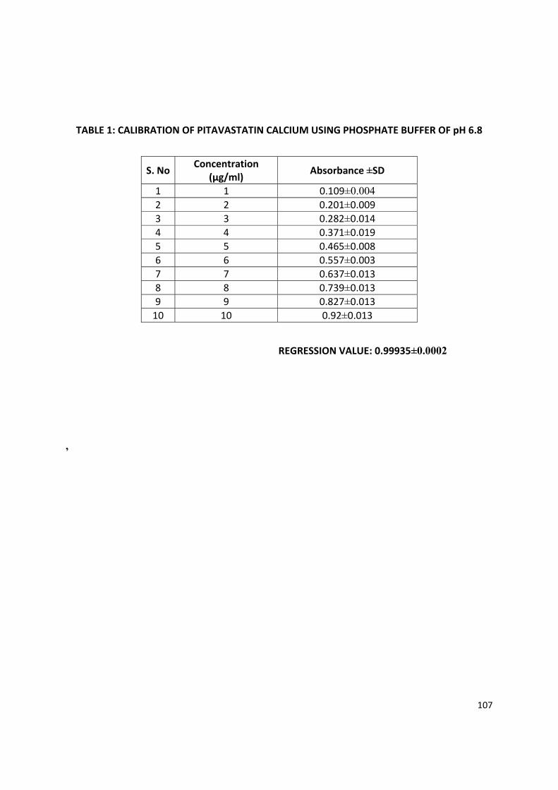
107
TABLE 1: CALIBRATION OF PITAVASTATIN CALCIUM USING PHOSPHATE BUFFER OF pH 6.8
REGRESSION VALUE: 0.99935±0.0002
,
S. No Concentration (μg/ml) Absorbance ±SD
1 1 0.109±0.004 2 2 0.201±0.009 3 3 0.282±0.014 4 4 0.371±0.019 5 5 0.465±0.008 6 6 0.557±0.003 7 7 0.637±0.0138 8 0.739±0.0139 9 0.827±0.013
10 10 0.92±0.013
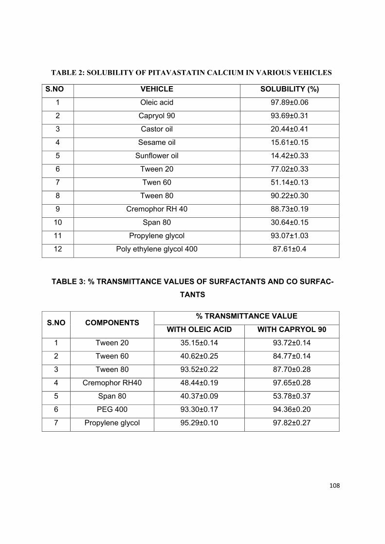
108
TABLE 2: SOLUBILITY OF PITAVASTATIN CALCIUM IN VARIOUS VEHICLES
S.NO VEHICLE SOLUBILITY (%)
1 Oleic acid 97.89±0.06
2 Capryol 90 93.69±0.31
3 Castor oil 20.44±0.41
4 Sesame oil 15.61±0.15
5 Sunflower oil 14.42±0.33
6 Tween 20 77.02±0.33
7 Twen 60 51.14±0.13
8 Tween 80 90.22±0.30
9 Cremophor RH 40 88.73±0.19
10 Span 80 30.64±0.15
11 Propylene glycol 93.07±1.03
12 Poly ethylene glycol 400 87.61±0.4
TABLE 3: % TRANSMITTANCE VALUES OF SURFACTANTS AND CO SURFAC-
TANTS
S.NO COMPONENTS % TRANSMITTANCE VALUE
WITH OLEIC ACID WITH CAPRYOL 90
1 Tween 20 35.15±0.14 93.72±0.14
2 Tween 60 40.62±0.25 84.77±0.14
3 Tween 80 93.52±0.22 87.70±0.28
4 Cremophor RH40 48.44±0.19 97.65±0.28
5 Span 80 40.37±0.09 53.78±0.37
6 PEG 400 93.30±0.17 94.36±0.20
7 Propylene glycol 95.29±0.10 97.82±0.27
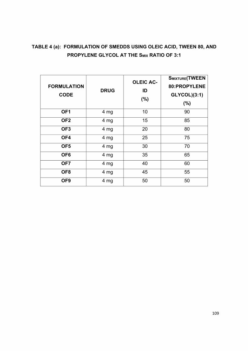
109
TABLE 4 (a): FORMULATION OF SMEDDS USING OLEIC ACID, TWEEN 80, AND
PROPYLENE GLYCOL AT THE SMIX RATIO OF 3:1
FORMULATION
CODE DRUG
OLEIC AC-
ID
(%)
SMIXTURE(TWEEN
80:PROPYLENE
GLYCOL)(3:1)
(%)
OF1 4 mg 10 90
OF2 4 mg 15 85
OF3 4 mg 20 80
OF4 4 mg 25 75
OF5 4 mg 30 70
OF6 4 mg 35 65
OF7 4 mg 40 60
OF8 4 mg 45 55
OF9 4 mg 50 50
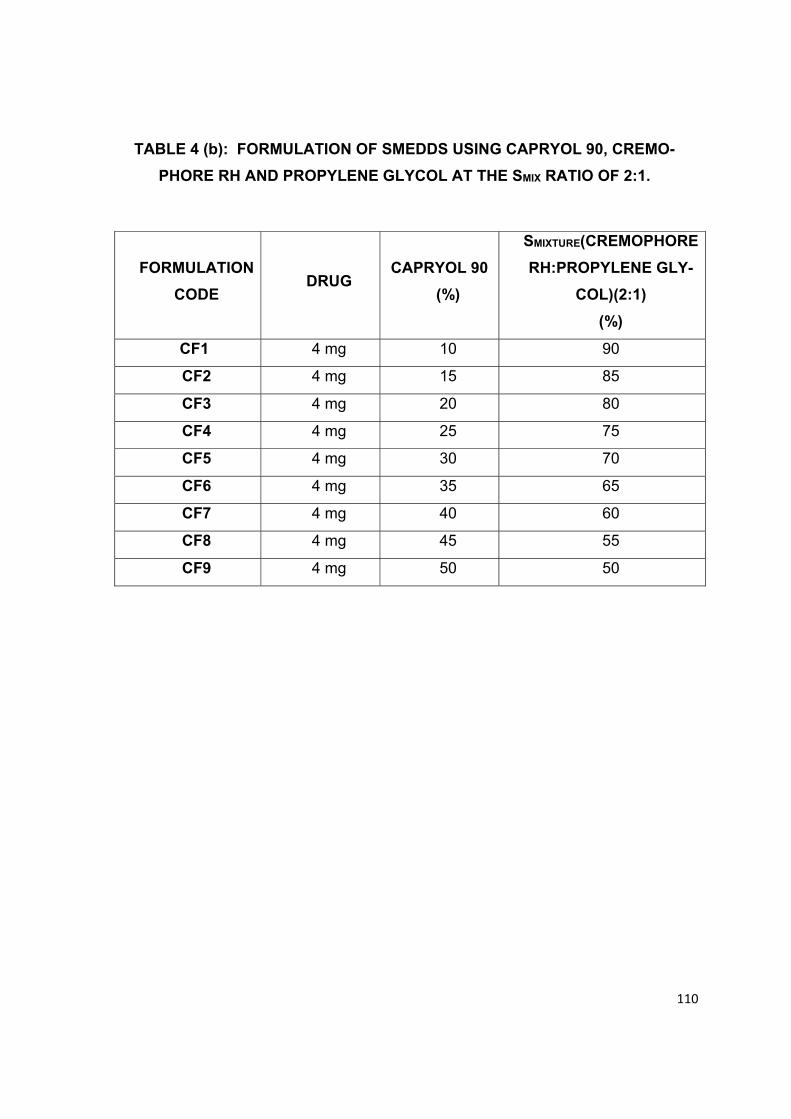
110
TABLE 4 (b): FORMULATION OF SMEDDS USING CAPRYOL 90, CREMO-
PHORE RH AND PROPYLENE GLYCOL AT THE SMIX RATIO OF 2:1.
FORMULATION
CODE DRUG
CAPRYOL 90
(%)
SMIXTURE(CREMOPHORE
RH:PROPYLENE GLY-
COL)(2:1)
(%)
CF1 4 mg 10 90
CF2 4 mg 15 85
CF3 4 mg 20 80
CF4 4 mg 25 75
CF5 4 mg 30 70
CF6 4 mg 35 65
CF7 4 mg 40 60
CF8 4 mg 45 55
CF9 4 mg 50 50
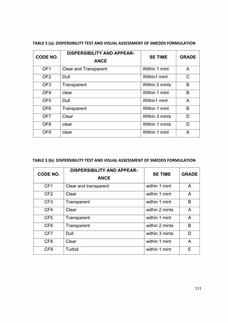
111
TABLE 5 (a): DISPERSIBILITY TEST AND VISUAL ASSESSMENT OF SMEDDS FORMULATION
CODE NO. DISPERSIBILITY AND APPEAR-
ANCE SE TIME GRADE
OF1 Clear and Transparent Within 1 mint A
OF2 Dull Within1 mint C
OF3 Transparent Within 2 mints B
OF4 clear Within 1 mint B
OF5 Dull Within1 mint A
OF6 Transparent Within 1 mint B
OF7 Clear Within 3 mints D
OF8 clear Within 1 mints D
OF9 clear Within 1 mint A
TABLE 5 (b): DISPERSIBILITY TEST AND VISUAL ASSESSMENT OF SMEDDS FORMULATION
CODE NO. DISPERSIBILITY AND APPEAR-
ANCE SE TIME GRADE
CF1 Clear and transparent within 1 mint A
CF2 Clear within 1 mint A
CF3 Transparent within 1 mint B
CF4 Clear within 2 mints A
CF5 Transparent within 1 mint A
CF6 Transparent within 2 mints B
CF7 Dull within 3 mints D
CF8 Clear within 1 mint A
CF9 Turbid within 1 mint E
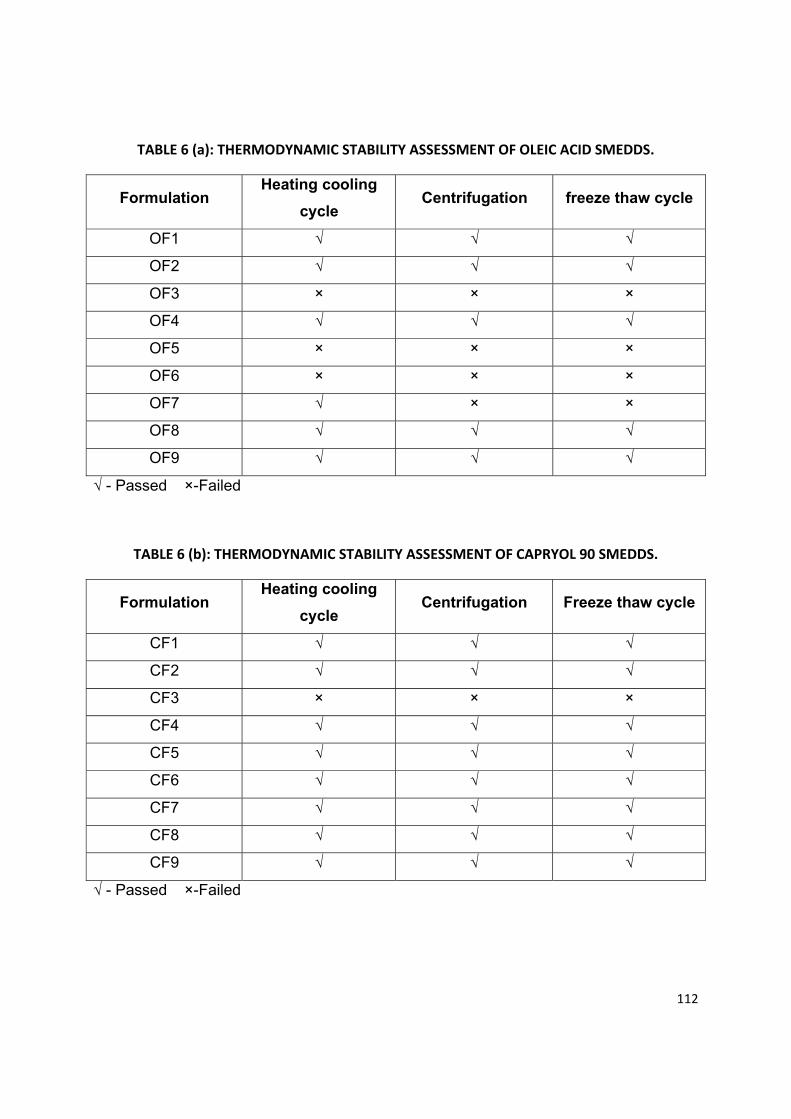
112
TABLE 6 (a): THERMODYNAMIC STABILITY ASSESSMENT OF OLEIC ACID SMEDDS.
Formulation Heating cooling
cycle Centrifugation freeze thaw cycle
OF1 √ √ √
OF2 √ √ √
OF3 × × ×
OF4 √ √ √
OF5 × × ×
OF6 × × ×
OF7 √ × ×
OF8 √ √ √
OF9 √ √ √
√ - Passed ×-Failed
TABLE 6 (b): THERMODYNAMIC STABILITY ASSESSMENT OF CAPRYOL 90 SMEDDS.
Formulation Heating cooling
cycle Centrifugation Freeze thaw cycle
CF1 √ √ √
CF2 √ √ √
CF3 × × ×
CF4 √ √ √
CF5 √ √ √
CF6 √ √ √
CF7 √ √ √
CF8 √ √ √
CF9 √ √ √
√ - Passed ×-Failed
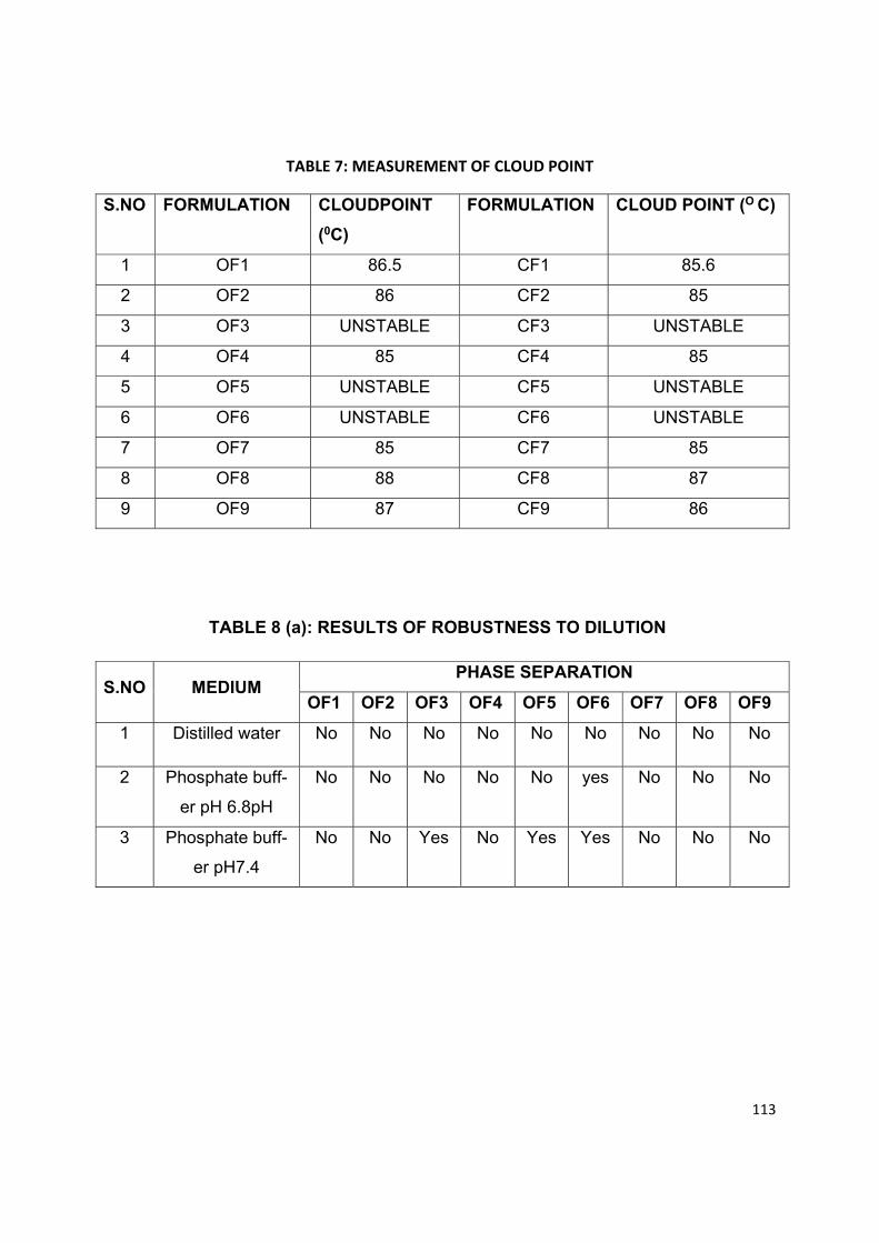
113
TABLE 7: MEASUREMENT OF CLOUD POINT
S.NO FORMULATION CLOUDPOINT
(0C)
FORMULATION CLOUD POINT (O C)
1 OF1 86.5 CF1 85.6
2 OF2 86 CF2 85
3 OF3 UNSTABLE CF3 UNSTABLE
4 OF4 85 CF4 85
5 OF5 UNSTABLE CF5 UNSTABLE
6 OF6 UNSTABLE CF6 UNSTABLE
7 OF7 85 CF7 85
8 OF8 88 CF8 87
9 OF9 87 CF9 86
TABLE 8 (a): RESULTS OF ROBUSTNESS TO DILUTION
S.NO MEDIUM PHASE SEPARATION
OF1 OF2 OF3 OF4 OF5 OF6 OF7 OF8 OF9
1 Distilled water No No No No No No No No No
2 Phosphate buff-
er pH 6.8pH
No No No No No yes No No No
3 Phosphate buff-
er pH7.4
No No Yes No Yes Yes No No No
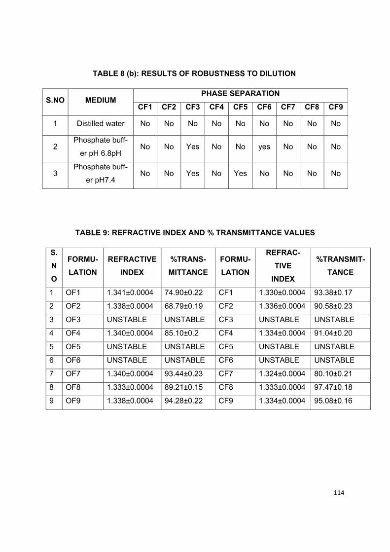
114
TABLE 8 (b): RESULTS OF ROBUSTNESS TO DILUTION
S.NO MEDIUM PHASE SEPARATION
CF1 CF2 CF3 CF4 CF5 CF6 CF7 CF8 CF9
1 Distilled water No No No No No No No No No
2 Phosphate buff-
er pH 6.8pH No No Yes No No yes No No No
3 Phosphate buff-
er pH7.4 No No Yes No Yes No No No No
TABLE 9: REFRACTIVE INDEX AND % TRANSMITTANCE VALUES
S.
N
O
FORMU-
LATION
REFRACTIVE
INDEX
%TRANS-
MITTANCE
FORMU-
LATION
REFRAC-
TIVE
INDEX
%TRANSMIT-
TANCE
1 OF1 1.341±0.0004 74.90±0.22 CF1 1.330±0.0004 93.38±0.17
2 OF2 1.338±0.0004 68.79±0.19 CF2 1.336±0.0004 90.58±0.23
3 OF3 UNSTABLE UNSTABLE CF3 UNSTABLE UNSTABLE
4 OF4 1.340±0.0004 85.10±0.2 CF4 1.334±0.0004 91.04±0.20
5 OF5 UNSTABLE UNSTABLE CF5 UNSTABLE UNSTABLE
6 OF6 UNSTABLE UNSTABLE CF6 UNSTABLE UNSTABLE
7 OF7 1.340±0.0004 93.44±0.23 CF7 1.324±0.0004 80.10±0.21
8 OF8 1.333±0.0004 89.21±0.15 CF8 1.333±0.0004 97.47±0.18
9 OF9 1.338±0.0004 94.28±0.22 CF9 1.334±0.0004 95.08±0.16
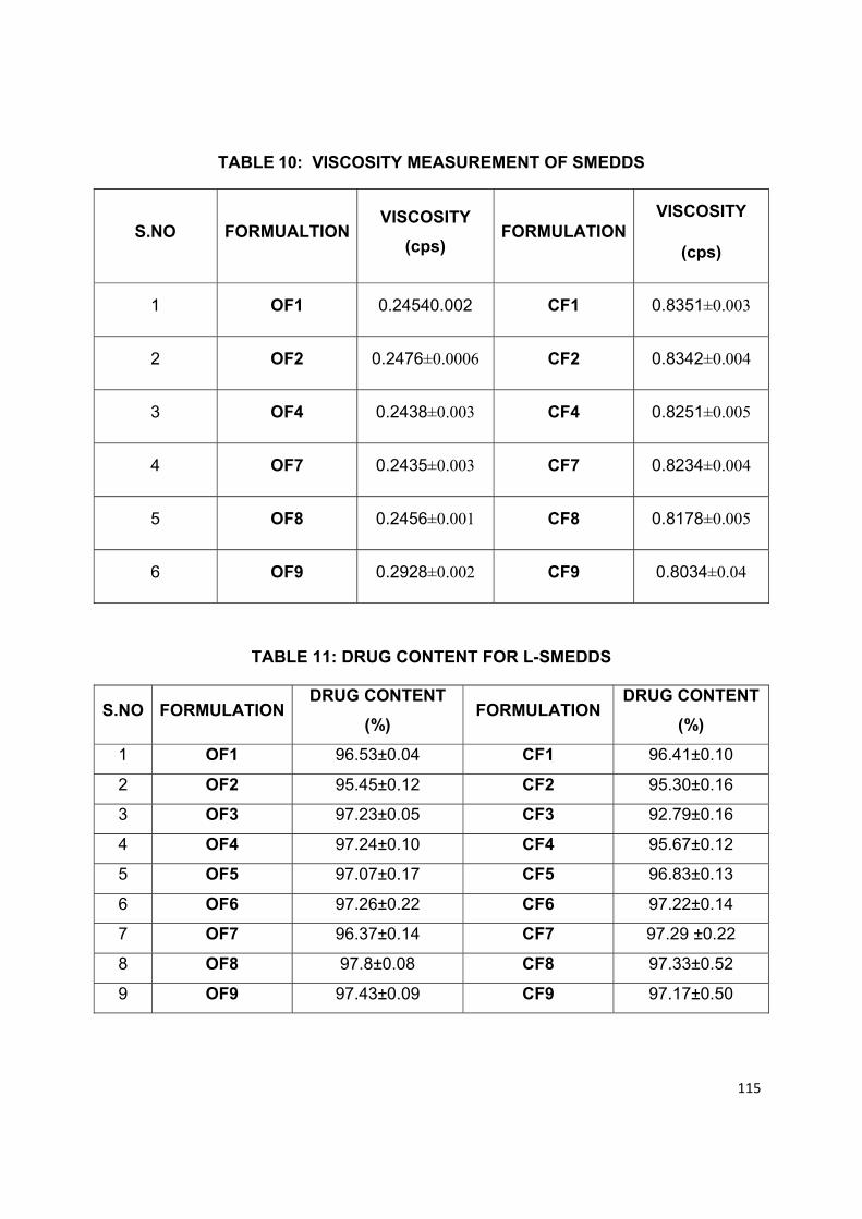
115
TABLE 10: VISCOSITY MEASUREMENT OF SMEDDS
S.NO FORMUALTION VISCOSITY
(cps) FORMULATION
VISCOSITY
(cps)
1 OF1 0.24540.002 CF1 0.8351±0.003
2 OF2 0.2476±0.0006 CF2 0.8342±0.004
3 OF4 0.2438±0.003 CF4 0.8251±0.005
4 OF7 0.2435±0.003 CF7 0.8234±0.004
5 OF8 0.2456±0.001 CF8 0.8178±0.005
6 OF9 0.2928±0.002 CF9 0.8034±0.04
TABLE 11: DRUG CONTENT FOR L-SMEDDS
S.NO FORMULATION DRUG CONTENT
(%) FORMULATION
DRUG CONTENT
(%)
1 OF1 96.53±0.04 CF1 96.41±0.10
2 OF2 95.45±0.12 CF2 95.30±0.16
3 OF3 97.23±0.05 CF3 92.79±0.16
4 OF4 97.24±0.10 CF4 95.67±0.12
5 OF5 97.07±0.17 CF5 96.83±0.13
6 OF6 97.26±0.22 CF6 97.22±0.14
7 OF7 96.37±0.14 CF7 97.29 ±0.22
8 OF8 97.8±0.08 CF8 97.33±0.52
9 OF9 97.43±0.09 CF9 97.17±0.50
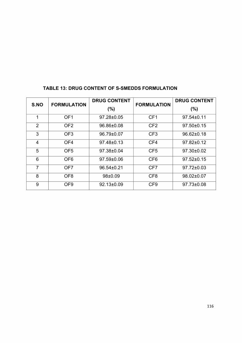
116
TABLE 13: DRUG CONTENT OF S-SMEDDS FORMULATION
S.NO FORMULATION DRUG CONTENT
(%) FORMULATION
DRUG CONTENT
(%)
1 OF1 97.28±0.05 CF1 97.54±0.11
2 OF2 96.86±0.08 CF2 97.50±0.15
3 OF3 96.79±0.07 CF3 96.62±0.18
4 OF4 97.48±0.13 CF4 97.82±0.12
5 OF5 97.38±0.04 CF5 97.30±0.02
6 OF6 97.59±0.06 CF6 97.52±0.15
7 OF7 96.54±0.21 CF7 97.72±0.03
8 OF8 98±0.09 CF8 98.02±0.07
9 OF9 92.13±0.09 CF9 97.73±0.08
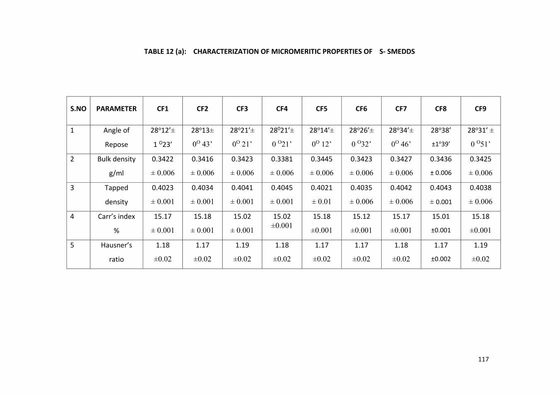
117
TABLE 12 (a): CHARACTERIZATION OF MICROMERITIC PROPERTIES OF S- SMEDDS
S.NO PARAMETER CF1 CF2 CF3 CF4 CF5 CF6 CF7 CF8 CF9
1 Angle of
Repose
28o12’±
1 O23’
28o13±
0O 43’
28o21’±
0O 21’
28021’±
0 O21’
28o14’±
0O 12’
28o26’±
0 O32’
28o34’±
0O 46’
28o38’
±1o39’
28o31’ ±
0 O51’
2 Bulk density
g/ml
0.3422
± 0.006
0.3416
± 0.006
0.3423
± 0.006
0.3381
± 0.006
0.3445
± 0.006
0.3423
± 0.006
0.3427
± 0.006
0.3436
± 0.006
0.3425
± 0.006
3 Tapped
density
0.4023
± 0.001
0.4034
± 0.001
0.4041
± 0.001
0.4045
± 0.001
0.4021
± 0.01
0.4035
± 0.006
0.4042
± 0.006
0.4043
± 0.001
0.4038
± 0.006
4 Carr’s index
%
15.17
± 0.001
15.18
± 0.001
15.02
± 0.001
15.02 ±0.001
15.18
±0.001
15.12
±0.001
15.17
±0.001
15.01
±0.001
15.18
±0.001
5 Hausner’s
ratio
1.18
±0.02
1.17
±0.02
1.19
±0.02
1.18
±0.02
1.17
±0.02
1.17
±0.02
1.18
±0.02
1.17
±0.002
1.19
±0.02
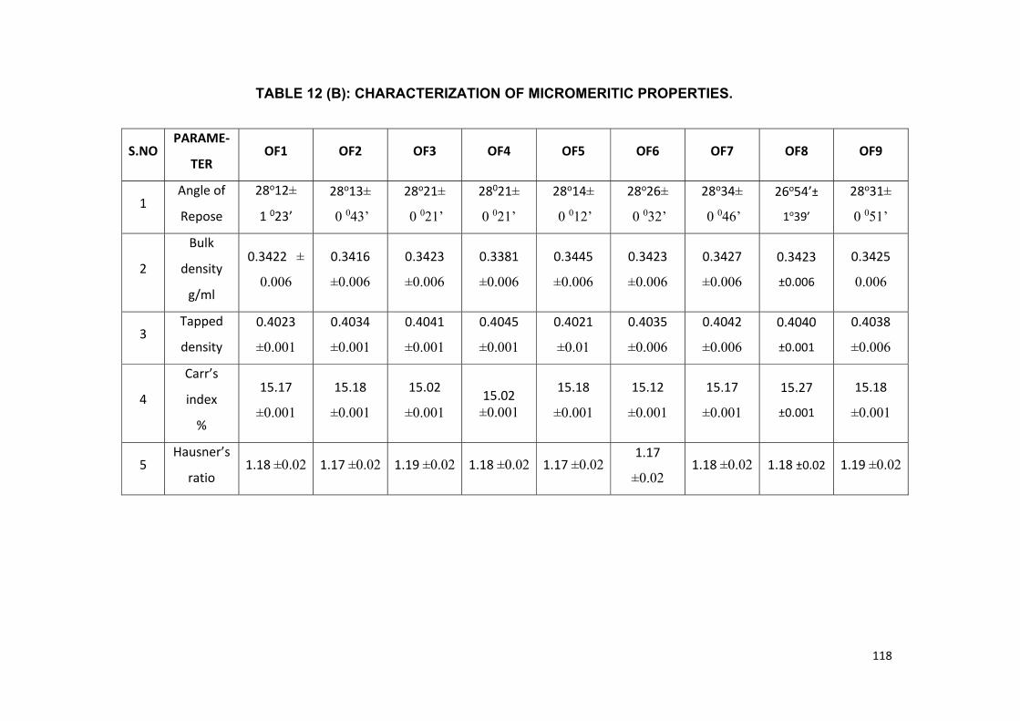
118
TABLE 12 (B): CHARACTERIZATION OF MICROMERITIC PROPERTIES.
S.NOPARAME-
TER OF1 OF2 OF3 OF4 OF5 OF6 OF7 OF8 OF9
1 Angle of
Repose
28o12±
1 023’
28o13±
0 043’
28o21±
0 021’
28021±
0 021’
28o14±
0 012’
28o26±
0 032’
28o34±
0 046’
26o54’±
1o39’
28o31±
0 051’
2
Bulk
density
g/ml
0.3422 ±
0.006
0.3416
±0.006
0.3423
±0.006
0.3381
±0.006
0.3445
±0.006
0.3423
±0.006
0.3427
±0.006
0.3423
±0.006
0.3425
0.006
3 Tapped
density
0.4023
±0.001
0.4034
±0.001
0.4041
±0.001
0.4045
±0.001
0.4021
±0.01
0.4035
±0.006
0.4042
±0.006
0.4040
±0.001
0.4038
±0.006
4
Carr’s
index
%
15.17
±0.001
15.18
±0.001
15.02
±0.001 15.02
±0.001
15.18
±0.001
15.12
±0.001
15.17
±0.001
15.27
±0.001
15.18
±0.001
5 Hausner’s
ratio 1.18 ±0.02 1.17 ±0.02 1.19 ±0.02 1.18 ±0.02 1.17 ±0.02
1.17
±0.02 1.18 ±0.02 1.18 ±0.02 1.19 ±0.02
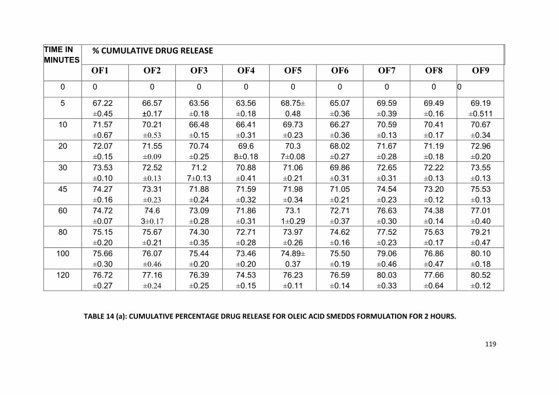
119
TABLE 14 (a): CUMULATIVE PERCENTAGE DRUG RELEASE FOR OLEIC ACID SMEDDS FORMULATION FOR 2 HOURS.
TIME IN MINUTES
% CUMULATIVE DRUG RELEASE
OF1 OF2 OF3 OF4 OF5 OF6 OF7 OF8 OF9 0 0 0 0 0 0 0 0 0 0
5 67.22 ±0.45
66.57 ±0.17
63.56 ±0.18
63.56 ±0.18
68.75± 0.48
65.07 ±0.36
69.59 ±0.39
69.49 ±0.16
69.19 ±0.511
10 71.57 ±0.67
70.21 ±0.53
66.48 ±0.15
66.41 ±0.31
69.73 ±0.23
66.27 ±0.36
70.59 ±0.13
70.41 ±0.17
70.67 ±0.34
20 72.07 ±0.15
71.55 ±0.09
70.74 ±0.25
69.6 8±0.18
70.3 7±0.08
68.02 ±0.27
71.67 ±0.28
71.19 ±0.18
72.96 ±0.20
30 73.53 ±0.10
72.52 ±0.13
71.2 7±0.13
70.88 ±0.41
71.06 ±0.21
69.86 ±0.31
72.65 ±0.31
72.22 ±0.13
73.55 ±0.13
45 74.27 ±0.16
73.31 ±0.23
71.88 ±0.24
71.59 ±0.32
71.98 ±0.34
71.05 ±0.21
74.54 ±0.23
73.20 ±0.12
75.53 ±0.13
60 74.72 ±0.07
74.6 3±0.17
73.09 ±0.28
71.86 ±0.31
73.1 1±0.29
72.71 ±0.37
76.63 ±0.30
74.38 ±0.14
77.01 ±0.40
80 75.15 ±0.20
75.67 ±0.21
74.30 ±0.35
72.71 ±0.28
73.97 ±0.26
74.62 ±0.16
77.52 ±0.23
75.63 ±0.17
79.21 ±0.47
100 75.66 ±0.30
76.07 ±0.46
75.44 ±0.20
73.46 ±0.20
74.89± 0.37
75.50 ±0.19
79.06 ±0.46
76.86 ±0.47
80.10 ±0.18
120 76.72 ±0.27
77.16 ±0.24
76.39 ±0.25
74.53 ±0.15
76.23 ±0.11
76.59 ±0.14
80.03 ±0.33
77.66 ±0.64
80.52 ±0.12

120
TIME IN MINTS
% CUMULATIVE DRUG RELEASE
CF1 CF2 CF3 CF4 CF5 CF6 CF7 CF8 CF9
0 0 0 0 0 0 0 0 0 0
5 68.98 ±0.31
63.75 ±0.32
67.05 ±0.09
66.46 ±0.10
70.59 ±0.24
67.05 ±0.33
71.22 ±0.14
71.85 ±0.34
72.43 ±0.17
10 70.31 ±0.17
65.62 ±0.23
68.18 ±0.32
67.81 ±0.26
71.58 ±0.35
68.12 ±0.25
72.42 ±0.22
73.36 ±0.06
73.74 ±0.20
20 70.98 ±0.12
67.25 ±0.43
70.04 ±0.44
69.67 ±0.17
72.70 ±0.24
69.13 ±0.10
73.70 ±0.35
74.65 ±0.18
75.26 ±0.24
30 71.63 ±0.08
68.59 ±0.13
70.66 ±0.41
70.42 ±0.12
74.36 ±0.65
70.80 ±0.27
75.69 ±0.29
76.25 ±0.59
77.06 ±0.32
45 73
±0.16 69.76 ±0.31
73.47 ±0.26
71.28 ±0.20
75.74 ±0.35
72.57 ±0.13
76.69 ±0.20
77.27 ±0.50
78.62 ±0.21
60 73.62 ±0.13
71.01 ±0.23
75.05 ±0.21
72.26 ±0.47
76.85 ±0.31
74.54 ±0.29
77.61 ±0.34
79.11 ±0.51
79.78 ±0.45
80 75.02 ±0.13
71.89 ±0.21
76.67 ±0.20
73.56 ±0.46
78.21 ±0.13
75.57 ±0.37
78.71±0 .35
80.14 ±0.18
80.59 ±0.22
100 75.72 ±0.16
72.85 ±0.27
78.46 ±0.35
74.53 ±0.34
79.40 ±0.22
76.72 ±0.16
80.64 ±0.21
80.93 ±0.17
81.58 ±0.31
120 76.46 ±0.10
74.79 ±0.32
79.59 ±0.16
77.02 ±0.24
80.45 ±0.18
78.51 ±0.38
81.77 ±0.21
82.14 ±0.15
81.98 ±0.25
TABLE 14 (b): CUMULATIVE PERCENTAGE DRUG RELEASE FOR CAPRYOL SMEDDS FORMULATION FOR 2 HOURS.
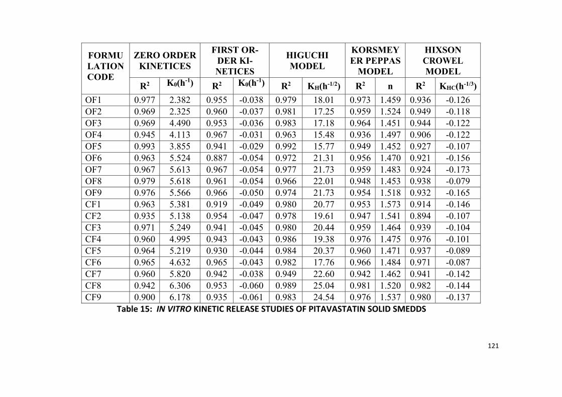
121
FORMU LATION CODE
ZERO ORDER KINETICES
FIRST OR-DER KI-NETICES
HIGUCHI MODEL
KORSMEY ER PEPPAS
MODEL
HIXSON CROWEL MODEL
R2 K0(h-1) R2 K0(h-1) R2 KH(h-1/2) R2 n R2 KHC(h-1/3)OF1 0.977 2.382 0.955 -0.038 0.979 18.01 0.973 1.459 0.936 -0.126 OF2 0.969 2.325 0.960 -0.037 0.981 17.25 0.959 1.524 0.949 -0.118 OF3 0.969 4.490 0.953 -0.036 0.983 17.18 0.964 1.451 0.944 -0.122 OF4 0.945 4.113 0.967 -0.031 0.963 15.48 0.936 1.497 0.906 -0.122 OF5 0.993 3.855 0.941 -0.029 0.992 15.77 0.949 1.452 0.927 -0.107 OF6 0.963 5.524 0.887 -0.054 0.972 21.31 0.956 1.470 0.921 -0.156 OF7 0.967 5.613 0.967 -0.054 0.977 21.73 0.959 1.483 0.924 -0.173 OF8 0.979 5.618 0.961 -0.054 0.966 22.01 0.948 1.453 0.938 -0.079 OF9 0.976 5.566 0.966 -0.050 0.974 21.73 0.954 1.518 0.932 -0.165 CF1 0.963 5.381 0.919 -0.049 0.980 20.77 0.953 1.573 0.914 -0.146 CF2 0.935 5.138 0.954 -0.047 0.978 19.61 0.947 1.541 0.894 -0.107 CF3 0.971 5.249 0.941 -0.045 0.980 20.44 0.959 1.464 0.939 -0.104 CF4 0.960 4.995 0.943 -0.043 0.986 19.38 0.976 1.475 0.976 -0.101 CF5 0.964 5.219 0.930 -0.044 0.984 20.37 0.960 1.471 0.937 -0.089 CF6 0.965 4.632 0.965 -0.043 0.982 17.76 0.966 1.484 0.971 -0.087 CF7 0.960 5.820 0.942 -0.038 0.949 22.60 0.942 1.462 0.941 -0.142 CF8 0.942 6.306 0.953 -0.060 0.989 25.04 0.981 1.520 0.982 -0.144 CF9 0.900 6.178 0.935 -0.061 0.983 24.54 0.976 1.537 0.980 -0.137
Table 15: IN VITRO KINETIC RELEASE STUDIES OF PITAVASTATIN SOLID SMEDDS
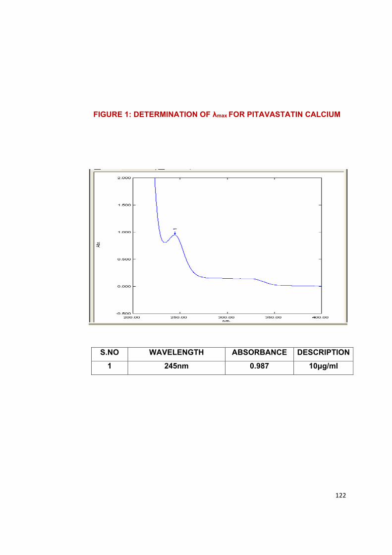
122
FIGURE 1: DETERMINATION OF λmax FOR PITAVASTATIN CALCIUM
S.NO WAVELENGTH ABSORBANCE DESCRIPTION
1 245nm 0.987 10μg/ml
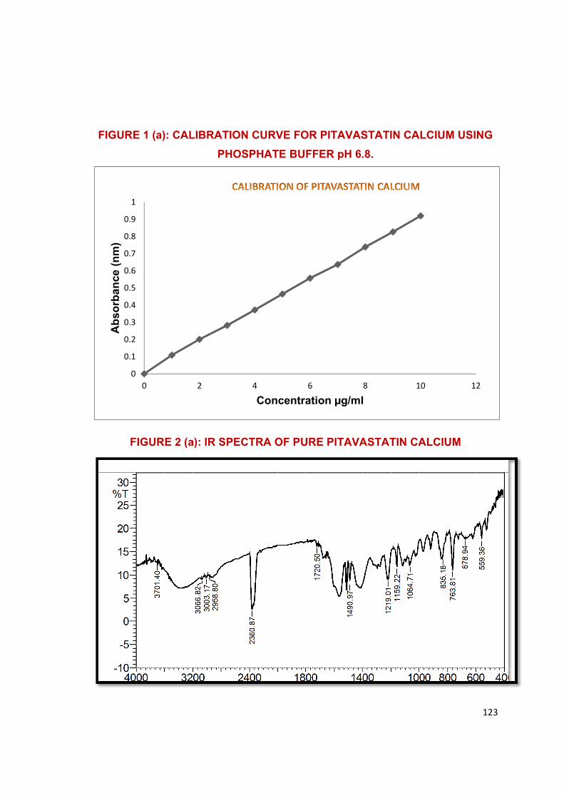
123
FIGURE 1 (a): CALIBRATION CURVE FOR PITAVASTATIN CALCIUM USING
PHOSPHATE BUFFER pH 6.8.
FIGURE 2 (a): IR SPECTRA OF PURE PITAVASTATIN CALCIUM
0
0.1
0.2
0.3
0.4
0.5
0.6
0.7
0.8
0.9
1
0 2 4 6 8 10 12
Ab
sorb
ance
(n
m)
Concentration µg/ml
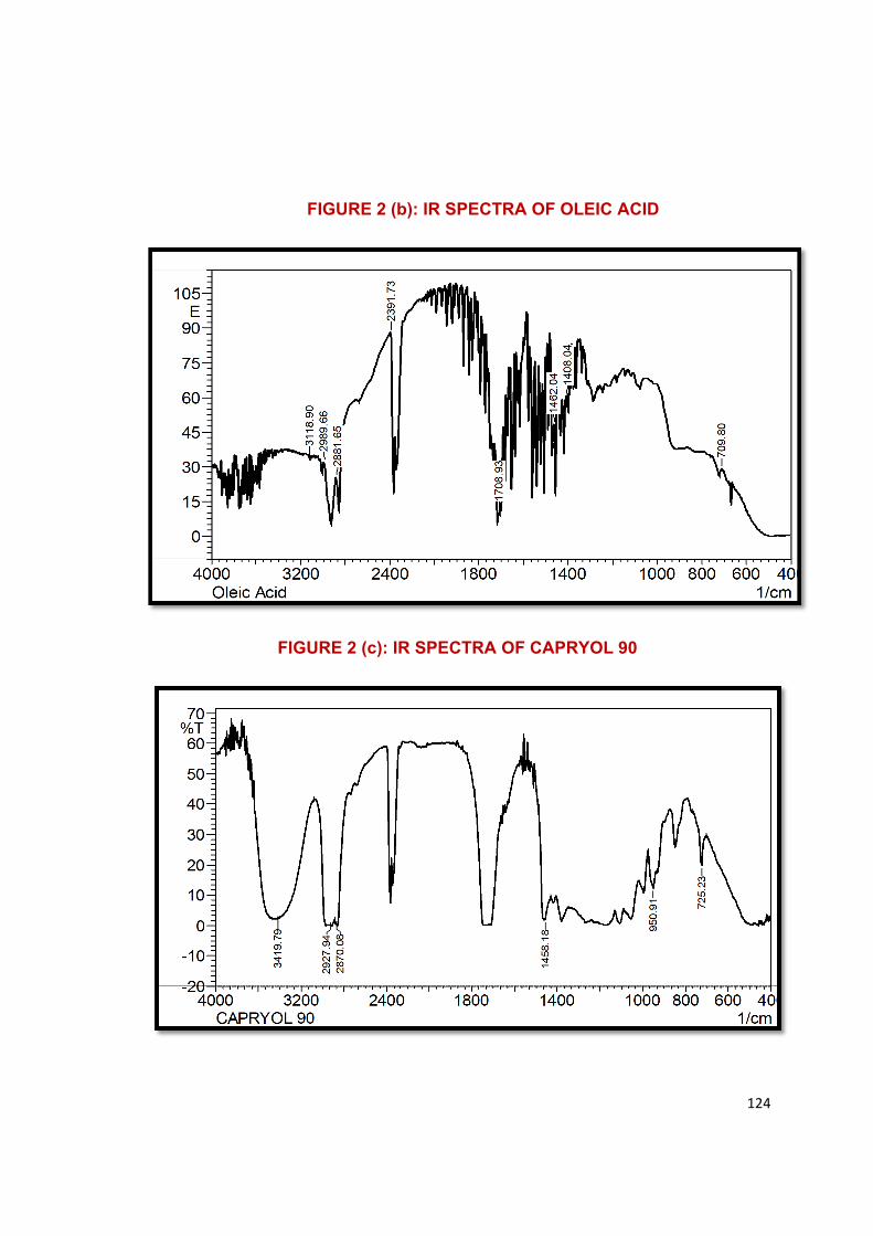
124
FIGURE 2 (b): IR SPECTRA OF OLEIC ACID
FIGURE 2 (c): IR SPECTRA OF CAPRYOL 90
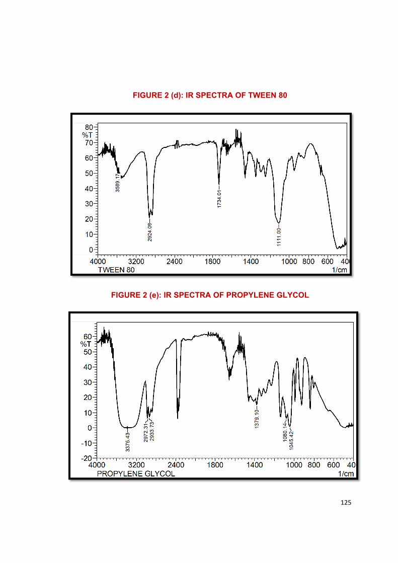
125
FIGURE 2 (d): IR SPECTRA OF TWEEN 80
FIGURE 2 (e): IR SPECTRA OF PROPYLENE GLYCOL
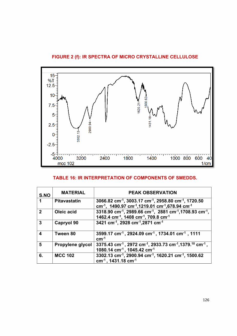
126
FIGURE 2 (f): IR SPECTRA OF MICRO CRYSTALLINE CELLULOSE
TABLE 16: IR INTERPRETATION OF COMPONENTS OF SMEDDS.
S.NO
MATERIAL PEAK OBSERVATION
1 Pitavastatin 3066.82 cm-1, 3003.17 cm-1, 2958.80 cm-1, 1720.50 cm-1, 1490.97 cm-1,1219.01 cm-1,678.94 cm-1
2 Oleic acid 3318.90 cm-1, 2989.66 cm-1, 2881 cm-1,1708.93 cm-1, 1462.4 cm-1, 1408 cm-1, 709.8 cm-1
3 Capryol 90 3421 cm-1, 2928 cm-1,2871 cm-1
4 Tween 80 3599.17 cm-1 , 2924.09 cm-1 , 1734.01 cm-1 , 1111 cm-1
5 Propylene glycol 3375.43 cm-1 , 2972 cm-1, 2933.73 cm-1,1379.10 cm-1 , 1080.14 cm-1 , 1045.42 cm-1
6. MCC 102 3302.13 cm-1, 2900.94 cm-1, 1620.21 cm-1, 1500.62 cm-1 , 1431.18 cm-1
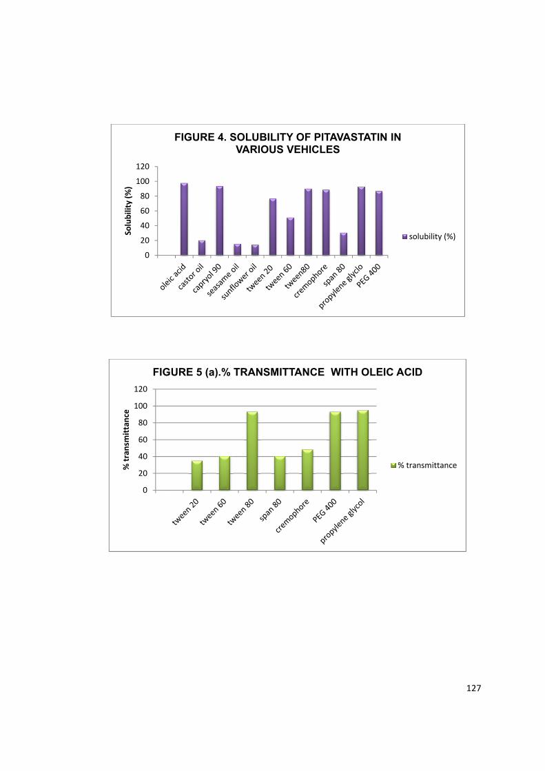
127
020406080
100120
Solu
bilit
y (%
)FIGURE 4. SOLUBILITY OF PITAVASTATIN IN
VARIOUS VEHICLES
solubility (%)
0
20
40
60
80
100
120
% tr
ansm
ittan
ce
FIGURE 5 (a).% TRANSMITTANCE WITH OLEIC ACID
% transmittance
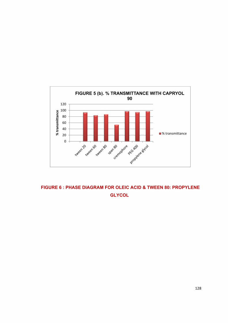
128
FIGURE 6 : PHASE DIAGRAM FOR OLEIC ACID & TWEEN 80: PROPYLENE
GLYCOL
020406080
100120
% tr
ansm
ittan
ce
FIGURE 5 (b). % TRANSMITTANCE WITH CAPRYOL 90
% transmittance
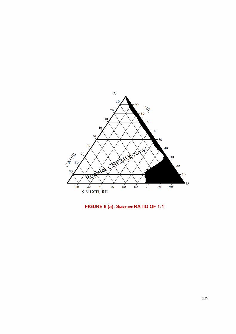
129
FIGURE 6 (a): SMIXTURE RATIO OF 1:1
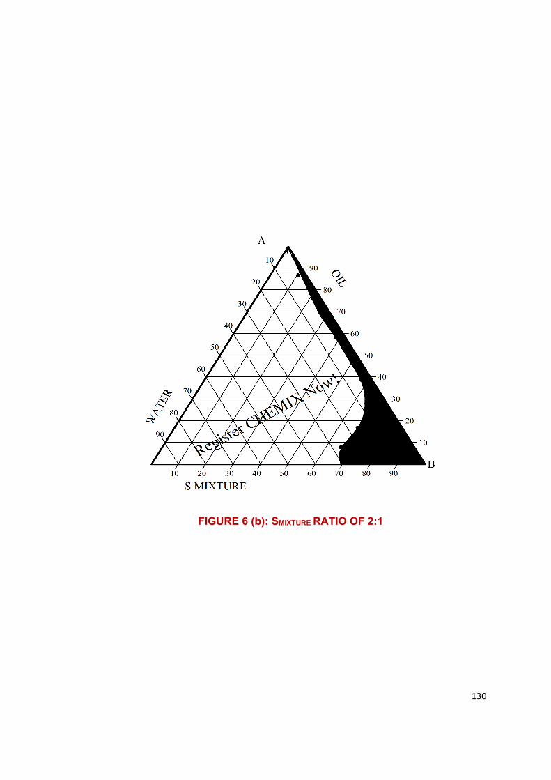
130
FIGURE 6 (b): SMIXTURE RATIO OF 2:1
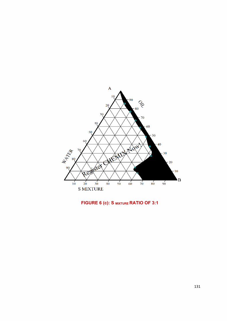
131
FIGURE 6 (c): S MIXTURE RATIO OF 3:1

132
FIGURE 7: PHASE DIAGRAM FOR CAPRYOL 90 AND CREMO-
PHORE:PROPYLENE GCOL
FIGURE 7 (a): SMIXTURE RATIO OF 1:1
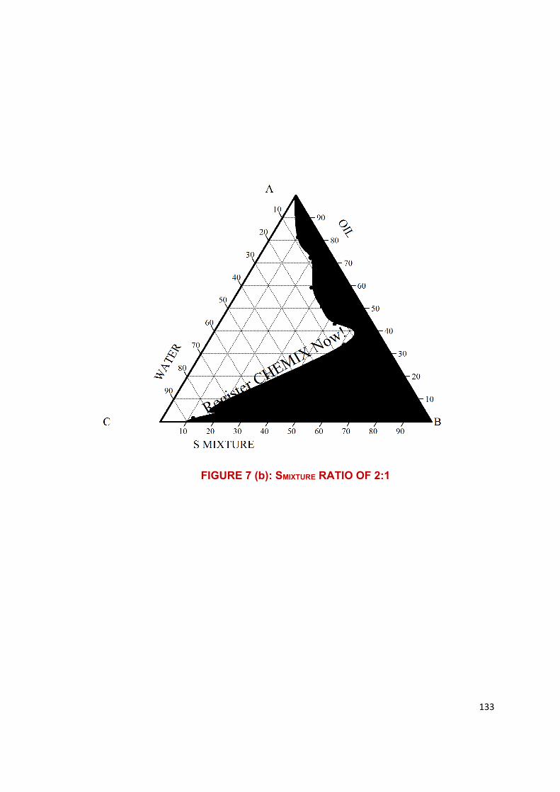
133
FIGURE 7 (b): SMIXTURE RATIO OF 2:1
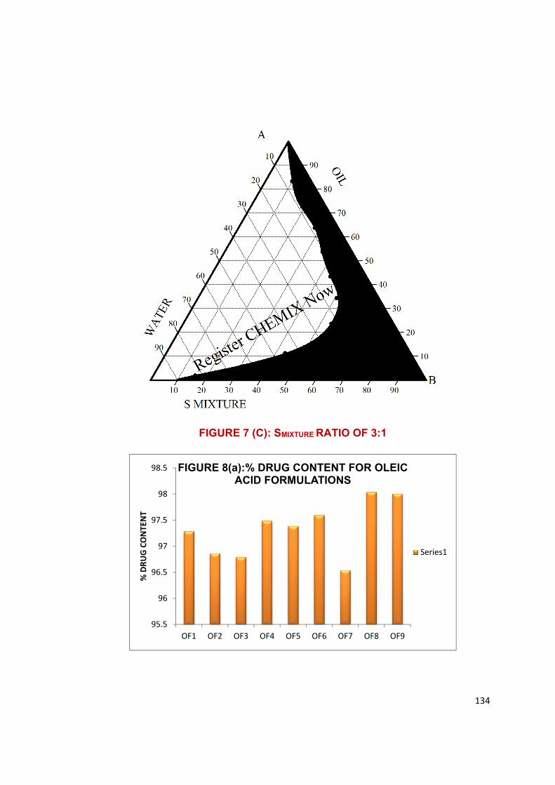
134
FIGURE 7 (C): SMIXTURE RATIO OF 3:1
95.5
96
96.5
97
97.5
98
98.5
OF1 OF2 OF3 OF4 OF5 OF6 OF7 OF8 OF9
% D
RUG
CONT
ENT
FIGURE 8(a):% DRUG CONTENT FOR OLEIC ACID FORMULATIONS
Series1

135
90
91
92
93
94
95
96
97
98
CF1 CF2 CF3 CF4 CF5 CF6 CF7 CF8 CF9
% D
RUG
CONT
ENT
Figure 8 (b.)% DRUG CONTENT FOR CAPRYOL FORMULATIONS
95.5
96
96.5
97
97.5
98
98.5
OF1 OF2 OF3 OF4 OF5 OF6 OF7 OF8 OF9
% D
RUG
CONT
ENT
Figure 9 (a).% DRUG CONTENT FOR OLEIC ACID FORMULATION
% DRUG CONTENT 0
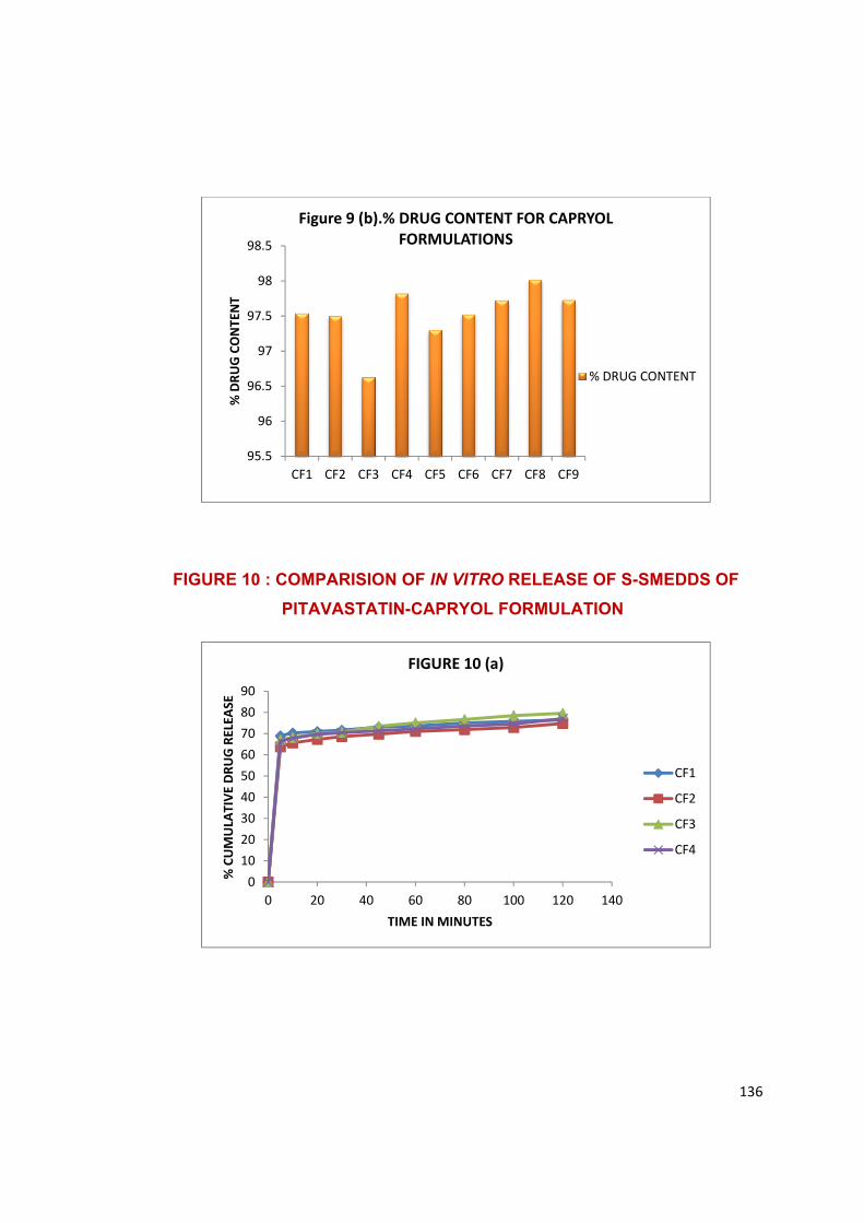
136
FIGURE 10 : COMPARISION OF IN VITRO RELEASE OF S-SMEDDS OF
PITAVASTATIN-CAPRYOL FORMULATION
95.5
96
96.5
97
97.5
98
98.5
CF1 CF2 CF3 CF4 CF5 CF6 CF7 CF8 CF9
% D
RUG
CONT
ENT
Figure 9 (b).% DRUG CONTENT FOR CAPRYOL FORMULATIONS
% DRUG CONTENT
0102030405060708090
0 20 40 60 80 100 120 140
% C
UMUL
ATIV
E DR
UG R
ELEA
SE
TIME IN MINUTES
FIGURE 10 (a)
CF1
CF2
CF3
CF4
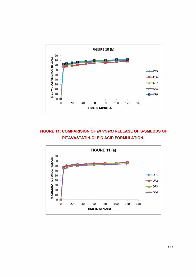
137
FIGURE 11: COMPARISION OF IN VITRO RELEASE OF S-SMEDDS OF
PITAVASTATIN-OLEIC ACID FORMULATION
0102030405060708090
0 20 40 60 80 100 120 140
% C
UMUL
ATIV
E DR
UG R
ELEA
SE
TIME IN MINUTES
FIGURE 10 (b)
CF5
CF6
CF7
CF8
CF9
0102030405060708090
0 20 40 60 80 100 120 140
% C
UMUL
ATIV
E DR
UG R
ELEA
SE
TIME IN MINUTES
FIGURE 11 (a)
OF1
OF2
OF3
OF4
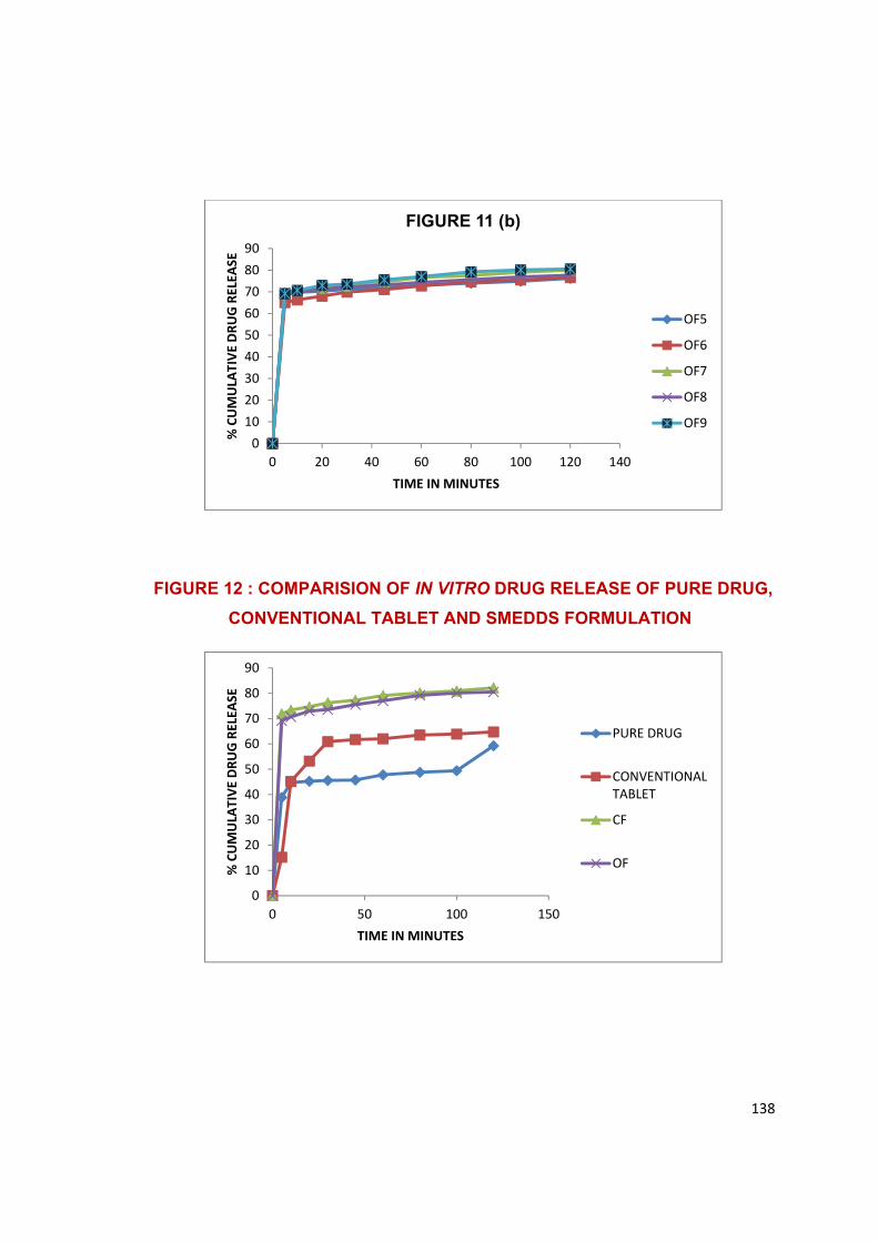
138
FIGURE 12 : COMPARISION OF IN VITRO DRUG RELEASE OF PURE DRUG,
CONVENTIONAL TABLET AND SMEDDS FORMULATION
0102030405060708090
0 20 40 60 80 100 120 140
% C
UMUL
ATIV
E DR
UG R
ELEA
SE
TIME IN MINUTES
FIGURE 11 (b)
OF5
OF6
OF7
OF8
OF9
0
10
20
30
40
50
60
70
80
90
0 50 100 150
% C
UMUL
ATIV
E DR
UG R
ELEA
SE
TIME IN MINUTES
PURE DRUG
CONVENTIONALTABLET
CF
OF
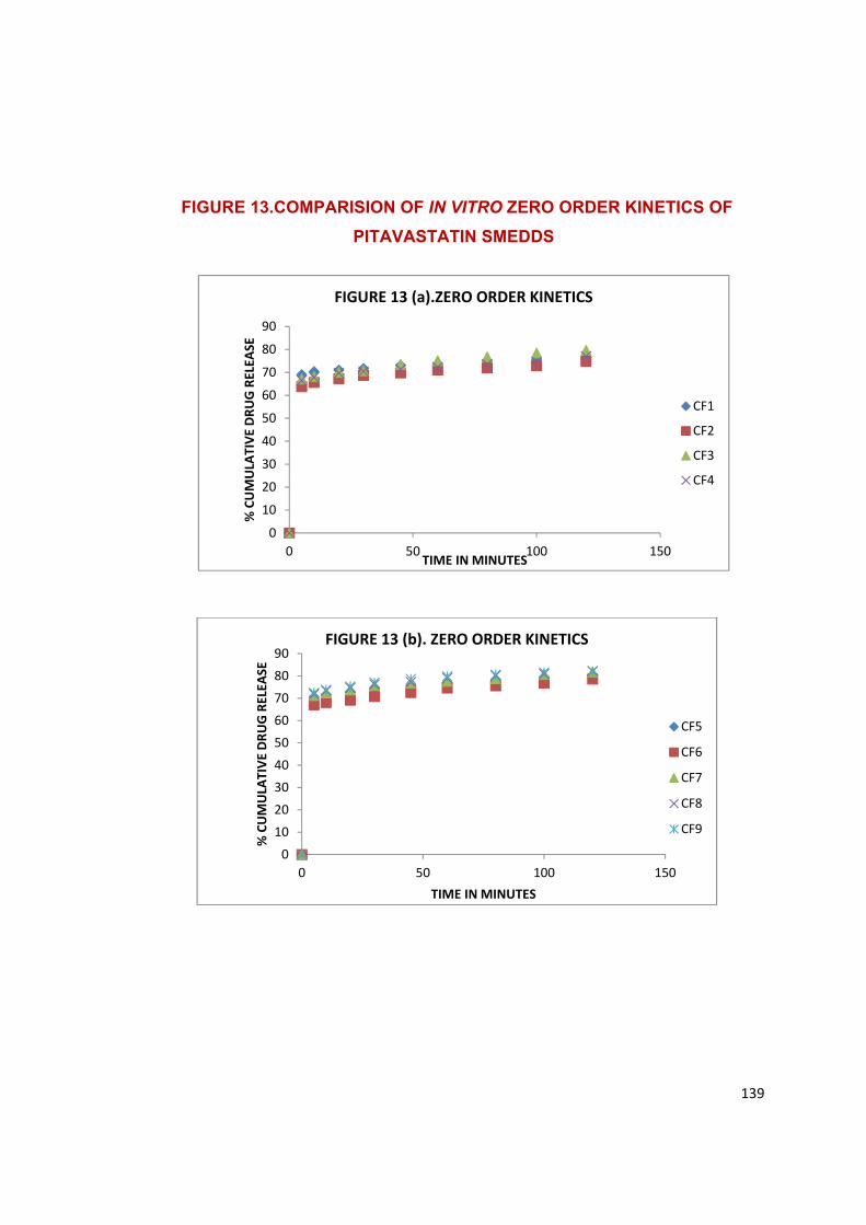
139
FIGURE 13.COMPARISION OF IN VITRO ZERO ORDER KINETICS OF
PITAVASTATIN SMEDDS
0102030405060708090
0 50 100 150
% C
UMUL
ATIV
E DR
UG R
ELEA
SE
TIME IN MINUTES
FIGURE 13 (b). ZERO ORDER KINETICS
CF5
CF6
CF7
CF8
CF9
0102030405060708090
0 50 100 150
% C
UMUL
ATIV
E DR
UG R
ELEA
SE
TIME IN MINUTES
FIGURE 13 (a).ZERO ORDER KINETICS
CF1
CF2
CF3
CF4
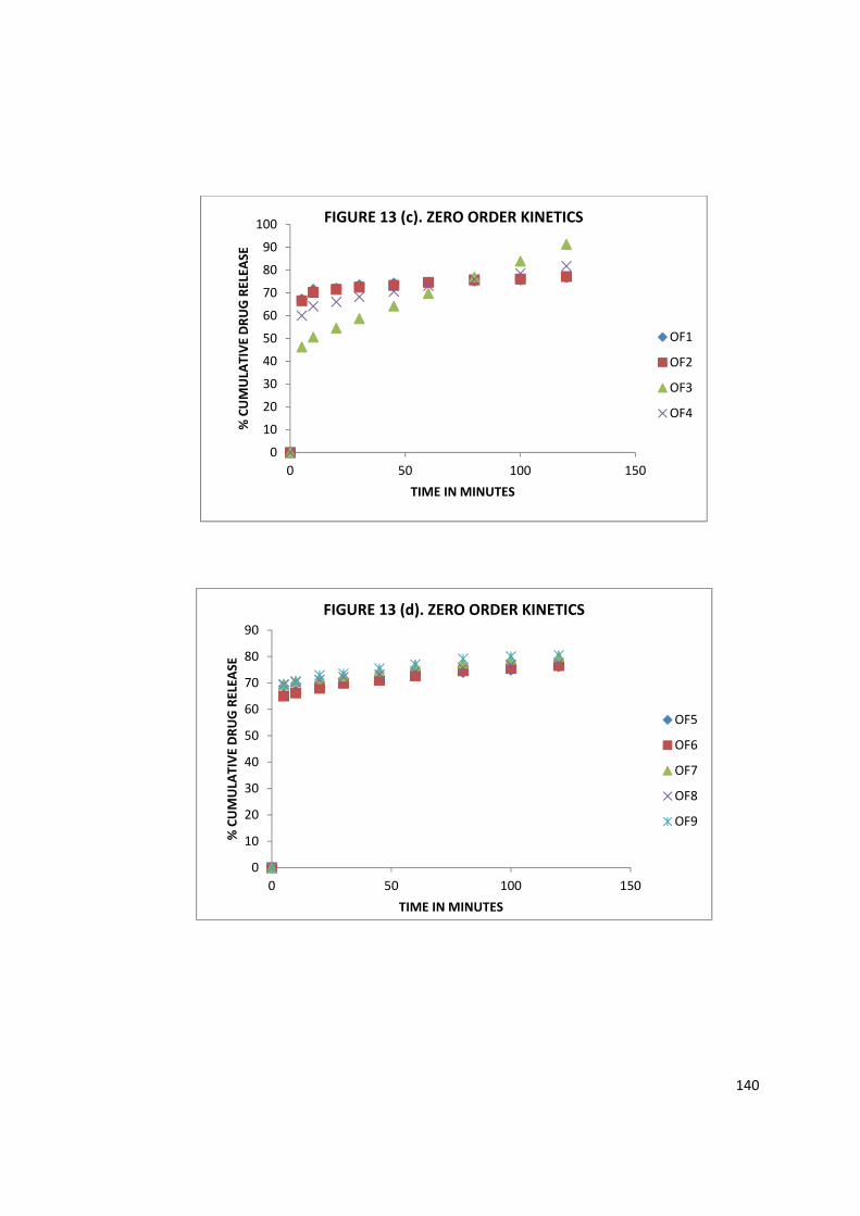
140
0102030405060708090
100
0 50 100 150
% C
UMUL
ATIV
E DR
UG R
ELEA
SE
TIME IN MINUTES
FIGURE 13 (c). ZERO ORDER KINETICS
OF1
OF2
OF3
OF4
0
10
20
30
40
50
60
70
80
90
0 50 100 150
% C
UMUL
ATIV
E DR
UG R
ELEA
SE
TIME IN MINUTES
FIGURE 13 (d). ZERO ORDER KINETICS
OF5
OF6
OF7
OF8
OF9
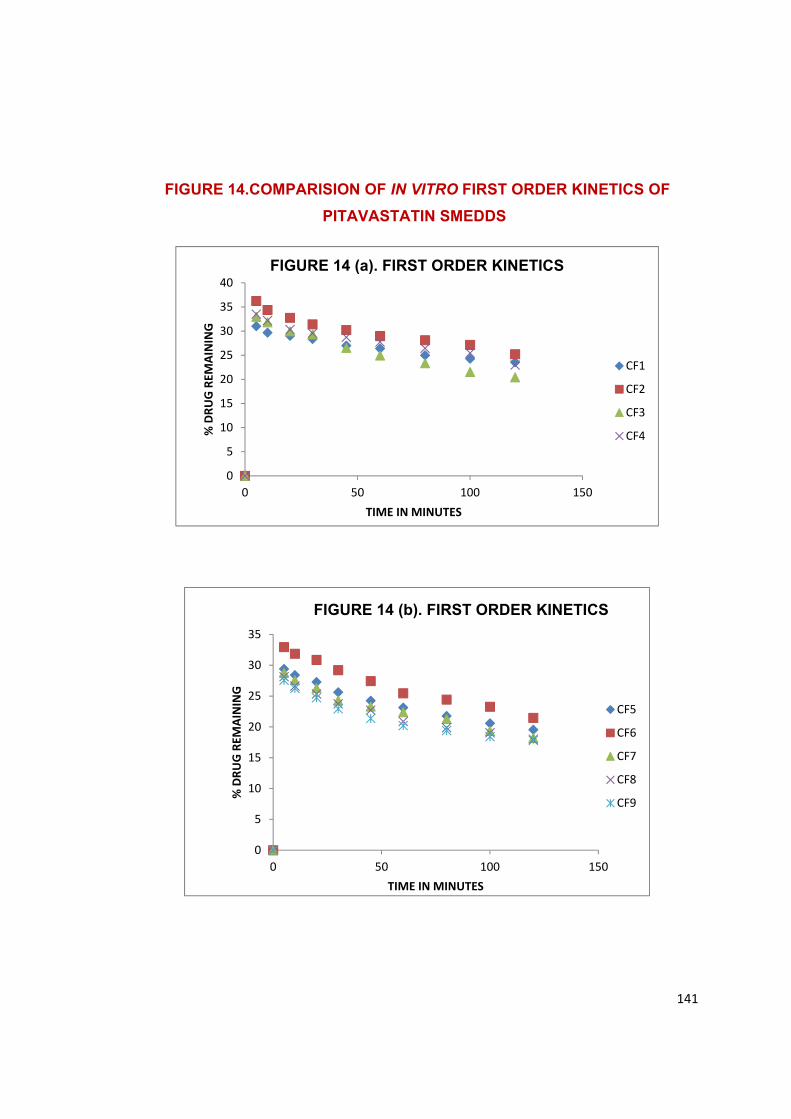
141
FIGURE 14.COMPARISION OF IN VITRO FIRST ORDER KINETICS OF
PITAVASTATIN SMEDDS
0
5
10
15
20
25
30
35
40
0 50 100 150
% D
RUG
REM
AINI
NG
TIME IN MINUTES
FIGURE 14 (a). FIRST ORDER KINETICS
CF1
CF2
CF3
CF4
0
5
10
15
20
25
30
35
0 50 100 150
% D
RUG
REM
AINI
NG
TIME IN MINUTES
FIGURE 14 (b). FIRST ORDER KINETICS
CF5
CF6
CF7
CF8
CF9
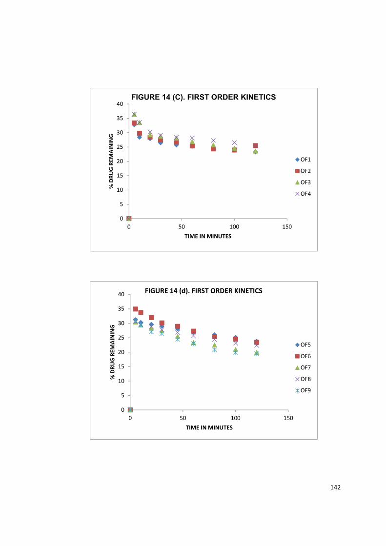
142
0
5
10
15
20
25
30
35
40
0 50 100 150
% D
RUG
REM
AINI
NG
TIME IN MINUTES
FIGURE 14 (C). FIRST ORDER KINETICS
OF1
OF2
OF3
OF4
0
5
10
15
20
25
30
35
40
0 50 100 150
% D
RUG
REM
AINI
NG
TIME IN MINUTES
FIGURE 14 (d). FIRST ORDER KINETICS
OF5
OF6
OF7
OF8
OF9
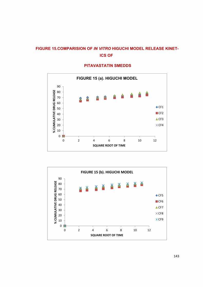
143
FIGURE 15.COMPARISION OF IN VITRO HIGUCHI MODEL RELEASE KINET-
ICS OF
PITAVASTATIN SMEDDS
0102030405060708090
0 2 4 6 8 10 12
% C
UMUL
ATIV
E DR
UG R
ELEA
SE
SQUARE ROOT OF TIME
FIGURE 15 (a). HIGUCHI MODEL
CF1
CF2
CF3
CF4
0102030405060708090
0 2 4 6 8 10 12
% C
UMUL
ATIV
E DR
UG R
ELEA
SE
SQUARE ROOT OF TIME
FIGURE 15 (b). HIGUCHI MODEL
CF5
CF6
CF7
CF8
CF9
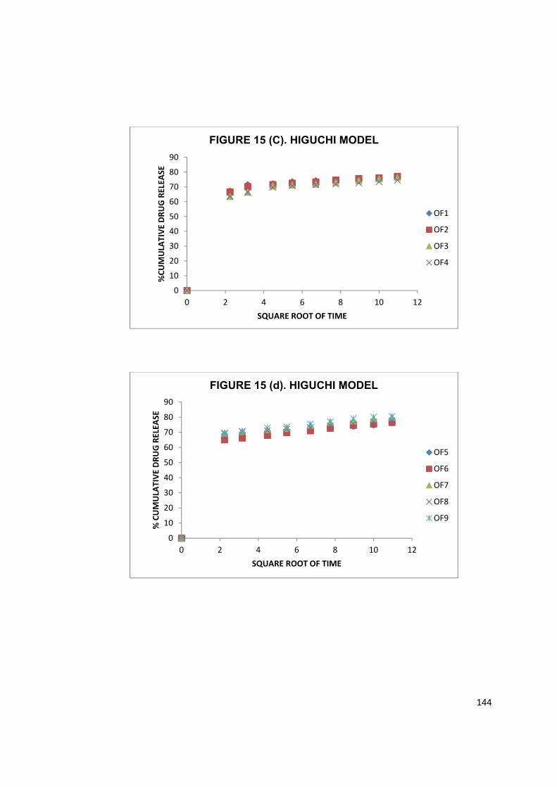
144
0102030405060708090
0 2 4 6 8 10 12
%CU
MUL
ATIV
E DR
UG R
ELEA
SE
SQUARE ROOT OF TIME
FIGURE 15 (C). HIGUCHI MODEL
OF1
OF2
OF3
OF4
0102030405060708090
0 2 4 6 8 10 12
% C
UMUL
ATIV
E DR
UG R
ELEA
SE
SQUARE ROOT OF TIME
FIGURE 15 (d). HIGUCHI MODEL
OF5
OF6
OF7
OF8
OF9
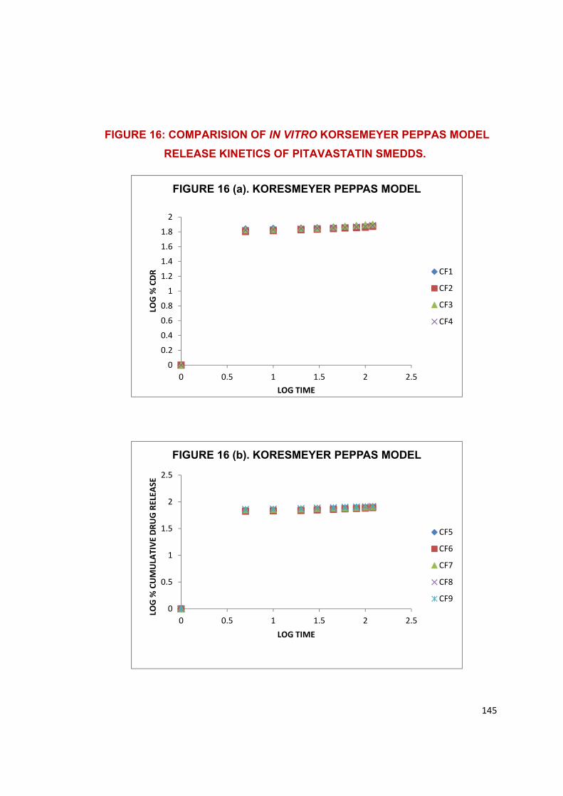
145
FIGURE 16: COMPARISION OF IN VITRO KORSEMEYER PEPPAS MODEL
RELEASE KINETICS OF PITAVASTATIN SMEDDS.
00.20.40.60.8
11.21.41.61.8
2
0 0.5 1 1.5 2 2.5
LOG
% C
DR
LOG TIME
FIGURE 16 (a). KORESMEYER PEPPAS MODEL
CF1
CF2
CF3
CF4
0
0.5
1
1.5
2
2.5
0 0.5 1 1.5 2 2.5
LOG
% C
UMUL
ATIV
E DR
UG R
ELEA
SE
LOG TIME
FIGURE 16 (b). KORESMEYER PEPPAS MODEL
CF5
CF6
CF7
CF8
CF9
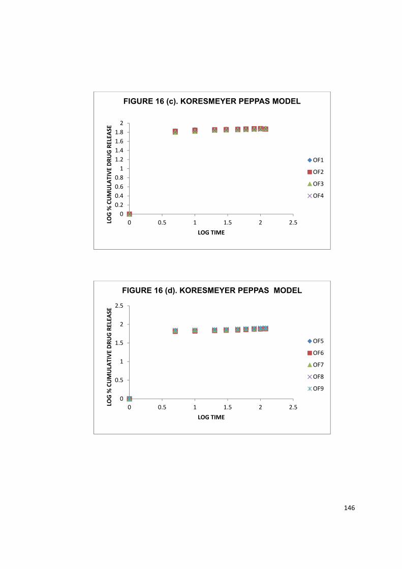
146
00.20.40.60.8
11.21.41.61.8
2
0 0.5 1 1.5 2 2.5LOG
% C
UMUL
ATIV
E DR
UG R
ELEA
SE
LOG TIME
FIGURE 16 (c). KORESMEYER PEPPAS MODEL
OF1
OF2
OF3
OF4
0
0.5
1
1.5
2
2.5
0 0.5 1 1.5 2 2.5LOG
% C
UMUL
ATIV
E DR
UG R
ELEA
SE
LOG TIME
FIGURE 16 (d). KORESMEYER PEPPAS MODEL
OF5
OF6
OF7
OF8
OF9

147
FIGURE 17. COMPARISION OF IN VITRO HIXON CROWEL MODEL RELEASE KINETICS OF PITAVASTATIN SMEDDS
0
0.5
1
1.5
2
2.5
3
3.5
0 20 40 60 80 100 120 140
CUBE
ROO
T OF
% D
RUG
REM
AINI
NG
TIME IN MINUTES
FIGURE 17 (a). HIXSON CROWEL MODEL
CF1
CF2
CF3
CF4
0
0.5
1
1.5
2
2.5
3
3.5
0 50 100 150
CUBE
ROO
T OF
% D
RUG
REM
AINI
NG
TIME IN MINUTES
FIGURE 17 (b). HIXSON CROWEL MODEL
CF5
CF6
CF7
CF8
CF9
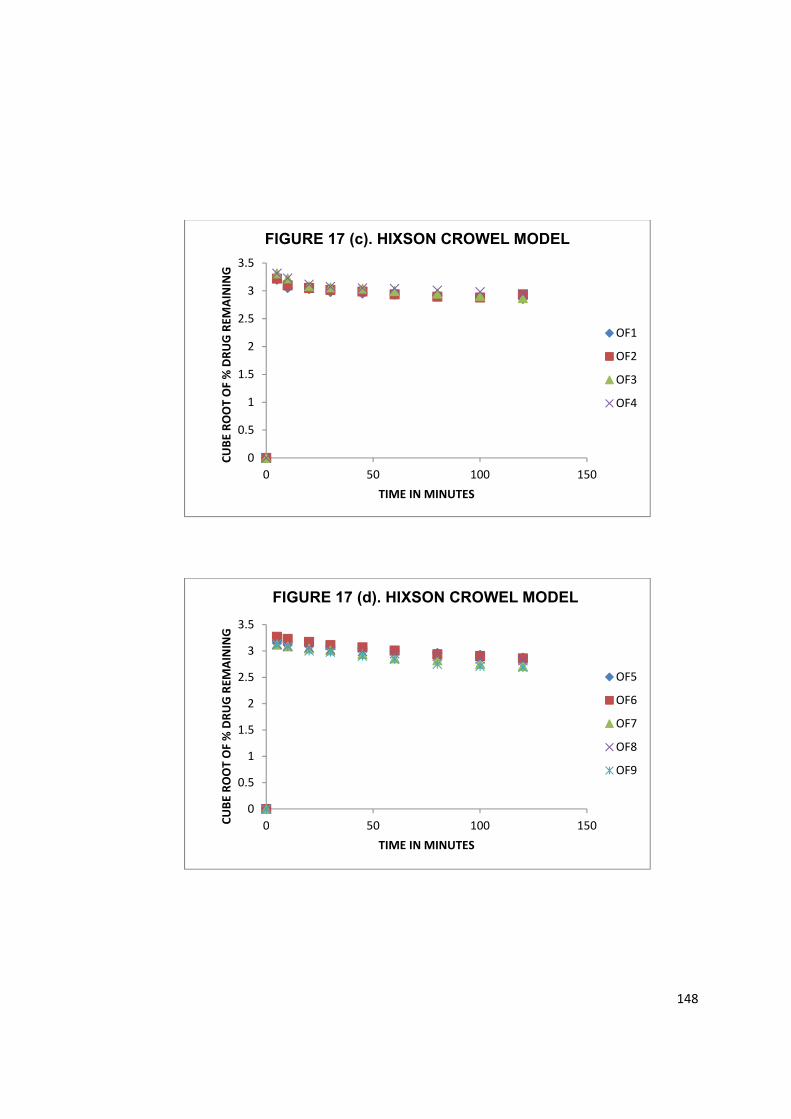
148
0
0.5
1
1.5
2
2.5
3
3.5
0 50 100 150
CUBE
ROO
T OF
% D
RUG
REM
AINI
NG
TIME IN MINUTES
FIGURE 17 (c). HIXSON CROWEL MODEL
OF1
OF2
OF3
OF4
0
0.5
1
1.5
2
2.5
3
3.5
0 50 100 150CUBE
ROO
T OF
% D
RUG
REM
AINI
NG
TIME IN MINUTES
FIGURE 17 (d). HIXSON CROWEL MODEL
OF5
OF6
OF7
OF8
OF9
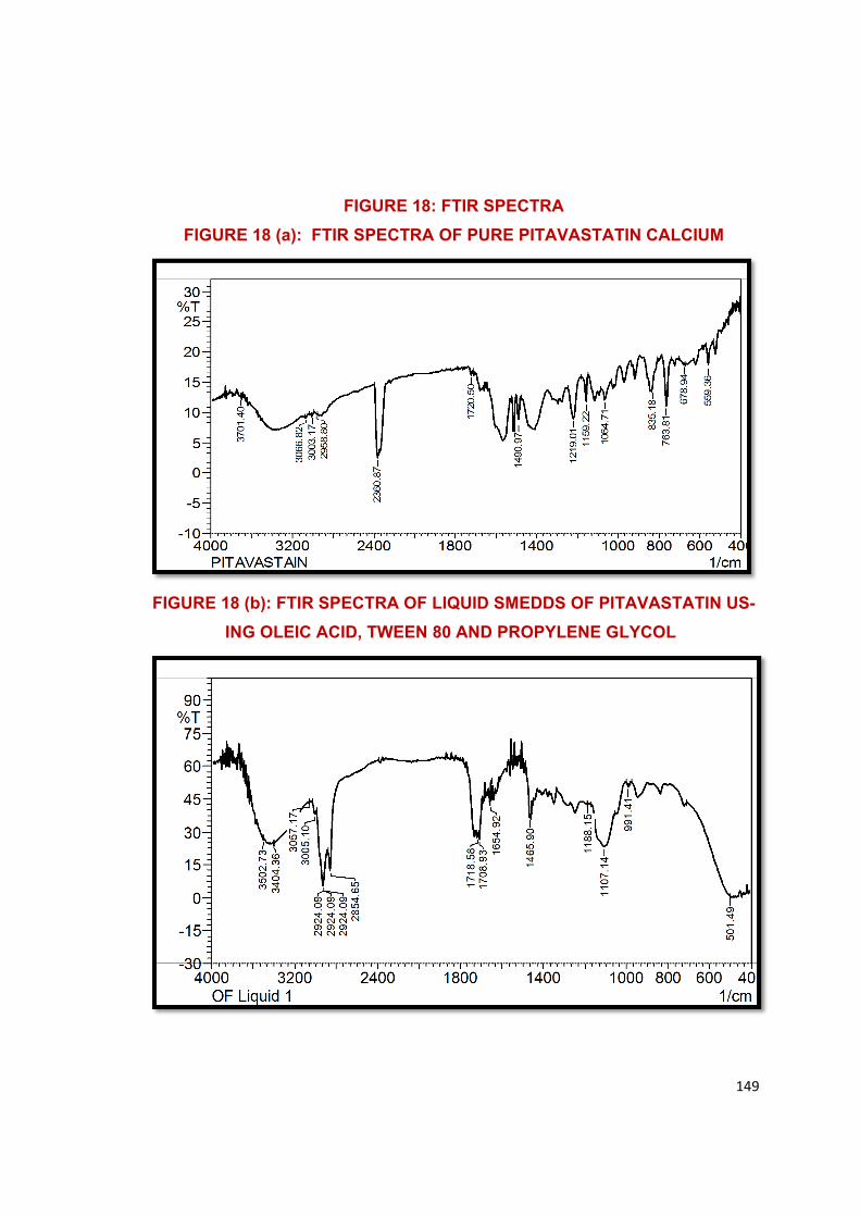
149
FIGURE 18: FTIR SPECTRA
FIGURE 18 (a): FTIR SPECTRA OF PURE PITAVASTATIN CALCIUM
FIGURE 18 (b): FTIR SPECTRA OF LIQUID SMEDDS OF PITAVASTATIN US-
ING OLEIC ACID, TWEEN 80 AND PROPYLENE GLYCOL
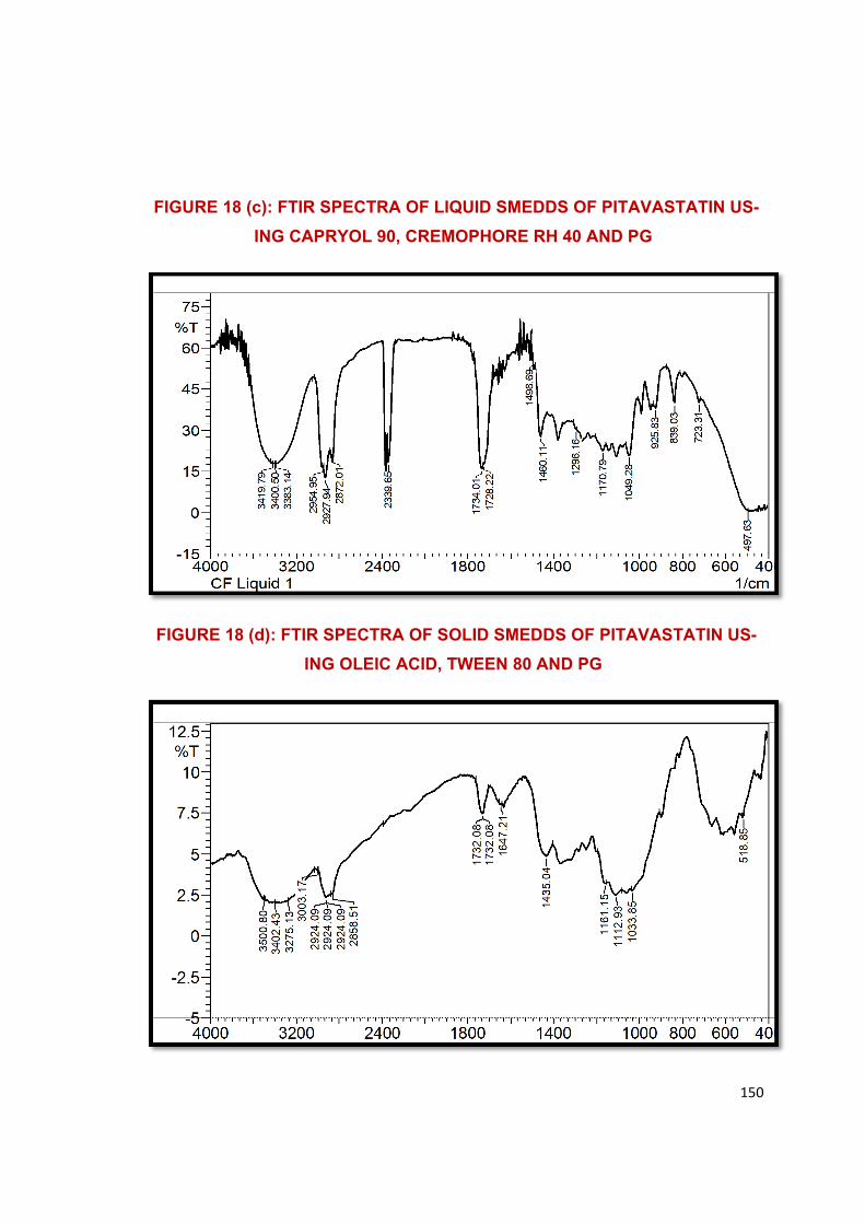
150
FIGURE 18 (c): FTIR SPECTRA OF LIQUID SMEDDS OF PITAVASTATIN US-
ING CAPRYOL 90, CREMOPHORE RH 40 AND PG
FIGURE 18 (d): FTIR SPECTRA OF SOLID SMEDDS OF PITAVASTATIN US-
ING OLEIC ACID, TWEEN 80 AND PG
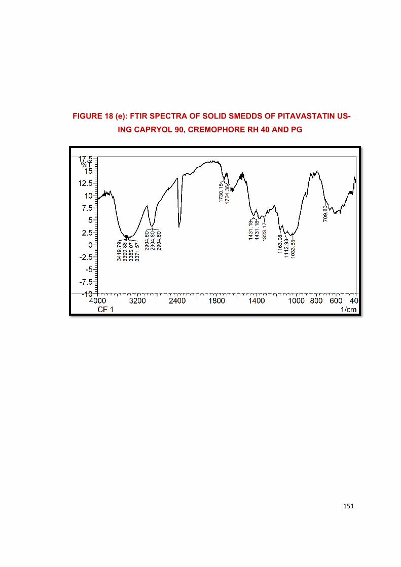
151
FIGURE 18 (e): FTIR SPECTRA OF SOLID SMEDDS OF PITAVASTATIN US-
ING CAPRYOL 90, CREMOPHORE RH 40 AND PG
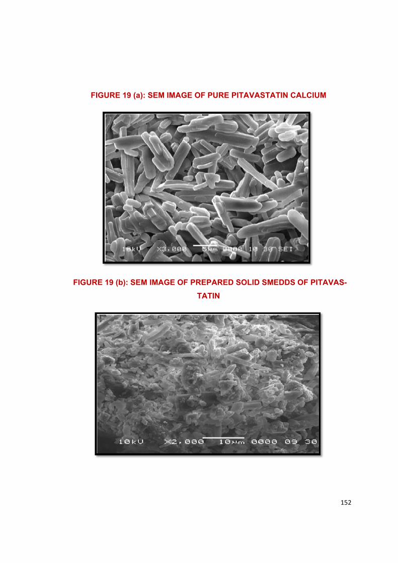
152
FIGURE 19 (a): SEM IMAGE OF PURE PITAVASTATIN CALCIUM
FIGURE 19 (b): SEM IMAGE OF PREPARED SOLID SMEDDS OF PITAVAS-
TATIN
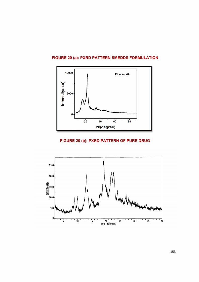
153
FIGURE 20 (a): PXRD PATTERN SMEDDS FORMULATION
FIGURE 20 (b): PXRD PATTERN OF PURE DRUG
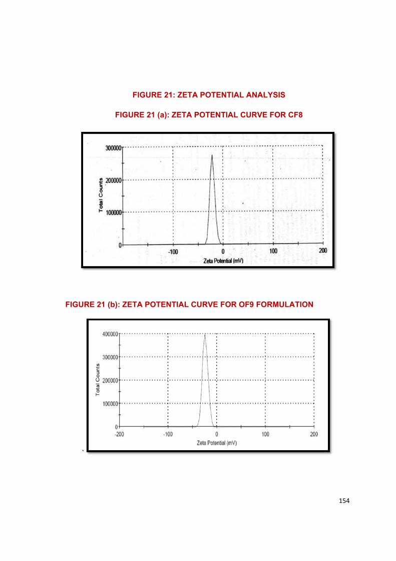
154
FIGURE 21: ZETA POTENTIAL ANALYSIS
FIGURE 21 (a): ZETA POTENTIAL CURVE FOR CF8
FIGURE 21 (b): ZETA POTENTIAL CURVE FOR OF9 FORMULATION
`
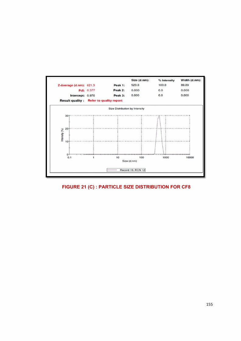
155
FIGURE 21 (C) : PARTICLE SIZE DISTRIBUTION FOR CF8
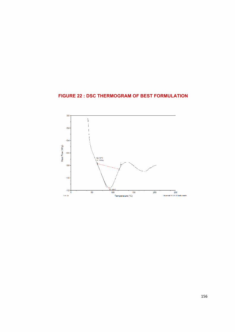
156
FIGURE 22 : DSC THERMOGRAM OF BEST FORMULATION
CHAPTER XI
SUMMARY AND CONCLUSION
CHAPTER XI SUMMARY AND CONCLUSION
DEPARTMENT OF PHARMACEUTICS, COLLEGE OF PHARMACY, MMC, MADURAI. 157
CHAPTER – XI
SUMMARY AND CONCLUSION
• In the present study, an attempt has been made to develop Self Micro
Emulsifying Drug Delivery system of Pitavastatin in order to enhance the
solubility and dissolution rate by incorporating the drug in a lipid vehicle.
• The results of compatability studies by Infrared spectroscopy and
differential scanning calorimetry (DSC), showed no interaction
between the drug and excipients.
• Various oils, surfactants and co-surfactants are optimized based on their
solulbilising capacity and % transmittance values.
• The solubility of Pitavastatin was found to higher in oleic acid
(97.89%±0.06) and Capryol 90 (93.69%±0.31)
• Pseudo ternary phase diagrams were constructed to get the maximum self
emulsification region of SMEDDS, and the optimum concentration of oil
was found to be 10-50% and Smixture was found to be 50-90% for both oils.
• Two different type of Pitavastatin liquid SMEDDS were successfully
prepared at different ratios of oil, surfactant and co-surfactant by simple
admixing method, namely CF1-CF9 and OF1-OF9.
• The prepared SMEDDS were evaluated for their physiochemical
parameters. (Dispersibility test, visual assessment, self emulsification
time, % transmittance and Refractive index).
• The RI of formulation ranges from 1.333 to 1.348.
• The % transmittance value for all the formulation ranges from 68 -97%.
And the formulation which shown % transmittance value of above 90%
provides clear emulsion. CF8 and OF8 shows maximum % transmittance
(97.47±0.18% and 94.28±0.22%)
CHAPTER XI SUMMARY AND CONCLUSION
DEPARTMENT OF PHARMACEUTICS, COLLEGE OF PHARMACY, MMC, MADURAI. 158
• The drug content of liquid SMEDDS ranges from 92- 98 %. Formulation
CF8 and OF8 shown maximum drug content. (98.02±0.07% and
98.23±0.17% respectively)
• Then the liquid SMEDDS were converted into S-SMEDDS by adsorption
technique by mixing it with Micro crystalline Cellulose at 1:1 w/w ratio.
• In vitro release study of all the formulations were shown an increased drug
release. Dissolution rate of all the formulations were improved when
compared to pure drug.
• The dissolution study was carried out in pH 6. 8 buffer for 2 hours. The
formulations shown higher rate of drug release than that of the pure drug
and marketed formulation, and all formulations showed 60-82% of drug
release within two hours. Formulation OF8 and CF8 shows
80.52±0.12% and 82.14±0.15% respectively, which is higher rate than
the pure drug (59.25±0.20%) and conventional tablet (64.72±0.21%)
• Particle size analyzer used to explore the particle size of Pitavastatin
SMEDDS showed a suitable particle size of 139.5 nm and 0.621nm for
OF8 and CF8 respectively.
• The polydispersibility index of selected SMEDDS formulations (CF8 & OF8)
were 0.377 and 0.291, which indicated a broad size distribution of
particles.
• Zeta potential value of Pitavastatin SMEDDS showed a negative surface
charge (- 23.9mv and -20.9mv).
• SEM studies confirmed the morphology of the solid SMEDDS.
• The crystalline state of the formulation was altered according to the XRPD
analysis. It confirms the solubilisation of drug.
CHAPTER XI SUMMARY AND CONCLUSION
DEPARTMENT OF PHARMACEUTICS, COLLEGE OF PHARMACY, MMC, MADURAI. 159
CONCLUSION
Pitavastain is a poorly water soluble drug, and its bioavailability is only
about 60% due it is poor solubility. When administering this poorly soluble
compound with hydrophilic carriers will enhance the solubility of the drug.
Hence, it was concluded that Self Micro Emulsifying drug delivery
system is a good approach to enhance the solubility and dissolution property
of Pitavastatin. The composition of optimized formulation [CF8 consist of
Capryol 90 as oil(45%), Cremophore RH(41.2%) as surfactant and Propylene
glycol(13.75%) as co-surfactant and OF8 consist of Oleic acid (45%), Tween
80 (41.2%) and Propylene glycol (13.75%)] containing 4mg of Pitavastatin
calcium showing drug release for solid SMEDDS formulation (82.14% &
80.52%), Particle size (139.5 nm & 621.3nm), Zeta potential (-23.29 & -20.9),
viscosity (0. 8824 cP). In-vitro drug release of the C8 and OF8 was highly
significant compared to pure drug and marketed conventional tablet. Here the
liquid SMEDDS are successfully converted into solid SMEDDS using MCC by
adsorption technique.

REFERENCES
REFERENCES
DEPARTMENT OF PHARMACEUTICS, COLLEGE OF PHARMACY, MMC, MADURAI. 161
• Nilesh Mahadeo Khutle ., Divya Kelan.,2016, Formulation and
Evaluation of Self-microemulsifying drug delivery system of
Cefpodoxime Proxetil, ejpmr, 2016, 3(3), P. No. 491-499.
• Maulik aj. Patel., Natvarlal M. Patel., Ritesh B. Patel., RAkesh P.
Patel.,2010, Formulation and evaluation of Self-microemulsifying drug
delivery system of Lovastatin, Asian Journal of Pharmaceutical sciences
2010, 5(6), P.No. 266-275.
• Ashok R. Patel., Pradeep R. Vavia., 2007, Preparation and I in vivo
Evaluation of SMEDDS containing Fenofibrate, AAPS Journal 2007; 9(3)
Article 41, P.No.344-352.
• Xianyi Sha., Juan Wu., Yanzuo Chen., Xiaoling Fang., 2012, Self-
microemulsifying drug-delivery system for improved oral bioavailability of
Probucol: preparation and evaluation, international Journal of
Nanomedicine2012:7, P.No. 705-712.
• Smita S. Pimple., Swapnil E. Yeole, Pravin D. Chaudhari., 2013,
Formulation and evaluation of Self Micro Emulsifying Drug Delivery
System for Poorly water Soluble Drug Risperidone, International Journal
of Pharmaceutical Sciences Review and Research, Nov-Dec2013, P.
No. 155-162.
• Ahmed A. Hussein., 2014, Preparation and Evaluation of Liquid and
Solid Self-Microemulsifying Drug Delivery System of Mebendazole,
Iraqui J Pharm Sci, Vol23(I), 2014, P.No.89-100.
• Tarkeshwar Prasad Shukla., Ashish Kumar Sharma., 2016,
Formulation Development and In vitro Evaluation of Acetazolamide Self
Microemulsifying Drug Delivery system to improve the solubility and
bioavailability, World Journal of Pharmacy and Pharmaceutical
Sciences, Vol 6, Issue 2, 2017, P.No. 1000-1027.
• Karunakara Reddy T., Yajaman Sudhakar., Devanna N., 2016,
Development and Evaluation of Atazanavir Solid SEDDS: In vitro–in vivo
REFERENCES
DEPARTMENT OF PHARMACEUTICS, COLLEGE OF PHARMACY, MMC, MADURAI. 162
Evaluation Studies, International Journal of Pharmaceutical Sciences
Review and Research, 41(2), Nov-Dec 2016; Article No.69, P. No. 380-
387
• Ramesh Jakki., Muzammil Afzal Syed., Prabhakar Kandadi., Kishan
Veerabrahma.,2012, Development of a Self-Microemulsifying Drug
Delivery System Of Domperidone: In Vitro And In Vivo
Characterization, 2012, Acta Pharm. 63 (2013), P.No. 241–251.
• Ashish Deshmukh., Shirishkumar Kulakrni., 2012, Novel Self
Micro-emulsifying Drug Delivery System (SMEDDS) of Efavirenz,
Journal of Chemical and Pharmaceutical Research, 2012, 4(8), P. No.
3914-3919.
• Sajal Kumar Jha., Roopa Karki., Venkatesh D.P., Geethalakshmi
A.,2011, Formulation Development and Characterizaion of
Microemulsion Drug Delivery systems containing Antiulcer drug
Famotidine, International Journal of Drug Development and Research,
Oct-Dec2011,Vol. 3. Issue 4, P. No. 336-343.
• Zh. Ch. KE., Z. Y. CH., 2012, Design and Evaluation of Self –
Microemulsifying Drug Delivery System of Naproxen, Latin American
Journal of Pharmacy, 31 (7): 1074-8 (2012), P. No. 1074.
• Gehan F Balata., Ebtessam A Essa., Hanan A Sharmardi., Samira
H Zaidan.,Mohamed AS Abourehab.,2016, Self-emulsifying drug
delivery system as a tool to improve solubility and bioavailability of
Resveratrol, Drug Design, Development and Therapy 2016:10, P. No.
117-128.
• Pradeep Patil., Vandana Patil., Anant Paradkar.,2007, Formulation
of a self-emulsifying system for oral delivery of Simvastatin: In vitro
and In vivo evaluation, Acta Pharm. 57 (2007), P. No. 111-122.
• Sarwar Beg., Suryakanta Swain., Harendra Pratap Singh., Ch
Niranjan Patra., ME Bhanoji Rao., 2012, Development, Optimization
and Characterization of Solid Self-Nano emulsifying Drug Delivery
System of Valsartan Using Porous Carriers, AAPS Pharm. SciTech,
Vol.13, No.4, December 2012, P. No. 1416-1427.
REFERENCES
DEPARTMENT OF PHARMACEUTICS, COLLEGE OF PHARMACY, MMC, MADURAI. 163
• Urvashi Goyal., Anit Gupta., Avtar Chand Rana ., Geeta
Aggarwal., Self MiceroEmulsifying Drug Delivery System: A Method
for Enhancement of Bioavailability
• Shukla Prachi., Prajapati S.K., Sharma Upendra K., Shivhare
Shipra., Akhtar Ali.,2012, A review on Self Micro Emulsifying Drug
Delivery System: An approach to enhance the oral bioavailability of
poorly water soluble drugs, Intrnational Research Journal of
Pharmacy,2012,3 (9), P. No. 1-6.
• Gupta Roop., Gupta Rakesh., Rathore Garvendra singh., 2009,
Enhancement of oral bioavailability of Lipophilic drugs from Self- Micro
Emulsifying Drug Delivery System (SMEDDS), 2009, International
Journal of Drug Development and Research, Sep-Dec 2009, Vol. 1,
Issue 1, P. No. 10-18.
• Shailesh T. Prajapati., Harsh A. Joshi., Chhaganbhai N,
Patel.,2013, Preparation and Characterization of Self-Microemulsifying
Drug Delivery System of Olmesartan Medoxomil for Bioavalilability
improvement, Journal of Pharmaceutics, Volume 2013, Article Id
728425, P. No.
• Vikas Panday., Seema Kohli., 2017, Formulation, In vitro evaluation
and SMEDDS of Pioglitazone, Future Journal of Pharmaceutical
Sciences, Vol. 3, Issue 1, P. No. 53-59.
• Khalid Akhter Ansari., Kunal Pratap Pagar., Shoeb Anwar.,2014,
Design and optimization of Self-microemulsifying drug delivery system
of Felodipine for chronotherapeutic application, Brazilian Journal of
Pharmaceutical Sciences, Vol. 50 no. 1, Jan- Mar.2014.
• Bhagwat Durgacharan A., D Souza John I.,2012, Development of
Solid Micro Emulsifying Drug Delivery System with Neusilin US2 for
Enhanced Dissolution Rate of Telmisartan, International Journal of
Drug Development and Research, Oct-Dec 2012,4(4): P. No. 398-407.
• Pankaj V. Dangre.,Ritu M., Gilhotra., Shashikant N. Dhole.,2016,
Formulation and Development of Solid Self micro-emulsifying drug
delivery system containing Chlorthlaidone for improvement of
REFERENCES
DEPARTMENT OF PHARMACEUTICS, COLLEGE OF PHARMACY, MMC, MADURAI. 164
dissolution, Journal of Pharmaceutical Investigation,Dec.2016, Vol. 46,
Issue7, P. No.633-644.
• Kommuru TR, Gurley B, Khan MA, Reddy IK .,Self-emulsifying drug
delivery systems (SEDDS) of coenzyme Q10: formulation development
and bioavailability assessment. Int J Pharm, 2001; 212: pg.233-46.
• Humberstone AJ, Charman WN., Lipid-based vehicles for the oral
delivery of poorly water soluble drugs. Adv Drug Del Rev, 1997; 25:
pg.103-28.
• Pouton CW., Formulation of self-emulsifying drug delivery systems. Adv
Drug Del Rev, 1997; 25: pg. 47-58.
• Gursoy RN, Benita S., Self-emulsifying drug delivery systems (SEDDS)
for improved oral delivery of lipophilic drugs. Biomed Pharmacother,
2004; 58: pg. 173-82.
• Stegemanna S, Leveillerb F., When poor solubility becomes an issue:
from early stage to proof of concept. Eur J Pharm Sci ,2007; 31:
pg.249-61.
• Murdandea SB, Gumkowskia MJ., Development of a self-emulsifying
formulation that reduces the food effect for torcetrapib. Int J of Pharm,
2008; 351: pg. 15-22.
• Pouton CW., Formulation of poorly water-soluble drugs for oral
administration: physicochemical and physiological issues and the lipid
formulation classification system. Eur J of Pharm Sci, 2006; 29:
pg.278-87.
• Cuine JF., McEvoy CL., Charman WN ., Pouton CW., Edwards GA.,
Benameur H., Porter CJ., Evaluation of the impact of surfactant
digestion on the bioavailability of danazol after oral administration of
lipidic selfemulsifying formulations to dogs. J of Pharm Sci, 2008; 97:
pg.993-1010.
• Gershanik T., Benita S., Self-dispersing lipid formulations for improving
oral absorption of lipophilic drugs. Eur J of Pharm Sci, 2000; 50: pg.179-
188.
REFERENCES
DEPARTMENT OF PHARMACEUTICS, COLLEGE OF PHARMACY, MMC, MADURAI. 165
• Pouton CW., Formulation of poorly water-soluble drugs for oral
administration: physicochemical and physiological issues and the lipid
formulation classification system. Eur J of Pharm Sci, 2006; 29:
pg.278-87.
• Strickley RG., Solubilizing excipients in oral and injectable formulations.
Pharm Res, 2004; 21: pg.201–230.
• Tang J., Self-Emulsifying Drug Delivery Systems: strategy for improving
oral delivery of poorly soluble drugs. Cur Drug Th, 2007; 2: pg. 85-93.
• Kawakami K, Yoshikawa T, Moroto Y, Kanakao E, Takahuani K,
Nishihara Y, Masuda K. Microemulsion formulation for enhanced
absorption of poorly soluble Drugs. I. Prescription design. J of Contr Rel ,
2002; 81: pg.75-82.
• Lawrence MJ, Rees GD., Microemulsion-based media as novel drug
delivery system. Adv Drug Deliv Rev, 2000; 45: pg.89-121.
• Gursoy RN, Benita S., Self-emulsifying drug delivery systems (SEDDS)
for improved oral delivery of lipophilic drugs. Biom & Pharma, 2004; 58:
pg.173–182.
• Pouton CW., Self-emulsifying drug delivery systems: Assessment of the
efficiency of emulsification. International Journal of Pharmaceutics, 1985;
27(2-3): 335-348.
Copyright © 2022 FDOKUMEN