Natural deep eutectic solvent supported targeted solid–liquid ...
16
Natural deep eutectic solvent supported targeted solid–liquid polymer carrier for breast cancer therapy† Xianfu Sun, a Periyakaruppan Pradeepkumar, * b Naresh Kumar Rajendran, c Harshavardhan Shakila, d Nicolette Nadene Houreld, c Dunia A. Al Farraj, e Yousif M. Elnahas, f Nandhakumar Elumalai g and Mariappan Rajan * b Solid–liquid nanocarriers (SLNs) are at the front of the rapidly emerging field of medicinal applications with a potential role in the delivery of bioactive agents. Here, we report a new SLN of natural deep eutectic solvent (NADES) and biotin-conjugated lysine–polyethylene glycol copolymer. The SLN system was analyzed for its functional groups, thermal stability, crystalline nature, particle size, and surface morphology through the instrumental analysis of FT-IR, TGA, XRD, DLS, SEM, and TEM. Encapsulation of PTX (paclitaxel) and 7-HC (7-hydroxycoumarin) with the SLN was carried out by dialysis, and UV-visible spectra evidenced the drug loading capacity and higher encapsulation efficiency obtained. The enhanced anticancer potential of PTX- and 7-HC-loaded SLN was assessed in vitro, and the system reduces the cell viability of MDA-MB-231 cells. The PTX- and 7-HC-loaded SLN system was investigated in a breast cancer-induced rat model via in vivo studies. It shows decreased lysosomal enzymes and increased levels of caspase to cure breast tumors. It very well may be reasoned that the designed PTX- and 7-HC-loaded SLN system has strong anticancer properties and exhibits potential for delivery of drug molecules in cancer treatment. Introduction Recent research development in medicinal applications has advanced through the improvement of new methods and materials that are more biodegradable, biocompatible, and less toxic. 1,2 There is an enormous attempt to explore the role/ function of synthetic biomaterials used in drug delivery appli- cations. 3 Currently, synthetic and natural polymers, metal complexes, peptides, proteins, and hydrogels are used for the development of anticancer drug delivery systems, 4–8 which reduce some disadvantages over conventional anticancer treatments such as poor solubility, low biodegradability, toxic nature, and poor intracellular penetration, retention time and sustainable release of encapsulated drugs. 9 A signicant number of nanoparticles such as inorganic nanoparticles or other polymeric/liquid nanoparticles have also been used in developing drug carrier systems. The drug carrier leads to the fabrication of a nanocarrier, and most of these are made up of synthetic polymers and not liquid-based carriers. 10 Researchers have paid more attention to the advancement of new carrier systems by using ILs (ionic liquids) and DESs (deep eutectic solvents). 11 Unfortunately, ILs and DESs have exhibited thermal instability, low drug loading levels, low drug release and solubility, and have a very weak interaction with and a toxic nature in biological systems. This issue can be overcome by the utilization of natural deep eutectic solvents (NADES). A NADES is a highly biocompatible material designed to serve as a carrier molecule that transports drugs to a specic site without any side effects; it is a non-toxic solvent prepared by secondary metabolites and does not affect the drug release mechanism. 12 Secondary metabolites like phenolics, terpenoids, avonoids, and other natural compounds are crucial for medicinal applications. 13,14 Compared to organic solvents, the use of NADES has gained attention in the synthesis of carrier systems. 15–18 a Department of Breast, The Affiliated Tumor Hospital of Zhengzhou University, Zhengzhou, Henan, 450008, China b Biomaterials in Medicinal Chemistry Laboratory, Department of Natural Products Chemistry, School of Chemistry, Madurai Kamaraj University, Madurai-625021, Tamil Nadu, India. E-mail: [email protected] c Laser Research Centre, Faculty of Health Sciences, University of Johannesburg, PO Box 17011, Doornfontein 2028, South Africa d Department of Molecular Microbiology, School of Biotechnology, Madurai Kamaraj University, Madurai-625021, India e Department of Botany and Microbiology, College of Science, King Saud University, Riyadh 11451, Saudi Arabia f Department of Surgery, College of Medicine, King Saud University Medical City, King Saud University, Riyadh, Saudi Arabia g Department of Biochemistry, Sri Muthukumaran Medical College and Research Institute, Chennai-600069, Tamil Nadu, India † Electronic supplementary information (ESI) available. See DOI: 10.1039/d0ra03790g Cite this: RSC Adv. , 2020, 10, 36989 Received 27th April 2020 Accepted 11th August 2020 DOI: 10.1039/d0ra03790g rsc.li/rsc-advances This journal is © The Royal Society of Chemistry 2020 RSC Adv. , 2020, 10, 36989–37004 | 36989 RSC Advances PAPER Open Access Article. Published on 07 October 2020. Downloaded on 5/30/2022 6:01:06 PM. This article is licensed under a Creative Commons Attribution 3.0 Unported Licence. View Article Online View Journal | View Issue
-
Upload
khangminh22 -
Category
Documents
-
view
1 -
download
0
Transcript of Natural deep eutectic solvent supported targeted solid–liquid ...
RSC Advances
PAPER
Ope
n A
cces
s A
rtic
le. P
ublis
hed
on 0
7 O
ctob
er 2
020.
Dow
nloa
ded
on 5
/30/
2022
6:0
1:06
PM
. T
his
artic
le is
lice
nsed
und
er a
Cre
ativ
e C
omm
ons
Attr
ibut
ion
3.0
Unp
orte
d L
icen
ce.
View Article OnlineView Journal | View Issue
Natural deep eut
aDepartment of Breast, The Affiliated Tu
Zhengzhou, Henan, 450008, ChinabBiomaterials in Medicinal Chemistry Labo
Chemistry, School of Chemistry, Madura
Tamil Nadu, India. E-mail: rajanm153@gmcLaser Research Centre, Faculty of Health Sci
17011, Doornfontein 2028, South AfricadDepartment of Molecular Microbiology, Sc
University, Madurai-625021, IndiaeDepartment of Botany and Microbiology, C
Riyadh 11451, Saudi ArabiafDepartment of Surgery, College of Medicine
Saud University, Riyadh, Saudi ArabiagDepartment of Biochemistry, Sri Muthuk
Institute, Chennai-600069, Tamil Nadu, Ind
† Electronic supplementary informa10.1039/d0ra03790g
Cite this: RSC Adv., 2020, 10, 36989
Received 27th April 2020Accepted 11th August 2020
DOI: 10.1039/d0ra03790g
rsc.li/rsc-advances
This journal is © The Royal Society o
ectic solvent supported targetedsolid–liquid polymer carrier for breast cancertherapy†
Xianfu Sun,a Periyakaruppan Pradeepkumar,*b Naresh Kumar Rajendran,c
Harshavardhan Shakila,d Nicolette Nadene Houreld,c Dunia A. Al Farraj,e
Yousif M. Elnahas,f Nandhakumar Elumalaig and Mariappan Rajan *b
Solid–liquid nanocarriers (SLNs) are at the front of the rapidly emerging field of medicinal applications with
a potential role in the delivery of bioactive agents. Here, we report a new SLN of natural deep eutectic
solvent (NADES) and biotin-conjugated lysine–polyethylene glycol copolymer. The SLN system was
analyzed for its functional groups, thermal stability, crystalline nature, particle size, and surface
morphology through the instrumental analysis of FT-IR, TGA, XRD, DLS, SEM, and TEM. Encapsulation of
PTX (paclitaxel) and 7-HC (7-hydroxycoumarin) with the SLN was carried out by dialysis, and UV-visible
spectra evidenced the drug loading capacity and higher encapsulation efficiency obtained. The
enhanced anticancer potential of PTX- and 7-HC-loaded SLN was assessed in vitro, and the system
reduces the cell viability of MDA-MB-231 cells. The PTX- and 7-HC-loaded SLN system was investigated
in a breast cancer-induced rat model via in vivo studies. It shows decreased lysosomal enzymes and
increased levels of caspase to cure breast tumors. It very well may be reasoned that the designed PTX-
and 7-HC-loaded SLN system has strong anticancer properties and exhibits potential for delivery of drug
molecules in cancer treatment.
Introduction
Recent research development in medicinal applications hasadvanced through the improvement of new methods andmaterials that are more biodegradable, biocompatible, and lesstoxic.1,2 There is an enormous attempt to explore the role/function of synthetic biomaterials used in drug delivery appli-cations.3 Currently, synthetic and natural polymers, metalcomplexes, peptides, proteins, and hydrogels are used for thedevelopment of anticancer drug delivery systems,4–8 which
mor Hospital of Zhengzhou University,
ratory, Department of Natural Products
i Kamaraj University, Madurai-625021,
ail.com
ences, University of Johannesburg, PO Box
hool of Biotechnology, Madurai Kamaraj
ollege of Science, King Saud University,
, King Saud University Medical City, King
umaran Medical College and Research
ia
tion (ESI) available. See DOI:
f Chemistry 2020
reduce some disadvantages over conventional anticancertreatments such as poor solubility, low biodegradability, toxicnature, and poor intracellular penetration, retention time andsustainable release of encapsulated drugs.9 A signicantnumber of nanoparticles such as inorganic nanoparticles orother polymeric/liquid nanoparticles have also been used indeveloping drug carrier systems. The drug carrier leads to thefabrication of a nanocarrier, and most of these are made up ofsynthetic polymers and not liquid-based carriers.10
Researchers have paid more attention to the advancementof new carrier systems by using ILs (ionic liquids) and DESs(deep eutectic solvents).11 Unfortunately, ILs and DESs haveexhibited thermal instability, low drug loading levels, low drugrelease and solubility, and have a very weak interaction withand a toxic nature in biological systems. This issue can beovercome by the utilization of natural deep eutectic solvents(NADES). A NADES is a highly biocompatible materialdesigned to serve as a carrier molecule that transports drugs toa specic site without any side effects; it is a non-toxic solventprepared by secondary metabolites and does not affect thedrug release mechanism.12 Secondary metabolites likephenolics, terpenoids, avonoids, and other naturalcompounds are crucial for medicinal applications.13,14
Compared to organic solvents, the use of NADES has gainedattention in the synthesis of carrier systems.15–18
RSC Adv., 2020, 10, 36989–37004 | 36989
RSC Advances Paper
Ope
n A
cces
s A
rtic
le. P
ublis
hed
on 0
7 O
ctob
er 2
020.
Dow
nloa
ded
on 5
/30/
2022
6:0
1:06
PM
. T
his
artic
le is
lice
nsed
und
er a
Cre
ativ
e C
omm
ons
Attr
ibut
ion
3.0
Unp
orte
d L
icen
ce.
View Article Online
Citrus limon peels are rich in bioactive compounds, includingantioxidants, avonoids, vitamins, and prolinebetaine (PB). PB isa natural bioactive compound, which has numerous biologicalproperties, including antifungal, antibacterial, and anticanceractivities.19,20 Anticancer activity is an essential property of PB andis due to the presence of amine which is a highly reactivecompound that can readily react with biological compoundmoieties such as hydroxyl groups. In this study, PB is isolated andused for the synthesis of NADES in combination with lactic acid(LA), which is an excellent electron donor/acceptor. The self-assembled drug carrier system with PB is reported to targettumor cells.21 However, there is the need for an extra efficient andactive targeting ligand to achieve better cellular uptake of drug-loaded carriers. Different ligands such as polysaccharides, folicacid, peptides, and biotin are involved in targeting drug carriersynthesis.22 Among these, biotin is predominantly utilized asa tumor-focusing group for numerous cancer chemotherapyapplications.23 Tumor cells need vitamins for their survival, andspeedily growing cancer cells overexpress the receptors for thisvitamin on their cell surface.
This paper reports for the rst time on the preparation ofNADES using secondary metabolites, and it is used as a biotin-conjugated solid–liquid nanocarrier (SLN) for the encapsulationof the anticancer drugs paclitaxel (PTX) and 7-hydroxycoumarin(7-HC). The advantage of SLNs is the ability to incorporatehydrophobic anticancer drugs in the inner core and hydrophilicanticancer drugs on the outer core. The incorporation of PTXand 7-HC in the SLN system will reduce side effects and improvebioavailability. The anticancer activity of the synthesized PTX-and 7-HC-loaded SLN system was investigated in vitro in a MDA-MB-231 breast cancer cell line, and in vivo in a DMBA-inducedbreast cancer rat model.
Experimental sectionMaterials
EDC (1-ethyl-3-(3-dimethylaminopropyl)carbodiimide), LA(lactic acid), NHS (N-hydroxysuccinimide), p-TsOH (p-toluene-sulfonic acid), tin(II) 2-ethylhexanoate (Sn(Oct)2), and 7,12-dimethylbenz[a] anthracene (DMBA) were procured fromSigma-Aldrich, Mumbai, India. Lysine, polyethylene glycol-6000, biotin, PTX, and 7-HC were purchased from Merck,Mumbai, India. Petroleum ether (PE), dichloromethane (DCM),ethyl acetate (EA), diethyl ether (DE), methanol, acetone,acetonitrile (AN) and dimethyl sulfoxide (DMSO) were receivedfrom Himedia Laboratories Pvt. Ltd, Mumbai, India (HPLCgrade). A human breast adenocarcinoma (MDA-MB-231) cellline and normal mouse broblast cell line (L929) obtained fromthe National Center for Cell Science, Pune, India were used forthis work. Caspase colorimetric assay kit was from R and DSystems Inc., USA. The chemicals and solvents used were ofanalytical grade and main clarity. In all the experiments,double-distilled (DD) water was used as a solvent and washingsolution. The other chemicals were purchased from thedifferent companies like BDH Division (Mumbai, India), GlaxoLaboratories, Sisco Research Laboratories, Sarabhai Chemicals(Vadodara, India), and SD Fine Chemicals (Mumbai, India).
36990 | RSC Adv., 2020, 10, 36989–37004
Isolation of prolinebetaine from Citrus limon peels
Citrus fruits (Citrus limon peels) were acquired from a nearbymarket at Madurai, Tamil Nadu, India.24 The fruits were cleanedwith DD water and acetone. The fruit peels were chopped intolittle pieces and dried at room temperature (27 �C). Driedsamples were powdered into ne particles utilizing a blender.Ground samples were then dissolved in various solvents such aspetroleum ether, chloroform, ethyl acetate, methanol, acetoni-trile, and water for solubility optimization. Samples were loadedonto a silica gel packed column (100–200 mesh). Pet ether wasused for initial elution. Fractions were removed for primarycompounds such as oils, fats, fatty acids, resin, and chlorophyllcompounds. Solvents were recovered through vacuum rotaryevaporation (Rotavapor® R-300).
Fractions were collected and used for further analysis.Optimized and stabilized ethyl acetate andmethanol in ratios of70 : 30 and 50 : 50 were used for the collection of 4-hydrox-yprolinebetaine and PB. The solvents from the collectedsamples were evaporated at room temperature (27 �C). Thederived compounds were identied by HPLC, 1H- and 13C-NMR,and FT-IR analysis.
Synthesis of NADESs
NADESs were prepared according to a previously reportedmethod.25 Isolated and puried PB and LA were mixed in molarratios of 1 : 1, 1 : 2, 1 : 3, and 2 : 1 at 40 �C. Mixtures weremagnetically stirred until a transparent homogenous liquid wasobtained. Aerward, the eutectic mixture was chilled to roomtemperature. Ratios of 1 : 2 and 1 : 3 formed a clear solution,while proportions of 1 : 1 and 2 : 1 did not create a clear solu-tion, as is shown in ESI Fig. 1.† The formation of NADESs andthe chemical structures were identied through 1H and 13CNMR and FT-IR spectroscopy, as shown in ESI Fig. 3 and 4.†
Synthesis of biotin-g-lysine
Biotin graed lysine was prepared by the condensation methodand carried out in the presence of EDC$HCl and NHS as perprevious literature.26 In general, biotin (0.2 g, 0.00136 mmol)and lysine (0.33 g, 0.00136 mmol) were solubilized in DCM (15mL). To this mixture, EDC$HCl (0.168 mL, 0.00136 mmol) andNHS (0.15 g, 0.00136 mmol) were added drop-wise. The pH ofthe reaction mixture was adjusted to pH 7.4 using 1 N NaOHand stirred for 2 h at 45 �C. Then, the reaction mixture wasprecipitated using an organic solvent (acetone). The precipitatewas ltered using Whatman (0.2 mm) lter paper and dried ina vacuum chamber. The obtained precipitate was dialyzed usinga dialysis membrane (MWCO-12000) in DD water for 22 h toeliminate unreacted constituents.
Synthesis of biotin-g-lysine-co-polyethylene glycol (biotin-g-lysine-co-PEG)
Typically, biotin-g-lysine (1 g, 0.00268 mmol), p-TsOH (0.4 g,0.00268 mmol), and polyethylene glycol-6000 (1 g) in 50 mLtoluene were placed in a double neck round reaction ask witha Dean–Stark apparatus.27 Then, Sn(Oct)2 (0.8 mL, 1.0857mmol)
This journal is © The Royal Society of Chemistry 2020
Paper RSC Advances
Ope
n A
cces
s A
rtic
le. P
ublis
hed
on 0
7 O
ctob
er 2
020.
Dow
nloa
ded
on 5
/30/
2022
6:0
1:06
PM
. T
his
artic
le is
lice
nsed
und
er a
Cre
ativ
e C
omm
ons
Attr
ibut
ion
3.0
Unp
orte
d L
icen
ce.
View Article Online
was added drop-wise, and heated at 125 �C for 24 h to reux themixture with continuous stirring and elimination of water. Thereaction mixture was cooled to room temperature (27 �C);subsequently, the reaction solution was dissolved in ether andltered to remove by-products. A Soxhlet apparatus was used forpolymer extraction for 24 h with an organic solvent (ethylacetate). Then, the obtained crude product was dried ina vacuum and the chemical structure of biotin-g-lysine-co-PEGwas conrmed through FT-IR spectroscopy.
NADES-based biotin-conjugated solid–liquid polymer insolvent emulsion method
NADES-based biotin-conjugated polymeric SLN was prepared bythe method mentioned in our previous report.28 Initially, 1 g ofbiotin-g-lysine-co-PEG was dissolved in 1 mL of NADES. Thisreaction mixture was kept under continuous stirring for 1 h atroom temperature until the mixture became a homogeneoussolution and attained a viscous consistency. Nanocarriers wereformed using dialysis in aqueous solution, following a previousreport.29 Briey, 100 mg of NADES-biotin-g-lysine-co-PEG systemwas dispersed in 1 mL DMSO solution and the solution wasplaced into a dialysis bag and dialyzed against 250 mL of DDwater for 24 h to eliminate the DMSO solvent. The separatedSLN was used for further processing.
Determination of self-assembly of carrier formation
The critical nanocarrier concentration (CMC) value of NADES-based biotin-conjugated solid–liquid polymer nanocarrier wasstudied by uorescence spectroscopy through pyrene as a uo-rescent moiety.30 In brief, various concentrations of NADES-based biotin-conjugated solid–liquid polymer nanocarrier(0.1 mL to 1.0 mL) were dissolved in 10 mL of DMSO solution.Then, pyrene (0.1 mg) was solubilized in 5 mg mL�1 methanoland added to each test container of NADES-based biotin-conjugated solid–liquid polymer nanocarrier solution. Thereaction solution wasmagnetically stirred at 27 �C for 1 day, andsolvents vaporized. Aerwards, 5 mL of distilled water wasadded individually in a sample tube. The uorescence of everypyrene-containing NADES-based biotin-conjugated solid–liquidpolymer nanocarrier solution was determined by emissionspectra (spectrouorometer, Shimadzu F-4500, Japan) ata wavelength of 472 nm at 21 � 1 �C.
Encapsulation efficiency (EE)
The NADES-based biotin-conjugated solid–liquid polymernanocarrier (100 mg) was taken in a 25 mL beaker and PTX(20 mgmL�1) and 7-HC (20mgmL�1) dispersed in ethanol wereadded to the SLN and stirred for 1 h with a magnetic stirrer. Fordetermining the drug encapsulation efficiency, the supernatantsolution was collected at 10 min intervals and analyzed by UV-visible spectroscopy at lmax values of 270 nm and 320 nm(Shimadzu UV 1800).16 EE was calculated with the followingequation:1
This journal is © The Royal Society of Chemistry 2020
Encapsulation efficiency ð%Þ
¼ total amount of drug� free amount of drug
total amount of drug� 100
(1)
Drug release study
The discharge of PTX and 7-HC from the nanocarrier wastransferred to the dialysis system and a physiological buffersolution at pH values of 2.8, 5.5, and 7.4 was used at 27 �C.Briey, PTX- and 7-HC-loaded nanocarrier samples (30mg) wereadded to 5 mL of fresh PBS (pH 2.8, 5.5, and 7.4) in the dialysisbag at room temperature. The dialysis membrane was incu-bated in 50 mL PBS. PTX- and 7-HC-loaded SLN samples werestirred using a magnetic stirrer at a constant rate (100 rpm), and3 mL of the supernatant solution was extracted at standard timeintervals and supplemented with 3 mL of new buffer solution.Drugs released into the medium were quantied by a UV-visiblespectrometer at lmax values of 270 nm and 320 nm (Shimadzu1600, Japan).
Swelling studies
The swelling behavior of PTX- and 7-HC-loaded SLN (30mg) wasanalyzed in various buffer solutions at pH 2.8, 5.5, and 7.4 atroom temperature (27 �C). At standard time intervals, sampleswere removed and weighed. A stable weight was achieved andthe following equation was used to determine the swellingbehavior:
Swelling ð%Þ ¼ We �Wd
Wd
(2)
where We and Wd are the weights of swollen polymer and driednanocarriers, respectively.
Biological characterizations
Cell culture. A human breast adenocarcinoma cell line(MDA-MB-231) and a mouse broblast cell line (L929) werepurchased from the NCCS (National Center for Cell Science,Pune, India). Cells were maintained in Dulbecco's modiedEagle's medium supplemented with 10% fetal bovine serum,and 1 unit per mL penicillin and 1 mg mL�1 streptomycin, andincubated at 37 �C with 5% CO2 in a humidied chamber. Cellswere passaged every 3 to 4 days using trypsin/ethylenediaminetetraacetic acid (EDTA) for detachment.
Cell viability. MDA-MB-231 human breast adenocarcinomaand L929 normal mouse broblast cells were used to interrogatethe cytotoxic effect of PTX- and 7-HC-loaded and unloaded SLNsystem. Cells were seeded into 96-well plates at a seedingdensity of 1 � 105 cells per well and incubated for 24 h at 37 �Cin 5% CO2. Various concentrations of PTX- and 7-HC-loadedSLN system (10, 25, 50, 75, and 100 mg mL�1) were added towells and cells were cultured for 24 h. Aerwards, 25 mL of MTT(3-(4,5-dimethylthiazol-2-yl)-2,5-diphenyltetrazolium bromide)assay solution was added to each well and incubated for 5 h at
RSC Adv., 2020, 10, 36989–37004 | 36991

RSC Advances Paper
Ope
n A
cces
s A
rtic
le. P
ublis
hed
on 0
7 O
ctob
er 2
020.
Dow
nloa
ded
on 5
/30/
2022
6:0
1:06
PM
. T
his
artic
le is
lice
nsed
und
er a
Cre
ativ
e C
omm
ons
Attr
ibut
ion
3.0
Unp
orte
d L
icen
ce.
View Article Online
37 �C. The cell culture medium was measured at an absorptionwavelength of 570 nm using a microplate reader (SpectraMaxM3, Molecular Devices, USA). The corresponding cell viability(%) was correlated to normal cells. The following equation forcell viability was used for IC50 calculation:
Cell viability ð%Þ ¼ OD of the treated cells
OD of the untreated cells ðcontrolÞ� 100 (3)
Mean OD of the untreated cells (control), is the mean of theuntreated cells used as the control.
Cellular uptake analysis by confocal microscope. MDA-MB-231 cells were cultured in 96-well plates and incubated withPTX- and 7-HC-loaded SLN for 24 h. Cells were washed threetimes with PBS, xed with 5% paraformaldehyde for 30 min,and stained with 4,6-diamidino-2-phenylindole (DAPI; 1 mgmL�1 in PBS) for 10 min. Cells were examined by confocal laserscanning microscopy (Olympus IX 81 under DU897 mode,Japan).
Animals and experimental design. Female Sprague-Dawleyrats (eight weeks old) were separated into four groups, witheach group having six animals. The regimen dose is shown inTable 1. All groups of animals had taken food and water, whichwas monitored daily for 14 days. A permanent amount of ratchow and uid was given to every rat and relled aer that day.Aer treatment periods of 6 weeks, the rats were fasted for twonights, anesthetized and sacriced by cervical decapitation.Whole blood samples were collected from both separations likeplasma/serum, respectively. The mammary tissue was cut andwashed in ice-cold saline and weighed. Tissue was cut up andhomogenized with 0.1 M Tris–HCl buffer (pH 7.4, and 10%) andcentrifuged (3000 � g for 10 min). The nally obtained super-natant solution was used for enzyme assays. The bodyweights ofthe whole animals were recorded before and aer the treatmentand sacrice.
The experimental work was performed with ethical normsapproved by committee members of the Animals (CPCSEA),Ministry of Environment and Forests (Animal Welfare Division),
Table 1 Experimental animal groups and treatment conditionsa
Group Group name
Group I Control
Group II Induced
Group III SLN unloaded
Group IV SLN loaded
a b.wt, body weight; DMBA, 7,12-dimethylbenz[a]anthracene.
36992 | RSC Adv., 2020, 10, 36989–37004
Government of India, and Institutional Animal EthicsCommittee (IEAC) Guidelines.
Gross observations and tumor volume. The change in thebody weight and tumor of animals was measured and recordedin grams. The following formula was used to calculate tumorvolume:
v ¼ 4/3pr12r2 (radius r1 < r2; r ¼ tumor diameter in mm/2)
Estimation of lysosomal marker enzymes. The enzymaticactivity of acid phosphatase and cathepsin-D was evaluated asreported previously,31,32 and values were expressed as phenol/protein min per mg and tyrosine/protein min per mg, respec-tively. b-D-Glucuronidase was assayed by the method of Kawaiand Anno,33 and the activity was expressed as p-nitrophenol (mper moles) formed per min per mg protein.
Measurement of caspase-3, -8, and -9 activities. Caspaseactivation is a common apoptotic mechanism by which anti-cancer agents induce apoptosis. Hence, the level of caspaseactivation was determined for PTX- and 7-HC-loaded SLNsystem. The activities of caspase-3, -8, and -9 were determinedusing a caspase colorimetric assay kit according to the manu-facturer's recommended protocol (R and D Systems Inc., USA).Spectrophotometric detection by the chromophore p-nitro-anilide (pNA) assay was used for the cleavage from the labeledsubstrate that recognizes an optimal tetrapeptide sequence ofthe individual activation sites. Briey, tissue homogenates werewashed with ice-cold PBS, lysed with 50 mL of cold lysis bufferand incubated on ice for 10 min. Protein concentrations of thehomogenates were assessed by the Bradford method, and 200mg of protein was diluted in 50 mL lysis buffer solution. Inaddition to that, 50 mL of 2� reaction buffer (containing 10 mMDTT) and 4 mM DEVD-pNA substrate were added to eachsample well. Aer incubation at 27 �C for 2 h, samples were readat 405 nm using a microplate reader (BioTek, USA). Changes incaspase activity were determined by comparing these resultswith the level of control.
Treatment condition
Intraperitoneal injection of 1 mL saline perweek for four weeksMammary carcinoma induced by a single doseof intraperitoneal injection of 25 mg per kg b.wtof DMBA dissolved in salineMammary carcinoma induced same as group IIand treated with an intraperitoneal injection ofunloaded nanocarriers (5 mg per kg b.wt perweek for four-six weeks)Mammary carcinoma induced same as group IIand treated with an intraperitoneal injection ofPTX- and 7-HC-loaded SLN (5 mg per kg b.wt perweek for six weeks)
This journal is © The Royal Society of Chemistry 2020

Paper RSC Advances
Ope
n A
cces
s A
rtic
le. P
ublis
hed
on 0
7 O
ctob
er 2
020.
Dow
nloa
ded
on 5
/30/
2022
6:0
1:06
PM
. T
his
artic
le is
lice
nsed
und
er a
Cre
ativ
e C
omm
ons
Attr
ibut
ion
3.0
Unp
orte
d L
icen
ce.
View Article Online
Histopathological analysis. The evaluation of mammary andliver tissues preserved in formaldehyde (10%) and embedded inparaffin was conducted through microscopic examination. Thetissues were separated at 4–5 mm thickness via a semi-automated microtome and consequently stained withhematoxylin/eosin. Sections were viewed under a microscope(20� magnication).
Statistical analysis
The results were analyzed using a single way of variance, andwhole results were obtained as mean� standard deviation (SD).All experimental work was performed in triplicate. Results with*P < 0.05 were considered statistically signicant.
ResultsSynthesis of NADES-supported biotin-conjugated solid–liquidpolymer
For the formulation of the NADES, 4-hydroxyprolinebetaine andPB were isolated from Citrus limon peels and identied byHPLC, 1H-NMR, 13C-NMR, and FT-IR spectroscopy and thespectroscopic results were compared with those of previous
Scheme 1 Schematic representation of (A) NADES synthesis and (B) syn
This journal is © The Royal Society of Chemistry 2020
reports.34 The purity of 4-hydroxyprolinebetaine and PB wasidentied by HPLC with 70 : 30 and 50 : 50 ratios of ethylacetate/methanol solvent, respectively. The HPLC trace of the70 : 30 ratio fractions revealed two peaks, one major peak andone minor peak. The major peak noted at a retention time (RT)of 25.6 corresponds to PB, and the minor peak observed at a RTof 23.6 corresponds to 4-hydroxyprolinebetaine (ESI Fig. 2†).These peaks were conrmed by standards and previously re-ported literature.34 Similarly, in the ethyl acetate/methanol50 : 50 ratio fraction, the major peak was noted at a RT of24.9, corresponding to PB.
The secondary metabolites PB and LA were used for thesynthesis of NADESs with various ratios of 1 : 1, 1 : 2, 1 : 3, and2 : 1. Ratios of 1 : 2 and 1 : 3 formed as a transparent solution,and the physicochemical properties were characterized,including the melting point and density of the solvents. NADESwas created into exact molar ratios of the components to reachthe eutectic point (Scheme 1A). NADES-based solid–liquidpolymeric nanocarrier was synthesized by the interaction of freeamine groups of the biotin-g-lysine-co-PEG polymer and –OHgroups of the PB–LA solvent. The synthetic route of the NADES-based biotin-g-lysine-co-PEG polymer is given in Scheme 1A.
thetic route of the biotin-conjugated solid–liquid polymer.
RSC Adv., 2020, 10, 36989–37004 | 36993

Table 2 The density of the NADESsa
Code CompositionTemperature(�C)
Density (gcm�3) Observation (RT)
1 PB : LA (1 : 1) 27 1.1571 Semisolid2 PB : LA (1 : 2) 27 1.1425 Transparent liquid3 PB : LA (1 : 3) 27 1.1713 Highly viscous4 PB : LA (2 : 1) 27 1.1358 Semisolid
a PB, prolinebetaine; LA, lactic acid; RT, room temperature.
RSC Advances Paper
Ope
n A
cces
s A
rtic
le. P
ublis
hed
on 0
7 O
ctob
er 2
020.
Dow
nloa
ded
on 5
/30/
2022
6:0
1:06
PM
. T
his
artic
le is
lice
nsed
und
er a
Cre
ativ
e C
omm
ons
Attr
ibut
ion
3.0
Unp
orte
d L
icen
ce.
View Article Online
Density analysis
Solvent density plays a critical role in chemical synthesis, and itis an essential physical property. In drug carrier synthesis usingILs, DES, and NADES, density plays a signicant role in theformation of drug delivery systems (DDSs).29 The densities ofthe NADES-1 : 1, NADES-1 : 2, NADES-1 : 3, and NADES-2 : 1solvents are mentioned in Table 2. The NADES density valuesranged from 1.1571 to 1.1713 g cm�3 at room temperature(27 �C). NADES-1:3 had the maximum density (1.1713 g cm�3) at27 �C. As the concentration of LA increased, simultaneously themolecular kinetic energy also increased because of the vibrationof the atoms which were available in the NADES. It is seen thatsince the mobility of the NADES improved, the densities of thesolvent decreased.
NMR analysis
The interaction of the PB : LA-based NADES formation wasexamined by using 1H- and 13C-NMR to conrm the structure.The HBA of PB characteristic signal of 1H values: 4.23 (t, CH),3.05 (s, 2CH3), 3.00 (q, CH2), 2.01 (p, CH2), 1.83 (q, CH2) and
13CNMR values: 185, 93, 55, 43, and 28 (ESI Fig. 3†). The HBD of LAshowed 1H NMR values: 4.67 (s, OH), 4.11 (s, OH), 1.47 (d, 3H),1.11 (1, 1H), and 13C NMR values: 185, 70, 25 (ESI Fig. 4†). Aerthe interaction of PB : LA (NADES-1:2) 1H values: 10.73 (s, 1H),5.21 (q, CH2), 4.21 (q, CH2), 3.36 (s, 2CH2), 3.42 (q, CH2), 2.56 (s,1H), 1.81 (p, CH2), 1.79 (p, CH2), 0.8 (s, CH2) and 13C NMRvalues: 170, 89, 77, 75, 58, 28, and 26 (ESI Fig. 5†). In ESI Fig. 6,†the formation of NADES-1:3 characteristic signal of the 1H NMRvalues: 10.71 (s, 1H), 6.51 (q, CH2), 5.51 (q, CH2), 4.45 (s, 2CH2),4.01 (q, CH2), 4.00 (s, 1H), 2.71 (p, CH2), 2.62 (p, CH2), 2.01 (s,CH2) and
13C NMR values: 173, 93, 79, 78, 56, 27, and 24. Thespectral results show that the hydrogen bonding of (O–H+/�O)group is indicative of interaction with HBA and HBD in NADESformation (Fig. S2C†). The characteristic 1H signals of NADESwere shied downeld when compared with free PB and LA,which is a result of excellent hydrogen bond interaction ofNADES.35
FT-IR analysis
PB, LA, NADES, biotin-g-lysine-co-PEG polymer, and PTX- and 7-HC-loaded NADES-based biotin-conjugated solid–liquid poly-mer nanocarrier were characterized by FT-IR spectroscopy. Theresults are shown in Fig. 1. Fig. 1A shows the absorption bandsof PB functional groups at 1355 cm�1 (–CH stretching vibration)
36994 | RSC Adv., 2020, 10, 36989–37004
and 1612 cm�1 (carbonyl stretching vibrations). Fig. 1B showsthe LAmoiety characteristic peaks at 3570, 1726, and 1081 cm�1
corresponding to the OH, C–O, and C–C group vibration peaks.Fig. 1C and D present the typical spectra of NADES-1:2 andNADES-1:3 solvents. The COO–, C–O, –CH and C–C bands wereobserved at 1360 cm�1, 1635 cm�1, 1646 cm�1, 1638 cm�1, 755and 752 cm�1.15,16 Broad peaks were observed at 2934 and3322 cm�1, which indicates that the hydroxyl group (OH) wasinvolved in strong intramolecular hydrogen bonding duringNADES formation. Also, the new characteristic peaks of NADESconrm the interaction of LA and PB moiety. As shown inFig. 1E, the peaks at 3295 cm�1 (O–H), 2935 cm�1 (C–H),1570 cm�1 (N–H), 1378 cm�1 (C–C), 1690 cm�1 (C]O), and1378 cm�1 (C–O) are revealed by biotin molecule. Fig. 1F showsa strong peak at 1666 cm�1 corresponding to amide bonds andrepresents the amide bond of the biotin graed lysine (biotin-g-lysine).17,36 The FT-IR spectrum of biotin-g-lysine-co-PEG (biotin-conjugated solid–liquid polymer) (Fig. 1G) demonstrates char-acteristic peaks at 1596 and 1726 cm�1, which represent theamide and ester stretching vibration of PEG and biotin-g-lysine,respectively.37 The characteristic peaks at 1095, 1241, 1513,1560, 1651, 1734, 2940, and 3376 cm�1 show a strong hydro-phobic interaction between nanocarriers and drugs (PTX and 7-HC). All the characteristic peaks conrm the synthesis of PTX-and 7-HC-loaded NADES-based biotin-conjugated solid–liquidpolymer nanocarrier (Fig. 1H).
XRD, Raman spectroscopy and TGA
The XRD patterns and Raman spectra of biotin-g-lysine-co-PEGpolymer, NADES-based biotin-conjugated solid–liquid polymer,and PTX- and 7-HC-loaded nanocarrier were studied. The XRDpattern of biotin-conjugated solid–liquid polymer nanocarrierexhibited peaks at 2q values of 23.11, 25.6, 26.5, 27.8, 29.9,31.56, 43.90, 44, 49.32, 50.1, 53.5 and 56.0�, indicating thegraed polymer is semi-crystalline (Fig. 2a(A)). As shown inFig. 2a(B), the intensity of the characteristic peaks wasdecreased due to the interaction between NADES biotin-g-lysine-co-PEG polymer in the formation of SLN, and the 2qvalues are 15.41, 22.47, 25.71, 31.70, and 45.31�. PTX showsmultiple peaks at 2q values of 23, 35, 39.01, 41.25, 44.56, 47.21,and 66� due to its crystalline nature (Fig. 2a(C)).38 The XRDpattern of 7-HC exhibited peaks appearing at 2q values of 28.15,30.15, 31.39, 32.20, 33.83, 44.37, 46.00, 54.92, 62.23, 65.83,67.52, and 69.97� (Fig. 2a(D)).39 Aer encapsulation of bothdrugs in the SLN system, the XRD pattern shows broad, lessintense peaks, with a minor shi. This indicates that the drug-encapsulating SLN system has excellent interactions witha semi-crystalline nature (Fig. 2a(E)). Generally, a semi-crystalline system has a good interaction with biologicalliquids, which may be tting for penetration through tissuelayers and might provide an excellent diffusion property for thenanocarriers with drug molecules. Since the nature of the PTX-and 7-HC-loaded SLN system allows for interaction with the cellmembranes, these materials have been used.
Raman spectra were investigated to study the interactionbetween PTX and 7-HC and SLN (Fig. 2b). The bands at
This journal is © The Royal Society of Chemistry 2020

Fig. 1 FT-IR spectra of (A) prolinebetaine, (B) lactic acid, (C) NADES-1:2, (D) NADES-1:3, (E) biotin, (F) biotin-g-lysine, (G) biotin-g-lysine-co-PEG,and (H) PTX- and 7-HC-loaded NADES-based biotin-conjugated solid–liquid polymer (PTX- and 7-HC-loaded nanocarrier).
Paper RSC Advances
Ope
n A
cces
s A
rtic
le. P
ublis
hed
on 0
7 O
ctob
er 2
020.
Dow
nloa
ded
on 5
/30/
2022
6:0
1:06
PM
. T
his
artic
le is
lice
nsed
und
er a
Cre
ativ
e C
omm
ons
Attr
ibut
ion
3.0
Unp
orte
d L
icen
ce.
View Article Online
1126 cm�1 are assigned to the C–N stretching vibration mode ofPB, and the band at 865 cm�1 is attributed to the strong C–COObending mode.26 The bands at 1319 cm�1, 1371 cm�1, and1069 cm�1 are attributed to the strong CH3 stretching mode.The band at 1680 cm�1 was assigned to the ester vibrationmode.40 The characteristic vibration bands at 1680 cm�1 cor-responded to the amide stretching vibration modes, as shownin Fig. 2b(A). Fig. 2b(B) shows the Raman spectrum of PTX- and7-HC-loaded nanocarrier and vibration bands appeared at 278,293, 309, 340, 366, 392, 926, 952, and 1066 cm�1. This revealsthe durable hydrophobic nature and hydrophobic interactionbetween nanocarrier and drugs.
The thermal stability and decomposition of the unloadedand PTX- and 7-HC-loaded nanocarrier were characterized byTGA. Fig. 2c reveals PTX- and 7-HC-loaded nanocarrier
This journal is © The Royal Society of Chemistry 2020
degradation occurs as a multistep process compared with thenanocarrier alone. Thermal stability is a powerful force ofmolecules for bond energy and interactions. The TGA curveshows a slight decrease in the range of 25–35 �C and corre-sponds to the elimination of moisture in the nanocarrier(Fig. 2c(A)). The 2nd stage of weight reduction is from 150 to165 �C due to CO and CO2,41 and the third stage of weight lossfrom 320 to 350 �C is related to the decomposition of ester andamide linkages. In contrast, from the weight loss of PTX- and 7-HC-loaded nanocarrier, it was found to have a higher thermalstability compared to the unloaded nanocarrier. At a tempera-ture of 210 �C, the mass had reduced by 85% in the carrier,followed by a gradual reduction of weight at 400–500 �C(Fig. 2c(B)).
RSC Adv., 2020, 10, 36989–37004 | 36995
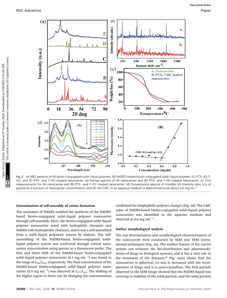
Fig. 2 (a) XRD patterns of (A) biotin-conjugated solid–liquid polymer, (B) NADES-based biotin-conjugated solid–liquid polymer, (C) PTX, (D) 7-HC, and (E) PTX- and 7-HC-loaded nanocarrier. (b) Raman spectra of (A) nanocarrier and (B) PTX- and 7-HC-loaded nanocarrier. (c) TGAmeasurements for (A) nanocarrier and (B) PTX- and 7-HC-loaded nanocarrier. (d) Fluorescence spectra of micelles (A) intensity ratio I1/I3 ofpyrene as a function of nanocarrier concentration, and (B) the CMC in an aqueous medium is determined to be about 0.6 mg mL�1.
RSC Advances Paper
Ope
n A
cces
s A
rtic
le. P
ublis
hed
on 0
7 O
ctob
er 2
020.
Dow
nloa
ded
on 5
/30/
2022
6:0
1:06
PM
. T
his
artic
le is
lice
nsed
und
er a
Cre
ativ
e C
omm
ons
Attr
ibut
ion
3.0
Unp
orte
d L
icen
ce.
View Article Online
Determination of self-assembly of carrier formation
The assistance of NADES enabled the synthesis of the NADES-based biotin-conjugated solid–liquid polymer nanocarrierthrough self-assembly. Here, the biotin-conjugated solid–liquidpolymer nanocarrier acted with hydrophilic character andNADES with hydrophobic character, and it was a self-assembledfrom a solid–liquid polymeric system by dialysis. The self-assembling of the NADES-based biotin-conjugated solid–liquid polymer system was conrmed through critical nano-carrier concentration using pyrene as a uorescent probe. Therst and third shi of the NADES-based biotin-conjugatedsolid–liquid polymer nanocarrier (0.1 mg mL�1) was found inthe range of I390/I445, respectively. The nal concentration of theNADES-based biotin-conjugated solid–liquid polymer nano-carrier (0.9 mg mL�1) was observed at I377/I442. The shiing ofthe higher region to lower one by changing the concentrations
36996 | RSC Adv., 2020, 10, 36989–37004
conrmed the amphiphilic polymer changes (Fig. 2d). The CMCvalue of NADES-based biotin-conjugated solid–liquid polymernanocarrier was identied in the aqueous medium andobserved at 0.6 mg mL�1.
Surface morphological analysis
The size determination and morphological characterization ofthe nanocarrier were conducted by SEM and TEM instru-mental techniques (Fig. 3a). The surface feature of the carriersystem can enhance the bio-distribution and pharmacoki-netics of drugs in biological systems, and it has a vital role inthe treatment of the diseases.42 Fig. 3a(A) shows that thenanocarrier is spherical, its size is increased with the incor-poration of drugs and it is semi-crystalline. The SLN particleobserved in the SEM image showed that the NADES liquid wascovering to stabilize of the solid particle, and the solid portion
This journal is © The Royal Society of Chemistry 2020
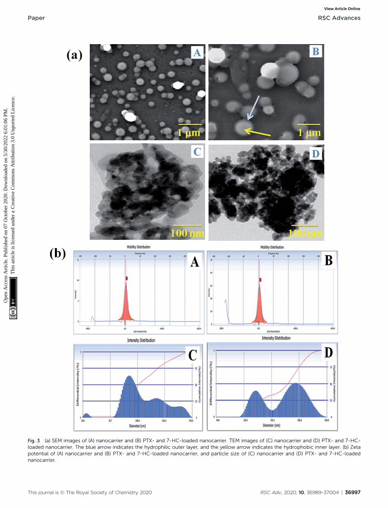
Fig. 3 (a) SEM images of (A) nanocarrier and (B) PTX- and 7-HC-loaded nanocarrier. TEM images of (C) nanocarrier and (D) PTX- and 7-HC-loaded nanocarrier. The blue arrow indicates the hydrophilic outer layer, and the yellow arrow indicates the hydrophobic inner layer. (b) Zetapotential of (A) nanocarrier and (B) PTX- and 7-HC-loaded nanocarrier, and particle size of (C) nanocarrier and (D) PTX- and 7-HC-loadednanocarrier.
This journal is © The Royal Society of Chemistry 2020 RSC Adv., 2020, 10, 36989–37004 | 36997
Paper RSC Advances
Ope
n A
cces
s A
rtic
le. P
ublis
hed
on 0
7 O
ctob
er 2
020.
Dow
nloa
ded
on 5
/30/
2022
6:0
1:06
PM
. T
his
artic
le is
lice
nsed
und
er a
Cre
ativ
e C
omm
ons
Attr
ibut
ion
3.0
Unp
orte
d L
icen
ce.
View Article Online

RSC Advances Paper
Ope
n A
cces
s A
rtic
le. P
ublis
hed
on 0
7 O
ctob
er 2
020.
Dow
nloa
ded
on 5
/30/
2022
6:0
1:06
PM
. T
his
artic
le is
lice
nsed
und
er a
Cre
ativ
e C
omm
ons
Attr
ibut
ion
3.0
Unp
orte
d L
icen
ce.
View Article Online
was highly dense in the drug-loaded SLN system (Fig. 3a(B)).Furthermore, the PTX- and 7-HC-loaded nanocarrier wasspherical, and the size of the particle increased due to thecharged drug molecules. Fig. 3a(C and D) show representativeTEM images of the nanocarrier, the particles of which arespherical with a mean particle size of 200–300 nm; thiscorrelates well with the SEM images.
Particle size analysis and zeta potential determinations
The surface charge and particle size of the unloaded and PTX-and 7-HC-loaded SLN system were tested using DLS, and theresults are presented in Fig. 3b. The zeta potential was observed
Fig. 4 UV-visible spectroscopy showing (A) encapsulation efficiency annanocarrier. In vitro drug discharge status of PTX- and 7-HC-loaded narelease pattern over 320 min.
36998 | RSC Adv., 2020, 10, 36989–37004
at�3� 0.5 mV and�0.2� 0.5 mV for unloaded and PTX- and 7-HC-loaded SLN systems, respectively. From these observations,drug-loaded SLN had enhanced stability over the unloadedcarrier, due to the interaction of the drugs and carrier mole-cules. The observed zeta potential was nearly zero (Fig. 3b(A andB)). The surface charge of the nanocarrier has a meaningfulrelevance for biological applications for stability, interactionwith cells, and also tissues.43 The size distribution of thenanocarrier was observed in the range of z240 nm for theunloaded and z290 nm for the loaded carrier. This indicatedthat molecule size expanded as PTX and 7-HC were incorpo-rated in the nanocarrier (Fig. 3b(C and D)).
d (B) percentage encapsulation efficiency of PTX- and 7-HC-loadednocarrier at (C) pH 2.8, (D) pH 5.5, and (E) pH 7.4. (F) Cumulative drug
This journal is © The Royal Society of Chemistry 2020
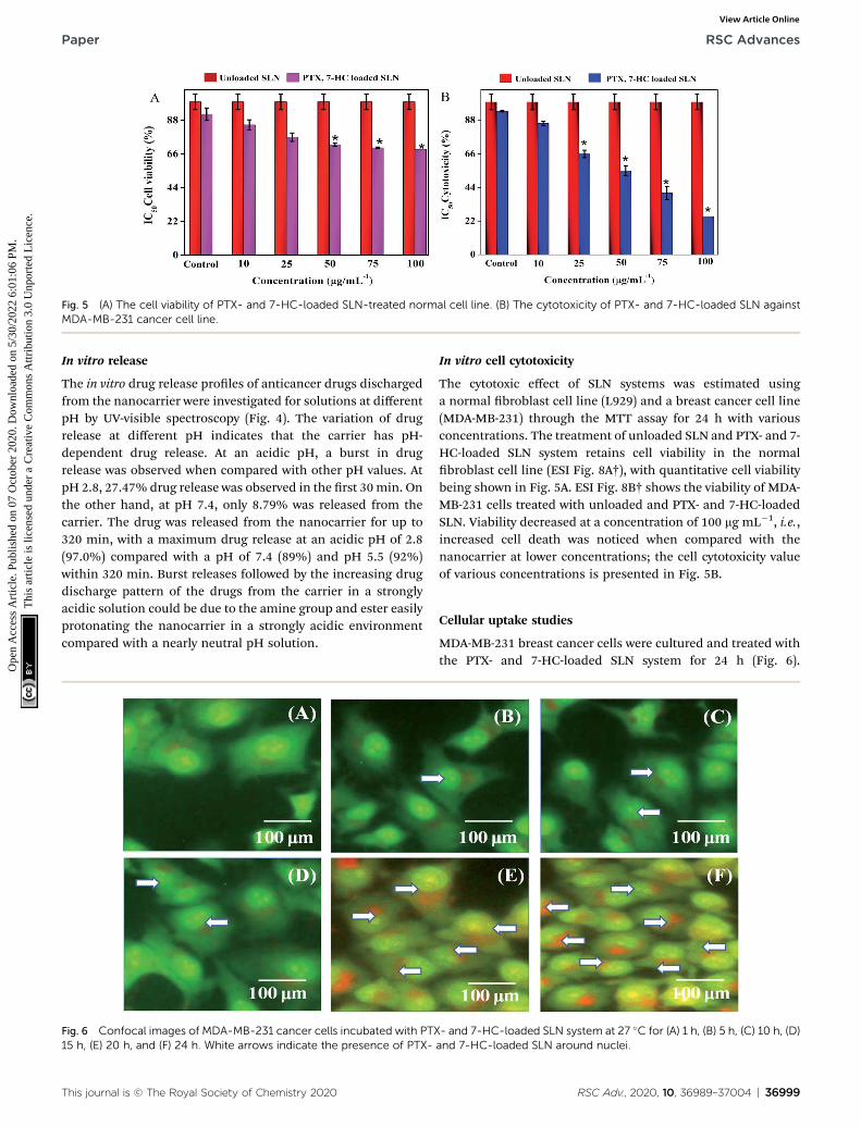
Fig. 5 (A) The cell viability of PTX- and 7-HC-loaded SLN-treated normal cell line. (B) The cytotoxicity of PTX- and 7-HC-loaded SLN againstMDA-MB-231 cancer cell line.
Paper RSC Advances
Ope
n A
cces
s A
rtic
le. P
ublis
hed
on 0
7 O
ctob
er 2
020.
Dow
nloa
ded
on 5
/30/
2022
6:0
1:06
PM
. T
his
artic
le is
lice
nsed
und
er a
Cre
ativ
e C
omm
ons
Attr
ibut
ion
3.0
Unp
orte
d L
icen
ce.
View Article Online
In vitro release
The in vitro drug release proles of anticancer drugs dischargedfrom the nanocarrier were investigated for solutions at differentpH by UV-visible spectroscopy (Fig. 4). The variation of drugrelease at different pH indicates that the carrier has pH-dependent drug release. At an acidic pH, a burst in drugrelease was observed when compared with other pH values. AtpH 2.8, 27.47% drug release was observed in the rst 30min. Onthe other hand, at pH 7.4, only 8.79% was released from thecarrier. The drug was released from the nanocarrier for up to320 min, with a maximum drug release at an acidic pH of 2.8(97.0%) compared with a pH of 7.4 (89%) and pH 5.5 (92%)within 320 min. Burst releases followed by the increasing drugdischarge pattern of the drugs from the carrier in a stronglyacidic solution could be due to the amine group and ester easilyprotonating the nanocarrier in a strongly acidic environmentcompared with a nearly neutral pH solution.
Fig. 6 Confocal images of MDA-MB-231 cancer cells incubated with PTX15 h, (E) 20 h, and (F) 24 h. White arrows indicate the presence of PTX-
This journal is © The Royal Society of Chemistry 2020
In vitro cell cytotoxicity
The cytotoxic effect of SLN systems was estimated usinga normal broblast cell line (L929) and a breast cancer cell line(MDA-MB-231) through the MTT assay for 24 h with variousconcentrations. The treatment of unloaded SLN and PTX- and 7-HC-loaded SLN system retains cell viability in the normalbroblast cell line (ESI Fig. 8A†), with quantitative cell viabilitybeing shown in Fig. 5A. ESI Fig. 8B† shows the viability of MDA-MB-231 cells treated with unloaded and PTX- and 7-HC-loadedSLN. Viability decreased at a concentration of 100 mg mL�1, i.e.,increased cell death was noticed when compared with thenanocarrier at lower concentrations; the cell cytotoxicity valueof various concentrations is presented in Fig. 5B.
Cellular uptake studies
MDA-MB-231 breast cancer cells were cultured and treated withthe PTX- and 7-HC-loaded SLN system for 24 h (Fig. 6).
- and 7-HC-loaded SLN system at 27 �C for (A) 1 h, (B) 5 h, (C) 10 h, (D)and 7-HC-loaded SLN around nuclei.
RSC Adv., 2020, 10, 36989–37004 | 36999
RSC Advances Paper
Ope
n A
cces
s A
rtic
le. P
ublis
hed
on 0
7 O
ctob
er 2
020.
Dow
nloa
ded
on 5
/30/
2022
6:0
1:06
PM
. T
his
artic
le is
lice
nsed
und
er a
Cre
ativ
e C
omm
ons
Attr
ibut
ion
3.0
Unp
orte
d L
icen
ce.
View Article Online
Fluorescence images were observed aer 1 h, 5 h, 10 h, 15 h,20 h, and 24 h. As the treatment time increased, uorescentintensities increased, which is due to a higher uptake of PTX-and 7-HC-loaded carriers via receptor-mediated endocytosis.The uorescence signal was very strong, and co-localizationaround the nucleus (white arrow) was observed at 20 h and24 h in the PTX- and 7-HC-loaded SLN system-treated samples.
In vivo studies
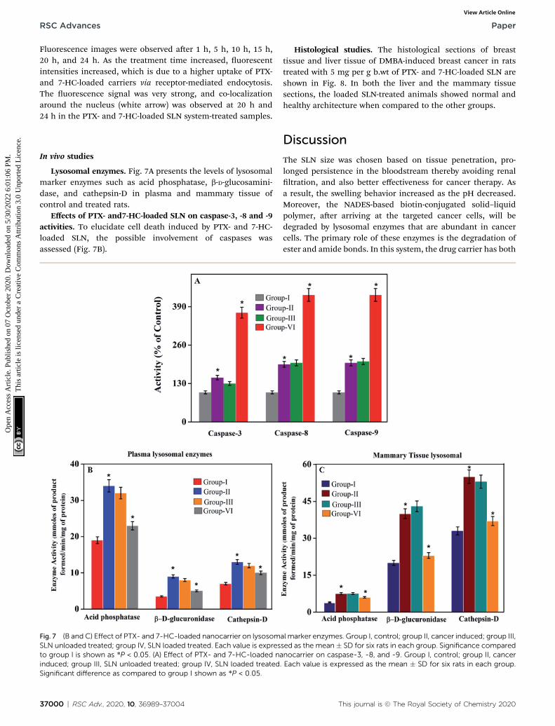
Lysosomal enzymes. Fig. 7A presents the levels of lysosomalmarker enzymes such as acid phosphatase, b-D-glucosamini-dase, and cathepsin-D in plasma and mammary tissue ofcontrol and treated rats.
Effects of PTX- and7-HC-loaded SLN on caspase-3, -8 and -9activities. To elucidate cell death induced by PTX- and 7-HC-loaded SLN, the possible involvement of caspases wasassessed (Fig. 7B).
Fig. 7 (B and C) Effect of PTX- and 7-HC-loaded nanocarrier on lysosomSLN unloaded treated; group IV, SLN loaded treated. Each value is expresto group I is shown as *P < 0.05. (A) Effect of PTX- and 7-HC-loaded nainduced; group III, SLN unloaded treated; group IV, SLN loaded treated.Significant difference as compared to group I shown as *P < 0.05.
37000 | RSC Adv., 2020, 10, 36989–37004
Histological studies. The histological sections of breasttissue and liver tissue of DMBA-induced breast cancer in ratstreated with 5 mg per g b.wt of PTX- and 7-HC-loaded SLN areshown in Fig. 8. In both the liver and the mammary tissuesections, the loaded SLN-treated animals showed normal andhealthy architecture when compared to the other groups.
Discussion
The SLN size was chosen based on tissue penetration, pro-longed persistence in the bloodstream thereby avoiding renalltration, and also better effectiveness for cancer therapy. Asa result, the swelling behavior increased as the pH decreased.Moreover, the NADES-based biotin-conjugated solid–liquidpolymer, aer arriving at the targeted cancer cells, will bedegraded by lysosomal enzymes that are abundant in cancercells. The primary role of these enzymes is the degradation ofester and amide bonds. In this system, the drug carrier has both
al marker enzymes. Group I, control; group II, cancer induced; group III,sed as the mean� SD for six rats in each group. Significance comparednocarrier on caspase-3, -8, and -9. Group I, control; group II, cancerEach value is expressed as the mean � SD for six rats in each group.
This journal is © The Royal Society of Chemistry 2020
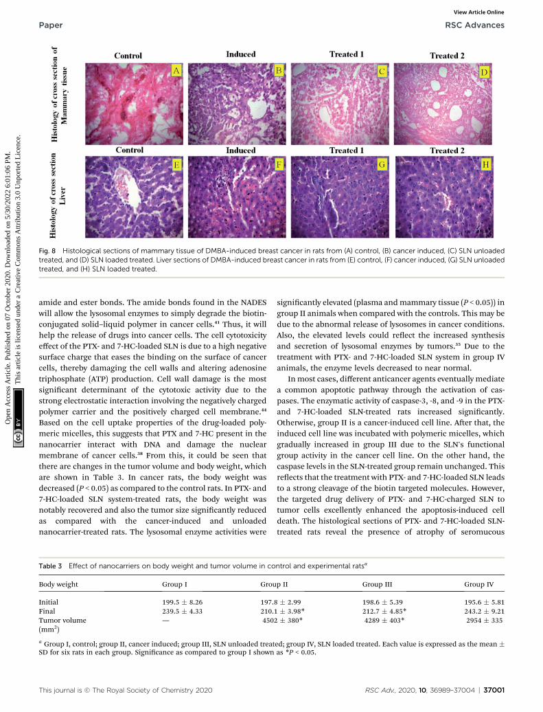
Fig. 8 Histological sections of mammary tissue of DMBA-induced breast cancer in rats from (A) control, (B) cancer induced, (C) SLN unloadedtreated, and (D) SLN loaded treated. Liver sections of DMBA-induced breast cancer in rats from (E) control, (F) cancer induced, (G) SLN unloadedtreated, and (H) SLN loaded treated.
Paper RSC Advances
Ope
n A
cces
s A
rtic
le. P
ublis
hed
on 0
7 O
ctob
er 2
020.
Dow
nloa
ded
on 5
/30/
2022
6:0
1:06
PM
. T
his
artic
le is
lice
nsed
und
er a
Cre
ativ
e C
omm
ons
Attr
ibut
ion
3.0
Unp
orte
d L
icen
ce.
View Article Online
amide and ester bonds. The amide bonds found in the NADESwill allow the lysosomal enzymes to simply degrade the biotin-conjugated solid–liquid polymer in cancer cells.41 Thus, it willhelp the release of drugs into cancer cells. The cell cytotoxicityeffect of the PTX- and 7-HC-loaded SLN is due to a high negativesurface charge that eases the binding on the surface of cancercells, thereby damaging the cell walls and altering adenosinetriphosphate (ATP) production. Cell wall damage is the mostsignicant determinant of the cytotoxic activity due to thestrong electrostatic interaction involving the negatively chargedpolymer carrier and the positively charged cell membrane.44
Based on the cell uptake properties of the drug-loaded poly-meric micelles, this suggests that PTX and 7-HC present in thenanocarrier interact with DNA and damage the nuclearmembrane of cancer cells.38 From this, it could be seen thatthere are changes in the tumor volume and body weight, whichare shown in Table 3. In cancer rats, the body weight wasdecreased (P < 0.05) as compared to the control rats. In PTX- and7-HC-loaded SLN system-treated rats, the body weight wasnotably recovered and also the tumor size signicantly reducedas compared with the cancer-induced and unloadednanocarrier-treated rats. The lysosomal enzyme activities were
Table 3 Effect of nanocarriers on body weight and tumor volume in co
Body weight Group I Grou
Initial 199.5 � 8.26 197.8Final 239.5 � 4.33 210.1Tumor volume(mm2)
— 4502
a Group I, control; group II, cancer induced; group III, SLN unloaded treatSD for six rats in each group. Signicance as compared to group I shown
This journal is © The Royal Society of Chemistry 2020
signicantly elevated (plasma andmammary tissue (P < 0.05)) ingroup II animals when compared with the controls. This may bedue to the abnormal release of lysosomes in cancer conditions.Also, the elevated levels could reect the increased synthesisand secretion of lysosomal enzymes by tumors.35 Due to thetreatment with PTX- and 7-HC-loaded SLN system in group IVanimals, the enzyme levels decreased to near normal.
Inmost cases, different anticancer agents eventually mediatea common apoptotic pathway through the activation of cas-pases. The enzymatic activity of caspase-3, -8, and -9 in the PTX-and 7-HC-loaded SLN-treated rats increased signicantly.Otherwise, group II is a cancer-induced cell line. Aer that, theinduced cell line was incubated with polymeric micelles, whichgradually increased in group III due to the SLN's functionalgroup activity in the cancer cell line. On the other hand, thecaspase levels in the SLN-treated group remain unchanged. Thisreects that the treatment with PTX- and 7-HC-loaded SLN leadsto a strong cleavage of the biotin targeted molecules. However,the targeted drug delivery of PTX- and 7-HC-charged SLN totumor cells excellently enhanced the apoptosis-induced celldeath. The histological sections of PTX- and 7-HC-loaded SLN-treated rats reveal the presence of atrophy of seromucous
ntrol and experimental ratsa
p II Group III Group IV
� 2.99 198.6 � 5.39 195.6 � 5.81� 3.98* 212.7 � 4.85* 243.2 � 9.21� 380* 4289 � 403* 2954 � 335
ed; group IV, SLN loaded treated. Each value is expressed as the mean �as *P < 0.05.
RSC Adv., 2020, 10, 36989–37004 | 37001
RSC Advances Paper
Ope
n A
cces
s A
rtic
le. P
ublis
hed
on 0
7 O
ctob
er 2
020.
Dow
nloa
ded
on 5
/30/
2022
6:0
1:06
PM
. T
his
artic
le is
lice
nsed
und
er a
Cre
ativ
e C
omm
ons
Attr
ibut
ion
3.0
Unp
orte
d L
icen
ce.
View Article Online
glands with surrounding stromal brosis, fatty tissue with smalllobules, and tumor regression. DMBA-induced rats showedtumors composed of hyperchromatic, pleomorphic, vesicularnuclei, and moderate cytoplasm arranged in nests, sheets, andacinar structures with numerous mitotic structures. Based onthe results, the PTX- and 7-HC-loaded SLN can reduce sideeffects. Ultimately, histological studies of the SLN and PTX- and7-HC-loaded SLN did not reveal any abnormal morphologicalchange to the mammary tissue and liver, further showing thatthey are safe for therapeutic use due to the PEG corona pro-tecting against protein absorption with highly stable SLN andPTX- and 7-HC-loaded SLN, while the PEGylated surface led toan uptake in the liver.
Conclusion
A novel approach in advanced cancer prevention and treat-ment includes the use of SLN systems for anticancer drugdelivery. A PTX- and 7-HC-loaded SLN was synthesized for therst time with a green solvent as a promising novel drugdelivery system for the enhancement of the therapeutic indexof cancer cells. The unloaded and PTX- and 7-HC-loaded SLNswere characterized in terms of size and crystalline properties.The higher toxicity of PTX- and 7-HC-loaded SLN system wasobserved by MTT assay, which promoted MDA-MB-231 celldeath. The effect of PTX- and 7-HC-loaded SLN on cell viabilitywas investigated with MTT assay and also by morphologicalobservations. The decreased cell viability observed in thisstudy could be due to direct inhibition of cell growth by PTX-and 7-HC-loaded SLN. Apoptosis was included in cell deathmechanisms observed in breast cancer-induced rats. Treat-ment with PTX- and 7-HC-loaded SLN system elevates caspaselevels that stimulate cell death mechanisms and damagetumor tissue. From these results, it is clear that the synthe-sized PTX- and 7-HC-loaded SLN system is highly valuable inpreventing cancer cell proliferation. It is concluded that thePTX- and 7-HC-packed SLN system might be a potentialcandidate for breast cancer treatment and also which canimprove the treatment of incurable diseases. Additionalstudies are suggested for estimating the effect of these nano-carriers on other cell death mechanisms.
Abbreviations
NADES
37002 | RSC Ad
Natural deep eutectic solvents
SLN Solid–liquid nanocarrier NMR Nuclear magnetic resonance FTIR Fourier-transform infrared spectroscopy TGA Thermogravimetric analysis XRD Powder X-ray diffraction DLS Dynamic light scattering SEM Scanning electron microscopy TEM Transmission electron microscopy PTX Paclitaxel 7-HC 7-Hydroxycoumarin MDA-MB-231 M. D. Anderson and metastasis breast cancerv., 2020, 10, 36989–37004
PB
T
Prolinebetaine
LA Lactic acid b.wt Bodyweight DMBA 7,12-Dimethylbenz[a]anthracene DDSs Drug delivery systems ILs Ionic liquids DESs Deep eutectic solvents DD water Double-distilled water ATP Adenosine triphosphate RT Retention timeConflicts of interest
The authors declare that they have no competing interests.
Acknowledgements
M. Rajan is grateful for nancial support under the plan of“ICMR” (F. No. 5/3/8/350/2018-ITR; New Delhi, India) andDepartment of Science and Technology, Science and Engi-neering Research Board (Ref: YSS/2015/001532; New Delhi,India) and also acknowledges the PURSE program for thepurchase of SEM and FT-IR and UPE programs for the purchaseof TEM. The authors extend their appreciation to theresearchers supporting project number RSP-2020/190, KingSaud University, Riyadh, Saudi Arabia.
References
1 J. Brindha and C. Kaushik, Evolutionary approaches inprotein engineering towards biomaterial construction, RSCAdv., 2019, 9, 34720–34734.
2 S. Mehnath, M. Arjama, M. Rajan, K. Premkumar,K. Karthikeyan and M. Jeyaraj, Mineralization of bioactivemarine sponge and electrophoretic deposition on Ti-6Al-4Vimplant for osteointegration, Surf. Coat. Technol., 2020,392, 125727.
3 F. Khan, M. Tanaka and S. R. Ahmad, Fabrication ofPolymeric Biomaterials: A Strategy for Tissue Engineeringand Medical Devices, J. Mater. Chem. B, 2015, 3, 8224–8249.
4 H. H. Tonnesen and J. Karlsen, Alginate in Drug DeliverySystems, Drug Dev. Ind. Pharm., 2002, 28, 621–630.
5 S. Palvai, L. Anandi, S. Sarkar, M. Augustus, M. Roy, M. Lahiriand S. Basu, Drug-Triggered Self-Assembly of Linear Polymerinto Nanoparticles for Simultaneous Delivery ofHydrophobic and Hydrophilic Drugs in Breast CancerCells, ACS Omega, 2017, 2, 8730–8740.
6 D. Govindaraj, P. Pradeepkumar and M. Rajan, Synthesis ofmorphology tuning multi mineral substituted apatitenanocrystals by novel natural deep eutectic solvents, Mater.Discovery, 2018, 9, 11–15.
7 N. Graf and S. J. Lippard, Redox Activation of Metal-BasedProdrugs as A Strategy for Drug Delivery, Adv. Drug DeliveryRev., 201, 264, 993–1004.
8 Y. Ishihara and N. Shimamoto, Involvement ofEndonuclease G in Nucleosomal DNA Fragmentation
his journal is © The Royal Society of Chemistry 2020
Paper RSC Advances
Ope
n A
cces
s A
rtic
le. P
ublis
hed
on 0
7 O
ctob
er 2
020.
Dow
nloa
ded
on 5
/30/
2022
6:0
1:06
PM
. T
his
artic
le is
lice
nsed
und
er a
Cre
ativ
e C
omm
ons
Attr
ibut
ion
3.0
Unp
orte
d L
icen
ce.
View Article Online
under Sustained Endogenous Oxidative Stress, J. Biol. Chem.,2006, 281, 6726–6733.
9 S. G. Arguelles, M. C. Serrano, M. C. Gutieerrez, M. LuisaFerrer, L. Yuste, F. Rojo and F. del Monte, Deep EutecticSolvents for the Self-Assembly of Gold Nanoparticles: ASAXS, UV�Vis, and TEM Investigation, Langmuir, 2013, 29,9525–9534.
10 H. T. Thai Thanh, H. P. Emily, N. Dai Hai, S. L. Jung, P. KiDong and P. T. Nghia, The Importance of Poly(ethyleneglycol) Alternatives for Overcoming PEG Immunogenicityin Drug Delivery and Bioconjugation, Polymers, 2020, 12,298.
11 S. Ksenia, S. Egorova, G. Evgeniv, G. Gordeev andP. Valentine, Biological Activity of Ionic Liquids and TheirApplication in Pharmaceutics and Medicine, Chem. Rev.,2017, 117, 7132–7189.
12 P. Pradeepkumar, A. M. Elgorban, A. H. Bahkali andM. Rajan, Natural Solvent-Assisted Synthesis ofAmphiphilic Co-Polymeric Nanonanocarrier for ProlongedRelease of Camptothecin Delivery, New J. Chem., 2018, 42,10366–10375.
13 R. Verpoorte, Chemodiversity and the Biological Role ofSecondary Metabolites, Some Thoughts for Selecting PlantMaterial for Drug Development, in Bioassay Methods inNatural Product Research and Drug Development, ed. L.Bohlin and J. G. Bruhn, Proceedings of the PhytochemicalSociety of Europe, Springer, Dordrecht, 1999, vol. 43, pp.11–23.
14 W. Czaban, J. Rasmussen, B. B. Laursen, N. H. Vidkjaer,R. Sapkota, M. Nicolaisen and S. Fomsgaard, MultipleEffects of Secondary Metabolites on Amino Acid Cycling inWhite Clover Rhizosphere, Soil Biol. Biochem., 2018, 123,54–63.
15 S. Ksenia, S. Egorova, G. Evgeniv, G. Gordeev andP. Valentine, Biological Activity of Ionic Liquids and TheirApplication in Pharmaceutics and Medicine, Chem. Rev.,2017, 117, 7132–7189.
16 P. Pradeepkumar, D. Govindaraj, M. Jeyaraj,M. A. Munusamy and M. Rajan, Assembling ofMultifunctional Latex-Based Hybrid Nanocarriers fromCalotropisgigantea for Sustained (Doxorubicin) DOXReleases, Biomed. Pharmacother., 2017, 87, 461–470.
17 C. Mukesh, J. Bhatt and K. Prasad, Preparation ofa NoncytotoxicHemocompatible Ion Gel by Self-Polymerization of HEMA in a Green Deep Eutectic Solvent,Macromol. Chem. Phys., 2014, 215, 1498–1504.
18 A. L. Wang, X. L. Zheng, Z. Z. Zhao, C. P. Li and X. F. Zheng,Deep Eutectic Solvent Catalyzed friedel–Cras Alkylation ofElectron-Rich Arenes with Aldehydes, RSC Adv., 2014, 5,59022–59026.
19 D. Monleon, J. M. Morales, A. Barrasa, J. A. Lopez, C. Vazquezand B. Celd, Metabolite Proling of Fecal Water ExtractsFrom Human Colorectal Cancer, NMR Biomed., 2009, 22,342–348.
20 C. R. Day and S. A. Kempson, Betaine Chemistry, Roles, andPotential Use in Liver Disease, Biochim. Biophys. Acta, Gen.Subj., 2016, 1860(6), 1098–1106.
This journal is © The Royal Society of Chemistry 2020
21 S. S. Heinzmann, I. J. Brown, Q. Chan, M. Bictash,M. E. Dumas, S. Kochhar and J. Stamler, Metabolicproling strategy for discovery of nutritional biomarkers:prolinebetaine as a marker of citrus consumption, Am. J.Clin. Nutr., 2010, 92, 436–443.
22 M. J. Joralemon, K. S. Murthy, E. E. Remsen, M. L. Beckerand K. L. Wooley, Synthesis, Characterization, andBioavailability of Mannosylated Shell Cross-LinkedNanoparticles, Biomacromolecules, 2004, 5, 903–913.
23 J. H. Kim, Y. Li, M. S. Kim, S. W. Kang, J. H. Jeong andD. Sung Lee, Synthesis and Evaluation of Biotin-Conjugated Ph-Responsive Polymeric Nanocarriers as DrugCarriers, Int. J. Pharm., 2012, 427, 435–442.
24 Y. Miyake, K. Yamamoto, Y. Morimitsu and T. Osawa,Isolation of C-Glucosylavone from Lemon Peel andAntioxidative Activity of Flavonoid Compounds in LemonFruit, J. Agric. Food Chem., 1997, 45(12), 4619–4623.
25 V. S. Raghuwanshi, M. Ochmann, A. Hoell, F. Polzer andK. Rademann, Deep Eutectic Solvents for the Self-Assemblyof Gold Nanoparticles: A SAXS, UV�Vis, and TEMInvestigation, Langmuir, 2014, 30, 6038–6046.
26 W. Zhu, Z. Song, P. Wei, N. Meng, T. Teng, F. Yang, N. Liuand R. Feng, Y-Shaped Biotin-Conjugated Poly (EthyleneGlycol)–Poly (Epsilon-Caprolactone) Copolymer for TheTargeted Delivery of Curcumin, J. Colloid Interface Sci.,2015, 443, 1–7.
27 A. S. Mikhail and C. Allen, (Poly(ethylene glycol)-b-poly(3-caprolactone)) Nanocarriers Containing ChemicallyConjugated and Physically Entrapped Docetaxel: Synthesis,Characterization, and the Inuence of the Drug onNanocarrier Morphology, Biomacromolecules, 2010, 11,1273–1280.
28 R. A. Praphakar, M. A. Munusamy, A. A. Alarfaj, S. SureshKumar and M. Rajan, Zn2+ Cross-Linked Sodium Alginate-G-AllylamineMannose Polymeric Carrier on Rifampicin forMacrophage Targeting Tuberculosis Nanotherapy, New J.Chem., 2017, 41, 11324–11334.
29 P. Pradeepkumar, R. Naresh Kumar, A. A. Alarfaj,M. A. Murugan and M. Rajan, Deep Eutectic Solvent-Mediated FA-g-b-Alanine-co-PCL Drug Carrier forSustainable and Site-Specic Drug Delivery, ACS Appl. BioMater., 2018, 1(6), 2094–2109.
30 C. Janas, Z. Mostaphaoui, L. Schmiederer, J. Bauer andM. G. Wacker, Novel Polymeric Nanocarriers for DrugDelivery: Material Characterization and FormulationScreening, Int. J. Pharm., 2016, 509, 197–207.
31 J. King, The hydrolases–acid and Alkaline Phosphatises, inPractical Clinical Enzymology, ed. J. C. King, D. VanNostrand Company Ltd., London, 1965, pp. 191–208.
32 A. I. Sapolsky, R. D. Atlman, D. S. Howell and D. Cathepsin,Activity in Normal and osteoarthritic human cartilage, Fed.Proc., 1973, 32, 1489–1493.
33 Y. Kawai and K. Anno, Mucopolysaccharide DegradingEnzymes from the Liver of SquidOmmastrephesSolaniPacicus. I. Hyaluronidase, Biochim.Biophys. Acta, Enzymol., 1971, 242, 428–436.
RSC Adv., 2020, 10, 36989–37004 | 37003
RSC Advances Paper
Ope
n A
cces
s A
rtic
le. P
ublis
hed
on 0
7 O
ctob
er 2
020.
Dow
nloa
ded
on 5
/30/
2022
6:0
1:06
PM
. T
his
artic
le is
lice
nsed
und
er a
Cre
ativ
e C
omm
ons
Attr
ibut
ion
3.0
Unp
orte
d L
icen
ce.
View Article Online
34 L. Servillo, N. D'Onofrio, A. Giovane, R. Casale, D. Cautela,G. Ferrari and D. Castaldo, The Betaine Prole of CerealFlours Unveils New And Uncommon Betaines, Food Chem.,2018, 239, 234–241.
35 H. Shinar, M. D. Battistel, M. Mandler, F. Lichaa andF. L. Freed berg, Chemical Exchange Saturation TransferNMR of Oligo- and Poly-Sialic Acids and the Assignment ofTheir Hydroxyl Groups Using Selective- and HSQC-TOCSY,Carbohydr. Res., 2014, 389, 165–173.
36 A. L. Wang, X. L. Zheng, Z. Z. Zhao, C. P. Li and X. F. Zheng,Deep Eutectic Solvent Catalyzed friedel–Cras Alkylation ofElectron-Rich Arenes with Aldehydes, RSC Adv., 2014, 5,59022–59026.
37 C. Chien, T. Ching, A. Lin, M. Zhang, S. L. Levengood andM. Zhang, PEG-Chitosan Hydrogel with Tunable Stiffnessfor Study of Drug Response of Breast Cancer Cells,Polymers, 2016, 8, 112.
38 H. Zhang, H. Hu, H. Zhang, W. Dai, X. Wang, X. Wang andQ. Zhang, Effects of Pegylated Paclitaxel Nanocrystals OnBreast Cancer and Its Lung Metastasis, Nanoscale, 2015, 7,10790–10800.
39 G. A. Shtukenberg, Q. Zhu, D. J. Carter, L. Vogt, J. Hoja,E. Schneider and H. Song, Powder Diffraction and CrystalStructure Prediction Identify Four New CoumarinPolymorphs, Chem. Sci., 2017, 8, 4926–4940.
37004 | RSC Adv., 2020, 10, 36989–37004
40 W. C. Harris and D. A. Coe, Vibrational spectra and structureof esters–H.* Raman spectra and potential functioncalculations for HC00CH3, DCOOCH3 and HCOOCD3,Spectrochim. Acta, Part A, 1976, 32, 1–10.
41 H. Gheybi and A. A. Entezami, NanosizedNanocarriers Self-assembled from Amphiphilicpoly(citric acid)–poly(e-caprolactone)–poly(citric acid) Copolymers, Polym. Bull.,2013, 70, 1875–1894.
42 S. S. Davis, Biomedical Applications of Nanotechnology-Implications for Drug Targeting and Gene Therapy, TrendsBiotechnol., 1997, 15, 217–224.
43 K. Song Yang, N. V. Mai, H. P. Emily, P. R. J. Angus,R. W. Michael, F. Q. John, P. T. Nghia and P. D. Thomas,Elucidating the Inuences of Size, Surface Chemistry, andDynamic Flow on Cellular Association of NanoparticlesMade by Polymerization-Induced Self-Assembly, Small,2018, 14(34), 1801702.
44 M. Rajan, P. Krishnan, P. Pradeepkumar, M. Jeyanthinath,M. Jeyaraj, M. P. Ling, P. Arulselvan, A. Higuchi,M. A. Munusamy, R. Arumugam, G. Benelli, K. Muruganand S. Suresh Kumar, Magneto-chemotherapy for cervicalcancer treatment with camptothecin loadedFe3O4functionalizedb-cyclodextrinnanovehicles, RSC Adv.,2017, 7, 46271.
This journal is © The Royal Society of Chemistry 2020