FMRFamide-related peptides in the gut of Locusta migratoria L.: a comprehensive map and...
14
Peptides 24 (2003) 1511–1524 FMRFamide-related peptides in the gut of Locusta migratoria L.: a comprehensive map and developmental profile Sharon R. Hill ∗ , Ian Orchard Department of Biology, University of Toronto at Mississauga, 3359 Mississauga Road North, Mississauga, Ont., Canada L5L 1C6 Received 3 June 2003; accepted 18 August 2003 Abstract The gut tissues and associated nervous system of the African migratory locust, Locusta migratoria, were found to contain FMRFamide-like immunoreactive (FLI) material throughout the five larval instars and 2 weeks into the adult stage in both males and females. FMRFamide-like immunoreactivity associated with the locust gut was described using camera lucida techniques. FMRFamide-like immunoreactivity is ob- served in the frontal connectives, recurrent nerve, and oesophageal nerves; projections from the ingluvial ganglion onto the anterior midgut, and from the proctodeal nerve onto the hindgut and posterior midgut; in the neuropils of the frontal ganglion, hypocerebral ganglion and ingluvial ganglia; 30 cell bodies in the frontal ganglion; multipolar sensory cells on the foregut; and endocrine-like cells in the gastric caecae and midgut. Radioimmunoassay (RIA) was used to determine the quantities of FLI material in foreguts, gastric caecae, anterior and posterior midguts, and hindgut of first–fifth instar larvae, 1–3- and 14–17-day male and female adult locusts. As expected, as the tissue size (assessed by total protein content) increases, so does the amount of FLI material in each tissue. Normalizing for tissue size reveals significant differences in FLI content among the stages for each tissue tested. Reversed phase-high pressure liquid chromatography (RP-HPLC) followed by RIA has identified four groups of FLI fractions present in the gut, and different members of these groups are present in the various gut tissues. © 2003 Elsevier Inc. All rights reserved. Keywords: FMRFamides; Insect; Foregut; Midgut; Hindgut; Gastric caecae; RIA; HPLC; Immunohistochemistry; Camera lucida; Development 1. Introduction Feeding and digestion in insects is under intense scrutiny in the scientific community, since crops are decimated and diseases are vectored from host to host via many insect feed- ing behaviors. One such agricultural pest is the migratory locust, Locusta migratoria L. Research in current years has focused on neuropeptides as a possible target for manipu- lation that is geared towards rapid modification of feeding behaviours [20]. One of the potential outcomes of this re- search is the development of safe, fast acting and highly specific feeding control methods. One superfamily of neuropeptides under investigation is the FMRFamide-related peptides (FaRPs). Found through- out invertebrates, FaRPs are peptides that terminate in the residues Phe-Met/Leu-Arg-Phe and are C-terminally ami- dated [18]. Members of the FaRP superfamily display a diverse array of functions (for review see [24]). Focusing ∗ Corresponding author. Tel.: +1-905-569-4752; fax: +1-905-828-3792. E-mail addresses: [email protected] (S.R. Hill), [email protected] (I. Orchard). our attention on the insect gut, FaRPs are implicated in the modulation of muscle contraction, enzyme secretion and the active transport of ions across the gut wall. For ex- ample, myosuppressins, a subgroup of decapeptide FaRPs with the consensus sequence (XDVXHXFLRFamide), in- hibit muscle contraction in the foregut of Phormia regina [27], and Drosophila melanogaster [13,22], the midgut of Agrius convolvuli [8], L. migratoria [16], and Diploptera punctata [9], and the hindgut of Leucophaea maderae [12]. Myosuppressins stimulate -amylase secretion in the midguts of D. punctata [10], Rhynchophorus ferrugineus [23] and Pecten maximus [7]. Two members of the extended FLRFamide sub-group of FaRPs in Manduca sexta, F7G (GNSFLRFamide) and F7D (DPSFLRFamide), stimulate hindgut contractions [14]. F7G and F7D inhibit the active transport of ions across the gut wall in a dose-dependent manner [17], and Orchard et al. [24] suggest that the demonstrated variability in active ion transport observed during larval development, paired with the ability of F7G and F7D to inhibit transport, may indicate that the short circuiting of active ion transport is under the regulation of FaRPs. 0196-9781/$ – see front matter © 2003 Elsevier Inc. All rights reserved. doi:10.1016/j.peptides.2003.08.013
Transcript of FMRFamide-related peptides in the gut of Locusta migratoria L.: a comprehensive map and...

Peptides 24 (2003) 1511–1524
FMRFamide-related peptides in the gut ofLocusta migratoria L.:a comprehensive map and developmental profile
Sharon R. Hill∗, Ian OrchardDepartment of Biology, University of Toronto at Mississauga, 3359 Mississauga Road North, Mississauga, Ont., Canada L5L 1C6
Received 3 June 2003; accepted 18 August 2003
Abstract
The gut tissues and associated nervous system of the African migratory locust,Locusta migratoria, were found to contain FMRFamide-likeimmunoreactive (FLI) material throughout the five larval instars and 2 weeks into the adult stage in both males and females. FMRFamide-likeimmunoreactivity associated with the locust gut was described usingcamera lucida techniques. FMRFamide-like immunoreactivity is ob-served in the frontal connectives, recurrent nerve, and oesophageal nerves; projections from the ingluvial ganglion onto the anterior midgut,and from the proctodeal nerve onto the hindgut and posterior midgut; in the neuropils of the frontal ganglion, hypocerebral ganglion andingluvial ganglia; 30 cell bodies in the frontal ganglion; multipolar sensory cells on the foregut; and endocrine-like cells in the gastriccaecae and midgut. Radioimmunoassay (RIA) was used to determine the quantities of FLI material in foreguts, gastric caecae, anteriorand posterior midguts, and hindgut of first–fifth instar larvae, 1–3- and 14–17-day male and female adult locusts. As expected, as thetissue size (assessed by total protein content) increases, so does the amount of FLI material in each tissue. Normalizing for tissue sizereveals significant differences in FLI content among the stages for each tissue tested. Reversed phase-high pressure liquid chromatography(RP-HPLC) followed by RIA has identified four groups of FLI fractions present in the gut, and different members of these groups arepresent in the various gut tissues.© 2003 Elsevier Inc. All rights reserved.
Keywords: FMRFamides; Insect; Foregut; Midgut; Hindgut; Gastric caecae; RIA; HPLC; Immunohistochemistry;Camera lucida; Development
1. Introduction
Feeding and digestion in insects is under intense scrutinyin the scientific community, since crops are decimated anddiseases are vectored from host to host via many insect feed-ing behaviors. One such agricultural pest is the migratorylocust,Locusta migratoria L. Research in current years hasfocused on neuropeptides as a possible target for manipu-lation that is geared towards rapid modification of feedingbehaviours[20]. One of the potential outcomes of this re-search is the development of safe, fast acting and highlyspecific feeding control methods.
One superfamily of neuropeptides under investigation isthe FMRFamide-related peptides (FaRPs). Found through-out invertebrates, FaRPs are peptides that terminate in theresidues Phe-Met/Leu-Arg-Phe and are C-terminally ami-dated [18]. Members of the FaRP superfamily display adiverse array of functions (for review see[24]). Focusing
∗ Corresponding author. Tel.:+1-905-569-4752; fax:+1-905-828-3792.E-mail addresses: [email protected] (S.R. Hill),
[email protected] (I. Orchard).
our attention on the insect gut, FaRPs are implicated inthe modulation of muscle contraction, enzyme secretionand the active transport of ions across the gut wall. For ex-ample, myosuppressins, a subgroup of decapeptide FaRPswith the consensus sequence (XDVXHXFLRFamide), in-hibit muscle contraction in the foregut ofPhormia regina[27], andDrosophila melanogaster [13,22], the midgut ofAgrius convolvuli [8], L. migratoria [16], and Diplopterapunctata [9], and the hindgut ofLeucophaea maderae[12]. Myosuppressins stimulate�-amylase secretion in themidguts of D. punctata [10], Rhynchophorus ferrugineus[23] andPecten maximus [7]. Two members of the extendedFLRFamide sub-group of FaRPs inManduca sexta, F7G(GNSFLRFamide) and F7D (DPSFLRFamide), stimulatehindgut contractions[14]. F7G and F7D inhibit the activetransport of ions across the gut wall in a dose-dependentmanner [17], and Orchard et al.[24] suggest that thedemonstrated variability in active ion transport observedduring larval development, paired with the ability of F7Gand F7D to inhibit transport, may indicate that the shortcircuiting of active ion transport is under the regulation ofFaRPs.
0196-9781/$ – see front matter © 2003 Elsevier Inc. All rights reserved.doi:10.1016/j.peptides.2003.08.013

1512 S.R. Hill, I. Orchard / Peptides 24 (2003) 1511–1524
In this paper, we will provide a comprehensive map ofFMRFamide-like immunoreactivity in the gut tissue and as-sociated neural processes ofL. migratoria; a detailed assess-ment of the quantity of FMRFamide-like immunoreactivityin each region of the gut (foregut, gastric caecae, anteriorand posterior midgut, and hindgut) in females and males asadults and throughout larval development; and an identifica-tion of different FaRP family members present in distinct re-gions of 2-week adult gut tissue through the use of reversedphase-high pressure liquid chromatography (RP-HPLC) fol-lowed by radioimmunoassay (RIA).
2. Materials and methods
2.1. Locust rearing conditions
Locusts were raised in crowded conditions under a12 h/12 h light/dark cycle. All animals had ad libitum accessto wheat bran, and were fed on wheat grass once daily. Lo-custs of both sexes were chosen 1–3 days post-ecdysis fromeach instar (first to fifth) and 1–3- and 14–17-day adults.
2.2. Immunohistochemistry
Locust wholeguts were exposed dorsally and were cutopen along the dorsal midline under normal physiological lo-cust saline (150 mM NaCl, 10 mM KCl, 4 mM CaCl2, 2 mMMgCl2, 4 mM NaHCO3, 5 mM HEPES, pH 7.2, 90 mM su-crose and 5 mM trehalose). The gastric caecae and the gutlumen were flushed with fresh physiological saline beforebeing fixed flat at 4◦C overnight. Tissues were washed withfrequent changes (every 20–30 min) of phosphate-bufferedsaline (PBS; 10 mM) at room temperature for 4–8 h.
Rabbit anti-FMRFamide primary antiserum (Incstar, MN)(dilution 1:1000) was preabsorbed at 4◦C overnight in PBSwith 4% Triton X-100, 2% BSA and 10% normal sheepserum (NSS). Tissues were incubated on a shaker tableat 8◦C in the preabsorbed primary antiserum for 18–36 h,washed overnight in PBS anti-rabbit IgG attached to FITC(Sigma) diluted 1:200 in 0.4% Triton X-100, 2% BSA and10% NSS, and incubated for 18 h at 4◦C. Tissues werewashed in PBS for 6 h (replacing the PBS 12–14 times) andmounted in 5%n-propylgalate in glycerol for viewing underepifluorescence and/or confocal microscopes.
2.3. Camera lucida
Camera lucida drawings were constructed using the epi-fluorescence microscope at ocular 10× and objective 16×.
2.4. Preparation for radioimmunoassay and RP-HPLC
Individual guts were separated into foregut, gastric cae-cae, anterior midgut, posterior midgut and hindgut undernormal locust physiological saline. Tissues were placedinto ice-cold methanol:acetic acid:water (90:9:1) and stored
below 0◦C. To isolate crude peptide fractions, the tissueswere sonicated on ice in 15 s bursts and centifuged at 8.8 Krpm for 10 min. The peptide-containing supernatant wastransferred to fresh tubes and dried in a Speed-Vac con-centrator (Savant, Farmingdale, NY). The protein pelletsremaining following the supernatant transfer were com-pletely dissolved in 1 M NaOH at 56◦C and assayed fortotal protein content using the BioRad protein assay systembased on the Bradford assay using human�-globulin as thestandard[4].
2.5. Reversed phase high pressure liquid chromatography(RP-HPLC)
The peptide-containing pellets following Speed-Vac dry-ing were resuspended in 0.1% trifluoroacetic acid (TFA)and pooled into groups of 10. Sep-Pak C-18 cartridges (Wa-ters Associates, Milford, MA) were equilibrated by passing8 ml of MeOH, water, and 0.1% TFA, and 2 ml of 0.1%TFA with 10�g BSA through the column sequentially. Thepooled peptide samples were passed over the columns threetimes before the columns were washed with 0.1% TFA. Thepeptides retained on the Sep-Pak C-18 column were elutedin 3 ml MeOH containing 0.1% TFA and dried down to∼50�l in the Speed Vac. The pooled peptide samples werediluted to 1 ml in HPLC start buffer (9% acetonitrile contain-ing 0.1% TFA). Samples were injected onto the BrownleeRP-18 Spheri-5 column (2.6 mm× 25 cm) over a period of5 min. Samples were eluted off the column over a period of34 min by a linear gradient of 9–60% acetonitrile contain-ing 0.1% TFA at a flow rate of 1 ml/min over the column.Fractions were collected at 1 min intervals and dried downin the Speed-Vac.
2.6. Radioimmunoassay
The peptide pellets were redissolved in 400�l radioim-munoassay (RIA) buffer (0.1 M PBS containing 0.3% NaCl,0.1% BSA, 0.01% NaN3 at pH 7.4). Commercially avail-able FMRFamide antibody (Incstar, MN) (100�l, 1:33,000dilution) was added to peptide samples (<20�l), followedby 100�l of 125I-YFMRFamide (∼12.5�Ci) and incubatedovernight at 4◦C. A standard curve was generated for theRIA using known concentrations of FMRFamide (Sigma,MO; 2.5, 5, 10, 25, 50, 125, 250, 500 and 1000 fmol). Theassay was terminated with the addition of 250�l activatedcharcoal (0.4 g in 50 ml RIA buffer with 0.04 g of dex-tran). Supernatant (400�l) was transferred to fresh tubes andcounted in a gamma counter. The FLI content determinedby RIA is presented per tissue, and per milligram of totalprotein. Normalization to total protein content is done tominimize variations in FMRFamide-like immunoreactivitydue to differences in tissue size. Non-normalized data is in-cluded for comparison as the number of FLI endocrine-likecells may not be size dependent.

S.R. Hill, I. Orchard / Peptides 24 (2003) 1511–1524 1513
3. Results
3.1. Two-week adults
3.1.1. Immunohistochemistry and camera lucidaBoth male and female 14–17-day locusts were exam-
ined. There were no apparent differences between thesexes with regard to immunohistochemical staining withanti-FMRFamide antiserum.
3.1.1.1. Frontal ganglion and hypocerebral ganglion.Processes extend from the tritocerebrum through frontalconnectives and into the frontal ganglion. There are 30 FLI
Fig. 1. Compositecamera lucida drawing showing FMRFamide-like immunoreativity over the gut of 2-week adultL. migratoria. The solid black lineacross the foregut indicates the transition from dorsal to ventral view in the diagram (i.e., anterior to the line is a dorsal view; and posterior to thelineis a ventral view.) The small arrow indicates the frontal ganglion; the large arrow indicates the hypocerebral ganglion; and the arrowhead indicatesoneof the two ingluvial ganglia. Only one of the six gastric caecae is represented here.
cell bodies in the frontal ganglion: three anterior dorsalpairs, four lateral dorsal pairs and eight lateral ventral pairsof cell bodies with tracts extending into the neuropil andsome tracts exiting through the recurrent nerve (Figs. 1 and2A and B). FaRP-like immunoreactive processes enter thehypocerebral ganglion through the recurrent nerve (Figs. 1and 2C and D), extend through the neuropil and enter theingluvial nerves (Figs. 1 and 2E and F).
3.1.1.2. Foregut. FLI sites are located along the inglu-vial nerves which appear to be neurohaemal sites. A fewfine short processes extend from the ingluvial nerves andproject to the foregut muscle (Fig. 1). FLI processes enter

1514 S.R. Hill, I. Orchard / Peptides 24 (2003) 1511–1524
Fig. 2. FMRFamide-like immunoreativity and associatedcamera lucida drawings. (A and B) Frontal ganglion; (C and D) hypocerebral ganglion; (Eand F) ingluvial ganglion from the 14–17-dayL. migratoria. Scale bars represent 200�m. Open circles represent ventral cell bodies and closed circlesrepresent dorsal cell bodies. The hatched regions in panels (B) and (F) represent the neuropil.
the ingluvial ganglia through the ingluvial nerves and travelinto the neuropil (Figs. 1 and 2E and F). The neuropil stainsintensely. No FLI cell bodies are evident in this ganglion.FaRP-like immunoreactive processes project into the nerveconnecting the two ingluvial ganglia across the ventral
foregut, and proceed through tracts, deviating both anteri-orly and posteriorly over the foregut surface (Fig. 1). Mostof these tracts appear to branch and project to the foregutmuscle. There are six FLI projections extending posteri-orly reaching over the anterior midgut; two extending from

S.R. Hill, I. Orchard / Peptides 24 (2003) 1511–1524 1515
each ingluvial ganglion, and two descending from the nerveconnecting the ingluvial ganglia (Fig. 1).
FLI processes cover the region between the ingluvial gan-glia. In addition to immunoreactive processes, there are alsoFLI multipolar neurons lying on the surface of the foregut
Fig. 3. Whole mount preparations of (A) corpus allatum, (B) endocrine-like cells of the gastric caecae, (C) endocrine-like cells and processes of thegastric caecae, (D) processes of the gastric caecae, (E) processes of the posterior foregut displaying FMRFamide-like immunoreactivity in the 14–17-dayadult L. migratoria. The large arrow in panels (A) and (D) indicates neural projections. The small arrows in panels (B) and (C) indicate endocrine cellsin the gastric caecae. The arrowhead in panel (D) indicates putative neurohaemal sites along the gastric caecae. The arrowheads in panel (E) indicatethecell bodies of multipolar neurons on the surface of the posterior foregut. Scale bars: 200�m for panels (A), (C) and (E); 100�m for panels (B) and (D).
in the region posterior to the nerve connecting the ingluvialganglia (Figs. 1 and 3E and F).
3.1.1.3. Gastric caecae. FLI projections travel over thegastric caecae sending fine lateral processes onto the

1516 S.R. Hill, I. Orchard / Peptides 24 (2003) 1511–1524
Fig. 4. FMRFamide-like immunoreactivity in 14–17-day adultL. migratoria in (A) and (B) midgut endocrine-like cells, (C) anterior midgut processes,(D) posterior midgut processes, and (E) endocrine-like cells associated with the ampulae. (B) A closer view of the dashed line square in panel (A). Smallarrows in panels (B) and (E) indicate endocrine-like cells. Large arrows in panels (C) and (D) indicate neural processes. Scale bars: 200�m for panels(A), (C) and (D); 100�m for panels (B) and (E).

S.R. Hill, I. Orchard / Peptides 24 (2003) 1511–1524 1517
Fig. 5. FMRFamide-like immunoreactivity in hindguts of 14–17-day adultL. migratoria in (A) anterior colon, (B) anterior rectum, (C) anterior ileum,(D) posterior rectum and (E and F) posterior ileum. The small solid arrow in panel (A) indicates the ampulae and the arrowhead indicates the bifurcationaround an ampulae of one of the six main processes (large arrow) originating in the proctodeal nerve. The large arrows in panels (C), (E) and (F) indicateone of the six main processes originating in the proctodeal nerve while the small arrows indicate fine processes extending from the main projections inpanels (B), (C) and (F). Scale bars: 200�m for panels (A)–(E); 100�m for panel (F).

1518 S.R. Hill, I. Orchard / Peptides 24 (2003) 1511–1524
tissue (Figs. 1 and 3C and D). Putative neurohaemal sites,‘putative’ because neither peptide release from these sitesnor ultrastructural characteristics have been demonstrated,are observed at some branches from the projections (Figs. 1and 3C and D). There are numerous endocrine-like cellspresent in the gastric caecae whose cell bodies are∼50�min length (Fig. 3B).
3.1.1.4. Midgut. Six main FLI projections extend from theforegut onto the anterior midgut (Fig. 1). These projectionsbranch in the anterior region of the midgut to form a looselattice of tracts over the muscle fibres (Figs. 1 and 4C).In the mid to posterior midgut, FLI processes appear to bemore structured and a grid-like formation of tracts is evident(Figs. 1 and 4D). The posterior region of the midgut receivestracts extending anteriorly from the hindgut region (Figs. 1and 5A).
There are numerous endocrine-like immunoreactive cellsassociated with the midgut (Figs. 1 and 4A, B and E). Thenumber of endocrine-like cells is variable from one individ-ual to another, but generally there are more endocrine-likecells in the anterior midgut than the posterior midgut. Themajority of these cells appear to be open endocrine cellswith fine processes extending from either end, or with shortwide contact processes on one end and a long fine processon the other (Figs. 3B and 4B). The approximate length ofthese midgut endocrine-like cell bodies is shorter than thoseof the gastric caecae, and is∼20�m.
3.1.1.5. Ampulae. The junction between the posterior endof the midgut and the Malpighian tubules is a structure calledthe ampulae. The ampulae contain many endocrine-likecells, and a large number of these cells are FLI (Figs. 1and 4E).
3.1.1.6. Hindgut. FLI processes over the hindgut extendanteriorly from the proctodeal nerve. There are six FLI pro-jections. Fine FLI processes extend over the rectum in anindistinguishable pattern (Figs. 1 and 5B and D). Anteriorto the rectum in the posterior and anterior ileum, the 6 FLItracts are distinguishable and send short fine lateral pro-cesses (Figs. 1 and 5C, E and F). In mid-ileum, these tractsdo not extend branches. The fine branches off the tracts pro-duce longer extensions in the anterior colon forming a ringof interconnected FLI processes just posterior to the ampu-lae (Fig. 1). The six FLI tracts divide and send processes oneither side of each ampulae to invade the posterior midgut(Figs. 1 and 5A).
3.1.2. RadioimmunoassayBoth female and male 14–17-day adult locust gut tissues
were assayed for FLI content by radioimmunoassay (RIA)(n = 11 and 6, respectively) and content expressed on aper tissue and per milligram total protein basis (Fig. 6). Theonly tissue that did not reveal differences in FLI contentbetween males and females was the gastric caecae (P >
Fig. 6. FMRFamide-like immunoreactivity in 14–17-day male and femaleadult L. migratoria as determined by radioimmunoassay is comparedamong gut tissues and the sexes normalized for tissue size (fmol/mg totalprotein). Inset: RIA comparison of FLI content not normalized for tissuesize (fmol per tissue). The values are mean± S.E. for six preparations.Solid bars represent females and hatched bars represent the males.
0.05). Student’st-tests performed on the other gut tissuesrevealed significant differences between the sexes. RIA re-vealed FLI content in the foregut, anterior and posteriormidgut of 2-week female locusts to be significantly largerthan males (P < 0.04, P < 0.03, P < 0.05) whereas theFLI content in the hindgut was significantly larger in malesthan females (P < 0.002) (Fig. 6).
3.1.3. RP-HPLCThere appear to be four distinguishable groups of FLI
fractions revealed by RP-HPLC (Fig. 7). Group I containsfraction 24 (37.5% acetonitrile containing 0.1% TFA), groupII is made up of fractions 26 and 27 (40.5–42% acetonitrilecontaining 0.1% TFA), group III contains fractions 29–31(45–48% acetonitrile containing 0.1% TFA) and group IVcontains fractions 37–38 (57–58.5 acetonitrile containing0.1% TFA). Fractions from group I are found in both ante-rior and posterior midguts of males and females, but in thegastric caecae of only males (Fig. 7B–D). Group II fractionsare found in all tissues but the foregut and the gastric cae-cae of female locusts (Fig. 7B–E). All tissues tested in bothsexes of 2-week locusts revealed FLI fractions from groupIII ( Fig. 5). Group IV fractions are observed in the posteriormidguts of both males and females and yet in the hindgutsof only males (Fig. 7D and E). The processes descendingfrom the foregut onto the gastric caecae and anterior midgut,therefore, must contain FaRP(s) from the group III fractions(Fig. 7A–C). Similarly, the processes of the posterior midgutextending over the hindgut and originating from the proc-todeal nerve contain FaRP(s) from group II and group III

S.R. Hill, I. Orchard / Peptides 24 (2003) 1511–1524 1519
Fig. 7. Representative profile of FLI of 14–17-day male (hatched bars) and female (solid bars) locusts (A) foreguts, (B) gastric caecae, (C) anterior midguts,(D) posterior midguts and (E) hindguts (n = 10) as determined by radioimmunoassay following reversed phase-high pressure liquid chromatography.
(Fig. 7D and E). In 2-week males these processes also con-tain FaRP(s) from group IV (Fig. 7E). Group I FaRP(s) arefound exclusively in regions where FLI endocrine-like cellsoccur (i.e., gastric caecae, anterior and posterior midgut)(Fig. 7B–D).
3.1.4. Developmental survey
3.1.4.1. Immunohistochemistry. Both males and femalesof all instars revealed similar patterns of staining to those ofthe 1–3-day and 2-week adults (data not shown).

1520 S.R. Hill, I. Orchard / Peptides 24 (2003) 1511–1524
S.R. Hill, I. Orchard / Peptides 24 (2003) 1511–1524 1521
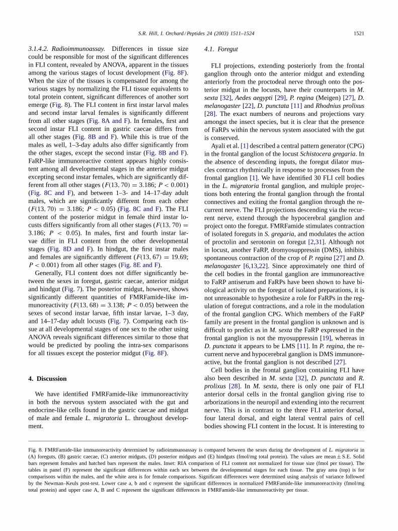
3.1.4.2. Radioimmunoassay. Differences in tissue sizecould be responsible for most of the significant differencesin FLI content, revealed by ANOVA, apparent in the tissuesamong the various stages of locust development (Fig. 8F).When the size of the tissues is compensated for among thevarious stages by normalizing the FLI tissue equivalents tototal protein content, significant differences of another sortemerge (Fig. 8). The FLI content in first instar larval malesand second instar larval females is significantly differentfrom all other stages (Fig. 8A and F). In females, first andsecond instar FLI content in gastric caecae differs fromall other stages (Fig. 8B and F). While this is true of themales as well, 1–3-day adults also differ significantly fromthe other stages, except the second instar (Fig. 8B and F).FaRP-like immunoreactive content appears highly consis-tent among all developmental stages in the anterior midgutexcepting second instar females, which are significantly dif-ferent from all other stages (F(13, 70) = 3.186;P < 0.001)(Fig. 8C and F), and between 1–3- and 14–17-day adultmales, which are significantly different from each other(F(13, 70) = 3.186; P < 0.05) (Fig. 8C and F). The FLIcontent of the posterior midgut in female third instar lo-custs differs significantly from all other stages (F(13, 70) =3.186; P < 0.05). In males, first and fourth instar lar-vae differ in FLI content from the other developmentalstages (Fig. 8D and F). In hindgut, the first instar malesand females are significantly different (F(13, 67) = 19.69;P < 0.001) from all other stages (Fig. 8E and F).
Generally, FLI content does not differ significantly be-tween the sexes in foregut, gastric caecae, anterior midgutand hindgut (Fig. 7). The posterior midgut, however, showssignificantly different quantities of FMRFamide-like im-munoreactivity (F(13, 68) = 3.138;P < 0.05) between thesexes of second instar larvae, fifth instar larvae, 1–3 day,and 14–17-day adult locusts (Fig. 7). Comparing each tis-sue at all developmental stages of one sex to the other usingANOVA reveals significant differences similar to those thatwould be predicted by pooling the intra-sex comparisonsfor all tissues except the posterior midgut (Fig. 8F).
4. Discussion
We have identified FMRFamide-like immunoreactivityin both the nervous system associated with the gut andendocrine-like cells found in the gastric caecae and midgutof male and femaleL. migratoria L. throughout develop-ment.
Fig. 8. FMRFamide-like immunoreactivity determined by radioimmunoassay is compared between the sexes during the development ofL. migratoria in(A) foreguts, (B) gastric caecae, (C) anterior midguts, (D) posterior midguts and (E) hindguts (fmol/mg total protein). The values are mean± S.E. Solidbars represent females and hatched bars represent the males. Inset: RIA comparison of FLI content not normalized for tissue size (fmol per tissue). Thetables in panel (F) represent the significant differences within each sex between the developmental stages for each tissue. The gray area (top) is forcomparisons within the males, and the white area is for female comparisons. Significant differences were determined using analysis of variance followedby the Newman–Keuls post-test. Lower case a, b and c represent the significant differences in normalized FMRFamide-like immunoreactivity (fmol/mgtotal protein) and upper case A, B and C represent the significant differences in FMRFamide-like immunoreactivity per tissue.
4.1. Foregut
FLI projections, extending posteriorly from the frontalganglion through onto the anterior midgut and extendinganteriorly from the proctodeal nerve through onto the pos-terior midgut in the locusts, have their counterparts inM.sexta [32], Aedes aegypti [29], P. regina (Meigen)[27], D.melanogaster [22], D. punctata [11] andRhodnius prolixus[28]. The exact numbers of neurons and projections varyamongst the insect species, but it is clear that the presenceof FaRPs within the nervous system associated with the gutis conserved.
Ayali et al.[1] described a central pattern generator (CPG)in the frontal ganglion of the locustSchistocera gregaria. Inthe absence of descending inputs, the foregut dilator mus-cles contract rhythmically in response to processes from thefrontal ganglion[1]. We have identified 30 FLI cell bodiesin the L. migratoria frontal ganglion, and multiple projec-tions both entering the frontal ganglion through the frontalconnectives and exiting the frontal ganglion through the re-current nerve. The FLI projections descending via the recur-rent nerve, extend through the hypocerebral ganglion andproject onto the foregut. FMRFamide stimulates contractionof isolated foreguts inS. gregaria, and modulates the actionof proctolin and serotonin on foregut[2,31]. Although notin locust, another FaRP, dromyosuppressin (DMS), inhibitsspontaneous contraction of the crop ofP. regina [27] andD.melanogaster [6,13,22]. Since approximately one third ofthe cell bodies in the frontal ganglion are immunoreactiveto FaRP antiserum and FaRPs have been shown to have bi-ological activity on the foregut of isolated preparations, it isnot unreasonable to hypothesize a role for FaRPs in the reg-ulation of foregut contractions, and a role in the modulationof the frontal ganglion CPG. Which members of the FaRPfamily are present in the frontal ganglion is unknown and isdifficult to predict as inM. sexta the FaRP expressed in thefrontal ganglion is not the myosuppressin[19], whereas inD. punctata it appears to be LMS[11]. In P. regina, the re-current nerve and hypocerebral ganglion is DMS immunore-active, but the frontal ganglion is not described[27].
Cell bodies in the frontal ganglion containing FLI havealso been described inM. sexta [32], D. punctata and R.prolixus [28]. In M. sexta, there is only one pair of FLIanterior dorsal cells in the frontal ganglion giving rise toarborizations in the neuropil and extending into the recurrentnerve. This is in contrast to the three FLI anterior dorsal,four lateral dorsal, and eight lateral ventral pairs of cellbodies showing FLI content in the locust. It is interesting to

1522 S.R. Hill, I. Orchard / Peptides 24 (2003) 1511–1524
note that parasitism ofM. sexta appears to increase the FLIcontent of the anterior dorsal cell bodies and their processes,and leads to the recruitment of a lateral dorsal pair of cellbodies producing FLI[32].
Developmental stage and sex have a bearing on the FLIcontent of the locust foregut suggesting that FaRPs mayfunction differently during the various stages in devel-opment. Although there were no apparent differences inFMRFamide-like immunoreactivity revealed by immuno-histochemistry. Radioimmunoassay revealed differences inFLI content of foreguts between males and females in thefirst three instars. The FLI content per tissue increased overdevelopmental time, however following normalization fortissue size by total protein content, FLI content was sig-nificantly increased in males of the first instar and femalesin the second instar, and significantly decreased in 2-weekmale adults.
Through all developmental stages and both sexes, FLImultipolar cells are observed on the surface of the mid toposterior region of the foregut. Multipolar cells on the sur-face of the foregut were first described in the beetle,Oryctes[25]. Wigglesworth[30] proposed that these multipolar cellsare proprioceptive organs, an afferent neural plexus sendinginformation on the degree of muscle fiber tension into thegut nervous system. The FLI multipolar cells we have de-scribed are consistent in morphology with those describedin the beetle[25] and appear to send projections into theneuropil of the ingluvial ganglia.
We observe one main peak of FLI in the foregut of 2-weeklocusts using RP-HPLC followed by RIA. A single peakmay indicate a single FaRP family member is present in theforeguts of 2-week female and male adult locusts. It is alsopossible, however, that more than one FaRP is isolated inthe main peak, and the other minor peaks, and so furtherpurification and isolation are underway.
4.2. Midgut and gastric caecae
FLI processes of the midgut and gastric caecae are shownto be a continuation of the projections descending from theingluvial ganglia on the foregut. The projections from theforegut extend over the gastric caecae and anterior midgut,but not into the posterior midgut. The projections over thehindgut extend over the posterior, but not anterior midgut.It is reasonable to suggest that the FMRFamide-like im-munoreactivity found in group I and group II HPLC frac-tions is a result of FLI content of the anterior midgut andgastric caecae endocrine-like cell populations, as only FLIfrom group III is observed in the foregut, which is devoid ofendocrine-like cells. Arguably, the same reasoning can beused to describe the presence of FLI in group I HPLC frac-tions in the posterior midgut: that is, it is due to the presenceof group I FLI content in posterior midgut endocrine-likecells, as the processes over the posterior midgut arise fromthe hindgut processes which contain groups II, III and IV,but not group I FLI fractions.
Endocrine-like cells in the midgut of insects have beendescribed previously[29,31]. FMRFamide-like immunore-activity in midgut endocrine-like cells has been observedin a range of insect species such asL. migratoria [15], D.punctata [10], M. sexta [19,32], Euborellia annulipes [26],D. melanogaster [21]. The midgut endocrine-like cells de-scribed are of the open-type with processes extending tothe lumen, and the cell bodies located adjacent to the dor-sal epithelial surface. In situ hybridization to myosuppressinmRNA in D. punctata [10], D. melanogaster [21], andM.sexta [19] has revealed that myosuppressin transcripts areexpressed in the anterior portion of the midgut. The pres-ence of large numbers of FLI endocrine-like cells in themidgut, and evidence that other peptides such as tackykininsand allatostatins exhibit positive immunoreactivity in midgutendocrine cells as well, provides further evidence that themidgut is acting as an endocrine gland in insects (see[24]).
The study of the insect gut has begun to expand in scoperecently, as there are more and more indications that thegut is not only acting as a digestive organ. The gut actsas a major endocrine gland; may play significant roles inthe regulation of the insects’ ionic balance (see[24]); andindeed may play a significant role in ecdysis. The secretionand eventual ingestion of ecdysial fluid, along with the airswallowing behavior observed at ecdysis is necessary forremoval of the cuticle and the inflation of wings duringthe final moult [3]. FaRPs may not simply be acting onthe gut to regulate digestion through modulation of musclecontraction and enzyme activity. FaRPs may be intricatelyinvolved in the myriad of other physiological processes ofthe gut including those mentioned above.
Significant differences in the amount of FLI contentobserved in the various tissues among the developmentalstages, once normalized for tissue size, appear to be stagespecific. By stage specific we mean that for each regionof the gut, individual stages during development will besignificantly different in FLI content from all other develop-mental stages. This stage specific variation in FLI contentsuggests that FaRPs may play different roles at times dur-ing locust development, and the differences in FLI contentbetween the sexes suggests that these roles may be sexu-ally dimorphic, either in developmental timing, or in thefunction of the peptides themselves.
Lange[15] assayed fifth instar, day 1 and day 14 adultmale L. migratoria midguts and gastric caecae for FLIcontent. When males from fifth instar, 1–3- and 14–18-dayadult locusts from this study were compared with thosefrom Lange [15] by analysis of variance, no significantdifferences in FMRFamide-like immunoreactivity of allmidgut tissues were observed (F(5, 26) = 15.19;P > 0.05)except for the comparisons between the day 1 males adults.The amount of FMRFamide-like immunoreactivity in thegastric caecae of day 1 adult males[15] is significantlydifferent from the 1–3-day males described in this study(F(5, 26) = 20.09; P < 0.001 comparing tissue equiva-lents andF(5, 26) = 15.19; P < 0.01 comparing tissue

S.R. Hill, I. Orchard / Peptides 24 (2003) 1511–1524 1523
equivalents normalized for amount of total protein). Thedifferences observed in FMRFamide-like immunoreactivityin the gastric caecae may be due to the pooling of day 1 today 3 adult males. A transient increase in FMRFamide-likeimmunoreactivity in day 1 animals may be masked in apooled sample containing 3 days worth of animals, or per-haps it is due to the differences in rearing between theseseparate two colonies of locusts.
4.3. Hindgut
In the hindgut of D. melanogaster, McCormick andNichols [21] describe two sensory cells containing FLImaterial throughout larval, pupal and adult stages. We didnot observe these sensory cells in the locust. The onlyFLI sensory cells we observe directly associated with guttissue are those multipolar cells covering the mid to poste-rior foregut. The distribution of processes over the hindgutof all developmental stages ofL. migratoria is similar tothat described forD. punctata [11] andM. sexta [32]. Theloose grid-like formation of FLI processes just posteriorto the midgut/hindgut border suggests the innervation ofthe pyloric valve similar to that described for the distribu-tion crustacean cardioactive peptide[5]. The pyloric valvemay control the flow of material from both midgut andmalpighian tubules into the hindgut, and the presence ofFLI processes closely associated with this valve suggestsa role for FaRPs in modulating its activity. The presenceof three groups of FLI HPLC fractions in the hindguts of2-week locusts suggests that there may be more than onerole for FaRPs along the surface of the hindgut. Myosup-pressins cause inhibition of hindgut muscle contractions inL. maderae [12] while other extended FLRFamides, such asManseFLRFamides II and III, can stimulate hindgut musclecontraction inM. sexta [14].
The current study indicates that FLI content is present innervous projections and endocrine-like cells associated withthe gut throughout larval development and until the adult lo-cust reaches at least 2 weeks of age. There are tissue, stageand sex specific variations in FMRFamide-like immunore-activity. These differences in FaRP expression patterns sug-gest that individual or groups of FaRP family members maybe acting in various ways, and at different times, during de-velopment. As different FaRP superfamily members havebeen shown previously to perform opposite actions on thesame tissue[24], and since we have shown that at least fourgroups of FaRP-like peptides are present within the gut tis-sues and associated nervous system, we suggest that FaRPsplay a major role in regulating and modulating the physio-logical processes associated with the locust gut.
Acknowledgments
This work was supported by the Natural Sciences andEngineering Research Council of Canada.
References
[1] Ayali A, Zilberstein Y, Cohen N. The locust frontal ganglion: acentral pattern generator network controlling foregut rhythmic motorpatterns. J Exp Biol 2002;205:2825–32.
[2] Banner SE, Osborne RH. Modulation of 5-HT and proctolin receptorsby FMRFamide in the foregut of the locustSchistocerca gregaria. JInsect Physiol 1989;35:887–92.
[3] Bestman JE, Booker R. Modulation of foregut synaptic activitycontrols resorption of molting fluid during larval molts of the mothManduca sexta. J Exp Biol 2003;206:1207–20.
[4] Bradford MM. A rapid, and sensitive method for the quantification ofmicrogram quantities of protein utilizing the principle of protein-dyebinding. Anal Biochem 1976;8:95–108.
[5] Donini A, Ngo C, Lange AB. Evidence for crustacean cardioactivepeptide-like innervation of the gut inLocusta migratoria. Peptides2002;23:1915–23.
[6] Duttlinger A, Berry K, Nichols R. The different effects of threeDrosophila melanogaster dFMRFamide-containing peptides on cropcontractions suggest these structurally related peptides do not playredundant functions in the gut. Peptides 2002;23:1953–7.
[7] Favrel P, Giard W, Benlimane N, Boucaud-Camou E, HenryM. A new biological activity for the neuropeptide FMRFamide:experimental evidence for a secretagogue effect on amylase secretionin the scallopPecten maximus. Experientia 1994;50:1106–10.
[8] Fujisawa Y, Shimoda M, Kiguchi K, Ichikawa T, Fujita N. Theinhibitory effect of a neuropeptide,ManducaFLRFamide, on themidgut activity of the sphingid moth,Agris convulvuli. Zool Sci1993;10:773–7.
[9] Fuse M, Orchard I. The muscular contractions of the midgut of thecockroachDiploptera puntata: effects of the insect neuropeptidesproctolin and leucomyosuppressin. Reg Pept 1998;77:163–8.
[10] Fuse M, Zhang JR, Partridge E, Nachman RJ, Orchard I, BendenaWG, et al. Effects of an allatostatin and a myosuppressin on midgutcarbohydrate enzyme activity in the cockroachDiploptera punctata.Peptides 1999;20:1285–93.
[11] Fuse M, Bendena WG, Donly BC, Tobe SS, Orchard I. In situhybridization analysis of leucomyosuppressin mRNA expression inthe cockroach,Diploptera punctata. J Comp Neurol 1998;395:328–41.
[12] Holman GM, Cook BJ, Nachman RJ. Primary structure and synthesisof a blocked myotropic neuropeptide isolated from the cockroach,Leucophaea maderae. Comp Biochem Physiol C 1986;85:219–24.
[13] Kaminski S, Orlowski E, Berry K, Nichols R. The effects of threeDrosophila melanogaster myotropins on the frequency of foregutcontractions differ. J Neurogenet 2002;16:125–34.
[14] Kingan T, Shabanowitz J, Hunt D, Witten J. Characterization oftwo myotropic neuropeptides in the FMRFamide family from thesegmental ganglia of the mothManduca sexta: candidate neuro-hormones and neuromodulators. J Exp Biol 1996;199:1095–104.
[15] Lange AB. Feeding state influences the content of FMRFamide- andtachykinin-related peptides in the endocrine-like cells of the midgutof Locusta migratoria. Peptides 2001;22:229–34.
[16] Lange AB, Orchard I. The effects of SchistoFLRFamide oncontractions of locust midgut. Peptides 1998;19:459–67.
[17] Lee KY, Horodyski FM, Chamberlin ME. Inhibition of midgut iontransport by allatotropin (Mas-AT) andManduca FLRFamides in thetobacco hornwormManduca sexta. J Exp Biol 1998;201:3067–74.
[18] Linacre A, Kellet A, Suanders S, Bright K, Benjamin PR, BurkeJF. Cardioactive neuropeptide Phe-Met-Arg-Phe-NH2 (FMRFamide)and novel related peptides are encoded in multiple copies by a singlegene in the snailLymnaea stagnalis. J Neurosci 1990;10:412–9.
[19] Lu D, Lee K-Y, Horodyski FM, Witten JL. Molecular characterizationand cell-specific expression of aManduca sexta FLRFamide gene.J Comp Neurol 2002;446:337–96.
[20] Maule AG, Mousley A, Marks NJ, Day TA, Thompson DP, GearyTG, et al. Neuropeptide signaling systems—potential drug targetsfor parasite and pest control. Curr Top Med Chem 2002;2:733–58.

1524 S.R. Hill, I. Orchard / Peptides 24 (2003) 1511–1524
[21] McCormick J, Nichols R. Spatial and temporal expression identifydromyosuppressin as a brain-gut peptide inDrosophila melanogaster.J Comp Neurol 1993;338:278–88.
[22] Merte J, Nichols R. Drosophila melanogaster FMRFamide-containing peptides: redundant or diverse functions? Peptides 2002;23:209–20.
[23] Nachman RJ, Favre P, Sreekumar S, Holman GM. Insectmyosuppressins and sulfakinins stimulate the release of thee digestiveenzyme�-amylase in two invertebrates: the scallopPecten maximusand the insectRhynchophorus ferrugineus. Ann NY Acad Sci1997;814:335–8.
[24] Orchard I, Lange AB, Bendena WG. FMRFamide-related peptides: amultifunctional family of structurally related neuropeptides in insects.Adv Insect Physiol 2001;28:267–329.
[25] Orlav J. Innervation of the gut: Lamellicorn larvae, Coleoptera. ZWiss Zool 1924;124:425–502.
[26] Rankin SM, Seymour SM. FMRFamide-like material in the earwig,Euborellia annulipes, and its functional significance. Arch InsectBiochem Physiol 2001;47:189–97.
[27] Richer S, Stoffolano Jr JG, Yin C-M, Nichols R. Innervation ofdromyosuppressin (DMS) immunoreactive processes and effect ofDMS and benzethonium chloride on thePhormia regina (Meigen)crop. J Comp Neurol 2000;421:136–42.
[28] Tsang PW, Orchard I. Distribution of FMRFamide-related peptidesin the blood-feeding bug,Rhodnius prolixus. J Comp Neurol1991;311:17–32.
[29] Veenstra JA, Lau GW, Agricola HJ, Petzel DH. Immunohistologicallocalization of regulatory peptides in the midgut of the femalemosquitoAedes aegypti. Histochem Cell Biol 1995;104(5):337–47.
[30] Wigglesworth VB. The principles of insect physiology. London:Methune & Co. Ltd.; 1965.
[31] Wood SJ, Osborne RH, Cattell KJ, Banner SE. FMRFamide andrelated peptides modulate the actions of 5-hydroxytryptamine andproctolin on the foregut of the locust,Schistocerca gregaria. BiochemSoc Trans 1990;18:384–5.
[32] Zitnan D, Kingan TG, Beckage NE. Parasitism-induced accumulationof FMRFamide-like peptides in the gut innervation and endocrinecells of Manduca sexta. Insect Biochem Mol Biol 1995;25:669–78.