An Investigation into the Feasibility and Viability of ... - CiteSeerX

This article was downloaded by: [Consiglio Nazionale delle Ricerche]On: 14 June 2013, At: 07:42Publisher: Taylor & FrancisInforma Ltd Registered in England and Wales Registered Number: 1072954 Registered office:Mortimer House, 37-41 Mortimer Street, London W1T 3JH, UK
Journal of Immunoassay andImmunochemistryPublication details, including instructions for authors and subscriptioninformation:http://www.tandfonline.com/loi/ljii20
Fluorescent Antibody‐Viability Staining andβ‐Glucuronidase Assay as Rapid Methodsfor Monitoring Escherichia coli Viability inCoastal Marine WatersG. Caruso a , F. De Pasquale a , M. Mancuso a , D. Zampino a & E. Crisafi aa Institute for Coastal Marine Environment (IAMC)‐CNR, Section of Messina,Messina, ItalyPublished online: 06 Feb 2007.
To cite this article: G. Caruso , F. De Pasquale , M. Mancuso , D. Zampino & E. Crisafi (2006): FluorescentAntibody‐Viability Staining and β‐Glucuronidase Assay as Rapid Methods for Monitoring Escherichia coliViability in Coastal Marine Waters, Journal of Immunoassay and Immunochemistry, 27:1, 1-13
To link to this article: http://dx.doi.org/10.1080/15321810500403599
PLEASE SCROLL DOWN FOR ARTICLE
Full terms and conditions of use: http://www.tandfonline.com/page/terms-and-conditions
This article may be used for research, teaching, and private study purposes. Any substantialor systematic reproduction, redistribution, reselling, loan, sub-licensing, systematic supply, ordistribution in any form to anyone is expressly forbidden.
The publisher does not give any warranty express or implied or make any representation that thecontents will be complete or accurate or up to date. The accuracy of any instructions, formulae,and drug doses should be independently verified with primary sources. The publisher shall notbe liable for any loss, actions, claims, proceedings, demand, or costs or damages whatsoever orhowsoever caused arising directly or indirectly in connection with or arising out of the use of thismaterial.

Fluorescent Antibody-Viability Stainingand b-Glucuronidase Assay as Rapid
Methods for Monitoring Escherichia coliViability in Coastal Marine Waters
G. Caruso, F. De Pasquale, M. Mancuso, D. Zampino,
and E. Crisafi
Institute for Coastal Marine Environment (IAMC)-CNR,
Section of Messina, Messina, Italy
Abstract: A faecal pollution monitoring of coastal Messina waters was performed by
comparing three (microscopic, enzyme, and culture) methods. Evidence of Escherichia
coli cells (29.99 to 96.79% of the total enteropathogenic serotypes) retaining their
viability into the marine environment was shown. b-Glucuronidase activity rates
suggested that living cells were also metabolically active. Heavily polluted sites
were detected, where improperly treated urban wastes were discharged. Significant
relationships between microscopic and enzymatic data proved both methods to be
suitable alternatives to the culture method for E. coli detection, improving environ-
mental quality assessment.
Keywords: Faecal pollution, Seawater monitoring, Escherichia coli, Viability,
Fluorescent antibody, b-Glucuronidase
INTRODUCTION
Bacterial pollution represents a threat for many coastal marine areas with
severe implications for water quality. Rapid and reliable analytical
protocols are needed for the microbiological control of aquatic environments;
improvement of the techniques available up to now constitutes the challenge
Address correspondence to G. Caruso, Institute for Coastal Marine Environment
(IAMC)-CNR, Section of Messina, Spianata S. Raineri 86, 98122, Messina, Italy.
E-mail: [email protected]
Journal of Immunoassay & Immunochemistry, 27: 1–13, 2006
Copyright # Taylor & Francis LLC
ISSN 1532-1819 print/1532-4230 online
DOI: 10.1080/15321810500403599
1
Dow
nloa
ded
by [
Con
sigl
io N
azio
nale
del
le R
icer
che]
at 0
7:42
14
June
201
3

for future developments in this field.[1] The antigen-antibody reaction, on which
the fluorescent antibody (FA) method relies, and the screening for selected
bacterial enzymes, e.g., b-glucuronidase, offer specific tools to detect and
quantify the abundance and activity of Escherichia coli in seawaters.[2] The
viability assessment of enteric bacteria is also of primary importance for
public health preservation because, in adverse environmental conditions,
they may survive in aquatic environments entering the viable but nonculturable
(VBNC) state.[3,4] In E. coli, this phase, characterised by lost of culturability,
with retention of cellular integrity and potential pathogenesis, has been
widely documented.[4 – 10] The severe sanitary consequences that could result
from the recovery of cell viability when environmental conditions become
favourable[11] support the increasing interest addressed to this specific topic.
In the framework of the MIUR-Cluster 10 research programme, aimed at
setting up new rapid methodologies for coastal marine monitoring, an investi-
gation of bacterial pollution along Messina shoreline was undertaken. The
aim of our research was to depict the occurrence of E. coli in seawater,
paying attention to changes in cell viability occurring in space and in time.
Improperly treated urban discharges have been recognised as the main source
of the worsening quality levels of coastal waters surrounding Messina. High
levels of faecal contamination occur, particularly along the Ionian coastline
between Capo Peloro and Tremestieri, but the particular hydrodynamic
conditions of the area leads to a strong reduction of the density of faecal
coliforms already present 200 m off shore.[12] The study provided a good oppor-
tunity to compare and test, in the field, three different analytical approaches to
detect the presence of E. coli as the main faecal pollution indicator: the
standard culture method (m-FC), the b-glucuronidase activity assay (through
the fluorogenic substrate 4-methylumbelliferyl-b-D-glucuronide, MUG), and
a specifically developed protocol that combines a viability staining (the
Live/Dead staining) with an indirect fluorescent antibody (FA) procedure.
EXPERIMENTAL
Collection and Treatment of Samples
From the 24th of March to the 5th of May, 2004, on a weekly basis, surface
seawater samples (n ¼ 34) were collected from five coastal sites (Tremestieri,
Pagliarisi, Europa, Annunziata, Tennis, Fig. 1). Sub-samples (100 mL or less)
were analysed using the following methods.
Plate Counts (m-FC)
Faecal coliforms (FC) were counted by membrane filtration (0.45mm) and
incubation on m-FC agar (Difco) plates.[13] Results were expressed as CFU
100 mL21 of water. The density of Escherichia coli was determined by
G. Caruso et al.2
Dow
nloa
ded
by [
Con
sigl
io N
azio
nale
del
le R
icer
che]
at 0
7:42
14
June
201
3

confirmation with James’ indole reagent (Biomerieux, Marcy l’Etoile, France)
of the colonies grown on m-FC agar.
MUG Assay
For the determination of b-glucuronidase activity, increasing amounts (5 to
50mmol L21) of the fluorogenic substrate 4-methylumbelliferyl-b-D-glucuro-
nide (MUG, Sigma, 0.5 mmol L21 stock solution) were added to 10 mL
aliquots of a 10-times concentrated sample.[14] The fluorescence released by
enzymatic cleavage of the substrate in the product methylumbelliferone was
measured at time 0, immediately after its addition, and after 3.5 h of incu-
bation at 448C, with a spectrofluorometer (Cary Eclipse Fluorescence spectro-
photometer, Varian Inc., Palo Alto, CA, USA) at 365 and 455 nm (excitation
and emission wavelengths, respectively). Calibration was performed with
known concentrations (200, 400, and 800 nmol L21) of the standard MUF
(Sigma, 0.5 mmol L21). Enzyme values were reported as maximum velocity
of hydrolysis (Vmax, in nmol of MUF released h21 100 mL21).
Combined FA-Live/Dead (FA-L/D) Staining Procedure
This protocol combined the indirect fluorescent antibody (FA) labelling
procedure[15] using isothiocyanate fluorescein (FITC)-conjugated immune
sera specific for E. coli enteropathogenic serotypes (Behring Diagnostics
Figure 1. Sampling stations.
Monitoring Escherichia coli Viability in Coastal Marine Waters 3
Dow
nloa
ded
by [
Con
sigl
io N
azio
nale
del
le R
icer
che]
at 0
7:42
14
June
201
3

Inc., Westwood, MA, USA) together with propidium iodide (PI) stain, a
component of the Live/Dead (L/D) BacLight bacterial viability kit
(Molecular Probes, Eugene, Oregon, USA). The use of PI, which penetrates
cells with damaged membranes only, allowed the distinction of dead cells,
fluorescing red, within the total enteropathogenic E. coli population. Cells
were enumerated by an epifluorescence microscope (Axioplan 2, Carl Zeiss
S.p.A., Arese, Milano, Italia) equipped with an image analysis system and
viewed separately with fluorescein (blue light, BP 450–490, FT 510, LP
520) and rhodamine (green light, BP 510–560, FT 580, LP 590) filter sets.
When excited under blue light, total (livingþ non-living) E. coli cells
showed green fluorescing outlines due to FITC (emission peak: 520 nm); by
switching to the green light, the dead (PI-positive) E. coli cells fluoresced
red, due to PI labelling (emission peak: 617–623 nm). The fraction of living
cells was determined as the difference between the total FITC-labelled
E. coli cells and PIþ dead cells. Prior to its application to the analysis of
natural samples, a suspension of killed E. coli (by boiling) was used as a
negative control, in order to verify that dead cells were efficiently stained
by our FA-L/D protocol. Counts were performed on a minimum of 20
randomly selected microscopic fields. Cell abundance was expressed as the
mean value of E. coli cells per 100 mL of sample.
Statistical Analysis
Values obtained by each method were log-converted to stabilize the variance
and attain normality; the differences between mean counts were tested for
statistical significance using an analysis of variance (ANOVA). Paired data
were analysed using Pearson correlation coefficients to determine if values
were statistically correlated.
RESULTS
The main steps of our FA-L/D labelling protocol are presented schematically
in Table 1. The concentration of 30mM L21 PI, chosen from a variety of con-
centrations ranging from 5 to 50mM L21, proved to be effective in labelling
dead cells without any interference with the antigen-antibody reaction. The
use of PI from a L/D BacLight bacterial viability kit was conceived to
answer to the need of standardizing the viability staining protocol. After
this treatment, microscopic fields observed, using fluorescein or rhodamine
filters, were also overlapped to observe the simultaneous labelling of both
fluorochromes and to discriminate, within the total enteropathogenic E. coli
cells, the ones that were dead.
Figure 2 shows the distribution, during each sampling, of FC values, as
obtained by plate count, and of total enteropathogenic E. coli, as determined
G. Caruso et al.4
Dow
nloa
ded
by [
Con
sigl
io N
azio
nale
del
le R
icer
che]
at 0
7:42
14
June
201
3

by FA-L/D; b-glucuronidase activity rates, measured by MUG assay, are also
reported. Total enteropathogenic E. coli values ranged from 1.31 � 103 to
4.49 � 105 cells 100 mL21, with peaks at Pagliarisi and Europa stations.
Except for one sampling (April, 30) only, their lowest concentrations were
found at Annunziata station, which differed significantly from those
recorded at the stations Pagliarisi (F ¼ 11.90, P , 0.01, n ¼ 14) and Europa
(F ¼ 19.13, P , 0.01, n ¼ 14).
FC showed values 1–2 orders of magnitude lower than E. coli, ranging
from 9.0 � 100 to 4 � 104 CFU 100 mL21, suggesting the existence of a
fraction of VNCB cells. However, similar trends were reported for FC and
E. coli values, highlighting no significant differences between culture and
microscopic methods. This finding was also confirmed by results of
ANOVA (F ¼ 2.82, P , 0.05, n ¼ 34). As far as the enzyme measurements
are concerned, the potential b-glucuronidase activity rates oscillated between
0.002 and 726.88 nmol h21 100 mL21, with the lowest values recorded at
Annunziata station. Their spatial distribution was similar to that observed
for E. coli and FC counts, as further demonstrated by ANOVA values
(F ¼ 3.35 and 2.57, P , 0.05, n ¼ 34, respectively).
Bacterial densities followed a variable temporal distribution, with the
highest culture and microscopic counts during the first samplings, particularly
at Europa station. During the following samplings, bacterial peaks moved
towards Pagliarisi station, more southern than Europa station. Bacterial
counts dropped to minimum values (on average 2.48 � 102 CFU 100 mL21
and 2.91 � 103 cells 100 mL21 for FC and E. coli) during the last sampling,
when they were significantly different from those of the first one
(F ¼ 5.637, P , 0.05, and 17.25, P , 0.01, n ¼ 14, by m-FC and FA-L/D,
respectively). In a similar way, the levels of b-glucuronidase activity
reached their minimum values during the May sampling.
For each sampling, significant relationships between m-FC and FA-L/D
(0.742 � r � 0.995, P , 0.01) and m-FC and MUG (0.746 � r � 0.999,
P , 0.01) results were detected by Pearson correlation coefficients calculated
comparing the culture vs FA-L/D combined staining counts and the
Table 1. Main steps of the combined FA-L/D protocol
Filtration through a 0.22mm Nuclepore black membrane
Incubation with hydrolysed gelatine (2% final concentration), 20 min
Washing 3 times with 5 mL PBS
Incubation with a 1 : 40 dilution in PBS of Murex E. coli antisera (pool Aþ Bþ C),
30 min
Washing 3 times with 5 mL PBS
Labelling with a 1 : 80 dilution of goat-anti rabbit–IgG-FITC, 30 min
Incubation with PI stain from L/D kit 15 min in the dark
Mounting with L/D BacLight mounting oil
Observation under epifluorescence microscope
Monitoring Escherichia coli Viability in Coastal Marine Waters 5
Dow
nloa
ded
by [
Con
sigl
io N
azio
nale
del
le R
icer
che]
at 0
7:42
14
June
201
3
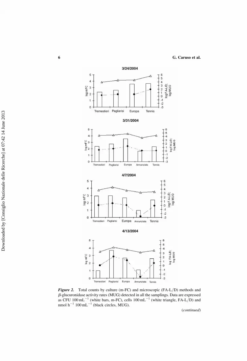
Figure 2. Total counts by culture (m-FC) and microscopic (FA-L/D) methods and
b-glucuronidase activity rates (MUG) detected in all the samplings. Data are expressed
as CFU 100 mL21 (white bars, m-FC), cells 100 mL21 (white triangle, FA-L/D) and
nmol h21 100 mL21 (black circles, MUG).
(continued)
G. Caruso et al.6
Dow
nloa
ded
by [
Con
sigl
io N
azio
nale
del
le R
icer
che]
at 0
7:42
14
June
201
3

culture counts vs MUG rates (data not shown in the figure). Overall, MUG and
FA-L/D values were also significantly related (r ¼ 0.673, P , 0.01).
The contribution of live and dead fractions to the total enteropathogenic
E. coli population at each station is reported in Fig. 3. No clear temporal trend
was observed in their distribution. Within the total enteropathogenic E. coli
population, identified by FITC labelling, living cells globally represented a
percentage ranging from 29.99 to 96.79%, with the highest mean values
recorded at Tremestieri and Tennis stations (98.21 and 91.92%, respectively).
Dead cells accounted for a similar fraction (15% of the total) at Pagliarisi and
Europa stations. As revealed by ANOVA (F ¼ 3.16, P , 0.05, n ¼ 34), the
values concerning the living fraction were not significantly different from
MUG values; this result suggested that both fluorescent-antibody/viability
Figure 2. Continued.
Monitoring Escherichia coli Viability in Coastal Marine Waters 7
Dow
nloa
ded
by [
Con
sigl
io N
azio
nale
del
le R
icer
che]
at 0
7:42
14
June
201
3

Figure 3. Percentages of live (white bars) and dead (black bars) E. coli cells obtained
by the combined FA-L/D protocol at the different stations: a, Tremestieri; b, Pagliarisi;
c, Europa; d, Annunziata; e, Tennis.
(continued)
G. Caruso et al.8
Dow
nloa
ded
by [
Con
sigl
io N
azio
nale
del
le R
icer
che]
at 0
7:42
14
June
201
3

and enzyme methods identified the same bacterial sub-population or, in other
words, that cells scored as living by epifluorescence microscope were also still
metabolically active.
Furthermore, the linear regression analysis of log-transformed m-FC vs
FA-L/D and m-FC vs MUG data (Fig. 4) showed that all the three methods
yielded statistically similar results; in particular, enzyme rates were correlated
with FC counts more significantly (R2 ¼ 0.6161) than FA-L/D (R2¼ 0.4873).
Moreover, FA-L/D data displayed a poor dispersion with respect to the
theoretical regression line.
DISCUSSION
The viability of faecal bacteria in seawaters is a critical issue for public health
preservation; in our study, living properties of E. coli cells have been detected
Figure 3. Continued.
Monitoring Escherichia coli Viability in Coastal Marine Waters 9
Dow
nloa
ded
by [
Con
sigl
io N
azio
nale
del
le R
icer
che]
at 0
7:42
14
June
201
3

in coastal samples by comparing different methods: culturability by the plate
method; metabolic (b-glucuronidase enzyme) activity by fluorogenic
measurement, and membrane integrity by an indirect FA assay combined
with PI. It is commonly recognised that standard culture methods for
the specific detection and quantification of E. coli involve some restrictions
in terms of both accuracy and sensitivity (i.e., failure to detect injured
bacteria), which make their application to seawater monitoring inadequate.[1,16]
Consequently, the search for rapid, highly sensitive methods is encouraged
as a reliable and effective tool for early warning of sewage pollution. Our
study aimed at proposing new analytical approaches, such as microscopic
and enzymatic methods, that are more specific, require less time and are
less cost-consuming than the standard culture method, involving the use of
basic laboratory facilities. Being growth independent, both FA-L/D and
MUG were able to identify viable but nonculturable cells; they differed in
their basic principles, the first relying on membrane integrity and the second
considering the expression of enzyme activity. On the other hand, it is recog-
nised that the parameter “cell activity” could not be used as a surrogate esti-
mation of “cell culturability” and, therefore, activity rates are not directly
comparable with abundance data. Furthermore, the assessment of b-glucuro-
nidase activity only could not be considered as an adequate predictor of the
complete metabolic activities, and this could result in a possible underestima-
tion of the amount of active cells. In spite of these limitations, our basic
opinion is that data concerning both bacterial metabolism and density, if
yielding reciprocally consistent results, could contribute to a more actual
and precise estimation of the viability of faecal bacteria in natural marine
samples. As previously stated,[17] in fact, the assay of one property of living
cells cannot provide sufficient proof of viability.
Figure 4. Regression analysis of m-FC values vs FA-L/D (white squares, solid line)
and MUG values (black triangles, dotted line).
G. Caruso et al.10
Dow
nloa
ded
by [
Con
sigl
io N
azio
nale
del
le R
icer
che]
at 0
7:42
14
June
201
3

In the samples examined, E. coli represented the main component within
the total FC population (data not shown); this result was confirmed by the sig-
nificant relationships between E. coli and FC values. Compared with FA-L/D,
the m-FC method, however, yielded an underestimate of bacterial concen-
trations; this quantitative discrepancy could be explained by the large percen-
tage of nonculturable cells present in our natural samples that were still
alive and also expressed b-glucuronidase activity in situ, as shown by the
significant relationship between FA-L/D and MUG. Analysis of total data
by linear regression suggested the reciprocal agreement of results obtained
by the three methods; enzyme rates were correlated with FC values at a
significance level higher than FA counts. This finding, previously observed,
both in freshwater[9] and highly-contaminated marine environments,[18] led
us to suppose that the high level of organic matter available at these sites
could support a great percentage of metabolically active cells. As a fact,
76.47% (on average) of the samples examined exceeded the threshold
values allowed by Italian laws for bathing waters (FC , 100 CFU
100 mL21), showing the occurrence of improperly treated urban sewage
along the shoreline.
In our study the FA-viability staining procedure previously applied for the
detection of E. coli in natural seawaters[19] was also modified: the propidium
iodide reagent came from a standardised L/D viability kit, making it easier to
set up the appropriate concentration of the stain and also resulting in
advantages for the operator safety. Our protocol has proved to be effective
in discriminating, in seawaters, the living and the dead fraction within a
specific (E. coli) bacterial population. Compared with b-glucuronidase
activity measurements, sometimes biased by the occurrence of false-
positive and false-negative results (i.e., coliforms other than E. coli or
b-glucuronidase negative E. coli, respectively), the microscopic method
offers the advantages of simplicity of execution, specificity, and sensitivity
of the immunological techniques, rapid and quantitative responses,
providing rapid information about the physiological state in which pathogenic
bacteria may persist in natural environments.[6,9] The high incidence (on
average 91.64%) of viable cells detected by the combined FA-L/D protocol
and the measured values of b-glucuronidase activity higher than the
maximum value (79.11 nmol MUF h21 100 mL21) previously reported in
the same Sicilian area,[2] suggested that anthropogenic inputs were recent
and that the large nutrient and organic matter inputs supported bacterial
survival and activity. If we compare the present data with counts obtained
in 2002, when dead cells accounted for 22% of the total bacterial density in
Messina Straits,[19] this result proved evidence of a worsening of the microbio-
logical quality of our coastal waters. It also confirmed the importance, for
human health, of rapid procedures able to detect, in situ, the fraction of
bacteria retaining their viable and/or active properties that could be scored
as negative and, therefore, underestimated by conventional plate counts.
The assessment of the viability of specific bacteria using a “battery” of
Monitoring Escherichia coli Viability in Coastal Marine Waters 11
Dow
nloa
ded
by [
Con
sigl
io N
azio
nale
del
le R
icer
che]
at 0
7:42
14
June
201
3

different methods may yield a more sensitive estimate of the persistence of
human faecal indicators in coastal waters, offering new, interesting perspec-
tives in the field of aquatic environment monitoring.
ACKNOWLEDGMENTS
The work has been funded by Italian MIUR (Ministero Universita e Ricerca)
within the Cluster 10-SAM “Realizzazione ed attivazione di una rete integrata
di piattaforme e mezzo mobile attrezzati per il monitoraggio dell’ambiente
marino costiero” Project.
REFERENCES
1. Sartory, D.P.; Watkins, J. Conventional culture for water quality assessment: isthere a future? J. Appl. Microbiol. Symp. Suppl. 1999, 85 (26), 225S–233S.
2. Caruso, G.; Denaro, R.; Genovese, M.; Giuliano, L.; Mancuso, M.; Yakimov, M.New methodological strategies for detecting bacterial indicators. Chem. Ecol.2004, 20 (3), 167–181.
3. Roszack, D.B.; Colwell, R.R. Metabolic activity of bacterial cells enumerated byDirect Viable Count. Appl. Environ. Microbiol. 1987, 53, 2889–2893.
4. Rozen, Y.; Belkin, S. Survival of enteric bacteria in seawater. FEMS Microbiol.Rev. 2001, 725, 1–17.
5. Xu, H.S.; Roberts, N.; Singleton, F.L.; Attwell, R.W.; Grimes, D.J.; Colwell, R.R.Survival and viability of nonculturable Escherichia coli and Vibrio cholerae in theestuarine and marine environment. Microb. Ecol. 1982, 8, 313–323.
6. Lopez-Amoros, R.; Comas, J.; Vives-Rego, J. Flow cytometric assessment ofEscherichia coli and Salmonella typhimurium starvation-survival in seawaterusing Rhodamine 123, propidium iodide, and oxonol. Appl. Environ. Microbiol.1995, 61, 2521–2526.
7. Muela, A.; Arana, I.; Justo, J.I.; Seco, C.; Barcina, I. Changes in DNA content andcellular death during a starvation-survival process of Escherichia coli in riverwater. Microb. Ecol. 1999, 37, 62–69.
8. Ericsson, M.; Hanstorp, D.; Hagberg, P.; Enger, J.; Nystrom, T. Sorting outbacterial viability with optical tweezers. J. Bacteriol. 2000, 182, 5551–5555.
9. Petit, M.; George, I.; Servais, P. Survival of Escherichia coli in freshwater: b-D-glucuronidase activity measurements and characterization of cellular states.Can. J. Microbiol. 2000, 46, 679–684.
10. Ohtomo, R.; Saito, M. Increase in the culturable cell number of Escherichia coliduring recovery from saline stress: possible implication for resuscitation fromthe VBNC state. Microb. Ecol. 2001, 42, 208–214.
11. Pommepuy, M.; Butin, M.; Derrien, A.; Gourmelon, M.; Colwell, R.R.;Cormier, M. Retention of enteropathogenicity by viable but nonculturableEscherichia coli exposed to seawater and sunlight. Appl. Environ. Microbiol.1996, 62, 4621–4626.
12. Caruso, G.; Zaccone, R.; Monticelli, L.; Crisafi, E.; Zampino, D. Bacterialpollution of Messina coastal waters: a one year study. Microbiologica 2000, 23,297–304.
G. Caruso et al.12
Dow
nloa
ded
by [
Con
sigl
io N
azio
nale
del
le R
icer
che]
at 0
7:42
14
June
201
3

13. American Public Health Association. Standard methods for the examination ofwater and waste water, 1992, 18th Ed.; American Public Health Association:Washington, DC, 1992.
14. Caruso, G.; Crisafi, E.; Mancuso, M. Development of an enzyme assay for rapidassessment of Escherichia coli in seawaters. J. Appl. Microbiol. 2002, 93 (4),548–556.
15. Caruso, G.; Crisafi, E.; Mancuso, M. Immunofluorescence detection ofEscherichia coli in seawater: a comparison of various commercial antisera.J. Immunoassay Immunochem. 2002, 23 (4), 479–496.
16. Volterra, L.; Garizio, M. Limiti delle determinazioni microbiologiche ambientali acarattere sanitario: la colimetria. Biologi Italiani 1997, 8/97, 28–34.
17. Weichart, D. How viable is the VBNC hypothesis? Logical Biol. 2000, 1, 21–24.18. Caruso, G.; Crisafi, E.; Mancuso, M. Coastal monitoring by MUG assay to the
detection of seawater pollution. In XXXVIII Congres CIESM; Proceedings of theCongress, Barcelona, Spain, Jun 7–11, 2004; CIESM: Monaco, 2004, 269.
19. Caruso, G.; Mancuso, M.; Crisafi, E. Combined fluorescent antibody assay andviability staining for the assessment of the physiological states of Escherichiacoli in seawaters. J. Appl. Microbiol. 2003, 95 (2), 225–233.
Received June 3, 2005
Accepted July 2, 2005
Manuscript 3180
Monitoring Escherichia coli Viability in Coastal Marine Waters 13
Dow
nloa
ded
by [
Con
sigl
io N
azio
nale
del
le R
icer
che]
at 0
7:42
14
June
201
3
Copyright © 2022 FDOKUMEN