Structural Role of Conserved Asn179 in the Short-Chain Dehydrogenase/Reductase Scaffold
Upload
teknologimalaysiaCategory
view
0download
0
Seediscussions,stats,andauthorprofilesforthispublicationat:https://www.researchgate.net/publication/5252126
Expression,purificationandcharacterizationofaphyAm-phyCsfusionphytase
ArticleinJournalofZhejiangUniversitySCIENCEB·August2008
DOI:10.1631/jzus.B0720006·Source:PubMed
CITATIONS
8
READS
39
9authors,including:
LikouZou
SichuanAgriculturalUniversity
34PUBLICATIONS188CITATIONS
SEEPROFILE
Guo-BaoTian
SunYat-SenUniversity
27PUBLICATIONS492CITATIONS
SEEPROFILE
QiWu
SichuanAgriculturalUniversity
44PUBLICATIONS314CITATIONS
SEEPROFILE
AllcontentfollowingthispagewasuploadedbyLikouZouon03December2016.
Theuserhasrequestedenhancementofthedownloadedfile.Allin-textreferencesunderlinedinblueareaddedtotheoriginaldocument
andarelinkedtopublicationsonResearchGate,lettingyouaccessandreadthemimmediately.
Zou et al. / J Zhejiang Univ Sci B 2008 9(7):536-545 536
Expression, purification and characterization of a phyAm-phyCs fusion phytase*
Li-kou ZOU†1,2, Hong-ning WANG†‡1, Xin PAN3, Guo-bao TIAN1, Zi-wen XIE1, Qi WU4, Hui CHEN4, Tao XIE4, Zhi-rong YANG1
(1College of Life Science, Bioengineering Research Center for Animal Disease Prevention and Control, Sichuan University, Chengdu 610065, China) (2Faculty of Biotechnology of Dujiangyan Campus, Sichuan Agricultural University, Dujiangyan 611830, China)
(3College of Earth Science, Chengdu University of Technology, Chengdu 610059, China) (4College of Life Science, Sichuan Agricultural University, Ya’an 625014, China)
†E-mail: [email protected]; [email protected] Received Dec. 7, 2007; revision accepted Apr. 26, 2008
Abstract: The phyAm gene encoding acid phytase and optimized neutral phytase phyCs gene were inserted into expression vector pPIC9K in correct orientation and transformed into Pichia pastoris in order to expand the pH profile of phytase and decrease the cost of production. The fusion phytase phyAm-phyCs gene was successfully overexpressed in P. pastoris as an active and ex-tracellular phytase. The yield of total extracellular fusion phytase activity is (25.4±0.53) U/ml at the flask scale and (159.1±2.92) U/ml for high cell-density fermentation, respectively. Purified fusion phytase exhibits an optimal temperature at 55 °C and an optimal pH at 5.5~6.0 and its relative activity remains at a relatively high level of above 70% in the range of pH 2.0 to 7.0. About 51% to 63% of its original activity remains after incubation at 75 °C to 95 °C for 10 min. Due to heavy glycosylation, the expressed fusion phytase shows a broad and diffuse band in SDS-PAGE (sodium dodecyl sulfate-polyacrylamide gel electrophoresis). After deglycosylation by endoglycosidase H (EndoHf), the enzyme has an apparent molecular size of 95 kDa. The characterization of the fusion phytase was compared with those of phyCs and phyAm. Key words: Expression, PhyAm, PhyCs, Fusion phytase, Pichia pastoris doi:10.1631/jzus.B0720006 Document code: A CLC number: Q55
INTRODUCTION
Phytate (myo-inositol hexakisphosphate) is the predominant form of storage of phosphorus in cereals, oilseeds and vegetables which are the major ingre-dients of animal feed (Reddy et al., 1982; Wodzinski and Ullah, 1996). Monogastric animals such as swine, poultry and fish are unable to utilize phytate due to the low levels of phytate-degrading enzyme activity in their digestive tracts and inorganic phosphate is added to the feed for the purpose of phosphorous
supplementation (Bitar and Reinhold, 1972; Common, 1989). Hence non-metabolized phytate and unab-sorbed phosphate pass through the intestinal tract and are excreted in manure causing phosphorous eutro-phication. Furthermore, phytate also acts as an antinutritional factor in monogastric animals by che-lating various metal ions needed by the animal, such as calcium, copper and zinc, therefore decreasing the dietary availability of these nutrients (Applegate et al., 2003; Bohn et al., 2008; Veum et al., 2006).
Phytase (myo-inositol hexaphosphate phospho-hydrolase, EC 3.1.3.8 or EC 3.1.3.26) hydrolyzes phytate, thereby releasing inorganic phosphate (Liu et al., 1998; Wodzinski and Ullah, 1996). Supplemental microbial phytase in meal diets for swine, poultry and fish effectively improves phytate phosphorus utiliza-tion by these animals, decreasing phosphorus excre-
Journal of Zhejiang University SCIENCE B ISSN 1673-1581 (Print); ISSN 1862-1783 (Online) www.zju.edu.cn/jzus; www.springerlink.com E-mail: [email protected]
‡ Corresponding author * Project supported by the National Key Technologies R & D Program of China during the 10th Five-Year Plan Period (No. 2002BA514A-12), the Education Department of Sichuan Province (No. 2006B014) and the Innovative Fund for Distinguished Young Scholars of Sichuan Agricultural University, China

Zou et al. / J Zhejiang Univ Sci B 2008 9(7):536-545 537
tion pollution (Augspurger et al., 2003; Rutherfurd et al., 2002; Olukosi et al., 2007).
There have been many reports on the cloning and expressions of phyA, phyC and appA phytases (Chen et al., 2004; Rodriguez et al., 2000; Kim et al., 1999; Zinin et al., 2004; Rodriguez et al., 1999; Wang et al., 2005; Wu et al., 2004a; 2004b; Han et al., 1999). Recently phyA was site-mutated to phyAm and phyCs was synthesized without altering the protein sequence of phyC according to the Pichia pastoris codon usage bias because of the low productions of wild-type phyA and phyC phytases in P. pastoris, and we overex-pressed them in yeast as an active and extracellular enzyme (Chen et al., 2005; Zou et al., 2006). The phyA (phyAm) phytase exhibits an optimum pH at 2.5 to 5.5, having a pH-activity profile ideally suited for maximal activity at low pH point (Chen et al., 2003; Kim et al., 2006). The phyC (phyCs) phytase exhibits an optimum pH around 6 to 9, suitable for neutral tracts and is more thermostable (Janne et al., 1998; Kim et al., 1998). However, the digestive tract of monogastric animals reveals pH between 2.0 to 7.0. For example, the stomach pH in many species is around 3.5, which happens to be the lowest activity point in the pH profile of phyC. And the small intes-tine pH is around 7.0, which is the lowest activity point in the pH profile of phyA. Thus a relatively high level of phytase supplementation is required in animal diets for adequate hydrolysis of phytate-phosphorus.
Therefore the objective of the present study is to expand the pH profile of phytase. The aim can be circumvented by fusion of phyAm and phyCs phytases, which can be a feasible undertaking. Hence we report the overexpression of phyAm-phyCs fusion phytase in P. pastoris. The enzyme was purified to homogeneity to study the thermostability and the effect of pH and temperature on its activity.
MATERIALS AND METHODS Primers, plasmids and strains
The genes were amplified using pUC18-phyAm and pUC18-phyCs vectors as templates (Chen et al., 2005; Zou et al., 2006). To amplify the phyAm gene, the upstream primer 5′-TACGTACTGGCAGTCC CCGCCTCGAG-3′ and the downstream primer 5′-CCTAGGTGGAGCAAAACACTCCGCCCAAT-3′
were designed with SnaBI and AvrII restriction en-zyme sites at 5′ and 3′ terminals, respectively. To amplify the phyCs gene, an upstream primer 5′-CCT AGGGGTGGCGGGGGTTCTGGTGGCGGTGGTTCCGGTGGCGGGGGTTCCAAGCACAAGTTGTC TGACCCTTACCACTTCA-3′ was synthesized with AvrII restriction enzyme site and linker (Gly4Ser)3 at the 5′ terminal, and a downstream primer 5′-GCGG CCGCTTACTTACCAGATCTGTCAGTCAACTTT CTTGGGTCGACTTGCTTGTTGAC-3′ was designed with NotI restriction enzyme site. The two PCR (po-lymerase chain reaction) fragments were cloned into pUCm-T vector (Sangon Bioengineering Co., Ltd., Shanghai, China), named pUCm-phyAm and pUCm-phyCs, respectively. Vector pPIC9K was from Introgen (Carlsbad, CA, USA) for expression. P. pastoris GS115 (Introgen, Carlsbad, CA, USA) was used as the host for expression, and Escherichia coli JM109 was used as the host for subcloning. P. pas-toris PP-NPm-8 (Chen et al., 2005) and P. pastoris PP9KCs3 (Zou et al., 2006) were positive strains for phyAm and phyCs phytase expressions.
Chemicals, enzymes and medium
Sodium phytate was purchased from the Sigma Chemical Co., Ltd. (St. Louis, MO, USA). Enzymes (TaqDNA polymerase, restriction endonucleases), protein markers, Agarose Gel DNA Purification Kit Version 2.0 and DNA Ligation Kit Version 2.1 were purchased from TaKaRa (Dalian, China). Endogly-cosidase H (EndoHf) was from New England Biolabs (Beverly, MA, USA). All other materials were from the Sangon Bioengineering Co., Ltd. (Shanghai, China) and of analytical reagent grade.
E. coli was cultured in Luria-Bertani (LB) broth (1% (w/v) tryptone, 0.5% (w/v) yeast extract, 0.5% (w/v) NaCl) or on LB agar plate. Yeast was cultivated in YPD (1% (w/v) yeast extract, 2% (w/v) peptone, 2% (w/v) dextrose, 2% (w/v) agar) medium. WBEG (wheat bran extract grown) and WBEI (wheat bran extract induction) media (Zou et al., 2006) were used for shake-flask growing and induction. The expres-sion strains were screened in MD (minimal dextrose) (1.34% (w/v) YNB (yeast nitrogen base), 4×10⁻⁵% (w/v) biotin, 2% (w/v) dextrose) and MM (minimal methanol) (1.34% (w/v) YNB, 4×10⁻⁵% (w/v) biotin, 0.5% (v/v) methanol). The WBSG (wheat bran silk-worm grown) (100 g/L malt powder and 15 g/L wheat

Zou et al. / J Zhejiang Univ Sci B 2008 9(7):536-545 538
bran prepared as WBEG, 8 g/L silkworm chrysalis, 17.8 ml H3PO4, 0.62 g/L CaSO4, 12.1 g/L K2SO4, 9.9 g/L MgSO4·7H2O, 2.75 g KOH) medium was used for phytase fed-batch fermentation (Xie and Wang, 2007).
Subcloning, construction of expression vector and transformation
The DNA manipulations were carried out by using standard procedures. The phyCs gene was ob-tained by digestion of pUCm-phyCs with restriction endonucleases AvrII and NotI, followed by agarose gel electrophoresis resolution and purification using TaKaRa Agarose Gel DNA Purification Kit Version 2.0. The purified phyCs fragment was ligated to the purified AvrII-NotI double-digested secretory ex-pression vector pPIC9K using TaKaRa DNA Ligation Kit Version 2.1. The recombinant vector, pPIC9K- phyCs, was prepared by a plasmid miniprep kit (Ω), identified by AvrII-NotI double-digested restriction endonucleases analysis and sequenced. The phyAm gene was inserted into the vector pPIC9K-phyCs at SnaBI and AvrII sites. The construct, pPIC9K- phyAmCs, was transformed into JM109 which was plated on LB medium containing 80 µg/ml kanamy-cin. The positive colonies were then grown to prepare DNA for transformation.
P. pastoris GS115 strain was grown in the YPD medium and prepared for transformation according to the manufacturer instructions. Ten µg of plasmid DNA was linearized using BspEI and then trans-formed into yeast by electroporation. Roughly 10 µg linearized DNA pPIC9K-phyAmCs and 80 µl compe-tent GS115 cells were used for each transformation. Transformants were plated onto an MD plate and incubated at 28 °C for 3~4 d. All plates should be checked daily. Using a sterile toothpick, pick one colony and patch the His+ transformant in a regular pattern on both MM and MD plates, making sure to patch the MM plate first. For testing the effectiveness of expression conditions, GS115 albumin and GS115 β-Gal were grown as control.
Shake-flask cultivation
Single colonies were inoculated in 10 ml WBEG medium and grown at 28 °C and vigorously shaken at 250 r/min in a 100-ml flask for 16~20 h. Next, 3% (v/v) cells were inoculated into 100 ml WBEG me-
dium. Cells were grown at 28 °C for 17~20 h and shaken at 250 r/min, then harvested by centrifugation at 5000×g and 4 °C for 5 min. The supernatant was decanted, and the pellet was resuspended and culti-vated in a 500-ml flask containing 100 ml induction medium WBEI. Yeast growth was monitored by measuring the optical density at 600 nm (OD600). 1.5% (v/v) methanol was added every 12 h in order to induce phytase production. The culture supernatant after induction for 0, 24, 48, 60, 72, 84, 96 and 120 h obtained by centrifugation was assayed for phytase activity.
Phytase activity assay
Phytase activity was assayed by measuring the rate of increase in inorganic orthophosphate (Pi) using the method as previously described (Janne et al., 1998; Kim et al., 1999; Tye et al., 2002; Kim and Lei, 2005). One unit of enzyme activity was defined as the activ-ity that releases 1 μmol of inorganic phosphorus per minute from sodium phytate at pH 5.5 and 37 °C. Briefly, the crude enzyme was diluted in a suitable volume with 50 mmol/L sodium acetate buffer (2 mmol/L CaCl2, 10% (v/v) glycerol, pH 5.5). A reac-tion mixture containing 250 µl diluted enzyme preparation and 1 ml 2 mmol/L sodium phytate in 100 mmol/L sodium acetate buffer (2 mmol/L CaCl2, pH 5.5) was incubated at 37 °C for 30 min. Then the reaction was stopped by adding 1.25 ml of 15% (w/v) trichloroacetic acid (TCA). Blanks were run by in-cubating the enzyme samples with TCA for 30 min before adding the substrate. The released inorganic phosphate was measured by adding 2.5 ml of a color reagent. The color reagent was freshly prepared by mixing four volumes of 1.5% (w/v) ammonium mo-lybdate in a 5.5% (v/v) sulfuric-acid solution and one volume of a 2.7% (w/v) ferrous sulfate solution. Following a 10 min color-development period, the liberated inorganic phosphate was measured at 700 nm.
Fermentation
A single colony of transformant, found to pro-duce the highest level of fusion phytase, was used for inoculation. A 30 ml YPD medium inoculated with fusion phytase-producing strain was grown in a 200-ml flask at 28 °C for 18 h. The 30 ml cultivated cells were then inoculated into a 2-L flask containing

Zou et al. / J Zhejiang Univ Sci B 2008 9(7):536-545 539
270 ml YPD medium and grown at 28 °C for a further 16 h, following which 300 ml of the seed culture was added into a 5-L fermentor (East Biotech Equipment and Technology Co., Ltd., Zhenjiang, China) con-taining 2.7 L WBSG medium supplemented with 4% (v/v) glycerol as the sole carbon source. The tem-perature during fermentation was maintained at 28 °C and the pH was adjusted to 5.5 with 50% (w/w) NH4OH. Dissolved oxygen was measured and main-tained at over 20% air saturation by regulating agita-tion to between 300 and 800 r/min. Aeration was maintained at 1.5~2 vvm. The fermentation was car-ried out in two phases, a glycerol batch culture phase for approximately 18~19 h, followed by a glycerol and methanol fed-batch induction culture phase for 96 h. The glycerol batch culture phase was maintained until the glycerol had been completely exhausted, as indicated by a sudden increase in the level of dis-solved oxygen. During the fed-batch induction cul-ture phase the 50% (v/v) glycerol was supplemented with a feeding rate of 12 ml/(L·h) for 12 h. The methanol feeding (100% (v/v) methanol with 12 ml/L PTM1 trace salts) was controlled and the methanol rate was maintained at 8 ml/(L·h). The fermentation product was taken periodically after induction for 0, 24, 48, 60, 72, 84 and 96 h for phytase activity assay and sodium dodecyl sulfate-polyacrylamide gel elec-trophoresis (SDS-PAGE) analysis.
Purification of recombinant fusion phytase
All purification steps were carried out at 4 °C unless otherwise stated (Janne et al., 1998; Zou et al., 2006; Zhang et al., 2007). The culture supernatant obtained by centrifugation at 5000×g for 10 min was supplemented with CaCl2 to a final concentration of 2 mmol/L. Initial purification was achieved by mixing the culture supernatant with 3 volumes of cold (−20 °C) enthanol and the enzyme was precipitated over-night at −20 °C. The precipitate was pelleted by cen-trifugation at 5000×g for 20 min, and washed once with cold (−20 °C) ethanol and once with cold (−20 °C) acetone. Protein pellets were air-dried at room temperature and re-dissolved in 10 mmol/L Tris-maleate buffer (2 mmol/L CaCl2, 10% (v/v) glycerol, pH 6.0). The dissolved enzyme was per-formed by Chromatography Systems BioLogic LP and BioFracTM Fraction Collector (Bio-Rad, Califor-nia, USA). An aliquot of the dissolved sample was
applied to a Q-sepharose fast flow (FF) anion- exchange column (Amersham Biosiences) equili-brated with Buffer A (10 mmol/L Tris-maleate, 2 mmol/L CaCl2, pH 6.0). The bound proteins were eluted with a linear NaCl gradient from 0 to 0.5 mol/L using Buffer B (50 mmol/L Tris-maleate, 2 mmol/L CaCl2, pH 6.0). A flow rate of 5 ml/min was used and protein elution was monitored by absorbance at 280 nm. The peak fractions were pooled and then dialyzed against 10 mmol/L sodium acetate buffer (2 mmol/L CaCl2, 10% (v/v) glycerol, pH 5.5). The portion after dialysis was concentrated by PEG (polyaethylene glycol) 20000 and stored at −20 °C for the next step analysis. The phyAm acid phytase and phyCs neutral phytase were purified as we reported previously (Chen et al., 2005; Zou et al., 2006).
Characterization of recombinant fusion phytase
Enzyme activity assays were performed in de-fined buffers for various pH and temperature. The optimal pH was determined at 37 °C using buffers of 0.1 mol/L glycine-HCl (pH 2.0, 2.5 and 3.0), 0.1 mol/L sodium acetate (pH 3.5, 4.0, 4.5, 5.0 and 5.5), 0.1 mol/L Tris-maleate (pH 6.0 and 6.5), 0.1 mol/L Tris-HCl (pH 7.0, 7.5, 8.0 and 9.0) and 0.1 mol/L glycine-NaOH (pH 10.0, 11.0, 12.0 and 13.0). All buffers contained 2 mmol/L sodium phytate and 2 mmol/L CaCl2. The optimal temperature at pH 5.5 was measured at 16, 26, 37, 45, 50, 55, 60, 65, 70, 75, 80, 90 and 100 °C. The sample was incubated in a water bath for 30 min, then the reaction was quenched with 1.25 ml of 15% (w/v) TCA and the phytase ac-tivity was monitored as previously outlined. For en-zyme thermostability tests the enzyme was incubated for 10 min in a water bath at seven different tem-peratures (37, 75, 80, 85, 90 and 95 °C) in a buffer of 50 mmol/L sodium acetate (2 mmol/L CaCl2, 10% (v/v) glycerol, pH 5.5) and then cooled to room temperature. The remaining phytase activity was measured at 37 °C, as described above.
Deglycosylation
EndoHf was used to deglycosylate the fusion phytase expressed. Deglycosylation of purified en-zyme was done according to the manufacturer’s in-structions (New England Biolabs, Beverly, MA, USA). After the reaction the mixture was subjected to SDS-PAGE, using 8% (w/v) separating gels with a

Zou et al. / J Zhejiang Univ Sci B 2008 9(7):536-545 540
4.4% (w/v) stacking gel. Gels were stained using Comassie brilliant blue R-250, methanol and glacial acetic acid.
RESULTS Construction of expression vector pPIC9K- phyAmCs and its expression in P. pastoris
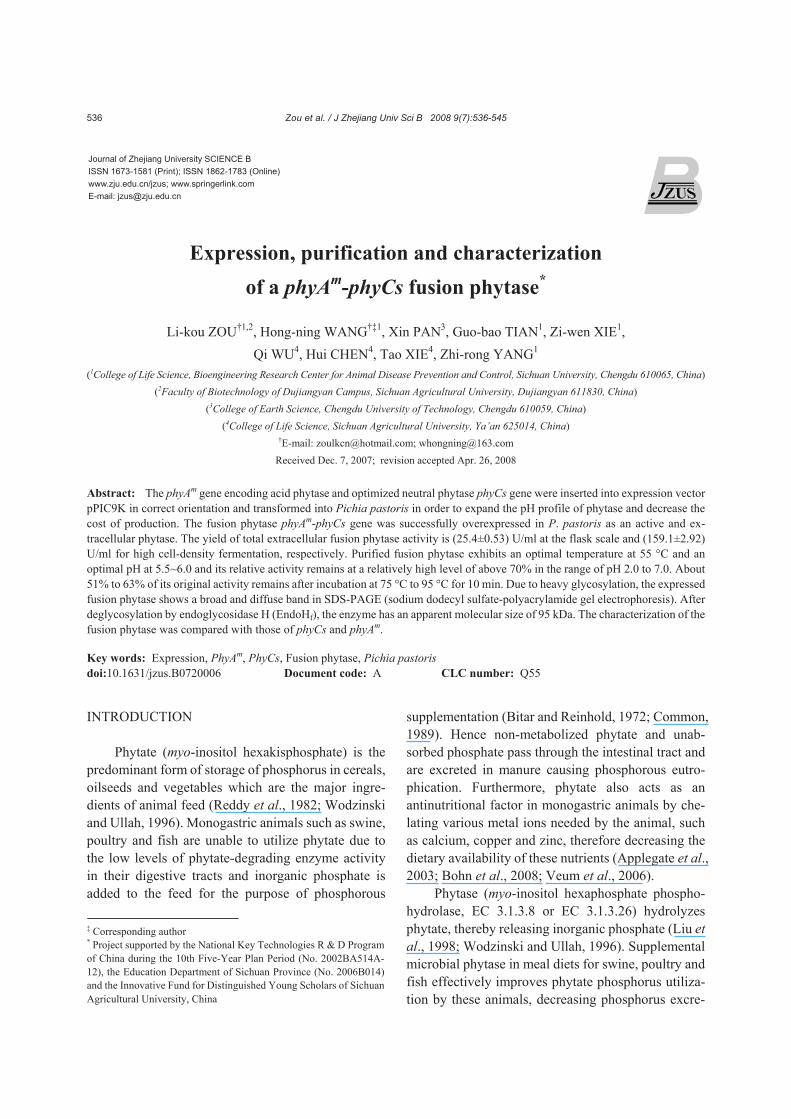
To create the construction of pPIC9K-phyCs, the phyCs gene was amplified by primers containing the linker (Gly4Ser)3 before subcloning into the expres-sion vector pPIC9K. The final expression vector pPIC9K-phyAmCs, with the expected cloning junction sequence phyAm-phyCs gene, was selected and se-quenced before transformation into P. pastoris. The expression vector containing the phyCs and fusion phyAm-phyCs genes in pPIC9K is shown in Fig.1. The BspEI-linearized pPIC9K-phyAmCs was transformed into competent P. pastoris strain GS115. By lin-earizing the recombinant vector at the unique site located in HIS4 region, Mut+ recombinants were generated. In this study a total of 40 transformants appeared and one colony, which exhibited the highest phytase activity among the colonies examined, was selected for shake-flask expression. The fusion phy-tase amino acid sequence is shown in Fig.2. As shown in Fig.3, after 96 h of methanol induction, the yield of total extracellular phytase activity reached its highest value at (25.4±0.53) U/ml in WBEI medium at the flask scale.
Purification and deglycosylation of the recombi-nant fusion phytase
The purification of recombinant phytase from WBEI medium by Q-sepharose FF anion-exchange
chromatography is shown in Fig.4. When applied to an anion-exchange chromatography column at pH 6.0, the fusion phytase was eluted as a single peak at about 33 ml. The active protein fraction which showed higher phytase activity was pooled and combined. The sample was then dialyzed against a 10 mmol/L sodium acetate buffer (2 mmol/L CaCl2, 10% (v/v) glycerol, pH 5.5) and concentrated by PEG 20 000, prior to the measurement of enzyme properties. The expressed purified fusion phytase showed a broad and diffuse band rather than a protein with an apparent molecular size (Fig.5c).
phyAm YVLAVPASRNQSTCDTVDQGYQCFSETSHLWGQYAPFFSLANESAISPDVPAGCRVTFAQVLSRHGARYPTDSKGKKYSALIEEIQQNATTFDGKYAFLKTYNYSLGADDLTPFGEQELVNSGIKFYQRYESLTRNIIPFIRSSGSSRVIASGKKFIEGFQSTKLKDPRAQPGQSSPKIDVVISEASSSNNTLDPGTCTVFEDSELADAVEANFTATFVPTIRQRLENDLSGVSLTDTEVTYLMDMCSFDTISTSTVDTKLSPFCDLFTHDEWINYDYLQSLKKYYGHGAGNPLGPTQGVGYANELIARLTHSPVHDDTSSNHTLDSNSATFPLNSTLYADFSHDNGIISILFALGLYNGTKPLSTTTVQNITQTDGFSSAWTVPFASRLYVEMMQCQAEQEPLVRVLVNDRVVPLHGCPVDALGR
linker phyCs CTRDSFVKGLSFARSGGDWAECFAPPRGGGGSGGGGSGGGGSKHKLSDPYHFTVNAAAETEPVDTAGDAADDPAIWLDPKNPQNSKLITTNKKSGLVVYSLEGKTLHSYHTGKLNNVDIRYDFPLNGKKVDIAAASNRSEGKNTIEIYAIDGKNGTLQSITDPDRPIASAIDEVYGFSLYHSQKTGKYYAMVTGKEGEFEQYELNADKNGYISGKKVRAFKMNSQTEGMAADDEYGSLYIAEEDEAIWKFSAEPDGGSNGTVIDRADGRHLTPDIEGLTIYYAADGKGYLLASSQGNSSYAIYERQGQNKYVADFQITDGPETDGTSDTDGIDVLGFGLGPEYPFGLFVAQDGENIDHGQKVNQNFKMVPWERIADKIGFHPQVNKQVDPRKLTDRSGK
Fig.2 Amino acid sequence of fusion phytase. The restriction enzyme sites amino acids are shown in bold, the linkeris shown in italic, and the phyAm and phyCs genes are shown by the arrows
Fig.1 Maps of recombinant expression plasmidspPIC9K-phyCs (a) and pPIC9K-phyAmCs (b)

Zou et al. / J Zhejiang Univ Sci B 2008 9(7):536-545 541
To prove that the fusion phytase gene was ex-
pressed successfully in P. pastoris, the phyAm and phyCs phytases were also purified and deglycosy-lated by EndoHf. As shown in Figs.5a, 5b and 5c, after deglycosylation by EndoHf the phyCs, phyAm and fusion phyAm-phyCs phytases had an apparent molecular size of 42, 51 and 95 kDa, respectively. The linker sequence encoding (Gly4Ser)3 had a mo-lecular size of about 2 kDa. Therefore the fusion phyAm-phyCs phytase gene was expressed success-fully in P. pastoris as an active and extracellular enzyme.
Characterization of recombinant fusion phytase Fig.6a shows the effect of temperature on fusion
phytase activity in defined buffers and the optimum temperature proved to be 55 °C. The relative activities of fusion phytase were 0.39, 0.50, 0.68, 0.75, 0.83, 1.00, 0.86, 0.78, 0.40, 0.25, 0.17, 0.10 and 0.06 at 16, 26, 37, 45, 50, 55, 60, 65, 70, 75, 80, 90 and 100 °C, respectively. As shown in Fig.6b, the purified fusion phytase had the optimum pH at 5.5 to 6.0, but the over 70% of enzyme activity remained in the range of pH 2.0 to 7.0 including three peaks at pH 2.0 to 2.5, pH 5.5 to 6.0 and pH 7.0. The relative activities of the
Fig.3 Effect of induction time on secretory expressedfusion phytase in flask scale. Pichia pastoris transfor-mants were grown in wheat bran extract induction(WBEI) and 1.5% (v/v) methanol was added for induc-tion. The phytase activity was expressed as mean±SD(n=3)
Fig.4 Purification of fusion phytase by anion-exchange chromatography. Five ml of dissolved samples con-taining fusion phytase were applied to a Q-sepharose FF anion-exchange chromatography column and eluted with a linear sodium chloride gradient. The fusion phytase was eluted as a single peak at an elution volume of approximately 33 ml
Fig.5 SDS-PAGE analysis of deglycosylated recombinant phytase. (a) Deglycosylation of phyCs phytase. 7.5 µl of purified phyCs phytase was loaded. Concentrations of protein were 0.968 μg/μl (Lane 1), 1.177 μg/μl (Lane 2, up-per), 1.029 μg/μl (Lane 2, lower) and 0.837 μg/μl (Lane 3). Lane M: Lower molecular weight protein marker from TaKaRa; Lane 1: EndoHf; Lane 2: EndoHf (upper) and deglycosylated phytase (lower); Lane 3: Purified phyCsphytase; (b) Deglycosylation of phyAm phytase. Six µl of purified phytase was loaded. Concentrations of protein were 1.036 μg/μl (Lane 1), 0.549 μg/μl (Lane 2, upper), 0.545 μg/μl (Lane 2, lower) and 0.312 μg/μl (Lane 3). Lane M: Higher molecular weight protein marker from TaKaRa; Lane 1: EndoHf; Lane 2: EndoHf (upper) and deglycosylated phytase (lower); Lane 3: Purified phyAm phytase; (c) Deglycosylation of phyAm-phyCs fusionphytase. Six µl of purified phytase was loaded. Concentrations of protein were 0.3 μg/μl (Lane 1, upper), 0.407 μg/μl(Lane 1, lower), 0.327 μg/μl (Lane 2) and 0.3 μg/μl (Lane 3). Lane M: Higher molecular weight protein marker from TaKaRa; Lane 1: Deglycosylated phytase (upper) and EndoHf (lower); Lane 2: EndoHf; Lane 3: PurifiedphyAm-phyCs fusion phytase

Zou et al. / J Zhejiang Univ Sci B 2008 9(7):536-545 542
fusion phytase were 0.67, 0.73, 0.49, 0.53, 0.60, 0.78, 0.94, 1.00, 0.99, 0.73, 0.74, 0.25, 0.08, 0.05, 0.02, 0, 0 and 0 at pH 2.0, 2.5, 3.0, 3.5, 4.0, 4.5, 5.0, 5.5, 6.0, 6.5, 7.0, 7.5, 8.0, 9.0, 10.0, 11.0, 12.0 and 13.0, respec-tively. To test the thermostability, the activity of the fusion phytase was measured after incubation at various temperatures for 10 min. The relative activity of the enzyme remains at the levels of 1.00, 0.63, 0.63, 0.57, 0.56 and 0.51 at 37, 75, 80, 85, 90 and 95 °C, respectively. In conclusion, 51% to 63% of its origi-nal activity remained after incubation at 75 to 95 °C for 10 min, as shown in Fig.6c. The properties of phyAm acid phytase and phyCs neutral phytase were measured as we reported previously (Chen et al., 2003; 2005; Zou et al., 2006).
Fermentation The high cell-density culturing was performed
by feeding 50% (v/v) glycerol (12 ml/h for 12 h) after the initial amount of glycerol had been exhausted, as evidenced by the sudden increase in the level of dis-solved oxygen. As shown in Fig.7b, for the glycerol batch phase the OD600 was 129.8±4.25. The OD600 reached 143.3±2.8 after the 24 h fed-batch induction cultivation. Maximum activity occurred within 84 h of post-induction and SDS-PAGE analysis indicated that phytase comprised the major protein present in the broth (Fig.7a). The enzyme secreted into the me-dium was approximately (159.1±2.92) U/ml, with the cell density reaching approximately OD600=178.4± 2.08.
Fig.6 Effects of temperature (a), pH (b) and temperature stability (c) on phytase activity. To test the thermosta-bility, the enzyme was pre-incubated at various temperatures for 10 min, and residual activity was measured at 37°C. The data were presented as relative activity at each pH and temperature (n=3)
Fig.7 Fusion phytase production in fermentation. (a) SDS-PAGE analysis. One µl of fermentation product was loaded. Concentrations of protein were 0.585, 0.738, 0.949, 1.326 and 1.335 μg/μl (from Lane 1 to Lane 5). Lane M:Higher molecular weight protein marker from TaKaRa as the same as that in Fig.5c; Lane 0: Culture supernatant before induction; Lanes 1~5: Culture supernatant after induction for 24, 48, 72, 84 and 96 h, respectively; (b) The correlation of culture concentration and activity of fusion phytase with induction time. The data were presented as mean±SD (n=3)

Zou et al. / J Zhejiang Univ Sci B 2008 9(7):536-545 543
DISCUSSION
The most striking success in the study was in proving the feasibility of expanding the pH profile of phytase to enhance its catalytic efficiency in the di-gestive tract. As shown in Fig.6a, the phyAm acid phytase exhibited an optimum pH at 2.5 or 5.5, but it lost most of its activity when the pH was below 2.0 or above 6.5. The phyCs neutral phytase exhibited an optimum pH at 7.5 but it lost most of its activity be-low pH 4.0 or above 10.0. The optimum pH of the fusion phytase was at 5.5 to 6.0 and over 70% of relative activity of the enzyme remained in the range of pH 2.0 to 7.0 including three peaks at pH 2.0 to 2.5, pH 5.5 to 6.0 and pH 7.0. Only 1%, 40% and 8% relative activity of the phyAm phytase remained at pH 2.0, 6.5 and 7.0, respectively. The wild-type phyA phytase showed optimum pH at 4.6 and lost most of its activity at pH 2.0 and 6.5 (Chen et al., 2003). But 79%, 71% and 84% of relative activity of the fusion phytase remained at pH 2.0, 6.5 and 7.0, respectively, higher than that of phyAm and wild-type phyA. Be-tween pH 2.0 and 4.5, 70% to 92% of relative activity of the fusion phytase remained, which was higher than that of phyCs (0% to 25% of relative activity). Therefore the two phytases may have an interactive effect which we explored by crystal structure com-parison (data not shown). However, the fusion phy-tase activity declined suddenly at pH 7.5, which was unexpectedly different from that of phyCs. Thus the fusion phytase could be not only suitable for the acid but also for the neutral digestive tracts of animals. Characterization of the recombinant fusion phytase revealed its higher relative activity and broader pH optimum, which was considered to be important for the application.
The fusion phytase had the same optimum tem-perature at 55 °C as the wild-type phyA phytase, but its residual activities at 16 °C and 37 °C were higher than those of phyAm acid phytase and phyCs neutral phytase (Chen et al., 2003; 2005; Zou et al., 2006). As shown in Fig.6a, the phyAm and phyCs phytases re-vealed optimum temperatures of 60 and 70 °C, re-spectively. Thermostability is a particularly important issue since feed pelleting is commonly performed at temperatures between 65 °C and 95 °C. About 51% to 63% of the original activity of fusion phytase re-mained after incubation at 75 °C to 95 °C for 10 min
without a protective agent, which was more thermo-stable than phyAm phytase (Fig.6c) and phyA mutant which retained 20% activity after being heated at 80 °C for 10 min (Zhang et al., 2007).
The expressed phyAm, phyCs and fusion phyAm-phyCs phytases showed a broad and diffuse band in SDS-PAGE due to the heavy glycosylation (Fig.5) as described by Markus et al.(1999) and Zou et al.(2006). After deglycosylation by EndoHf, the phyAm, phyCs and fusion phyAm-phyCs phytases had an apparent molecular size of 51, 42 and 95 kDa, respectively. The glycosylation may have a number of effects on the properties of an enzyme, and it may have a positive effect on the thermostability (Yoshi-masu et al., 2004; Han and Lei, 1999). As we previ-ously reported, the glycosylation of P. pastoris phy-tase appeared to have increased with induction time and the glycosylated phytase resulted in the en-hancement of thermostability (Zou et al., 2006), so we deduced that deglycosylation may lead to the decrease of fusion phytase thermostability. The puri-fied phytase showed streaking in SDS-PAGE, but after deglycosylation the streaking was eliminated as shown in Fig.5.
Vuolanto et al.(2001) reported recombinant phyC neutral phytase activity of 48 U/ml with Bacil-lus subtilis expression system at a 2-L fermentor us-ing a fed-batch protocol, which was lower than the phyCs phytase activity of 54 U/ml expressed in P. pastoris (data not shown). The modified phyA-sh acid phytase activity expressed in P. pastoris only reached 125 U/ml at a 5-L fermentor reported by Xiong et al. (2005). In this paper, after an induction period of 84 h, the fusion phytase activity reached (159.1±2.92) U/ml at a 5-L fermentor using a fed-batch protocol. This is the first report of a fusion phytase gene in P. pastoris which has been shown to be suitable for high-level expression of various heterologous proteins, either intracellular or secretory. Under fermentation condi-tions P. pastoris has been shown to be able to grow to high cell densities capable of giving high levels of expressed protein with levels greater than 10 g/L reported (Cregg and Higginns, 1995; Romanos, 1995; André et al., 2006). So there is a huge potential for the fermentation of fusion phytase if optimizing the cul-tivation conditions. According to our calculation, the cost of the BMMY (buffered methanol-complex me-dium), BSM (basal salts medium) and WBSG media

Zou et al. / J Zhejiang Univ Sci B 2008 9(7):536-545 544
was 7.5, 2.5 and 0.5 Yuan/L, respectively. Hence compared with the expensive BMMY and BSM (Xiong et al., 2005) media, WBSG is an economical medium for the growth of P. pastoris and phytase expression.
In conclusion, the phyAm-phyCs gene was suc-cessfully expressed as an active and extracellular phytase in P. pastoris. The fusion phytase shows an extended pH profile and favourable thermostability suitable for production and application.
References André, N., Cherouati, N., Prual, C., Steffan, T., Zeder-Lutz, G.,
Magnin, T., Pattus, F., Michel, H., Wagner, R., Reinhart, C., 2006. Enhancing functional production of G pro-tein-coupled receptors in Pichia pastoris to levels re-quired for structural studies via a single expression screen. Protein Science, 15(5):1115-1126. [doi:10.1110/ps.06209 8206]
Applegate, T.J., Webel, D.M., Lei, X.G., 2003. Efficacy of a phytase derived from Escherichia coli and expressed in yeast on phosphorus utilization and bone mineralization in turkey poults. Poultry Science, 82(11):1726-1732.
Augspurger, N.R., Webel, D.M., Lei, X.G., Baker, D.H., 2003. Efficacy of an E. coli phytase expressed in yeast for re-leasing phytate-bound phosphorus in young chicks and pigs. J. Anim. Sci., 81(2):474-483.
Bitar, K., Reinhold, J.G., 1972. Phytase and alkaline phos-phatase activities in intestinal mucosae of rat, chicken, calf, and man. Biochem. Biophys. Acta, 268(2):442-452.
Bohn, L., Meyer, A.S., Rasmussen, S.K., 2008. Phytate: im-pact on environment and human nutrition. A challenge for molecular breeding. J. Zhejiang Univ. Sci. B, 9(3): 165-191. [doi:10.1631/jzus.B0710640]
Chen, C.C., Wu, P.H., Huang, C.T., Cheng, K.J., 2004. A Pichia pastoris fermentation strategy for enhancing the heterologous expression of an Escherichia coli phytase. Enzyme and Microbial Technology, 35(4):315-320. [doi:10.1016/j.enzmictec.2004.05.007]
Chen, H., Zhao, H.X., Wang, H.N., 2003. The research of phytase’s characteristics from gene engineering yeast. Microbiology, 30(3):38-41 (in Chinese).
Chen, H., Zhao, H.X., Wang, H.N., 2005. Increasing expres-sion level of phytase gene (phyA) in Pichia pastoris by changing rare codons. Chinese Journal of Biochemistry and Molecular Biology, 21(2):171-175 (in Chinese).
Common, F.H., 1989. Biological availability of phosphorus for pigs. Nature, 143:370-380.
Cregg, J.M., Higginns, D.R., 1995. Production of foreign proteins in the yeast Pichia pastoris. Can. J. Bot., 73(Suppl.):5891-5987.
Han, Y., Lei, X.G., 1999. Role of glycosylation in the func-tional expression of an Aspergillus niger phytase (phyA) in Pichia pastoris. Archives of Biochemistry and Bio-physics, 364(1):83-90. [doi:10.1006/abbi.1999.1115]
Han, Y., Wilson, D.B., Lei, X.G., 1999. Expression of an Aspergillus niger phytase gene (phyA) in Saccharomyces cerevisiae. Applied and Environmental Microbiology, 65(5):1915-1918.
Janne, K., Marko, L., Paivi, N., Nisse, K., Juha, A., 1998. Isolation, characterization, molecular gene cloning, and sequencing of a novel phytase from Bacillus subtilis. Applied and Environmental Microbiology, 64(6): 2079-2085.
Kim, T., Mullaney, E.J., Porres, J.M., Roneker, K.R., Crowe, S., Rice, S., Ko, T., Ullah, A.H.J., Daly, C.B., Welch, R., Lei, X.G., 2006. Shifting the pH profile of Aspergillus niger phyA phytase to match the stomach pH enhances its effectiveness as an animal feed additive. Applied and Environmental Microbiology, 72(6):4397-4403 [doi:10. 1128/AEM.02612-05]
Kim, T.W., Lei, X.G., 2005. An improved method for a rapid determination of phytase activity in animal feed. J. Anim. Sci., 83:1062-1067.
Kim, Y.O., Lee, J.K., Kim, H.K., Yu, J.H., Oh, T.K., 1998. Cloning of the thermostable phytase gene (phy) from Bacillus sp. DS11 and its expression in E. coli. FEMS Microbiology Letters, 162(1):185-191. [doi:10.1111/j. 1574-6968.1998.tb12997.x]
Kim, Y.O., Lee, J.K., Oh, B.C., Oh, T.K., 1999. High level expression of a recombinant thermostable phytase in Ba-cillus subtilis. Biosci. Biotechnol. Biochem., 63(12): 2205-2207. [doi:10.1271/bbb.63.2205]
Liu, B.L., Rafiq, A., Tzeng, Y.M., Rob, A., 1998. The induc-tion and characterization of phytase and beyond. Enzyme and Microbial Technology, 22(5):415-424. [doi:10.1016/ S0141-0229(97)00210-X]
Markus, W., Luis, P., Arno, F., Roland, R., Michek, T., Al-exandra, K., Anke, M., Martin, L., Line, S., Ursro, T., et al., 1999. Biophysical characterization of fungal phytases (myo-inositol hexakisphosphate phosphohydrolases): molecular size, glycosylation pattern, and engineering of proteolytic resistance. Applied and Environmental Mi-crobiology, 65(2):359-366.
Olukosi, O.A., Sands, J.S., Adeola, O., 2007. Supplementation of carbohydrases or phytase individually or in combina-tion to diets for weanling and growing-finishing pigs. J. Anim. Sci., 855(7):1702-1711. [doi:10.2527/jas.2006-709]
Reddy, N.R., Sathe, S.K., Salunkhe, D.K., 1982. Phytates in legumes and cereals. Adv. Food Res., 28:1-92.
Rodriguez, E., Han, Y., Lei, X.G., 1999. Cloning, sequencing, and expression of an Escherichia coli acid phos-phatase/phytase gene (appA2) isolated from pig colon. Biochem. Biophys. Res. Commun., 257(1):117-123. [doi: 10.1006/bbrc.1999.0361]
Rodriguez, E., Mullaney, E.J., Lei, X.G., 2000. Expression of the Aspergillus fumigatus phytase gene in Pichia pastoris and characterization of the recombinant enzyme. Bio-chemical and Biophysical Research Communications, 268(2):373-378. [doi:10.1006/bbrc.2000.2121]
Romanos, M., 1995. Advances in the use of Pichia pastoris for high-level expression. Curr. Opin. Biotechnol., 6(5):

Zou et al. / J Zhejiang Univ Sci B 2008 9(7):536-545 545
527-533. [doi:10.1016/0958-1669(95)80087-5] Rutherfurd, S.M., Chung, T.K., Moughan, P.J., 2002. The
effect of microbial phytase on ileal phosphorus and amino acid digestibility in the broiler chicken. Br. Poultry Sci., 43(4):598-606. [doi:10.1080/0007166022000004516]
Tye, A.J., Siu, F.K.Y., Leung, T.Y.C., Lim, B.L., 2002. Mo-lecular cloning and the biochemical characterization of two novel phytases from B. subtilis 168 and B. licheni-formis. Appl. Microbiol. Biotechnol., 59(2):190-197. [doi:10.1007/s00253-002-1033-5]
Veum, T.L., Bollinger, D.W., Buff, C.E., Bedford, M.R., 2006. A genetically engineered Escherichia coli phytase im-proves nutrient utilization, growth performance, and bone strength of young swine fed diets deficient in available phosphorus. J. Anim. Sci., 84:1147-1158.
Vuolanto, A., von Weymarn, N., Kerovuo, J., Ojamo, H., Leisola, M., 2001. Phytase production by high cell den-sity culture of recombinant Bacillus subtilis. Biotech-nology Letters, 23(10):761-766. [doi:10.1023/A:1010369 325558]
Wang, H.N., Wu, Q., Zhao, H.X., Zou, L.K., Liu, S.G., 2005. Secretory expression of Bacillus subtilis phytase phyC in Pichia pastoris. Journal of Zhejiang University (Agric. & Life Sci.), 31(5):621-627 (in Chinese).
Wodzinski, R.J., Ullah, A.H., 1996. Phytase. Adv. Appl. Microbiol., 42:263-302.
Wu, Q., Wang, H.N., Zou, L.K., Zhao, H.X., Liu, S.G., 2004a. Expression of phytase phyC from B. subtilis in E. coli and its effect in enzymetic thermostability. High Technology Letters, 14(5):23-27 (in Chinese).
Wu, Q., Wang, H.N., Zou, L.K., Zhao, H.X., Liu, S.G., 2004b.
Intrcellular expression of Bacillus subtilis phytase phyC in Pichia patoris. Journal of Jishou University (Natural Science Edition), 25(1):36-41 (in Chinese).
Xie, Z.W., Wang, H.N., 2007. Preliminary study on flask fermentation condition of producing phytase by engi-neering bacterium. China Feed, 3:13-16 (in Chinese).
Xiong, A.S., Yao, Q.H., Peng, R.H., Han, P.L., Cheng, Z.M., Li, Y., 2005. High level expression of a recombinant acid phytase gene in Pichia pastoris. Journal of Applied Mi-crobiology, 98(2):418-428. [doi:10.1111/j.1365-2672.2004. 02476.x]
Yoshimasu, M.A., Tanaka, T., Ahn, J.K., Yada, R.Y., 2004. Effect of N-linked glycosylation on the aspartic pro-teinase porcine pepsin expressed from Pichia pastoris. Glycobiology, 14(5):417-429. [doi:10.1093/glycob/cwh024]
Zhang, W., Mullaney, E.J., Lei, X.G., 2007. Adopting selected hydrogen bonding and ionic interactions from Aspergillus fumigatus phytase structure improves the thermostability of Aspergillus niger phyA phytase. Applied and Envi-ronmental Microbiology, 73(9):3069-3076. [doi:10.1128/ AEM.02970-06]
Zinin, N.V., Serkina, A.V., Gelfand, M.S., Shevelev, A.B., Sineoky, S.P., 2004. Gene cloning, expression and char-acterization of novel phytase from Obesumbacterium proteus. FEMS Microbiology Letters, 236(2):283-290. [doi:10.1111/j.1574-6968.2004.tb09659.x]
Zou, L.K., Wang, H.N., Pan, X., Xie, T., Wu, Q., Xie, Z.W., Zhou, W.R., 2006. Design and expression of s synthetic phyC gene encoding the neutral phytase in Pichia pastoris. Acta Biochimica et Biophysica Sinica, 38(11):803-811. [doi:10.1111/j.1745-7270.2006.00231.x]
Copyright © 2022 FDOKUMEN


![Exposure to benzo[a]pyrene of Hepatic Cytochrome P450 Reductase Null (HRN) and P450 Reductase Conditional Null (RCN) mice: Detection of benzo[a]pyrene diol epoxide-DNA adducts by immunohistochemistry](https://static.fdokumen.com/doc/165x107/63259f17c9c7f5721c022d3b/exposure-to-benzoapyrene-of-hepatic-cytochrome-p450-reductase-null-hrn-and-p450.jpg)