Organization and evolution of the flavin-containing monooxygenase genes of human and mouse:...
14
Organization and evolution of the flavin-containing monooxygenase genes of human and mouse: identification of novel gene and pseudogene clusters Diana Hernandez a , Azara Janmohamed a , Pritpal Chandan a , Ian R. Phillips b and Elizabeth A. Shephard a Objectives To date, six flavin-containing monooxygenase (FMO) genes have been identified in humans, FMOs 1, 2, 3, 4 and 6, which are located within a cluster on chromosome 1, and FMO5, which is located outside the cluster. The objectives were to review and update current knowledge of the structure and expression profiles of these genes and of their mouse counterparts and to determine, via a bioinformatics approach, whether other FMO genes are present in the human and mouse genomes. Results and conclusions We have identified, for the first time, a mouse Fmo6 gene. In addition, we describe a novel human FMO gene cluster on chromosome 1, located 4 Mb telomeric of the original cluster. The novel cluster contains five genes, all of which exhibit characteristics of pseudogenes. We propose the names FMO 7P, 8P, 9P, 10P and 11P for these genes. We also describe a novel mouse gene cluster, located approximately 3.5 Mb distal of the original gene cluster on Chromosome 1. The novel mouse cluster contains three genes, all of which contain full- length open-reading frames and possess no obvious features characteristic of pseudogenes. One of the genes is apparently a functional orthologue of human FMO9P. We propose the names Fmo9, 12 and 13 for the novel mouse genes. Orthologues of these genes are also present in rat. Sequence comparisons and phylogenetic analyses indicate that the novel human and mouse gene clusters arose, not from duplications of the known gene cluster, but via a series of independent gene duplication events. The mammalian FMO gene family is thus more complex than previously realised. Pharmacogenetics 14:117–130 & 2004 Lippincott Williams & Wilkins Pharmacogenetics 2004, 14:117–130 Keywords: FMO, flavin-containing monooxygenase, gene family, pseudogene, chromosome 1, evolution, human, mouse a Department of Biochemistry and Molecular Biology, University College London, London, UK and b School of Biological Sciences, Queen Mary, University of London, UK. This study was supported by the Wellcome Trust, grant number 053590; P.C. is a recipient of a PhD studentship from the MRC, UK. Correspondence to Elizabeth Shephard, Department of Biochemistry and Molecular Biology, University College London, Gower Street, London WC1E 6BT, UK. Tel: + 44 20 76792321; fax: +44 20 76797193; e-mail: [email protected] Or Ian Phillips, School of Biological Sciences, Queen Mary, University of London, Mile End Road, London E1 4NS, U.K. Tel: +44 20 78826338; Fax +44 20 89830973; email: [email protected] Received 20 August 2003 Accepted 2 November 2003 Introduction The flavin-containing monooxygenases (FMOs) are, after the cytochromes P450 (CYPs), the second largest group of enzymes involved in the Phase I metabolism of drugs and other xenobiotics [1–3]. FMOs catalyse the NADPH-dependent oxidation of a wide range of structurally diverse compounds that contain, as the site of oxidation, a soft nucleophilic heteroatom, usually nitrogen, phosphorus or sulphur [1]. Substrates for FMO include drugs, such as tricyclic antidepressants, antihistamines and inhibitors of monoamine oxidase A and B [4]; dietary-derived compounds (e.g., trimethyla- mine) [5]; and neurotoxins (e.g. N-methyl-1,2,3,6-tetra- hydropyridine, MPTP) [6]. Cysteamine is an endogenous substrate for both pig liver FMO1 [7] and yeast FMO [8] and it has been suggested that the oxidation of this compound to the disulphide may provide a significant source of disulphide to maintain the cellular thiol:disulphide balance [9]. Although the FMOs exhibit activity towards a wide substrate range their specificity is determined by the size of their substrate cleft, which dictates entry or exclusion of a particular chemical [2,10,11]. In humans, mutations in FMO3 cause the inherited disorder trimethylaminuria [12]. Affected individuals are unable to metabolize dietary-derived trimethyla- mine (TMA) into TMA N-oxide, and consequently excrete relatively large amounts of the malodorous free amine in their urine, sweat and breath, which imparts an unpleasant body odour reminiscent of rotting fish. The disorder has thus been colloquially named fish-odour syndrome. Mutations of this gene are described in the trimethylaminuria database at http://human-fmo3.biochem.ucl.ac.uk/Human_FMO3/ [13]. For recent reviews on the clinical and bio- chemical aspects of trimethylaminuria see [14] and [15]. Copyright © Lippincott Williams & Wilkins. Unauthorized reproduction of this article is prohibited. Original article 117 0960-314X & 2004 Lippincott Williams & Wilkins DOI: 10.1097/01.fpc.0000054152.92680.ea
Transcript of Organization and evolution of the flavin-containing monooxygenase genes of human and mouse:...

Organization and evolution of the flavin-containingmonooxygenase genes of human and mouse: identificationof novel gene and pseudogene clustersDiana Hernandeza, Azara Janmohameda, Pritpal Chandana, Ian R. Phillipsb andElizabeth A. Shepharda
Objectives To date, six flavin-containing monooxygenase
(FMO) genes have been identified in humans, FMOs 1, 2, 3,
4 and 6, which are located within a cluster on chromosome
1, and FMO5, which is located outside the cluster. The
objectives were to review and update current knowledge of
the structure and expression profiles of these genes and
of their mouse counterparts and to determine, via a
bioinformatics approach, whether other FMO genes are
present in the human and mouse genomes.
Results and conclusions We have identified, for the first
time, a mouse Fmo6 gene. In addition, we describe a novel
human FMO gene cluster on chromosome 1, located 4 Mb
telomeric of the original cluster. The novel cluster contains
five genes, all of which exhibit characteristics of
pseudogenes. We propose the names FMO 7P, 8P, 9P, 10P
and 11P for these genes. We also describe a novel mouse
gene cluster, located approximately 3.5 Mb distal of the
original gene cluster on Chromosome 1. The novel mouse
cluster contains three genes, all of which contain full-
length open-reading frames and possess no obvious
features characteristic of pseudogenes. One of the genes
is apparently a functional orthologue of human FMO9P. We
propose the names Fmo9, 12 and 13 for the novel mouse
genes. Orthologues of these genes are also present in rat.
Sequence comparisons and phylogenetic analyses
indicate that the novel human and mouse gene clusters
arose, not from duplications of the known gene cluster, but
via a series of independent gene duplication events. The
mammalian FMO gene family is thus more complex than
previously realised. Pharmacogenetics 14:117–130 &
2004 Lippincott Williams & Wilkins
Pharmacogenetics 2004, 14:117–130
Keywords: FMO, flavin-containing monooxygenase, gene family,pseudogene, chromosome 1, evolution, human, mouse
aDepartment of Biochemistry and Molecular Biology, University College London,London, UK and bSchool of Biological Sciences, Queen Mary, University ofLondon, UK.
This study was supported by the Wellcome Trust, grant number 053590; P.C. isa recipient of a PhD studentship from the MRC, UK.
Correspondence to Elizabeth Shephard, Department of Biochemistry andMolecular Biology, University College London, Gower Street, London WC1E6BT, UK.Tel: + 44 20 76792321; fax: +44 20 76797193; e-mail: [email protected] Ian Phillips, School of Biological Sciences, Queen Mary, University of London,Mile End Road, London E1 4NS, U.K.Tel: +44 20 78826338; Fax +44 20 89830973; email: [email protected]
Received 20 August 2003Accepted 2 November 2003
IntroductionThe flavin-containing monooxygenases (FMOs) are,
after the cytochromes P450 (CYPs), the second largest
group of enzymes involved in the Phase I metabolism
of drugs and other xenobiotics [1–3]. FMOs catalyse
the NADPH-dependent oxidation of a wide range of
structurally diverse compounds that contain, as the site
of oxidation, a soft nucleophilic heteroatom, usually
nitrogen, phosphorus or sulphur [1]. Substrates for
FMO include drugs, such as tricyclic antidepressants,
antihistamines and inhibitors of monoamine oxidase A
and B [4]; dietary-derived compounds (e.g., trimethyla-
mine) [5]; and neurotoxins (e.g. N-methyl-1,2,3,6-tetra-
hydropyridine, MPTP) [6]. Cysteamine is an
endogenous substrate for both pig liver FMO1 [7] and
yeast FMO [8] and it has been suggested that the
oxidation of this compound to the disulphide may
provide a significant source of disulphide to maintain
the cellular thiol:disulphide balance [9]. Although the
FMOs exhibit activity towards a wide substrate range
their specificity is determined by the size of their
substrate cleft, which dictates entry or exclusion of a
particular chemical [2,10,11].
In humans, mutations in FMO3 cause the inherited
disorder trimethylaminuria [12]. Affected individuals
are unable to metabolize dietary-derived trimethyla-
mine (TMA) into TMA N-oxide, and consequently
excrete relatively large amounts of the malodorous
free amine in their urine, sweat and breath, which
imparts an unpleasant body odour reminiscent of
rotting fish. The disorder has thus been colloquially
named fish-odour syndrome. Mutations of this gene
are described in the trimethylaminuria database at
http://human-fmo3.biochem.ucl.ac.uk/Human_FMO3/
[13]. For recent reviews on the clinical and bio-
chemical aspects of trimethylaminuria see [14] and
[15].
Copyright © Lippincott Williams & Wilkins. Unauthorized reproduction of this article is prohibited.
Original article 117
0960-314X & 2004 Lippincott Williams & Wilkins DOI: 10.1097/01.fpc.0000054152.92680.ea

To date, six FMO genes, named FMOs 1, 2, 3, 4, 5 and
6, have been identified in humans [16] (http://www.
sanger.ac.uk/HGP/Chr1/). In this paper we review and
update our current knowledge of these genes and of
their counterparts in the mouse, which is increasingly
being used as a model organism to investigate the role
of specific gene products in health and disease. We
demonstrate that the six genes have been conserved in
the two species during evolution. We also describe
novel FMO gene clusters identified on chromosome 1
of human and mouse.
MethodsRNA isolation and northern blot hybridization
Human FMO mRNA expression in endocrine tissues
was analysed using the Human Endocrine System
MTN Blot (BD CLONTECH UK, Basingstoke, UK).
Hybridization probes were cDNAs for FMO1 [17],
FMO2 [18], FMO3 [19], FMO4 [20] and FMO5 [21].
Total RNA was isolated from liver, lung, kidney, brain,
heart, testis and ovary of 129Sv and C57BL/6 adult (8–
9 weeks postpartum) or juvenile (5 weeks postpartum)
mice through the use of the ULTRASPEC RNA
isolation system (Biotecx, Houston, TX, USA). RNA
samples (30 �g) were denatured in formaldehyde and
electrophoresed through a 1% (w/v) agarose gel [22].
RNA was transferred to an optimized nylon membrane
(BDH, Lutterworth, UK). The hybridization probe
used to detect mouse FMO1 mRNA was a PCR-
amplified product derived from the 39UTR of the gene.
Hybridization conditions were as previously described
[17]. Expression data for human and mouse FMO
mRNAs were also obtained from the internet sites at
http://bioinformatics.weizmann.ac.il/cards/; http://www.
ncbi.nlm.nih.gov/UniGene and from the BLASTN
facility at http://www.ncbi.nih.gov/
Isolation of the mouse Fmo gene cluster
A mouse genomic DNA PAC library (RPC121) [23] was
obtained from the UK Human Genome Mapping
Project Resource Centre (HGMP) (Hinxton, UK) as
gridded filters. Each filter was screened, under moder-
ate stringency conditions (hybridization at 508C, final
wash at 508C in 0.1 3 standard saline citrate (SSC),
0.1% sodium dodecyl sulphate (SDS)), with a mixture
of human FMO1, 2, 3, 4 and 5 cDNAs [17–21]. Sixteen
positive PAC clones were identified and subsequently
obtained from the HGMP. DNA was isolated from all
16 clones and used to prepare five replica, dot-blot
filters. These were hybridized, under high stringency
conditions (hybridization at 658C, final wash at 658C in
0.1 3 SSC, 0.1% SDS), to individual known mouse
FMO cDNAs. Mouse I.M.A.G.E. cDNA clones were
obtained from the HGMP. FMO1: I.M.A.G.E: 1921107,
Accession No. AI316256; FMO2: I.M.A.G.E: 1432164,
Accession No. AA986385; FMO3: I.M.A.G.E: 1891165,
Accession No. AI226472; FMO4: I.M.A.G.E: 692387,
Accession No. AI390626; FMO5: I.M.A.G.E: 351766,
Accession No. AI322352. Plasmid DNAs were isolated
and their inserts sequenced (MWG Biotech, Germany).
The I.M.A.G.E. clones for FMOs 1, 2, 3 and 5 each
contain the entire coding sequence for the correspond-
ing protein. However, I.M.A.G.E: 692387 contains only
sequences derived from exons 1–6 of FMO4, plus 465
bp of intron 6.
PAC clones that gave a positive signal when hybridized
with individual mouse cDNAs were characterized by
restriction endonuclease digestion and Southern blot
analyses.
Identification and analysis of novel FMO genes of human,
mouse and rat
Novel FMO sequences were identified by a BLASTN
search of the human (http://www.ensembl.org/Homo_
sapiens/) and mouse (http://www.ensembl.org/Mus_
musculus/) genomes with cDNA sequences encoding
FMO5 of human (Accession No. NM_001461) and
mouse (Accession No. AI322352). The genomic region
containing the mouse Fmo6 gene was identified using
the human FMO6 putative cDNA as the search query.
Contig sequences identified by positive BLAST hits
were analysed using the ClustalW and Pustell matrix
programs of the MacVector 6.5.3 analysis package
(Oxford Molecular Ltd., Oxford, UK). Exonic se-
quences were located by comparison to the sequences
encoding cDNAs of human FMO5, Accession No.
NM_001461 [24]; mouse FMO5, Accession No.
U90535 [25]; human FMO3, Accession No. NM006894
[19]; and mouse FMO3, Accession No. U87147 [26].
Alignment of exonic sequences was optimized by
visual inspection and the putative cDNA sequences
assembled. In the case of pseudogenes exon 9 se-
quences were terminated at a position equivalent to
that of the stop codon of FMO5 of human and mouse.
The rat genome (http://www.ensembl.org/Rattus_nor-
vegicus/) was searched by BLASTN with assembled
cDNA sequences corresponding to the novel mouse
genes.
Splice sites were analysed and scored for their corre-
spondence to a consensus sequence using the Splice
Predictor Program [27] (available at http://www.fruitfly.
org/seq_tools/splice.html). This program scores sequen-
ces on a scale of 0 to 1.0, with scores closest to 1.0
being closest to the consensus.
Alignment of multiple protein or DNA sequences was
done with ClustalW (MacVector 6.5.3). Alignments
were edited manually to ensure the correct placement
of conserved protein motifs, and of exon boundaries of
pseudogenes that lack particular exons. The file was
converted for phylogeny analysis using the READSEQ
program developed by D.G. Gilbert (Indiana Univer-
Copyright © Lippincott Williams & Wilkins. Unauthorized reproduction of this article is prohibited.
118 Pharmacogenetics 2004, Vol 14 No 2

sity, Bloomingdale, IN, USA) (available at http://
bimas.dcrt.nih.gov/molbio/readseq/). The PAUP*4.0
beta 10 software package [28] was used for phyloge-
netic analyses and drawing of trees. The DNA tree was
constructed using a heuristic search and maximum
likelihood with settings corresponding to the HKY85
model.
Results and discussionThe FMO gene family of human and mouse – update
In this section we review and update our knowledge,
gained from experimental evidence and genome se-
quencing projects, of the structure and expression
profiles of FMO 1, 2, 3, 4, 5 and 6 genes. In mouse the
orthologous genes are denoted Fmo1, 2, 3, 4, 5 and 6.The identification of a gene and putative cDNA for
mouse FMO6 is described below.
cDNAs have been isolated for human FMOs 1 [17], 2
[18], 3 [19,29], 4 [20] and 5 [24]. FMO6 was identified
from the genome sequencing project (see below).
When first identified, the cDNA for human FMO4 was
given the name FMO2 [20]. Subsequently, FMOs were
re-named to reflect the chronological order in which the
proteins were identified [30].
Mouse FMO1, 3 and 5 cDNAs were cloned by conven-
tional means [25,26,31], FMO2 was identified from an
I.M.A.G.E. clone [32] and we have isolated a cDNA for
FMO4 (Accession No. AF461145) [33].
FMO gene structure
Experimental evidence and information derived from
genome sequencing projects shows that the FMO1, 2, 3,4 and 5 genes contain eight coding exons (numbered 2
to 9), the size and boundaries of which are highly
conserved, and at least one 59 non-coding exon (num-
bered 1) (Table 1) [34].
In contrast to other FMO mRNAs, FMO4 mRNAs
contain sequences derived from 10, not 9, exons, the
first two of which are entirely non-coding (Table 1). To
maintain the correlation with exons of other mamma-
lian FMOs, we have named the most upstream of the
FMO4 exons, exon 0. In the FMO4 gene of humans,
exons 0 and 1 are separated by an intron of 1772 bp.
Sequences derived from all 10 exons of FMO4 are
spliced to form the 4.3-kb mRNA present in human
liver [20]. However, the transcript of FMO4 is subject
to differential tissue-specific processing: in the pancreas
the predominant mRNA is 2.4 kb, whereas in other
tissues examined the major species is 4.3 kb (Fig. 1). It
is not known, however, whether the FMO4 transcript
undergoes tissue type-specific alternative splicing in
human brain, as has been observed in rat brain [35].
Analysis of mouse FMO4 cDNA clones shows that
alternative promoters are used in the transcription of
the Fmo4 gene. In a cDNA isolated from the amnion of
a 17-day-pregnant female (RIKEN clone, Accession
No. BY738979), sequences derived from exon 1 are
spliced to sequences from an upstream exon, which we
have named exon 0a. Exon 1 and exon 0a are separated
by an intron of 926 bp. A second cDNA, isolated from
mouse kidney (I.M.A.G.E. clone 4234388; Accession
No. BF782473), does not contain sequences from exon
0a, but instead has 59 sequence encoded by an exon,
which we have named 0b, which lies upstream of exon
0a. The two alternative exons (0a and 0b) are separated
by an intron of 42 bp.
The human FMO1 gene contains two non-coding exons
Copyright © Lippincott Williams & Wilkins. Unauthorized reproduction of this article is prohibited.
Table 1 FMO genes of human and mouse
Gene Non-coding exons Coding exonsAmino acid
residuesMajor expressionsite in adult Other sites of expression
FMO1 0, 1 2–9 532 Kidney Gut, pancreas, adrenal cortex and medulla, thymus, thyroid, testis,placenta, lung, hypothalamus, breast, brain tumours, fetal kidney, liver.
Fmo1 1 2–9 532 Kidney Embryonic and mammary tissues, brain, lung, heart, ovaries, testis, fetalkidney, liver.
FMO2 1 2–9 471a or 535b Lung Skeletal muscle, kidney, prostate gland, blood vessels, tumours.Fmo2 1 2–9 535 Kidney Liver, lung, mammary tissue, tumours.FMO3 1 2–9 532 Liver Lung, kidney, adrenal medulla and cortex, pancreas, thyroid, gut, brain.Fmo3 1 2–9 534 Liver (female) Lung, retina, blood vessels, fetal liver, male liver (up to 5 weeks of age).FMO4 0, 1 2–9 558 Kidney Liver, lung, spleen, testis, gut, adrenal medulla and cortex, pancreas,
thyroid, thymus, placenta, brain, tumours (parathyroid, melanoma, lung,adenocarcinoma).
Fmo4 0b, 0a, 1 2–9 561 Kidney Liver, colon, egg, amnion.FMO5 1 2–9 533 Liver Kidney, lung, gut, mammary gland, adipose tissue, spleen, pituitary,
placenta, brain, testis, pancreas.Fmo5 1 2–9 534 Liver Kidney, gut, lung, adrenal and pituitary glands, thymus, skin, eyeball,
uterus, ovary, bone marrow, bladder, eggs, epididymis, blastocysts,embryonic stem cells, fertilized eggs, brain, spinal cord.
aFMO2*2A allele; bFMO2*1 allele.
FMO genes and pseudogenes of human and mouse Hernandez et al. 119
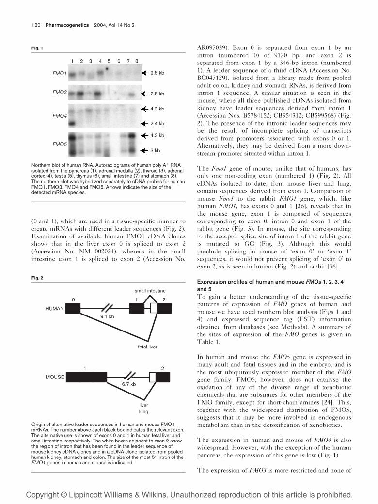
(0 and 1), which are used in a tissue-specific manner to
create mRNAs with different leader sequences (Fig. 2).
Examination of available human FMO1 cDNA clones
shows that in the liver exon 0 is spliced to exon 2
(Accession No. NM 002021), whereas in the small
intestine exon 1 is spliced to exon 2 (Accession No.
AK097039). Exon 0 is separated from exon 1 by an
intron (numbered 0) of 9120 bp, and exon 2 is
separated from exon 1 by a 346-bp intron (numbered
1). A leader sequence of a third cDNA (Accession No.
BC047129), isolated from a library made from pooled
adult colon, kidney and stomach RNAs, is derived from
intron 1 sequence. A similar situation is seen in the
mouse, where all three published cDNAs isolated from
kidney have leader sequences derived from intron 1
(Accession Nos. B5784152; CB954312; CB599568) (Fig.
2). The presence of the intronic leader sequences may
be the result of incomplete splicing of transcripts
derived from promoters associated with exons 0 or 1.
Alternatively, they may be derived from a more down-
stream promoter situated within intron 1.
The Fmo1 gene of mouse, unlike that of humans, has
only one non-coding exon (numbered 1) (Fig. 2). All
cDNAs isolated to date, from mouse liver and lung,
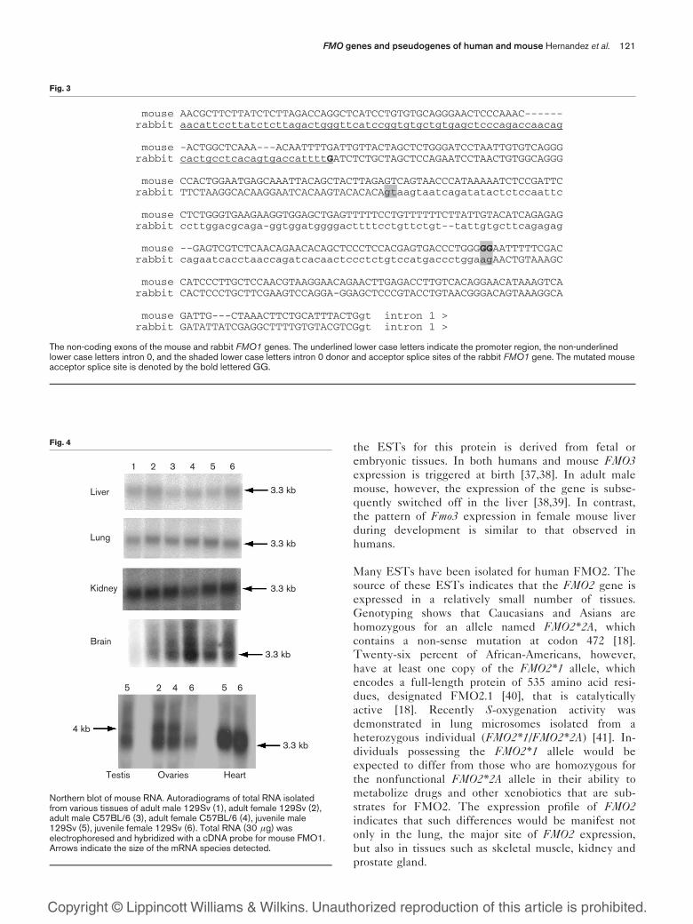
contain sequences derived from exon 1. Comparison of
mouse Fmo1 to the rabbit FMO1 gene, which, like
human FMO1, has exons 0 and 1 [36], reveals that in
the mouse gene, exon 1 is composed of sequences
corresponding to exon 0, intron 0 and exon 1 of the
rabbit gene (Fig. 3). In mouse, the site corresponding
to the acceptor splice site of intron 1 of the rabbit gene
is mutated to GG (Fig. 3). Although this would
preclude splicing in mouse of ‘exon 0’ to ‘exon 1’
sequences, it would not prevent splicing of ‘exon 0’ to
exon 2, as is seen in human (Fig. 2) and rabbit [36].
Expression profiles of human and mouse FMOs 1, 2, 3, 4
and 5
To gain a better understanding of the tissue-specific
patterns of expression of FMO genes of human and
mouse we have used northern blot analysis (Figs 1 and
4) and expressed sequence tag (EST) information
obtained from databases (see Methods). A summary of
the sites of expression of the FMO genes is given in
Table 1.
In human and mouse the FMO5 gene is expressed in
many adult and fetal tissues and in the embryo, and is
the most ubiquitously expressed member of the FMOgene family. FMO5, however, does not catalyse the
oxidation of any of the diverse range of xenobiotic
chemicals that are substrates for other members of the
FMO family, except for short-chain amines [24]. This,
together with the widespread distribution of FMO5,
suggests that it may be more involved in endogenous
metabolism than in the detoxification of xenobiotics.
The expression in human and mouse of FMO4 is also
widespread. However, with the exception of the human
pancreas, the expression of this gene is low (Fig. 1).
The expression of FMO3 is more restricted and none of
Copyright © Lippincott Williams & Wilkins. Unauthorized reproduction of this article is prohibited.
FMO1
FMO3
FMO4
FMO5
2.8 kb
2.8 kb
4.3 kb
2.4 kb
4.3 kb
3 kb
1 2 3 4 5 6 7 8
Fig. 1
Northern blot of human RNA. Autoradiograms of human poly Aþ RNAisolated from the pancreas (1), adrenal medulla (2), thyroid (3), adrenalcortex (4), testis (5), thymus (6), small intestine (7) and stomach (8).The northern blot was hybridized separately to cDNA probes for humanFMO1, FMO3, FMO4 and FMO5. Arrows indicate the size of thedetected mRNA species.
HUMAN
MOUSE
0 1 2
1 2
9.1 kb
fetal liver
small intestine
6.7 kb
liverlung
Fig. 2
Origin of alternative leader sequences in human and mouse FMO1mRNAs. The number above each black box indicates the relevant exon.The alternative use is shown of exons 0 and 1 in human fetal liver andsmall intestine, respectively. The white boxes adjacent to exon 2 showthe region of intron that has been found in the leader sequence ofmouse kidney cDNA clones and in a cDNA clone isolated from pooledhuman kidney, stomach and colon. The size of the most 59 intron of theFMO1 genes in human and mouse is indicated.
120 Pharmacogenetics 2004, Vol 14 No 2
the ESTs for this protein is derived from fetal or
embryonic tissues. In both humans and mouse FMO3expression is triggered at birth [37,38]. In adult male
mouse, however, the expression of the gene is subse-
quently switched off in the liver [38,39]. In contrast,
the pattern of Fmo3 expression in female mouse liver
during development is similar to that observed in
humans.
Many ESTs have been isolated for human FMO2. The
source of these ESTs indicates that the FMO2 gene is
expressed in a relatively small number of tissues.
Genotyping shows that Caucasians and Asians are
homozygous for an allele named FMO2*2A, which
contains a non-sense mutation at codon 472 [18].
Twenty-six percent of African-Americans, however,
have at least one copy of the FMO2*1 allele, which
encodes a full-length protein of 535 amino acid resi-
dues, designated FMO2.1 [40], that is catalytically
active [18]. Recently S-oxygenation activity was
demonstrated in lung microsomes isolated from a
heterozygous individual (FMO2*1/FMO2*2A) [41]. In-
dividuals possessing the FMO2*1 allele would be
expected to differ from those who are homozygous for
the nonfunctional FMO2*2A allele in their ability to
metabolize drugs and other xenobiotics that are sub-
strates for FMO2. The expression profile of FMO2indicates that such differences would be manifest not
only in the lung, the major site of FMO2 expression,
but also in tissues such as skeletal muscle, kidney and
prostate gland.
Copyright © Lippincott Williams & Wilkins. Unauthorized reproduction of this article is prohibited.
mouse AACGCTTCTTATCTCTTAGACCAGGCTCATCCTGTGTGCAGGGAACTCCCAAAC------ rabbit aacattccttatctcttagactgggttcatccggtgtgctgtgagctcccagaccaacag
mouse -ACTGGCTCAAA---ACAATTTTGATTGTTACTAGCTCTGGGATCCTAATTGTGTCAGGG rabbit cactgcctcacagtgaccattttGATCTCTGCTAGCTCCAGAATCCTAACTGTGGCAGGG
mouse CCACTGGAATGAGCAAATTACAGCTACTTAGAGTCAGTAACCCATAAAAATCTCCGATTC rabbit TTCTAAGGCACAAGGAATCACAAGTACACACAgtaagtaatcagatatactctccaattc
mouse CTCTGGGTGAAGAAGGTGGAGCTGAGTTTTTCCTGTTTTTTCTTATTGTACATCAGAGAG rabbit ccttggacgcaga-ggtggatggggacttttcctgttctgt--tattgtgcttcagagag
mouse --GAGTCGTCTCAACAGAACACAGCTCCCTCCACGAGTGACCCTGGGGGAATTTTTCGAC rabbit cagaatcacctaaccagatcacaactccctctgtccatgaccctggaagAACTGTAAAGC
mouse CATCCCTTGCTCCAACGTAAGGAACAGAACTTGAGACCTTGTCACAGGAACATAAAGTCA rabbit CACTCCCTGCTTCGAAGTCCAGGA-GGAGCTCCCGTACCTGTAACGGGACAGTAAAGGCA
mouse GATTG---CTAAACTTCTGCATTTACTGgt intron 1 > rabbit GATATTATCGAGGCTTTTGTGTACGTCGgt intron 1 >
Fig. 3
The non-coding exons of the mouse and rabbit FMO1 genes. The underlined lower case letters indicate the promoter region, the non-underlinedlower case letters intron 0, and the shaded lower case letters intron 0 donor and acceptor splice sites of the rabbit FMO1 gene. The mutated mouseacceptor splice site is denoted by the bold lettered GG.
Liver
Lung
Kidney
Brain
1 2 3 4 5 6
3.3 kb
3.3 kb
3.3 kb
3.3 kb
3.3 kb
4 kb
5 2 4 6 5 6
Testis Ovaries Heart
Fig. 4
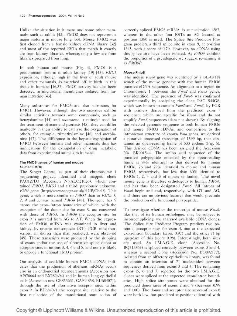
Northern blot of mouse RNA. Autoradiograms of total RNA isolatedfrom various tissues of adult male 129Sv (1), adult female 129Sv (2),adult male C57BL/6 (3), adult female C57BL/6 (4), juvenile male129Sv (5), juvenile female 129Sv (6). Total RNA (30 �g) waselectrophoresed and hybridized with a cDNA probe for mouse FMO1.Arrows indicate the size of the mRNA species detected.
FMO genes and pseudogenes of human and mouse Hernandez et al. 121
Unlike the situation in humans and some other mam-
mals, such as rabbit [42], FMO2 does not represent a
major isoform in mouse lung [33]. Mouse FMO2 was
first cloned from a female kidney cDNA library [32]
and most of the reported ESTs that match it exactly
are from kidney libraries, whereas only a few are from
libraries prepared from lung.
In both human and mouse (Fig. 4), FMO1 is a
predominant isoform in adult kidney [19] [43]. FMO1expression, although high in the liver of adult mouse
and other mammals, is switched off at birth in this
tissue in humans [16,37]. FMO1 activity has also been
detected in microsomal membranes isolated from hu-
man intestine [43].
Many substrates for FMO3 are also substrates for
FMO1. However, although the two enzymes exhibit
similar activities towards some compounds, such as
benzydamine [44] and tazarotene, a retinoid used for
the treatment of acne and psoriasis [45], they can differ
markedly in their ability to catalyse the oxygenation of
others, for example, trimethylamine [46] and methio-
nine [47]. The difference in the hepatic expression of
FMO3 between humans and other mammals thus has
implications for the extrapolation of drug metabolic
data from experimental animals to humans.
The FMO6 genes of human and mouse
Human FMO6
The Sanger Centre, as part of their chromosome 1
sequencing project, identified and mapped clone
PAC127D3 (Accession No.ALO21026), which con-
tained FMO2, FMO3 and a third, previously unknown,
FMO gene (http://www.sanger.ac.uk/HGP/Chr1/). This
gene, which is more similar to FMO3 than to FMOs 1,2, 4 and 5, was named FMO6 [48]. The gene has 9
exons, the exon–intron boundaries of which, with the
exception of the donor site for exon 9, are conserved
with those of FMO3. In FMO6 the acceptor site for
exon 9 is mutated from AG to AT. When the expres-
sion of FMO6 mRNA was examined in liver and
kidney, by reverse transcriptase (RT)–PCR, nine tran-
scripts, all shorter than that predicted, were observed
[49]. These transcripts were produced by the skipping
of exons and/or the use of alternative splice donor or
acceptor sites in introns 3, 4, 6 and 8, and none is likely
to encode a functional FMO protein.
Our analysis of available human FMO6 cDNAs indi-
cates that the production of aberrant mRNAs occurs
also in an endometrial adenocarcinoma (Accession nos.
AI978664 and BX282694) and in human lung epithelial
cells (Accession nos. CB853631, CA944858, BU684071),
through the use of alternative acceptor sites within
exon 9. In BU684071 the acceptor site, relative to the
first nucleotide of the translational start codon of
correctly spliced FMO3 mRNA, is at nucleotide 1287,
whereas in the other four ESTs an AG located at
position 1380 is used. The Splice Site Predictor Pro-
gram predicts a third splice site in exon 9, at position
1345, with a score of 0.70. However, no cDNAs using
this splice site have been isolated. As FMO6 exhibits
the properties of a pseudogene we suggest re-naming it
as FMO6P.
Mouse Fmo6
The mouse Fmo6 gene was identified by a BLASTN
search of the mouse genome with the human FMO6
putative cDNA sequence. An alignment to a region on
Chromosome 1, between the Fmo2 and Fmo3 genes,
was identified. The presence of Fmo6 was confirmed
experimentally by analysing the clone PAC 544G8,
which was known to contain Fmo2 and Fmo3, by PCR
with primers derived from the predicted exon 7
sequence, which are specific for Fmo6 and do not
amplify Fmo3 sequences (data not shown). By aligning
the selected genomic sequence to both human FMO6
and mouse FMO3 cDNAs, and comparison to the
intron/exon structure of known Fmo genes, we derived
a putative processed transcript, of 1599 bp, that con-
tained an open-reading frame of 533 codons (Fig. 5).
This derived cDNA has been assigned the Accession
No. BK001544. The amino acid sequence of the
putative polypeptide encoded by the open-reading
frame is 84% identical to that derived for human
FMO6, 76 and 72% identical to mouse and human
FMO3, respectively, but less than 60% identical to
FMOs 1, 2, 4 and 5 of mouse or human. The novel
mouse gene is therefore orthologous to human FMO6,and has thus been designated Fmo6. All introns of
Fmo6 begin and end, respectively, with GT and AG,
and there are no obvious features that would preclude
the production of a functional polypeptide.
To investigate whether the transcript of mouse Fmo6,like that of its human orthologue, may be subject to
incorrect splicing, we analysed available cDNA clones.
The Splice Site Predictor Program predicts two po-
tential acceptor sites for exon 4, one at the expected
exon-intron boundary (score 0.97) and the other 71 bp
upstream of this (score 0.98). Interestingly, both sites
are used. An I.M.A.G.E. clone (Accession No.
BQ715367) is spliced correctly between exons 3 and 4,
whereas a second clone (Accession No. BQ892177),
isolated from an olfactory epithelium library, was found
to contain an insertion of 71 nucleotides between
sequences derived from exons 3 and 4. The remaining
exons (5, 6 and 7) reported for the two I.M.A.G.E.
clones were spliced at the expected exon-intron bound-
aries. High splice site scores were obtained for the
predicted donor sites of exons 2 and 9 (between 0.99
and 1.00). The donor and acceptor site scores of exon 8
were both low, but predicted at positions identical with
Copyright © Lippincott Williams & Wilkins. Unauthorized reproduction of this article is prohibited.
122 Pharmacogenetics 2004, Vol 14 No 2
those of other FMOs. Further experimental analysis is
required to establish whether the Fmo6 gene of mouse
should be classified as a gene or a pseudogene.
The FMO1, 2, 3, 4 and 6 genes are located on chromosome
1 of human and mouse
FMOs 1, 2, 3 and 4 were mapped experimentally to the
long arm of human chromosome 1, in the region 1q23–
25 (Fig. 6) [17,20,50,51], and, as described above,
FMO6 was identified by the Sanger Centre, as part of
their chromosome 1 sequencing project. The order of
the genes was determined experimentally by Southern
blot hybridization of independent YAC clones (data not
shown) and is the same as that determined by the
human genome sequencing project. The five genes
span a region of about 220 000 bp (Fig. 6). The most
Copyright © Lippincott Williams & Wilkins. Unauthorized reproduction of this article is prohibited.
FAD FMO3 MKKK-VAIIGAGVSGLAAIRSCLEEGLEPTCFERSDDVGGLWKFSDHIEEGRASIYQSVFTNSSKEMMCFPDFPYPDDFP FMO6 MGKK-VAIVGAGVSGLAAIRCCLEEGLDPICFERSIDVGGLWKFSSHAEEGRASIYQSVFTNSSKEMMCFPDFPYPDDFP FMO9 MVKKQIAVIGAGISGLGAIKCCLDEDLEPTCFERNDDIGGLWKFQKNASEKMPSIYRSVTINTSKEMMCFSDFPIPDHFP FMO12 MGVKRIAVIGAGVSGLGAIKCCLEEGLEPTCFEKKSDIGGLWKYEEIPKSGNLGIYKSLTCNTSKEMTAFSDYPIPDHYP FMO13 MEVKQIAIIGAGVSGLGAIKSCLEEGLEPTCFEKSNDIGGLWRYKETPENGRPGIYKSLTCNTSKEMTTFSDYPIPDHYP FMO5 MAKKRIAVIGAGASGLTCIKCCLEEGLEPVCFERSGDIGGLWRFQEAPEEGRASIYQSVVINTSKEMMCFSDYPIPDHYP
FMO3 NFMHHSKLQEYITSFAKEKNLLKYIQFETPVTSINKCPNFSTTGKWEVTTEKHGKKETAVFDATMICSGHHIFPHVPKDS FMO6 NYMHHSKLQEYITSFAQKKGLLRYIQFETLVSSIKKCSSFLTTGQWVVVTEKEGKQESVLFDAVMICSGHHVYPNMPTDS FMO9 NYMHNSKLMDYFRMYAKRFSLLDYIRFKTTVRSVRKRPDFHIHGQWDVVVETDGKQESLVFDGVLVCSGHHTDPHLPLKS FMO12 NYMHHSKMMEYLRMYARHFGLMKHIQFQTNVCNIKKRPDFSSSGQWDVVVETEEMQKTYIFDGIMVCSGHYTEKYFPLQD FMO13 NYMHHSKMMEYLRMYARHFGLMKHIQFQTRVCVVRKRPDFSSSGQWDVVVEADGKQKNYIFDGVMVCSGHYTEKYLPLQD FMO5 NYMHNSQVLEYFRMYAKEFDLLKYIQFKTTVCSVKKQPDFSTSGQWQVVTECEGKQQVDVFDGVLVCTGHHTDAHLPLES
NADPH FMO3 FPGLNRFKGKCFHSRDYKEPGIWKGKRVLVIGLGNSGCDIAAELSHVAQKVTISSRSGSWVMSRVWDDGYPWDMVVLTRF FMO6 FPGLEHFRGKCLHSRDYKGPGAFQGKKVLVIGLGNSASDIAVELSRLATQVIISTRSGSWIMSRVWNDGYPWDMVYVTRF FMO9 FPGIEKFEGCYFHSREYKSPEDYVGKRIIVVGIGNSGVDIAVELGRVAKQVFLSTRRGSWILHRVWNNGYPMDSSFFTRF FMO12 FEGISKFQGSYLHTWEYKHPDNFVGKRVAVIGLGNSGADVAGEISRVADQVFLSTRQGAWIWNRVWDHGEPMDTTVFTRY FMO13 FAGISKFQGSCLHSWEYKHPDSFVGKRVVVIGIGNSGADVANEISCVTEQVFLSTRRGTWIWNRVWDNGDPLDIALFTRY FMO5 FPGIEKFKGKYFHSRDYKNPVEFTGKRVIVIGIGNSGGDLAVEISHTAKQVFLSTRRGAWILNRVGKHGYPIDLLLSSRI
FMO3 QTFLKNNLPTAISDWWYTRQMNARFKHENYGLVPLNRTLRKEPVFNDELPARILCGMVTIKPNVKEFTETSAVFEDGTMF FMO6 TSFLRNILPSFVSDWLYIKKMNTWFKHENYGLMPLNGPLRKEPVFNDELPSRILCGMVTIKPIVTKFTETSAVFEDGTVF FMO9 HSFLQKILTTEAVNKYLEKTLNSRFNHAHYGLQPQHRPLSQHPTISDDLPNHIISGKVQVKPNVKEFTGTDVHFDDGTVE FMO12 NRAVQKICPRYIINRQMEKKLNGRFNHANYGLLPTHRILEQRTVLSDDLPNRIIIGKVKIKPNVKEFTSTSAIFEDG-TK FMO13 NRTVKSFYPTFLINRWTENKLNLRFNHANYGLQAKHRFLSHQSIFSDDLPNRIISGRVLVKTNVREFTSTSAIFEDG-SE FMO5 MYYLSRICGPSLKNNYMEKQMNQRFDHEMFGLKPKHRALSQHPTVNDDLPNRIIAGLVKVKGNVKEFTETAAIFEDGSRE
FMO3 EAIDCVIFATGYGYAYPFLDDSIIKSRNNEVTLYKGVFPPQLEKPTMAVIGLVQSLGATIPITDLQARWAAQVIKGTCTLL FMO6 EAIDCVIFATGYGYAYPFLDDSIIKSRNNEVTLYKGIFPPQLEKPTMAVIGLVQSLGAAIPTADLQARWAAKVFTSTCVLL FMO9 ENIDVVIFATGYSISFPFLGDLIA-VTDNEVSLYKLMFPPDLEKPTLAVIGLIQPLGIILPIAELQSRWAVRVFKGLSKLL FMO12 ENIDVVIFATGYKLSFPFLSDDSG-VLDNQYSMFKYVFPPELEKPTLAFIGILQPAGAILPTSELQSRWVVHVFKGIKKLL FMO13 EIVDVVVFATGYTLSFPFLDDSSE-ILDSKHTMFKFVFPPQLEKPTLAFIGILQPIGATIPTSELQSRWVTRVFAGLQKLL FMO5 DGIDVVIFATGYSFAFPFLEDSVK-VVKNKVSLYKKVFPPNLEKPTLAIIGLIQPLGAIMPISELQGRWATQVFKGLKKL
FMO3 PSVNDMMDDIDEKMGEKFKWYG---NSTTIQTDYIVYMDELASFIGAKPNLLWLFLKDPRLAVEVFFGPCSPYQFRLVGP FMO6 PTTNEMMDDIDEKMGKKLKWFG---QSHTLQTDYITYMDELSSFIGAKPNIPWLFLTDPQLALEVYFGPCSPYQFRLMGP FMO9 PSVKAMKADMDQR-KKAMEKRYVKTARHTIQVDHIEYMDEIASLAGVKPNLLLLFLSDPTLAMEVFFGPCTPYQYRLQGP FMO12 PSRRAMIADINRKNHQIMAKGSKKILQDHRRVTFVDYMDEIASEIGVKPNLLSLLLWDTKLAKEVFCGPCTSYQYRLQGP FMO13 PSQSNMMADINRK-KRKMEKEFVKSPRDVRRVPYIDYMDEIASEIGVKPNLLSFFLWDTKLAKEIFWGPCTPYQYRLQGP FMO5 PSQSEMMAEINKA-REEMAKRYVDSQRHTIQGDYIDTMEEIADLVGVRPNIQPLVFTDPRLALRLLLGPCTPVQYRLQGP
FMO3 GKWSGARNAILTQWDRSLKPMKTRVVSKVQKSC--SHFYSRLLRLLAVPVLLIALFLVLI FMO6 GKWDGARNAILTQWKRTVKPTRTRAVGEAQR----PRHLYDLLRMLFFPVFFLAVLLTFY FMO9 GKWDGARRAILTQRERIIKPLKTRITSEKSRSAPGLFWIKMALFGLAFLVPSLTYFSYICQ FMO12 GKWDGARAAILTQRERMLKPLRTRVVKQSHLS--HLSWVKSACVVVFFFISVVVIMHITSH FMO13 GKWAGARAAILTQRDRILKPLRSRVLKNSETSSSSLFWVRCICAVIFPFVSVFAIIHAIYQ FMO5 GKWAGARKTILTTEDRVRKPLMTRVVERDSSGG-SLVTVRVLMLAVAFFAVILAYF
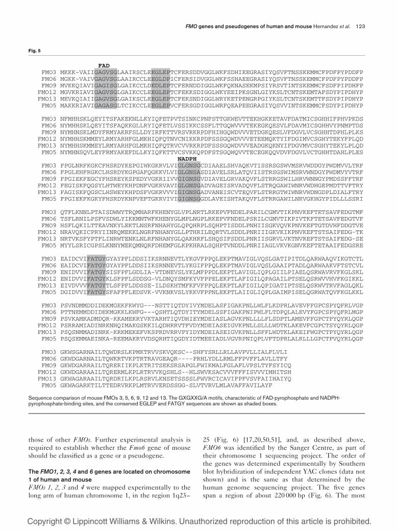
Fig. 5
Sequence comparison of mouse FMOs 3, 5, 6, 9, 12 and 13. The GXGXXG/A motifs, characteristic of FAD-pyrophosphate and NADPH-pyrophosphate-binding sites, and the conserved EGLEP and FATGY sequences are shown as shaded boxes.
FMO genes and pseudogenes of human and mouse Hernandez et al. 123

Copyright © Lippincott Williams & Wilkins. Unauthorized reproduction of this article is prohibited.
FMO3 FMO6 FMO2 FMO1 FMO4
26.9 kb
20 kb
23.2 kb 25.6 kb 37.4 kb 27.6 kb
24 kb 40 kb 20 kb
�220 kb
q21.1 q24.2 q24.3
�22 Mb �4 Mb
FMO5
39 kb
Human Chromosome 1
FMO7p FMO8p FMO9p FMO10p FMO11p
�320 kb
? kb 14.5 kb 18.9 kb 17.1 kb 29.4 kb
Mouse Chromosome 1
Fmo13 Fmo12 Fmo9150 kb 30 kb
13 kb 13.2 kb 17 kb�210 kb
H2H1
�3.5 Mb
�200 kb
Fmo4 Fmo1 Fmo2 Fmo6 Fmo320.5 kb 10.5 kb 28.3 kb 6.9 kb
15.9 kb 36.7 kb 23.8 kb 20.5 kb 29.1 kb
Mouse Chromosome 3F2
Fmo5
24.3 kb
(a)
(b)
83 kb 81 kb 18 kb 81 kb
Fig. 6
Diagrammatic representation of the chromosomal location of human and mouse FMO genes. (a) The location of FMO5 and of the two gene clustersare shown in their cytogenetic bands on human chromosome 1. Each gene is represented by a filled square, with its size given below. Theapproximate sizes of each cluster and the distances between them (www.ensembl.org) are shown. (b) The location of Fmo5 on Chromosome 3 andof the two Fmo clusters on Chromosome 1 are shown in their cytogenetic bands. Each gene is represented by a filled square, with its size givenbelow. The size of the two clusters and the distance between them are also given. The arrows represent the direction in which the genes aretranscribed. The diagrams are not drawn to scale.
124 Pharmacogenetics 2004, Vol 14 No 2

centromeric gene in the cluster is FMO3, followed by
FMO6, FMO2, FMO1 and FMO4. The genes are all
transcribed in the centromeric to telomeric direction.
To determine the chromosomal location of the mouse
genes, Fmos1, 2, 3, 4 and 6, we identified PAC clones,
from a mouse genomic library, that hybridized to mouse
Fmo cDNA sequences. The complement of Fmo genes
in each PAC was assessed by PCR, using primers based
on the mouse cDNA sequences. Three clones were
found to be positive for both Fmo1 and Fmo4, two were
positive for both Fmo2 and Fmo3, and two were posi-
tive for Fmo4 only (data not shown), indicating that, as
is the case in humans, in the mouse Fmo4 lies next to
Fmo1, and Fmo2 lies next to Fmo3. Analysis by
fluorescence in situ hybridization (FISH), using the
PAC clones as probes, located the Fmo 1, 2, 3 and 4genes in a cluster towards the distal end of mouse
Chromosome 1 (data not shown) in a region that is
syntenic with human chromosome 1 [52].
From comparative mapping data [52], it would appear
that the whole of the region of synteny between human
chromosome 1 and mouse Chromosome 1 is inverted in
relation to the telomere and centromere. Thus, in this
region, genes that are more centromeric in humans are
more distal in mice and this appears to be the case for
the FMO clusters of mouse and man. The order and
orientation of the five genes within the cluster, as
indicated by our analysis of YAC and PAC clones and
the order and orientation derived by the Sanger Centre,
is cent-FMO3-FMO6-FMO2-FMO1-FMO4-tel in humans,
whereas in mouse the genes are orientated cent-Fmo4-Fmo1-Fmo2-Fmo6-Fmo3-tel and are transcribed from
the telomere to the centromere (Fig. 6).
The FMO5 genes of human and mouse
Of the three independent PAC clones that were posi-
tive for Fmo5, none contained any other Fmo gene
(data not shown), indicating that in mouse, as is the
case in humans, this gene is separated from the main
Fmo cluster. FISH analysis mapped Fmo5 to mouse
Chromosome 3 (data not shown). In humans FMO5 is
located on the long arm of chromosome 1 in the region
1q21.1 [51,53], between the genes PRKAB2 and BCL9,a region syntenic to mouse Chromosome 3.
Identification of novel FMO genes in human and mouse
A second FMO gene cluster on human chromosome 1
Through an analysis of the human genome we have
identified a second FMO gene cluster on the long arm
of chromosome 1, located approximately 4.27 Mb closer
to the centromere than the cluster that contains FMOs1, 2, 3, 4 and 6. This second cluster contains five genes,
which span a region of about 330 000 bp. By comparison
with both FMO5 and FMO3 exonic sequences, potential
exons were identified in each of the five genes. All five
genes have the characteristics of pseudogenes (see
below). The potential DNA coding sequence of each
pseudogene is between 57 and 65% identical to the
genes for FMO1, FMO2, FMO3, FMO4, FMO5 and
FMO6 (Table 2). No pseudogene is therefore obviously
derived from any of the six known FMO genes. We
therefore suggest the names FMO7P, FMO8P, FMO9P,FMO10P and FMO11P for these novel sequences.
FMO7P is the most centromeric and FMO11P the most
telomeric of the genes in this cluster (Fig. 6A). The
evidence for the existence of these genes and the
features that characterize them as pseudogenes are
described below. Unless stated otherwise, the reference
numbers given to locate each of the genes on chromo-
some 1 (http://www.sanger.ac.uk/HGP/Chr1/) are as
follows: the first indicates the A of the ATG start codon
and designates the beginning of the potential protein-
coding sequence; the second defines the position, in
exon 9 of the pseudogene, that corresponds to that of
the last nucleotide of the stop codon in FMO5.
Human FMO7P This gene is the most centromeric in
the cluster. Only sequences corresponding to exons 3, 4,
5 and 7 could be assigned with confidence. The 59 end
of exon 3 and the 39 end of exon 7 correspond, respec-
tively, to nucleotides 162 094 790 and 162 101 690 of
chromosome 1. No sequence corresponding to exon 6
was identified, even after a rigorous analysis of the 2769
nucleotides that separate exons 5 and 7. Translation of
sequences with some similarity to exons 2, 8 and 9
indicated possible remnants of these three exons. How-
ever, as they could not be assigned with confidence,
these regions were not used in the calculation of nucleo-
tide identities or for phylogenetic analyses (Table 2 and
Fig. 7).
Human FMO8P Eight potential exons, that together
are 57 to 60% identical to the 8 coding exons of the
known human FMO genes (Table 2), were identified
for the second gene in the cluster, FMO8P. This gene
is located between nucleotides 162 185 557 and
162 200 081 of chromosome 1. In this region of the
chromosome, a novel transcript, ENST0000031389,
comprising four potential exonic sequences is predicted
by the Sanger Centre. The first two exons of this
transcript correspond to exons 4 and 5 of a FMO gene,
whereas the last two exons do not correspond to FMOsequences. The predicted transcript contains a 19 449-
bp intron between its third and fourth exons. Closer
analysis of this ‘intronic’ sequence revealed that it con-
tained sequences corresponding to exons 6, 7, 8 and 9 of
a FMO gene. Exon 3 of FMO8P is located within the
predicted transcript ENST0000031389 and is separated
from exon 4 by an intron of 455 bp. Exon 2, which is not
contained within ENST0000031389, is separated from
exon 3 by an intron of 3637 bp. The acceptor splice site
for exon 7 is mutated to CG and thus a transcript
Copyright © Lippincott Williams & Wilkins. Unauthorized reproduction of this article is prohibited.
FMO genes and pseudogenes of human and mouse Hernandez et al. 125

encoding a functional FMO protein would not be pro-
duced. In addition, exons 6 and 7 contain in-frame stop
codons, exon 8 a frame-shift mutation and exon 9 a
premature stop codon.
Human FMO9P FMO9P lies between nucleotides
162 231 846 and 162 250 846. This region of the genome
is annotated with two overlapping transcripts,
NM_138784 and a novel predicted transcript ENST
00000322141 (http://www.ensembl.org/Homo_sapiens/).
NM_138784 is the transcript of the Ensembl gene
ENSG00000143151 and comprises six exons. Sequence
comparisons reveal that four of these correspond to
exons 2, 3, 4 and 5 of a FMO gene, whereas the other
two do not correspond to FMO sequences. The novel
predicted transcript ENST00000322141 contains exons
7, 8 and part of 9 of a FMO gene. Further examination of
the genomic region between these two transcripts re-
vealed a FMO exon 6 sequence, located between the
predicted sixth exon of NM_138784 and the predicted
first exon of ENST00000322141. Exon 6 of FMO9P is
flanked by both mutated acceptor (AT) and donor (AT)
splice sites, which accounts for the lack of detection of
exon 6 by the automated transcript assembly programs
used by the genome consortium. In addition, exon 2 of
FMO9P has a 1-bp deletion, which causes a shift in the
reading frame and a premature stop signal at codon 7.
The gene is thus unable to produce a functional FMO
protein.
Transcript NM_138784 is related to I.M.A.G.E. clone
3997249, which was isolated from the bladder carci-
Copyright © Lippincott Williams & Wilkins. Unauthorized reproduction of this article is prohibited.
Table
2DNAse
quence
identity
betw
eenFMOsofhumanandmouse
FMO1
FMO2
FMO3
FMO4
FMO5
FMO6P
FMO7P
FMO8P
FMO9P
FM010P
FMO11P
FMO1
FMO2
FMO3
FMO4
FMO5
FMO6
FMO9
FMO12
FMO1
–FMO2
65
–FMO3
62
63
–FMO4
61
63
60
–FMO5
60
63
61
61
–FMO6P
63
63
74
60
60
–FMO7P
59
61
57
57
65
56
–FMO8P
57
60
59
59
60
57
59
–FMO9P
59
61
60
62
65
59
63
65
–FMO10P
57
59
58
57
62
57
61
71
65
–FMO11P
60
59
59
58
64
59
55
78
67
69
–FMO1
85
62
62
59
58
62
57
56
57
56
56
–FMO2
62
83
62
61
62
62
57
59
62
58
59
61
–FMO3
63
64
83
60
61
75
56
58
60
57
60
63
64
–FMO4
60
63
60
83
60
59
55
57
60
56
57
59
61
60
–FMO5
60
63
61
60
84
60
64
59
65
61
61
58
61
60
58
–FMO6
62
61
74
60
59
84
54
57
59
56
58
61
61
76
59
60
–FMO9
59
61
61
61
65
60
62
64
83
64
67
57
62
61
59
64
60
–FMO12
58
59
59
57
62
58
58
69
64
75
73
57
58
59
57
63
58
65
–FMO13
59
60
58
57
62
60
59
69
65
77
74
59
60
59
57
63
58
66
80
Sha
ded
area
s¼
hum
an;u
nsha
ded
area
s¼
mo
use.
Per
cent
iden
titie
sw
ere
bas
edo
nth
ep
rote
in-c
od
ing
reg
ions
ofa
llkn
ow
nex
ons
.The
sew
ere
3to
5an
d7
forFMO7P
;2to
7an
d9
forFMO10P
;2,3
and
6to
9fo
rFMO11P
;an
d2
to9
for
allo
ther
gen
es.
FMO4Fmo4
FMO5
Fmo5
Fmo9
FMO9P
FMO10P
Fmo12
Fmo13FMO8P
FMO11p
FMO7PFmo1
FMO1
Fmo6
FMO6P
FMO3
Fmo3
Fmo2FMO2
Fig. 7
Phylogenetic tree of the FMO genes of human and mouse.
126 Pharmacogenetics 2004, Vol 14 No 2

noma clone library NIH_MCG_53. Because of the
frame-shift in exon 2 of FMO9P the predicted start
codon of the I.M.A.G.E. clone lies downstream, within
exon 3 of FMO9P, and is out of frame. Thus no section
of the amino acid sequence predicted from this cDNA
corresponds to a FMO protein.
Human FMO10P The FMO10P gene lies between
nucleotides 162 269 228 and 162 302 376. Analysis of this
genomic sequence revealed seven regions, which to-
gether have 57 to 62% nucleotide sequence identities to
exons 2 to 7 and exon 9 of known FMOs (Table 2). Exon
8 could not be assigned with certainty and is not
included in the phylogenetic analyses described below.
An in-frame stop signal at codon 152 precludes the
formation of functional protein. A number of other
mutations are present in exons 7 and 9.
Human FMO11P FMO11P, the most telomeric gene in
the cluster, is located between nucleotides 162 383 413
and 162 413 114 of chromosome 1. Exons 2 to 9 were
identified. However, exon 4 is corrupted by the insertion
of a short, 38-bp repeat sequence that is present many
times in the human genome, and therefore this exon
was not included in further analyses described below.
Exon 5 too, is poorly conserved, although the last 17
codons can be aligned with exon-5 sequences of other
FMO genes. This exon also was excluded from phyloge-
netic analyses. The gene contains a number of other
mutations. For example, exon 2 has a 1-bp deletion
resulting in a change in the reading frame, from codon
36, and the formation of an in-frame stop codon in exon
3.
A second mouse Fmo gene cluster on Chromosome 1
We have identified three novel Fmo genes within
the mouse genome (http://www.ensembl.org/Mus_
musculus/). The three genes span about 210 000 bp and
form a cluster on mouse Chromosome 1 in band H2.
They lie about 3.5 Mb, closer to the telomere than the
known Fmo cluster and are transcribed from the
telomeric to centromeric direction.
As described below, all three of the genes encode full-
length open-reading frames (Fig. 5) and possess no
obvious features that would categorize them as pseudo-
genes. Thus, until experimental evidence proves other-
wise, they should be regarded as functional and,
consequently, we propose that their names should not
be appended with a ‘p’ for pseudogene. Exonic regions
of the most distal gene of the cluster (Fig. 6B) are 83%
identical at the nucleotide level to exonic sequences of
the human pseudogene FMO9P. This is the same
degree of identity exhibited by orthologous FMOs of
human and mouse (Table 2). The mouse gene is
apparently a functional orthologue of human FMO9Pand we therefore propose that it be designated Fmo9.
The exonic regions of the other two genes in the
cluster are 80% identical to each other at the nucleotide
level. The human genes to which they have greatest
similarity are the pseudogenes FMO10P and 11P. The
exonic regions of the most proximal and middle genes
in the cluster, respectively, are 77 and 75% identical to
FMO10P and 74 and 73% identical to FMO11P. It is
not clear from this degree of sequence identity whether
either of the mouse genes is orthologous to one of the
human pseudogenes, and, if so, which would be the
orthologous pair(s). Therefore we propose that they be
named Fmo12, for the middle gene of the cluster, and
Fmo13, for the most proximal gene (Fig. 6B). Each of
the three genes is described below. We give the
location of the genes by chromosome nucleotide num-
bers. The first number indicates the A of the ATG start
codon and designates the beginning of the potential
protein-coding sequence; the second defines the posi-
tion of the last nucleotide of the stop codon (http://
www.ensembl.org/Mus_musculus/).
Mouse Fmo9 The Fmo9 gene lies between nucleotides
167 349 105 and 167 332 118 and is designated as tran-
script ENSMUST00000027843. A cDNA clone (Acces-
sion No. NM_172844), isolated from a 0-day neonate
head library, corresponds exactly to the eight exonic
sequences of Fmo9 and contains an open-reading frame
of 539 codons (Fig. 5). The deduced amino acid se-
quence of the predicted protein (Fig. 5) is about 65%
identical to that of FMO5. This demonstrates that Fmo9can produce a correctly spliced mRNA and is therefore
not a pseudogene in the mouse.
Three other transcripts have been predicted for this
region of the genome (http://www.ensembl.org/Mus_
musculus/genome). Two of these are partial transcripts,
ENS MUST00000027843 and ENSMUST0000002784,
which overlap with NM_172844, and lack, respectively,
exons 3 and 8 and exons 6 and 8. The third, EN-
SMUST00000027842, comprises 10 exons. Analysis of
this sequence showed that the predicted transcript is a
chimera of exons 2 to 7 of Fmo9 and exons 8 and 9 of
Fmo12, with a predicted intron of 189019 bp between
exons 7 and 8 (see below). None of these transcripts is
likely to exist.
Mouse Fmo12 Fmo12, the middle gene of the cluster,
lies between nucleotides 167 312 150 and 167 298 987
and is located within the 59 region of the predicted
189 019-bp intronic sequence of transcript EN-
SMUST00000027842 (see above). The nucleotide se-
quences of the 8 coding exons of Fmo12 have overall
identities of 57 to 63% to the coding exons of Fmos 1, 2,3, 4, 5 and 6 (Table 2). There are no obvious aberrant
splice donor or acceptor sites. The gene is predicted to
encode a polypeptide of 537 amino acid residues (Fig. 5)
that is 46 to 58% identical to FMOs 1, 2, 3, 4, 5, 6 and 9
Copyright © Lippincott Williams & Wilkins. Unauthorized reproduction of this article is prohibited.
FMO genes and pseudogenes of human and mouse Hernandez et al. 127

(data not shown). The derived cDNA has been assigned
the Accession No. BK001546.
A novel transcript, ENSMUST00000046662, predicted
from the region in which Fmo12 lies, is derived from 6
exons. Five of these correspond to exons 2, 3, 4, 5 and
7 of the Fmo12 gene, whereas the sixth is 53 bp and is
not related to a Fmo sequence. The Splice Site
Predictor Program gives donor site scores for exons 2, 3,
4, 5, 6, 7 and 8 as 0.96, 0.99, 0.75, 0.65, 0.90, 0.98 and
1.00, respectively. Relatively high scores, of 0.85 and
0.79, respectively, are predicted for sites 179 bp down-
stream of exon 4 and 43 bp downstream of exon 8. As
no ESTs have been reported for Fmo12 it is not known
whether these predicted alternative splice sites are
used. The splice donor sites for exons 3, 4, 5, 6, 8 and 9
have scores of 0.92, 0.95, 0.99, 0.83, 0.95 and 0.60,
respectively. The program did not predict a donor site
for exon 7, although a consensus AG is present at the
correct position in the gene. A site with a high score of
0.91 was predicted 35 bp upstream of the beginning of
exon 7. However, the expected splice junctions for this
exon are identified in the predicted transcript EN-
SMUST00000046662.
Mouse Fmo13 Fmo13 lies at the proximal end of the
gene cluster between nucleotides 167 160 135 and
167 144 915 (Fig. 6B). Three transcripts have been pre-
dicted from the region in which this gene is located:
ENSMUST00000027842 contains exons 8 and 9 of a
Fmo gene; ENSMUST00000027844 has 4 exons, three
correspond to exons 6, 7 and 8 of a Fmo gene, whereas
the fourth does not correspond to a Fmo sequence;
ENSMUST00000061918 has 4 exons, corresponding to
Fmo exons 3, 4, an extended 5 and 7. Close analysis of
the genomic region from which these transcripts are
predicted revealed a Fmo exon 2, 1370 bp upstream of
exon 3. All donor splice sites begin with GT and all
acceptor sites end with AG. The Splice Site Predictor
Program gives a donor splice site score for exon 2 of
0.98. It is possible, therefore, that a mRNA derived from
exons 2 to 9 of Fmo13 could be produced. The mRNA
would encode a polypeptide of 539 amino acid residues
that is 47 to 58% identical to FMOs 1, 2, 3, 4, 5, 6 and 9,
and 73% identical to FMO12. The derived cDNA has
been assigned Accession No. BK001545. No EST clones
derived from this gene have been reported.
Evolution of FMO genes
The nucleotide sequences of members of the human
pseudogene cluster (FMOs 7P, 8P, 9P, 10P and 11P) aremore similar to each other than to members of the
known gene cluster (FMOs 1, 2, 3, 4 and 6) (Table 2).
This indicates that, although both clusters contain five
genes, the pseudogene cluster did not arise via a
complete duplication of the gene cluster. Instead, it
appears to have arisen via a series of independent gene
duplication events. This also seems to be the case for
members of the novel mouse gene cluster (Fmos 9, 12and 13) (Table 2). The novel human and mouse genes
are more similar to FMO5 than to FMOs 1, 2, 3, 4 and 6(Table 2).
Phylogenetic analyses of human and mouse FMOs,
based on nucleotide (Fig. 7) and amino acid sequences
(data not shown), suggest that an ancestral gene gave
rise, via a series of duplications, to five genes, FMO1,FMO2, the precursor of FMOs 3 and 6, FMO4, and
another gene. Soon after, this last gene gave rise to
FMO5, FMO7P, and a third gene, which subsequently
duplicated to yield FMO9 and the precursor of the
remaining FMO genes and pseudogenes. A series of
duplications of the latter gene gave rise to FMOs 8P,10P, 11P, 12 and 13.
From the calculated rate of evolution of FMOs [16] andtheir amino acid sequence identities, we can estimate
the time at which some of the gene duplications took
place. The duplications that gave rise to FMOs 1, 2, theprecursor of 3 and 6, 4, and the precursor of the
remaining FMO genes are estimated to have occurred
some 275 million years ago (mya). FMOs 5 and 9diverged about 210 to 240 mya, whereas FMOs 3 and 6,and 12 and 13, arose some 140 mya. Thus, the most
recent common ancestor of human and mouse, and
indeed of all placental mammals (about 80 mya), is
predicted to have had a cluster containing genes for
FMOs 1, 2, 3, 4 and 6, a separate gene for FMO5, and
a second cluster, containing genes for FMOs 9, 12 and
13. It is possible that the second cluster contained
additional genes, subsequently lost in the mouse line-
age, or that genes arose specifically in the human
lineage and then became pseudogenes.
Human FMO9P and mouse Fmo9 are clearly ortholo-
gues (Table 2, Fig. 7). Although it is not possible to
assign orthologues of Fmos 12 and 13 unambiguously,
FMO10P and FMO11P are, respectively, approximately
76 and 74% identical to Fmos 12 and 13 (Table 2).
Given that 10P and 11P are pseudogenes, and thus
would have accumulated mutations more rapidly since
losing their function, it is possible that they may
represent human orthologues of mouse Fmos 12 and 13.
Analysis of the rat genome reveals that it contains a
cluster of orthologues of mouse Fmos 9, 12 and 13 (data
not shown). Two of these, rat FMOs 9 and 12, containopen-reading frames that encode polypeptides that are
92 and 89% identical to their mouse orthologues.
FMO13, however, contains a premature stop codon and
is apparently a pseudogene in rat and thus should be
designated FMO13P.
The mammalian FMO gene family is thus more com-
Copyright © Lippincott Williams & Wilkins. Unauthorized reproduction of this article is prohibited.
128 Pharmacogenetics 2004, Vol 14 No 2

plex than previously realized. In addition to the main
gene cluster of FMOs 1, 2, 3, 4 and 6, and the separate
FMO5, which appear to be present in all mammals
investigated to date, a second cluster, containing genes
encoding three novel FMOs (9, 12 and 13), is present
in rodents, and a pseudogene cluster containing five
members (FMOs 7P, 8P, 9P, 10P and 11P), is present inhumans.
References1 Ziegler DM. Recent studies on the structure and function of multi-
substrate flavin-containing monooxygenases. Ann Rev Pharmacol Toxicol1993; 33:179–199.
2 Rettie AE, Meier GP, Sadeque AJ. Prochiral sulfides as in vitro probes formultiple forms of the flavin-containing monooxygenase. Chem Biol Inter-act 1995; 96:3–15.
3 Cashman JR. Stereoselectivity in S- and N-oxygenation by the mammalianflavin-containing and cytochrome P-450 monooxygenases. Drug Met Rev1998; 30:675–707.
4 Cashman JR. Human flavin-containing monooxygenase: substrate specifi-city and role in drug metabolism. Curr Drug Metab 2000; 1:181–191.
5 Marks R, Greaves MW, Prottey C, Hartop PJ. Trimethylaminuria: the useof choline as an aid to diagnosis. Br J Dermatol 1977; 96:399–402.
6 Cashman JR, Ziegler DM. Contribution of N-oxygenation to the metabo-lism of MPTP (1-methyl-4-phenyl-1,2,3,6-tetrahydropyridine) by variousliver preparations. Mol Pharmacol 1986; 29:163–167.
7 Duffel MW, Logan DJ, Ziegler DM. Cysteamine and cystamine. MethodsEnzymol 1987; 143:149–154.
8 Suh JK, Poulsen LL, Ziegler DM, Robertus JD. Redox regulation of yeastflavin-containing monooxygenase. Arch Biochem Biophys 2000;381:317–321.
9 Ziegler DM, Duffel MW, Poulsen LL. Studies on the nature and regulationof the cellular thio:disulphide potential. Ciba Found Symp 1979; 2:191–204.
10 Guo WX, Ziegler DM. Estimation of flavin-containing monooxygenaseactivities in crude tissue preparations by thiourea-dependent oxidation ofthiocholine. Anal Biochem 1991; 198:143–148.
11 Kim YM, Ziegler DM. Size limits of thiocarbamides accepted as sub-strates by human flavin-containing monooxygenase 1. Drug Metab Dispos2000; 28:1003–1006.
12 Dolphin CT, Janmohamed A, Smith RL, Shephard EA, Phillips IR.Missense mutation in flavin-containing mono-oxygenase 3 gene, FMO3,underlies fish-odour syndrome. Nat Genet 1997; 17:491–494.
13 Hernandez D, Addou S, Lee D, Orengo C, Shephard EA, Phillips IR.Trimethylaminuria and a human FMO3 mutation database. Hum Mutat2003; 22:201–213.
14 Mitchell SC, Smith RL. Trimethylaminuria: the fish malodor syndrome.Drug Metab Dispos 2001; 29:517–521.
15 Cashman JR, Camp K, Fakharzadeh SS, Fennessey PV, Hines RN,Mamer OA, et al. Biochemical and clinical aspects of the human flavin-containing monooxygenase form 3 (FMO3) related to trimethylaminuria.Curr Drug Metab 2003; 4:151–170.
16 Phillips IR, Dolphin CT, Clair P, Hadley MR, Hutt AJ. McCombie RR,et al. The molecular biology of the flavin-containing monooxygenases ofman. Chem Biol Interact 1995; 96:17–32.
17 Dolphin C, Shephard EA, Povey S, Palmer CN, Ziegler DM, Ayesh R,et al. Cloning, primary sequence, and chromosomal mapping of a humanflavin-containing monooxygenase (FMO1). J Biol Chem, 1991;266:12379–12385.
18 Dolphin CT, Beckett DJ, Janmohamed A, Cullingford TE, Smith RL,Shephard EA, et al. The flavin-containing monooxygenase 2 gene (FMO2)of humans, but not of other primates, encodes a truncated, nonfunctionalprotein. J Biol Chem 1998; 273:30599–30607.
19 Dolphin CT, Cullingford TE, Shephard EA, Smith RL, Phillips IR.Differential developmental and tissue-specific regulation of expression ofthe genes encoding three members of the flavin-containing monooxygen-ase family of man, FMO1, FMO3 and FM04. Eur J Biochem 1996;235:683–693.
20 Dolphin CT, Shephard EA, Povey S, Smith RL, Phillips IR. Cloning,primary sequence and chromosomal localization of human FMO2, a newmember of the flavin-containing mono-oxygenase family. Biochem J 1992;287:261–267.
21 Janmohamed A, Dolphin CT, Phillips IR, Shephard EA. Quantification andcellular localization of expression in human skin of genes encoding flavin-
containing monooxygenases and cytochromes P450. Biochem Pharma-col 2001; 62:777–786.
22 Fourney RM, Miyakoshi J, Day RS, Paterson MC. Northern blotting:efficient RNA staining and transfer. Focus (BRL) 1988; 10:5–7.
23 Osoegawa K, Tateno M, Woon PY, Frengen E, Mammoser AG, CataneseJJ, et al. Bacterial artificial chromosome libraries for mouse sequencingand functional analysis. Genome Res 2000; 10: 116–128.
24 Overby LH, Buckpitt AR, Lawton MP, Atta-Asafo-Adjei E, Schulze J,Philpot RM. Characterization of flavin-containing monooxygenase 5(FMO5) cloned from human and guinea pig: evidence that the uniquecatalytic properties of FMO5 are not confined to the rabbit ortholog. ArchBiochem Biophys 1995; 317:275–284.
25 Cherrington NJ, Falls JG, Rose RL, Clements KM, Philpot RM, Levi PE,et al. Molecular cloning, sequence, and expression of mouse flavin-containing monooxygenases 1 and 5 (FMO1 and FMO5). J Biochem MolToxicol 1998; 12:205–212.
26 Falls JG, Cherrington NJ, Clements KM, Philpot RM, Levi PE, Rose RL,et al. Molecular cloning, sequencing, and expression in Escherichia coli ofmouse flavin-containing monooxygenase 3 (FMO3): comparison with thehuman isoform. Arch Biochem Biophys 1997; 347:9–18.
27 Reese MG, Eeckman FH, Kulp D, Haussler D. Improved splice sitedetection in Genie. J Comput Biol 1997; 4:311–323.
28 Swofford, DL, PAUP*: Phylogenetic Analysis Using Parsimony (and OtherMethods) 4.0 Beta. 2002, Sunderland, MA: Sinauer Associates, Inc.
29 Lomri N, Gu Q, Cashman JR. Molecular cloning of the flavin-containingmonooxygenase (form II) cDNA from adult human liver. Proc Natl AcadSci U S A 1992; 89:1685–1689.
30 Lawton MP, Cashman JR, Cresteil T, Dolphin CT, Elfarra AA, Hines RN,et al. A nomenclature for the mammalian flavin-containing monooxygenasegene family based on amino acid sequence identities. Arch BiochemBiophys 1994; 308:254–257.
31 Itoh K, Nakamura K, Kimura T, Itoh S, Kamataki T. Molecular cloning ofmouse liver flavin containing monooxygenase (FMO1) cDNA and char-acterization of the expression product: metabolism of the neurotoxin,1,2,3,4-tetrahydroisoquinoline (TIQ). J Toxicol Sci 1997; 22:45–56.
32 Karoly ED, Rose RL. Sequencing, expression, and characterization ofcDNA expressed flavin-containing monooxygenase 2 from mouse.J Biochem Mol Toxicol 2001; 15:300–308.
33 Janmohamed A, Hernandez D, Chandan P, Phillips IR, Shephard EA. Celltype-specific expression of the flavin-containing mono-oxygenase family ofmouse. submitted.
34 Dolphin CT, Riley JH, Smith RL, Shephard EA, Phillips IR. Structuralorganization of the human flavin-containing monooxygenase 3 gene(FMO3), the favored candidate for fish-odor syndrome, determineddirectly from genomic DNA. Genomics 1997; 46:260–267.
35 Lattard V, Longin-Sauvageon C, Benoit E. Cloning, sequencing andtissue distribution of rat flavin-containing monooxygenase 4: two differentforms are produced by tissue-specific alternative splicing. Mol Pharmacol2003; 63:253–261.
36 Luo Z, Hines RN. Identification of multiple rabbit flavin-containing mono-oxygenase form 1 (FMO1) gene promoters and observation of tissue-specific DNase I hypersensitive sites. Arch Biochem Biophys 1996;336:251–260.
37 Koukouritaki SB, Simpson P, Yeung CK, Rettie AE, Hines RN. Humanhepatic flavin-containing monooxygenases 1 (FMO1) and 3 (FMO3)developmental expression. Pediatr Res 2002; 51:236–243.
38 Falls JG, Blake BL, Cao Y, Levi PE, Hodgson E. Gender differences inhepatic expression of flavin-containing monooxygenase isoforms (FMO1,FMO3, and FMO5) in mice. J Biochem Toxicol 1995; 10:171–177.
39 Falls JG, Ryu DY, Cao Y, Levi PE, Hodgson E. Regulation of mouse liverflavin-containing monooxygenases 1 and 3 by sex steroids. Arch BiochemBiophys 1997; 342:212–223.
40 Whetstine JR, Yueh M, McCarver DG, Williams DE, Park C, Kang JH,et al. Ethnic differences in human flavin-containing monooxygenase 2(FMO2) polymorphisms: detection of expressed protein in African-Amer-icans. Toxicol Appl Pharmacol 2000; 168:216–224.
41 Krueger SK, Martin SR, Yueh MF, Pereira CB, Williams DE. Identificationof active flavin-containing monooxygenase isoform 2 in human lung andcharacterization of expressed protein. Drug Metab Dispos 2002; 30:34–41.
42 Williams DE, Ziegler DM, Nordin DJ, Hale SE, Masters BS. Rabbit lungflavin-containing monooxygenase is immunochemically and catalyticallydistinct from the liver enzyme. Biochem Biophys Res Commun 1984;125:116–122.
43 Yeung CK, Lang DH, Thummel KE, Rettie AE. Immunoquantitation ofFMO1 in human liver, kidney, and intestine. Drug Metab Dispos 2000;28:1107–1111.
44 Lang DH, Rettie AE. In vitro evaluation of potential in vivo probes for
Copyright © Lippincott Williams & Wilkins. Unauthorized reproduction of this article is prohibited.
FMO genes and pseudogenes of human and mouse Hernandez et al. 129

human flavin-containing monooxygenase (FMO): metabolism of benzyda-mine and caffeine by FMO and P450 isoforms. Br J Clin Pharmacol2000; 50:311–314.
45 Attar M, Dong D, Ling KH, Tang-Liu DD. Cytochrome P450 2C8 andFlavin-containing monooxygenases are involved in the metabolism oftazarotenic acid in humans. Drug Metab Dispos 2003; 31:476–481.
46 Lang DH, Yeung CK, Peter RM, Ibarra C, Gasser R, Itagaki K, et al.Isoform specificity of trimethylamine N-oxygenation by human flavin-containing monooxygenase (FMO) and P450 enzymes: selective catalysisby FMO3. Biochem Pharmacol 1998; 56:1005–1012.
47 Ripp SL, Itagaki K, Philpot RM, Elfarra AA. Methionine S-oxidation inhuman and rabbit liver microsomes: evidence for a high-affinity methionineS-oxidase activity that is distinct from flavin-containing monooxygenase 3.Arch Biochem Biophys 1999; 367:322–332.
48 Phillips IR, Dolphin CT, McCombie RR, Beckett DJ, Janmohamed A,Smith RL, et al. Molecular genetics of the flavin-containing monooxy-genases. In First International Workshop on Trimethylaminuria 1999;Bethesda, MD, USA.
49 Hines RN, Hopp KA, Franco J, Saeian K, Begun FP. Alternative proces-sing of the human FMO6 gene renders transcripts incapable of encodinga functional flavin-containing monooxygenase. Mol Pharmacol 2002;62:320–325.
50 Shephard EA, Dolphin CT, Fox MF, Povey S, Smith R, Phillips IR.Localization of genes encoding three distinct flavin-containing mono-oxygenases to human chromosome 1q. Genomics 1993; 16:85–89.
51 McCombie RR, Dolphin CT, Povey S, Phillips IR, Shephard EA. Localiza-tion of human flavin-containing monooxygenase genes FMO2 and FMO5to chromosome 1q. Genomics 1996; 34:426–429.
52 Carver EA, Stubbs L. Zooming in on the human-mouse comparative map:genome conservation re-examined on a high resolution scale. GenomeRes 1997; 7:1123–1137.
53 Gelb BD, Zhang J, Cotter PD, Gershin IF, Desnick RJ. Physical mappingof the human connnexin 40 (GJA5), flavin-containing monooxygenase 5,and natriuretic peptide receptor genes on 1q21. Genomics 1997;39:409–411.
Copyright © Lippincott Williams & Wilkins. Unauthorized reproduction of this article is prohibited.
130 Pharmacogenetics 2004, Vol 14 No 2