Exempting Homologous Pseudogene Sequences from Polymerase Chain Reaction Amplification Allows...
16
25 Chapter 2 Exempting homologous pseudogene sequences from PCR amplification allows genomic keratin 14 hotspot mutation analysis Petra H.L. Hut 1 , Pieter v.d. Vlies 1 , Marcel F. Jonkman 2 , Edwin Verlind 1 , Hiroshi Shimizu 3 , Charles H.C.M. Buys 1 , Hans Scheffer 1 1 Dept. of Medical Genetics, University of Groningen, Groningen, The Netherlands 2 Dept. of Dermatology, University Hospital Groningen, Groningen, The Netherlands 3 Dept. of Dermatology, Hokkaido University School of Medicine, Sapporo, Japan Published in Journal of Investigative Dermatology (2000) 114:616-619
Transcript of Exempting Homologous Pseudogene Sequences from Polymerase Chain Reaction Amplification Allows...

25
Chapter 2
Exempting homologous pseudogenesequences from PCR amplification allows
genomic keratin 14hotspot mutation analysis
Petra H.L. Hut1, Pieter v.d. Vlies1, Marcel F. Jonkman2, Edwin Verlind1,
Hiroshi Shimizu3, Charles H.C.M. Buys1, Hans Scheffer1
1 Dept. of Medical Genetics, University of Groningen, Groningen, The Netherlands2 Dept. of Dermatology, University Hospital Groningen, Groningen, The Netherlands
3 Dept. of Dermatology, Hokkaido University School of Medicine, Sapporo, Japan
Published in Journal of Investigative Dermatology (2000) 114:616-619

Chapter 2
26
Abstract
In patients with the major forms of epidermolysis bullosa simplex (EBS), either of the
keratin genes KRT5 or KRT14 is mutated. This causes a disturbance of the filament
network resulting in skin fragility and blistering. For KRT5, a genomic mutation
detection system has been described previously. Mutation detection of KRT14 on a
DNA level is, however, hampered by the presence of a highly homologous but
nontranscribed keratin 14 pseudogene. Consequently, mutation detection in EBS
has mostly been carried out on cDNA synthesised from KRT5 and KRT14 transcripts
in mRNA isolated from skin biopsies. Here we present a genomic mutation detection
system for exons 1, 4 and 6 of KRT14 that encode the 1A, L1-2 and 2B domains of
the keratin 14 protein containing the mutation hot spots. After cutting the keratin 14
pseudogene genomic sequences with restriction enzymes while leaving the
homologous genomic sequences of the functional gene intact, only the mutation hot
spot-containing exons of the functional KRT14 gene are amplified. This is followed
by direct sequencing of the PCR products. In this way, three novel mutations could
be identified, Y415H, L419Q and E422K, all located in the helix termination motif of
the keratin 14 rod domain 2B, resulting in moderate, severe and mild EBS
phenotype, respectively. By obviating the need of KRT14 cDNA synthesis from RNA
isolated from skin biopsies, this approach substantially facilitates the detection of
KRT14 hot spot mutations.

Genomic KRT14 mutation detection
27
Introduction
Epidermolysis bullosa simplex (EBS) is characterised by intra-epidermal skin
blistering as a result of mild trauma. According to clinical criteria, EBS is classified
into three major subgroups (Fine et al., 1991). The most severe form, EBS-Dowling-
Meara (DM), is characterised by generalised blistering with a circinate configuration.
At the electron microscopy level, EBS-DM is identifiable by the presence of large
cytoplasmic clumps of tonofilaments in the basal keratinocytes (Anton and Schnyder,
1982; Niemi et al., 1983; McGrath et al., 1992). EBS-Koebner (K) and EBS-Weber-
Cockayne (WC) are milder forms. In EBS-K blistering is generalised whereas in
EBS-WC it is confined to hands and feet (Haneke and Anton, 1982). In the majority
of families, EBS is inherited as an autosomal dominant disorder. In rare cases,
however, the disease is transmitted in an autosomal recessive mode [recessive
epidermolysis bullosa simplex, REBS] (Chan et al., 1994; Rugg et al., 1994;
Jonkman et al., 1996).
The major subtypes of EBS are caused by molecular defects in either the keratin
14 (KRT14) gene, located at 17q12-q21, or the keratin 5 (KRT5) gene, located at
12q13. The gene products are the epidermal keratins K14 and K5, respectively. K14
belongs to the type I keratins, or acidic keratins, and has a molecular weight of 50
kDa, whereas K5 belongs to the type II keratins, or basic keratins, and has a
molecular weight of 58 kDa (Nelson and Sun, 1983; Conway and Parry, 1988). The
keratins K14 and K5 are mainly expressed in basal keratinocytes, the basal cell layer
of the epidermis. Together these proteins form a 10 nm keratin intermediate filament
structure composed of dimeric molecules each containing a K5 and a K14 chain
(Hatzfeld and Weber, 1990; Steiner, 1990; Fuchs, 1996). The keratin intermediate
filament coiled coil helical-rod is subdivided into helices 1A, 1B, 2A and 2B
connected by three short nonhelical linker segments, linkers L1, L1-2 and L2,
respectively (Conway and Parry, 1988) (Fig 1). A single mutation in KRT14 or KRT5
can lead to disturbance of the filament network resulting in skin fragility and
blistering. KRT5 and KRT14 mutations cluster within the rod ends of 1A and 2B and
Chapter 2
28
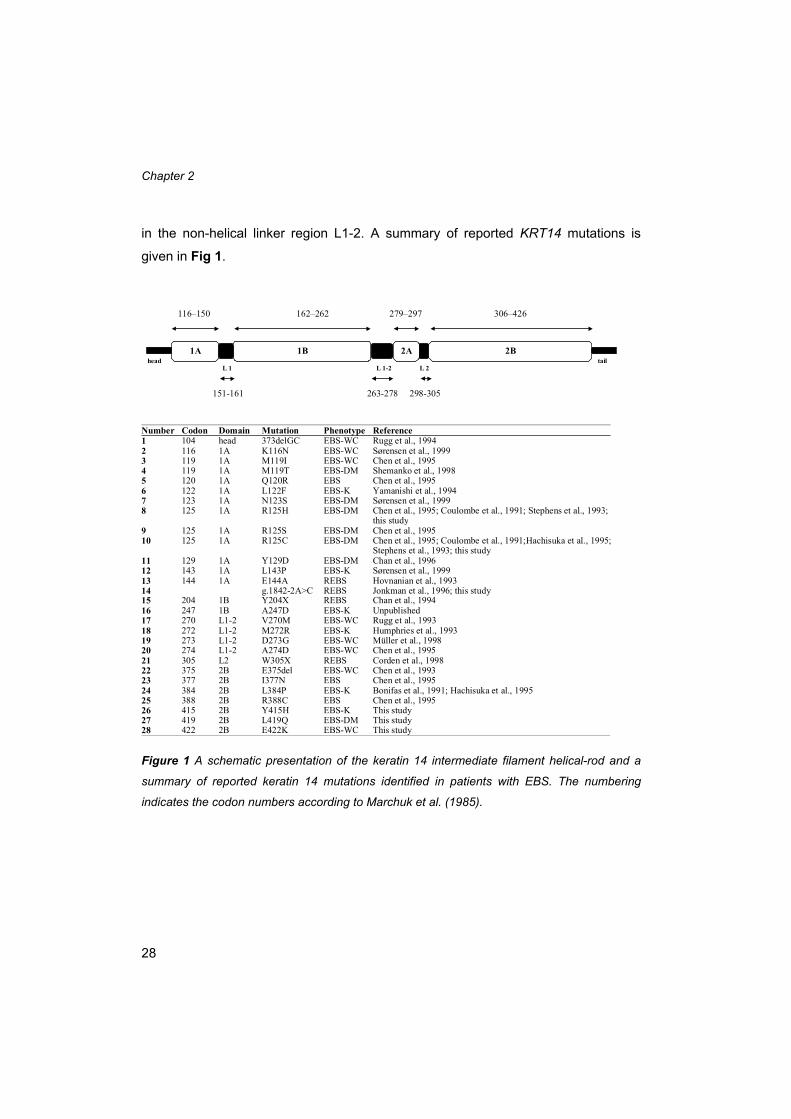
in the non-helical linker region L1-2. A summary of reported KRT14 mutations is
given in Fig 1.
Figure 1 A schematic presentation of the keratin 14 intermediate filament helical-rod and a
summary of reported keratin 14 mutations identified in patients with EBS. The numbering
indicates the codon numbers according to Marchuk et al. (1985).
116–150 162–262 279–297 306–426
Number Codon Domain Mutation Phenotype Reference1 104 head 373delGC EBS-WC Rugg et al., 19942 116 1A K116N EBS-WC Sørensen et al., 19993 119 1A M119I EBS-WC Chen et al., 19954 119 1A M119T EBS-DM Shemanko et al., 19985 120 1A Q120R EBS Chen et al., 19956 122 1A L122F EBS-K Yamanishi et al., 19947 123 1A N123S EBS-DM Sørensen et al., 19998 125 1A R125H EBS-DM Chen et al., 1995; Coulombe et al., 1991; Stephens et al., 1993;
this study9 125 1A R125S EBS-DM Chen et al., 199510 125 1A R125C EBS-DM Chen et al., 1995; Coulombe et al., 1991;Hachisuka et al., 1995;
Stephens et al., 1993; this study11 129 1A Y129D EBS-DM Chan et al., 199612 143 1A L143P EBS-K Sørensen et al., 199913 144 1A E144A REBS Hovnanian et al., 199314 g.1842-2A>C REBS Jonkman et al., 1996; this study15 204 1B Y204X REBS Chan et al., 199416 247 1B A247D EBS-K Unpublished17 270 L1-2 V270M EBS-WC Rugg et al., 199318 272 L1-2 M272R EBS-K Humphries et al., 199319 273 L1-2 D273G EBS-WC Müller et al., 199820 274 L1-2 A274D EBS-WC Chen et al., 199521 305 L2 W305X REBS Corden et al., 199822 375 2B E375del EBS-WC Chen et al., 199323 377 2B I377N EBS Chen et al., 199524 384 2B L384P EBS-K Bonifas et al., 1991; Hachisuka et al., 199525 388 2B R388C EBS Chen et al., 199526 415 2B Y415H EBS-K This study27 419 2B L419Q EBS-DM This study28 422 2B E422K EBS-WC This study
L 1 L 1-2 L 2
1B 2A1A
151-161 263-278 298-305
2Bhead tail

Genomic KRT14 mutation detection
29
KRT14 mutation detection on a DNA level is hampered by the presence of a
highly homologous keratin 14 pseudogene. The exon and the intron sequences of
the functional KRT14 gene and the keratin 14 pseudogene are 95% and 93%
identical, respectively. However, the keratin 14 pseudogene is not being transcribed
or translated because of the presence of some deleterious mutations, including
frameshift mutations, alterations in intron/ exon boundaries and disruption of the
polyadenylation signal (Savtchenko et al., 1988). Consequently, mutation analysis in
EBS has mostly been carried out on cDNA synthesised from KRT5 and KRT14
transcripts in mRNA isolated from skin biopsies. For KRT5, a genomic mutation
detection system has already been described (Stephens et al., 1997). Here we
present a method for specific amplification of genomic DNA of KRT14 exons 1, 4
and 6 that code for the 1A, L1-2 and 2B domains containing the mutation hot spots.
Our method thus obviates the need of KRT14 cDNA synthesis from keratinocytes.
Materials and methods
The clinical phenotypes of the patients included in this study are summarised in
Table I. DNA was isolated from peripheral blood by the high salt/chloroform
extraction method (Miller et al., 1988). Prior to amplification of exons 1, 4 and 6 by
the polymerase chain reaction (PCR), the genomic DNA was digested using the
restriction enzyme TaqI, SphI or MseI respectively, that cleaves the homologous
keratin 14 pseudogene sequences while leaving the KRT14 gene sequences intact.
The primer sequences, the PCR programmes for amplification of the particular
amplicons and the restiction enzymes used prior to PCR are given in Table II. PCR
was carried out using 37 pmol of each primer, 500 ng digested genomic DNA, 0.6 U
of rTaq DNA polymerase (Pharmacia Biotech, Piscataway, NJ), 0.2 mM dNTP’s in a
reaction mix containing 10 mM Tris-HCl, 1.5 mM MgCl2, 50 mM KCl, 0.01% w/v
gelatin and 0.1% v/v Triton x-100 in a total volume of 50 µl under paraffin oil. In case
of exon 1, 10% v/v of DMSO was added to the PCR mix. After amplification, the
PCR products were purified using the high pure PCR product purification kit

Chapter 2
30
(Boehringer Mannheim, Basel, Switzerland). Subsequently, the PCR fragments were
subjected to direct sequencing using an automated DNA sequencer (ABI 377, Perkin
Elmer, Norwalk, CT).
In patients with REBS, mutation analysis of KRT14 exon 2 containing the 1B
domain of K14 was carried out using a similar method as previously described
(Jonkman et al., 1996).
When a mutation was detected its presence was confirmed by sequencing of the
antisense strand. In those cases where the mutation abolished or created a
restriction site the presence of the mutation was confirmed by restriction analysis.
Table I Clinical phenotypes and KRT14 mutations of patients included in this study
Patient Clinical phenotype KRT14 mutation Inheritance
Patient I EBS-DM R125C AD a de novo
Patient II EBS-DM R125C AD a de novo
Patient III EBS-DM R125H AD a familial
Patient IV EBS-DM L419Q AD a de novo
Patient V EBS-K Y415H AD a de novo
Patient VI EBS-WC E422K AD a familial
Patient VII EBS-WC E422K AD a unknown
Patient VIII REBS g.1842-2A→C AR b familial
Patient IX REBS g.1842-2A→C AR b familial
__________________________________________________________________________
a Autosomal dominantb Autosomal recessive

Genomic KRT14 mutation detection
31
Table II Primers and PCR programs for keratin 14 hotspot mutation detection
Results
Genomic KRT14 mutation analysis
When using the primers given in Table II, TaqI, cleaving the pseudogene sequence
at the end of exon 1, SphI, cleaving the pseudogene sequence at the end of intron 3
and MseI, cleaving the pseudogene sequence at the start of intron 6 leave the
KRT14 sequences intact (Fig. 2). From the few KRT14 polymorphisms described in
the literature, none actually creates a restriction site for the enzyme used to cleave
the pseudogene sequence of that particular amplicon. Thus, we could amplify the
A: 95oC 3’ 60oC 1’ 72oC 1’ 1x
94oC 1.5’ 60oC 1’ 72oC 1’ 3x
94oC 1.5’ 58oC 1’ 72oC 1’ 4x
94oC 1.5’ 56oC 1’ 72oC 1’ 4x
94oC 1.5’ 55oC 1’ 72oC 1’ 18x
B: 94oC 4’ 1x
94oC 1’ 52oC 1’ 72oC 1’ 30x
72oC 6’ 1x
20oC 10’ 1x
C: 94oC 45” 56oC 1’ 72oC 45” 30x
Exon Enzyme Forward primer Reverse primer PCR program Amplicon
1 TaqI 5’-cagctccatgaagggctcc-3’ 5’-gagctagctggaatggtgcc-3’ A 93-626
2 AluI 5’-gacaaattacctgtgccttt-3’ 5’-gcccaagagtcttattcttt-3’ B 1810-2061
4 SphI 5’-caggcctaaggaacaccaat-3’ 5’-gagaatgccattcacaccag-3’ C 2910-3212
6 MseI 5’-gaacggccacactcactaat-3’ 5’-cattagatacatggtggggc-3’ C 3347-3732

Chapter 2
32
KRT14 exons 1, 4 and 6, while the pseudogene sequences were exempted from
PCR amplification.
Figure 2 A schematic presentation of the position of the recognition sites of the particular
restriction enzymes, cleaving the pseudogene sequences while leaving the functional KRT14
sequences intact.
Mutation detection
In KRT14 exon 1 of patients I and II (EBS-DM), a C to T transition at nucleotide 433
was identified (data not shown). The transition changes codon 125 for arginine
(CGC) into a codon for cysteine (TGC). This mutation is designated R125C (codon
numbering according to Marchuk et al., 1985). The mutation was confirmed by
digestion with AciI (data not shown). Its presence was excluded in the parents of
these patients (data not shown), implying a de novo occurrence in both cases.
exon 1
TaqI
KRT14 functional
keratin 14 pseudogene
exon 4
SphI
KRT14 functional
keratin 14 pseudogene
exon 6
MseI
KRT14 functional
keratin 14 pseudogene

Genomic KRT14 mutation detection
33
In KRT14 exon 1 of patient III (EBS-DM), a different transition was shown in
codon 125 (data not shown). In this case, a G to A transition at nucleotide position
434 was identified by which the codon 125 sequence CGC coding for arginine
changes into the sequence CAC coding for histidine. This R125H mutation was also
identified in the patient’s mother who also had EBS-DM (data not shown).
In KRT14 exon 6 of patient IV (EBS-DM), a T to A transversion at nucleotide
position 3647 was identified (Fig. 3). This transversion changes codon 419 for
leucine (CTG) into a codon for glutamine (CAG). This novel mutation, L419Q, was
confirmed by digestion with PstI (data not shown). Its absence in both unaffected
parents implies that it had occurred as a de novo event.
In patient V (EBS-K), a T to C transition in KRT14 exon 6 at nucleotide 3634 was
identified (Fig. 3), resulting in the change of the codon 415 sequence TAC coding for
tyrosine into the sequence CAC coding for histidine. This novel mutation, Y415H,
was excluded in both parents. Thus, it must have occurred de novo.
In KRT14 exon 6 of the unrelated patients VI and VII (EBS-WC), a G to A
transition at nucleotide position 3655 was identified (Fig. 3). Due to this transition,
codon 422 for glutamic acid (GAG) had changed into a codon for lysine (AAG). This
again novel mutation, E422K, was also present in an affected sibling of patient VI.
DNA from family members of patient VII was not available for analysis.
In the DNA of unrelated patients VIII and IX (REBS) identical homozygous
mutations were identified (data not shown). It concerns an A to C transition at
nucleotide position 1840 (g.1842-2A>C), situated at the junction of intron 1 and exon
2 of KRT14. This mutation is a known splice site mutation and has been described
previously in three REBS patients in an apparently unrelated family (Jonkman et al.,
1996).
In this study about 45% of the mutations occurred as a de novo event, 45% of
the mutations are familial and of 10% of the mutations their possible occurrence as a
de novo event is unknown. Of all KRT14 mutations described in the literature about
25% are documented as a de novo event, 60% of the mutations are familial, and of
approximately 15% of the mutations their occurrence as a possible de novo event
was not documented.

Chapter 2
34
Figure 3 Novel KRT14 exon 6 mutations. The identified base changes are indicated by arrows
and are designated L419Q (patient IV, EBS-DM), Y415H (patient V, EBS-K) and E422K
(patients VI & VII, EBS-WC) respectively.
Discussion
The presence of a highly homologous but nontranscribed keratin 14 pseudogene
has hampered genomic mutation analysis of KRT14. For this reason, KRT14
mutation analysis has thusfar been carried out on the transcribed KRT14 gene, i.e.
on cDNA synthesised from KRT14 mRNA isolated from skin biopsies. The method
described here makes use of restriction enzymes that cleave the keratin 14
pseudogene genomic sequences, while leaving the homologous genomic sequences
of the functional gene intact. Subsequently, the mutation hotspot-containing exons of
the functional KRT14 gene are amplified, followed by direct sequencing of the PCR
products. Using this approach in combination with the KRT5 mutation analysis
(Stephens et al., 1997), we could detect all mutations in 16 patients with EBS.

Genomic KRT14 mutation detection
35
Consequently, EBS mutation analysis of the KRT14 gene might initially be limited to
the above-mentioned amplicons.
A recently published alternative (Sørensen et al., 1999) based on the principle of
mismatching with the keratin 14 pseudogene sequences, makes use of primers
specific for the amplification of the functional KRT14 gene sequences. A drawback of
this method may be that no optimal specificity of the amplification of the functional
gene can be obtained, since the mismatches are not always located at the 3’ end of
the primers.
Using our novel method, we identified 6 different KRT14 mutations in 16
patients. In the two REBS patients the mutation g.1842-2A>C, a known splice site
mutation (Jonkman et al., 1996), is identified. Haplotyping (results not shown)
indicated that this mutation is most likely a founder mutation in the northeastern part
of The Netherlands.
In three EBS-DM patients, codon 125, situated in domain 1A of the K14 protein,
appeared to be mutated. The mutations involved, R125H and R125C, have
previously been described in association with EBS-DM (Coulombe et al., 1991;
Stephens et al., 1993; Chen et al., 1995; Hachisuka et al., 1995;).
The three novel mutations Y415H (EBS-K), L419Q (EBS-DM) and E422K (EBS-
WC), identified in four unrelated EBS patients, occur in highly conserved residues at
the end of the K14 rod domain 2B (Fuchs, 1996). The conservation of these residues
is assumed to reflect functional importance. A number of studies, including cross-
linking experiments (Steinert et al., 1993), cell-based assays (Letai et al., 1992) and
structural analysis of synthetic peptides (Geisler et al., 1993) have indicated that this
region, within the helix termination motif, is implicated in end-to-end interactions in
keratin filament formation. Whereas mutations in KRT5 helix termination motifs have
already been described (Lane et al., 1992; Irvine et al., 1997; Müller et al., 1999) no
such mutations had as yet been reported for the helix termination motifs of KRT14.
It is remarkable that not all of the identified novel mutations in the
phylogenetically highly conserved helix termination motif cause the severe EBS-DM
phenotype (L419Q), but also result in the milder EBS-K (Y415H), and EBS-WC
(E422K) phenotypes. The phenotype is constant between affected members of the

Chapter 2
36
same family. The clinical phenotype does not correlate with the position of the
changed residue in the heptad repeat, since all changes occur at either the a or the
d positions of the heptad repeat, that are both oriented inwards in the helix coiled-
coil. However, it can be questioned whether the helix termination motif has a true
coiled-coil conformation. The mild Koebner phenotype of the patient carrying the
Y415H mutation may reflect the fact that both the tyrosine (Y) as well as the histidine
(H) residues occur frequently at the heptad repeat d position in the keratin gene
family (Conway and Parry, 1988).
Acknowledgements
We thank the patients for their collaboration. This study was supported by a grant
from DEBRA UK.
References
Anton LI, Schnyder UW (1982) Epidermolysis bullosa herpetiformis Dowling-Meara. Report of
a case and pathomorphogenesis. Dermatologica 164:221-235.
Bonifas JM, Rothman AL, Epstein EH (1991) Epidermolysis bullosa simplex: evidence in two
families for keratin gene abnormalities. Science 254: 1202-1205.
Chan Y, Anton-Lamprecht I, Yu QC, Jäckel A, Zabel B, Ernst JP, Fuchs E (1994) A human
keratin 14 “knock out”: the absence of K14 leads to severe epidermolysis bullosa simplex
and a function for an intermediate filament protein. Genes Dev. 8:2574-2587.
Chan YM, Cheng J, Gedde DTJ, Niemi KM, Fuchs E (1996) Genetic analysis of a severe case
of Dowling-Meara epidermolysis bullosa simplex. J.Invest.Dermatol. 105:629-632.
Chen MA, Bonifas JM, Matsumura K, Blumenfeld A, Epstein EH (1993) A novel three-
nucleotide deletion in the helix 2B region of keratin 14 in epidermolysis bullosa simplex:
delta E375. Hum.Mol.Genet. 2:1971-1972.
Chen H, Bonifas JM, Matsumura K, Ikeda S, Leyden WA, Epstein EH (1995) Keratin 14 gene
mutations in patients with epidermolysis bullosa simplex. J.Invest.Dermatol. 105:629-632.

Genomic KRT14 mutation detection
37
Conway JF, Parry DAD (1988) Intermediate filament structure: 3. Analysis of sequence
homologies. Int.J.Biol.Macromol. 10:79-98.
Corden LD, Mellerio JE, Gratian MJ, Eady RAJ, Harper JI, Lacour M, Magee G, Lane EB,
McGrath JA, McLean WHI (1998) Homozygous nonsense mutation in helix 2 of K14 causes
severe recessive epidermolysis bullosa simplex. Hum.Mutat. 11:279-285.
Coulombe PA, Hutton ME, Letai A, Hebert A, Paller AS, Fuchs E (1991) Point mutations in
human keratin 14 genes of epidermolysis bullosa simplex patients: genetic and functional
analyses. Cell 66:1301-1311.
Fine JD, Bauer EA, Briggaman RA, Carter DM, Eady RAJ, Esterly NB, Holbrook KA, Hurwitz
S, Johnson L, Lin A, Pearson R, Sybert VP (1991) Revised clinical and laboratory criteria
for subtypes of inherited epidermolysis bullosa. A consensus report by the Subcommittee
on Diagnosis and Classification of the National Epidermolysis Bullosa Registry.
J.Am.Acad.Dermatol. 24:119-135.
Fuchs E (1996) The cytoskeleton and disease: genetic disorders of intermediate filaments.
Annu.Rev.Genet. 30:197-231.
Geisler N, Heimburg T, Schuenemann J, Weber K (1993) Peptides from the conserved ends
of the rod domain of desmin disassemble intermediate filaments and reveal unexpected
structural features: a circular dichroism, Fourier transform infrared, and electron
microscopic study. J.Struct.Biol. 110: 205-214.
Hachisuka H, Morita M, Karashima T, Sasai Y (1995) Keratin 14 gene point mutation in the
Kobner and Dowling-Meara types of epidermolysis bullosa simplex as detected by the
PASA method. Arch.Dermatol.Res. 287:142-145.
Haneke E, Anton LI (1982) Ultrastructure of blister formation in epidermolysis bullosa
hereditaria: V. Epidermolysis bullosa simplex localisata type Weber-Cockayne.
J.Invest.Dermatol. 78:219-223.
Hatzfeld M, Weber K (1990) The coiled coil of in vitro assembled keratin filaments is a
heterodimer of type I and II keratins: use of site-specific mutagenesis and recombinant
protein expression. J.Cell.Biol. 110:1199-1210.
Hovnanian A, Pollack E, Hilal L, Rochat A, Prost C, Barrandon Y, Goossens M (1993) A
missense mutation in the rod domain of keratin 14 associated with recessive epidermolysis
bullosa simplex. Nat.Genet. 3:327-332.
Humphries MM, Sheils DM, Farrar GJ, Kumar Singh R, Kenna PF, Mansergh FC, Jordan SA,
Young M, Humphries P (1993) A mutation (Met→Arg) in the type I keratin (K14) gene
responsible for autosomal dominant epidermolysis bullosa simplex. Hum.Mutat. 2:37-42.

Chapter 2
38
Irvine AD, McKenna KE, Bingham A, Nevin NC, Hughes AE (1997) A novel mutation in the
helix termination peptide of keratin 5 causing epidermolysis bullosa simplex Dowling-Meara.
J.Invest.Dermatol. 109(6):815-816.
Jonkman MF, Heeres K, Pas HH, van Luyn MJA, Elema JD, Corden LD, Smith FJD, McLean
WHI, Ramaekers FCS, Burton M, Scheffer H (1996) Effects of keratin 14 ablation on the
clinical and cellular phenotype in a kindred with recessive epidermolysis bullosa simplex.
J.Invest.Dermatol. 107:764-769.
Lane EB, Rugg EL, Navsaria H, Leigh IM, Heagerty AH, Ishida-Yamamoto A, Eady RA (1992)
A mutation in the conserved helix termination peptide of keratin 5 in hereditary skin
blistering. Nature 356(6366):244-246
Letai A, Coulombe PA, Fuchs E (1992) Do the ends justify the means? Proline mutations at
the ends of the keratin coiled-coil rod segment are more disruptive than internal mutations.
J.Cell.Biol. 116:1181-1195.
Marchuk D, McCrohon S, Fuchs E (1985) Complete sequence of a gene encoding a human
type I keratin: sequences homologous to enhancer elements in the regulatory region of the
gene. Proc.Natl.Acad.Sci. U.S.A. 82:1609-1613.
McGrath JA, Ishida YA, Tidman MJ, Heagerty AH, Schofield OM, Eady RA (1992)
Epidermolysis bullosa simplex (Dowling-Meara). A clinicopathological review.
Br.J.Dermatol. 126:421-430.
Miller SA, Dykes DD, Polesky HF (1988) A simple salting out procedure for extracting DNA
from human nucleated cells. Nucleic Acids Res. 16:1215.
Müller FB, Küster W, Bruckner TL, Korge BP (1998) Novel K5 and K14 mutations in German
patients with the Weber-Cockayne variant of epidermolysis bullosa simplex.
J.Invest.Dermatol. 111:900-902.
Müller FB, Anton-Lamprecht I, Küster W, Korge BP (1999) A premature stop codon mutation
in the 2B helix termination peptide of keratin 5 in a German epidermolysis bullosa simplex
Dowling-Meara case. J.Invest.Dermatol. 112:988-990
Nelson WG, Sun TT (1983) The 50- and 58-kdalton keratin classes as molecular markers for
stratified squamous epithelia: cell culture studies. J.Cell.Biol. 97:244-251.
Niemi KM, Kero M, Kanerva L, Mattila R (1983) Epidermolysis bullosa simplex. A new
histologic subgroup. Arch.Dermatol. 119:138-141.
Rugg EL, Morley SM, Smith FJ, Boxer M, Tidman MJ, Navsaria H, Leigh IM, Lane EB (1993)
Missing links: Weber-Cockayne keratin mutations implicate the L12 linker domain in
effective cytoskeleton function. Nat.Genet. 5:294-300.

Genomic KRT14 mutation detection
39
Rugg EL, McLean WH, Lane EB, Pitera R, McMillan JR, Dopping-Hepenstal PJ, Navsaria HA,
Leigh IM, Eady RA (1994) A functional "knockout" of human keratin 14. Genes Dev.
8(21):2563-2573
Savtchenko ES, Freedberg IM, Choi IY, Blumenberg M (1988) Inactivation of human keratin
genes: the spectrum of mutations in the sequence of an acidic keratin pseudogene.
Mol.Biol.Evol. 5:97-108.
Shemanko CS, Mellerio JE, Tidman MJ, Lane EB, Eady RA (1998) Severe palmo-plantar
hyperkeratosis in Dowling-Meara epidermolysis bullosa simplex caused by a mutation in the
keratin 14 gene (KRT14). J.Invest.Dermatol. 111:893-895.
Sørensen CB, Ladekjær-Mikkelsen AS, Andresen BS, Brandrup F, Veien NK, Buus SK,
Anton-Lamprecht I, Kruse TA, Jensen PKA, Eiberg H, Bolund L, Gregersen N (1999)
Identification of novel and known mutations in the genes for keratin 5 and 14 in Danisch
patients with epidermolysis bullosa simplex: correlation between genotype and phenotype.
J.Invest.Dermatol. 112:184-190.
Steinert PM (1990) The two-chain coiled-coil molecule of native epidermal keratin
intermediate filaments is a type I-type II heterodimer. J.Biol.Chem. 265:8766-8774.
Steinert PM, Marekov LN, Fraser RDB, Parry DAD (1993) Keratin intermediate filament
structure; crosslinking studies yield quantitative information on molecular dimensions and
mechanism of assembly. J.Molec.Biol. 230:436-452.
Stephens K, Sybert VP, Wijsman EM, Ehrlich P, Spencer A (1993) A keratin 14 mutational hot
spot for epidermolysis bullosa simplex, Dowling-Meara: Implications for diagnosis.
J.Invest.Dermatol. 101:240-243.
Stephens K, Ehrlich P, Weaver M, Le R, Spencer A, Sybert VP (1997) Primers for exon-
specific amplification of the KRT5 gene: identification of novel and recurrent mutations in
epidermolysis bullosa simplex patients. J.Invest.Dermatol. 108:349-353.
Yamanishi K, Matsuki M, Konishi K, Yasuno H (1994) A novel mutation of Leu122 to Phe at a
highly conserved hydrophobic residue in the helix initiation motif of keratin 14 in
epidermolysis bullosa simplex. Hum.Mol.Genet. 3:1171-1172.

40