Elimination of Protein Kinase MK5/PRAK Activity by Targeted Homologous Recombination
10
MOLECULAR AND CELLULAR BIOLOGY, Nov. 2003, p. 7732–7741 Vol. 23, No. 21 0270-7306/03/$08.000 DOI: 10.1128/MCB.23.21.7732–7741.2003 Copyright © 2003, American Society for Microbiology. All Rights Reserved. Elimination of Protein Kinase MK5/PRAK Activity by Targeted Homologous Recombination Yu Shi, 1 Alexey Kotlyarov, 1 Kathrin Laa, 1 Achim D. Gruber, 2 Elke Butt, 3 Katrin Marcus, 4 Helmut E. Meyer, 4 Anke Friedrich, 5 Hans-Dieter Volk, 5 and Matthias Gaestel 1 * Institute of Biochemistry, Medical School Hannover, 30625 Hannover, 1 Department of Pathology, School of Veterinary Medicine Hannover, 30559 Hannover, 2 Institute of Clinical Biochemistry and Pathobiochemistry, Medical University Clinic, 97080 Wu ¨rzburg, 3 Medical Proteome Center, Ruhr University of Bochum, 44780 Bochum, 4 and Charite Campus Mitte, Institute of Medical Immunology, Humboldt University, 5 10098 Berlin, Germany Received 12 May 2003/Returned for modification 30 June 2003/Accepted 7 July 2003 MK5 (mitogen-activated protein kinase [MAPK]-activated protein kinase 5), also designated PRAK (p38- regulated and -activated kinase), was deleted from mice by homologous recombination. Although no MK5 full-length protein and kinase activity was detected in the MK5 knockout mice, the animals were viable and fertile and did not display abnormalities in tissue morphology or behavior. In addition, these mice did not show increased resistance to endotoxic shock or decreased lipopolysaccharide-induced cytokine production. Hence, MK5 deletion resulted in a phenotype very different from the complex inflammation-impaired phenotype of mice deficient in MK2, although MK2 and MK5 exhibit evolutional, structural, and apparent extensive functional similarities. To explain this discrepancy, we used wild-type cells and embryonic fibroblasts from both MK2 and MK5 knockout mice as controls to reexamine the mechanism of activation, the interaction with endogenous p38 MAPK, and the substrate specificity of both enzymes. In contrast to MK2, which shows interaction with and chaperoning properties for p38 MAPK and which is activated by extracellular stresses such as arsenite or sorbitol treatment, endogenous MK5 did not show these properties. Furthermore, endog- enous MK5 is not able to phosphorylate Hsp27 in vitro and in vivo. We conclude that the differences between the phenotypes of MK5- and MK2-deficient mice result from clearly different functional properties of both enzymes. In mouse and human genomes, a group of three mitogen- activated protein kinase (MAPK)-activated protein (MAP- KAP) kinases (MKs), MK2, MK3/3pK, and MK5/PRAK (p38- regulated and -activated kinase), has been identified and the three kinases have been assigned neighboring positions in the phylogenetic tree (14). These three kinases also show apparent functional similarities: they carry a nuclear localization signal, show nuclear localization in resting cells (3, 17), and are ex- ported from the nucleus upon activation (1, 3, 23, 26). Fur- thermore, the three kinases seem to have similar positions in the stress-activated p38 MAPK signaling pathway, i.e., all three seem to bind to p38 MAPK-/SAPK2a (15, 26) and are phos- phorylated and activated by p38 MAPK- and -/SAPK-2a and -2b (2, 15, 19, 21). The substrate specificities of these kinases also seem very similar. The small heat shock protein Hsp25/27 has been described as an efficient substrate for all three kinases in vitro (15, 19, 25) and also for MK2 in vivo (10). Taking into account these similarities and the ubiquitous expression of these enzymes in the different cells and tissues analyzed (4, 19, 24), it was rather surprising that mice lacking only one of the three kinases, MK2, showed a clear phenotype: deletion of the gene for MK2 led to a defect in lipopolysac- charide (LPS)-induced biosynthesis of cytokines such as tumor necrosis factor (TNF), interleukin-6 (IL-6), and gamma inter- feron (IFN-) (10) and also to changes in cell migration (7, 11). Although the lack in catalytic activity of MK2 was responsible for the defects in cytokine biosynthesis, the lack of MK2 pro- tein also resulted in decreased levels of p38 MAPK in several mouse tissues, probably due to MK2’s transport or chaperone properties for p38 MAPK (11). This was unexpected, since both MK3 and MK5, which are coexpressed in most of the tissues analyzed, apparently also bind p38 MAPK and are also translocated from nucleus to cytoplasm. Hence, these kinases should be able to compensate for the loss of MK2 to at least a certain degree. The reduction of biosynthesis of cytokines in the absence of MK2 is caused by a decrease of mRNA stability and translat- ability and depends on AU-rich elements in the 3 nontrans- lated region of the cytokine mRNAs (16, 29). Interestingly, destabilization of the AU-rich element-containing mRNA of urokinase plasminogen activators in cells in which p38 MAPK signaling is inhibited could be reverted by overexpression of constitutively active MK2 but not by that of the constitutively active mutant of MK5 (6). Similarly, MK2 but not MK5 stim- ulates phosphorylation-dependent interaction between tuberin and 14-3-3 protein in transfected HEK293 cells (13). These data indicate that although MK2, MK3, and MK5 are similar in structure and assumed position in signaling, there could be significant differences in their functions in vivo. To analyze this possibility further, we deleted the MK5 gene from mice and compared the phenotypes of the resulting MK5 knockout mice and the MK2-deficient mice. MATERIALS AND METHODS Isolation of the mouse gene for MK5. Two pairs of oligonucleotide PCR primers derived from MK5 cDNA (AF039840) were used to screen a 129SvJ P1 genomic library (Genome Systems, St. Louis, Mo.) from which one positive clone was obtained. The clone contained a part (about 13 kb) of the MK5 gene * Corresponding author. Mailing address: MHH, Institute of Bio- chemistry, Carl-Neuberg-Str. 1, D-30625 Hannover, Germany. Phone: 49 511 532 2825. Fax: 49 511 532 2827. E-mail: gaestel.matthias@mh -hannover.de. 7732
-
Upload
independent -
Category
Documents
-
view
0 -
download
0
Transcript of Elimination of Protein Kinase MK5/PRAK Activity by Targeted Homologous Recombination

MOLECULAR AND CELLULAR BIOLOGY, Nov. 2003, p. 7732–7741 Vol. 23, No. 210270-7306/03/$08.00�0 DOI: 10.1128/MCB.23.21.7732–7741.2003Copyright © 2003, American Society for Microbiology. All Rights Reserved.
Elimination of Protein Kinase MK5/PRAK Activity by TargetedHomologous Recombination
Yu Shi,1 Alexey Kotlyarov,1 Kathrin Laa�,1 Achim D. Gruber,2 Elke Butt,3 Katrin Marcus,4Helmut E. Meyer,4 Anke Friedrich,5 Hans-Dieter Volk,5 and Matthias Gaestel1*
Institute of Biochemistry, Medical School Hannover, 30625 Hannover,1 Department of Pathology, School of Veterinary MedicineHannover, 30559 Hannover,2 Institute of Clinical Biochemistry and Pathobiochemistry, Medical University Clinic,
97080 Wurzburg,3 Medical Proteome Center, Ruhr University of Bochum, 44780 Bochum,4 andCharite Campus Mitte, Institute of Medical Immunology, Humboldt University,5 10098 Berlin, Germany
Received 12 May 2003/Returned for modification 30 June 2003/Accepted 7 July 2003
MK5 (mitogen-activated protein kinase [MAPK]-activated protein kinase 5), also designated PRAK (p38-regulated and -activated kinase), was deleted from mice by homologous recombination. Although no MK5full-length protein and kinase activity was detected in the MK5 knockout mice, the animals were viable andfertile and did not display abnormalities in tissue morphology or behavior. In addition, these mice did not showincreased resistance to endotoxic shock or decreased lipopolysaccharide-induced cytokine production. Hence,MK5 deletion resulted in a phenotype very different from the complex inflammation-impaired phenotype ofmice deficient in MK2, although MK2 and MK5 exhibit evolutional, structural, and apparent extensivefunctional similarities. To explain this discrepancy, we used wild-type cells and embryonic fibroblasts fromboth MK2 and MK5 knockout mice as controls to reexamine the mechanism of activation, the interaction withendogenous p38 MAPK, and the substrate specificity of both enzymes. In contrast to MK2, which showsinteraction with and chaperoning properties for p38 MAPK and which is activated by extracellular stressessuch as arsenite or sorbitol treatment, endogenous MK5 did not show these properties. Furthermore, endog-enous MK5 is not able to phosphorylate Hsp27 in vitro and in vivo. We conclude that the differences betweenthe phenotypes of MK5- and MK2-deficient mice result from clearly different functional properties of both enzymes.
In mouse and human genomes, a group of three mitogen-activated protein kinase (MAPK)-activated protein (MAP-KAP) kinases (MKs), MK2, MK3/3pK, and MK5/PRAK (p38-regulated and -activated kinase), has been identified and thethree kinases have been assigned neighboring positions in thephylogenetic tree (14). These three kinases also show apparentfunctional similarities: they carry a nuclear localization signal,show nuclear localization in resting cells (3, 17), and are ex-ported from the nucleus upon activation (1, 3, 23, 26). Fur-thermore, the three kinases seem to have similar positions inthe stress-activated p38 MAPK signaling pathway, i.e., all threeseem to bind to p38 MAPK-�/SAPK2a (15, 26) and are phos-phorylated and activated by p38 MAPK-� and -�/SAPK-2aand -2b (2, 15, 19, 21). The substrate specificities of thesekinases also seem very similar. The small heat shock proteinHsp25/27 has been described as an efficient substrate for allthree kinases in vitro (15, 19, 25) and also for MK2 in vivo (10).
Taking into account these similarities and the ubiquitousexpression of these enzymes in the different cells and tissuesanalyzed (4, 19, 24), it was rather surprising that mice lackingonly one of the three kinases, MK2, showed a clear phenotype:deletion of the gene for MK2 led to a defect in lipopolysac-charide (LPS)-induced biosynthesis of cytokines such as tumornecrosis factor (TNF), interleukin-6 (IL-6), and gamma inter-feron (IFN-�) (10) and also to changes in cell migration (7, 11).Although the lack in catalytic activity of MK2 was responsible
for the defects in cytokine biosynthesis, the lack of MK2 pro-tein also resulted in decreased levels of p38 MAPK in severalmouse tissues, probably due to MK2’s transport or chaperoneproperties for p38 MAPK (11). This was unexpected, sinceboth MK3 and MK5, which are coexpressed in most of thetissues analyzed, apparently also bind p38 MAPK and are alsotranslocated from nucleus to cytoplasm. Hence, these kinasesshould be able to compensate for the loss of MK2 to at least acertain degree.
The reduction of biosynthesis of cytokines in the absence ofMK2 is caused by a decrease of mRNA stability and translat-ability and depends on AU-rich elements in the 3� nontrans-lated region of the cytokine mRNAs (16, 29). Interestingly,destabilization of the AU-rich element-containing mRNA ofurokinase plasminogen activators in cells in which p38 MAPKsignaling is inhibited could be reverted by overexpression ofconstitutively active MK2 but not by that of the constitutivelyactive mutant of MK5 (6). Similarly, MK2 but not MK5 stim-ulates phosphorylation-dependent interaction between tuberinand 14-3-3 protein in transfected HEK293 cells (13). Thesedata indicate that although MK2, MK3, and MK5 are similar instructure and assumed position in signaling, there could besignificant differences in their functions in vivo. To analyze thispossibility further, we deleted the MK5 gene from mice andcompared the phenotypes of the resulting MK5 knockout miceand the MK2-deficient mice.
MATERIALS AND METHODS
Isolation of the mouse gene for MK5. Two pairs of oligonucleotide PCRprimers derived from MK5 cDNA (AF039840) were used to screen a 129SvJ P1genomic library (Genome Systems, St. Louis, Mo.) from which one positive clonewas obtained. The clone contained a part (about 13 kb) of the MK5 gene
* Corresponding author. Mailing address: MHH, Institute of Bio-chemistry, Carl-Neuberg-Str. 1, D-30625 Hannover, Germany. Phone:49 511 532 2825. Fax: 49 511 532 2827. E-mail: [email protected].
7732

encoding exons 3 (homologous to genome locations 8162573 to 8162670 of theminus strand of the C57BL/6J strain) to 11 (genome locations 8149536 to8149666 of the minus strand of C57BL/6J strain; NCBI Mouse Genome Re-sources) of the MK5 gene.
Construction of the targeting vector and transfection of ES cells. SacI frag-ments of the P1 clone were subcloned into pBluescript II KS(�). The neomycincassette was inserted between a 2.0-kb SacI fragment containing exon 5 and a9.0-kb SpeI/SacI fragment containing exons 7 to 11 (Fig. 1), replacing parts ofintrons 5 and 6 and the entirety of exon 6. The targeting vector was linearized bydigestion with ClaI. E14-1 embryonic stem (ES) cells which were derived fromthe 129/Ola substrain were a generous gift from Thomas Muller (MDC, Berlin,Germany). Trypsinized ES cells in suspension with the linearized targeting vectorwere electroporated using a Bio-Rad Gene Pulser II apparatus at 500 �F and 240V/cm and subsequently seeded on neomycin-resistant mouse embryonic fibro-blast (MEF) feeder cells. Selection of transfected ES cells in cell medium con-taining 385 �g of G-418/ml proceeded for 7 days. Subsequently, clones were
picked with 50-�l pipette tips and placed into individual wells of a 96-well plate.After 5 days of cultivation in selection medium, half of the cells were frozen andthe other half were allowed to grow to confluence on gelatin-coated 48-wellplates for 5 to 7 days before Southern blot analysis was performed.
Southern blot analysis. Genomic DNA was prepared from ES cells or mousetail, digested by BamHI, separated by agarose gel electrophoresis, blotted ontonitrocellulose, and hybridized using the external probe P1 (BamHI-SacI frag-ment; Fig. 1A). Homologous recombination was detected by the appearance ofan additional fragment of about 4.4 kb (Fig. 1B).
Generation of chimeras. Two ES cell clones (60 and 149) were used forinjection into C57BL/6 blastocysts at the EMBL Transgenic Service, where allfurther steps for generation of chimeras were also carried out by Kristina Vin-tersten. Coat-color chimera mice with chimerism levels between 80 to 100% weremated to C57BL/6 mice.
Genotyping. Mouse tail genomic DNA was used for genotyping by PCR usingprimers 5�-cgtaacactagccacagttgtaactga and 5�-catatacttgtaagcacagctctgagtt. A
FIG. 1. Generation of MK5-deficient mice by homologous recombination. (A) Schematic structure of the MK5 gene, the targeting vector, andthe targeted locus. Restriction enzymes are indicated as follows: B, BamHI; E, EcoRV; N, NotI; S, SacI; Spe, SpeI. The neomycin cassette (neo)was cloned between SacI and SpeI sites, deleting exon 6 of MK5, which codes for part of kinase subdomains VIa and VIb. (B) Southern blot andPCR analysis of the F1 generation from the chimeric mice generated. For Southern hybridization after BamHI digestion, the external probeindicated in panel A was used. The targeted allele is represented by a 4.4-kb fragment, while the wild-type allele is represented by a 6.5-kbfragment. PCR was carried out using primers at both sides flanking the neo-cassette insertion and the same DNA as for Southern analysis. Insertionwas detected by amplification of a 1.2-kb fragment, while experiments using the wild-type allele resulted in amplification of a 0.97-kb fragment.(C) Detection of MK5 mRNA isolated from macrophages of wild-type (�/�) and MK5�/� mice. As an equal loading control, actin mRNA wasdetected. (D) RT-PCR using macrophage total RNA (as analyzed in panel C) as the template. The fragment amplified from wild-type (�/�)macrophage mRNA was about 1.4 kb, while the fragment from MK5-deficient mRNA was about 100 bp shorter. (E and F) Western blot detection(using a polyclonal antiserum against recombinant MK5 [catalog no. 06-960; Upstate Biotechnology]) of MK5 protein from macrophages (E) andimmortalized MEFs (F) derived from MK5-deficient (MK5�/�) mice and, as controls, from wild-type (wt) and MK2-deficient (MK2�/�) animals.The specific immunoreactive band of MK5 migrated with an apparent molecular mass of about 54 kDa. C, positive control (A431 cell lysate)(catalog no. 12-301; Upstate Biotechnology).
VOL. 23, 2003 MK5/PRAK ACTIVITY ELIMINATION BY HOMOLOGOUS RECOMBINATION 7733

970-bp fragment was characteristic for the wild-type allele, while a 1.2-kb frag-ment represented the targeted allele (Fig. 1B).
Northern hybridization. Total RNA of 5 � 106 macrophages was isolated byusing a peqGold RNA Pure kit (PEQLAB), separated electrophoretically in1.25% agarose–formaldehyde gels, and transferred to nitrocellulose. MK5mRNA was detected by hybridization to 32P-labeled MK5 cDNA fragments (bp707 to 2128 of AF039840).
RT-PCR. Reverse transcription (RT) was performed with a 20-�l reactionmixture consisting of 1� display THERMO-RT buffer (Qiagen), 0.5 mM eachdeoxynucleoside triphosphate, 0.8 �g of total RNA, 1 �M T25 primer, and 2 �lof display THERMO-RT terminator mix. The reaction was carried out at 42°Cfor 40 min and stopped by exposure to 65°C for 10 min. RT solution (1 �l) wasused for PCR in a 25-�l reaction mixture containing 1� PCR buffer (Qiagen),0.2 mM each deoxynucleoside triphosphate, 0.2 �M primers atgtcggaggacagcgacatggagaaag and ctactggggctcgtggggaagggtctgc, and 2 U of HotStarTag polymer-ase (Qiagen), with thermal cycling as follows: first, 96°C for 15 min; then, 35cycles of 96°C for 1 min, 50°C for 1 min, and 72°C for 1 min; and a finalelongation step at 72°C for 5 min. Products are analyzed using 1.5% agarose gelelectrophoresis. PCR products were extracted from the gel, cloned into TOPOvector, and sequenced.
Mouse tissue preparation. During mouse autopsy, all tissues were fixated inneutral-buffered formalin, embedded in paraffin through graded series of alco-hol, sectioned at 5-�m intervals, and stained with hematoxylin and eosin.
MEFs. To immortalize primary MEFs from MK5�/� mice, cells were cotrans-fected with pSV40Tag encoding simian virus 40 large T antigen and pREP8plasmid (Invitrogen) in a 10:1 mixture; colonies were selected with 3 mM his-tidinol (Sigma). MK2�/� and wild-type MEFs were obtained as described pre-viously (10).
Western blotting. For detection of MK5, proteins were extracted from 2 � 105
macrophages or MEFs, separated in sodium dodecyl sulfate (SDS)-polyacryl-amide gel electrophoresis (12% acrylamide), transferred to nitrocellulose, andanalyzed by Western blotting using anti-PRAK antibodies (catalog no. 06-960;Upstate Biotechnology) and a secondary rabbit anti-sheep horseradish peroxi-dase-conjugated immunoglobulin G (IgG) antibody which was detected by en-hanced chemiluminescence. As a positive control, A431 cell lysate (catalog no.12-301; Upstate Biotechnology) was used. For determination of p38 MAPKlevels, 50 �g of tissue lysate was separated for each lane by SDS-polyacrylamidegel electrophoresis and blotted onto nitrocellulose. p38 MAPK was detected
using an affinity-purified polyclonal rabbit pan-p38 MAPK antiserum (catalogno. 9212; Cell Signaling Technology) diluted 1:1,000 in Odyssey blocking buffer(Li Cor). As a secondary antibody, Alexa Fluor 680 goat anti-rabbit IgG (Mo-lecular Probes) was used in a dilution of 1:2,000. Blots were scanned and quan-tified using channel 700 of an Odyssey Infrared Imager and Odyssey 1.0 software(Li Cor).
Endotoxic shock and cytokine enzyme-linked immunosorbent assays. LPSfrom Escherichia coli O26:B6 (catalog no. L8274; Sigma) and D-galactosidase(D-Gal) (Sigma) were diluted in pyrogen-free saline and injected intraperitone-ally in a combination of LPS at 50 �g/kg of body weight and D-Gal at 1 g/kg.Spleen cells (107 cells/ml) from zymosan (Sigma)-primed animals were stimu-lated in vitro with 5 �g of LPS/ml. Release of cytokines was measured in theculture supernatant by enzyme-linked immunosorbent assays, as described pre-viously (10).
TAP. The cDNAs for mouse MK2 and MK5 were cloned into pcDNA3.1/NT-GFP-TOPO-TAP (Cellzome, Heidelberg) coding for eukaryotic expression ofgreen fluorescent protein (GFP)––calmodulin-binding protein (CBP)–tobaccoetch virus–protease cleavage site–protein A fusions. 293 cells were transientlytransfected with the constructs and lysed at 48 h after transfection, and GFPA-CBP fusion protein interacting partner complexes were purified by subsequentbinding to rabbit IgG beads (Sigma), TEV protease (Qiagen) cleavage, andbinding to calmodulin affinity resin (Stratagene). Endogenous p38 MAPK boundto fusion protein was detected by Western blotting using pan-p38 MAPK anti-bodies (Cell Signaling Technology). Expression of equal amounts of endogenousp38 MAPK and tandem affinity purification (TAP) fusion protein in each celllysate was demonstrated by Western blotting using the p38 MAPK antibody andthe specific binding of the secondary antibody to the protein A domain of thefusion protein, respectively.
MK5 and MK2 kinase assay. MEFs (2 � 106) were washed with ice-coldphosphate-buffered saline (PBS), lysed in 500 �l of lysis buffer (3), vortexed for10 s, and put on ice for 5 min. After centrifugation at 10,000 � g at 4°C for 10min, the supernatant was transferred to a new tube and the protein concentra-tions were determined using a Bio-Rad protein determination kit. Where indi-cated, kinase was immunoprecipitated from the lysate containing 1 mg of totalprotein by using 2 �g of PRAK antibody (catalog no. 06-960; Upstate Biotech-nology) or 5 �l of a MK2-specific antiserum (3); otherwise, lysate (1 �l) wasdirectly used in the kinase assay. For immunoprecipitations (IP), after 1 h ofmixing at 4°C, 30 �l of protein G-agarose (Pharmacia) (a 50% slurry preequili-
FIG. 2. Comparison of morphologies of selected tissues (heart muscle, skeletal muscle, and pancreas tissue [including an island]) fromMK5-deficient (MK5�/�), MK2-deficient (MK2�/�), and wild-type (wt) mice. Tissues were stained with hematoxylin and eosin. Bars, 100 �m.
7734 SHI ET AL. MOL. CELL. BIOL.

brated in lysis buffer) was added and the combination was mixed for anotherhour. Agarose was washed three times with lysis buffer containing 0.5 M NaCland two times with 50 mM Tris-HCl (pH 7.5). Then, 40 �l of substrate buffer (0.5mM EGTA, 0.5 mg of bovine serum albumin/ml, 30 �M PRAK substrate pep-tide, 50 mM Tris-HCl, pH 7.5) was added. After preheating at 30°C for 3 min, 10�l of hot buffer (75 mM MgCl2, 0.5 mM ATP, 2 �l of [�-33P]ATP) was added anda sample was incubated at 30°C for 20 min. A 20-�l aliquot was spotted onWhatman p81 paper and washed extensively in 1% phosphoric acid beforeradioactivity was measured. Detection of kinase activity in cell lysates was carriedout using Hsp25 as substrate as described previously (5).
In vivo phosphate labeling, two-dimensional (2D) electrophoresis, phospho-rimaging, and detection of phospho-Hsp25. Wild-type MEF, MK2�/�, andMK5�/� cells were grown to 90% confluence on 10-cm-diameter dishes in 90%Dulbecco’s minimal Eagle’s medium supplemented with 10% fetal calf serumand antibiotic-antimycotic in a humidified atmosphere of 95% air and 5% CO2
at 37°C. The medium was changed to phosphate-free Dulbecco’s minimal Eagle’smedium (Sigma), and cells were incubated with 500 �Ci of HCl-free[32P]orthophosphate (DuPont) for 2 h at 37°C. Cells were stimulated with 100�M sodium arsenite for 45 min, washed with PBS, and lysed in 350 �l of lysisbuffer containing 7 M urea, 2 M thiourea, 4% (wt/vol) CHAPS, 15 mM dithio-threitol (electrophoresis grade), 0.5% carrier ampholytes (pH 3 to 10), proteaseinhibitors (Roche), and 10 nM calyculin A (Calbiochem). The homogenate(about 350 �g) was solubilized by sonication on ice for 15 min followed by 20 minof centrifugation at 14,000 � g. Isoelectric focusing for 2D gel electrophoresiswas performed using a Protean isoelectric focusing cell from Bio-Rad accordingto the instructions of the manufacturer. Supernatant was loaded on a 17-cm-long
immobilized pH gradient strip (pH 3 to 10) and reswollen overnight at 50 V.Focusing was carried out for 1 h at 250 V, 1 h at 500 V, and 15 h at 7,000 V.
After equilibration in 50 mM Tris (pH 8.9), 6 M urea, 30% (wt/vol) glycerol,and 2% (wt/vol) SDS, gels were immediately applied to a vertical 12% (wt/vol)SDS gel without a stacking gel. Electrophoresis was carried out at 8°C with aconstant current of 40 mA per gel. The gels of radioactively labeled MEFproteins were fixed in 30% ethanol–10% acetic acid and exposed. Radioactivespots visualized by autoradiography were excised. Gel pieces were washed se-quentially for 10 min in tryptic digestion buffer (10 mM NH4HCO3) and diges-tion buffer-acetonitrile (1:1). These steps were repeated three times and led to ashrinking of the gel. It was reswollen with 2 �l of protease solution (Promega)(trypsin at 0.05 �g/�l) in digestion buffer and incubated overnight at 37°C.
Analysis of the tryptic peptides was carried out using an electrospray ion-trapmass spectrometer (MS) (LCQ Classic; Thermo Finnigan) directly coupled to aNano–high-pressure liquid chromatography (HPLC) system (LC Packings; Di-onex). The peptides were automatically transferred from the autosampler (Fa-mos; Dionex) to the preconcentration column (C18 PepMap Nano Precolumn[0.3-mm i.d. by 1 mm long]). After preconcentration and washing for 10 min (40�l/min in 0.1% trifluoroacetic acid), the peptides were automatically injectedinto a C18 PepMap Nano-HPLC column (LC Packings; Dionex) (75-�m i.d. by250 mm long; 300-A pore size; 5-�m-diameter particle size) using the Switchossystem (LC Packings; Dionex). The inert HPLC pump (Ultimate; Dionex) wasdriven with a flow rate of 160 nl/min. The gradient (solution A, 0.1% formicacid–84% acetonitrile; solution B, 0.1% formic acid–84% acetonitrile) started at5% of solution B and rose to 50% of solution B in 90 min. A dual-channel UVdetector (LC-Packings; Dionex) with a 3-nl flow cell (LC Packings; Dionex) was
FIG. 3. Comparison of the phenotype of MK5 deficiency with the MK2-deficient inflammatory phenotype. (A) Survival of LPS-galactosamine-induced endotoxic shock. wt, wild type. (B) LPS-induced cytokine (TNF, IL, and IFN) production of spleen cell cultures from MK5- andMK2-deficient mice plotted as percentages of cytokine production of wild-type spleen cells. Results are shown as means standard errors of themeans (n 24 in each group).
VOL. 23, 2003 MK5/PRAK ACTIVITY ELIMINATION BY HOMOLOGOUS RECOMBINATION 7735

used at wavelengths of 215 nm (peptide bond) and 295 nm (tryptophan sidechains). Separated peptides were directly transferred to the MS via a heatedcapillary and a metal-backed coated glass needle (PicoTip [catalog no. FS360-20-10]; New Objective Incorporated). The following electrospray ion-trap pa-rameters were used: spray voltage, 1.8 to 2.15 kV; capillary temperature, 200°C;capillary voltage, 42 V; tube lens offset, 30 V; and electron multiplier, �950 V.The collision time was set automatically, depending on the mass of the parention. The trapping time was set to 200 ms, and the automatic gain control was setto 107. The data were collected in centroid mode, with one full-MS experimentfollowed by three experiments investigating the MS-MS spectra of the three mostintensive ions (intensity, at least 3 � 105). Dynamic exclusion was used for thedata collection, with an exclusion duration of 5 min and an exclusion mass of 1.5 Da.
Hsp27 in-gel kinase assay. For in-gel kinase assays 107 cells were treated witharsenite (150 �M for 45 min) or phorbol myristate acetate (100 nM for 45 min)in the presence of freshly prepared pervanadate solution (final concentration, 40mM), washed with PBS, and lysed, and MK2 or MK5 was immunoprecipitatedusing immunoaffinity-purified non-cross-reacting MK5-specific antibodies (cata-log no. 06-960; Upstate Biotechnology) (8), MK5 phosphorylation-specific anti-bodies (kind gift of Sir P. Cohen, Dundee, Scotland), anti-PRAK-antibodies(kind gift from J. Han, La Jolla, Calif.), and anti-MK2 antibodies (3) as describedabove. IPs were separated by electrophoresis in SDS-polyacrylamide gels con-taining 0.5 mg of recombinant Hsp27/ml or 0.5 mg of bovine serum albumin/mlas a control. After an in-gel kinase assay (27) was performed, phosphorylatedproteins were visualized with a Fuji 1500 BAS phosphorimager and the signalwas quantified by the use of TINA 2.09 software.
RESULTS
Deletion of MK5. By homologous recombination in mice, wedeleted MK5 exon 6, which codes for parts of the proteinkinase catalytic core subdomains VIa and VIb (amino acids
[aa] 131 to 161) required for kinase activity (Fig. 1). The MK5exon 6-deficient allele was detected by Southern hybridizationand PCR (Fig. 1B) and segregated nearly completely withMendelian genetics. MK5�/� mice are viable and fertile anddo not show behavioral abnormalities.
Total RNA was isolated from MK5 exon 6-deficient(MK5�/�) and wild-type (�/�) macrophages and analyzed byNorthern blotting. In the analyses of both the wild-type andMK5�/� macrophages, an MK5-specific mRNA of about 2.4kb was detected (Fig. 1C). RT-PCR of MK5 mRNA demon-strated that the mRNA of MK5�/� cells was lacking about 100bp (Fig. 1D). Sequencing of the PCR fragment revealed thatthe fragment lacked the coding region corresponding to exon 6(bases 1100 to 1189 of the MK5 cDNA [AF039840]; data notshown). The sequence of the neomycin cassette was removedfrom the transcript, obviously by alternative splicing. The de-letion of parts of the strongly conserved protein kinase subdo-mains VIa and VIb (aa 132 to 161, containing the motif DLK-PEN) led not only to a catalytically inactive enzyme but also toan instable protein probably caused by misfolding. In someexperiments, there was a weaker and slightly faster-migratingband cross-reacting with the MK5 antiserum (catalog no.06960; Upstate Biotechnology) which seemed specific forMK5-deficient cells (Fig. 1E and F). The possibility could benot completely excluded that this band represents some re-maining truncated MK5 protein. However, from the results oftransfection experiments performed using the deletion mutant
FIG. 4. Analysis of interaction of MK5 with endogenous p38 MAPK. (A) Western blot detection of p38 MAPK levels in MK5-deficient mousetissues and, as controls, in MK2-deficient and wild-type (wt) mouse tissues. Numbers below the bands represent the results of band quantificationusing channel 700 of an Odyssey Infrared Imager and Odyssey 1.0 software (Li Cor). (B) Western blot detection of endogenous p38 MAPK afterTAP of proteins interacting with MK5 or MK2 from 293 cells. V, vector control without kinase fusion; C, control without transfection of TAPconstruct. (C) Expression control for TAP fusion proteins and p38 MAPK in 293 cell lysates before affinity purification. Lanes correspond to thoseshown in panel B.
7736 SHI ET AL. MOL. CELL. BIOL.

(aa 132 to 161), we know that this truncated protein lacking aa132 to 161 does not have any catalytic activity (data not shown).
Analysis of tissues of MK5�/� mice. For a first analysis ofthe phenotype of the MK5�/� mice, all tissues of the mice wereexamined by a European College of Veterinary PathologistsBoard-certified pathologist (A.D.G.) and compared to those ofwild-type mice. In addition, we also decided to compareMK5�/� mice to MK2�/� mice (10) since MK2 and MK5 show45% sequence identity (21) and because of the apparent func-tional similarities described in the introduction. However, nochanges were found in the morphology of the tissues analyzedfor either MK5-deficient or MK2-deficient animals. Compari-sons of heart and skeletal as well as pancreas tissues, threetissues in which MK5 is highly expressed in wild-type mice (19,21), are shown in Fig. 2.
Susceptibility to endotoxic shock and LPS-induced inflam-mation. A characteristic phenotype of MK2-deficient mice istheir resistance to endotoxic shock due to a decreased biosyn-thesis of certain inflammatory cytokines, such as TNF andIFN-�. We analyzed whether MK5-deficient mice show simi-larities in this regard. When injected with a combination ofLPS (50 �g per kg of body weight) and D-Gal (1 g per kg),MK5-deficient mice showed the same susceptibility to endo-toxin-mediated liver failure as wild-type mice (Fig. 3A). Ac-cordingly, there was also no significant reduction of biosynthe-sis of TNF, IL-6, or IFN-� in LPS-stimulated spleen cells fromMK5-deficient mice such as has been known to occur with
MK2-deficient mice (Fig. 3B). Only IL-10 production wasslightly decreased in both MK5�/� and in MK2�/� mice.There was even a slight increase in the production of IFN-�and IL-12 p40 in the MK5-deficient mice. Since the mice werestill in a mixed genetic background, these slight changes shouldbe interpreted with caution.
MK5 did not stabilize p38 MAPK in vivo. The lack of MK2in mice leads to a significant reduction of p38 MAPK proteinlevels, which is caused by lack of chaperoning and/or nucleo-cytoplasmatic cotransport of p38 MAPK by MK2. Since p38MAPK is supposed to be the activator for MK5 also and,hence, should also form a transient complex with endogenousMK5 (19, 23), we analyzed p38 MAPK protein levels in theMK5-deficient tissues. In contrast to MK2-deficient mice, inwhich Kotlyarov et al. detected the characteristic two- to three-fold reduction of p38 MAPK protein levels in heart, muscle,and lung tissues (11), in the same tissues from MK5-deficientmice no significant reductions of p38 MAPK protein levelswere observed (Fig. 4A). This indicates that MK5 does notfulfill the same chaperoning or transport function for p38MAPK as MK2.
MK5 does not specifically bind to endogenous p38 MAPK invivo. The failure of MK5 to stabilize p38 MAPK let us askwhether MK5 specifically binds to endogenous p38 MAPK. Totest this, we used tandem-affinity purification of MK5- and (asa control) MK2-interacting proteins from 293 cells (22). GFP-CBP-protein A fusion proteins were expressed in transientlytransfected 293 cells. After the two purification steps, endog-enous p38 MAPK bound to GFP-MK2-CBP-protein A orGFP-MK5-CBP-protein A was detected using Western blot-ting (Fig. 4B). As an expression control, the hybrid bait pro-teins were detected by the IgG-binding properties of the pro-tein A part and the endogenous p38 MAPK was detected byanti-pan-p38 MAPK Western blotting of the lysate proteins(Fig. 4C). Although expressed to a comparable level, only theMK2 fusion protein was able to bind detectable amounts ofendogenous p38 MAPK of transfected 293 cells (Fig. 4B).
MK5 activity is not significantly increased by stimulation ofthe p38 MAPK cascade. Since our data did not confirm aspecific interaction of MK5 with p38 MAPK, we decided toanalyze p38 MAPK-dependent activation of endogenous MK5.For that we used immortalized MEFs from the MK5-deficientmice and, as a positive control, MEFs derived from MK2-deficient and wild-type animals. Using a commercially avail-able substrate for MK5, the “PRAKtide” KKLRRTLSVA(which was derived from glycogen synthase), and an IP kinaseassay, MK5 activity was detected in nonstimulated wild-typeand MK2-deficient cells but not in MK5-deficient cells. Inter-estingly, both treatment with arsenite and treatment with sor-bitol (both of which are well known to stimulate the p38MAPK) did not induce increased MK5 activity in these cells(Fig. 5A). As a control for p38 MAPK stimulation and MEFintegrity, we also used MK2 IP and kinase assays to analyzeMK2 reactivity with the peptide KKLRRTLSVA in these cells.As expected, no MK2 activity was detected in MK2-deficientcells. However, in wild-type and MK5-deficient cells, MK2activity was greatly stimulated by the stress treatments used(Fig. 5B).
MK5 is not a major Hsp27 kinase. Catalytic reactivity ofMK5 with Hsp27 (19), with a peptide derived from myosin
FIG. 5. Analysis of stress-activated stimulation of MK5 and MK2 inMEFs by combined IP-kinase assays. Wild-type (wt) and MK2- andMK5-deficient MEFs were stimulated for 60 min with 250 �M arsenite(lanes A) or 300 mM sorbitol (lanes S) or left untreated (lanes C).Cells were lysed, and kinases were immunoprecipitated by MK2 (A)-and MK5 (B)-specific antibodies. Kinase activity in the IP was deter-mined using [�-33P]ATP and the peptide KKLRRTLSVA as the sub-strate. Incorporation of radioactive phosphate into the peptide wasmeasured after binding to phosphocellulose filters was performed.
VOL. 23, 2003 MK5/PRAK ACTIVITY ELIMINATION BY HOMOLOGOUS RECOMBINATION 7737

light chain II (21), and with the PRAKtide KKLNRTLSVA orKKLRRTLSVA (derived from glycogen synthase) (8) hasbeen described previously. In our experiments, the peptideKKLRRTLSVA was used to detect MK5 basal activity (seeabove). Although recombinant GST-MK5 was activated by p38MAPK in vitro and then phosphorylated Hsp27, we were notable to phosphorylate Hsp27 with immunoprecipitated endog-enous MK5 in vitro (data not shown). We then analyzed Hsp27kinase activity in lysates from serum-starved MEFs lackingMK2 or MK5 (or serum-starved wild-type MEFs as a control)left unstimulated or stimulated by arsenite treatment. An ap-proximately 3- to 10-fold stimulation of Hsp27 phosphorylatingactivity in wild-type and MK5-deficient cells was observed (Fig.6A). Comparable results for other p38 MAPK-activating stim-uli such as UV radiation were obtained with these cells (datanot shown). Interestingly, a complete reduction of phosphor-ylation of exogenous Hsp27 after arsenite treatment was de-tected only in lysates from MK2-deficient cells (Fig. 6A).
In a second approach, wild-type, MK2�/�, and MK5�/�
MEFs were incubated with [32P]orthophosphate and stimu-lated by arsenite and proteins of the cell lysates were separatedby 2D gel electrophoresis. The results determined using dif-ferential phosphoproteomes (Fig. 6B) demonstrated the phos-phorylation of a protein identified by mass spectrometry asHsp25, the mouse homologue of human Hsp27, in wild-type
and MK5-deficient cells, indicating that in vivo MK2 was theonly Hsp25 kinase stimulated by arsenite in these cells.
Since Hsp27 phosphorylation by MK5 has been shown usingan in-gel assay, a remaining possibility was that the denatur-ation-renaturation procedure makes Hsp27 a better substrate.We immunoprecipitated MK5 with different antibody prepa-rations and (as a control) MK2 from wild-type MEFs andMK2- and MK5-deficient cells and analyzed kinase activity inthe IP with an in-gel assay using recombinant Hsp27 as thesubstrate (Fig. 7). In the anti-MK2 IP with wild-type and MK5-deficient but not with MK2-deficient cells, we could detect thetwo major bands (about 45 and 54 kDa) of Hsp27 phosphor-ylating activity known for MK2 (10) (Fig. 7B). In contrast, inthe anti-MK5 IP using polyclonal affinity-purified sheep anti-bodies (8) (Fig. 7A) and a phosphorylation-specific antibody(kind gift of Sir P. Cohen; Dundee, Scotland; data not shown),no Hsp27 kinase activity could be detected. Interestingly, theantibody preparation against PRAK used for the first descrip-tion of PRAK activity (19) (kind gift of J. Han, La Jolla, Calif.)was able to IP “MK5/PRAK” activity from MK5-deficient cells,indicating a cross-reactivity of this reagent with other sHspkinases (Fig. 7C). Since these antibodies were not able toprecipitate stress-induced Hsp27 kinase activity from MK2-deficient cells (Fig. 7C), it is questionable whether MK5 is
FIG. 6. Hsp25 phosphorylation in vitro and in vivo. (A) Cell lysates from wild-type (wt) and MK2- and MK5-deficient MEFs which were leftnonstimulated (lanes C) or were stimulated by arsenite treatment (250 �M for 45 min) (lanes Ars) were incubated in vitro with recombinant Hsp25and [�-32P]ATP, and Hsp25 phosphorylating activity was detected by phosphorimaging. (B) 2D phosphoproteomics of wild-type and MK2- andMK5-deficient cells before and after stimulation by arsenite. The spot for phospho-Hsp25 was identified by mass spectroscopy and is indicated byan arrowhead. The autoradiograms shown are representative of the results of four separate experiments.
7738 SHI ET AL. MOL. CELL. BIOL.
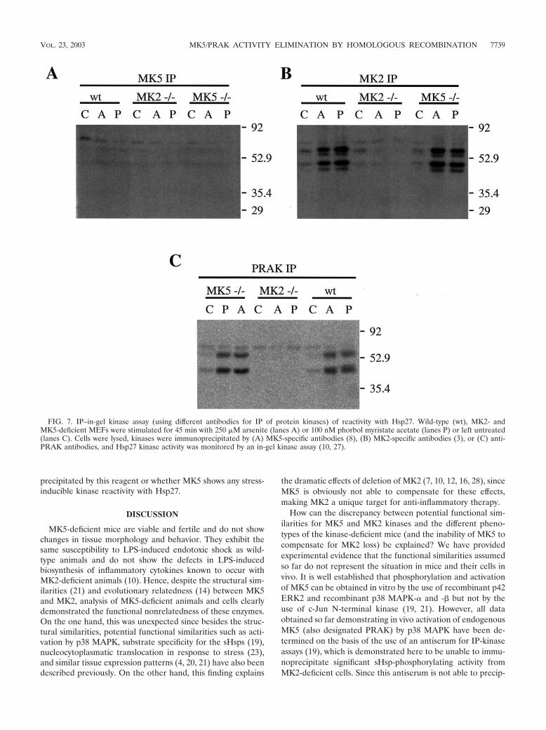
precipitated by this reagent or whether MK5 shows any stress-inducible kinase reactivity with Hsp27.
DISCUSSION
MK5-deficient mice are viable and fertile and do not showchanges in tissue morphology and behavior. They exhibit thesame susceptibility to LPS-induced endotoxic shock as wild-type animals and do not show the defects in LPS-inducedbiosynthesis of inflammatory cytokines known to occur withMK2-deficient animals (10). Hence, despite the structural sim-ilarities (21) and evolutionary relatedness (14) between MK5and MK2, analysis of MK5-deficient animals and cells clearlydemonstrated the functional nonrelatedness of these enzymes.On the one hand, this was unexpected since besides the struc-tural similarities, potential functional similarities such as acti-vation by p38 MAPK, substrate specificity for the sHsps (19),nucleocytoplasmatic translocation in response to stress (23),and similar tissue expression patterns (4, 20, 21) have also beendescribed previously. On the other hand, this finding explains
the dramatic effects of deletion of MK2 (7, 10, 12, 16, 28), sinceMK5 is obviously not able to compensate for these effects,making MK2 a unique target for anti-inflammatory therapy.
How can the discrepancy between potential functional sim-ilarities for MK5 and MK2 kinases and the different pheno-types of the kinase-deficient mice (and the inability of MK5 tocompensate for MK2 loss) be explained? We have providedexperimental evidence that the functional similarities assumedso far do not represent the situation in mice and their cells invivo. It is well established that phosphorylation and activationof MK5 can be obtained in vitro by the use of recombinant p42ERK2 and recombinant p38 MAPK-� and -� but not by theuse of c-Jun N-terminal kinase (19, 21). However, all dataobtained so far demonstrating in vivo activation of endogenousMK5 (also designated PRAK) by p38 MAPK have been de-termined on the basis of the use of an antiserum for IP-kinaseassays (19), which is demonstrated here to be unable to immu-noprecipitate significant sHsp-phosphorylating activity fromMK2-deficient cells. Since this antiserum is not able to precip-
FIG. 7. IP–in-gel kinase assay (using different antibodies for IP of protein kinases) of reactivity with Hsp27. Wild-type (wt), MK2- andMK5-deficient MEFs were stimulated for 45 min with 250 �M arsenite (lanes A) or 100 nM phorbol myristate acetate (lanes P) or left untreated(lanes C). Cells were lysed, kinases were immunoprecipitated by (A) MK5-specific antibodies (8), (B) MK2-specific antibodies (3), or (C) anti-PRAK antibodies, and Hsp27 kinase activity was monitored by an in-gel kinase assay (10, 27).
VOL. 23, 2003 MK5/PRAK ACTIVITY ELIMINATION BY HOMOLOGOUS RECOMBINATION 7739

itate sHsp-kinase activity from MK2-deficient cells but precip-itates such activity from MK5-deficient cells, a cross-reactionof this antibody preparation with other sHsp-phosphorylatingkinases such as MK2 or MK3 is likely. This means that the invivo properties described for MK5/PRAK using this antibodypreparation (19) result from IP of cross-reacting kinases.
Recently, thrombin-induced activation of PRAK in humanplatelets was analyzed by immunoaffinity-purified non-cross-reacting specific antibodies (8). In this case, relatively highbasal activity but no significant stimulation (1.2-fold stimula-tion after 150 min) of PRAK was measured whereas significantstimulation of p38 MAPK and MNK1 was detected after 25min. This result also makes activation of endogenous MK5/PRAK by p38 MAPK unlikely.
No significant stimulation of endogenous MK5 in living cellshas been detected using the specific IP kinase assay so far. Thismight mean that the action of basal activity is regulated byprotein-protein interaction and subcellular localization or sim-ply that the right physiological stimuli or situations where MK5is activated have not been identified so far. The recently avail-able antiserum specific for phosphorylation of the regulatorysite in the activation loop of the kinase (kind gift of P. Cohen)should help to answer this question in the future.
We further analyzed protein-protein interactions betweenp38 MAPK and MK5 and provided additional evidence thatendogenous p38 MAPK is probably not acting the same wayfor activation of MK5 as for activation of MK2. In contrast toMK2-deficient tissues, in which p38 MAPK levels are reduceddue to the missing chaperone and/or transporter function ofMK2 (9), in full-length MK5-deficient tissues, for which signif-icantly (at least) reduced MK5 truncated protein levels havebeen detected, no reduction of p38 MAPK levels has beenobserved. This indicates the existence of specific p38 MAPK-stabilizing properties of MK2. Expression levels of MK2 higherthan those of MK5 could theoretically explain this observationalso, but in the tissues analyzed a significant expression of MK5mRNA (19, 21) comparable to the expression of MK2 mRNA(4) was detected. Another analysis of interaction of MK5 withendogenous p38 MAPK was carried out using TAP. Weshowed that endogenous p38 MAPK from 293 cells copurifieswith MK2 but not with MK5. This strengthens the notion thatthere is no specific interaction between endogenous p38MAPK and MK5.
Interaction between MK5 and p38 MAPK in vitro and forcells overexpressing tagged versions of both proteins has beendescribed previously (18, 23). Furthermore, overexpression ofboth active and inactive p38 MAPK changes subcellular local-ization of MK5 (18, 23). Since artificial overexpression of sig-naling components can titrate out and quench other relevantendogenous signaling molecules, these data do not necessarilyreflect the in vivo situation. It is interesting that the stress-induced kinetics of translocation of GFP-tagged MK5 is muchslower than translocation of GFP-tagged MK2 and does notcorrelate with kinetics of p38 MAPK activation (18, 23). Thisalso indicates different mechanisms of regulation of these pro-cesses by different upstream kinases.
In the evolutionarily conserved group of MAPKAP kinases,there is a third enzyme present in mice and humans that hasbeen designated MK3 or 3pK (15, 24) (14). Since no mouseknockout is available for this kinase, the physiological role for
this enzyme has not been defined so far. Since in MK2/MK5double-knockout animals LPS-induced production of TNF is(in similarity to results with the MK2 knockout alone) reducedto about 10% of that of the wild type and is not furtherimpaired (A. Kotlyarov and M. Gaestel, unpublished data), thequestion remains open whether MK3 activity is responsible forthe remaining 10% of LPS-induced TNF biosynthesis. Wewould speculate that in regard to its functional properties MK3seems more clearly related to MK2, since its primary structureincludes the second regulatory phosphorylation site outside thecatalytic domain (15) and the kinetics of its nuclear export(Kotlyarov and Gaestel, unpublished) is clearly more similar tothat of MK2 than to that of MK5.
ACKNOWLEDGMENTS
We thank Kristina Vintersten for her excellent work at the EMBL-Heidelberg Transgenic Service, Ole Morten Seternes and Ugo Moensfor help with the MK5 kinase assay, Sir Philip Cohen (Dundee, Scot-land) and Jiahuai Han (La Jolla, Calif.) for MK5 and PRAK antibod-ies, Michael Kracht for support in the TAP experiments, and DorotheaKrone and Stefanie Feldhege for excellent technical help.
The work was supported by grants Ga 453/7, Bu740/5, SFB621TP-A3, and SFB 421 TP-B2 from the DFG.
REFERENCES
1. Ben-Levy, R., S. Hooper, R. Wilson, H. F. Paterson, and C. J. Marshall.1998. Nuclear export of the stress-activated protein kinase p38 mediated byits substrate MAPKAP kinase-2. Curr. Biol. 8:1049–1057.
2. Clifton, A. D., P. R. Young, and P. Cohen. 1996. A comparison of thesubstrate specificity of MAPKAP kinase-2 and MAPKAP kinase-3 and theiractivation by cytokines and cellular stress. FEBS Lett. 392:209–214.
3. Engel, K., A. Kotlyarov, and M. Gaestel. 1998. Leptomycin B-sensitive nu-clear export of MAPKAP kinase 2 is regulated by phosphorylation. EMBOJ. 17:3363–3371.
4. Engel, K., K. Plath, and M. Gaestel. 1993. The MAP kinase-activated pro-tein kinase 2 contains a proline-rich SH3-binding domain. FEBS Lett. 336:143–147.
5. Engel, K., H. Schultz, F. Martin, A. Kotlyarov, K. Plath, M. Hahn, U.Heinemann, and M. Gaestel. 1995. Constitutive activation of mitogen-acti-vated protein kinase-activated protein kinase 2 by mutation of phosphory-lation sites and an A-helix motif. J. Biol. Chem. 270:27213–27221.
6. Han, Q., J. Leng, D. Bian, C. Mahanivong, K. A. Carpenter, Z. K. Pan, J.Han, and S. Huang. 2002. Rac1-MKK3-p38-MAPKAPK2 pathway promotesurokinase plasminogen activator mRNA stability in invasive breast cancercells. J. Biol. Chem. 277:48379–48385.
7. Hannigan, M. O., L. Zhan, Y. Ai, A. Kotlyarov, M. Gaestel, and C. K. Huang.2001. Abnormal migration phenotype of mitogen-activated protein kinase-activated protein kinase 2�/� neutrophils in zigmond chambers containingformyl-methionyl-leucyl-phenylalanine gradients. J. Immunol. 167:3953–3961.
8. Hefner, Y., A. G. Borsch-Haubold, M. Murakami, J. I. Wilde, S. Pasquet, D.Schieltz, F. Ghomashchi, J. R. Yates III, C. G. Armstrong, A. Paterson, P.Cohen, R. Fukunaga, T. Hunter, I. Kudo, S. P. Watson, and M. H. Gelb.2000. Serine 727 phosphorylation and activation of cytosolic phospholipaseA2 by MNK1-related protein kinases. J. Biol. Chem. 275:37542–37551.
9. Kotlyarov, A., and M. Gaestel. 2002. Is MK2 (mitogen-activated proteinkinase-activated protein kinase 2) the key for understanding post-transcrip-tional regulation of gene expression? Biochem. Soc. Trans. 30:959–963.
10. Kotlyarov, A., A. Neininger, C. Schubert, R. Eckert, C. Birchmeier, H. D.Volk, and M. Gaestel. 1999. MAPKAP kinase 2 is essential for LPS-inducedTNF-alpha biosynthesis. Nat. Cell Biol. 1:94–97.
11. Kotlyarov, A., Y. Yannoni, S. Fritz, K. Laa�, J.-B. Telliez, D. Pitman, L.-L.Lin, and M. Gaestel. 2002. Distinct cellular functions of MK2. Mol. Cell.Biol. 22:4827–4835.
12. Lehner, M. D., F. Schwoebel, A. Kotlyarov, M. Leist, M. Gaestel, and T.Hartung. 2002. Mitogen-activated protein kinase-activated protein kinase2-deficient mice show increased susceptibility to Listeria monocytogenes in-fection. J. Immunol. 168:4667–4673.
13. Li, Y., K. Inoki, P. Vacratsis, and K. L. Guan. 2003. The p38 and MK2 kinasecascade phosphorylates tuberin, the tuberous sclerosis 2 gene product, andenhances its interaction with 14-3-3. J. Biol. Chem. 278:13663–13671.
14. Manning, G., D. B. Whyte, R. Martinez, T. Hunter, and S. Sudarsanam.2002. The protein kinase complement of the human genome. Science 298:1912–1934.
15. McLaughlin, M. M., S. Kumar, P. C. McDonnell, S. Van Horn, J. C. Lee,
7740 SHI ET AL. MOL. CELL. BIOL.

G. P. Livi, and P. R. Young. 1996. Identification of mitogen-activated protein(MAP) kinase-activated protein kinase-3, a novel substrate of CSBP p38MAP kinase. J. Biol. Chem. 271:8488–8492.
16. Neininger, A., D. Kontoyiannis, A. Kotlyarov, R. Winzen, R. Eckert, H. D.Volk, H. Holtmann, G. Kollias, and M. Gaestel. 2002. MK2 targets AU-richelements and regulates biosynthesis of tumor necrosis factor and interleu-kin-6 independently at different post-transcriptional levels. J. Biol. Chem.277:3065–3068.
17. Neufeld, B., A. Grosse-Wilde, A. Hoffmeyer, B. W. Jordan, P. Chen, D. Dinev,S. Ludwig, and U. R. Rapp. 2000. Serine/threonine kinases 3pK and MAPK-activated protein kinase 2 interact with the basic helix-loop-helix transcrip-tion factor E47 and repress its transcriptional activity. J. Biol. Chem. 275:20239–20242.
18. New, L., Y. Jiang, and J. Han. 2003. Regulation of PRAK subcellular loca-tion by p38 MAP kinases. Mol. Biol. Cell 14:2603–2616.
19. New, L., Y. Jiang, M. Zhao, K. Liu, W. Zhu, L. J. Flood, Y. Kato, G. C. Parry,and J. Han. 1998. PRAK, a novel protein kinase regulated by the p38 MAPkinase. EMBO J. 17:3372–3384.
20. New, L., M. Zhao, Y. Li, W. W. Bassett, Y. Feng, S. Ludwig, F. D. Padova, H.Gram, and J. Han. 1999. Cloning and characterization of RLPK, a novelRSK-related protein kinase. J. Biol. Chem. 274:1026–1032.
21. Ni, H., X. S. Wang, K. Diener, and Z. Yao. 1998. MAPKAPK5, a novelmitogen-activated protein kinase (MAPK)-activated protein kinase, is a sub-strate of the extracellular-regulated kinase (ERK) and p38 kinase. Biochem.Biophys. Res. Commun. 243:492–496.
22. Rigaut, G., A. Shevchenko, B. Rutz, M. Wilm, M. Mann, and B. Seraphin.1999. A generic protein purification method for protein complex character-ization and proteome exploration. Nat. Biotechnol. 17:1030–1032.
23. Seternes, O. M., B. Johansen, B. Hegge, M. Johannessen, S. M. Keyse, and
U. Moens. 2002. Both binding and activation of p38 mitogen-activated pro-tein kinase (MAPK) play essential roles in regulation of the nucleocytoplas-mic distribution of MAPK-activated protein kinase 5 by cellular stress. Mol.Cell. Biol. 22:6931–6945.
24. Sithanandam, G., F. Latif, F. M. Duh, R. Bernal, U. Smola, H. Li, I. Kuzmin,V. Wixler, L. Geil, and S. Shrestha. 1996. 3pK, a new mitogen-activatedprotein kinase-activated protein kinase located in the small cell lung cancertumor suppressor gene region. Mol. Cell. Biol. 16:868–876.
25. Stokoe, D., K. Engel, D. G. Campbell, P. Cohen, and M. Gaestel. 1992.Identification of MAPKAP kinase 2 as a major enzyme responsible for thephosphorylation of the small mammalian heat shock proteins. FEBS Lett.313:307–313.
26. Tanoue, T., R. Maeda, M. Adachi, and E. Nishida. 2001. Identification of adocking groove on ERK and p38 MAP kinases that regulates the specificityof docking interactions. EMBO J. 20:466–479.
27. van Dam, H., D. Wilhelm, I. Herr, A. Steffen, P. Herrlich, and P. Angel. 1995.ATF-2 is preferentially activated by stress-activated protein kinases to me-diate c-jun induction in response to genotoxic agents. EMBO J. 14:1798–1811.
28. Wang, X., L. Xu, H. Wang, P. R. Young, M. Gaestel, and G. Z. Feuerstein.2002. Mitogen-activated protein kinase-activated protein (MAPKAP) kinase2 deficiency protects brain from ischemic injury in mice. J. Biol. Chem.277:43968–43972.
29. Winzen, R., M. Kracht, B. Ritter, A. Wilhelm, C. Y. Chen, A. B. Shyu, M.Muller, M. Gaestel, K. Resch, and H. Holtmann. 1999. The p38 MAP kinasepathway signals for cytokine-induced mRNA stabilization via MAP kinase-activated protein kinase 2 and an AU-rich region-targeted mechanism.EMBO J. 18:4969–4980.
VOL. 23, 2003 MK5/PRAK ACTIVITY ELIMINATION BY HOMOLOGOUS RECOMBINATION 7741