The MCM-Binding Protein ETG1 Aids Sister Chromatid Cohesion Required for Postreplicative Homologous...
13
The MCM-Binding Protein ETG1 Aids Sister Chromatid Cohesion Required for Postreplicative Homologous Recombination Repair Naoki Takahashi 1,2,3 , Mauricio Quimbaya 1,2,4,5 , Veit Schubert 6 , Tim Lammens 1,2 , Klaas Vandepoele 1,2 , Ingo Schubert 6 , Minami Matsui 3 , Dirk Inze ´ 1,2 , Geert Berx 4,5 , Lieven De Veylder 1,2 * 1 Department of Plant Systems Biology, Flanders Institute for Biotechnology (VIB), Gent, Belgium, 2 Department of Plant Biotechnology and Genetics, Ghent University, Gent, Belgium, 3 Plant Functional Genomics Research Group, RIKEN Plant Science Center, Yokohama, Kanagawa, Japan, 4 Department for Molecular Biomedical Research, Molecular and Cellular Oncology Unit, Flanders Institute for Biotechnology (VIB), Gent, Belgium, 5 Department of Biomedical Molecular Biology, Ghent University, Gent, Belgium, 6 Leibniz Institute of Plant Genetics and Crop Plant Research (IPK), Gatersleben, Germany Abstract The DNA replication process represents a source of DNA stress that causes potentially spontaneous genome damage. This effect might be strengthened by mutations in crucial replication factors, requiring the activation of DNA damage checkpoints to enable DNA repair before anaphase onset. Here, we demonstrate that depletion of the evolutionarily conserved minichromosome maintenance helicase-binding protein ETG1 of Arabidopsis thaliana resulted in a stringent late G2 cell cycle arrest. This arrest correlated with a partial loss of sister chromatid cohesion. The lack-of-cohesion phenotype was intensified in plants without functional CTF18, a replication fork factor needed for cohesion establishment. The synergistic effect of the etg1 and ctf18 mutants on sister chromatid cohesion strengthened the impact on plant growth of the replication stress caused by ETG1 deficiency because of inefficient DNA repair. We conclude that the ETG1 replication factor is required for efficient cohesion and that cohesion establishment is essential for proper development of plants suffering from endogenous DNA stress. Cohesion defects observed upon knockdown of its human counterpart suggest an equally important developmental role for the orthologous mammalian ETG1 protein. Citation: Takahashi N, Quimbaya M, Schubert V, Lammens T, Vandepoele K, et al. (2010) The MCM-Binding Protein ETG1 Aids Sister Chromatid Cohesion Required for Postreplicative Homologous Recombination Repair. PLoS Genet 6(1): e1000817. doi:10.1371/journal.pgen.1000817 Editor: Mathilde Grelon, Institut Jean-Pierre Bourgin, INRA de Versailles, France Received September 1, 2009; Accepted December 16, 2009; Published January 15, 2010 Copyright: ß 2010 Takahashi et al. This is an open-access article distributed under the terms of the Creative Commons Attribution License, which permits unrestricted use, distribution, and reproduction in any medium, provided the original author and source are credited. Funding: This work was supported by grants from the Interuniversity Poles of Attraction Programne (IUAP VI/33), initiated by the Belgian State, Science Policy Office and the Research Foundation-Flanders (grant no. G008306). TL is indebted to the Institute for Promotion of Innovation by Science and Technology in Flanders for a predoctoral fellowship. LDV and KV are postdoctoral fellows of the Research Foundation-Flanders. The funders had no role in study design, data collection and analysis, decision to publish, or preparation of the manuscript. Competing Interests: The authors have declared that no competing interests exist. * E-mail: [email protected] Introduction For one single cell to generate two cells, numerous events must be coordinated, in particular, faithful DNA replication and partitioning of the sister chromatids to each of the daughter cells. It is of utmost importance to ensure the error-free duplication of the replicated DNA during each cell cycle, preventing the transmission of potentially harmful mutations to the daughter cells, which otherwise might result in developmental defects or even cancer. DNA damage and replication errors might originate from DNA stress provoked by either exogenous (such as c- irradiation and UV-B light) or endogenous (such as metabolic byproducts) sources. The latter include the replication process itself that necessitates the cooperation of many different proteins in a highly complex manner. Errors arisen during replication are preferentially repaired through homologous recombination be- tween the replicated sister chromatids that lay in close proximity thanks to cohesion. This sister chromatid cohesion is mediated by cohesin that consists of four subunits in budding yeast (Saccharo- myces cerevisiae): two structural maintenance of chromosome (SMC) proteins, designated SMC1 and SMC3, and two non-SMC subunits, designated SCC1 (also known as Mcd1/Rad21) and SCC3 [1–5]. SMC1 and SMC3 are self-folded by antiparallel coiled-coil interactions, creating a rod-shaped molecule with an ATP-binding ‘‘head’’ at one end and a ‘‘hinge’’ domain at the other. The two SMC subunits associate with each other through their hinge domains, producing a V-shaped dimer [6,7]. Cohesin forms a tripartite ring in which the open-V structure of the SMC heterodimer is closed by the simultaneous binding of the N- and C-terminal regions of SCC1 to the head domains of SMC3 and SMC1, respectively [8]. Cohesin is deposited on unreplicated chromatin in a reaction requiring ATP hydrolysis by the SMC heads and the cohesin- loading complex SCC2/SCC4 [9–11]. Whereas cohesion loading occurs well before S phase, the process of cohesion establishment is intimately connected with DNA replication. In budding yeast, cohesion depends on an acetyltransferase, designated Eco1/Ctf7, that acetylates two lysine residues on the ATPase head domain of SMC3 [12–14]. Eco1/Ctf7 interacts physically and genetically with the proliferating cell nuclear antigen (PCNA) and the replication factor C (RFC) and has been found to travel along the DNA with replication forks [15–17], suggesting that PLoS Genetics | www.plosgenetics.org 1 January 2010 | Volume 6 | Issue 1 | e1000817
Transcript of The MCM-Binding Protein ETG1 Aids Sister Chromatid Cohesion Required for Postreplicative Homologous...

The MCM-Binding Protein ETG1 Aids Sister ChromatidCohesion Required for Postreplicative HomologousRecombination RepairNaoki Takahashi1,2,3, Mauricio Quimbaya1,2,4,5, Veit Schubert6, Tim Lammens1,2, Klaas Vandepoele1,2,
Ingo Schubert6, Minami Matsui3, Dirk Inze1,2, Geert Berx4,5, Lieven De Veylder1,2*
1 Department of Plant Systems Biology, Flanders Institute for Biotechnology (VIB), Gent, Belgium, 2 Department of Plant Biotechnology and Genetics, Ghent University,
Gent, Belgium, 3 Plant Functional Genomics Research Group, RIKEN Plant Science Center, Yokohama, Kanagawa, Japan, 4 Department for Molecular Biomedical Research,
Molecular and Cellular Oncology Unit, Flanders Institute for Biotechnology (VIB), Gent, Belgium, 5 Department of Biomedical Molecular Biology, Ghent University, Gent,
Belgium, 6 Leibniz Institute of Plant Genetics and Crop Plant Research (IPK), Gatersleben, Germany
Abstract
The DNA replication process represents a source of DNA stress that causes potentially spontaneous genome damage. Thiseffect might be strengthened by mutations in crucial replication factors, requiring the activation of DNA damagecheckpoints to enable DNA repair before anaphase onset. Here, we demonstrate that depletion of the evolutionarilyconserved minichromosome maintenance helicase-binding protein ETG1 of Arabidopsis thaliana resulted in a stringent lateG2 cell cycle arrest. This arrest correlated with a partial loss of sister chromatid cohesion. The lack-of-cohesion phenotypewas intensified in plants without functional CTF18, a replication fork factor needed for cohesion establishment. Thesynergistic effect of the etg1 and ctf18 mutants on sister chromatid cohesion strengthened the impact on plant growth ofthe replication stress caused by ETG1 deficiency because of inefficient DNA repair. We conclude that the ETG1 replicationfactor is required for efficient cohesion and that cohesion establishment is essential for proper development of plantssuffering from endogenous DNA stress. Cohesion defects observed upon knockdown of its human counterpart suggest anequally important developmental role for the orthologous mammalian ETG1 protein.
Citation: Takahashi N, Quimbaya M, Schubert V, Lammens T, Vandepoele K, et al. (2010) The MCM-Binding Protein ETG1 Aids Sister Chromatid CohesionRequired for Postreplicative Homologous Recombination Repair. PLoS Genet 6(1): e1000817. doi:10.1371/journal.pgen.1000817
Editor: Mathilde Grelon, Institut Jean-Pierre Bourgin, INRA de Versailles, France
Received September 1, 2009; Accepted December 16, 2009; Published January 15, 2010
Copyright: � 2010 Takahashi et al. This is an open-access article distributed under the terms of the Creative Commons Attribution License, which permitsunrestricted use, distribution, and reproduction in any medium, provided the original author and source are credited.
Funding: This work was supported by grants from the Interuniversity Poles of Attraction Programne (IUAP VI/33), initiated by the Belgian State, Science PolicyOffice and the Research Foundation-Flanders (grant no. G008306). TL is indebted to the Institute for Promotion of Innovation by Science and Technology inFlanders for a predoctoral fellowship. LDV and KV are postdoctoral fellows of the Research Foundation-Flanders. The funders had no role in study design, datacollection and analysis, decision to publish, or preparation of the manuscript.
Competing Interests: The authors have declared that no competing interests exist.
* E-mail: [email protected]
Introduction
For one single cell to generate two cells, numerous events must
be coordinated, in particular, faithful DNA replication and
partitioning of the sister chromatids to each of the daughter cells.
It is of utmost importance to ensure the error-free duplication of
the replicated DNA during each cell cycle, preventing the
transmission of potentially harmful mutations to the daughter
cells, which otherwise might result in developmental defects or
even cancer. DNA damage and replication errors might originate
from DNA stress provoked by either exogenous (such as c-
irradiation and UV-B light) or endogenous (such as metabolic
byproducts) sources. The latter include the replication process
itself that necessitates the cooperation of many different proteins in
a highly complex manner. Errors arisen during replication are
preferentially repaired through homologous recombination be-
tween the replicated sister chromatids that lay in close proximity
thanks to cohesion. This sister chromatid cohesion is mediated by
cohesin that consists of four subunits in budding yeast (Saccharo-
myces cerevisiae): two structural maintenance of chromosome (SMC)
proteins, designated SMC1 and SMC3, and two non-SMC
subunits, designated SCC1 (also known as Mcd1/Rad21) and
SCC3 [1–5]. SMC1 and SMC3 are self-folded by antiparallel
coiled-coil interactions, creating a rod-shaped molecule with an
ATP-binding ‘‘head’’ at one end and a ‘‘hinge’’ domain at the
other. The two SMC subunits associate with each other through
their hinge domains, producing a V-shaped dimer [6,7]. Cohesin
forms a tripartite ring in which the open-V structure of the SMC
heterodimer is closed by the simultaneous binding of the N- and
C-terminal regions of SCC1 to the head domains of SMC3 and
SMC1, respectively [8].
Cohesin is deposited on unreplicated chromatin in a reaction
requiring ATP hydrolysis by the SMC heads and the cohesin-
loading complex SCC2/SCC4 [9–11]. Whereas cohesion loading
occurs well before S phase, the process of cohesion establishment is
intimately connected with DNA replication. In budding yeast,
cohesion depends on an acetyltransferase, designated Eco1/Ctf7,
that acetylates two lysine residues on the ATPase head domain of
SMC3 [12–14]. Eco1/Ctf7 interacts physically and genetically
with the proliferating cell nuclear antigen (PCNA) and the
replication factor C (RFC) and has been found to travel along
the DNA with replication forks [15–17], suggesting that
PLoS Genetics | www.plosgenetics.org 1 January 2010 | Volume 6 | Issue 1 | e1000817

replication fork progression and sister chromatid cohesion are
coupled events. This model is supported by the observation that
cohesion defects are caused by mutations in replisome compo-
nents, e.g., the DNA polymerase a-binding protein Ctf4 [18–19],
the Chl1 helicase [20], and RFC components, such as Ctf18 [21].
In budding yeast and humans, Ctf18 associates with Rfc2, Rfc3,
Rfc4, and Rfc5 to form the RFCCtf18 complex, which, in turn,
couples with two additional subunits, Dcc1 and Ctf8, creating a
heptameric complex with the PCNA that has a loading and
unloading activity and plays a role in sister chromatid cohesion
[19–23].
E2F transcription factors control the expression of many
genes involved in DNA replication and DNA repair. By
studying Arabidopsis thaliana E2F target genes, we have
previously identified the E2F TARGET GENE 1 (ETG1) as
a novel evolutionarily conserved replisome factor. ETG1 binds
with the minichromosome maintenance (MCM) complex and
is crucial for efficient DNA replication [24]. Similarly to
ETG1, the human orthologous MCM-BP protein binds to
replication origins as part of the MCM complex [25]. Plants
lacking the ETG1 gene have serrated leaves because of cell
cycle inhibition triggered by the DNA replication checkpoints,
as shown by the transcriptional induction of DNA stress and
checkpoint genes [24]. Here we demonstrate that the ETG1
protein plays an additional role in the establishment of sister
chromatid cohesion. Cohesion along chromosome arms was
impaired in ETG1-deficient cells. Strikingly, the growth
inhibition and the DNA stress phenotype of etg1 mutant plants
were strongly enhanced in cohesion mutant backgrounds.
Decreased DNA repair kinetics illustrate that the lack of
cohesion accounts for severe growth defects in plants suffering
from endogenous DNA stress, emphasizing the importance of
cohesion for correct plant development. Finally, we show that
the knockdown of the human MCM-BP results in a loss of
cohesion phenotype as well, suggesting an equally important
developmental role for the orthologous mammalian ETG1
proteins.
Results
Upregulation of mitosis-specific genes in etg1 mutantsETG1-deficient plants suffer from endogenous DNA stress and
display a transient cell cycle arrest [24]. To gain more insight into
this defective cell cycle, we examined transcript levels of 22,750
genes by using Affymetrix ATH1 GeneChip arrays. Triplicate
batches of proliferating first leaves of 9-day-old wild-type and two
independent etg1-1 and etg1-2 mutant plants were harvested for
total RNA preparation. Statistical analysis identified a total of 219
genes differentially expressed between wild-type and etg1 plants at
a P-value ,0.01, among which 89% upregulated and 11%
downregulated and displaying a 1.3- to 14.8-fold change in
expression. Strikingly, of the 195 upregulated genes, 103 (53%)
showed an expression peak during mitosis (Table S1 and Table S2;
Figure S1). Transcription of genes expressed specifically during
mitosis is regulated by a common upstream cis-acting element
(ynCAACGG), designated mitosis-specific activator (MSA). In
total, 82 upregulated genes in etg1 plants possessed an MSA
element within the first 1 kb region upstream of the translation
start, which is significantly more than expected by chance (P-value
,0.001) and indicative for an arrest in late G2 or mitosis. This
hypothesis was corroborated by an overall transcriptional
induction of G2 and M-phase expressed genes in etg1 knockout
plants, as demonstrated by plotting the average signal log ratios
(SLRs) between the expression levels in wild-type and etg1 mutant
plants of 9,910 genes previously defined as cell cycle regulated [26]
(Figure 1A).
To identify the biological processes that might be affected in
the etg1 mutant plants, the up- and downregulated genes were
analyzed for gene ontology (GO) enrichment [27]. Among the
upregulated genes, regulation of progression through cell cycle,
mitotic cell cycle, and microtubule-based movement genes were
significantly overrepresented (Figure 1B), indicating that lack of
ETG1 resulted in mitotic defects. This result was unexpected
because ETG1 is an E2F-dependent target gene expressed during
the S phase and its gene product is required for DNA replication.
Additionally, ETG1-deficient plants had been found to interact
synergistically with the wee1 and atr1 replication checkpoint
mutants and to suffer from DNA replication stress [24], which is
anticipated to arrest the cell cycle during S or early G2. Indeed,
when the etg1 microarray data set was compared with that of
plants exposed to UV-B light [28] or to the radiomimetic drug
bleomycin [29], the distribution of the significantly upregulated
cell cycle phase markers was different. In the latter cases, the set
of modified genes was clearly enriched for S-phase genes
(Figure 1C), suggesting that the cell cycle arrest in the etg1
mutants occurs downstream of that one triggered by UV-B or
bleomycin.
ETG1 is required for sister chromatid arm cohesionSister chromatid cohesion is essential for the appropriate
distribution of chromosomes into the daughter cells upon cell
division. Several observations indicate that cohesion establishment
is coupled with DNA replication [18–21]. Therefore, we analyzed
by fluorescent in situ hybridization (FISH) whether the cell cycle
arrest in etg1 mutant plants might be linked with a loss of cohesion.
The two adjacent overlapping bacterial artificial chromosomes
(BACs) T2P11 and T7N9 that label the upper mid-arm position of
chromosome 1 were used for FISH analysis on flow-sorted 4C leaf
nuclei to compare the frequency of sister arm alignment between
the wild-type and etg1-1. The number of FISH signals indicates
whether sister chromatids are linked at the position tested: one
(pairing of both homologs) or two FISH signals mark the positional
Author Summary
DNA replication is a highly complex process and thesource of potential DNA damage. It is of utmostimportance that the damaged DNA is repaired beforecells proceed through mitosis, because the genome holdsall the information required for correct development. DNAreplication results in two identical sister chromatids. A trickapplied by cells to overcome damaged DNA is homolo-gous recombination, using the undamaged copy of thesister chromatid as a template to repair the damaged one.This process is aided by keeping the two sister chromatidsin close proximity after the replication process by thedeposition of a molecular glue, called cohesin. In thepresent work, we identified the Arabidopsis thaliana ETG1protein as a novel evolutionarily conserved replicationfactor that is needed for maintaining the sister chromatidsphysically aligned. In plants without ETG1, DNA damagebuilds up due to inefficient DNA repair. As a consequence,cell division is impaired with a huge impact on plantgrowth, highlighting the importance of cohesin for thecorrect development of eukaryotic organisms. Cohesionphenotypes observed upon the depletion of the ortholo-gous human ETG1 protein indicate equally prominentroles for this particular factor during mammalian develop-ment.
ETG1 Aids Sister Chromatid Cohesion
PLoS Genetics | www.plosgenetics.org 2 January 2010 | Volume 6 | Issue 1 | e1000817

alignment of sister chromatids at the corresponding region and
three or four signals the sister chromatid separation at one or both
homologous chromosomes, respectively (Figure 2B–2D). When
compared to the wild-type nuclei, the etg1-1 nuclei showed a
significant increase (P-value ,0.001) in sister chromatid separation
(Figure 2A and 2E). Positional sister chromatid separation
occurred in 28.1% of the homologs in 4C wild-type leaf nuclei,
whereas etg1-1 leaf nuclei displayed 42.7% sister chromatid
separation per homolog (Figure 2A). By contrast, FISH applied
to the 178-bp centromere-specific sequence (pAL) revealed 4–12
signals in both the wild-type and the etg1-1 mutant 4C nuclei
(Figure 2A and 2F). These findings suggest that ETG1 is required
for correct establishment of sister chromatid cohesion along
chromosome arms, but not at centromeres.
Arabidopsis CTF18 is required for sister chromatidcohesion
The ETG1 gene had originally been identified by microarray
analysis as a transcript induced in plants that ectopically expressed the
heterodimeric E2Fa-DPa transcription factor [30] and had been
demonstrated to be directly controlled by the E2Fa and E2Fb
transcription factors [24]. Interestingly, putative orthologous genes
encoding proteins involved in cohesion establishment during the
replication process were also transcriptionally upregulated in E2Fa-
DPa-overexpressing plants, such as ECO1 (also known as CTF7)
(At4g31400), CHL1 (At1g79890), and CTF18 (At1g04730) (Figure S2).
In budding yeast, Ctf18 localizes at the replication fork and is a critical
factor for establishment of cohesion [16,19]. Putative orthologous
proteins were found in human, mouse, Arabidopsis, rice, fruitfly,
Figure 1. Upregulation of G2/M-specific genes in ETG1-deficient plants. (A) Plot of average signal log ratios (SLRs; logarithms to the base 2)between wild-type and etg1 mutant plants for 9,910 genes during the 22-h cell cycle of Arabidopsis [26]. Genes were sorted into 10 bins representing the 10time points of measurements, based on their maximal expression during the cell cycle. Cells were synchronized by a release from an aphidicolin arrest.Synchronized cells completed S phase in 5 h and went through mitosis from 9 to 14 h. The solid line represents the best-fitted curve. (B) GO analysis of the196 upregulated genes in the first leaves of etg1 mutant plants. The yellow-to-orange color of the circles correspond to the level of significance of theoverrepresented GO category of #0.01 according to a multiple t test with false discovery rate–corrected P value. The size of the circle is proportional to thenumber of genes in the category. (C) Comparison of the distribution of cell cycle phase-dependent upregulated genes in etg1 plants and plants treated withUV-B- or bleomycin. Microarray data sets of UV-B, and bleomycin treatment were imported from [28] and [29], respectively. S (red), G2 (blue), M (yellow), andG1 (green) phase-specific gene expression patterns were defined by Menges et al. [26].doi:10.1371/journal.pgen.1000817.g001
ETG1 Aids Sister Chromatid Cohesion
PLoS Genetics | www.plosgenetics.org 3 January 2010 | Volume 6 | Issue 1 | e1000817

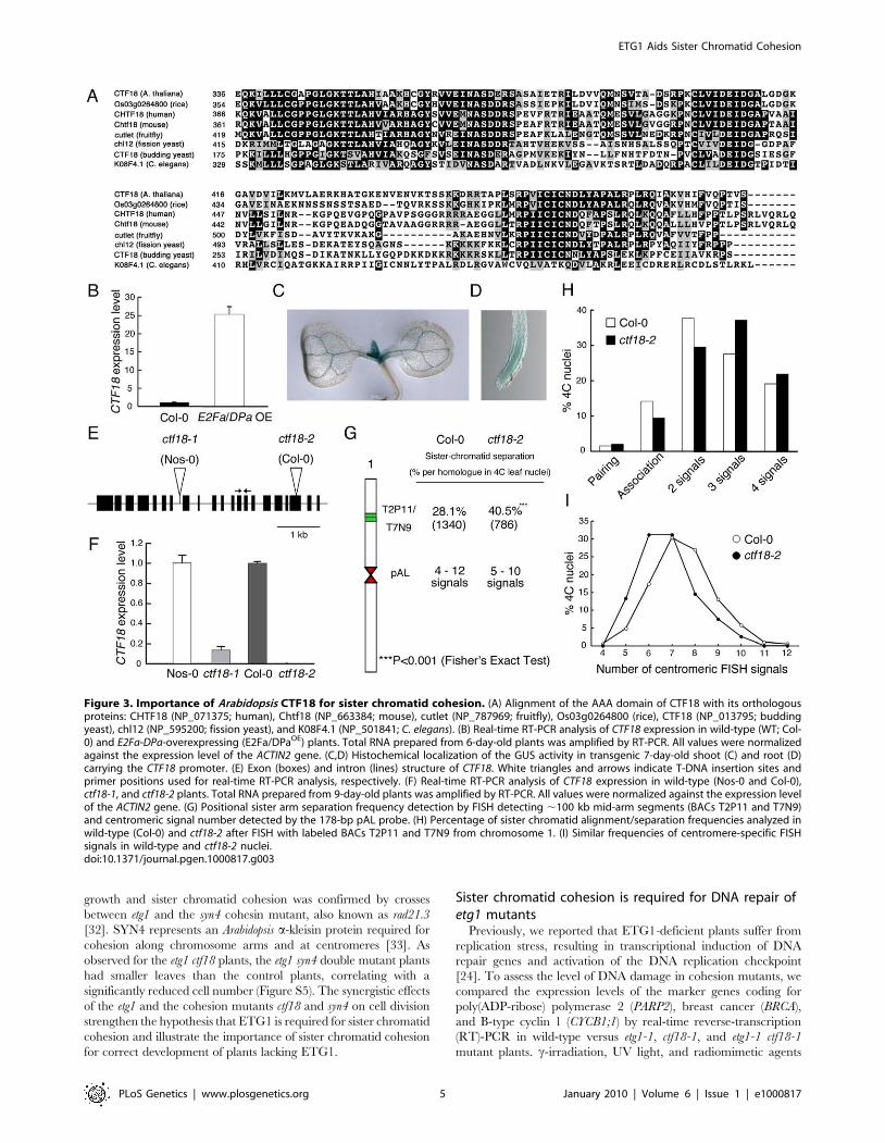
worm, and fission yeast (Figure 3A; Figure S3). Transcriptional
activation of the Arabidopsis CTF18 gene in E2Fa-DPa-overexpressing
plants was confirmed by quantitative real-time PCR analysis (Figure
3B). The spatial expression pattern of CTF18 was analyzed in more
than six independent transgenic lines expressing the b-glucuronidase
(GUS) reporter gene under control of the CTF18 promoter. In 7-day-
old seedlings, the levels of CTF18 expression were high at the shoot
apical and root meristems (Figure 3C and 3D), corresponding with an
anticipated role for CTF18 during cell cycle progression.
To investigate the role of the plant CTF18 in the establishment of
sister chromatid cohesion, we investigated the loss-of-function effect
of CTF18 in T-DNA insertion mutant nuclei. In plants with the T-
DNA inserted into the 7th intron (ctf18-1; ecotype Nossen-0 [Nos-0]),
the CTF18 transcript level was reduced by 85% compared to that in
control plants, whereas in plants with the T-DNA inserted into the
19th exon (ctf18-2; ecotype Columbia-0 [Col-0]) (Figure 3E), no
transcripts were detectable (Figure 3F). FISH analysis with the BAC
clones T2P11 and T7N9 on ctf18-2 leaf nuclei revealed sister
chromatid separation in 40.5% of homologous chromosomes versus
28.1% in the wild type (P-value ,0.001; Figure 3G and 3H),
demonstrating that CTF18 contributes to sister chromatid cohesion.
These observations reveal that the function of the CTF18 protein is
highly conserved in eukaryotes. Again, as observed for the etg1-1
mutants, the frequency of centromeric signals did not change
significantly (Figure 3I).
Sister chromatid cohesion is crucial for development ofETG1-deficient plants
To analyze the genetic interaction between etg1 and other sister
chromatid cohesion mutants, double mutants were constructed for
etg1 and ctf18. Single mutants were viable and developed normally
(Figure 4A–4D). By contrast, etg1-1 ctf18-1 double mutants had
distinctly smaller leaves (Figure 4E), a phenotype observed for etg1-2
ctf18-1 and etg1-2 ctf18-2 as well (Figure S4). When the first leaf pair
from each mature plant was compared, the leaf blade area of etg1-1
ctf18-1 double mutants was strongly reduced, whereas those of the
etg1-1 and ctf18-1 single mutants was nearly identical to those of the
control lines (Figure 4F). Furthermore, the total abaxial pavement
cell number per leaf was considerably lower in etg1-1 ctf18-1 mutants
than that in control plants (Figure 4G), whereas their cell area was
similar to that of the etg1-1 single mutant (Figure 4H), indicating a
stringent cell division arrest in the double mutant. The growth
defect was also observed in etg1-1 ctf18-1 roots, with a significantly
reduced root growth rate (Figure 4I and 4J) and a significantly lower
number of dividing cells in the root meristem (metaphase, anaphase,
and telophase) in the etg1-1 ctf18-1 mutant than observed in the
single mutant and wild-type plants (Figure 4K).
FISH analysis revealed that the cohesion phenotype observed in
the etg1-2 and ctf18-2 single mutants was aggravated in the double
mutant, displaying sister arm separation in up to 54.7% of
homologous chromosomes (Figure 5A). Moreover, in contrast to
the single mutants, centromere cohesion was clearly impaired in
the double mutant, as indicated by the increase in number of
centromeric signals per nucleus in etg1-2 ctf18-2. Up to 22 signals
could be observed (compared to up to maximum 12 signals in
wild-type nuclei), indicating a strong defect in centromere
cohesion (Figure 5A–5C).
Arabidopsis cohesins contain four alternative SCC1 homologs
(SYN1 to SYN4) with different functions during somatic and
meiotic cell cycles (reviewed in [31]). The impact of etg1 on plant
Figure 2. Requirement of ETG1 for establishment of sister chromatid arm cohesion. (A) Percentage of positional separation frequenciesper homologous chromosome (number of investigated nuclei in parentheses) analyzed in wild-type (Col-0) and etg1-1 mutant plants after FISH withthe labeled T2P11 or T7N9 BACs from chromosome 1. The FISH probe pAL detects the centromeric 178-bp repeats. (B–D) Structural arrangement ofFISH signal positions in 4C nuclei counterstained with DAPI. (B) Positional alignment (T2P11) at both chromosome 1 homologs. Two of 10centromeric signals associated (arrow). (C) Positional sister chromatid separation at both homologs. (D) Positional association of both homologs. (E)Percentage of sister chromatid alignment/separation frequencies analyzed in wild-type (Col-0) and etg1-1. (F) Identical frequencies of centromere-specific FISH signals in wild-type and etg1-1 nuclei.doi:10.1371/journal.pgen.1000817.g002
ETG1 Aids Sister Chromatid Cohesion
PLoS Genetics | www.plosgenetics.org 4 January 2010 | Volume 6 | Issue 1 | e1000817
growth and sister chromatid cohesion was confirmed by crosses
between etg1 and the syn4 cohesin mutant, also known as rad21.3
[32]. SYN4 represents an Arabidopsis a-kleisin protein required for
cohesion along chromosome arms and at centromeres [33]. As
observed for the etg1 ctf18 plants, the etg1 syn4 double mutant plants
had smaller leaves than the control plants, correlating with a
significantly reduced cell number (Figure S5). The synergistic effects
of the etg1 and the cohesion mutants ctf18 and syn4 on cell division
strengthen the hypothesis that ETG1 is required for sister chromatid
cohesion and illustrate the importance of sister chromatid cohesion
for correct development of plants lacking ETG1.
Sister chromatid cohesion is required for DNA repair ofetg1 mutants
Previously, we reported that ETG1-deficient plants suffer from
replication stress, resulting in transcriptional induction of DNA
repair genes and activation of the DNA replication checkpoint
[24]. To assess the level of DNA damage in cohesion mutants, we
compared the expression levels of the marker genes coding for
poly(ADP-ribose) polymerase 2 (PARP2), breast cancer (BRCA),
and B-type cyclin 1 (CYCB1;1) by real-time reverse-transcription
(RT)-PCR in wild-type versus etg1-1, ctf18-1, and etg1-1 ctf18-1
mutant plants. c-irradiation, UV light, and radiomimetic agents
Figure 3. Importance of Arabidopsis CTF18 for sister chromatid cohesion. (A) Alignment of the AAA domain of CTF18 with its orthologousproteins: CHTF18 (NP_071375; human), Chtf18 (NP_663384; mouse), cutlet (NP_787969; fruitfly), Os03g0264800 (rice), CTF18 (NP_013795; buddingyeast), chl12 (NP_595200; fission yeast), and K08F4.1 (NP_501841; C. elegans). (B) Real-time RT-PCR analysis of CTF18 expression in wild-type (WT; Col-0) and E2Fa-DPa-overexpressing (E2Fa/DPaOE) plants. Total RNA prepared from 6-day-old plants was amplified by RT-PCR. All values were normalizedagainst the expression level of the ACTIN2 gene. (C,D) Histochemical localization of the GUS activity in transgenic 7-day-old shoot (C) and root (D)carrying the CTF18 promoter. (E) Exon (boxes) and intron (lines) structure of CTF18. White triangles and arrows indicate T-DNA insertion sites andprimer positions used for real-time RT-PCR analysis, respectively. (F) Real-time RT-PCR analysis of CTF18 expression in wild-type (Nos-0 and Col-0),ctf18-1, and ctf18-2 plants. Total RNA prepared from 9-day-old plants was amplified by RT-PCR. All values were normalized against the expression levelof the ACTIN2 gene. (G) Positional sister arm separation frequency detection by FISH detecting ,100 kb mid-arm segments (BACs T2P11 and T7N9)and centromeric signal number detected by the 178-bp pAL probe. (H) Percentage of sister chromatid alignment/separation frequencies analyzed inwild-type (Col-0) and ctf18-2 after FISH with labeled BACs T2P11 and T7N9 from chromosome 1. (I) Similar frequencies of centromere-specific FISHsignals in wild-type and ctf18-2 nuclei.doi:10.1371/journal.pgen.1000817.g003
ETG1 Aids Sister Chromatid Cohesion
PLoS Genetics | www.plosgenetics.org 5 January 2010 | Volume 6 | Issue 1 | e1000817

(such as bleomycin) are known to induce PARP2, BRCA, and
CYCB1;1 expression [29,34]. The expression level of these DNA
stress genes was significantly upregulated in the etg1-1 mutant,
confirming previous data [24]. By contrast, the cohesion mutant
ctf18-1 showed no signs of DNA stress (Figure 6A). Interestingly,
expression of the PARP2, BRCA, and CYCB1;1 genes was hyper-
induced in etg1-1 ctf18-1 double mutant plants (Figure 6A),
indicating severe DNA stress. To examine the level of DNA
damage, 8-day-old seedlings were assayed by comet assay. In
agreement with previous analyses, etg1-1, but not ctf18-1, mutants,
exhibited significant DNA damage in comparison with control
plants, whereas the DNA damage level of the etg1-1 ctf18-1 double
mutant seedlings was much higher than that observed in the single
etg1-1 mutants (Figure 6B and 6C).
In Arabidopsis, the S phase-established cohesion is seemingly a
prerequisite for double-strand break (DSB)-dependently enforced
cohesion that, in turn, is required for homologous recombination
repair between sister chromatids [35]. Therefore, the DSB repair,
kinetics of etg1-1 and wild-type plants were compared during the
recovery from bleomycin treatment by calculating the extent of
the remaining DNA damage from the percentage of DNA in the
comet tails. Whereas in wild-type seedlings almost 80% of all
DSBs was repaired within 1 h, in etg1-1 plants, it was significantly
delayed (Figure 6D). Moreover, the etg1-1 mutants displayed
increased sensitivity to methyl methane sulfonate (a monofunc-
tional alkylating agent) and mitomycin C (a multifunctional DNA
cross-linking agent), both triggering DSBs indirectly by interfer-
ing of DNA excision repair with DNA replication (Figures 6E–
6G). In replicating cells, such DSBs are preferentially repaired
through homologous recombination that needs an intact sister
chromatid in physical proximity. These data substantiate a role
for ETG1 in sister chromatid cohesion and support the idea that
cohesion might be important for homologous recombination
repair.
Figure 4. Inhibition of plant growth by loss of sister chromatid cohesion. (A–E) Seedling phenotypes of 21-day-old wild-type (Col-0) (A),etg1-1 (B), wild-type (Nos-0) (C), ctf18-1 (D), and etg1-1 ctf18-1 (E) grown on MS plates. (F-H) Leaf growth of the first leaf pair of wild-type (Col-0), etg1-1, wild-type (Nos-0), ctf18-1, and etg1-1 ctf18-1 plants. Leaf blade area (F), epidermal cell number (G), and average epidermal cell size (H) on the abaxialside of the leaf. Data represent average 6 SD (n = 5). (I) Root phenotype of 8-day-old wild-type (Col-0), etg1-1, wild-type (Nos-0), ctf18-1, and etg1-1ctf18-1 plants grown on MS plates (two plants each). (J) Kinematic root growth analysis of the root elongation rate of wild-type (Col-0), etg1-1, wild-type (Nos-0), ctf18-1, and etg1-1 ctf18-1 plants. Plants were grown on MS agar plates. Data represent average 6 SD (n = 20). (K) Number of mitotic cellsper root tip of 7-day-old wild-type (Col-0), etg1-1, wild-type (Nos-0), ctf18-1, and etg1-1 ctf18-1 seedlings. Data represent average 6 SD (n = 20 to 30).Asterisk marks statistically significant differences by Student’s t-test (P-value ,0.05).doi:10.1371/journal.pgen.1000817.g004
ETG1 Aids Sister Chromatid Cohesion
PLoS Genetics | www.plosgenetics.org 6 January 2010 | Volume 6 | Issue 1 | e1000817

Knockdown of the human ETG1 results in defectivechromatid cohesion
A role for MCM-BP, the human ETG1 homolog, in sister
chromatid cohesion was assessed with the RNA interference
technique by means of pooled short interference RNAs (siRNAs).
In human embryonic kidney 293T (HEK-293T) cells transfected with
siRNAs that targeted MCM-BP, the MCM-BP expression was much
lower than that of untransfected or transfected control cells (Figure
7A). Protein gel blotting with a specific antibody confirmed that the
decrease in MCM-BP transcription was accompanied by a reduced
MCM-BP protein abundance (Figure 7B). No change in protein level
was seen for MCM4, MCM6, and MCM7, which are the most
conserved and core subunits of the MCM complex, demonstrating
that the siRNAs targeted specifically MCM-BP without affecting the
abundance of other proteins of the MCM complex. As seen in mitotic
spreads (Figure 7C–7F), depletion of MCM-BP strongly affected
sister chromatid cohesion, resulting in a higher proportion of nuclei
with completely separated sister chromatids (38% versus 2% or 3% in
untransfected or transfected control cells, respectively). Thus, as
observed for its plant counterpart, MCM-BP seems to be important
for sister chromatid cohesion.
Discussion
ETG1 is a replisome component involved inestablishment of sister chromatid cohesion
Previously, we have demonstrated that the ETG1 protein binds
to replisome components, is required for efficient DNA replica-
tion, and its absence causes DNA replication stress [24]. Here, we
showed that ETG1 plays an additional role in sister chromatid
cohesion. The effects of ETG1 deficiency on cohesion might result
from impaired DNA replication, rather than vice versa, because
no DNA stress was observed in ctf18 mutants displaying a loss in
sister chromatid cohesion similar to that of etg1 knockout plants.
Cohesion was lost at mid-arm positions, but not at the
centromeres. Only when the etg1 mutation was introgressed into
the ctf18 mutant background, additional loss of sister centromere
cohesion was observed. Cohesin binding at centromeric DNA is
particularly important because centromeres are directly exposed to
spindle pulling forces during mitosis and must resist sister
chromatid segregation until all chromosomes are bipolarly
attached to the spindle. Enhanced cohesion at centromeres
compared to chromosome arms has been observed in yeasts and
Figure 5. Synergistic effect of etg1 and ctf18 on sister chromatid cohesion in 4C leaf nuclei. (A) Percentage of positional separationfrequencies (number of investigated nuclei in parentheses) analyzed in wild-type (Col-0) and etg1-2 ctf18-2 mutant plants after FISH with the labeledT2P11 BAC from chromosome 1. The FISH probe pAL detected the centromeric 178-bp repeats. More than the expected 20 signals from separatedsister centromeres in 4C nuclei might be caused by signal splitting due to pAL repeat elongation along the centromeres. (B) Frequency ofcentromeric signals in the etg1 ctf18 double mutant compared to wild-type nuclei. (C) Sister centromere separation in an etg1 ctf18 nucleus (left)compared to aligned sister centromeres in a wild-type nucleus (right). Due to occasional centromere fusion, the average signal number in the wild-type is 8 instead of 10.doi:10.1371/journal.pgen.1000817.g005
ETG1 Aids Sister Chromatid Cohesion
PLoS Genetics | www.plosgenetics.org 7 January 2010 | Volume 6 | Issue 1 | e1000817

mammals [5]. In wild-type nuclei of Arabidopsis, cohesion along the
arms is less consistent than at the centromeres, suggesting that also
in plants centromere cohesion is enforced [36,37]. The preferen-
tial release of sister arm cohesion after ETG1 depletion might be
explained by a lower cohesin concentration at the arms than at the
centromeres. Alternatively, additional proteins that are not
affected by the loss of ETG1 might be needed for centromere
cohesion. In yeasts and mammals, the shugoshin (SGO) protein is
essential for protection of centromere cohesion [38]. The
recruitment of SGO to centromeres in these species depends on
the checkpoint protein BUB1 and on HP1a [39–41]. Interestingly,
the putative Arabidopsis ortholog of BUB1 is transcriptionally
upregulated in ETG1-deficient plants. This finding suggests that
the cohesion defect in etg1 activates the spindle checkpoint to
prevent centromeres from segregating.
Several observations suggest that the cohesion establishment is
coupled to the replication process and occurs in close vicinity to the
replisome. Cohesion is initiated by the acetyltransferase ECO1/
Ctf7 that travels along the DNA together with replication forks [16].
Furthermore, in yeasts, the depletion or mutation of several
nonessential components of the replisome, such as the replication
factor Ctf18, causes cohesion defects [19–21]. Knockout mutation
of the Arabidopsis CTF18 homolog impairs sister chromatid cohesion
as well. Through its association with MCM proteins, ETG1 is very
likely a component of the replisome and plays a role in establishing
cohesion during the replication process rather than in cohesin
Figure 6. Inefficient DNA repair upon loss of sister chromatid cohesion. (A) Real-time RT-PCR analysis of DNA stress-inducible genes PARP2,BRCA, and CYCB1;1 in wild-type (Col-0), etg1-1, wild-type (Nos-0), ctf18-1, and etg1-1 ctf18-1 plants. Total RNA prepared from 8-day-old seedlings wasamplified by RT-PCR. All values were normalized against the expression level of the ACTIN2 gene. (B) Statistical analysis of a comet assay. The average%-values of DNA in tails of nuclei of 7-day-old wild-type (Col-0), etg1-1, wild-type (Nos-0), ctf18-1, and etg1-1 ctf18-1 seedlings. Error bars indicate SD.(C) Examples of comets from plant nuclei with undamaged (top) or damaged (bottom) DNA. (D) Kinetics of DSB repair in wild-type versus etg1-1mutant plants. Fractions of remaining DSB were calculated for 0, 5, 10, 20, and 60 min recovery time after treatment with 50 mg/ml bleomycin for 1 h.Maximum damage was normalized as 100% at t = 0. (E–G) Wild-type (Col-0, left) and etg1-1 (right) plants were grown on medium holding 50 ppmmethyl methane sulfonate (E), 3 mg/ml mitomycin C (F), or no supplement (G). Plants were photographed 3 weeks after sowing.doi:10.1371/journal.pgen.1000817.g006
ETG1 Aids Sister Chromatid Cohesion
PLoS Genetics | www.plosgenetics.org 8 January 2010 | Volume 6 | Issue 1 | e1000817

loading. This model is supported by co-regulated expression of
ETG1 and other genes involved in cohesion establishment, such as
ECO1/CTF7, CHL1, and CTF18 that are expressed during S phase
and, interestingly, are transcriptionally upregulated in E2Fa-DPa-
overexpressing plants [30]. A role in cohesion establishment is also
suggested by the synergetic effect between etg1 and ctf18 on sister
chromatid cohesion. Additionally, the human MCM-BP was found
to associate preferentially with chromatin during G1/S and S,
corresponding with the timing of cohesion establishment, whereas
cohesion loading occurred immediately after formation of the
nuclear envelope in telophase [25,42]. Taken together, these results
imply that ETG1 functions during S phase for cohesion
establishment and that it represents a novel important link between
DNA replication and sister chromatid cohesion. As ETG1 is part of
the MCM complex, it is not unlikely that the MCM subunits
contribute to cohesion establishment as well, however, but until
now, no such role has been reported for MCMs in yeasts and
mammals.
Impaired sister chromatid cohesion enhances pre-mitoticarrest after DNA damage
Cohesion is essential after replication to allow homologous
recombination repair of DNA DSBs that might have arisen by
genotoxic impact directly or indirectly by interference of excision
repair with replication-mediated gaps. In fission yeast (Schizosac-
charomyces pombe), the cohesion component SCC1/RAD21 was first
identified in genetic screens for mutants that are hypersensitive to
DNA damage [43]. Also SYN2/RAD21.1-deficient plants are
hypersensitive to ionizing radiation [32] and SMC5/6 complex
proteins, as well as the S phase-dependent cohesin component
SYN1, are needed to enhance sister chromatid alignment required
for homologous recombination after induction of DSBs by X-
irradiation [35]. Although CTF18 is necessary for sister chromatid
cohesion, the corresponding knockout plants are viable and
develop normally, without any significant effect on cell cycle
progression. These data indicate that mild chromosome cohesion
defects (up to 40% of sister chromatid separation) are not that
critical for cell cycle progression and plant development. However,
the more pronounced cohesion deficiency in the etg1 ctf18 double
mutant has a strong impact on plant growth: leaf size is severely
reduced and root growth is inhibited as a consequence of restricted
cell division. This phenotype correlates with an increased loss of
sister chromatid cohesion and a strong increase in the expression
of DNA damage response genes. While in the etg1 single mutants
the developmental disturbance caused by endogenous DNA stress
is weak, in the etg1 ctf18 double mutant, the synergetic effect on
Figure 7. Induction of cohesion defects by downregulation of human MCM-BP. (A) MCM-BP transcript levels in siRNA transfected HEK-293Tcells versus untransfected and transfected control cells. Samples were harvested 48 h after transfection. All values were normalized against theexpression level of the TBP and UBC genes. (B) MCM-BP, MCM4, MCM6, and MCM7 protein levels in samples as described in (A). (C) Quantification ofnuclei showing totally separated sister chromatids (n.100 per condition, obtained from two independent transfection experiments). The asteriskmarks a significant difference by Student’s t-test (P-value ,0.05). (C–E) Representative images of mitotic spread of untransformed (D), controltransfected (E), and MCM-BP siRNA-transfected HEK-293T cells (F). Insets show higher magnification images of single sister chromatid pairs.doi:10.1371/journal.pgen.1000817.g007
ETG1 Aids Sister Chromatid Cohesion
PLoS Genetics | www.plosgenetics.org 9 January 2010 | Volume 6 | Issue 1 | e1000817

development indicates that cohesion of the sister chromatids is
highly important for correct DNA repair. We assume that the etg1
ctf18 double mutants are less efficient in correct DNA repair by
homologous recombination because of the reduced sister arm
cohesion, reminiscent of the smc5/6 mutants that are hypersen-
sitive to genotoxins and defective in homologous recombination
[35]. This model is supported by slower DSB repair kinetics in
ETG1-deficient seedlings than in wild-type plants and by their
increased sensitivity to genotoxins. The presumed disturbance of
homologous recombination repair in etg1 ctf18 plants might
enforce a more stringent checkpoint than that in the single etg1
mutants, thus inhibiting growth by strongly reduced cell division.
These results underscore the importance of cohesion for the
development of DNA damage-suffering plants. In mammals and
yeasts, the spindle checkpoint arrests cells in mitosis upon incorrect
mitotic spindle attachment to the chromosomes [44]. Depletion of
the origin recognition complex (Orc2) protein of budding yeast
delays progression through mitosis because of impaired sister
chromatid cohesion and activation of both the DNA damage and
the Mad2 spindle checkpoints [45]. Whether the induction of
spindle checkpoint or rather of DNA damage checkpoint in the
single etg1 and in etg1 ctf18 double mutants contributes to the
observed G2/M arrest remains to be tested.
Except for mediating DNA repair, cohesion is essential for
diverse biological processes, including chromosome segregation
and gene expression [46]. The importance of cohesion is
illustrated by the observations that defects in cohesion factors
associate with human genetic disorders, including colorectal
cancer and developmental diseases, such as the Cornelia de Lange
syndrome [47,48]. Studies from a number of organisms have
shown that defects in sister chromatid cohesion lead to
chromosome mis-segregation, with aneuploidy, a hallmark of
cancer progression, as a consequence [48,49]. Similarly to its plant
counterpart, the depletion of the human MCM-BP protein caused
chromatid cohesion defects, suggesting that aberrant MCM-BP
expression might result in chromosome instability that increases
the organism’s risk of neoplastic transformation.
Materials and Methods
Plant growth conditions and plasmid constructionArabidopsis thaliana (L.) Heyhn. (ecotypes Columbia-0 [Col-0] and
Nossen-0 [Nos-0]) plants were grown under long-day conditions
(16 h/8 h light/darkness) at 22uC on half-strength Murashige and
Skoog (MS) agar plates [50]. The ctf18-1 (13-0845-1) and ctf18-2
(SALK_126071) alleles were retrieved from the Salk Institute
Genomic Analysis Laboratory engine (http://signal.salk.edu/cgi-
bin/tdnaexpress) and the seeds were acquired from the RIKEN
BioResource Centre and Arabidopsis Biological Research Center,
respectively. To screen for homozygous insertion alleles, the
following primer pairs were designed: 59-TCACATTGCAGC-
TAAGCATTG-39 and 59-GCTAACGTGTACCGGAGACAG-39
for ctf18-1, and 59-ACAACTGGCGGGTTTGGTCATG-39
and 59-TTCTAACGGGTCTCTTCACAGC-39 for ctf18-2. The
CTF18 promoter sequence was amplified from Arabidopsis genomic
DNA by PCR with the 59-GGGGACAAGTTTGTACAAAAAAG-
CAGGCTTATGAGTTCATAGCTGACTCATCC-39 and 59-
GGGGACCACTTTGTACAAGAAAGCTGGGTCCTCCTCC-
GGCAATGGGATATCG-39 primers. The PCR fragment was
cloned into the pDONR201 entry vector by BP recombination
reaction and subsequently transferred into the pKGWFS7 destina-
tion vector [51] by LR recombination reaction, resulting in a
transcriptional fusion between the CTF18 promoter and the
enhanced green fluorescent protein and GUS (eGFP::GUS) gene.
The construct was transferred into the Agrobacterium tumefaciens
C58C1RifR strain harboring plasmid pMP90. The obtained
Agrobacterium strains were used to generate stably transformed
Arabidopsis with the floral dip transformation method [52].
Transgenic plants were grown on kanamycin-containing medium
and later transferred to soil. The etg1-1, etg1-2, and atrad21.3/syn4
mutants and the E2Fa/DPa-overexpressing plants have been
described previously [24,32,53].
Phenotypic analysisPlants were germinated and grown in round 12-cm Petri
dishes filled with 100 ml of half-strength MS medium (Duchefa,
Haarlem, The Netherlands) and 0.8% plant tissue culture agar
(Lab M, Bury, UK). Three-week-old plants were harvested,
cleared overnight in 100% ethanol, and subsequently stored in
lactic acid for microscopy. The leaf primordia were observed
under a microscope fitted with differential interference contrast
optics (DMLB; Leica, Wetzlar, Germany). The total (blade) area
of the first leaves of each seedling was determined from
drawing-tube images with the public domain image analysis
program ImageJ (version 1.30v; http://rsb.info.nih.gov/ij/).
The primordia were digitized directly with a charge-coupled
device camera mounted on a binocular (Stemi SV11; Zeiss,
Jena, Germany), connected to a personal computer fitted with a
frame-grabber board LG3 (Scion Corp., Frederick, MD, US2).
Cell density was determined from scanned drawing-tube images
of outlines of at least 30 cells of the abaxial epidermis located
25% and 75% from the distance between the tip and the base of
the leaf primordium, halfway between the midrib and the leaf
margin. The following parameters were determined: total area
of all cells in the drawing, total number of cells, and number of
guard cells. From these data, the average cell area was
calculated and the total number of cells per leaf estimated by
dividing the leaf area by the average cell area (averaged between
the apical and basal positions).
Quantitative PCR analysisRNA was extracted from Arabidopsis tissues with RNeasy Plant
Mini Kit (Qiagen, Hilden, Germany). First-stranded cDNA was
prepared from total RNA with the Superscript III First-Strand
Synthesis System (Invitrogen, Carlsbad, CA, USA) according to the
manufacturer’s instructions. For quantitative PCR, a LightCycler 480
SYBR Green I Master (Roche Diagnostics, Brussels, Belgium) was
used with 100 nM primers and 0.1 mg of RT reaction product.
Reactions were run and analyzed on the LightCycler 480 Real-Time
PCR System (Roche Diagnostics) according to the manufacturer’s
instructions. Quantitative reactions were done in triplicate and
averaged. Primers used were 59-GGCTCCTCTTAACCCAA-
AGGC-39 and 59-CACACCATCACCAGAATCCAGC-39 for AC-
TIN2, 59-TAATGACGCTTCTGGCAGTG-39 and 59-CATGG-
TAGTGGAGCTGCAAA-39 for CTF18, 59-ATGGCGTTC-
TGCTCCTCTGC-39 and 59-GGTGCTGTTTTCCCCACACC-
39 for PARP2, 59-TGTTCCCTCTTTCAGCGATTTGA-
TG-39 and 59-GGCCTCTGAGTCCATTCAAACA-39 for BRCA,
59-CTCAAAATCCCACGCTTCTTGTGG-39 and 59-CACGTC-
TACTACCTTTGGTTTCCC-39 for CYCB1;1, 59-GCTAGCT-
CCATGGGACAGAG-39 and 59-CCCCAAACTCCAAATGTC-
AC-39 for NQK1, 59- TAGGAGCAGCAATGCATCAG-39 and 59-
CTGCAATGTCAAGCCCTCTT-39 for PLEIADE, 59-TCTG-
CGGCTCTACGGTTACT-39 and 59-CTCTAGCCAATGACG-
CAACA-39 for AURORA2, 59-GTGGCAAGCCTTCTTCACTC-39
and 59-TCCTTTTCCCTGACATTTGC-39 for MYB3R4, 59-
TTGATTGCTAATCCACAGATGG-39 and 59-AAGCGTGTC-
GACTTTGTGAA-39 for MAD2, and 59- CCTAGGATCTCAT-
ETG1 Aids Sister Chromatid Cohesion
PLoS Genetics | www.plosgenetics.org 10 January 2010 | Volume 6 | Issue 1 | e1000817

CATTACTCTACACC-39 and 59- CCATGTATCCTCGTACG-
GAGTTCC -39 for CDKA;1.
Histochemical GUS measurementsHistochemical GUS assays were carried out according to
standard protocols [54]. The young seedlings were incubated
in 80% acetone for 2 h at 4uC. After the material had been
washed in phosphate buffer, it was immersed in the enzymatic
reaction mixture (1 mg/mL of 5-bromo-4-chloro-3-indolyl b-
D-glucuronide, 2 mM ferricyanide, and 0.5 mM of ferrocya-
nide in 100 mM phosphate buffer, pH 7.4). The reaction was
carried out overnight at 37uC in the dark. The material was
cleared with chlorolactophenol (chloral hydrate/phenol/lactic
acid 2:1:1) and observed under a light microscope or a
stereoscope.
Root growth analysisFor root growth experiments, seedlings were grown in square
plates in vertical position in half-strength MS medium containing
10 g/L plant tissue culture agar. Root growth was marked every
24 h on plates that were photographed and was measured with
ImageJ software by calculating the distance between successive
marks along the root axis.
Determination of the mitotic indexRoots were fixed in a solution of formaldehyde, ethanol, and
acetic acid (2:17:1) for 12 h at 4uC, washed twice in water, and
mounted under cover slips. The samples were crushed, snap-
frozen with liquid nitrogen to remove the cover slip, and mounted
in Vectashield (Vector Laboratories, Burlingame, CA, USA)
containing 1 mg/ml 49,6-diamidino-2-phenylindole (DAPI). The
roots were analyzed for mitotic stages with an Axiovert
fluorescence microscope (Zeiss).
Microarray and GO analysisFor the microarray experiment, RNA was extracted from 9-
day-old Arabidopsis leaf primordia with the RNeasy Plant Mini Kit
(Qiagen). The microarray experiment was done by the VIB
MicroArrays Facility (Leuven, Belgium; http://www.microar-
rays.be/) with the ATH1 GeneChip array (Affymetrix, Santa
Clara, CA, USA) of 23,800 probe sets designed for Arabidopsis.
The experimental design comprised three replicates of each
genotype, with one replicate corresponding to one RNA
extraction from an independent pool of plants. Raw data
obtained by microarray were analyzed as described [55]. To
determine significantly overrepresented GO categories among
up- and down-regulated genes, we used the BiNGO plugin for
Cytoscape (http://www.psb.ugent.be/cbd/papers/BiNGO/)
[27]. Promoter motif enrichment was calculated with the
hypergeometric distribution based on MSA motif instances as
reported [56].
FISH analysis, microscopic evaluation, image processing,and statistics
Preparation of nuclei, probe labeling, and fluorescent in situ
hybridization (FISH) were as described [57]. FISH signals were
analyzed with an epifluorescence microscope Axiophot (Zeiss) with
a 100x/1.45 a-plan-fluar objective and a 3-chip color camera
(DXC-950P; Sony, Tokyo, Japan). The microscope was integrated
into a Digital Optical 3D Microscope system (Schwertner GbR,
Jena, Germany) to check signal separation/distances along x-, y-,
and z-axes. Images were captured separately for each fluoro-
chrome with appropriate excitation and emission filters. The
images were merged with Adobe Photoshop 6.0 software (Adobe
Systems, San Jose, CA, USA). FISH signals indicating positional
sister chromatid separation were compared against those of the
Col-0 wild-type by the one-sided Fisher’s exact test.
Comet assay for DNA damage measurementDNA damage was detected by comet using a CometAssay kit
(Trevigen, Gaithersburg, MD, USA). Samples were prepared as
described [58]. The percentage of DNA in each comet tail was
evaluated with Comet Score software (http://www.autocomet.
com). DNA damage was calculated by averaging the values for the
percentage of DNA in tails from three individual slides, scoring 80
comets per slide. The percentage of the remaining damage after a
given post-treatment recovery time is defined as: % of DSB
remaining = (mean % tail-DNA (tx) - mean % tail-DNA (control))
/ (mean % tail-DNA (t0) - mean % tail-DNA (control))6100.
HEK-293T cell culture and transfectionHEK-293T cell cultures were grown in 5 ml of complete
medium (Dulbecco’s modified Eagle medium with 10% fetal calf
serum; Invitrogen) at 37uC and 5% CO2. siRNAs were transfected
into HEK-293T cells grown in 6-well plates according to the
manufacturer’s instructions (DharmaFECT, Thermo Fisher Sci-
entific, Waltham, MA, USA). Final concentrations of each siRNA
were 30 nM. The following siRNA sequences were used: human
MCM-BP (C10ORF119) (SMARTpool; J-014474-09, J-014474-
10, J-014474-11, and J-014474-12) and control (SMARTpool
non-targeting pool).
Protein gel blottingProtein extracts were prepared from 2-day-old transfected
HEK293T cells. Protein gel blotting was carried out according to
standard procedures with primary anti-MCM4 (ab4459), anti-
MCM6 (ab4458), and anti-MCF7 (ab52489) antibodies (Abcam,
Cambridge, UK) at a dilution of 1:2,000, 1:2,000, and 1:10,000,
respectively. The MCM-BP antibody [25] was used at a 1:1,000
dilution and a horseradish peroxidase–conjugated donkey anti-
rabbit (GE-Healthcare) diluted 1:10,000 as a secondary antibody.
Proteins were detected with Western Lightning Plus-ECL luminol
reagent (Perkin Elmer, Massachusetts, USA) according to the
manufacturer’s instructions.
Chromosome spreads and DAPI stainingSub-confluent HEK cells were treated for 48 h after transfection
with KaryoMAX colcemid (Invitrogen) to enrich for mitotic
chromosomes. The complete medium was replaced by 2 ml of
medium at a final concentration of KaryoMAX of 0.6 mg/ml.
Cells were incubated at 37uC with 5% CO2 for 5 h before
harvesting, trypsinized, pelleted (110 g for 5 min), and resus-
pended in 1 ml of a hypotonic solution of KCl at a final
concentration of 60 mM for 30 min at room temperature. After
incubation, HEK cells were twice pelleted (110 g for 5 min) and
resuspended in freshly made methanol:glacial acetic acid (3:1)
added drop-wise. Two or 3 drops of suspended cells were applied
to precleaned smear glass slides (Menzel-Glazer, Braunschweig,
Germany) and chromosomes were counterstained with Vecta-
Shield (Vector Laboratories, Burlingame, CA, USA) containing
DAPI. A minimum of 200 mitotic spreads were imaged for each
control or siRNA-treated cell population with the DAPI channel
of a BX61 Olympus epifluorescence microscope equipped with
a 1006/1.30 UPlan FLN objective coupled to a U-C MAD
3 imaging system with a Cell‘M imaging software (Olympus,
Tokyo, Japan).
ETG1 Aids Sister Chromatid Cohesion
PLoS Genetics | www.plosgenetics.org 11 January 2010 | Volume 6 | Issue 1 | e1000817

Quantitative PCR analysis of MCM-BP knocked-down cellsHEK-293T cells were collected 48 h after transfection with a
rubber policeman. RNA was extracted with an RNeasy animal
Mini Kit (Qiagen) and cDNA was prepared with the cDNA
Synthesis System according to manufacturer’s instructions (Roche
Diagnostics, Indianapolis, USA). For quantitative PCR, a Light-
Cycler 480 SYBR Green I Master (Roche Diagnostics) was used
with 100 nM primers and 0.1 mg of reverse transcription reaction
product. Reactions were run and analyzed on the LightCycler 480
RealTime PCR System according to manufacturer’s instructions
(Roche Diagnostics). All quantifications were normalized to the
TATA Binding Protein (TBP) and Ubiquitin C (UBC) expression levels.
Quantitative reactions were done in triplicate and averaged.
Primers used were 59ACTCTCCACGAAATACCACTTTG39
and 59GTAGGATGTTGAGGGACTGACTCG39 for MCM-BP,
59CGGCTGTTTAACTTCGCTTC39 and 59CACACGCCAA-
GAAACAGTGA39 for TBP, and 59ATTTGGGTCGCGGT-
TCTTG39 and 59TGCCTTGACATTCTCGATGGT39 for UBC.
Supporting Information
Figure S1 Upregulation of mitotis-specific genes in etg1 mutants.
Real-time RT-PCR analysis of mitosis-specific genes PLEIADE
(PLE), KNOLLE (KN) , AURORA 2 (AUR2) , MYB3R4, and CDKA;1
(as a control) in wild-type (Col-0; white bars) and etg1-1 (black bars)
plants. Total RNA prepared from the first leaf of 8-day-old
seedlings was amplified by RT-PCR. All values were normalized
against the expression level of the ACTIN2 gene.
Found at: doi:10.1371/journal.pgen.1000817.s001 (0.11 MB TIF)
Figure S2 Upregulation of cohesion establishment genes in
E2Fa-DPa-overexpressing plants. Relative expression level of
cohesion establishment genes ECO1, CHL1, and CTF18 in wild-
type (black) and E2Fa-DPa -overexpressing (white) plants. Data
were imported from [25].
Found at: doi:10.1371/journal.pgen.1000817.s002 (0.07 MB TIF)
Figure S3 Conservation of the CTF18 protein in eukaryotes.
Alignment of Arabidopsis CTF18 (ATCTF18) and its orthologous
proteins: Os03g0264800 (rice), CHTF18 (NP_071375; human),
Chtf18 (NP_663384; mouse), cutlet (NP_787969; fruitfly),
K08F4.1 (NP_501841; C. elegans), CTF18 (NP_013795; budding
yeast), and chl12 (NP_595200; fission yeast). Amino acid
similarity between ATCTF18 and its orthologous proteins is
45% for rice, 30% for human, 30% for mouse, 29% for fruitfly,
27% for C. elegans, 26% for budding yeast, and 24% for fission
yeast.
Found at: doi:10.1371/journal.pgen.1000817.s003 (3.44 MB TIF)
Figure S4 Genetic interaction between etg1 and ctf18 mutants on
plant growth. (A-D) Seedling phenotypes of 21-day-old wild-type
(Col-0) (A), etg1-2 (Col-0 background) (B), ctf18-2 (Col-0
background) (C), and etg1-2 ctf18-2 (D) plants. (E-I) Seedlings
phenotype of 21-day-old wild-type (Col-0) (E), etg1-2 (Col-0
background) (F), wild-type (Nos-0) (G), ctf18-1 (Nos-0 background)
(H), and etg1-2 ctf18-1 (I) plants.
Found at: doi:10.1371/journal.pgen.1000817.s004 (2.33 MB TIF)
Figure S5 Genetic interaction between etg1 and cohesin mutant
syn4. (A-D) Seedling phenotypes of 21-day-old wild-type (Col-0)
(A), etg1-2 (Col-0 background) (B), syn4 (Col-0 background) (C),
and etg1-2 syn4 (D) plants. (E-G) Leaf growth of the first leaf pair of
21-day-old wild-type (Col-0), etg1-1, syn4, and etg1-1 syn4 plants.
Leaf blade area (E), epidermal cell size on the abaxial side of the
leaf (F), and epidermal cell number on the abaxial side of the leaf
(G). Data represent average 6 SD (n = 5).
Found at: doi:10.1371/journal.pgen.1000817.s005 (1.86 MB TIF)
Table S1 Upregulated genes in etg1 compared with the wild-type
genes (Col-0).
Found at: doi:10.1371/journal.pgen.1000817.s006 (0.20 MB
DOC)
Table S2 Downregulated genes in etg1 compared with the wild-
type genes (Col-0).
Found at: doi:10.1371/journal.pgen.1000817.s007 (0.06 MB
DOC)
Acknowledgments
We thank all members of the cell cycle group for fruitful discussions and
suggestions, the Arabidopsis Biological Research Center and RIKEN
BioResource Centre for providing T-DNA insertion lines, Dr. Lori
Frappier for the MCM-BP antiserum, Martina Kuhne for technical
assistance, and Martine De Cock for help in preparing the manuscript.
Author Contributions
Conceived and designed the experiments: NT VS IS MM DI GB LDV.
Performed the experiments: NT MQ VS TL. Analyzed the data: NT MQ
VS TL KV IS MM GB LDV. Wrote the paper: NT LDV.
References
1. Guacci V, Koshland D, Strunnikov A (1997) A direct link between sister
chromatid cohesion and chromosome condensation revealed through the
analysis of MCD1 in S. cerevisiae. Cell 91: 47–57.
2. Michaelis C, Ciosk R, Nasmyth K (1997) Cohesins: chromosomal proteins that
prevent premature separation of sister chromatids. Cell 91: 35–45.
3. Losada A, Hirano M, Hirano T (1998) Identification of Xenopus SMC protein
complexes required for sister chromatid cohesion. Genes Dev 12: 1986–1997.
4. Nasmyth K, Haering CH (2005) The structure and function of SMC and kleisin
complexes. Annu Rev Biochem 74: 595–648.
5. Peters J-M, Tedeschi A, Schmitz J (2008) The cohesin complex and its roles in
chromosome biology. Genes Dev 22: 3089–3114.
6. Melby TE, Ciampaglio CN, Briscoe G, Erickson HP (1998) The symmetrical
structure of structural maintenance of chromosomes (SMC) and MukB proteins:
long, antiparallel coiled coils, folded at a flexible hinge. J Cell Biol 142: 1595–1604.
7. Anderson DE, Losada A, Erickson HP, Hirano T (2002) Condensin and cohesin
display different arm conformations with characteristic hinge angles. J Cell Biol
156: 419–424.
8. Haering CH, Lowe J, Hochwagen A, Nasmyth K (2002) Molecular architecture
of SMC proteins and the yeast cohesin complex. Mol Cell 9: 773–788.
9. Ciosk R, Shirayama M, Shevchenko A, Tanaka T, Toth A, et al. (2000)
Cohesin’s binding to chromosomes depends on a separate complex consisting of
Scc2 and Scc4 proteins. Mol Cell 5: 243–254.
10. Arumugam P, Gruber S, Tanaka K, Haering CH, Mechtler K, et al. (2003) ATP
hydrolysis is required for cohesin’s association with chromosomes. Curr Biol 13:
1941–1953.
11. Weitzer S, Lehane C, Uhlmann F (2003) A model for ATP hydrolysis-dependent
binding of cohesin to DNA. Curr Biol 13: 1930–1940.
12. Unal E, Heidinger-Pauli JM, Kim W, Guacci V, Onn I, et al. (2008) A
molecular determinant for the establishment of sister chromatid cohesion.
Science 321: 566–569.
13. BenShahar TR, Heeger S, Lehane C, East P, Flynn H, et al. (2008) Eco1-
dependent cohesin acetylation during establishment of sister chromatid
cohesion. Science 321: 563–566.
14. Zhang J, Shi X, Li Y, Kim B.-J, Jia J, et al. (2008) Acetylation of Smc3 by Eco1 is
required for S phase sister chromatid cohesion in both human and yeast. Mol
Cell 31: 143–151.
15. Kenna MA, Skibbens RV (2003) Mechanical link between cohesion establishment
and DNA replication: Ctf7p/Eco1p, a cohesion establishment factor, associates with
three different replication factor C complexes. Mol Cell Biol 23: 2999–3007.
16. Lengronne A, Mclntyre J, Katou Y, Kanoh Y, Hopfner K-P, et al. (2006)
Establishment of sister chromatid cohesion at the S. cerevisiae replication fork.
Mol Cell 23: 787–799.
17. Moldovan G-L, Pfander B, Jentsch S (2006) PCNA controls establishment of
sister chromatid cohesion during S phase. Mol Cell 23: 723–732.
ETG1 Aids Sister Chromatid Cohesion
PLoS Genetics | www.plosgenetics.org 12 January 2010 | Volume 6 | Issue 1 | e1000817

18. Miles J, Formosa T (1992) Evidence that POB1, a Saccharomyces cerevisiae protein
that binds to DNA polymerase a acts in DNA metabolism in vivo. Mol Cell Biol12: 57245735.
19. Hanna JS, Kroll ES, Lundblad V, Spencer FA (2001) Saccharomyces cerevisiae
CTF18 and CTF4 are required for sister chromatid cohesion. Mol Cell Biol 21:3144–3158.
20. Petronczki M, Chwalla B, Siomos MF, Yokobayashi S, Helmhart W, et al.(2004) Sister-chromatid cohesion mediated by the alternative RFCCtf18/Dcc1/Ctf8
the helicase Chl1 and the polymeraseaassociated protein Ctf4 is essential for
chromatid disjunction during meiosis II. J Cell Sci 117: 3547–3559.21. Mayer ML, Pot I, Chang M, Xu H, Aneliunas V, et al. (2004) Identification of
protein complexes required for efficient sister chromatid cohesion. Mol Biol Cell15: 1736–1745.
22. Naiki T, Kondo T, Nakada D, Matsumoto K, Sugimoto K (2001) Chl12 (Ctf18)forms a novel replication factor C-related complex and functions redundantly
with Rad24 in the DNA replication checkpoint pathway. Mol Cell Biol 21:
5838–5845.23. Bylund GO, Burgers PMJ (2005) Replication protein A-directed unloading of
PCNA by the Ctf18 cohesion establishment complex. Mol Cell Biol 25:5445–5455.
24. Takahashi N, Lammens T, Boudolf V, Maes S, Yoshizumi T, et al. (2008) The
DNA replication checkpoint aids survival of plants deficient in the novelreplisome factor ETG1. EMBO J 27: 1840–1851.
25. Sakwe AM, Nguyen T, Athanasopoulos V, Shire K, Frappier L (2007)Identification and characterization of a novel component of the human
minichromosome maintenance complex. Mol Cell Biol 27: 3044–3055.26. Menges M, Hennig L, Gruissem W, Murray JAH (2003) Genome-wide gene
expression in an Arabidopsis cell suspension. Plant Mol Biol 53: 423–442.
27. Maere S, Heymans K, Kuiper M (2005) BiNGO: a Cytoscape plugin to assessoverrepresentation of Gene Ontology categories in Biological Networks.
Bioinformatics 21: 3448–3449.28. Kilian J, Whitehead D, Horak J, Wanke D, Weinl S, et al. (2007) The
AtGenExpress global stress expression data set: protocols, evaluation and model
data analysis of UV-B light, drought and cold stress responses. Plant J 50:347–363.
29. Molinier J, Oakeley EJ, Niederhauser O, Kovalchuk I, Hohn B (2005) Dynamicresponse of plant genome to ultraviolet radiation and other genotoxic stresses.
Mutat Res 571: 235–247.30. Vandepoele K, Vlieghe K, Florquin K, Hennig L, Beemster GTS, et al. (2005)
Genome-wide identification of potential plant E2F target genes. Plant Physiol
139: 316–328.31. Schubert V (2009) SMC proteins and their multiple functions in higher plants.
Cytogenet Genome Res 124: 202–214.32. da CostaNunes JA, Bhatt AM, O’Shea S, West CE, Bray CM, et al. (2006)
Characterization of the three Arabidopsis thaliana RAD21 cohesins reveals
differential responses to ionizing radiation. J Exp Bot 57: 971–983.33. Schubert V, Weibleder A, Ali H, Fuchs J, Lermontova I, et al. (2009) Cohesin
gene defects may impair sister chromatid alignment and genome stability inArabidopsis thaliana. Chromosoma 118: 591–605.
34. Culligan KM, Robertson CE, Foreman J, Doerner P, Britt AB (2006) ATR andATM play both distinct and additive roles in response to ionizing radiation.
Plant J 48: 947–961.
35. Watanabe K, Pacher P, Dukowic S, Schubert V, Puchta H, Schubert I (2009)The STRUCTURAL MAINTENANCE OF CHROMOSOMES 5/6 complex
promotes sister chromatid alignment and homologous recombination after DNAdamage in Arabidopsis thaliana. Plant Cell 21: 2688–2699.
36. Schubert V, Klatte M, Pecinka A, Meister A, Jasencakova Z, et al. (2006) Sister
chromatids are often incompletely aligned in meristematic and endopolyploidinterphase nuclei of Arabidopsis thaliana. Genetics 172: 467–475.
37. Schubert V, Kim YM, Berr A, Fuchs J, Meister A, et al. (2007) Random
homologous pairing and incomplete sister chromatid alignment are common in
angiosperm interphase nuclei. Mol Genet Genomics 278: 167–176.
38. McGuinness BE, Hirota T, Kudo NR, Peters J-M, Nasmyth K (2005) Shugoshin
prevents dissociation of cohesin from centromeres during mitosis in vertebrate
cells. PLoS Biol 3: e86. doi:10.1371/journal.pbio.0030086.
39. Tang Z, Sun Y, Harley SE, Zou H, Yu H (2004) Human Bub1 protects
centromeric sister-chromatid cohesion through Shugoshin during mitosis. Proc
Natl Acad Sci USA 101: 18012–18017.
40. Kitajima TS, Hauf S, Ohsugi M, Yamamoto T, Watanabe Y (2005) Human
Bub1 defines the persistent cohesion site along the mitotic chromosome by
affecting shugoshin localization. Curr Biol 15: 353–359.
41. Yamagishi Y, Sakuno T, Shimura M, Watanabe Y (2008) Heterochromatin links
to centromeric protection by recruiting shugoshin. Nature 455: 251–255.
42. Gerlich D, Koch B, Dupeux F, Peters J-M, Ellenberg J (2006) Live-cell imaging
reveals a stable cohesin-chromatin interaction after but not before DNA
replication. Curr Biol 16: 1571–1578.
43. Birkenbihl RP, Subramani S (1992) Cloning and characterization of rad21 an
essential gene of Schizosaccharomyces pombe involved in DNA double-strand-break
repair. Nucleic Acids Res 20: 6605–6611.
44. Burke DJ, Stukenberg PT (2008) Linking kinetochore-microtubule binding to
the spindle checkpoint. Dev Cell 14: 474–479.
45. Shimada K, Gasser SM (2007) The origin recognition complex functions in
sister-chromatid cohesion in Saccharomyces cerevisiae. Cell 128: 85–99.
46. Watrin E, Peters J-M (2006) Cohesin and DNA damage repair. Exp Cell Res
312: 2687–2693.
47. Dorsett D (2007) Roles of the sister chromatid cohesion apparatus in gene
expression, development, and human syndromes. Chromosoma 116: 113.
48. Barber TD, McManus K, Yuen KWY, Reis M, Parmigiani G, et al. (2008)
Chromatid cohesion defects may underlie chromosome instability in human
colorectal cancers. Proc Natl Acad Sci USA 105: 3443–3448.
49. Skibbens RV (2008) Cell biology of cancer: BRCA1 and sister chromatid pairing
reactions? Cell Cycle 7: 449–452.
50. Valvekens D, Van Montagu M, Van Lijsebettens M (1988) Agrobacterium
tumefaciens-mediated transformation of Arabidopsis thaliana root explants by using
kanamycin selection. Proc Natl Acad Sci USA 85: 5536–5540.
51. Karimi M, Inze D, Depicker A (2002) GATEWAY vectors for Agrobacterium-
mediated plant transformation. Trends Plant Sci 7: 193–195.
52. Clough SJ, Bent AF (1998) Floral dip: a simplified method for Agrobacterium-
mediated transformation of Arabidopsis thaliana. Plant J 16: 735–743.
53. De Veylder L, Beeckman T, Beemster GTS, de Almeida Engler J, Ormenese S,
et al. (2002) Control of proliferation, endoreduplication and differentiation by
the Arabidopsis E2Fa-DPa transcription factor. EMBO J 21: 1360–1368.
54. Beeckman T, Engler G (1994) An easy technique for the clearing of
histochemically stained plant tissue. Plant Mol Biol Rep 12: 37–42.
55. Lammens T, Boudolf V, Kheibarshekan L, Zalmas LP, Gaamouche T, et al.
(2008) Atypical E2F activity restrains APC/CCCS52A2 function obligatory for
endocycle onset. Proc Natl Acad Sci USA 105: 14721–14726.
56. Vandepoele K, Casneuf T, Van de Peer Y (2006) Identification of novel
regulatory modules in dicotyledonous plants using expression data and
comparative genomics. Genome Biol 7: R103.1–R103.15.
57. Schubert V, Kim YM, Schubert I (2008) Arabidopsis sister chromatids often show
complete alignment or separation along a 1.2Mb euchromatic region but no
cohesion ‘‘hot spots’’. Chromosoma 117: 261–266.
58. Wang C, Liu Z (2006) Arabidopsis ribonucleotide reductases are critical for cell
cycle progression, DNA damage repair, and plant development. Plant Cell 18:
350–365.
ETG1 Aids Sister Chromatid Cohesion
PLoS Genetics | www.plosgenetics.org 13 January 2010 | Volume 6 | Issue 1 | e1000817