Identification and functional characterization of a new member of the human Mcm protein family:...
10
Identification and functional characterization of a new member of the human Mcm protein family: hMcm8 Devrim Gozuacik, Mounia Chami, David Lagorce, Jamila Faivre, Yoshiki Murakami, Olivier Poch 1 , Esther Biermann 2 , Rolf Knippers 2 , Christian Bre ´chot and Patrizia Paterlini-Bre ´ chot* INSERM Unit 370/Pasteur Institute, Faculte ´ de Me ´ decine Necker-Enfants Malades, 156 rue de Vaugirard, 75730, Paris, France, 1 PUPR 9005 CNRS, Institut de Biologie Mole ´ culaire et Cellulaire, 15 rue Rene ´ Descartes, 67084 Strasbourg-Ce ´dex, France and 2 Department of Biology, Universita ¨t Konstanz, D-78457 Konstanz, Germany Received October 16, 2002; Revised and Accepted November 6, 2002 DDBJ/EMBL/GenBank accession no. AJ439063 ABSTRACT The six minichromosome maintenance proteins (Mcm2–7) are required for both the initiation and elongation of chromosomal DNA, ensuring that DNA replication takes place once, and only once, during the S phase. Here we report on the cloning of a new human Mcm gene (hMcm8) and on characterisation of its protein product. The hMcm8 gene contains the central Mcm domain conserved in the Mcm2–7 gene family, and is expressed in a range of cell lines and human tissues. hMcm8 mRNA accumulates during G 1 /S phase, while hMcm8 protein is detectable throughout the cell cycle. Immunoprecipitation- based studies did not reveal any participation of hMcm8 in the Mcm3/5 and Mcm2/4/6/7 subcom- plexes. hMcm8 localises to the nucleus, although it is devoid of a nuclear localisation signal, suggest- ing that it binds to a nuclear protein. In the nucleus, the hMcm8 structure-bound fraction is detectable in S, but not in G 2 /M, phase, as for hMcm3. However, unlike hMcm3, the hMcm8 structure-bound fraction is not detectable in G 1 phase. Overall, our data iden- tify a new Mcm protein, which does not form part of the Mcm2–7 complex and which is only structure- bound during S phase, thus suggesting its specific role in DNA replication. INTRODUCTION DNA replication is an organised and strictly coordinated biological process, which ensures the continuity and preser- vation of genetic material. This requires that DNA replication takes place once and only once per cell cyle in the S phase and that a given DNA fragment is not amplified more than once (1). The strategy of eukaryotic cells to restrict DNA replication to one round per cell cycle can be viewed as a concerted effort to regulate the many activities of the Mcm complex (2). The key to this strategy is the periodic recruitment and discharge of the Mcm complex at replication origins, and the temporal separation of pre-replication complex assembly from initiation of DNA synthesis (3). In fact, the current view of replication initiation is that the origins are licensed for firing during G 1 , but are only activated under cellular conditions that preclude their relicensing (4). A series of limiting steps has been described, which renders chromatin competent for replication (reviewed in 1,3,5). Formation of the pre-replication complex during the G 1 phase of the cell cycle results from the sequential loading of: (i) six origin recognition complex (ORC) proteins, which act as a replication ‘landing pad’ (6–8); (ii) CDC6p and Cdt1 (9,10), which interact with the ORC and are required for loading of minichromosome maintenance (Mcm) proteins; (iii) Mcm proteins on specific DNA regions called ‘origins of replica- tion’ (11,12). Recruitement of the Mcm complex is restricted to the period of time between the exit from mitosis and the initiation of DNA synthesis. It occurs indiscriminately to all replication origins, potentially licensing them for replication, although not all licensed origins are activated at the same time. In fact, the initiation of DNA synthesis appears to be controlled locally, the earliest events taking place at the G 1 to S phase transition and later events occurring throughout the S phase. All six Mcm proteins contain a central region of approxi- mately 200 amino acids of extensive similarity. One element similar to the A motif of the Walker-type NTP-binding sequence and the Mcm signature motif IDEFDKM (13) are found in this region. In addition, an N-terminal zinc finger- type motif CX 2 CX 18/19 CX 2/4 C is present in four of the Mcm *To whom correspondence should be addressed. Tel: +33 1 40615644; Fax: +33 1 40615581; Email: [email protected] Present addresses: Devrim Gozuacik, Molecular Genetics Department, Weizmann Institute of Science, 76100 Rehovot, Israel Mounia Chami, Department of Experimental and Diagnostic Medicine, Section of General Pathology, 44100 Ferrara, Italy The authors wish it to be known that, in their opinion, the first two authors should be regarded as joint First Authors 570–579 Nucleic Acids Research, 2003, Vol. 31, No. 2 ª 2003 Oxford University Press DOI: 10.1093/nar/gkg136 by guest on July 6, 2015 http://nar.oxfordjournals.org/ Downloaded from
-
Upload
independent -
Category
Documents
-
view
0 -
download
0
Transcript of Identification and functional characterization of a new member of the human Mcm protein family:...

Identi®cation and functional characterization of anew member of the human Mcm protein family:hMcm8Devrim Gozuacik, Mounia Chami, David Lagorce, Jamila Faivre, Yoshiki Murakami,
Olivier Poch1, Esther Biermann2, Rolf Knippers2, Christian BreÂchot and
Patrizia Paterlini-BreÂchot*
INSERM Unit 370/Pasteur Institute, Faculte de MeÂdecine Necker-Enfants Malades, 156 rue de Vaugirard, 75730,Paris, France, 1PUPR 9005 CNRS, Institut de Biologie MoleÂculaire et Cellulaire, 15 rue Rene Descartes,67084 Strasbourg-CeÂdex, France and 2Department of Biology, UniversitaÈ t Konstanz, D-78457 Konstanz, Germany
Received October 16, 2002; Revised and Accepted November 6, 2002 DDBJ/EMBL/GenBank accession no. AJ439063
ABSTRACT
The six minichromosome maintenance proteins(Mcm2±7) are required for both the initiation andelongation of chromosomal DNA, ensuring that DNAreplication takes place once, and only once, duringthe S phase. Here we report on the cloning of a newhuman Mcm gene (hMcm8) and on characterisationof its protein product. The hMcm8 gene contains thecentral Mcm domain conserved in the Mcm2±7 genefamily, and is expressed in a range of cell lines andhuman tissues. hMcm8 mRNA accumulates duringG1/S phase, while hMcm8 protein is detectablethroughout the cell cycle. Immunoprecipitation-based studies did not reveal any participation ofhMcm8 in the Mcm3/5 and Mcm2/4/6/7 subcom-plexes. hMcm8 localises to the nucleus, although itis devoid of a nuclear localisation signal, suggest-ing that it binds to a nuclear protein. In the nucleus,the hMcm8 structure-bound fraction is detectable inS, but not in G2/M, phase, as for hMcm3. However,unlike hMcm3, the hMcm8 structure-bound fractionis not detectable in G1 phase. Overall, our data iden-tify a new Mcm protein, which does not form part ofthe Mcm2±7 complex and which is only structure-bound during S phase, thus suggesting its speci®crole in DNA replication.
INTRODUCTION
DNA replication is an organised and strictly coordinatedbiological process, which ensures the continuity and preser-vation of genetic material. This requires that DNA replicationtakes place once and only once per cell cyle in the S phase
and that a given DNA fragment is not ampli®ed more thanonce (1).
The strategy of eukaryotic cells to restrict DNA replicationto one round per cell cycle can be viewed as a concerted effortto regulate the many activities of the Mcm complex (2). Thekey to this strategy is the periodic recruitment and discharge ofthe Mcm complex at replication origins, and the temporalseparation of pre-replication complex assembly from initiationof DNA synthesis (3). In fact, the current view of replicationinitiation is that the origins are licensed for ®ring during G1,but are only activated under cellular conditions that precludetheir relicensing (4).
A series of limiting steps has been described, which renderschromatin competent for replication (reviewed in 1,3,5).Formation of the pre-replication complex during the G1 phaseof the cell cycle results from the sequential loading of: (i) sixorigin recognition complex (ORC) proteins, which act as areplication `landing pad' (6±8); (ii) CDC6p and Cdt1 (9,10),which interact with the ORC and are required for loading ofminichromosome maintenance (Mcm) proteins; (iii) Mcmproteins on speci®c DNA regions called `origins of replica-tion' (11,12). Recruitement of the Mcm complex is restrictedto the period of time between the exit from mitosis and theinitiation of DNA synthesis. It occurs indiscriminately to allreplication origins, potentially licensing them for replication,although not all licensed origins are activated at the same time.In fact, the initiation of DNA synthesis appears to becontrolled locally, the earliest events taking place at the G1
to S phase transition and later events occurring throughout theS phase.
All six Mcm proteins contain a central region of approxi-mately 200 amino acids of extensive similarity. One elementsimilar to the A motif of the Walker-type NTP-bindingsequence and the Mcm signature motif IDEFDKM (13) arefound in this region. In addition, an N-terminal zinc ®nger-type motif CX2CX18/19CX2/4C is present in four of the Mcm
*To whom correspondence should be addressed. Tel: +33 1 40615644; Fax: +33 1 40615581; Email: [email protected] addresses:Devrim Gozuacik, Molecular Genetics Department, Weizmann Institute of Science, 76100 Rehovot, IsraelMounia Chami, Department of Experimental and Diagnostic Medicine, Section of General Pathology, 44100 Ferrara, Italy
The authors wish it to be known that, in their opinion, the ®rst two authors should be regarded as joint First Authors
570±579 Nucleic Acids Research, 2003, Vol. 31, No. 2 ã 2003 Oxford University PressDOI: 10.1093/nar/gkg136
by guest on July 6, 2015http://nar.oxfordjournals.org/
Dow
nloaded from
proteins (Mcm2, Mcm4, Mcm6 and Mcm7). Homologies inthe N- and C-terminals are interspersed. The overall identitybetween the different Mcm proteins reaches ~30% and thesimilarity ~50% (11). The divergency between the primarystructure of the Mcm proteins highlights the specialisedfunctions of each member of the Mcm family. Indeed,inactivation or loss of a member inhibits DNA replication,both in vitro (14±16) and in vivo (17±19).
Cell extracts contain subcomplexes of Mcm proteins, theMcm3/5 dimer and the Mcm2/4/6/7 tetramer. SubcomplexMcm4/6/7, but not the six-membered Mcm complex, hasexhibited limited helicase activity, only unwinding very shortduplex DNA (20,21). However, further support for thehypothesis of Mcm helicase function was provided by the®nding that the archeal hexameric Mcm complex behaves likea processive DNA helicase (22±24) and, more recently, by thedemonstration that the processive DNA helicase activity of theMcm4/6/7 complex requires forked DNA structures (25).Mcm proteins have been reported to be involved in transcrip-tional control through their interaction with RNA II poly-merase holoenzyme (26), and to activate transcription throughinteraction with the transcription activating domain of STAT1(27). Furthermore, recent evidence has highlighted the roleof the Mcm complex in the elongation phase of DNAreplication (17).
To date, all human Mcm genes have been cloned as thehuman counterparts of yeast Mcm genes. In a screeningprogram aimed at isolating cancer-related genes (28), we haveanalysed the hepatitis B virus (HBV) DNA integration sites inthe cellular DNA extracted from human hepatocellularcarcinomas. In one tumor, the HBV genome was found tobe integrated into a cellular sequence identical to part of aMcm gene-like sequence on chromosome 20p12.3±13. Weshow here that the protein encoded by this gene is a newmember of the Mcm2/7 protein family which, although it has acentral conserved Mcm domain, does not participate in theMcm3/5 and Mcm2/4/6/7 subcomplexes and only binds tochromatin in the S phase.
MATERIALS AND METHODS
Cloning of a new human Mcm cDNA
Total RNA was extracted from human normal liver usingTRIzol (Life Technologies, Gibco BRL, Bethesda, MD).Poly(A)+ RNA was obtained using Dynabeads oligo(dT)(Dynal, France). Double-stranded cDNA was synthetized withthe poly(T) primer according to the Marathon Kit instructions(Clontech, Palo Alto, CA). Ampli®cation of the hMcm8coding sequence was achieved using 5¢ATG (5¢-ATGAAT-GGAGAGTATAGAGGC-3¢) and 3¢STOP (5¢-TTACAT-AGTTTGAAGCTGGTAA-3¢) speci®c primers (ATG andSTOP sequences are in bold), designed on the Mcm2/Mcm3/Mcm5 gene-like genomic sequence (accession no.AL035461). Non-coding hMcm8 sequences (5¢ and 3¢) wereobtained using RACE±PCR with primers close to the 5¢ATGand 3¢STOP primers (5¢-CACATTTACCATTGTTTCTCCA-TCA-3¢ and 5¢-CTGATGAATTTGGGAACCTAGATTT-TGA-3¢, respectively) and AP1 adaptor Marathon Kitprimers, according to the manufacturer's instructions. PCRbands were puri®ed using the QiaQuick spin PCR puri®cation
kit (Qiagen, Hilden, Germany), cloned into the pGEM-Tvector (Boehringer Mannheim) and sequenced to selectthe longest clones overlapping with the coding sequence.The hMcm8 coding sequence was subcloned into thepcDNA3.1/Myc-His eukaryotic expression vector (Invitrogen,NV Leek, The Netherlands) containing the CMV promoterand a C-terminal Myc epitope (hMcm8-Myc) or intothe pET21b prokaryotic expression vector (Novagen,Madison, WI).
Sequence comparisons and phylogenetic analysis
Sequence comparisons and phylogenetic analysis of Mcmfamily proteins were performed as described elsewhere, usingDbclustal software with manual adjustment of the ®nalalignment (29).
Cell culture, cell synchronisation and transienttransfection
The following cell lines were used: Hs27 cells (CRL1634,American Type Culture Collection) derived from humannewborn foreskin primary ®broblasts; HeLa cells (CCL-2,American Type Culture Collection, ATCC) derived fromhuman cervix epithelioid carcinoma; HEK-293 (CRL-1573,ATCC) derived from a human embryonic kidney cell line;CCL13 cells (ATCC) derived from liver tumor cells; HuH7cells derived from a human hepatocellular carcinoma. Thecells were grown in DMEM medium supplemented with 10%foetal calf serum (FCS) containing 10 U/ml penicillin and0.1 mg/ml streptomycin (Life Technologies). Hs27 cells weresynchronised in the G0 phase by growth to con¯uence inDMEM medium supplemented with 10% FCS. Cells werethen released into the cell cycle by splitting them in a 1:3 ratio.HeLa cells were blocked in mitosis by exposure for 20 h to50 ng/ml nocodazole (Sigma). Mitotic cells were detached bygentle pipetting and then re-seeded in drug-free DMEMmedium with 10% FCS after three washes in phosphate-buffered saline (PBS). The synchronisation of HeLa cellsbefore entry into the S phase was achieved by addingmimosine (0.5 mM ®nal concentration) (Sigma) for 25 h.Aphidicoline (10 mM ®nal concentration) (Sigma) was addedto the medium for 16 h to synchronise cells at early S phase.The synchronisation in the cell cycle was checked bypropidium iodide staining of the cells followed by ¯owcytometry using FACScan analysis (Becton Dickinson) andLysis II software. Data are presented as histograms showingrelative DNA content (x-axis) and cell number (y-axis).Transient transfections were carried out using the Ca2PO4
co-precipitation method (20 mg/10 cm dish).
Northern blot analysis
Northern blot analysis was performed using poly(A)+ RNA, asdescribed elsewhere (30). A PCR fragment corresponding tonucleotides 1±856 of the hMcm8 cDNA sequence or to full-lenght hMcm4 was labelled radioactively with [32P]dCTPusing the Megaprime labelling kit (Amersham Pharmacia,UK) and used as a probe.
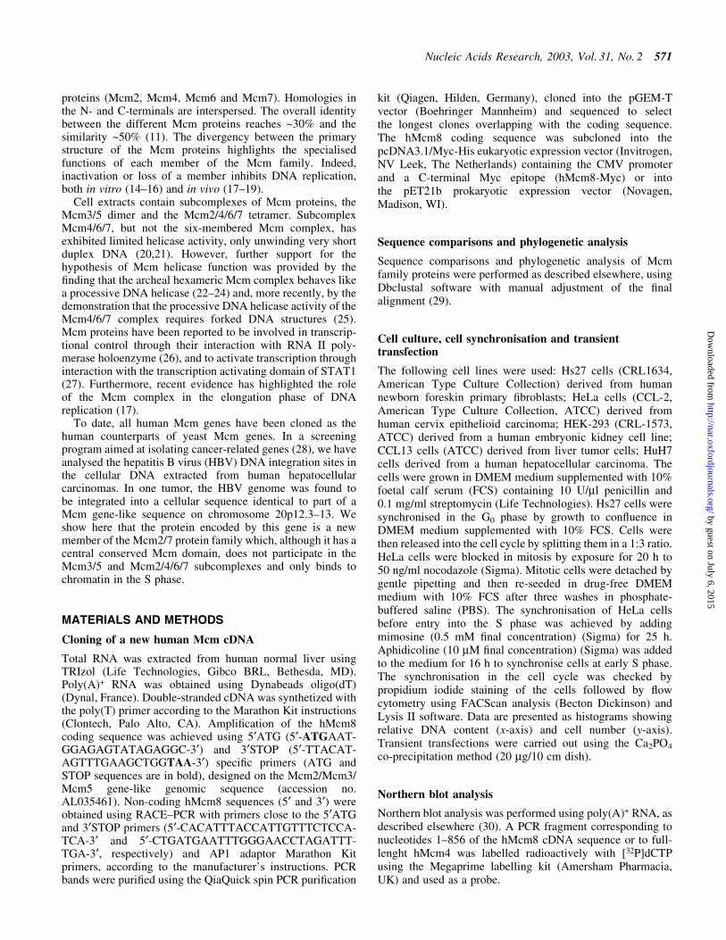
Nucleic Acids Research, 2003, Vol. 31, No. 2 571
by guest on July 6, 2015http://nar.oxfordjournals.org/
Dow
nloaded from

Production of anti-hMcm8 antibody and recombinantprotein expression in Escherichia coli
Polyclonal antibody was raised against a synthetic hMcm8N-terminal oligopeptide (amino acids 2±17: NGEYRGRGF-GRGRFQS) derived from the hMcm8 predicted proteinsequence. The 16 amino acid peptide was synthesised andcoupled to keyhole limpet haemocyanin. An aliquot of 250 mgof peptide was emulsi®ed in Freund's complete adjuvant (forthe ®rst injection) and subsequently in incomplete adjuvant(for the three other injections) and injected into New Zealandrabbits at 14 day intervals. The speci®c antibody waspuri®ed by af®nity chromatography (Hi-Trap SepharoseNHS column).
BL21/Lys E.coli were transformed using the hMcm8-pET21b construct. Expression of the recombinant proteinwas induced with 0.1 mM isopropyl-b-D-thiogalactopyrano-side (IPTG) at 37°C for 3 h. His-tagged recombinantprotein (His-hMcm8) was then puri®ed by chromatographyon a NTA±agarose column (Qiagen), according to themanufacturer's instructions.
Immunoprecipitation and western blotting
For immunoprecipitation, 5 3 106 exponentially growingHeLa cells were washed three times with PBS, scraped andresuspended in ice-cold hypotonic buffer A (1 mM HEPES,pH 7.3, with a mixture of protease inhibitors). After 20 minincubation on a wheel at 4°C and homogenisation with aDounce homogeniser (100 strokes), cells were centrifuged at20 000 g for 30 min at 4°C. The cell extract was then clearedby two more centrifugations (20 000 g at 4°C for 30 min). Thesupernatant was converted to buffer B (10 mM HEPES, pH 7.3,50 mM NaCl, 10% glycerol) and incubated at 4°C for 2 h withprotein A±Sepharose beads (Boehringer Mannheim) and anti-hMcm4 (0.2 mg/ml) or anti-hMcm3 (0.24 mg/ml) antibody(31). After ®ve washes with 1 ml buffer B, 23 SDS±Laemmlibuffer was added and the samples were vortexed, boiled for5 min at 100°C and used for western blotting.
Lysates were loaded at an equal protein concentration,subjected to 8% SDS±PAGE and transferred onto Immobilon-P+ membranes (Millipore, Bedford, MA). The ®lter wasblocked with TBS±Tween buffer (10 mM Tris±HCl, pH 7.5,100 mM NaCl, 0.2% Tween 20) containing 5% non-fat driedmilk, incubated for 1 h with the primary antibody (anti-hMcm8, 1:500; anti-Mcm2±7 antibodies, 1:1000 dilution) andwashed three times with TBS±Tween buffer. After incubationfor 1 h with the secondary antibody coupled to horseradishperoxidase (1:2000 dilution; Amersham), three TBS±Tweenwashes were performed. The blots were revealed using theECL Kit (Amersham Pharmacia) and exposed to Biomax-ML®lm (Kodak).
Immuno¯uorescence
HeLa cells were grown on coverslips, ®xed with 3.7%paraformaldehyde for 10 min and permeabilised with 0.2%Triton X-100 for 2 min. Non-speci®c binding sites wereblocked with 10% goat serum (Sigma). Immunostaining wasperformed with anti-hMcm8 antibody (diluted 1:50) for 1 h.After three PBS washes, immunodetection was carried outusing secondary antibody conjugated to ¯uorescein isothio-cyanate (FITC) diluted 1:200. After washing three times with
PBS, the nuclei were stained with 7-amino-actinomycin D (7-AAD) (Sigma) and the coverslips were mounted and analysedusing ¯uorescence microscopy (Olympus BX60) and confocallaser scanning microscopy (Zeiss LSM 510).
Chromatin binding assay
HeLa cells (7 3 105) were harvested during the exponentialphase of growth and incubated for 30 min on ice in apermeabilisation buffer (10 mM HEPES±KOH, pH 7.5, 7 mMMgCl2, 1.5 mM KCl, 1 mM DTT and 1% Triton X-100) with amixture of protease inhibitors (Boehringer). After centrifuga-tion (2000 g for 30 min at 4°C) the pellet was resuspended inthe same volume of permeabilisation buffer with 0, 0.1, 0.2,0.5, 1 or 1.5 M NaCl and then incubated on ice for 30 min.After centrifugation at 20 000 g for 30 min at 4°C, identicalvolumes of the supernatant were blotted.
RESULTS
Cloning of the cDNA encoding the hMcm8 protein
In a human hepatocellular carcinoma tissue, we identi®ed theintegration of HBV DNA next to a 485 bp sequence identical toa part of the GenBank genomic clone HS967N21 on chromo-some 20p12.3±13 (28). FGENES, GENSCAN, CpG island andpoly(A) feature analysis of this genomic clone by the SangerCentre chromosome 20 mapping group showed that this part ofthe sequence could correspond to a hypothetical gene encodingfor a novel Mcm2/Mcm3/Mcm5 family member.
We cloned the cDNA sequence (ATG±STOP) of hMcm8from human normal liver poly(A)+ RNA using RT±PCR andobtained the 5¢ and 3¢ non-coding regions by RACE±PCR. Thetotal length of hMcm8 cDNA is 2523 bases (ATG±STOP).Analysis of the hMcm8 cDNA and its amino acid sequenceshowed a central Mcm conserved domain (Mcm box)containing the Mcm signature IDEFDKM and the WalkerA-type NTP-binding site GXXGXGKS (SupplementaryMaterial, Fig. S1). hMcm8 protein also contains a zinc ®ngermotif CX2CX18CX4C, but neither a classical SV40 large Tantigen type of nuclear localisation signal (NLS) nor abipartite type of NLS. The overall content of the basic residuesis 12.3%. We have made the mRNA sequence of hMcm8available in the EMBL Nucleotide Sequence Database(accession no. AJ439063).
The longest open reading frame encodes an 840 amino acidprotein highly homologous with P1/Mcm2±7 protein familymembers (Fig. S1). The homology between hMcm8 proteinand other hMcm is 61.1% (hMcm4), 59.3% (hMcm2), 55.7%(hMcm3), 55.0% (hMcm5), 54.5% (hMcm6) and 52.1%(hMcm7). The identity between hMcm8 and other hMcm is26.5% (hMcm2), 26.4% (hMcm4), 22.6% (hMcm3), 24.0%(hMcm5), 21.7% (hMcm6) and 21.3% (hMcm7). The highesthomology between hMcm8 and other hMcms was found in thecentral region of 210 amino acids (residues 401±611), whichcontains the Walker A-type NTP-binding site `GXXGXGKS'and the Mcm signature sequence IDEFDKM.
hMcm8 is conserved during evolution
Sequence comparison and phylogenetic analysis of the Mcmprotein family and its new member, hMcm8, was performedusing Dbclustal software with manual adjustment of the ®nal
572 Nucleic Acids Research, 2003, Vol. 31, No. 2
by guest on July 6, 2015http://nar.oxfordjournals.org/
Dow
nloaded from

alignment (29). The tree shown in Figure 1 is based on full-length protein sequences. A reduced version of the multiplealignment excluding the variable N- and C-termini resulted ina similar tree (data not shown). Homologues of hMcm8protein were found in Encephalitozoon cuniculi, Leishmaniamajor, Arabidopsis thaliana and Drosophila. Furthermore, wewere able to identify an EST sequence of Xenopus laevis and apeptide sequence of Mus musculus highly homologous tothe hMcm8 sequence. We could not ®nd any yeast orCaenorhabditis elegans homologues of the hMcm8 protein.The accession numbers of the Mcm sequences used for thisanalysis and the sequence alignments are available on the website www-igbmc.u-strasbg.fr/BioInfo/Mcm.
Characterisation of anti-hMcm8 antibodies
In order to detect the protein expression of hMcm8, weproduced a polyclonal antibody raised against a 16 amino acid
synthetic peptide located in the N-terminal part of the protein(amino acids 2±17). This antibody was then characterised bysubcloning the hMcm8 cDNA sequence into a prokaryotic anda eukaryotic expression vector. The anti-hMcm8 antibodyrecognised the recombinant hMcm8 protein produced inbacteria (Fig. 2A) or overexpressed as a myc-tagged protein inthe CCL13 cell line (data not shown).
To rule out any cross-reaction between the anti-hMcm8antibody and human Mcm2±7 proteins, we performed awestern blot analysis using HeLa total cell extract, and thenhybridised the stripes corresponding to parallel wells usinganti-hMcm8 antibody or antibodies against other known Mcmproteins. The anti-hMcm8 antibody detected a single 95 kDaband (Fig. 2C), and no other band corresponding to the size ofthe other hMcm was detected after longer exposures (data notshown). The hMcm of a most similar size was hMcm4, with amolecular weight of 97 kDa; however, the hMcm4 band
Figure 1. Phylogenetic tree of Mcm protein sequences. The bootstrapped non-rooted tree was created from the NJ algorithm implemented in the Clustal soft-ware with manual adjustment of the ®nal alignment. The bootstrap values of the branch-root of each Mcm sub-family are shown when these values are higherthan 800 over the 1000 bootstraps performed.
Nucleic Acids Research, 2003, Vol. 31, No. 2 573
by guest on July 6, 2015http://nar.oxfordjournals.org/
Dow
nloaded from

appeared as a double band corresponding to the differentiallyphosphorylated forms of hMcm4 (32), and the unique size ofhMcm8 was slightly smaller than the lower band of hMcm4.Furthermore, the adsorption of anti-hMcm8 antibody with the16 amino acid synthetic peptide (used to raise the antibody)abolished detection of both the recombinant and endogenoushMcm8 proteins (Fig. 2B). We therefore concluded that thepolyclonal anti-hMcm8 antibody speci®cally recognises thehMcm8 protein.
Using this antibody, we detected a unique, endogenous95 kDa band in ®broblasts (Hs27) and tumor-derived humancell lines (HeLa, HEK-293 and Huh7) as well as in normalhuman liver and skeletal muscle (Fig. 2D), which could beabolished by the adsorption test (data not shown). It isnoteworthy that the accumulation of hMcm8 protein washigher in a proliferative, liver-derived, tumor cell line (HuH7)than in the corresponding non-proliferative tissue (normalliver).
hMcm8 mRNA is expressed during late G1 to S phase
Cells from the human ®broblast cell line Hs27 were densityarrested in G0, trypsinised when no mitosis was apparent andthen released by re-seeding cells at a lower density. Flowcytometric analysis revealed that 93% of the cells werearrested in G0. Under these conditions, 35% of cells enteredthe S phase 24 h after release and 28% reached the G2/M phase32 h later (Fig. 3A). The mRNA of both hMcm4, used as acontrol, and hMcm8 were absent in cells analysed 0, 6 and 12 hafter release (Fig. 3A and B). hMcm8 and hMcm4 mRNA
accumulation were ®rst observed 18 h after release andcontinued to increase during the S and G2/M phases (Fig. 3B).Since it is known that hMcm4 accumulation occurs in G1 (andnot in G0) and since the accumulation patterns of hMcm8 andhMcm4 were identical, we conclude that, like for hMcm4,hMcm8 mRNA accumulation begins in G1 and continues intothe S and G2/M phases of the cell cycle (Fig. 3B). This resultalso indicates that, in our experimental conditions, synchron-ised Hs27 cells are in G0 phase at 0, 6 and 12 h after release,and in G1 phase 18 h after release.
hMcm8 protein levels remain stable during the cell cycle
We then evaluated the level of hMcm8 protein during the cellcycle. All known mammalian Mcm proteins exhibit onlyminor variations in their protein levels during the cell cycle.This is probably because of their stability and low turnover(33,34), the exception being the hMcm6 protein, the level ofwhich rises dramatically during G1/S phase (35).
Using western blots, we analysed the total protein extractsof the same synchronised Hs27 cells used for northern blotanalysis, and re-hybridised the same western blot using anti-tubulin antibody as a reference for protein loading (Fig. 3D).hMcm8 protein expression is very low in G0 phase (0 and 12 hafter release) while its level increases as cells enter G1 andremains stable during the S and G2/M phases.
hMcm8 is a nuclear protein
Mammalian Mcm2±7 proteins are nuclear throughout the cellcycle. However, only Mcm2 and Mcm3 proteins contain a
Figure 2. Characterisation of the anti-hMcm8 antibody. (A) Immunoblotting with anti-hMcm8 antibody of E.coli whole cell lysates harbouring the hMcm8full-length prokaryotic expression vector after (+IPTG) or before (±IPTG) induction with IPTG. (B) Immunoblotting of the E.coli whole cell lysate expressingthe hMcm8 recombinant protein (lanes 1, 1¢, 2 and 2¢) and Hs27 whole cell lysate (30 mg) (lanes 3 and 3¢) with the anti-Mcm8 antibody. hMcm8protein detection without (lanes 1, 2 and 3) or with (lanes 1¢, 2¢ and 3¢) adsorption of the antibody using the 16 amino acid synthetic hMcm8 peptide.(C) Immunoblotting of the HeLa whole cell lysates (30 mg/lane) with human Mcm speci®c antibodies: anti-hMcm2 (top band) and anti-hMcm3, anti-hMcm4,anti-hMcm5, anti-hMcm8, anti-hMcm6 and anti-hMcm7. (D) Immunoblot analysis of whole cell lysates of ~1 3 105 cells (Hs27, HeLa, HEK-293 and Huh7cell lines) or 50 mg human tissue protein extracts (NL, human normal liver; SM, human skeletal muscle) with anti-hMcm8 antibody.
574 Nucleic Acids Research, 2003, Vol. 31, No. 2
by guest on July 6, 2015http://nar.oxfordjournals.org/
Dow
nloaded from

NLS. In fact, the murine homologues of Mcm4±Mcm7 aremainly cytoplasmic when overexpressed in COS cells, whiletheir co-expression with Mcm2 or Mcm3 leads to a shift intheir localisation from the cytoplasm to the nucleus (36).
Furthermore, the association of Mcm proteins in a hexamericcomplex is a prerequisite for their nuclear localisation in yeast(37). It is thus plausible that the Mcm2 and Mcm3 NLS isinvolved in translocating the complex from the cytoplasm tothe nucleus.
The subcellular localisation of hMcm8 was determined bylaser confocal microscopy. Non-transfected Hs27 or HeLacells were stained by immuno¯uorescence (FITC) with theanti-hMcm8 antibody. The nuclei were counterstained with7-AAD. As shown in Figure 4, the anti-hMcm8 signal wasmainly nuclear, while no speci®c staining was observed usingthe preimmune serum (negative control). The absence of aNLS in the hMcm8 protein sequence suggests that this proteincould be carried to the nucleus as a result of its interaction withother nuclear proteins. The variable pattern of hMcm8punctate nuclear staining could be related to differences inthe expression level of hMcm8 and/or to changes in theproportion of structure-bound versus nucleosolic hMCM8 indifferent phases of the cell cycle.
hMcm8 protein has a structure-bound and a nucleosolicfraction
It has been shown that Mcm proteins comprise two nuclearfractions, a nucleosolic soluble fraction, which is released bypermeabilising the nuclei with non-ionic detergents, and achromatin-bound fraction which can be extracted by increas-ing the salt concentration (38). In order to determine whetherthis is also the case for hMcm8, HeLa cells in the exponentialphase of growth were treated with a hypotonic buffercontaining Triton X-100. After centrifugation, we obtainedthe soluble hMcm8 nucleosolic fraction in the supernatant andthe structure-bound fraction in the pellet. As indicated in
Figure 3. Expression of the hMcm8 mRNA and protein during the cellcycle. Hs27 human ®broblast cells were arrested in G0 by con¯uence andreleased by splitting cells in a 1:3 ratio. (A) Cell cycle pattern analysed by¯ow cytometry. Cells were harvested at the indicated times. (B) Northernblot of total RNA (20 mg) probed with a 32P-labelled hMcm8 or hMcm4probe and ethidium bromide staining of the RNA before blotting (bottom).Positions of 28S and 18S rRNA are indicated on the right. (C) Whole celllysates (15 mg) of the same Hs27 population were analysed by immuno-blotting with the anti-hMcm8 antibody. The bottom part of the same blotwas revealed with anti-tubulin antibody used as reference for proteinloading.
Figure 4. hMcm8 protein localises to the nucleus. Paraformaldehyde-®xedasynchronous HeLa cells were permeabilised with 0.2% Triton X-100, andincubated with either the anti-hMcm8 antibody or the preimmune serum (ascontrol) followed by incubation with FITC-conjugated donkey anti-rabbitIgG antibodies. After immunostaining, the nuclei were stained with 7-AADand the samples were analysed by confocal microscopy.
Nucleic Acids Research, 2003, Vol. 31, No. 2 575
by guest on July 6, 2015http://nar.oxfordjournals.org/
Dow
nloaded from

Figure 5A, a fraction of total hMcm8 is structure-bound.Subsequent extractions of this fraction using a buffercontaining increasing salt concentrations dissociated hMcm8from the chromatin (Fig. 5A, NaCl). Therefore, hMcm8localises to the nucleus, where it exists as soluble, nucleosolicand structure-bound protein.
hMcm8 protein is structure-bound in the S phase, afterthe establishment of DNA replication forks
Chromatin binding of Mcm2±Mcm7 proteins is tightlyregulated during the cell cycle (1). In order to analyse thebehaviour of hMcm8 during the cell cycle, HeLa cells weresynchronised with nocodazole and then released to enter thecell cycle. At 0, 5, 10 and 20 h after release, cells were
harvested and equal amounts of nucleosolic and structure-bound fractions were blotted (Fig. 5B). The relative propor-tions of nucleosolic and structure-bound hMcm3 and hMcm8protein were assessed by image analysis of speci®c bandsobtained by western blot. The majority of hMcm3 protein wasnucleosolic in cells arrested in the G2/M phase by nocodazole.An increasing fraction of hMcm3 protein was structure-boundstarting from G1 phase (29% of total protein), reaching 52% atS phase. The hMcm8 protein was also nucleosolic innocodazole-arrested cells. However, unlike hMcm3, thestructure-bound fraction of hMcm8 in G1 phase was barelydetectable (0.2%). A fraction (11%) of hMcm8 protein startedto be structure-bound in S phase, then reaching 32% of thetotal protein. In order to consider this point in greater depth,we repeated the same experiment using HeLa cells treatedwith 0.5 mM mimosine for 25 h or with aphidicoline (Fig. 5C).Under these conditions, mimosine blocks the cells in the lateG1 phase (thus before the establishment of active replicationforks) (39). Aphidicoline is an inhibitor of DNA polymerase aand it suppresses the migration of the replication fork, but itdoes not prevent the initiation of replication. Therefore,aphidicoline causes a cell cycle arrest in the beginning of the Sphase. In mimosine-arrested cells, an important fraction (20%)of hMcm3 is structure-bound. Strikingly, all the hMcm8protein is nucleosolic in these cells, indicating that, unlikeother Mcm proteins, hMcm8 is not structure-bound in cellsarrested in late G1 phase. In cells arrested at the beginning of Sphase with aphidicoline, 29% of hMcm3 protein is structure-bound, while only a small fraction (9%) of hMcm8 protein isstructure-bound. These data are consistent with the view that,at variance with the other members of the Mcm protein family,hMcm8 is structure-bound in the early S phase after theestablishment of replication forks, and not in G1 phase.
hMcm8 protein does not co-immunoprecipitate with theMcm2±7 complex
Mcm2±7 proteins are found as a heterohexameric complex inHeLa cells. In addition to the Mcm2±7 complex (31), Mcm2/4/6/7, Mcm4/6/7 and Mcm3/5 subcomplexes have also beenidenti®ed (21,40). To investigate whether hMcm8 can asso-ciate with one of these Mcm subcomplexes, we performedimmunoprecipitation and western blot analyses using expo-nentially growing, asynchronous HeLa cell extracts andpolyclonal antibodies speci®c to hMcm3 or hMcm4. Asshown in Figure 6A, anti-hMcm3 antibody immunoprecipi-tated both hMcm3 and hMcm5, but not hMcm8. Using theanti-hMcm4 antibody, we were able to immunoprecipitehMcm4, hMcm6 and hMcm2, but not hMcm8 (Fig. 6B).Although we do not rule out any possible interaction betweenhMcm8 and other Mcm proteins, hMcm8 does not seem to beinvolved in the Mcm2±7 complex.
DISCUSSION
Our understanding of the mechanisms involved in eukaryoticreplication origins has been greatly improved in the last fewyears and several key proteins have been identi®ed. Thesestudies have revealed a sophisticated programme determiningthat initiation occurs only once at every cell division and®xing the time at which individual origins ®re during the Sphase (1,3,5). However, this ®nely tuned process has not yet
Figure 5. Binding to chromatin of hMcm8 protein occurs in S phase.(A) The Triton X-100-extractable fraction (Tx100/SN) of HeLa cells wasobtained as described (see Materials and Methods). The remaining pelletfraction (Tx100/P) was extracted with permeabilization buffer adjusted tothe indicated NaCl concentrations. Immunoblot analysis with anti-hMcm8antibodies of equal amounts of each fraction is shown. (B) HeLa cells weresynchronised with nocodazole and then released to enter the cell cycle. Attime points 0, 5, 10 and 20 h after release cells were harvested and equalamounts of nucleosolic (Sol) and chromatin-bound (Str) fractions were blot-ted. The relative proportions of nucleosolic and structure-bound hMcm3 andhMcm8 protein were assessed by image analysis of speci®c bands obtainedby western blot. (C) HeLa cells were synchronised with nocodazole (blockin G2/M), mimosine (block in late G1) or aphidicoline (block in early S) andequal amounts of nucleosolic (Sol) and chromatin-bound (Str) fractionswere blotted. Quanti®cations were performed as described in (B).
576 Nucleic Acids Research, 2003, Vol. 31, No. 2
by guest on July 6, 2015http://nar.oxfordjournals.org/
Dow
nloaded from

been completely elucidated, and the identi®cation of newplayers may throw further light on its complexity.
Mcm genes were ®rst discovered in budding and ®ssionyeast as genes required to enable progression through the celldivision cycle or to maintain minichromosomes containingautonomously replicating sequences (ARS). Characterisationof the Saccharomyces cerevisiae genes Mcm2 (41), Mcm3(42) and Cdc46/Mcm5 (43,44) suggested their involvement inDNA replication. The protein family grew rapidly withcloning of the Schizosaccharomyces pombe genes cdc21/Mcm4 (45) and mis5/Mcm6 (46) and of the S.cerevisiaeCdc47/Mcm7 genes (47). The evolutionarily preservedhomologues of the six yeast Mcm proteins were rapidlycharacterized in fruit ¯y, frog, mouse and humans. No otherhomologues of these genes have been identi®ed in thecomplete genome of S.cerevisiae. Therefore, since the repli-cation initiation mechanisms seem to be highly conservedfrom yeast to humans, it has been hypothesised that the sixmembers of the Mcm family did not diversify any further (11).
In contrast with this view, we describe here a new memberof the human Mcm protein family, hMcm8. It contains thetypical domains and functional motifs of the Mcm2±7 proteinfamily. hMcm8 mRNA accumulates during the G1/S±G2/Mphase, while its protein product is detectable throughout thenormal cell cycle. Its nuclear localisation and association withchromatin in a cell cycle-regulated manner suggest its role inDNA replication. Database searches have revealed that theMcm8 protein is evolutionarily conserved, thus allowing theprediction of its potentially relevant biological function.
However, the precise role of hMcm8 remains unknown. Ourresults show that hMcm8 does not participate in the Mcm3±5and Mcm2/4/6/7 subcomplexes, and that its binding tochromatin is restricted to the S phase. The nuclear localisationof hMcm8 protein in the absence of a NLS suggests thathMcm8 may have a nuclear protein partner(s) which couldcause its translocation to the nucleus. However, the data weobtained in HeLa cells show that this partner is not a knownmember of the Mcm family. At present, we can only speculateas to the biological function of Mcm8. It may be involved inconformational changes to the Mcm complex during the Sphase and/or in the melting of origin DNA. Mcm proteins havealso been shown to be involved in transcriptional control (26)and activation (27). Furthermore, the role of the Mcm complexin the elongation phase of DNA replication has recently beendemonstrated (17). hMcm8 may participate in one or severalof these activities, and further study is required to explore itsfunction.
It has recently been shown that the down-regulation of Mcmproteins is a common downstream mechanism causing a lossof proliferative capacity in human cells (48). In fact, a loss ofreplication licensing coincides with the commitment ofprecursor cells to a speci®c lineage of differentiation or withcell quiescence. Mcm proteins have been proposed as newbiomarkers capable of detecting G1 cells `with growthpotential'. These properties of Mcm proteins highlight theirpotential usefulness in cancer biology as tumor markers(49,50), and in studies focusing on stem cell differentiation.Our preliminary data show that hMcm8 expression is higher ina proliferative cell line than in the corresponding (liver) non-proliferative tissue. However, these results need to beextended to a wider range of tissues and completed by cellcycle analyses.
In this context, it is also noteworthy that we recentlyreported on an in vivo hMcm8 gene mutation occurringselectively in a tumorous tissue and not in adjacent, non-tumorous cells, suggesting that hMcm8 mutations may play arole in cell transformation (see the mutated hMcm8 proteinexpressed in the tumor in Fig. 3C) (28).
In conclusion, we have described a new member of theMcm protein family, evolutionarily conserved, which, unlikethe other known Mcm proteins, displays restriction of nuclearstructure binding to the S phase and does not participate in theMcm complex. Although further work is required to elucidatethe biological function of this new gene, its mutation inclonally expanded human tumor cells suggests its potentialimpact on the control of cell proliferation.
SUPPLEMENTARY MATERIAL
Supplementary Material is available at NAR Online.
ACKNOWLEDGEMENTS
This work was supported by grants from INSERM (InstitutNational de Sante et Recherche MeÂdicale), LNC (LigueNationale contre le Cancer) and ARC (Association pour laRecherche contre le Cancer).
Figure 6. hMcm8 protein does not immunoprecipitate with the P1/Mcm2±7protein complexes. The extracts of exponentially growing HeLa cells wereprepared as described. An aliquot of one-tenth of the extract was loaded asinput. The extracts were immunoprecipitated with anti-hMcm3 or anti-hMcm4 antibodies or with rabbit immunoglobin as control (control).(A) Immunoprecipitation using anti-hMcm3 antibody. The blots wererevealed with anti-hMcm3, anti-hMcm5 or anti-hMcm8 antibodies.(B) Immunoprecipitation using anti-hMcm4 antibody. The blots wererevealed with anti-hMcm4, anti-hMcm6, anti-hMcm2 or anti-hMcm-8antibodies.
Nucleic Acids Research, 2003, Vol. 31, No. 2 577
by guest on July 6, 2015http://nar.oxfordjournals.org/
Dow
nloaded from
REFERENCES
1. Ritzi,M. and Knippers,R. (2000) Initiation of genome replication:assembly and disassembly of replication-competent chromatin. Gene,245, 13±20.
2. Tye,B.K. (2000) Insights into DNA replication from the third domain oflife. Proc. Natl Acad. Sci. USA, 97, 2399±2401.
3. Lei,M. and Tye,B.K. (2001) Initiating DNA synthesis: from recruiting toactivating the MCM complex. J. Cell Sci., 114, 1447±1454.
4. Donaldson,A.D. and Blow,J.J. (1999) The regulation of replication originactivation. Curr. Opin. Genet. Dev., 9, 62±68.
5. Tye,B.K. (1999) MCM proteins in DNA replication. Annu. Rev.Biochem., 68, 649±686.
6. Coleman,T.R., Carpenter,P.B. and Dunphy,W.G. (1996) The XenopusCdc6 protein is essential for the initiation of a single round of DNAreplication in cell-free extracts. Cell, 87, 53±63.
7. Rowles,A., Chong,J.P., Brown,L., Howell,M., Evan,G.I. and Blow,J.J.(1996) Interaction between the origin recognition complex and thereplication licensing system in Xenopus. Cell, 87, 287±296.
8. Romanowski,P., Madine,M.A., Rowles,A., Blow,J.J. and Laskey,R.A.(1996) The Xenopus origin recognition complex is essential for DNAreplication and MCM binding to chromatin. Curr. Biol., 6,1416±1425.
9. Maiorano,D., Moreau,J. and Mechali,M. (2000) XCDT1 is required forthe assembly of pre-replicative complexes in Xenopus laevis. Nature,404, 622±625.
10. Nishitani,H., Lygerou,Z., Nishimoto,T. and Nurse,P. (2000) The Cdt1protein is required to license DNA for replication in ®ssion yeast. Nature,404, 625±628.
11. Kearsey,S.E. and Labib,K. (1998) MCM proteins: evolution, propertiesand role in DNA replication. Biochim. Biophys. Acta, 1398, 113±136.
12. Maiorano,D., Lemaitre,J.M. and Mechali,M. (2000) Stepwise regulatedchromatin assembly of MCM2-7 proteins. J. Biol. Chem., 275,8426±8431.
13. Koonin,E.V. (1993) A common set of conserved motifs in a vast varietyof putative nucleic acid-dependent ATPases including MCM proteinsinvolved in the initiation of eukaryotic DNA replication. Nucleic AcidsRes., 21, 2541±2547.
14. Chong,J.P., Mahbubani,H.M., Khoo,C.Y. and Blow,J.J. (1995)Puri®cation of an MCM-containing complex as a component of the DNAreplication licensing system. Nature, 375, 418±421.
15. Kubota,Y., Mimura,S., Nishimoto,S., Takisawa,H. and Nojima,H. (1995)Identi®cation of the yeast MCM3-related protein as a component ofXenopus DNA replication licensing factor. Cell, 81, 601±609.
16. Madine,M.A., Khoo,C.Y., Mills,A.D. and Laskey,R.A. (1995) MCM3complex required for cell cycle regulation of DNA replication invertebrate cells. Nature, 375, 421±424.
17. Labib,K., Tercero,J.A. and Dif¯ey,J.F. (2000) Uninterrupted MCM2-7function required for DNA replication fork progression. Science, 288,1643±1647.
18. Todorov,I.T., Attaran,A. and Kearsey,S.E. (1995) BM28, a humanmember of the MCM2-3-5 family, is displaced from chromatin duringDNA replication. J. Cell Biol., 129, 1433±1445.
19. Fujita,M., Kiyono,T., Hayashi,Y. and Ishibashi,M. (1996) Inhibition ofS-phase entry of human ®broblasts by an antisense oligomer againsthCDC47. Biochem. Biophys. Res. Commun., 219, 604±607.
20. Lee,J.K. and Hurwitz,J. (2000) Isolation and characterization of variouscomplexes of the minichromosome maintenance proteins ofSchizosaccharomyces pombe. J. Biol. Chem., 275, 18871±18878.
21. Ishimi,Y. (1997) A DNA helicase activity is associated with an MCM4,-6 and -7 protein complex. J. Biol. Chem., 272, 24508±24513.
22. Kelman,Z., Lee,J.K. and Hurwitz,J. (1999) The single minichromosomemaintenance protein of Methanobacterium thermoautotrophicum DeltaHcontains DNA helicase activity. Proc. Natl Acad. Sci. USA, 96,14783±14788.
23. Chong,J.P., Hayashi,M.K., Simon,M.N., Xu,R.M. and Stillman,B. (2000)A double-hexamer archaeal minichromosome maintenance protein is anATP-dependent DNA helicase. Proc. Natl Acad. Sci. USA, 97,1530±1535.
24. Shechter,D.F., Ying,C.Y. and Gautier,J. (2000) The intrinsic DNAhelicase activity of Methanobacterium thermoautotrophicum delta Hminichromosome maintenance protein. J. Biol. Chem., 275,15049±15059.
25. Lee,J.K. and Hurwitz,J. (2001) Processive DNA helicase activity of theminichromosome maintenance proteins 4, 6 and 7 complex requiresforked DNA structures. Proc. Natl Acad. Sci. USA, 98, 54±59.
26. Yankulov,K., Todorov,I., Romanowski,P., Licatalosi,D., Cilli,K.,McCracken,S., Laskey,R. and Bentley,D.L. (1999) MCM proteins areassociated with RNA polymerase II holoenzyme. Mol. Cell. Biol., 19,6154±6163.
27. DaFonseca,C.J., Shu,F. and Zhang,J.J. (2001) Identi®cation of tworesidues in MCM5 critical for the assembly of MCM complexes andStat1-mediated transcription activation in response to IFN-gamma.Proc. Natl Acad. Sci. USA, 98, 3034±3039.
28. Gozuacik,D., Murakami,Y., Saigo,K., Chami,M., Mugnier,C.,Lagorce,D., Okanoue,T., Urashima,T., Brechot,C. andPaterlini-Brechot,P. (2001) Identi®cation of human cancer-related genesby naturally occurring Hepatitis B Virus DNA tagging. Oncogene, 20,6233±6240.
29. Thompson,J.D., Plewniak,F., Thierry,J. and Poch,O. (2000) DbClustal:rapid and reliable global multiple alignments of protein sequencesdetected by database searches. Nucleic Acids Res., 28, 2919±2926.
30. Church,G.M. and Gilbert,W. (1984) Genomic sequencing. Proc. NatlAcad. Sci. USA, 81, 1991±1995.
31. Richter,A. and Knippers,R. (1997) High-molecular-mass complexes ofhuman minichromosome-maintenance proteins in mitotic cells.Eur. J. Biochem., 247, 136±141.
32. Musahl,C., Schulte,D., Burkhart,R. and Knippers,R. (1995) A humanhomologue of the yeast replication protein Cdc21. Interactions with otherMcm proteins. Eur. J. Biochem., 230, 1096±1101.
33. Schulte,D., Burkhart,R., Musahl,C., Hu,B., Schlatterer,C., Hameister,H.and Knippers,R. (1995) Expression, phosphorylation and nuclearlocalization of the human P1 protein, a homologue of the yeast Mcm 3replication protein. J. Cell Sci., 108, 1381±1389.
34. Musahl,C., Holthoff,H.P., Lesch,R. and Knippers,R. (1998) Stability ofthe replicative Mcm3 protein in proliferating and differentiating humancells. Exp. Cell Res., 241, 260±264.
35. Tsuruga,H., Yabuta,N., Hosoya,S., Tamura,K., Endo,Y. and Nojima,H.(1997) HsMCM6: a new member of the human MCM/P1 family encodesa protein homologous to ®ssion yeast Mis5. Genes Cells, 2, 381±399.
36. Kimura,H., Ohtomo,T., Yamaguchi,M., Ishii,A. and Sugimoto,K. (1996)Mouse MCM proteins: complex formation and transportation to thenucleus. Genes Cells, 1, 977±993.
37. Pasion,S.G. and Forsburg,S.L. (1999) Nuclear localization ofSchizosaccharomyces pombe Mcm2/Cdc19p requires MCM complexassembly. Mol. Biol. Cell, 10, 4043±4057.
38. Kimura,H., Nozaki,N. and Sugimoto,K. (1994) DNA polymerase alphaassociated protein P1, a murine homolog of yeast MCM3, changes itsintranuclear distribution during the DNA synthetic period. EMBO J., 13,4311±4320.
39. Krude,T. (1999) Mimosine arrests proliferating human cells before onsetof DNA replication in a dose-dependent manner. Exp. Cell Res., 247,148±159.
40. Burkhart,R., Schulte,D., Hu,D., Musahl,C., Gohring,F. and Knippers,R.(1995) Interactions of human nuclear proteins P1Mcm3 and P1Cdc46.Eur. J. Biochem., 228, 431±438.
41. Yan,H., Gibson,S. and Tye,B.K. (1991) Mcm2 and Mcm3, two proteinsimportant for ARS activity, are related in structure and function.Genes Dev., 5, 944±957.
42. Gibson,S.I., Surosky,R.T. and Tye,B.K. (1990) The phenotype of theminichromosome maintenance mutant mcm3 is characteristic of mutantsdefective in DNA replication. Mol. Cell. Biol., 10, 5707±5720.
43. Hennessy,K.M., Clark,C.D. and Botstein,D. (1990) Subcellularlocalization of yeast CDC46 varies with the cell cycle. Genes Dev., 4,2252±2263.
44. Hennessy,K.M., Lee,A., Chen,E. and Botstein,D. (1991) A group ofinteracting yeast DNA replication genes. Genes Dev., 5, 958±969.
45. Coxon,A., Maundrell,K. and Kearsey,S.E. (1992) Fission yeast cdc21+belongs to a family of proteins involved in an early step of chromosomereplication. Nucleic Acids Res., 20, 5571±5577.
46. Takahashi,K., Yamada,H. and Yanagida,M. (1994) Fission yeastminichromosome loss mutants mis cause lethal aneuploidy andreplication abnormality. Mol. Biol. Cell, 5, 1145±1158.
47. Dalton,S. and Whitbread,L. (1995) Cell cycle-regulated nuclear importand export of Cdc47, a protein essential for initiation of DNA replicationin budding yeast. Proc. Natl Acad. Sci. USA, 92, 2514±2518.
578 Nucleic Acids Research, 2003, Vol. 31, No. 2
by guest on July 6, 2015http://nar.oxfordjournals.org/
Dow
nloaded from

48. Stoeber,K., Tlsty,T.D., Happer®eld,L., Thomas,G.A., Romanov,S.,Bobrow,L., Williams,E.D. and Williams,G.H. (2001) DNA replicationlicensing and human cell proliferation. J. Cell Sci., 114,2027±2041.
49. Freeman,A., Morris,L.S., Mills,A.D., Stoeber,K., Laskey,R.A.,Williams,G.H. and Coleman,N. (1999) Minichromosome maintenance
proteins as biological markers of dysplasia and malignancy. Clin. CancerRes., 5, 2121±2132.
50. Todorov,I.T., Werness,B.A., Wang,H.Q., Buddharaju,L.N.,Todorova,P.D., Slocum,H.K., Brooks,J.S. and Huberman,J.A. (1998)HsMCM2/BM28: a novel proliferation marker for human tumors andnormal tissues. Lab. Invest., 78, 73±78.
Nucleic Acids Research, 2003, Vol. 31, No. 2 579
by guest on July 6, 2015http://nar.oxfordjournals.org/
Dow
nloaded from