Identification of Mycobacterium tuberculosis complex based on amplification and sequencing of the...
7
Nakajima et al. BMC Infectious Diseases 2010, 10:118 http://www.biomedcentral.com/1471-2334/10/118 Open Access RESEARCH ARTICLE BioMed Central © 2010 Nakajima et al; licensee BioMed Central Ltd. This is an Open Access article distributed under the terms of the Creative Commons Attribution License (http://creativecommons.org/licenses/by/2.0), which permits unrestricted use, distribution, and reproduction in any medium, provided the original work is properly cited. Research article Identification of Mycobacterium tuberculosis clinical isolates in Bangladesh by a species distinguishable multiplex PCR Chie Nakajima* 1 , Zeaur Rahim †2 , Yukari Fukushima 1 , Isamu Sugawara 3 , Adri GM van der Zanden 4 , Aki Tamaru 5 and Yasuhiko Suzuki †1 Abstract Background: Species identification of isolates belonging to the Mycobacterium tuberculosis complex (MTC) seems to be important for the appropriate treatment of patients, since M. bovis is naturally resistant to a first line anti-tuberculosis (TB) drug, pyrazinamide, while most of the other MTC members are susceptible to this antimicrobial agent. A simple and low-cost differentiation method was needed in higher TB burden countries, such as Bangladesh, where the prevalence of M. bovis among people or cattle has not been investigated. Methods: Genetic regions cfp32, RD9 and RD12 were chosen as targets for a species distinguishable multiplex PCR and the system was evaluated with twenty reference strains of mycobacterial species including non-tubercular mycobacteria (NTM). A total of 350 clinical MTC isolates obtained in Bangladesh were then analyzed with this multiplex PCR. Results: All of the MTC reference strains gave expected banding patterns and no non-specific amplifications were observed in the NTM strains. Out of 350 clinical isolates examined by this method, 347 (99.1%) were positive for all of the cfp32, RD9 and RD12 and determined as M. tuberculosis. Two isolates lacked cfp32 PCR product and one lacked RD12, however, those three samples were further examined and identified as M. tuberculosis by the sequence analyses of hsp65 and gyrB. Conclusions: The MTC-discrimination multiplex PCR (MTCD-MPCR) developed in this study showed high specificity and was thought to be very useful as a routine test because of its simplicity. In the current survey, all the 350 MTC isolates obtained from Bangladesh TB patients were determined as M. tuberculosis and no other MTC were detected. This result suggested the general TB treatment regimen including pyrazinamide to be the first choice in Bangladesh. Background Mycobacterium tuberculosis complex (MTC), including M. tuberculosis, M. bovis, M. africanum, M. microti, M. pinnipedii, M. caprae, "M. canettii" and other closely related strains, is a group of causative agents for human and animal tuberculosis (TB) [1,2]. Although the myco- bacterial species in MTC are highly similar to each other in DNA level, MTC members differ widely in terms of host tropism, phenotype and pathogenicity [1,3-5]. No further differentiation is usually performed with isolates determined as MTC, however, it seems to be important in some cases for the appropriate management of patients or for an epidemiological purpose. Especially, in the case of M. bovis infection, to identify the species in the early stage of diagnosis is essential to avoid inappropriate treat- ment, since M. bovis is naturally resistant to a major anti- TB drug, pyrazinamide [3,4,6], and the standard regimen including this drug has to be altered. Several rapid identification methods using nucleic acid amplification techniques have been developed and used for the diagnosis of TB [7-9], however, they do not differ- entiate M. tuberculosis from other members of MTC. Recent comparative genomic analyses have provided valuable information on the region of difference (RD) in * Correspondence: [email protected] 1 Department of Global epidemiology, Hokkaido University Research Center for Zoonosis Control, Kita20-Nishi10, Kita-ku, Sapporo 001-0020, Japan † Contributed equally Full list of author information is available at the end of the article
-
Upload
independent -
Category
Documents
-
view
3 -
download
0
Transcript of Identification of Mycobacterium tuberculosis complex based on amplification and sequencing of the...

Nakajima et al. BMC Infectious Diseases 2010, 10:118http://www.biomedcentral.com/1471-2334/10/118
Open AccessR E S E A R C H A R T I C L E
Research articleIdentification of Mycobacterium tuberculosis clinical isolates in Bangladesh by a species distinguishable multiplex PCRChie Nakajima*1, Zeaur Rahim†2, Yukari Fukushima1, Isamu Sugawara3, Adri GM van der Zanden4, Aki Tamaru5 and Yasuhiko Suzuki†1
AbstractBackground: Species identification of isolates belonging to the Mycobacterium tuberculosis complex (MTC) seems to be important for the appropriate treatment of patients, since M. bovis is naturally resistant to a first line anti-tuberculosis (TB) drug, pyrazinamide, while most of the other MTC members are susceptible to this antimicrobial agent. A simple and low-cost differentiation method was needed in higher TB burden countries, such as Bangladesh, where the prevalence of M. bovis among people or cattle has not been investigated.
Methods: Genetic regions cfp32, RD9 and RD12 were chosen as targets for a species distinguishable multiplex PCR and the system was evaluated with twenty reference strains of mycobacterial species including non-tubercular mycobacteria (NTM). A total of 350 clinical MTC isolates obtained in Bangladesh were then analyzed with this multiplex PCR.
Results: All of the MTC reference strains gave expected banding patterns and no non-specific amplifications were observed in the NTM strains. Out of 350 clinical isolates examined by this method, 347 (99.1%) were positive for all of the cfp32, RD9 and RD12 and determined as M. tuberculosis. Two isolates lacked cfp32 PCR product and one lacked RD12, however, those three samples were further examined and identified as M. tuberculosis by the sequence analyses of hsp65 and gyrB.
Conclusions: The MTC-discrimination multiplex PCR (MTCD-MPCR) developed in this study showed high specificity and was thought to be very useful as a routine test because of its simplicity. In the current survey, all the 350 MTC isolates obtained from Bangladesh TB patients were determined as M. tuberculosis and no other MTC were detected. This result suggested the general TB treatment regimen including pyrazinamide to be the first choice in Bangladesh.
BackgroundMycobacterium tuberculosis complex (MTC), includingM. tuberculosis, M. bovis, M. africanum, M. microti, M.pinnipedii, M. caprae, "M. canettii" and other closelyrelated strains, is a group of causative agents for humanand animal tuberculosis (TB) [1,2]. Although the myco-bacterial species in MTC are highly similar to each otherin DNA level, MTC members differ widely in terms ofhost tropism, phenotype and pathogenicity [1,3-5]. Nofurther differentiation is usually performed with isolates
determined as MTC, however, it seems to be importantin some cases for the appropriate management of patientsor for an epidemiological purpose. Especially, in the caseof M. bovis infection, to identify the species in the earlystage of diagnosis is essential to avoid inappropriate treat-ment, since M. bovis is naturally resistant to a major anti-TB drug, pyrazinamide [3,4,6], and the standard regimenincluding this drug has to be altered.
Several rapid identification methods using nucleic acidamplification techniques have been developed and usedfor the diagnosis of TB [7-9], however, they do not differ-entiate M. tuberculosis from other members of MTC.Recent comparative genomic analyses have providedvaluable information on the region of difference (RD) in
* Correspondence: [email protected] Department of Global epidemiology, Hokkaido University Research Center for Zoonosis Control, Kita20-Nishi10, Kita-ku, Sapporo 001-0020, Japan† Contributed equallyFull list of author information is available at the end of the article
BioMed Central© 2010 Nakajima et al; licensee BioMed Central Ltd. This is an Open Access article distributed under the terms of the Creative CommonsAttribution License (http://creativecommons.org/licenses/by/2.0), which permits unrestricted use, distribution, and reproduction inany medium, provided the original work is properly cited.
Nakajima et al. BMC Infectious Diseases 2010, 10:118http://www.biomedcentral.com/1471-2334/10/118
Page 2 of 7
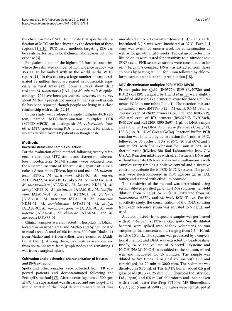
the chromosome of MTC to indicate that specific identi-fication of MTC can be achieved by the detection of theseregions [1-3,10]. PCR-based methods targeting RDs canbe easily performed in local clinical laboratories with lowexpense [3].
Bangladesh is one of the highest TB burden countries,where the estimated number of TB incidence in 2007 was353,000 to be ranked sixth in the world in the WHOreport [11]. In this country, a large number of cattle esti-mated 23 million heads are reared in households espe-cially in rural areas [12]. Some surveys about drugresistant M. tuberculosis [13,14] or M. tuberculosis epide-miology [15] have been performed, however, no surveyabout M. bovis prevalence among humans as well as cat-tle has been reported though people are living in a closerelationship with cattle [12,16].
In this study, we developed a simple multiplex-PCR sys-tem, named MTC-discrimination multiplex PCR(MTCD-MPCR), to distinguish M. tuberculosis fromother MTC species using RDs, and applied it for clinicalisolates derived from TB patients in Bangladesh.
MethodsBacterial strains and sample collectionFor the evaluation of the method, following twenty refer-ence strains, four MTC strains and sixteen nontubercu-lous mycobacteria (NTM) strains, were obtained fromthe Research Institute of Tuberculosis, Japan Anti-Tuber-culosis Association (Tokyo, Japan) and used: M. tubercu-losis H37Rv, M. africanum KK13-02, M. microtiATCC19422, M. bovis BCG Tokyo, M. avium JATA51-01,M. intracellulare JATA52-01, M. kansasii KK21-01, M.xenopi KK42-02, M. fortuitum JATA61-01, M. lentifla-vum JATA9N-01, M. simiae KK23-01, M. gordonaeJATA33-01, M. marinum JATA22-01, M. asiaticumKK24-01, M. scrofulaceum JATA31-01, M. szulgaiJATA32-01, M. nonchromogenicum JATA45-01, M. mal-moense JATA47-01, M. chelonae JATA62-01 and M.abscessus JATA63-01.
Clinical samples were collected in hospitals in Dhaka,located in an urban area, and Matlab and Sylhet, locatedin rural areas. A total of 350 isolates, 300 from Dhaka, 41from Matlab and 9 from Sylhet, were examined (Addi-tional file 1). Among them, 327 isolates were derivedfrom sputa, 22 were from lymph nodes and remaining 1was from a surgical injury.
Cultivation and biochemical characterization of isolates and DNA extractionSputa and other samples were collected from TB sus-pected patients and decontaminated following thePetropff 's method [17]. After a centrifugation at 500 rpmat 4°C, the supernatant was discarded and one loop-full (5mm diameter of the loop) decontaminated pellet was
inoculated onto 2 Lowenstein-Jensen (L-J) slants each.Inoculated L-J slants were incubated at 37°C. Each L-Jslant was examined once a week for contamination aswell as for growth until 8 weeks. Typical mycobacterium-like colonies were tested for sensitivity to p-nitrobenzoic(PNB) acid. PNB sensitive strains were considered to beM. tuberculosis complex. DNA was extracted from thosecolonies by heating at 95°C for 5 min followed by chloro-form extraction and ethanol precipitation [18].
MTC-discrimination multiplex PCR (MTCD-MPCR)Primer pairs for cfp32 (Rv0577), RD9 (Rv2073c) andRD12 (Rv3120) designed by Huard et al [3] were slightlymodified and used as a primer mixture for three simulta-neous PCRs in one tube (Table 1). The reaction mixturecontained 1 mM dNTPs (0.25 mM each), 0.5 M betaine,750 nM each of cfp32 primers (Rv0577F and Rv0577R),250 nM each of RD primers (Rv2073cF, Rv2073cR,Rv3120F and Rv3120R (390-369)), 1 μL of DNA sampleand 1 U of GoTaq DNA Polymerase (Promega Corp., WI,U.S.A.) in 20 μL of Green GoTaq Reaction Buffer. PCRreaction was initiated by denaturation for 1 min at 96°C,followed by 35 cycles of 10 s at 96°C, 20 s at 60°C and 1min at 72°C with final extension for 5 min at 72°C in athermalcycler (iCycler, Bio Rad Laboratories Inc., CA,U.S.A.). Reaction mixtures with M. tuberculosis DNA andwithout template DNA were also run simultaneously withsamples every time as a positive control and a negativecontrol to evaluate the MTCD-MPCR system. The prod-ucts were electrophoresed in 2.0% agarose gel in TAEbuffer, and stained with ethidium bromide.
The sensitivity of the method was determined usingserially diluted purified genomic-DNA solutions, ten-folddilution from 5 ng/μL to 50 fg/μL, extracted from M.tuberculosis H37Rv and M. bovis BCG Tokyo. For thespecificity study, the concentration of the DNA solutionfrom each reference strain was adjusted to 5 ng/μL andused.
A detection study from sputum samples was performedwith M. tuberculosis H37Rv spiked sputa. Serially dilutedbacteria were spiked into healthy volunteer's sputumsamples to final concentrations ranging from 1.5 × 10/mLto 1.5 × 10⁶/mL. The sputum was processed by a conven-tional method and DNA was extracted by bead-beating.Briefly, twice the volume of N-acetyl-L-cysteine andNaOH (NALC-NaOH) was added to the sputum, mixedwell and incubated for 15 minutes. The sample wasdiluted to five times its original volume with PBS andcentrifuged for 20 min at 3000 rpm. The sediment wasdissolved in 0.75 mL of Tris-EDTA buffer, added 0.5 g ofglass beads (0.15 - 0.25 mm, Fuji Chemical Industry Co.,Ltd., Japan) and 0.5 mL of chloroform and then shakenwith a bead-beater (FastPrep FP100A, MP Biomedicals,U.S.A.) for 5 min at 5500 rpm. Tubes were centrifuged at
Nakajima et al. BMC Infectious Diseases 2010, 10:118http://www.biomedcentral.com/1471-2334/10/118
Page 3 of 7
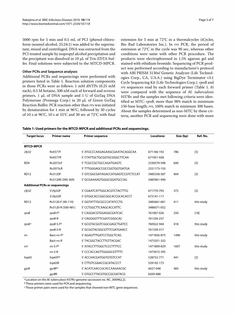
Table 1: Used primers for the MTCD-MPCR and additional PCRs and sequencings.
Target locus Primer name Primer sequence Locationa Size (bp) Ref. No.
MTCD-MPCR
cfp32 Rv0577F 5' ATGCCCAAGAGAAGCGAATACAGGCAA 671166-192 786 [3]
Rv0577R 5' CTATTGCTGCGGTGCGGGCTTCAA 671951-928
RD9 Rv2073cF 5' TCGCCGCTGCCAGATGAGTC 2330579-598 600 [3]
Rv2073cR 5' TTTGGGAGCCGCCGGTGGTGATGA 2331173-150
RD12 Rv3120F 5' GTCGGCGATAGACCATGAGTCCGTCTCCAT 3485558-587 404 [3]
Rv3120R (390-369) 5' GCGAAAAGTGGGCGGATGCCAG 3485961-940
Additional PCRs or sequencings
cfp32 3'cfp32F 5' CGAATCATTGGCACGTCTACTTTG 671770-793 372 [2]
3'cfp32R 5' GTGGCACCGGCGGCACCGCACACCT 672141-117
RD12 Rv3120-F (90-110) 5' GGTATTTGCGCCCATATCCTG 3485661-681 411 this study
Rv3120-R (500-481) 5' CCTGGCTTCAAGCACCATTC 3486071-052
rpoB rpoB-Fb 5' CAGGACGTGGAGGCGATCAC 761007-026 250 [18]
rpoB-R 5' CAGGGGTTTCGATCGGGCAC 761256-237
rpoB c rpoB-S-Fb 5' GCGTACGGTCGGCGAGCTGATCC 760922-944 418 this study
rpoB-S-R 5' GCGGTACGGCGTTTCGATGAACC 761339-317
rrs Bact-rrs-Fb 5' AGAGTTTGATCCTGGCTCAG 1471856-875 1496 this study
Bact-rrs-R 5' TACGGCTACCTTGTTACGAC 1473351-332
rrsc rrs-S-Fb 5' ATACCTTTGGCTCCCTTTTCC 1471809-829 1607 this study
rrs-S-R 5' CCCACCAGTTGGGGCGTTTTC 1473415-395
hsp65 hsp65Fb 5' ACCAACGATGGTGTGTCCAT 528752-771 441 [2]
hsp65R 5' CTTGTCGAACCGCATACCCT 529192-173
gyrB gyrBFb 5' ACATCAACCGCACCAAGAACGC 6027-048 483 this study
gyrBR 5' GTGCCTTACGTGCCGCGATACG 6509-488
a Location on the M. tuberculosis H37Rv genome (accession no. NC_000962.2).b These primers were used for PCR and sequencing.c Those primer pairs were used for the samples that showed non-MTC gene sequences.
5000 rpm for 5 min and 0.5 mL of PCI (phenol-chloro-form-isoamyl alcohol, 25:24:1) was added to the superna-tant, mixed and centrifuged. DNA was extracted from thePCI treated sample by isopropyl alcohol precipitation andthe precipitant was dissolved in 10 μL of Tris-EDTA buf-fer. Final solutions were subjected to the MTCD-MPCR.
Other PCRs and Sequence analysesAdditional PCRs and sequencings were performed withprimers listed in Table 1. Reaction solution componentsin those PCRs were as follows: 1 mM dNTPs (0.25 mMeach), 0.5 M betaine, 500 nM each of forward and reverseprimers, 1 μL of DNA sample and 1 U of GoTaq DNAPolymerase (Promega Corp.) in 20 μL of Green GoTaqReaction Buffer. PCR reaction other than rrs was initiatedby denaturation for 1 min at 96°C, followed by 30 cyclesof 10 s at 96°C, 10 s at 55°C and 30 sec at 72°C with final
extension for 5 min at 72°C in a thermalcycler (iCycler,Bio Rad Laboratories Inc.). In rrs PCR, the period ofextension at 72°C in the cycle was 90 sec, whereas otherconditions were same with other PCR procedure. Theproducts were electrophoresed in 1.5% agarose gel andstained with ethidium bromide. Sequencing of PCR prod-uct was performed according to manufacturer's protocolwith ABI PRISM 3130xl Genetic Analyzer (Life Technol-ogies Corp., CA, U.S.A.) using BigDye Terminator v3.1Cycle Sequencing Kit (Life Technologies Corp.). rpoB andrrs sequences read by each forward primer (Table 1. b)were compared with the sequence of M. tuberculosisH37Rv and the samples met following criteria were iden-tified as MTC: rpoB, more than 98% match in minimum150-base length; rrs, 100% match in minimum 300 bases.About the samples determined as non-MTC by these cri-teria, another PCR and sequencing were done with more
Nakajima et al. BMC Infectious Diseases 2010, 10:118http://www.biomedcentral.com/1471-2334/10/118
Page 4 of 7
MTC specific primers (Table 1. c) with the same criteria.The samples identified as MTC with those specific prim-ers were determined as mixed-culture samples. Speciesidentifications by hsp65 or gyrB sequences were doneaccording to previous publications [2,19,20].
Ethical ClearanceThe original research project was approved by theResearch Review Committee and Ethical Review Com-mittee of the International Centre for Diaddroeal DiseaseResearch, Bangladesh (ICDDR, B). Signed informed con-sent was obtained from each patient and volunteerrecruited for the study.
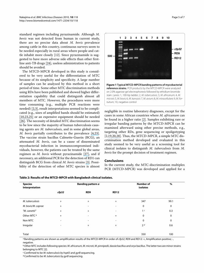
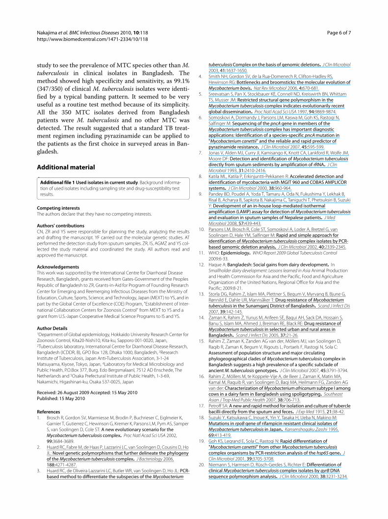
Results and discussionMTCD-MPCRThree genetic regions were selected as the targets for themultiplex PCR: cfp32, RD9 and RD12. cfp32 is an MTC-restricted gene and used to confirm isolates to belong toMTC [3,21]. RD9 is the region that can be found in onlyM. tuberculosis and "M. canettii", and RD12 is found in allMTC members except M. bovis, M. caprae and "M. canet-tii" [2,3]. By a trial with several patterns of primer con-centrations, the best combination was determined to be750 nM each of cfp32 primers and 250 nM each of RD9and RD12 primers in the multiplex PCR. With this PCR,an isolate possessing all the three regions can be identi-fied as M. tuberculosis whereas a strain showing only oneamplified band, cfp32, will be classified as M. bovis or M.caprae (Figure 1, Table 2). Other banding patterns areinterpreted as described in Table 2. "M. canettii" isanother MTC that clinical isolates reported so far werenaturally pyrazinamide resistant [6,19]. Thus, this PCRsystem was thought to be useful for the discrimination ofMTC species, especially for the screening of naturallypyrazinamide-resistant species with only one PCR reac-tion per sample.
The sensitivities of the method were determined as 500fg genomic DNA for M. tuberculosis (H37Rv) and 50 fgfor M. bovis (BCG Tokyo), which were assumed to beequivalent to 100 bacilli and 10 bacilli, respectively (datanot shown). The specificity of the MTCD-MPCR wasconfirmed with DNA templates extracted from mycobac-terial reference strains. Typical gel electrophoresis resultsof the MTC and NTM strains are shown in Figure 1. Allof the PCR products from the MTC strains gave expectedbanding patterns in correct sizes (Table 1). On the con-trary, no bands were obtained from the NTM samples.These results demonstrated the specificity and applicabil-ity of the MTCD-MPCR for the differentiation of M.tuberculosis in clinical isolates.
In the study using bacterium-spiked sputa, the detec-tion limit of M. tuberculosis was 1.5 × 10⁴ cells in 1 mL ofsputum (data not shown). This bacterial concentration is
similar or a little higher than the detection limit of thebacteria by the Zeal-Nelsen staining. This means speciesdiscrimination by the MTCD-MPCR is applicable forclinical samples if the sputum is diagnosed as smear posi-tive. The discrepancy of the detection limit between puri-fied DNA and bacterium-spiked sputum seems to dependon the extraction or purification procedure, which is tobe improved.
Discrimination of MTC in Bangladesh clinical isolatesA total of 350 clinical isolates obtained in Bangladeshwere analyzed to see the prevalence of MTC species otherthan M. tuberculosis. All samples were subjected to rpoBor rrs sequencing to confirm as MTC [22]. By thissequence analysis, 18 samples were revealed as mixturesof MTC and other bacteria, mainly mycobacterial spe-cies. Those mixed samples were also subjected to theMTCD-MPCR study.
Out of the 350 isolates, 347 (99.1%) showed the typicalbanding pattern of M. tuberculosis in the electrophoresisby the MTCD-MPCR (Table 2). Among three remainingisolates, one (0.3%, isolate ATP138) lacked RD12 bandshowing "M. canettii" pattern and two (0.6%, isolateM2000 and S2247) did not have cfp32 band, presented asirregular in Table 2. Those samples were further analyzedto detect the target region, cfp32 or RD12, with other setsof primers (Table 1) [2]. With this trial, sample S2247gave an expected sized cfp32 band, suggesting a partialdeletion or a mutation in the primer-binding site. Othertwo samples, ATP138 and M2000, still failed to amplifythe expected DNA fragment indicating that larger dele-tion event had occurred in the target genomic area.ATP138, the "M. canettii" pattern sample, was subjectedto hsp65 sequencing and confirmed not to be "M. canet-tii" [2,19]. These three isolates were further confirmed tobe M. tuberculosis by the sequencing of gyrB [20]. No iso-lates out of the 350 lacked RD9 band, concordant with theobservations by former researchers [2,10,23], possiblyindicating the high stability of this region in M. tuberculo-sis. The lack of cfp32 in an MTC isolate has been reportedby Huard et al [2] with a similar incidence (1/125, 0.8%)to the current study. Some of the mixed-culture samplesshowed correct but faint banding patterns, suggesting aninhibitory effect of contaminated DNA. The result indi-cated that despite its decreased sensitivity, the MTCD-MPCR was able to detect MTC from mixed-culture sam-ples, which sometimes are observed in primary cultures[8].
All of the 350 MTC isolates obtained from clinicalspecimens in Bangladesh were M. tuberculosis and noother MTC species were detected. This information isvery helpful for the management of patients to determinetreatment regimens. Patients suffered from MTC in sur-veyed area in Bangladesh can possibly be subjected to the
Nakajima et al. BMC Infectious Diseases 2010, 10:118http://www.biomedcentral.com/1471-2334/10/118
Page 5 of 7
standard regimen including pyrazinamide. Although M.bovis was not detected from human in current study,there are no precise data about M. bovis prevalenceamong cattle in this country, continuous surveys seem tobe needed especially in rural areas where people and cat-tle inhabit more closely [12]. Since pyrazinamide is sug-gested to have more adverse side effects than other first-line anti-TB drugs [24], useless administration to patientsshould be avoided.
The MTCD-MPCR developed in this study is consid-ered to be very useful for the differentiation of MTCbecause of its simplicity and specificity. A large numberof samples can be analyzed by this method in a shortperiod of time. Some other MTC discrimination methodsusing RDs have been published and showed higher differ-entiation capability that could distinguish almost allmembers of MTC. However, the procedures were moretime consuming (e.g., multiple PCR reactions wereneeded) [2,3], result interpretations seemed to be compli-cated (e.g., sizes of amplified bands should be estimated)[10,23,25] or an expensive equipment should be needed[26]. The necessity of detailed MTC discrimination seemsto be low since the majority of human tuberculosis caus-ing agents are M. tuberculosis, and in some global areas,M. bovis partially contributes to the prevalence [4,23].The vaccine strain bacillus Calmette-Guerin (BCG), anattenuated M. bovis, can be a cause of disseminatedmycobacterial infection in immunocompromised indi-viduals, however, the patients can be treated by the sameregimen as M. bovis without pyrazinamide [27], and ifnecessary, an additional PCR for the detection of RD1 candistinguish BCG from clinical M. bovis strains [3]. Possi-bility of the detection of other MTC species is almost
negligible in routine laboratory diagnoses, except for thecases in some African countries where M. africanum canbe found in a higher ratio [2]. Samples exhibiting rare orirregular banding patterns by the MTCD-MPCR can beexamined afterward using other precise methods, e.g.,targeting other RDs, gene sequencing or spoligotyping[2,19,20,28]. Thus, the MTCD-MPCR, a simple MTC dis-crimination method developed and evaluated in thisstudy seemed to be very useful as a screening tool forclinical isolates to distinguish M. tuberculosis from M.bovis for the prompt decision of treatment regimen.
ConclusionsIn the current study, the MTC-discrimination multiplexPCR (MTCD-MPCR) was developed and applied for a
Table 2: Results of the MTCD-MPCR with Bangladesh clinical isolates.
Species Interpretation
Banding pattern a Number of isolates
%
cfp32 RD9 RD12
M. tuberculosis + + + 347 99.1
M. bovis/M. caprae + - - 0 0
"M. canettii" + + - 1 c 0.3
Other MTC b + - + 0 0
Non MTC - - - 0 0
Irregular - + + 2 d 0.6
Total 350 100
a Banding patterns are shown as amplification results of the MTCD-MPCR in order of cfp32, RD9 and RD12: +, Amplification positive; -, negative.b Other MTC includes following species: M. africanum, M. microti, M. pinnipedii, dassie bacillus and oryx bacillus. The latter two are minor strains belonging to MTC [2].c Confirmed to be M. tuberculosis by hsp65 and gyrB sequencing.d Confirmed to be M. tuberculosis by gyrB sequencing.
Figure 1 Typical MTCD-MPCR banding patterns of mycobacterial reference strains. PCR products by the MTCD-MPCR were analyzed on 2.0% agarose gel electrophoresis followed by ethidium bromide stain. Lanes: 1, 100-bp ladder; 2, M. tuberculosis; 3, M. africanum; 4, M. microti; 5, M. bovis; 6, M. kansasii; 7, M. avium; 8, M. intracellulare; 9, M. for-tuitum; 10, negative control
1 2 3 4 5 6 7 8 9 10
500 ---
-- cfp32
-- RD9
RD12-- RD12
Nakajima et al. BMC Infectious Diseases 2010, 10:118http://www.biomedcentral.com/1471-2334/10/118
Page 6 of 7
study to see the prevalence of MTC species other than M.tuberculosis in clinical isolates in Bangladesh. Themethod showed high specificity and sensitivity, as 99.1%(347/350) of clinical M. tuberculosis isolates were identi-fied by a typical banding pattern. It seemed to be veryuseful as a routine test method because of its simplicity.All the 350 MTC isolates derived from Bangladeshpatients were M. tuberculosis and no other MTC wasdetected. The result suggested that a standard TB treat-ment regimen including pyrazinamide can be applied tothe patients as the first choice in surveyed areas in Ban-gladesh.
Additional material
Competing interestsThe authors declare that they have no competing interests.
Authors' contributionsCN, ZR and YS were responsible for planning the study, analyzing the resultsand drafting the manuscript. YF carried out the molecular genetic studies. ATperformed the detection study from sputum samples. ZR, IS, AGMZ and YS col-lected the study material and coordinated the study. All authors read andapproved the manuscript.
AcknowledgementsThis work was supported by the International Centre for Diarrhoeal Disease Research, Bangladesh, grants received from Gates-Government of the Peoples Republic of Bangladesh to ZR, Grants-in-Aid for Program of Founding Research Center for Emerging and Reemerging Infectious Diseases from the Ministry of Education, Culture, Sports, Science, and Technology, Japan (MEXT) to YS, and in part by the Global Center of Excellence (COE) Program, "Establishment of Inter-national Collaboration Centers for Zoonosis Control" from MEXT to YS and a grant from U.S.-Japan Cooperative Medical Science Programs to IS and YS.
Author Details1Department of Global epidemiology, Hokkaido University Research Center for Zoonosis Control, Kita20-Nishi10, Kita-ku, Sapporo 001-0020, Japan, 2Tuberculosis laboratory, International Centre for Diarrhoeal Disease Research, Bangladesh (ICDDR, B), GPO Box 128, Dhaka 1000, Bangladesh, 3Research Institute of Tuberculosis, Japan Anti-Tuberculosis Association, 3-1-24 Matsuyama, Kiyose, Tokyo, Japan, 4Laboratory for Medical Microbiology and Public Health, P.O.Box 377, Burg. Edo Bergsmalaanl, 7512 AD Enschede, The Netherlands and 5Osaka Prefectural Institute of Public Health, 1-3-69, Nakamichi, Higashinari-ku, Osaka 537-0025, Japan
References1. Brosch R, Gordon SV, Marmiesse M, Brodin P, Buchrieser C, Eiglmeier K,
Garnier T, Gutierrez C, Hewinson G, Kremer K, Parsons LM, Pym AS, Samper S, van Soolingen D, Cole ST: A new evolutionary scenario for the Mycobacterium tuberculosis complex. Proc Natl Acad Sci USA 2002, 99:3684-3689.
2. Huard RC, Fabre M, de Haas P, Lazzarini LC, van Soolingen D, Cousins D, Ho JL: Novel genetic polymorphisms that further delineate the phylogeny of the Mycobacterium tuberculosis complex. J Bacteriology 2006, 188:4271-4287.
3. Huard RC, de Oliveira Lazzarini LC, Butler WR, van Soolingen D, Ho JL: PCR-based method to differentiate the subspecies of the Mycobacterium
tuberculosis Complex on the basis of genomic deletions. J Clin Microbiol 2003, 41:1637-1650.
4. Smith NH, Gordon SV, de la Rua-Domenech R, Clifton-Hadley RS, Hewinson RG: Bottlenecks and broomsticks: the molecular evolution of Mycobacterium bovis. Nat Rev Microbiol 2006, 4:670-681.
5. Sreevatsan S, Pan X, Stockbauer KE, Connell ND, Kreiswirth BN, Whittam TS, Musser JM: Restricted structural gene polymorphism in the Mycobacterium tuberculosis complex indicates evolutionarily recent global dissemination. Proc Natl Acad Sci USA 1997, 94:9869-9874.
6. Somoskovi A, Dormandy J, Parsons LM, Kaswa M, Goh KS, Rastogi N, Salfinger M: Sequencing of the pncA gene in members of the Mycobacterium tuberculosis complex has important diagnostic applications: Identification of a species-specific pncA mutation in "Mycobacterium canettii" and the reliable and rapid predictor of pyrazinamide resistance. J Clin Microbiol 2007, 45:595-599.
7. Jonas V, Alden MJ, Curry JI, Kamisango K, Knott CA, Lankford R, Wolfe JM, Moore DF: Detection and identification of Mycobacterium tuberculosis directly from sputum sediments by amplification of rRNA. J Clin Microbiol 1993, 31:2410-2416.
8. Katila ML, Katila P, Erkinjuntti-Pekkanen R: Accelerated detection and identification of mycobacteria with MGIT 960 and COBAS AMPLICOR systems. J Clin Microbiol 2000, 38:960-964.
9. Pandey BD, Poudel A, Yoda T, Tamaru A, Oda N, Fukushima Y, Lekhak B, Risal B, Acharya B, Sapkota B, Nakajima C, Taniguchi T, Phetsuksiri B, Suzuki Y: Development of an in-house loop-mediated isothermal amplification (LAMP) assay for detection of Mycobacterium tuberculosis and evaluation in sputum samples of Nepalese patients. J Med Microbiol 2008, 57:439-443.
10. Parsons LM, Brosch R, Cole ST, Somoskovi A, Loder A, Bretzel G, van Soolingen D, Hale YM, Salfinger M: Rapid and simple approach for identification of Mycobacterium tuberculosis complex isolates by PCR-based genomic deletion analysis. J Clin Microbiol 2002, 40:2339-2345.
11. WHO: Epidemiology. WHO Report 2009 Global Tuberculosis Control 2009:6-33.
12. Haque A: Bangladesh: Social gains from dairy development. In Smallholder dairy development: Lessons learned in Asia Animal Production and Health Commission for Asia and the Pacific, Food and Agriculture Organization of the United Nations, Regional Office for Asia and the Pacific; 2009:8-21.
13. Storla DG, Rahim Z, Islam MA, Plettner S, Begum V, Myrvang B, Bjune G, Rønnild E, Dahle UR, Mannsåker T: Drug resistance of Mycobacterium tuberculosis in the Sunamganj District of Bangladesh. Scand J Infect Dis 2007, 39:142-145.
14. Zaman K, Rahim Z, Yunus M, Arifeen SE, Baqui AH, Sack DA, Hossain S, Banu S, Islam MA, Ahmed J, Breiman RE, Black RE: Drug resistance of Mycobacterium tuberculosis in selected urban and rural areas in Bangladesh. Scand J Infect Dis 2005, 37:21-26.
15. Rahim Z, Zaman K, Zanden AG van der, Möllers MJ, van Soolingen D, Raqib R, Zaman K, Begum V, Rigouts L, Portaels F, Rastogi N, Sola C: Assessment of population structure and major circulating phylogeographical clades of Mycobacterium tuberculosis complex in Bangladesh suggests a high prevalence of a specific subclade of ancient M. tuberculosis genotypes. J Clin Microbiol 2007, 45:3791-3794.
16. Rahim Z, Möllers M, te Koppele-Vije A, de Beer J, Zaman K, Matin MA, Kamal M, Raquib R, van Soolingen D, Baqi MA, Heilmann FG, Zanden AG van der: Characterization of Mycobacterium africanum subtype I among cows in a dairy farm in Bangladesh using spoligotyping. Southeast Asian J Trop Med Public Health 2007, 38:706-713.
17. Petroff SA: A new and rapid method for isolation and culture of tubercle bacilli directly from the sputum and feces. J Exp Med 1915, 21:38-42.
18. Suzuki Y, Katsukawa C, Inoue K, Yin Y, Tasaka H, Ueba N, Makino M: Mutations in rpoB gene of rifampicin resistant clinical isolates of Mycobacterium tuberculosis in Japan. Kansenshogaku Zasshi 1995, 69:413-419.
19. Goh KS, Legrand E, Sola C, Rastogi N: Rapid differentiation of "Mycobacterium canettii" from other Mycobacterium tuberculosis complex organisms by PCR-restriction analysis of the hsp65 gene. J Clin Microbiol 2001, 39:3705-3708.
20. Niemann S, Harmsen D, Rüsch-Gerdes S, Richter E: Differentiation of clinical Mycobacterium tuberculosis complex isolates by gyrB DNA sequence polymorphism analysis. J Clin Microbiol 2000, 38:3231-3234.
Additional file 1 Used isolates in current study. Background informa-tion of used isolates including sampling site and drug-susceptibility test results.
Received: 26 August 2009 Accepted: 15 May 2010 Published: 15 May 2010This article is available from: http://www.biomedcentral.com/1471-2334/10/118© 2010 Nakajima et al; licensee BioMed Central Ltd. This is an Open Access article distributed under the terms of the Creative Commons Attribution License (http://creativecommons.org/licenses/by/2.0), which permits unrestricted use, distribution, and reproduction in any medium, provided the original work is properly cited.BMC Infectious Diseases 2010, 10:118
http://www.ncbi.nlm.nih.gov/entrez/query.fcgi?cmd=Retrieve&db=PubMed&dopt=Abstract&list_uids=9275218
http://www.ncbi.nlm.nih.gov/entrez/query.fcgi?cmd=Retrieve&db=PubMed&dopt=Abstract&list_uids=8408564
Nakajima et al. BMC Infectious Diseases 2010, 10:118http://www.biomedcentral.com/1471-2334/10/118
Page 7 of 7
21. Huard RC, Chitale S, Leung M, Lazzarini LC, Zhu H, Shashkina E, Laal S, Conde MB, Kritski AL, Belisle JT, Kreiswirth BN, Lapa e Silva JR, Ho JL: The Mycobacterium tuberculosis complex-restricted gene cfp32 encodes an expressed protein that is detectable in tuberculosis patients and is positively correlated with pulmonary interleukin-10. Infect Immun 2003, 71:6871-6883.
22. Kim BJ, Lee SH, Lyu MA, Kim SJ, Bai GH, Chae GT, Kim EC, Cha CY, Kook YH: Identification of mycobacterial species by comparative sequence analysis of the RNA polymerase gene (rpoB). J Clin Microbiol 1999, 37:1714-1720.
23. Das S, Das SC, Verma R: Occurrence of RD9 region and 500 bp fragment among clinical isolates of Mycobacterium tuberculosis and Mycobacterium bovis. Microbiol Immunol 2007, 51:231-234.
24. Yee D, Valiquette C, Pelletier M, Parisien I, Rocher I, Menzies D: Incidence of serious side effects from first-line antituberculosis drugs among patients treated for active tuberculosis. Am J Respir Crit Care Med 2003, 167:1472-1477.
25. Warren RM, Gey van Pittius NC, Barnard M, Hesseling A, Engelke E, de Kock M, Gutierrez MC, Chege GK, Victor TC, Hoal EG, van Helden PD: Differentiation of Mycobacterium tuberculosis complex by PCR amplification of genomic regions of difference. Int J Tuberc Lung Dis 2006, 10:818-822.
26. Pinsky BA, Banaei N: Multiplex real-time PCR assay for rapid identification of Mycobacterium tuberculosis complex members to the species level. J Clin Microbiol 2008, 46:2241-2246.
27. Rezai MS, Khotaei G, Mamishi S, Kheirkhah M, Parvaneh N: Disseminated Bacillus Calmette-Guerin infection after BCG vaccination. J Trop Pediatr 2008, 54:413-416.
28. Kamerbeek J, Schouls L, Kolk A, van Agterveld M, van Soolingen D, Kuijper S, Bunschoten A, Molhuizen H, Shaw R, Goyal M, van Embden J: Simultaneous detection and strain differentiation of Mycobacterium tuberculosis for diagnosis and epidemiology. J Clin Microbiol 1997, 35:907-914.
Pre-publication historyThe pre-publication history for this paper can be accessed here:http://www.biomedcentral.com/1471-2334/10/118/prepub
doi: 10.1186/1471-2334-10-118Cite this article as: Nakajima et al., Identification of Mycobacterium tubercu-losis clinical isolates in Bangladesh by a species distinguishable multiplex PCR BMC Infectious Diseases 2010, 10:118