Human papillomavirus viral load in predicting high-grade CIN in women with cervical smears showing...
6
ABSTRACT INTRODUCTION The clinical significance and management practices adopted in women with atypical sq- uamous cells (ASC) or low-grade squamous intraepithelial lesion (LSIL) have not been uniformly accepted. 1-8 Repeated cervical smears, addition of tests to detect human papillomavirus (HPV) DNA or referral of the women for immediate colposcopy are some of the different options, although they have clearly different cost-effectiveness. The pre- liminary results of the ASCUS/LSIL Triage Study (ALTS), 1 an American national prospec- tive randomized trial based on 3,488 women with atypical squamous cells of undetermined significance (ASCUS) and 1,572 women with LSIL, showed that hybrid capture II (HCII) testing for high-risk HPV DNA was fre- quently positive (around 85%) in women with LSIL. This therefore demonstrated the lim- ited usefulness of the HCII assay in the man- agement of these cases, because the vast ma- jority of these women would be referred for (unnecessary) colposcopy due to the positive HPV tests. Regarding the women with AS- CUS smears, Solomon et al. (2001) concluded that HCII testing for high-risk HPV DNA types was positive in 50.6% and should be a viable option for the management of these women. HPV DNA had greater sensitivity and specificity in detecting CIN3 (cervical intraepithelial neoplasia grade 3, carcinoma in situ or severe dysplasia) or above, in com- parison with a single additional cervical test showing ASCUS or above. 1 These results are not uniformly accepted, however. 4,9 Recently, the Bethesda system modified their ASCUS category to make it two-tier: ASC–US (of undetermined signifi- cance) and ASC-H (cannot exclude high-grade squamous intraepithelial lesion - HSIL). 10 According to the ASCCP (American So- ciety for Colposcopy and Cervical Pathology) 2001 Consensus Guidelines for the Manage- ment of Women with Cervical Cytological Abnormalities, 8 women with ASC-US should be managed using a program of two repeated cytology tests, immediate colposcopy or HPV DNA testing for high-risk HPV types. Women with ASC-H, LSIL, HSIL and atypi- cal glandular cells should be referred for im- mediate colposcopy evaluation, regardless of the result of HPV testing. However, HPV test- ing for routine clinical management of ASC- US cytological abnormalities is not yet war- ranted 9-11 and continues to be an investiga- tional tool. Furthermore, evaluation of the clinical effectiveness of HPV testing combined with cervical smears needs to be completed in developing countries. The aim of the present study was to shed further light on the issue and investigate whether HPV testing and viral load quantifi- cation are clinically useful tools for women who present with ASC or LSIL cervical smears in our settings in Brazil. MATERIAL AND METHODS Patients In this cross-sectional study, we analyzed a series of 119 women aged 16 to 63 (median of 31 years), referred for the Colposcopy Clinic due to an abnormal cervical smear consistent • André Luis Ferreira Santos • Sophie Françoise Mauricette Derchain • Marcos Roberto Martins • Luís Otávio Zanatta Sarian • Edson Zangiacome Martinez • Kari Juhani Syrjänen Human papillomavirus viral load in predicting high-grade CIN in women with cervical smears showing only atypical squamous cells or low-grade squamous intraepithelial lesion Department of Obstetrics and Gynecology, Universidade Estadual de Campinas, Campinas, and Universidade de Taubaté, Taubaté, São Paulo, Brazil, and in association with the National Health Institute, Rome, Italy Original Article Sao Paulo Med J 2003; 121(6):238-243. CONTEXT: Human papillomavirus (HPV) viral load may have an important role in predicting high-grade cervical intraepithelial neoplasia (CIN) in women with cervical smears showing atypical squamous cells or LSIL. OBJECTIVE: To determine whether the assessment of the viral load of high-risk HPV DNA is useful in predicting the detection of high-grade cervical intraepithelial neoplasia (CIN2 and 3) in women referred because of cervical smears showing only atypical squamous cells or LSIL. TYPE OF STUDY: Cross-sectional SETTING: Colposcopy Clinic in a University hospital. METHODS: A series of 119 women referred because of atypical squamous cells or LSIL between August 2000 and April 2001 were included. All women were subjected to a new cervical smear, HPV test- ing for the high-risk types using hybrid capture II (HCII), viral load measurement in relative light units (RLU) and colposcopy, with cervical biopsies (n = 97). Cervical lesions were graded using the CIN classification. RESULTS: Cervical biopsies revealed CIN2 or CIN3 in 11% of the cases, equally among women referred because of atypical squamous cells or LSIL. The HCII test was positive in 16% of women with atypical squamous cells and 52% of those with LSIL (OR = 5.8; 95% CI 1.4 to 26.7). There was strong corre- lation between CIN2 or CIN3 and positivity for HPV DNA when this group was compared with women with only CIN1 or normal cervix (OR = 7.8; 95% CI 1.5 to 53.4). In ROC analysis for HCII in diag- nosing CIN2 and CIN3, the area under the ROC curve was 0.784, and the viral load cutoff point of 10.0 RLU/cutoff presented 77% sensitivity and 73% specificity. Second cytology showing at least atypi- cal squamous cells did not accurately detect CIN2 or CIN3 (OR = 6.4; 95% CI 1.0 to 50.9). The sensitivities of the second cervical smear and HCII were similar, although the specificity of HCII was significantly higher than the second cervical smear. CONCLUSIONS: The viral load of high-risk HPV types was significantly associated with the di- agnosis of CIN2 or CIN3 in women referred because of atypical squamous cells and LSIL ab- normalities in their cervical smear. KEY WORDS: Human papillomavirus. Viral load. Cer- vical intraepithelial neoplasia.
-
Upload
independent -
Category
Documents
-
view
0 -
download
0
Transcript of Human papillomavirus viral load in predicting high-grade CIN in women with cervical smears showing...
○ ○ ○ ○ ○ ○ ○ ○ ○ ○ ○ ○ ○ ○ ○ ○ ○ ○ ○
ABSTRACT○ ○ ○ ○ ○ ○ ○ ○ ○ ○ ○ ○ ○ ○ ○ ○ ○ ○ ○ ○
INTRODUCTION
The clinical significance and managementpractices adopted in women with atypical sq-uamous cells (ASC) or low-grade squamousintraepithelial lesion (LSIL) have not beenuniformly accepted.1-8 Repeated cervicalsmears, addition of tests to detect humanpapillomavirus (HPV) DNA or referral of thewomen for immediate colposcopy are someof the different options, although they haveclearly different cost-effectiveness. The pre-liminary results of the ASCUS/LSIL TriageStudy (ALTS),1 an American national prospec-tive randomized trial based on 3,488 womenwith atypical squamous cells of undeterminedsignificance (ASCUS) and 1,572 women withLSIL, showed that hybrid capture II (HCII)testing for high-risk HPV DNA was fre-quently positive (around 85%) in women withLSIL. This therefore demonstrated the lim-ited usefulness of the HCII assay in the man-agement of these cases, because the vast ma-jority of these women would be referred for(unnecessary) colposcopy due to the positiveHPV tests. Regarding the women with AS-CUS smears, Solomon et al. (2001) concludedthat HCII testing for high-risk HPV DNAtypes was positive in 50.6% and should be aviable option for the management of thesewomen. HPV DNA had greater sensitivity andspecificity in detecting CIN3 (cervicalintraepithelial neoplasia grade 3, carcinomain situ or severe dysplasia) or above, in com-parison with a single additional cervical testshowing ASCUS or above.1
These results are not uniformly accepted,however.4,9 Recently, the Bethesda system
modified their ASCUS category to make ittwo-tier: ASC–US (of undetermined signifi-cance) and ASC-H (cannot exclude high-gradesquamous intraepithelial lesion - HSIL).10
According to the ASCCP (American So-ciety for Colposcopy and Cervical Pathology)2001 Consensus Guidelines for the Manage-ment of Women with Cervical CytologicalAbnormalities,8 women with ASC-US shouldbe managed using a program of two repeatedcytology tests, immediate colposcopy or HPVDNA testing for high-risk HPV types.Women with ASC-H, LSIL, HSIL and atypi-cal glandular cells should be referred for im-mediate colposcopy evaluation, regardless ofthe result of HPV testing. However, HPV test-ing for routine clinical management of ASC-US cytological abnormalities is not yet war-ranted9-11 and continues to be an investiga-tional tool. Furthermore, evaluation of theclinical effectiveness of HPV testing combinedwith cervical smears needs to be completed indeveloping countries.
The aim of the present study was to shedfurther light on the issue and investigatewhether HPV testing and viral load quantifi-cation are clinically useful tools for womenwho present with ASC or LSIL cervical smearsin our settings in Brazil.
○ ○ ○ ○ ○ ○ ○ ○ ○ ○ ○ ○ ○ ○ ○ ○ ○ ○ ○ ○
MATERIAL AND METHODS
PatientsIn this cross-sectional study, we analyzed a
series of 119 women aged 16 to 63 (median of31 years), referred for the Colposcopy Clinicdue to an abnormal cervical smear consistent
• André Luis Ferreira Santos
• Sophie Françoise MauricetteDerchain
• Marcos Roberto Martins
• Luís Otávio Zanatta Sarian
• Edson Zangiacome Martinez
• Kari Juhani Syrjänen
Human papillomavirus viral load inpredicting high-grade CIN inwomen with cervical smearsshowing only atypical squamouscells or low-grade squamousintraepithelial lesionDepartment of Obstetrics and Gynecology, Universidade Estadual deCampinas, Campinas, and Universidade de Taubaté, Taubaté, São Paulo,Brazil, and in association with the National Health Institute, Rome, Italy
Ori
gina
l Art
icle
Sao Paulo Med J 2003; 121(6):238-243.
CONTEXT: Human papillomavirus (HPV) viral load mayhave an important role in predicting high-gradecervical intraepithelial neoplasia (CIN) in womenwith cervical smears showing atypical squamouscells or LSIL.
OBJECTIVE: To determine whether the assessment ofthe viral load of high-risk HPV DNA is useful inpredicting the detection of high-grade cervicalintraepithelial neoplasia (CIN2 and 3) in womenreferred because of cervical smears showing onlyatypical squamous cells or LSIL.
TYPE OF STUDY: Cross-sectional
SETTING: Colposcopy Clinic in a University hospital.
METHODS: A series of 119 women referred becauseof atypical squamous cells or LSIL between August2000 and April 2001 were included. All womenwere subjected to a new cervical smear, HPV test-ing for the high-risk types using hybrid capture II(HCII), viral load measurement in relative light units(RLU) and colposcopy, with cervical biopsies (n =97). Cervical lesions were graded using the CINclassification.
RESULTS: Cervical biopsies revealed CIN2 or CIN3 in11% of the cases, equally among women referredbecause of atypical squamous cells or LSIL. The HCIItest was positive in 16% of women with atypicalsquamous cells and 52% of those with LSIL (OR =5.8; 95% CI 1.4 to 26.7). There was strong corre-lation between CIN2 or CIN3 and positivity for HPVDNA when this group was compared with womenwith only CIN1 or normal cervix (OR = 7.8; 95%CI 1.5 to 53.4). In ROC analysis for HCII in diag-nosing CIN2 and CIN3, the area under the ROCcurve was 0.784, and the viral load cutoff point of10.0 RLU/cutoff presented 77% sensitivity and 73%specificity. Second cytology showing at least atypi-cal squamous cells did not accurately detect CIN2or CIN3 (OR = 6.4; 95% CI 1.0 to 50.9). Thesensitivities of the second cervical smear and HCIIwere similar, although the specificity of HCII wassignificantly higher than the second cervical smear.
CONCLUSIONS: The viral load of high-risk HPVtypes was significantly associated with the di-agnosis of CIN2 or CIN3 in women referredbecause of atypical squamous cells and LSIL ab-normalities in their cervical smear.
KEY WORDS: Human papillomavirus. Viral load. Cer-vical intraepithelial neoplasia.
São Paulo Medical Journal — Revista Paulista de Medicina 239
Ori
gina
l Art
iclewith ASC (n = 19) or LSIL (HPV-suggestive
changes in 37 women and CIN1 in 63), be-tween August 2000 and April 2001. Womenwere excluded from the study if: a) they had aprevious history of CIN or cervical, vaginal orvulvar cancer; b) they were referred because ofan HSIL cervical smear; c) they had immuno-suppression; or d) they were pregnant. The Ethi-cal Committee of the Medical School Hospitalapproved the study protocol, and all partici-pants gave their written informed consent.
All women were offered a questionnaireasking for their key sociodemographic data,and a new conventional cervical smear wastaken, using the Ayre spatula and endocervi-cal brush. This was fixed in 95% ethanol andstained by means of the modified Papanico-laou method. A second specimen was obtainedfrom the endocervix using a Dacron swab andplaced in 1.0 ml of specimen transport me-dium (Digene Corporation) for DNA HPVtesting using the HCII test.
All women were subjected to colposcopicexamination, according to routine practice,and the results were classified according tothe International Federation of Cervical Pa-thology and Colposcopy (IFCCP) classifica-tion.12 The cervix was considered colposco-pically normal when all of the squamous co-lumnar junction was shown, and abnormalwhen the cervix presented some area ofacetowhite epithelium, mosaicism, punctua-tion, leukoplasia or abnormal vessels. Abnor-mal areas were classified as major or minorabnormalities. In 90 women, colposcopicallytargeted biopsies were taken from suspiciousareas. In addition, for seven women withunsatisfactory colposcopy, a large loop exci-sion of the transformation zone (LLETZ) wasdone using a diathermy procedure. Twenty-two women with entirely normal colposcopyand a totally visible squamous columnarjunction had no biopsy taken, and they wereconsidered as having a normal cervix.
Cytology and histologyReferral cervical smears were available for
review from all the patients. The final cyto-logical diagnoses for both the referral and thesecond cervical smear were obtained using theBethesda System (2002)10 and were classifiedas negative, ASC, LSIL or HSIL (the secondcervical smear only), based on a consensus re-view by two pathologists. Cervical biopsieswere fixed in 10% phosphate-buffered forma-lin, embedded in paraffin, and stained withhematoxylin and eosin (HE). Biopsies wereanalyzed according to the World Health Or-ganization criteria13 and classified as negative,
CIN1, CIN2 or CIN3. Women with biopsy-confirmed cervicitis and those with normalcolposcopy were classified as having a normalcervix in this study.
Hybrid captureThe specimens for HCII were tested for
probe B (high-risk HPVs: types 16, 18, 31,33, 35, 39, 45, 51, 52, 56, 58, 59 and 68),and the test was classified as positive when therelative light unit/cutoff (RLU/CO) ratio(RLU of specimen/mean RLU of two posi-tive controls) was 1 pg/ml or greater. TheseRLU/CO ratios also provided an estimate ofthe amount of HPV DNA in the specimens,i.e. the viral load in the sample. The storageof the specimens and all reagents, as well asthe conduct of the tests, took place at theMedical School Hospital Laboratory, follow-ing the manufacturer’s instructions (DigeneDiagnostics Inc., USA).
Statistical analysisMean HPV viral loads with standard de-
viation (SD) were calculated in relation tocervical smears and histological results. Forethical reasons, invasive diagnostic biopsy wasindicated only in cases of repeated positivecervical smears or abnormal colposcopy. Begg-corrected estimate values for the sensitivity andspecificity of HCII (at standard 1.0 RLU/COcutoffs) and second cervical smears were cal-culated.14,15 Receiver operating characteristic(ROC) analysis was used for testing the diag-nostic performance of the HCII test at cut-offs of 1.0 RLU/CO, 2.0 RLU/CO, 5.0 RLU/CO, 10.0 RLU/CO, 50.0 RLU/CO, 100.0
RLU/CO and 500.0 RLU/CO, and above thecutoff point, in order to detect histologically-confirmed CIN2 or CIN3 lesions.
All statistical analyses were done using theSAS software, version 8.0. Odds ratios (OR)with 95% confidence interval (95% CI) wereused for evaluating the association betweenthe HPV test result, cervical smear result anddisease status.
○ ○ ○ ○ ○ ○ ○ ○ ○ ○ ○ ○ ○ ○ ○ ○ ○ ○ ○ ○
RESULTS
Altogether, 23 women (19%) were foundto have a normal cervix via colposcopy, 91(76%) presented with minor abnormalitiesand 5 (4%) with major abnormalities. Of thewomen with colposcopically guided biopsyareas, 6 (5%) presented with cervicitis in thehistological analysis, 78 (66%) showed CIN1and 13 (11%) had CIN2 or CIN3 (data notshown). Considering the distribution of theseresults according to the first cervical smears,only CIN1 was significantly higher in womenreferred because of LSIL cervical smears (OR4.14; 95% CI 1.25 to 13.9). CIN 2 or CIN3were found in the same proportions in womenwith ASC or LSIL (Table 1).
The HCII test was positive for the high-risk HPV DNA in 16% of the women (3women) who had a cervical smear with ASCand 52% of those with LSIL. The HPV DNAdetection rate was significantly higher (OR5.8; 95% CI 1.4 to 26.7) in women with cy-tological changes consistent with LSIL thanin those with ASC (Table 2).
The HCII and histology results are sum-marized in Table 3. It is noteworthy that 85%
Table 1. Histological diagnosis and first cervical smear result in 119women referred for colposcopy
Final disease status First cervical smear OR (95% CI)
ASC (%) LSIL (%)Normal cervix 9 (48%) 19 (19%) REFCIN 1 8 (42%) 70 (70%) 4.14 (1.25 to 13.9)CIN 2 or 3 2 (10%) 11 (11%) 2.1 (0.4 to 21.2)Total 19 (100%) 100 (100%)
CIN = cervical intraepithelial neoplasia; ASC = atypical squamous cells, LSIL = low-grade squamous intraepithelial lesion; OR = odds ratio; REF=reference values for odds ratios; CI = confidence interval.
Table 2. HPV DNA detection according to first cervical smearresult in 119 women referred for colposcopy
Hybrid Capture II (1.0 RLU/CO) First cervical smear OR (95% CI)
ASC (%) LSIL (%)Negative 16 (84%) 48 (48%)Positive 3 (16%) 52 (52%) 5.8 (1.4 to 26.7)Total 19 100
ASC = atypical squamous cells; LSIL = low-grade squamous intraepithelial lesion; OR = odds ratio; CI= confidence interval.
Sao Paulo Med J 2003; 121(6):238-243.
São Paulo Medical Journal — Revista Paulista de Medicina240
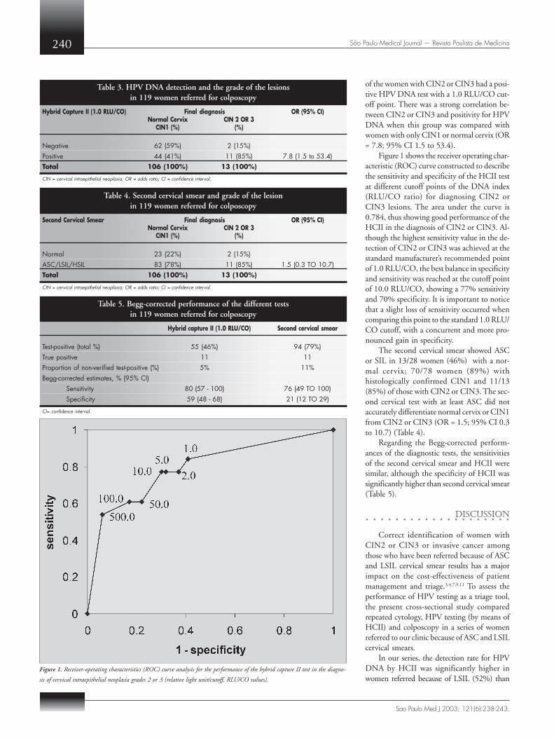
of the women with CIN2 or CIN3 had a posi-tive HPV DNA test with a 1.0 RLU/CO cut-off point. There was a strong correlation be-tween CIN2 or CIN3 and positivity for HPVDNA when this group was compared withwomen with only CIN1 or normal cervix (OR= 7.8; 95% CI 1.5 to 53.4).
Figure 1 shows the receiver operating char-acteristic (ROC) curve constructed to describethe sensitivity and specificity of the HCII testat different cutoff points of the DNA index(RLU/CO ratio) for diagnosing CIN2 orCIN3 lesions. The area under the curve is0.784, thus showing good performance of theHCII in the diagnosis of CIN2 or CIN3. Al-though the highest sensitivity value in the de-tection of CIN2 or CIN3 was achieved at thestandard manufacturer’s recommended pointof 1.0 RLU/CO, the best balance in specificityand sensitivity was reached at the cutoff pointof 10.0 RLU/CO, showing a 77% sensitivityand 70% specificity. It is important to noticethat a slight loss of sensitivity occurred whencomparing this point to the standard 1.0 RLU/CO cutoff, with a concurrent and more pro-nounced gain in specificity.
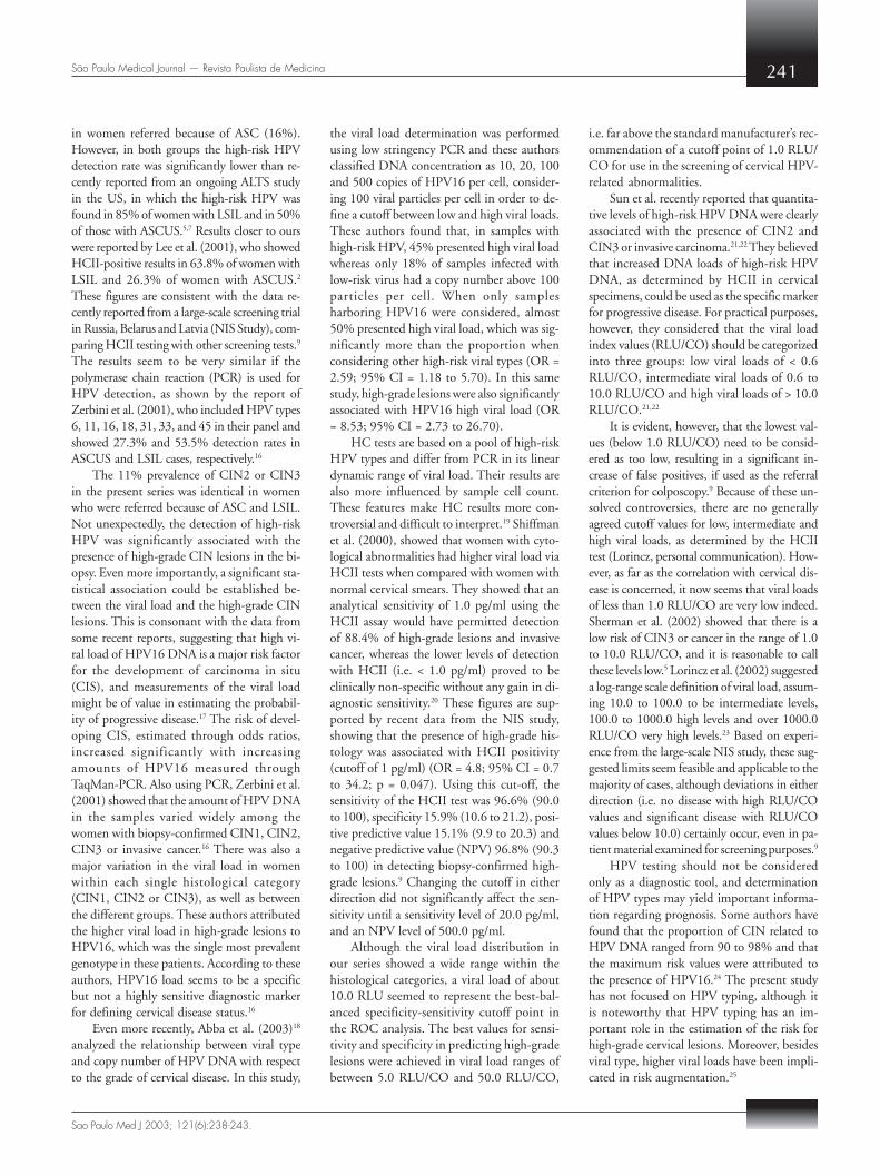
The second cervical smear showed ASCor SIL in 13/28 women (46%) with a nor-mal cervix; 70/78 women (89%) withhistologically confirmed CIN1 and 11/13(85%) of those with CIN2 or CIN3. The sec-ond cervical test with at least ASC did notaccurately differentiate normal cervix or CIN1from CIN2 or CIN3 (OR = 1.5; 95% CI 0.3to 10.7) (Table 4).
Regarding the Begg-corrected perform-ances of the diagnostic tests, the sensitivitiesof the second cervical smear and HCII weresimilar, although the specificity of HCII wassignificantly higher than second cervical smear(Table 5).
○ ○ ○ ○ ○ ○ ○ ○ ○ ○ ○ ○ ○ ○ ○ ○ ○ ○ ○ ○
DISCUSSION
Correct identification of women withCIN2 or CIN3 or invasive cancer amongthose who have been referred because of ASCand LSIL cervical smear results has a majorimpact on the cost-effectiveness of patientmanagement and triage.3,4,7,9,11 To assess theperformance of HPV testing as a triage tool,the present cross-sectional study comparedrepeated cytology, HPV testing (by means ofHCII) and colposcopy in a series of womenreferred to our clinic because of ASC and LSILcervical smears.
In our series, the detection rate for HPVDNA by HCII was significantly higher inwomen referred because of LSIL (52%) than
Table 3. HPV DNA detection and the grade of the lesionsin 119 women referred for colposcopy
Hybrid Capture II (1.0 RLU/CO) Final diagnosis OR (95% CI)Normal Cervix CIN 2 OR 3
CIN1 (%) (%)
Negative 62 (59%) 2 (15%)Positive 44 (41%) 11 (85%) 7.8 (1.5 to 53.4)Total 106 (100%) 13 (100%)
CIN = cervical intraepithelial neoplasia; OR = odds ratio; CI = confidence interval.
Figure 1. Receiver-operating characteristics (ROC) curve analysis for the performance of the hybrid capture II test in the diagno-
sis of cervical intraepithelial neoplasia grades 2 or 3 (relative light unit/cutoff, RLU/CO values).
Table 4. Second cervical smear and grade of the lesionin 119 women referred for colposcopy
Second Cervical Smear Final diagnosis OR (95% CI)Normal Cervix CIN 2 OR 3
CIN1 (%) (%)
Normal 23 (22%) 2 (15%)ASC/LSIL/HSIL 83 (78%) 11 (85%) 1.5 (0.3 TO 10.7)Total 106 (100%) 13 (100%)
CIN = cervical intraepithelial neoplasia; OR = odds ratio; CI = confidence interval.
Table 5. Begg-corrected performance of the different testsin 119 women referred for colposcopy
Hybrid capture II (1.0 RLU/CO) Second cervical smear
Test-positive (total %) 55 (46%) 94 (79%)True positive 11 11Proportion of non-verified test-positive (%) 5% 11%Begg-corrected estimates, % (95% CI)
Sensitivity 80 (57 - 100) 76 (49 TO 100)Specificity 59 (48 - 68) 21 (12 TO 29)
CI= confidence interval.
Sao Paulo Med J 2003; 121(6):238-243.
São Paulo Medical Journal — Revista Paulista de Medicina 241
in women referred because of ASC (16%).However, in both groups the high-risk HPVdetection rate was significantly lower than re-cently reported from an ongoing ALTS studyin the US, in which the high-risk HPV wasfound in 85% of women with LSIL and in 50%of those with ASCUS.5,7 Results closer to ourswere reported by Lee et al. (2001), who showedHCII-positive results in 63.8% of women withLSIL and 26.3% of women with ASCUS.2
These figures are consistent with the data re-cently reported from a large-scale screening trialin Russia, Belarus and Latvia (NIS Study), com-paring HCII testing with other screening tests.9
The results seem to be very similar if thepolymerase chain reaction (PCR) is used forHPV detection, as shown by the report ofZerbini et al. (2001), who included HPV types6, 11, 16, 18, 31, 33, and 45 in their panel andshowed 27.3% and 53.5% detection rates inASCUS and LSIL cases, respectively.16
The 11% prevalence of CIN2 or CIN3in the present series was identical in womenwho were referred because of ASC and LSIL.Not unexpectedly, the detection of high-riskHPV was significantly associated with thepresence of high-grade CIN lesions in the bi-opsy. Even more importantly, a significant sta-tistical association could be established be-tween the viral load and the high-grade CINlesions. This is consonant with the data fromsome recent reports, suggesting that high vi-ral load of HPV16 DNA is a major risk factorfor the development of carcinoma in situ(CIS), and measurements of the viral loadmight be of value in estimating the probabil-ity of progressive disease.17 The risk of devel-oping CIS, estimated through odds ratios,increased significantly with increasingamounts of HPV16 measured throughTaqMan-PCR. Also using PCR, Zerbini et al.(2001) showed that the amount of HPV DNAin the samples varied widely among thewomen with biopsy-confirmed CIN1, CIN2,CIN3 or invasive cancer.16 There was also amajor variation in the viral load in womenwithin each single histological category(CIN1, CIN2 or CIN3), as well as betweenthe different groups. These authors attributedthe higher viral load in high-grade lesions toHPV16, which was the single most prevalentgenotype in these patients. According to theseauthors, HPV16 load seems to be a specificbut not a highly sensitive diagnostic markerfor defining cervical disease status.16
Even more recently, Abba et al. (2003)18
analyzed the relationship between viral typeand copy number of HPV DNA with respectto the grade of cervical disease. In this study,
the viral load determination was performedusing low stringency PCR and these authorsclassified DNA concentration as 10, 20, 100and 500 copies of HPV16 per cell, consider-ing 100 viral particles per cell in order to de-fine a cutoff between low and high viral loads.These authors found that, in samples withhigh-risk HPV, 45% presented high viral loadwhereas only 18% of samples infected withlow-risk virus had a copy number above 100particles per cell. When only samplesharboring HPV16 were considered, almost50% presented high viral load, which was sig-nificantly more than the proportion whenconsidering other high-risk viral types (OR =2.59; 95% CI = 1.18 to 5.70). In this samestudy, high-grade lesions were also significantlyassociated with HPV16 high viral load (OR= 8.53; 95% CI = 2.73 to 26.70).
HC tests are based on a pool of high-riskHPV types and differ from PCR in its lineardynamic range of viral load. Their results arealso more influenced by sample cell count.These features make HC results more con-troversial and difficult to interpret.19 Shiffmanet al. (2000), showed that women with cyto-logical abnormalities had higher viral load viaHCII tests when compared with women withnormal cervical smears. They showed that ananalytical sensitivity of 1.0 pg/ml using theHCII assay would have permitted detectionof 88.4% of high-grade lesions and invasivecancer, whereas the lower levels of detectionwith HCII (i.e. < 1.0 pg/ml) proved to beclinically non-specific without any gain in di-agnostic sensitivity.20 These figures are sup-ported by recent data from the NIS study,showing that the presence of high-grade his-tology was associated with HCII positivity(cutoff of 1 pg/ml) (OR = 4.8; 95% CI = 0.7to 34.2; p = 0.047). Using this cut-off, thesensitivity of the HCII test was 96.6% (90.0to 100), specificity 15.9% (10.6 to 21.2), posi-tive predictive value 15.1% (9.9 to 20.3) andnegative predictive value (NPV) 96.8% (90.3to 100) in detecting biopsy-confirmed high-grade lesions.9 Changing the cutoff in eitherdirection did not significantly affect the sen-sitivity until a sensitivity level of 20.0 pg/ml,and an NPV level of 500.0 pg/ml.
Although the viral load distribution inour series showed a wide range within thehistological categories, a viral load of about10.0 RLU seemed to represent the best-bal-anced specificity-sensitivity cutoff point inthe ROC analysis. The best values for sensi-tivity and specificity in predicting high-gradelesions were achieved in viral load ranges ofbetween 5.0 RLU/CO and 50.0 RLU/CO,
i.e. far above the standard manufacturer’s rec-ommendation of a cutoff point of 1.0 RLU/CO for use in the screening of cervical HPV-related abnormalities.
Sun et al. recently reported that quantita-tive levels of high-risk HPV DNA were clearlyassociated with the presence of CIN2 andCIN3 or invasive carcinoma.21,22 They believedthat increased DNA loads of high-risk HPVDNA, as determined by HCII in cervicalspecimens, could be used as the specific markerfor progressive disease. For practical purposes,however, they considered that the viral loadindex values (RLU/CO) should be categorizedinto three groups: low viral loads of < 0.6RLU/CO, intermediate viral loads of 0.6 to10.0 RLU/CO and high viral loads of > 10.0RLU/CO.21,22
It is evident, however, that the lowest val-ues (below 1.0 RLU/CO) need to be consid-ered as too low, resulting in a significant in-crease of false positives, if used as the referralcriterion for colposcopy.9 Because of these un-solved controversies, there are no generallyagreed cutoff values for low, intermediate andhigh viral loads, as determined by the HCIItest (Lorincz, personal communication). How-ever, as far as the correlation with cervical dis-ease is concerned, it now seems that viral loadsof less than 1.0 RLU/CO are very low indeed.Sherman et al. (2002) showed that there is alow risk of CIN3 or cancer in the range of 1.0to 10.0 RLU/CO, and it is reasonable to callthese levels low.5 Lorincz et al. (2002) suggesteda log-range scale definition of viral load, assum-ing 10.0 to 100.0 to be intermediate levels,100.0 to 1000.0 high levels and over 1000.0RLU/CO very high levels.23 Based on experi-ence from the large-scale NIS study, these sug-gested limits seem feasible and applicable to themajority of cases, although deviations in eitherdirection (i.e. no disease with high RLU/COvalues and significant disease with RLU/COvalues below 10.0) certainly occur, even in pa-tient material examined for screening purposes.9
HPV testing should not be consideredonly as a diagnostic tool, and determinationof HPV types may yield important informa-tion regarding prognosis. Some authors havefound that the proportion of CIN related toHPV DNA ranged from 90 to 98% and thatthe maximum risk values were attributed tothe presence of HPV16.24 The present studyhas not focused on HPV typing, although itis noteworthy that HPV typing has an im-portant role in the estimation of the risk forhigh-grade cervical lesions. Moreover, besidesviral type, higher viral loads have been impli-cated in risk augmentation.25
Sao Paulo Med J 2003; 121(6):238-243.
São Paulo Medical Journal — Revista Paulista de Medicina242
1. Solomon D, Schiffman M, Tarone R. Comparison of three man-
agement strategies for patients with atypical squamous cells of
undetermined significance: baseline results from a randomized
trial. J Natl Cancer Inst 2001;93(4):293-9.
2. Lee NW, Kim D, Park JT, Kim A. Is the human papillomavirus
test combination with the Papanicolaou test useful for manage-
ment of patients with diagnoses of atypical squamous cells of
undetermined significance/low-grade squamous intraepithelial
lesions? Arch Pathol Lab Med 2001;125(11):1453-7.
3. Herbst AL, Pickett KE, Follen M, Noller KL. The management
of ASCUS cervical cytologic abnormalities and HPV testing: a
cautionary note. Obstet Gynecol 2001;98(5 Pt 1):849-51.
4. Paraskevaidis E, Malamou-Mitsi V, Koliopoulos G, et al. Ex-
panded cytological referral criteria for colposcopy in cervical
screening: comparison with human papillomavirus testing.
Gynecol Oncol 2001;82(2):355-9.
5. Sherman ME, Schiffman M, Cox JT. Effects of age and human
papilloma viral load on colposcopy triage: data from the
randomized Atypical Squamous Cells of Undetermined Signifi-
cance/Low-Grade Squamous Intraepithelial Lesion Triage Study
(ALTS). J Natl Cancer Inst 2002;94(2):102-7.
6. Hughes SA, Sun D, Gibson C, et al. Managing atypical squa-
mous cells of undetermined significance (ASCUS): human
papillomavirus testing, ASCUS subtyping, or follow-up cytol-
ogy? Am J Obstet Gynecol 2002;186(3):396-403.
7. Human papillomavirus testing for triage of women with
cytologic evidence of low-grade squamous intraepithelial lesions:
baseline data from a randomized trial. The Atypical Squamous
Cells of Undetermined Significance/Low-Grade Squamous
Intraepithelial Lesions Triage Study (ALTS) Group. J Natl Can-
cer Inst 2000;92(5)397-402.
8. Wright TC, Cox JT, Massad LS, Twiggs LB, Wilkinson EJ. 2001
○ ○ ○ ○ ○ ○ ○ ○ ○ ○ ○ ○ ○ ○ ○ ○ ○ ○ ○ ○ ○ ○ ○ ○ ○ ○ ○ ○ ○ ○ ○ ○ ○ ○ ○ ○ ○ ○ ○ ○ ○ ○ ○ ○ ○ ○ ○ ○ ○ ○ ○ ○ ○ ○ ○ ○ ○ ○ ○ ○ ○ ○ ○ ○
REFERENCES
Consensus Guidelines for the management of women with cer-
vical cytological abnormalities. JAMA 2002;287(16):2120-9.
9. Syrjänen S, Shabalova IP, Petrovichev N, et al. Human
papillomavirus testing and conventional Pap smear cytology as
optional screening tools of women at different risks for cervical
cancer in the countries of the former Soviet Union. J Lower
Genital Tract Dis 2002;6(2):97-110.
10. Solomon D, Davey D, Kurman R, et al. The 2001 Bethesda
System: terminology for reporting results of cervical cytology.
JAMA 2002;287(16):2114-9.
11. Gamzu R, Almog B, Levin I, et al. Clinical and economic im-
plication of adding HPV tests to the routine cytology follow-
up and management of patients with histologically defined cer-
vical intraepithelial neoplasia grade 1. Gynecol Oncol
2002;86(2):129-33.
12. Stafl A, Wilbanks GD. An international terminology of colpos-
copy: report of the Nomenclature Committee of the Interna-
tional Federation of Cervical Pathology and Colposcopy. Obstet
Gynecol 1991;77(2):313-4.
13. Scully RE, Bonfiglio TA, Kurman RJ, Silverberg SG, Wilkins
EJ. Histological typing of female genital tract tumors. World
Health Organization - International histological classification
of tumors. 2nd ed. Berlin: Springer-Verlag; 1994.
14. Begg CB, Greenes RA. Assessment of diagnostic tests when dis-
ease verification is subject to selection bias. Biometrics
1983;39(1):207-15.
15. Schneider A, Hoyer H, Lotz B, et al. Screening for high-grade
cervical intra-epithelial neoplasia and cancer by testing for high-
risk HPV, routine cytology or colposcopy. Int J Cancer
2000;89(6):529-34.
16. Zerbini M, Venturoli S, Cricca M, et al. Distribution and viral
load of type specific HPVs in different cervical lesions as de-
tected by PCR-ELISA. J Clin Pathol 2001;54(5):377-80.
17. Josefsson AM, Magnusson PK, Ylitalo N, et al. Viral load of
human papilloma virus 16 as a determinant for development of
cervical carcinoma in situ: a nested case-control study. Lancet
2000;355(9222):2189-93.
18. Abba MC, Mourón SA, Gómez MA, Dulout FN, Golijow CD.
Association of human papillomavirus viral load with HPV16
and high-grade intraepithelial lesion. Int J Gynecol Cancer
2003;13(2):154-8.
19. Gravitt PE, Burk RD, Lorincz A, et al. A comparison between
real-time polymerase chain reaction and hybrid capture 2 for
human papillomavirus DNA quantitation. Cancer Epidemiol
Biomarkers Prev 2003;12(6):477-84.
20. Schiffman M, Herrero R, Hildesheim A, et al. HPV DNA test-
ing in cervical cancer screening: result from women in a high-
risk province of Costa Rica. JAMA 2000;283(1):87-93.
21. Sun CA, Lai HC, Chang CC, Neih S, Yu CP, Chu TY. The
significance of human papillomavirus viral load in prediction
of histologic severity and size of squamous intraepithelial le-
sions of uterine cervix. Gynecol Oncol 2001;83(1):95-9.
22. Sun CA, Liu JF, Wu DM, Nieh S, Yu CP, Chu TY. Viral load of
high-risk human papillomavirus in cervical squamous
intraepithelial lesions. Int J Gynaecol Obstet 2002;76(1):41-7.
23. Lorincz AT, Castle PE, Sherman ME, et al. Viral load of human
papillomavirus and risk of CIN3 or cervical cancer. Lancet
2002;360(9328):228-9.
24. Muñoz N, Bosch FX, de Sanjosé S, et al. Epidemiologic classi-
fication of human papillomavirus types associated with cervical
cancer. N Engl J Med 2003;348(6):518-27.
25. Schelecht NF, Trevisan A, Duarte-Franco E, et al. Viral load as
a predictor of the risk of cervical intraepithelial neoplasia. Int J
Cancer 2003;103(4):519-24.
○ ○ ○ ○ ○ ○ ○ ○ ○ ○ ○ ○ ○ ○ ○ ○ ○ ○ ○ ○
CONCLUSIONS
In the studied population, referred for ex-amination due to an ASC or LSIL cytological
smear, HCII testing for high-risk HPV typeswas a sensitive tool in detecting CIN2 orCIN3, with better diagnostic performancethan repeated cervical smears. As the detec-
tion rate for high-risk HPV DNA was around50% in women with LSIL smears (lower thanin many other series), the HCII test performedparticularly well in this group of women.
Sao Paulo Med J 2003; 121(6):238-243.

São Paulo Medical Journal — Revista Paulista de Medicina 243
Carga viral do papilomavírus humano como fa-tor preditivo de neoplasia intra-epitelial dealto grau em mulheres com células esca-mosas atípicas ou lesão escamosa intra-epite-lial de baixo grau na colpocitologia
CONTEXTO: A determinação da carga viral dopapilomavírus humano (HPV) pode ter im-portante papel na detecção de neoplasia intra-epitelial cervical (NIC) de alto grau em mu-lheres com colpocitologia apresentando cé-lulas escamosas atípicas ou sugestivas de le-são escamosa de baixo grau.
OBJETIVO: Avaliar se a determinação da cargaviral do DNA HPV é útil para predizer adetecção da neoplasia intra-epitelial de altograu (NIC2 e 3) em mulheres referidas porcolpocitologias mostrando apenas célulasescamosas atípicas ou lesão intra-epitelial debaixo grau.
TIPO DE ESTUDO: TransversalLOCAL: Serviço de colposcopia de hospital uni-
versitário.MÉTODOS: Foram incluídas 119 mulheres en-
caminhadas por células escamosas atípicas elesão intra-epitelial de baixo grau entre agos-to de 2000 e abril de 2001. De todas as mu-lheres foi coletada nova colpocitologia, espé-cime para teste de HPV usando captura dehíbridos II (CHII), carga viral medida emunidades relativas de luz (URL). Foi realiza-da colposcopia com biópsia cervical em 97mulheres. As lesões cervicais foram classifi-cadas usando a classificação NIC. Para diag-nóstico final, a colposcopia normal ou a pre-sença de cervicite confirmada por biópsia fo-
ram classificadas como colo normal. Paraanálise estatística foram calculados o oddsratio (OR), com intervalo de confiança em95%, e foi traçada uma curva “receiveroperator characteristic” (ROC).
RESULTADOS: As biópsias cervicais mostraramNIC2 ou 3 em 11% dos casos, igualmentedistribuídas entre as mulheres encaminhadaspor causa da presença de células escamosasatípicas ou lesão intra-epitelial de baixo grau.A CHII foi positiva em 16% das mulherescom células escamosas atípicas e em 52%daquelas com lesão intra-epitelial de baixograu (OR = 5,8; IC 95% 1,4 a 26,7). Entreas mulheres com CHII positiva, 7% tinhamcérvice normal, 73% NIC 1 (OR = 6,3; IC95% 1,8 a 23,8) e 20% tinham NIC2 ou 3(OR = 33,0; IC 95% 4,2 a 347,8). Na análi-se da curva ROC para CH II, diagnostican-do NIC2 e 3, a área sob a curva foi de 0,784e o ponto de corte da carga viral de 10.0 URLmostrou sensibilidade de 77% e especifi-cidade de 70%. A segunda colpocitologiamostrando ao menos células escamosasatípicas não apresentou boa performance nadetecção NIC 2 ou 3 (OR = 6,4%; IC 95%1,0 a 50,9).
CONCLUSÕES: A carga viral do DNA-HPVde alto risco oncológico foi significativa-mente associada com o diagnóstico deNIC2 e 3 em mulheres encaminhadas pordetecção de células escamosas atípicas e le-são intra-epitelial escamosa de baixo grauna colpocitologia.
PALAVRAS-CHAVE: Papilomavírus humano.Carga viral. Neoplasia intra-epitelial cervical.
○ ○ ○ ○ ○ ○ ○ ○ ○ ○ ○ ○ ○ ○ ○ ○ ○ ○ ○ ○ ○ ○ ○ ○ ○ ○ ○ ○ ○ ○ ○ ○ ○ ○ ○ ○ ○ ○ ○ ○ ○ ○
RESUMORESUMORESUMORESUMORESUMO○ ○ ○ ○ ○ ○ ○ ○ ○ ○ ○ ○ ○ ○ ○ ○ ○ ○ ○ ○
PUBLISHING INFORMATION
ACKNOWLEDGMENTS: The skillful technical assistance ofDenise da Rocha Pitta Lima de Moraes, Lúcia Maria Fagiande Carvalho and Elisabete Aparecida Campos is grate-fully acknowledged.
André Luis Ferreira Santos. MSc in Gynecology,Universidade de Taubaté, Taubaté, São Paulo, Brazil.
Sophie Françoise Mauricette Derchain. Associate pro-fessor, Department of Obstetrics and Gynecology,Universidade Estadual de Campinas, Campinas, São Paulo,Brazil.
Marcos Roberto Martins. MSc in Gynecology,Universidade de Taubaté, Taubaté, São Paulo, Brazil.
Luís Otávio Zanatta Sarian. MSc in Gynecology,Universidade Estadual de Campinas, Campinas, São Paulo,Brazil.
Edson Zangiacome Martinez. PhD in Statistics,Universidade Estadual de Campinas, Campinas, São Paulo,Brazil.
Kari Juhani Syrjänen. Associate professor, National In-stitute of Health, Rome, Italy.
Conflict of interest: Not declared
Sources of funding: Fundação Amparo a Pesquisa doEstado de São Paulo (FAPESP), grant number 99/11264-0,and Conselho Nacional de Desenvolvimento Científico eTecnológico (CNPq), grant number 300354/01-0.
Date of first submission: April 22, 2003
Last received: June 30, 2003
Accepted: August 7, 2003
Address for correspondence:Luís Otávio Zanatta Sarian
R. Alexander Fleming, 848Campinas/SP — Brasil — CEP 13092-340Tel. (+55 19) 3252-4948E-mail: [email protected]
COPYRIGHT © 2003, Associação Paulista de Medicina
Sao Paulo Med J 2003; 121(6):238-243.