Time-dependent quantum wave-packet description of the 1??* photochemistry of pyrrole
Bioorganic & Medicinal Chemistry Letters 17 (2007) 4538–4544
Design and synthesis of hepatoselective, pyrrole-basedHMG-CoA reductase inhibitors
Jeffrey A. Pfefferkorn,* Yuntao Song, Kuai-Lin Sun, Steven R. Miller,Bharat K. Trivedi, Chulho Choi, Roderick J. Sorenson, Larry D. Bratton,
Paul C. Unangst, Scott D. Larsen, Toni-Jo Poel, Xue-Min Cheng,Chitase Lee, Noe Erasga, Bruce Auerbach, Valerie Askew, Lisa Dillon,
Jeffrey C. Hanselman, Zhiwu Lin, Gina Lu, Andrew Robertson, Karl Olsen,Thomas Mertz, Catherine Sekerke, Alexander Pavlovsky, Melissa S. Harris,Graeme Bainbridge, Nicole Caspers, Huifen Chen and Matthias Eberstadt
Pfizer Global Research & Development, Michigan Laboratories, 2800 Plymouth Road, Ann Arbor, MI 48105, USA
Received 25 April 2007; revised 24 May 2007; accepted 30 May 2007
Available online 6 June 2007
Abstract—This manuscript describes the design and synthesis of a series of pyrrole-based inhibitors of HMG-CoA reductase for thetreatment of hypercholesterolemia. Analogs were optimized using structure-based design and physical property considerationsresulting in the identification of 44, a hepatoselective HMG-CoA reductase inhibitor with excellent acute and chronic efficacy ina pre-clinical animal models.� 2007 Elsevier Ltd. All rights reserved.
Coronary heart disease (CHD) is the leading cause ofdeath in the United States at an annual cost of morethan $150 billion.1 Given that hypercholesterolemia, orelevated serum cholesterol, is a key risk factor forCHD, substantial efforts have been undertaken to miti-gate this condition.2 Currently, the standard of care fortreating hypercholesterolemia is the use of HMG-CoAreductase inhibitors, also known as statins, which blockthe rate-limiting step of cholesterol biosynthesis.3 As aclass, statins have proven remarkably safe and effectivefor both primary prevention of coronary heart diseaseand secondary prevention of coronary events.4
In recent years, results from several clinical trials(TNT, MIRACAL, PROVE-IT, and others) have dem-onstrated that increasingly aggressive LDL-C-loweringtherapy may offer additional protection against CHDrelative to earlier, less aggressive, treatment standards.5
In light of this evidence, the National Cholesterol
0960-894X/$ - see front matter � 2007 Elsevier Ltd. All rights reserved.
doi:10.1016/j.bmcl.2007.05.096
Keywords: HMG-CoA reductase inhibitor; Statin; Hypercholesterolemia.* Corresponding author. Tel.: +1 860 686 3421; fax: +1 860 715
4608; e-mail: [email protected]
Education Program (NCEP-ATP-III) has modified itstreatment guidelines recommending that highest riskCHD patients reach a treatment goal of LDL-C<70 mg/dl, down from the previous treatment goal of100 mg/dl for this patient group.6 Moreover, otherresearchers have suggested that optimal LDL-C levelsto prevent atherosclerosis and CHD might be even low-er, in the range of 50–70 mg/dl.7
Achieving such aggressive LDL-C reductions in patientstypically requires the use of high dose statins alone or incombination with complimentary agents such as thecholesterol absorption inhibitor ezetimibe.8 A potentiallimitation of high dose statin therapy is statin-inducedmyalgia, the muscle pain or weakness that sometimesaccompanies statin therapy.9 While the overall incidenceof myalgia is low (2–7% of patients in trials), the likeli-hood of occurrence increases with drug dose, and it canbe a key factor in preventing patient compliance with atreatment regimen.9
The mechanism of statin-induced myalgia is complexbut thought to involve, in part, inhibition of HMG-CoA reductase in non-hepatic tissues (particularly
Br
F
O2N
F
a
CHOb
NBu
c NO2
F
d
HN CO2Et
F
HN CO2Et
F
e
6 8
10
H
N CO2H
F
OHCN CO2Et
F
gOHC
f
1112
13 14
4 5
F F
F
FF
F F
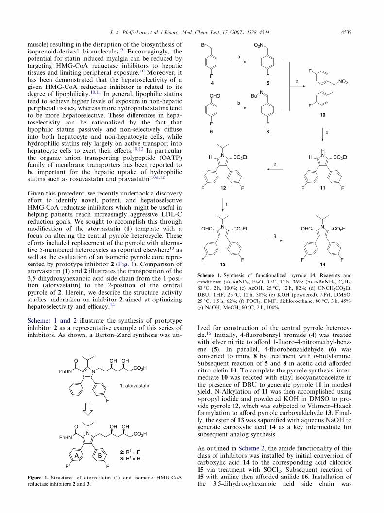
Scheme 1. Synthesis of functionalized pyrrole 14. Reagents and
conditions: (a) AgNO2, Et2O, 0 �C, 12 h, 36%; (b) n-BuNH2, C6H6,
80 �C, 2 h, 100%; (c) AcOH, 25 �C, 12 h, 82%; (d) CNCH2CO2Et,
DBU, THF, 25 �C, 12 h, 38%; (e) KOH (powdered), i-PrI, DMSO,
25 �C, 1.5 h, 62%; (f) POCl3, DMF, dichloroethane, 80 �C, 3 h, 45%;
(g) NaOH, MeOH, 60 �C, 2 h, 100%.
J. A. Pfefferkorn et al. / Bioorg. Med. Chem. Lett. 17 (2007) 4538–4544 4539
muscle) resulting in the disruption of the biosynthesis ofisoprenoid-derived biomolecules.9 Encouragingly, thepotential for statin-induced myalgia can be reduced bytargeting HMG-CoA reductase inhibitors to hepatictissues and limiting peripheral exposure.10 Moreover, ithas been demonstrated that the hepatoselectivity of agiven HMG-CoA reductase inhibitor is related to itsdegree of lipophilicity.10,11 In general, lipophilic statinstend to achieve higher levels of exposure in non-hepaticperipheral tissues, whereas more hydrophilic statins tendto be more hepatoselective. These differences in hepa-toselectivity can be rationalized by the fact thatlipophilic statins passively and non-selectively diffuseinto both hepatocyte and non-hepatocyte cells, whilehydrophilic statins rely largely on active transport intohepatocyte cells to exert their effects.10,12 In particularthe organic anion transporting polypeptide (OATP)family of membrane transporters has been reported tobe important for the hepatic uptake of hydrophilicstatins such as rosuvastatin and pravastatin.10d,12
Given this precedent, we recently undertook a discoveryeffort to identify novel, potent, and hepatoselectiveHMG-CoA reductase inhibitors which might be useful inhelping patients reach increasingly aggressive LDL-Creduction goals. We sought to accomplish this throughmodification of the atorvastatin (1) template with afocus on altering the central pyrrole heterocycle. Theseefforts included replacement of the pyrrole with alterna-tive 5-membered heterocycles as reported elsewhere13 aswell as the evaluation of an isomeric pyrrole core repre-sented by prototype inhibitor 2 (Fig. 1). Comparison ofatorvastatin (1) and 2 illustrates the transposition of the3,5-dihydroxyhexanoic acid side chain from the 1-posi-tion (atorvastatin) to the 2-position of the centralpyrrole of 2. Herein, we describe the structure–activitystudies undertaken on inhibitor 2 aimed at optimizinghepatoselectivity and efficacy.14
Schemes 1 and 2 illustrate the synthesis of prototypeinhibitor 2 as a representative example of this series ofinhibitors. As shown, a Barton–Zard synthesis was uti-
NO
PhHN
OH OH
CO2H
F
N
O
PhHN
OH OH
CO2H
F
2: R1 = F3: R1 = H
1: atorvastatin
R1
A B
Figure 1. Structures of atorvastatin (1) and isomeric HMG-CoA
reductase inhibitors 2 and 3.
lized for construction of the central pyrrole heterocy-cle.15 Initially, 4-fluorobenzyl bromide (4) was treatedwith silver nitrite to afford 1-fluoro-4-nitromethyl-benz-ene (5). In parallel, 4-fluorobenzaldehyde (6) wasconverted to imine 8 by treatment with n-butylamine.Subsequent reaction of 5 and 8 in acetic acid affordednitro-olefin 10. To complete the pyrrole synthesis, inter-mediate 10 was reacted with ethyl isocyanatoacetate inthe presence of DBU to generate pyrrole 11 in modestyield. N-Alkylation of 11 was then accomplished usingi-propyl iodide and powdered KOH in DMSO to pro-vide pyrrole 12, which was subjected to Vilsmeir–Haackformylation to afford pyrrole carboxaldehyde 13. Final-ly, the ester of 13 was saponified with aqueous NaOH togenerate carboxylic acid 14 as a key intermediate forsubsequent analog synthesis.
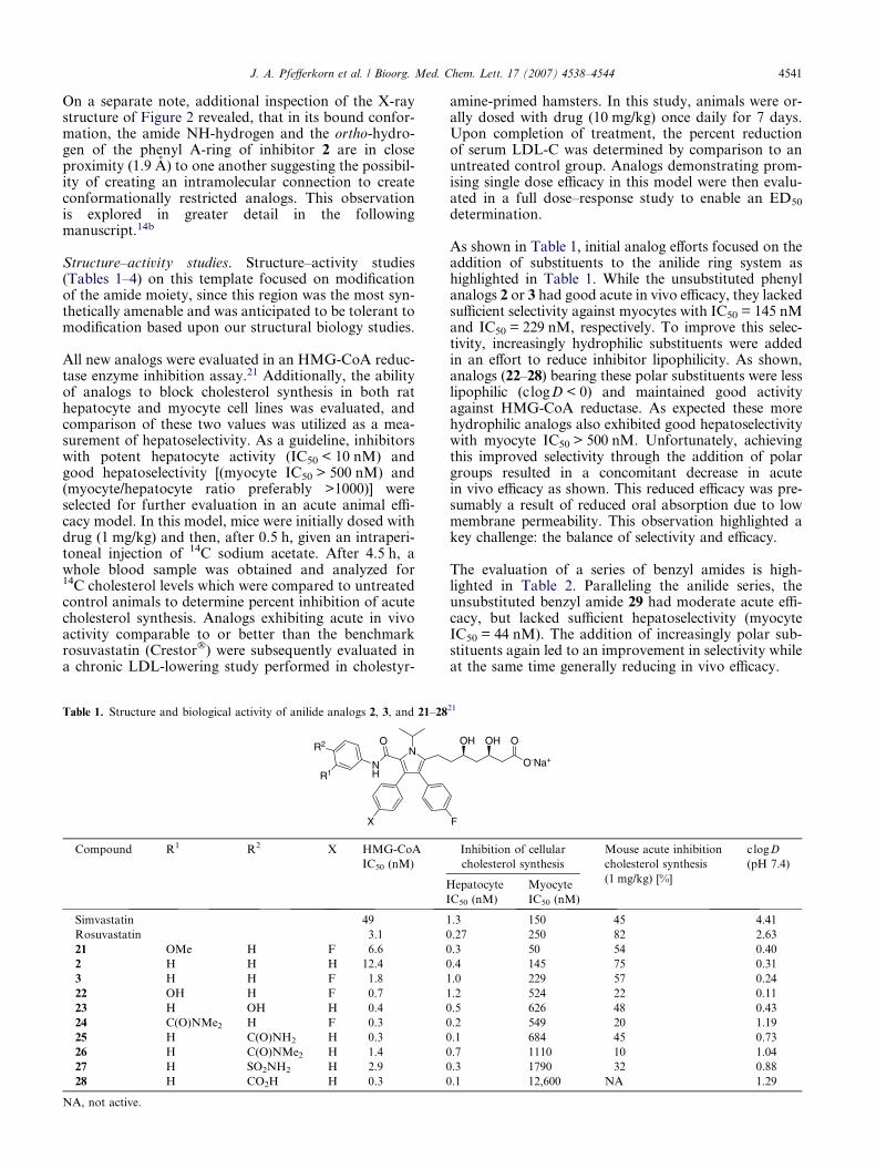
As outlined in Scheme 2, the amide functionality of thisclass of inhibitors was installed by initial conversion ofcarboxylic acid 14 to the corresponding acid chloride15 via treatment with SOCl2. Subsequent reaction of15 with aniline then afforded anilide 16. Installation ofthe 3,5-dihydroxyhexanoic acid side chain was
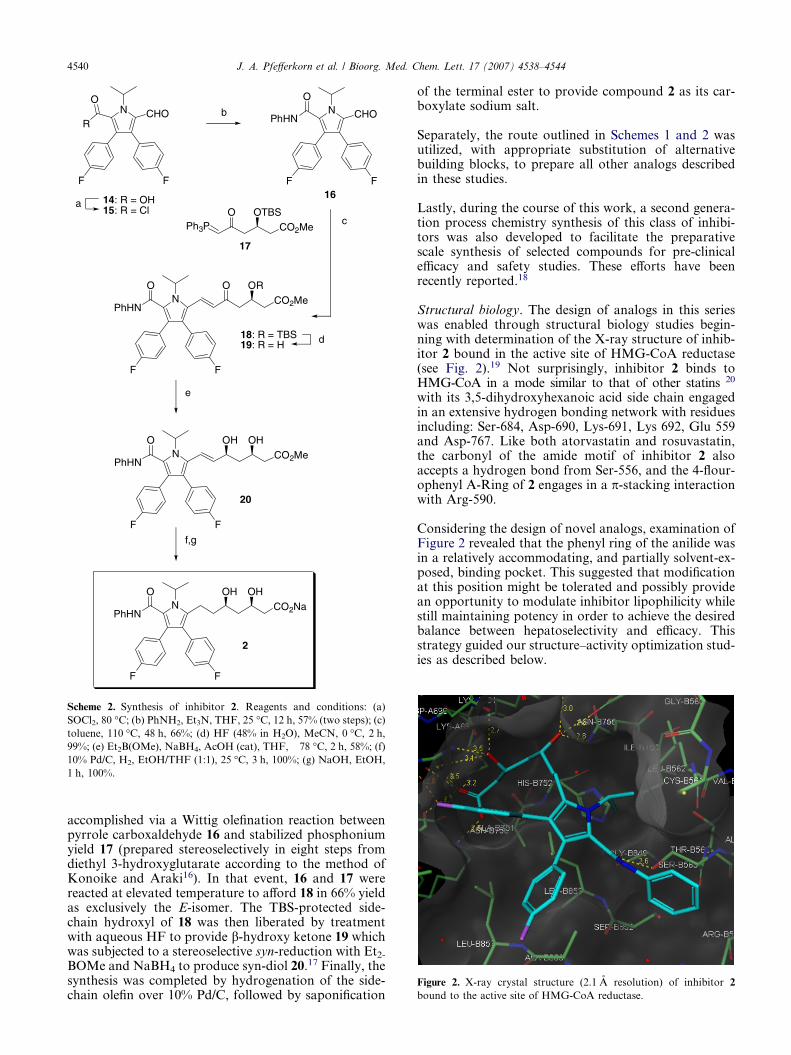
Figure 2. X-ray crystal structure (2.1 A resolution) of inhibitor 2
bound to the active site of HMG-CoA reductase.
N
F
CHO b N
F
CHOPhHN
O
14: R = OH15: R = Cl
R
O
F F
a
NO
PhHN
O OR
CO2Me
F
Ph3PO OTBS
CO2Me
18: R = TBS19: R = H
NO
PhHN
OH OH
CO2Me
F
NO
PhHN
OH OH
CO2Na
F
16
2
e
F
F
F
c
d
f,g
20
17
Scheme 2. Synthesis of inhibitor 2. Reagents and conditions: (a)
SOCl2, 80 �C; (b) PhNH2, Et3N, THF, 25 �C, 12 h, 57% (two steps); (c)
toluene, 110 �C, 48 h, 66%; (d) HF (48% in H2O), MeCN, 0 �C, 2 h,
99%; (e) Et2B(OMe), NaBH4, AcOH (cat), THF, �78 �C, 2 h, 58%; (f)
10% Pd/C, H2, EtOH/THF (1:1), 25 �C, 3 h, 100%; (g) NaOH, EtOH,
1 h, 100%.
4540 J. A. Pfefferkorn et al. / Bioorg. Med. Chem. Lett. 17 (2007) 4538–4544
accomplished via a Wittig olefination reaction betweenpyrrole carboxaldehyde 16 and stabilized phosphoniumyield 17 (prepared stereoselectively in eight steps fromdiethyl 3-hydroxyglutarate according to the method ofKonoike and Araki16). In that event, 16 and 17 werereacted at elevated temperature to afford 18 in 66% yieldas exclusively the E-isomer. The TBS-protected side-chain hydroxyl of 18 was then liberated by treatmentwith aqueous HF to provide b-hydroxy ketone 19 whichwas subjected to a stereoselective syn-reduction with Et2-
BOMe and NaBH4 to produce syn-diol 20.17 Finally, thesynthesis was completed by hydrogenation of the side-chain olefin over 10% Pd/C, followed by saponification
of the terminal ester to provide compound 2 as its car-boxylate sodium salt.
Separately, the route outlined in Schemes 1 and 2 wasutilized, with appropriate substitution of alternativebuilding blocks, to prepare all other analogs describedin these studies.
Lastly, during the course of this work, a second genera-tion process chemistry synthesis of this class of inhibi-tors was also developed to facilitate the preparativescale synthesis of selected compounds for pre-clinicalefficacy and safety studies. These efforts have beenrecently reported.18
Structural biology. The design of analogs in this serieswas enabled through structural biology studies begin-ning with determination of the X-ray structure of inhib-itor 2 bound in the active site of HMG-CoA reductase(see Fig. 2).19 Not surprisingly, inhibitor 2 binds toHMG-CoA in a mode similar to that of other statins 20
with its 3,5-dihydroxyhexanoic acid side chain engagedin an extensive hydrogen bonding network with residuesincluding: Ser-684, Asp-690, Lys-691, Lys 692, Glu 559and Asp-767. Like both atorvastatin and rosuvastatin,the carbonyl of the amide motif of inhibitor 2 alsoaccepts a hydrogen bond from Ser-556, and the 4-flour-ophenyl A-Ring of 2 engages in a p-stacking interactionwith Arg-590.
Considering the design of novel analogs, examination ofFigure 2 revealed that the phenyl ring of the anilide wasin a relatively accommodating, and partially solvent-ex-posed, binding pocket. This suggested that modificationat this position might be tolerated and possibly providean opportunity to modulate inhibitor lipophilicity whilestill maintaining potency in order to achieve the desiredbalance between hepatoselectivity and efficacy. Thisstrategy guided our structure–activity optimization stud-ies as described below.
J. A. Pfefferkorn et al. / Bioorg. Med. Chem. Lett. 17 (2007) 4538–4544 4541
On a separate note, additional inspection of the X-raystructure of Figure 2 revealed, that in its bound confor-mation, the amide NH-hydrogen and the ortho-hydro-gen of the phenyl A-ring of inhibitor 2 are in closeproximity (1.9 A) to one another suggesting the possibil-ity of creating an intramolecular connection to createconformationally restricted analogs. This observationis explored in greater detail in the followingmanuscript.14b
Structure–activity studies. Structure–activity studies(Tables 1–4) on this template focused on modificationof the amide moiety, since this region was the most syn-thetically amenable and was anticipated to be tolerant tomodification based upon our structural biology studies.
All new analogs were evaluated in an HMG-CoA reduc-tase enzyme inhibition assay.21 Additionally, the abilityof analogs to block cholesterol synthesis in both rathepatocyte and myocyte cell lines was evaluated, andcomparison of these two values was utilized as a mea-surement of hepatoselectivity. As a guideline, inhibitorswith potent hepatocyte activity (IC50 < 10 nM) andgood hepatoselectivity [(myocyte IC50 > 500 nM) and(myocyte/hepatocyte ratio preferably >1000)] wereselected for further evaluation in an acute animal effi-cacy model. In this model, mice were initially dosed withdrug (1 mg/kg) and then, after 0.5 h, given an intraperi-toneal injection of 14C sodium acetate. After 4.5 h, awhole blood sample was obtained and analyzed for14C cholesterol levels which were compared to untreatedcontrol animals to determine percent inhibition of acutecholesterol synthesis. Analogs exhibiting acute in vivoactivity comparable to or better than the benchmarkrosuvastatin (Crestor�) were subsequently evaluated ina chronic LDL-lowering study performed in cholestyr-
Table 1. Structure and biological activity of anilide analogs 2, 3, and 21–28
NNH
O
X
R2
R1
Compound R1 R2 X HMG-CoA
IC50 (nM)
H
I
Simvastatin 49 1
Rosuvastatin 3.1 0
21 OMe H F 6.6 0
2 H H H 12.4 0
3 H H F 1.8 1
22 OH H F 0.7 1
23 H OH H 0.4 0
24 C(O)NMe2 H F 0.3 0
25 H C(O)NH2 H 0.3 0
26 H C(O)NMe2 H 1.4 0
27 H SO2NH2 H 2.9 0
28 H CO2H H 0.3 0
NA, not active.
amine-primed hamsters. In this study, animals were or-ally dosed with drug (10 mg/kg) once daily for 7 days.Upon completion of treatment, the percent reductionof serum LDL-C was determined by comparison to anuntreated control group. Analogs demonstrating prom-ising single dose efficacy in this model were then evalu-ated in a full dose–response study to enable an ED50
determination.
As shown in Table 1, initial analog efforts focused on theaddition of substituents to the anilide ring system ashighlighted in Table 1. While the unsubstituted phenylanalogs 2 or 3 had good acute in vivo efficacy, they lackedsufficient selectivity against myocytes with IC50 = 145 nMand IC50 = 229 nM, respectively. To improve this selec-tivity, increasingly hydrophilic substituents were addedin an effort to reduce inhibitor lipophilicity. As shown,analogs (22–28) bearing these polar substituents were lesslipophilic (c logD < 0) and maintained good activityagainst HMG-CoA reductase. As expected these morehydrophilic analogs also exhibited good hepatoselectivitywith myocyte IC50 > 500 nM. Unfortunately, achievingthis improved selectivity through the addition of polargroups resulted in a concomitant decrease in acutein vivo efficacy as shown. This reduced efficacy was pre-sumably a result of reduced oral absorption due to lowmembrane permeability. This observation highlighted akey challenge: the balance of selectivity and efficacy.
The evaluation of a series of benzyl amides is high-lighted in Table 2. Paralleling the anilide series, theunsubstituted benzyl amide 29 had moderate acute effi-cacy, but lacked sufficient hepatoselectivity (myocyteIC50 = 44 nM). The addition of increasingly polar sub-stituents again led to an improvement in selectivity whileat the same time generally reducing in vivo efficacy.
21
F
OH OH
O-Na+
O
Inhibition of cellular
cholesterol synthesis
Mouse acute inhibition
cholesterol synthesis
(1 mg/kg) [%]
c logD
(pH 7.4)
epatocyte
C50 (nM)
Myocyte
IC50 (nM)
.3 150 �45 4.41
.27 250 �82 �2.63
.3 50 �54 0.40
.4 145 �75 0.31
.0 229 �57 0.24
.2 524 �22 �0.11
.5 626 �48 �0.43
.2 549 �20 �1.19
.1 684 �45 �0.73
.7 1110 �10 �1.04
.3 1790 �32 �0.88
.1 12,600 NA �1.29
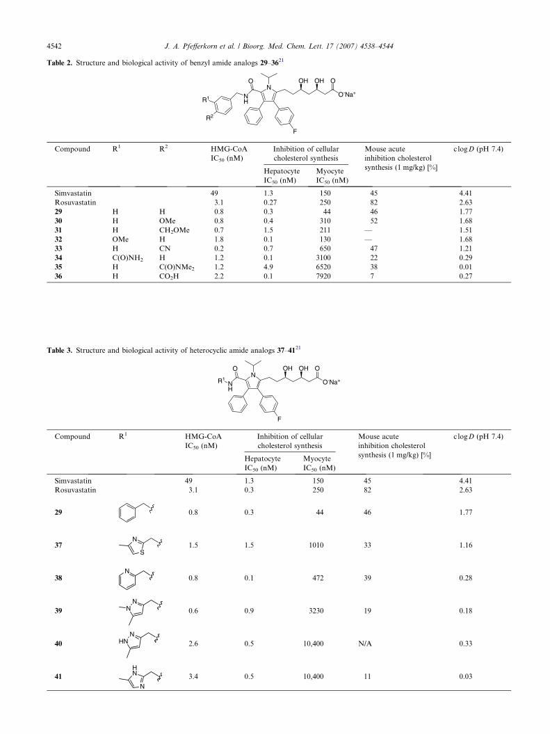
Table 3. Structure and biological activity of heterocyclic amide analogs 37–4121
N
F
NH
O OH OH
O-Na+
O
R1
Compound R1 HMG-CoA
IC50 (nM)
Inhibition of cellular
cholesterol synthesis
Mouse acute
inhibition cholesterol
synthesis (1 mg/kg) [%]
c logD (pH 7.4)
Hepatocyte
IC50 (nM)
Myocyte
IC50 (nM)
Simvastatin 49 1.3 150 �45 4.41
Rosuvastatin 3.1 0.3 250 �82 �2.63
29 0.8 0.3 44 �46 1.77
37N
S1.5 1.5 1010 �33 1.16
38N
0.8 0.1 472 �39 0.28
39
NN 0.6 0.9 3230 �19 0.18
40
NHN 2.6 0.5 10,400 N/A 0.33
41
HN
N
3.4 0.5 10,400 �11 �0.03
Table 2. Structure and biological activity of benzyl amide analogs 29–3621
N
F
NH
O OH OH
O-Na+
O
R2
R1
Compound R1 R2 HMG-CoA
IC50 (nM)
Inhibition of cellular
cholesterol synthesis
Mouse acute
inhibition cholesterol
synthesis (1 mg/kg) [%]
c logD (pH 7.4)
Hepatocyte
IC50 (nM)
Myocyte
IC50 (nM)
Simvastatin 49 1.3 150 �45 4.41
Rosuvastatin 3.1 0.27 250 �82 �2.63
29 H H 0.8 0.3 44 �46 1.77
30 H OMe 0.8 0.4 310 �52 1.68
31 H CH2OMe 0.7 1.5 211 — 1.51
32 OMe H 1.8 0.1 130 — 1.68
33 H CN 0.2 0.7 650 �47 1.21
34 C(O)NH2 H 1.2 0.1 3100 �22 0.29
35 H C(O)NMe2 1.2 4.9 6520 �38 0.01
36 H CO2H 2.2 0.1 7920 �7 �0.27
4542 J. A. Pfefferkorn et al. / Bioorg. Med. Chem. Lett. 17 (2007) 4538–4544
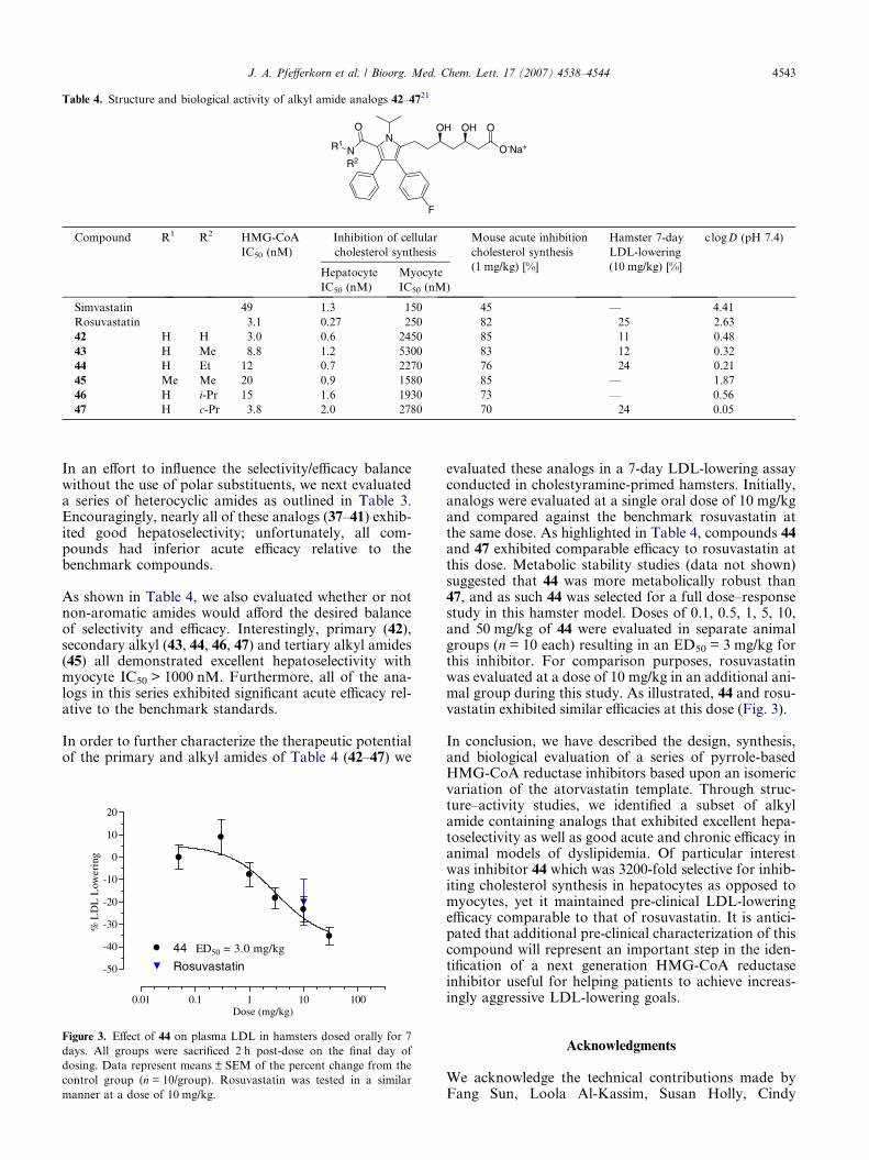
Table 4. Structure and biological activity of alkyl amide analogs 42–4721
N
F
N
O OH OH
O-Na+
O
R1
R2
Compound R1 R2 HMG-CoA
IC50 (nM)
Inhibition of cellular
cholesterol synthesis
Mouse acute inhibition
cholesterol synthesis
(1 mg/kg) [%]
Hamster 7-day
LDL-lowering
(10 mg/kg) [%]
c logD (pH 7.4)
Hepatocyte
IC50 (nM)
Myocyte
IC50 (nM)
Simvastatin 49 1.3 150 �45 — 4.41
Rosuvastatin 3.1 0.27 250 �82 �25 �2.63
42 H H 3.0 0.6 2450 �85 �11 �0.48
43 H Me 8.8 1.2 5300 �83 �12 �0.32
44 H Et 12 0.7 2270 �76 �24 �0.21
45 Me Me 20 0.9 1580 �85 — �1.87
46 H i-Pr 15 1.6 1930 �73 — 0.56
47 H c-Pr 3.8 2.0 2780 �70 �24 0.05
J. A. Pfefferkorn et al. / Bioorg. Med. Chem. Lett. 17 (2007) 4538–4544 4543
In an effort to influence the selectivity/efficacy balancewithout the use of polar substituents, we next evaluateda series of heterocyclic amides as outlined in Table 3.Encouragingly, nearly all of these analogs (37–41) exhib-ited good hepatoselectivity; unfortunately, all com-pounds had inferior acute efficacy relative to thebenchmark compounds.
As shown in Table 4, we also evaluated whether or notnon-aromatic amides would afford the desired balanceof selectivity and efficacy. Interestingly, primary (42),secondary alkyl (43, 44, 46, 47) and tertiary alkyl amides(45) all demonstrated excellent hepatoselectivity withmyocyte IC50 > 1000 nM. Furthermore, all of the ana-logs in this series exhibited significant acute efficacy rel-ative to the benchmark standards.
In order to further characterize the therapeutic potentialof the primary and alkyl amides of Table 4 (42–47) we
0.01 0.1 1 10 100
-50
-40
-30
-20
-10
0
10
20
44 ED50 = 3.0 mg/kg
Rosuvastatin
Dose (mg/kg)
% L
DL
Low
erin
g
Figure 3. Effect of 44 on plasma LDL in hamsters dosed orally for 7
days. All groups were sacrificed 2 h post-dose on the final day of
dosing. Data represent means ± SEM of the percent change from the
control group (n = 10/group). Rosuvastatin was tested in a similar
manner at a dose of 10 mg/kg.
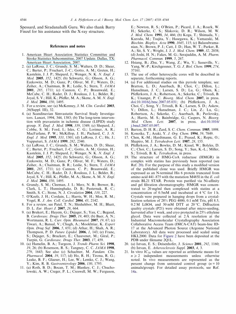
evaluated these analogs in a 7-day LDL-lowering assayconducted in cholestyramine-primed hamsters. Initially,analogs were evaluated at a single oral dose of 10 mg/kgand compared against the benchmark rosuvastatin atthe same dose. As highlighted in Table 4, compounds 44and 47 exhibited comparable efficacy to rosuvastatin atthis dose. Metabolic stability studies (data not shown)suggested that 44 was more metabolically robust than47, and as such 44 was selected for a full dose–responsestudy in this hamster model. Doses of 0.1, 0.5, 1, 5, 10,and 50 mg/kg of 44 were evaluated in separate animalgroups (n = 10 each) resulting in an ED50 = 3 mg/kg forthis inhibitor. For comparison purposes, rosuvastatinwas evaluated at a dose of 10 mg/kg in an additional ani-mal group during this study. As illustrated, 44 and rosu-vastatin exhibited similar efficacies at this dose (Fig. 3).
In conclusion, we have described the design, synthesis,and biological evaluation of a series of pyrrole-basedHMG-CoA reductase inhibitors based upon an isomericvariation of the atorvastatin template. Through struc-ture–activity studies, we identified a subset of alkylamide containing analogs that exhibited excellent hepa-toselectivity as well as good acute and chronic efficacy inanimal models of dyslipidemia. Of particular interestwas inhibitor 44 which was 3200-fold selective for inhib-iting cholesterol synthesis in hepatocytes as opposed tomyocytes, yet it maintained pre-clinical LDL-loweringefficacy comparable to that of rosuvastatin. It is antici-pated that additional pre-clinical characterization of thiscompound will represent an important step in the iden-tification of a next generation HMG-CoA reductaseinhibitor useful for helping patients to achieve increas-ingly aggressive LDL-lowering goals.
Acknowledgments
We acknowledge the technical contributions made byFang Sun, Loola Al-Kassim, Susan Holly, Cindy
4544 J. A. Pfefferkorn et al. / Bioorg. Med. Chem. Lett. 17 (2007) 4538–4544
Spessard, and Siradanahalli Guru. We also thank BarryFinzel for his assistance with the X-ray structure.
References and notes
1. American Heart Association Statistics Committee andStroke Statistics Subcommittee, 2007 Update. Dallas, TX:American Heart Association, 2007.
2. (a) LaRosa, J. C.; Grundy, S. M.; Walters, D. D.; Shear,C.; Barter, P.; Fruchart, J.-C.; Gotto, A. M.; Greten, H.;Kastelein, J. J. P.; Shepard, J.; Wenger, N. K. N. Engl. J.Med. 2005, 352, 1425; (b) Schwartz, G.; Olsson, A. G.;Ezekowitz, M. D.; Ganz, P.; Oliver, M. F.; Waters, D.;Zeiher, A.; Chaitman, B. R.; Leslie, S.; Stern, T. JAMA2001, 285, 1711; (c) Cannon, C. P.; Braunwald, E.;McCabe, C. H.; Rader, D. J.; Rouleau, J. L.; Belder, R.;Joyal, S. V.; Hill, K.; Pfeffer, M. A.; Skene, A. M. N. Engl.J. Med. 2004, 350, 1495.
3. For a review, see: (a) McKenney, J. M. Clin. Cardiol. 2003,26(Suppl. III), 32.
4. (a) Scandinavian Simvastatin Survival Study Investiga-tors. Lancet, 1994, 344, 1383; (b) The long-term interven-tion with pravastatin in ischemic disease (LIPID) studygroup. N. Engl. J. Med. 1998, 339, 1349; (c) Shepard, J.;Cobbe, S. M.; Ford, I.; Isles, C. G.; Lorimer, A. R.;MacFarlane, P. W.; McKillop, J. H.; Pachard, C. J. N.Engl. J. Med. 1995, 333, 1301; (d) LaRosa, J. C.; He, J.;Vupputuri, S. JAMA 1999, 282, 2340.
5. (a) LaRosa, J. C.; Grundy, S. M.; Walters, D. D.; Shear,C.; Barter, P.; Fruchart, J.-C.; Gotto, A. M.; Greten, H.;Kastelein, J. J. P.; Shepard, J.; Wenger, N. K. N. Engl. J.Med. 2005, 352, 1425; (b) Schwartz, G.; Olsson, A. G.;Ezekowitz, M. D.; Ganz, P.; Oliver, M. F.; Waters, D.;Zeiher, A.; Chaitman, B. R.; Leslie, S.; Stern, T. JAMA2001, 285, 1711; (c) Cannon, C. P.; Braunwald, E.;McCabe, C. H.; Rader, D. J.; Rouleau, J. L.; Belder, R.;Joyal, S. V.; Hill, K.; Pfeffer, M. A.; Skene, A. M. N. Engl.J. Med. 2004, 350, 1495.
6. Grundy, S. M.; Cleeman, J. I.; Merz, N. B.; Brewer, B.;Clark, L. T.; Hunninghake, D. B.; Pasternak, R. C.;Smith, S. C.; Stone, N. J. Circulation 2004, 110, 227.
7. O’Keefe, J. H.; Cordain, L.; Harris, W. H.; Moe, R. M.;Vogel, R. J. Am. Coll. Cardiol. 2004, 43, 2142.
8. For a review, see Patel, T. N.; Shishehbor, M. H.; Bhatt,D. L. Eur. Heart J. 2007, 28, 664.
9. (a) Brukert, E.; Hayem, G.; Dejager, S.; Yau, C.; Begaud,B. Cardiovasc. Drugs Ther. 2005, 19, 403; (b) Baer, A. N.;Wortmann, R. L. Curr. Opin. Rheumatol. 2007, 19, 67; (c)Tiwari, A.; Bansal, V.; Chugh, A.; Mookhtiar, K. ExpertOpin. Drug Saf. 2006, 5, 651; (d) Athar, H.; Shah, A. R.;Thompson, P. D. Future Lipidol. 2006, 1, 143; (e) Franc,S.; Dejager, S.; Bruckert, E.; Chauvenet, M.; Giral, P.;Turpin, G. Cardiovasc. Drugs Ther. 2003, 17, 459.
10. (a) Hamelin, B. A.; Turgeon, J. Trends Pharm Sci. 1998,19, 26; (b) Rosenson, R. S.; Tangney, C. C. JAMA 1998,279, 1643; See also (c) Schachter, M. Fundam. Clin.Pharmacol. 2004, 19, 117; (d) Ho, R. H.; Tirona, R. G.;Leake, B. F.; Glaeser, H.; Lee, W.; Lemke, C. J.; Wang,Y.; Kim, R. B. Gastroenterology 2006, 130, 1793.
11. (a) Roth, B. D.; Bocan, T. M.; Blankey, C. J.; Chucho-lowski, A. W.; Creger, P. L.; Creswell, M. W.; Ferguson,
E.; Newton, R. S.; O’Brien, P.; Picard, J. A.; Roark, W.H.; Sekerke, C. S.; Sliskovic, D. R.; Wilson, M. W.J. Med. Chem. 1991, 34, 466; (b) Koga, T.; Shimada, Y.;Kuroda, M.; Tsujita, Y.; Hasegawa, K.; Yamazaki, M.Biochim. Biophys. Acta 1990, 1045, 115; (c) Balasubrama-nian, N.; Brown, P. J.; Catt, J. D.; Han, W. T.; Parker, R.A.; Sit, S. Y.; Wright, J. J. J. Med. Chem. 1989, 32, 2038;(d) Joshi, H. N.; Fakes, M. G.; Serajuddin, A. M. Pharm.Pharmacol. Commun. 1999, 5, 267.
12. Hsiang, B.; Zhu, Y.; Wang, Z.; Wu, Y.; Sasseville, V.;Yang, W.-P.; Kirchgessner, T. G. J. Biol. Chem. 1999, 274,37161.
13. The use of other heterocyclic cores will be described inseparate, forthcoming reports.
14. (a) For additional studies on this pyrrole template, see:Bratton, L. D.; Auerbach, B.; Choi, C.; Dillon, L.;Hanselman, J. C.; Larsen, S. D.; Lu, G.; Olsen, K.;Pfefferkorn, J. A.; Robertson, A.; Sekerke, C.; Trivedi, B.K.; Unangst, P. C. Bioorg. Med. Chem. 2007, in press.doi:10.1016/j.bmc.2007.05.031; (b) Pfefferkorn, J. A.;Choi, C.; Song, Y.; Trivedi, B. K.; Larsen, S. D.; Askew,V.; Dillon, L.; Hanselman, J. C.; Lin, Z.; Lu, G.;Robertson, A.; Sekerke, C.; Auerbach, B.; Pavlovsky,A.; Harris, M. S.; Bainbridge, G.; Caspers, N. Bioorg.Med. Chem. Lett. 2007, in press. doi:10.1016/j.bmcl.2007.05.097.
15. Barton, D. H. R.; Zard, S. C. Chem. Commun. 1985, 1098.16. Konoike, T.; Araki, Y. J. Org. Chem. 1994, 59, 7849.17. Chen, K.-M.; Hardtmann, G. E.; Prasad, K.; Repic, O.;
Shapiro, M. J. Tetrahedron Lett. 1987, 28, 155.18. Pfefferkorn, J. A.; Bowles, D. M.; Kissel, W.; Bolyles, D.
C.; Choi, C.; Larsen, S. D.; Song, Y.; Sun, K.-L.; Miller,S.; Trivedi, B. K. Tetrahedron, in press.
19. The structure of HMG-CoA reductase (HMGR) incomplex with statins has previously been reported (seeRef. 20). For the purpose of this study a modified versionof the published clone was used. Briefly HMGR wasexpressed as an N-terminal His 6 protein truncated fromamino acid 441–875 with the mutation M485I in the E. colistrain BL21 STAR. Protein was purified via Ni-affinityand gel filtration chromatography. HMGR was concen-trated to 20 mg/ml then complexed with statins at aconcentration of 0.5mM and incubated at 4 �C for 1 h.Crystals were prepared in hanging drops from a crystal-lization solution of 28% PEG 4000, 0.1 mM Tris, pH 8.5,0.2 M LiSO4, and 50 mM DTT at 20 �C. Diffractionquality crystals (P21) were obtained after micro-seeding,harvested after 1 week, and cryo protected in 25% ethyleneglycol. Data were collected at 2 A resolution at theIndustrial Macromolecular Crystallography AssociationCollaborative Access Team (IMCA-CAT) beam-line ID-17 at the Advanced Photon Source (Argonne NationalLaboratory). All data were processed and scaled usingHKL2000. Data for Figure 2 have been deposited at thePDB under filename 2Q1L.
20. (a) Istvan, E. S.; Deisenhofer, J. Science 2001, 292, 1160;(b) Istvan, E. Atherosclerosis Suppl. 2003, 4, 3.
21. In vitro IC50 values are reported as arithmetic means forn P 2 independent measurements unless otherwisenoted. In vivo measurements are represented as thepercent change from untreated control group (n = 10animals/group). For detailed assay protocols, see Ref.14a.
Copyright © 2022 FDOKUMEN