Expression of the Ubiquitin-Conjugating DNA Repair Enzymes HHR6A and B Suggests a Role in...
14
DEVELOPMENTAL BIOLOGY 173, 119 –132 (1996) Article No. 0011 Expression of the Ubiquitin-Conjugating DNA Repair Enzymes HHR6A and B Suggests a Role in Spermatogenesis and Chromatin Modification Marcel H. M. Koken,* ,1 Jos W. Hoogerbrugge,² Iris Jaspers-Dekker,* Jan de Wit,* Rob Willemsen,* Henk P. Roest,* J. Anton Grootegoed,² and Jan H. J. Hoeijmakers* ,2 *Department of Cell Biology and Genetics, Medical Genetics Centre, Erasmus University, P.O. Box 1738 3000DR Rotterdam, The Netherlands; and ²Department of Endocrinology and Reproduction, Erasmus University, Rotterdam, The Netherlands RAD6, a member of the expanding family of ubiquitin-conjugating (E2) enzymes, functions in the so-called ‘‘N-rule’’ protein breakdown pathway of Saccharomyces cerevisiae. In vitro, the protein can attach one or multiple ubiquitin (Ub) moieties to histones H2A and B and trigger their E3-dependent degradation. Rad6 mutants display a remarkably pleiotropic phenotype, implicating the protein in DNA damage-induced mutagenesis, postreplication repair, repres- sion of retrotransposition, and sporulation. RAD6 transcription is strongly induced upon UV exposure and in meiosis, suggesting that it is part of a damage-induced response pathway and that it is involved in meiotic recombination. It is postulated that the protein exerts its functions by modulating chromatin structure. Previously, we have cloned two human homologs of this gene (designated HHR6A and HHR6B) and demonstrated that they partially complement the yeast defect. Here we present a detailed characterisation of their expression at the transcript and protein levels. Both HHR6 proteins, resolved by 2-dimensional immunoblot analysis, are expressed in all mammalian tissues and cell types examined, indicating that both genes are functional and constitutively expressed. Although the proteins are highly conserved, the UV induction present in yeast is not preserved, pointing to important differences in damage response between yeast and mammals. Absence of alterations in HHR6 transcripts or protein upon heat shock and during the cell cycle suggests that the proteins are not involved in stress response or cell cycle regulation. Elevated levels of HHR6 transcripts and proteins were found in testis. Enhanced HHR6 expression did not coincide with meiotic recombination but with the replacement of histones by transition proteins. Immunohistochemistry demon- strated that the HHR6 proteins are located in the nucleus, consistent with a functional link with chromatin. Electron microscopy combined with immunogold labeling revealed a preferential localisation of HHR6 in euchromatin areas, suggesting that the protein is associated with transcriptionally active regions. Our findings support the idea that both HHR6 genes have overlapping, constitutive functions related to chromatin conformation and that they have a specific role in spermatogenesis, involving Ub-mediated histone degradation. q 1996 Academic Press, Inc. INTRODUCTION pression of retrotransposition and sporulation (for a recent review see Lawrence, 1994). The RAD6(UBC2) gene en- codes a 172-amino-acid protein (M r 19.7 kDa) (Reynolds et Saccharomyces cerevisiae rad6 mutants display an ex- al., 1985) which was shown to be a ubiquitin-conjugating tremely pleiotropic phenotype: hypersensitivity to a re- (E 2 ) enzyme (Jentsch et al., 1987). Ubiquitin (Ub), a highly markably wide spectrum of genotoxic agents, defects in conserved 76-amino-acid polypeptide, can be attached to damage-induced mutagenesis, postreplication repair, re- many cellular proteins, after which it functions as a signal for selective degradation, (re)folding, or stabilisation. This implicates the protein in a variety of processes, ranging from 1 Present address: CNRS UPR43 Ho ˆ pital St. Louis, Ba ˆ timent IN- cellular stress response, mitochondrial protein import, and SERM, 16 Rue de la Grange aux Belles, 75010 Paris, France. 2 To whom correspondence should be addressed. peroxisomal biogenesis to apoptosis (for reviews see: Cie- 119 0012-1606/96 $12.00 Copyright q 1996 by Academic Press, Inc. All rights of reproduction in any form reserved.
Transcript of Expression of the Ubiquitin-Conjugating DNA Repair Enzymes HHR6A and B Suggests a Role in...
DEVELOPMENTAL BIOLOGY 173, 119–132 (1996)Article No. 0011
Expression of the Ubiquitin-Conjugating DNARepair Enzymes HHR6A and B Suggests a Role inSpermatogenesis and Chromatin Modification
Marcel H. M. Koken,*,1 Jos W. Hoogerbrugge,† Iris Jaspers-Dekker,*Jan de Wit,* Rob Willemsen,* Henk P. Roest,* J. Anton Grootegoed,†and Jan H. J. Hoeijmakers*,2
*Department of Cell Biology and Genetics, Medical Genetics Centre, Erasmus University,P.O. Box 1738 3000DR Rotterdam, The Netherlands; and †Department of Endocrinologyand Reproduction, Erasmus University, Rotterdam, The Netherlands
RAD6, a member of the expanding family of ubiquitin-conjugating (E2) enzymes, functions in the so-called ‘‘N-rule’’protein breakdown pathway of Saccharomyces cerevisiae. In vitro, the protein can attach one or multiple ubiquitin(Ub) moieties to histones H2A and B and trigger their E3-dependent degradation. Rad6 mutants display a remarkablypleiotropic phenotype, implicating the protein in DNA damage-induced mutagenesis, postreplication repair, repres-sion of retrotransposition, and sporulation. RAD6 transcription is strongly induced upon UV exposure and in meiosis,suggesting that it is part of a damage-induced response pathway and that it is involved in meiotic recombination.It is postulated that the protein exerts its functions by modulating chromatin structure. Previously, we have clonedtwo human homologs of this gene (designated HHR6A and HHR6B) and demonstrated that they partially complementthe yeast defect. Here we present a detailed characterisation of their expression at the transcript and protein levels.Both HHR6 proteins, resolved by 2-dimensional immunoblot analysis, are expressed in all mammalian tissues andcell types examined, indicating that both genes are functional and constitutively expressed. Although the proteinsare highly conserved, the UV induction present in yeast is not preserved, pointing to important differences in damageresponse between yeast and mammals. Absence of alterations in HHR6 transcripts or protein upon heat shock andduring the cell cycle suggests that the proteins are not involved in stress response or cell cycle regulation. Elevatedlevels of HHR6 transcripts and proteins were found in testis. Enhanced HHR6 expression did not coincide withmeiotic recombination but with the replacement of histones by transition proteins. Immunohistochemistry demon-strated that the HHR6 proteins are located in the nucleus, consistent with a functional link with chromatin. Electronmicroscopy combined with immunogold labeling revealed a preferential localisation of HHR6 in euchromatin areas,suggesting that the protein is associated with transcriptionally active regions. Our findings support the idea thatboth HHR6 genes have overlapping, constitutive functions related to chromatin conformation and that they have aspecific role in spermatogenesis, involving Ub-mediated histone degradation. q 1996 Academic Press, Inc.
INTRODUCTION pression of retrotransposition and sporulation (for a recentreview see Lawrence, 1994). The RAD6(UBC2) gene en-codes a 172-amino-acid protein (Mr 19.7 kDa) (Reynolds etSaccharomyces cerevisiae rad6 mutants display an ex-al., 1985) which was shown to be a ubiquitin-conjugatingtremely pleiotropic phenotype: hypersensitivity to a re-(E2) enzyme (Jentsch et al., 1987). Ubiquitin (Ub), a highlymarkably wide spectrum of genotoxic agents, defects inconserved 76-amino-acid polypeptide, can be attached todamage-induced mutagenesis, postreplication repair, re-many cellular proteins, after which it functions as a signalfor selective degradation, (re)folding, or stabilisation. Thisimplicates the protein in a variety of processes, ranging from1 Present address: CNRS UPR43 Hopital St. Louis, Batiment IN-cellular stress response, mitochondrial protein import, andSERM, 16 Rue de la Grange aux Belles, 75010 Paris, France.
2 To whom correspondence should be addressed. peroxisomal biogenesis to apoptosis (for reviews see: Cie-
119
0012-1606/96 $12.00Copyright q 1996 by Academic Press, Inc.All rights of reproduction in any form reserved.
/ m4450f8065 12-09-95 00:25:48 dba Dev Bio
120 Koken et al.
chanover, 1994; Finley and Chau, 1991; Hershko and Cie- which meiotic recombination and extensive chromatin re-modeling take place. Finally, this study intended to providechanover, 1992; Jentsch, 1992; Jentsch et al., 1990; Re-
chsteiner, 1988). Ubiquitination occurs by a cascade of clues as to whether the two highly homologous humangenes have acquired distinct functions detectable in the wayreactions: ATP-dependent activation of Ub by binding to a
Ub-activating enzyme (E1), transfer of the Ub moiety to a they are expressed, thus giving an evolutionary rationalefor the gene duplication that has occurred at the beginningmember of a family of Ub-conjugating enzymes (UBC, or
E2 enzymes), which in turn covalently attaches its activated of the origin of mammals.Ub to the target protein, with or without the assistance ofa ubiquitin–protein ligase (E3). Experiments have shownthat yeast RAD6 is able to link in vitro one (Jentsch et al., MATERIALS AND METHODS1987) or multiple (Sung et al., 1988) ubiquitin molecules toa specific lysine residue in the carboxyl terminus of histones
RNA and DNA ManipulationsH2A and H2B (Thorne et al., 1987). In vitro, the presenceof an E3 molecule causes tagged artificial substrates to be Standard DNA manipulations were done as describedquickly broken down (Dohmen et al., 1991; Sung et al., (Sambrook et al., 1989). Total RNA was isolated by means1991). RAD6 is one of the E2s involved in breakdown of of the LiCl/urea precipitation procedure (Sambrook et al.,proteins via the so-called ‘‘N-rule’’ (Bachmair et al., 1986; 1989). RNA samples were fractionated on 1% agarose form-Dohmen et al., 1991; Varshavsky, 1992; Watkins et al., aldehyde gels (Fourney et al., 1988) and transferred onto1993). The N-rule, which is conserved throughout evolution nylon membranes (Zetaprobe, Hybond). RNA hybridisa-from bacteria to mammals, is derived from the observation tion’s took place overnight at 567C in 31 SSC and washingsthat the N-terminal amino acid of a protein is one of the until 0.31 SSC were performed as detailed elsewhere (Sam-determinants for its in vivo half-life (Bachmair et al., 1986). brook et al., 1989). Although HHR6A and B are very homol-When histones are also one of the targets of RAD6 in vivo, ogous at the protein level the genes do not show any cross-it is likely that this E2 enzyme mediates the chromatin hybridisation at the nucleotide level under the conditionschanges required for postreplication repair and sporulation. specified above (as shown by Figs. 1A and 2A).
Via evolutionary walking using nucleotide homology wehave isolated homologous RAD6 genes from Schizosaccha-romyces pombe (named rhp6/) (Reynolds et al., 1990), Dro- UV-Light and Heat Shock Treatmentsophila melanogaster (Dhr6) (Koken et al., 1991a), and a
HeLa cells, primary human fibroblasts, or primary humanduplicated locus in man, HHR6A and HHR6B (Koken et al.,keratinocytes (Gibbs et al., 1990) were grown in a 1:1 mix-1991b). HHR6A is located on the X chromosome at positionture of Dulbecco–Vogt medium (DMEM) and Ham’s F10q24–25, and HHR6B on 5q23–31 (Koken et al., 1992). Themedium supplemented with 10% foetal calf serum, 0.75RAD6 homologs of these species share ú68% sequencemg/ml penicillin, 1.25 mg/ml streptomycin, and 2.92 mg/identity. A unique feature of yeast RAD6, not found in theml glutamine. When reaching near confluency cells wereother homologs, is the presence of a 20-amino-acid acidicwashed twice in phosphate-buffered saline (PBS) and irradi-C-terminus, which is essential for sporulation and the poly-ated with 1, 5, or 10 J/m2. Keratinocytes were cultured asubiquitination of histones in vitro, but not for monoubiqui-described (Gibbs et al., 1990) (40 J/m2). After UV exposuretination (Sung et al., 1988). Due to their high structuraland two additional washings with PBS, the conditioned me-conservation, all RAD6 homologues are able to functionallydium was added to the cells. For the heat-shock treatment,correct yeast rad6D mutants with respect to UV survivalcells were placed at 417C for 2 hr, after which they wereand UV-induced mutagenesis, but not with respect to thereturned to 377C. The time point of irradiation or transferdefect in sporulation, which requires the acidic tail (Kokento the 417C incubator was considered t Å 0. At differentet al., 1991a,b; Reynolds et al., 1990). RAD6 in yeast istime points cells were isolated by scraping, washed in PBS,UV-inducible consistent with the idea that RAD6 plays anand quickly frozen in liquid nitrogen.important role in an inducible cellular response to geno-
toxic insult. In addition, the gene is induced in meiosiscoinciding with meiotic recombination. In mammals dam- Cell Cycle Synchronisation by Double Thymidineage-induced cellular responses have received considerable Blockattention in recent years. Links observed with cell cycleprogression, DNA repair processes and genome stability, The synchronisation of HeLa cells was done with a double
thymidine (TdR) block (Galavazi et al., 1966). The syn-replication and transcription, as well as apoptosis have beenthe topic of intensive research. Therefore, it was of consider- chronisation was checked on a fluorescence activated cell
sorter; a cell sample was stained with propidium iodide andable interest to investigate whether the strong structuraland functional conservation of RAD6 also includes its gene- the DNA content was determined (Vindelov et al., 1983).
The S phase peak appeared 1 hr after the second TdR blockregulatory properties. In view of the presumed role of RAD6in chromatin transactions and sporulation we examined in had been released. After 8 hr the peak of G2 cells appeared
(74% pure), after 9 hr that of mitotic cells (58% pure) ap-detail the involvement of HHR6 in spermatogenesis, in
Copyright q 1996 by Academic Press, Inc. All rights of reproduction in any form reserved.
/ m4450f8065 12-09-95 00:25:48 dba Dev Bio
121Expression Analysis of Human RAD6
peared, and finally after 14.5 hr the G1 cells (84% pure) Immunohistochemistrycould be collected.
Light microscopy. Small tissue samples (f0.5cm) wereextracted from mouse or rat and fixed in 4% paraformalde-
Isolation of Different Cell Types from Rat Testis hyde–PBS for 4 hr at 47C, dehydrated, and embedded inparaffin (Fluka, Switzerland) according to standard proce-Spermatocytes, round and elongating spermatids. Sper-dures (Zeller et al., 1987, 1991). (For optimal detection ofmatogenic cells were isolated from 40- to 50-day-old ratsRAD6 protein in tissue sections we determined that theusing collagenase and trypsin treatment, and purified usingfixation time should not exceed 4 hr). Seven-micrometersedimentation at unit gravity (StaPut procedure) followedtissue sections were made, collected on gelatin"CrK-by density gradient centrifugation (Percoll gradients)(SO4)2r12H2O-coated microscopic slides, and dried over-(Grootegoed et al., 1986). The purity of the cell preparationsnight. After deparaffination in a xylol–ethanol–PBS se-was analysed using DNA-flow cytometry as described (Toe-quence, endogenous peroxidase was blocked by incubationbosch et al., 1989); the preparations enriched in spermato-in 100% methanol/1% H2O2 for 30 min. Slides were rinsedcytes and spermatids contained more than 90% of cells within PBS/0.5% Tween 20, 31 5 min, and incubated with pri-a 4 or 1 C amount of DNA per cell, respectively.mary antibody (total anti-RAD6 serum 1:600, affinity-puri-Sertoli cells from young (21-day-old) and adult rats.fied serum 1:40) in a moist incubation chamber at roomHighly purified Sertoli cells were isolated from immaturetemperature (RT) for 1 hr. After extensive washing (31 5rats as described (Themmen et al., 1991). Essentially themin) with PBS/Tween, the slides were incubated withsame method was used to obtain a preparation of adult Ser-horseradish peroxidase-labeled swine anti-rabbit conjugatetoli cells which was contaminated with 50% germinal cells.(DAKO, Denmark) (1:100 diluted) as second step antibodyThe mouse Sertoli cell line TM4, which was used in somefor 45 min at RT. After washing (31 5 min PBS/Tween)of the experiments, is described elsewhere (Mather, 1980).peroxidase was visualised with 0.1% 3,3 *-diaminobenzi-dinerHCl (Serva, FRG), 0.01% H2O2. Counterstaining was
Peptide Synthesis and Coupling to Affi-gel 10 done with haematoxilin for half a minute, after which theslides were passed through a PBS–ethanol–xylol sequenceBecause the original antiserum against yeast RAD6 (Mor-and embedded in Entellan (Merck, FRG).rison et al., 1988) shows some cross-reactivity with higher
Immunoelectron microscopy. Mouse testis were fixedmolecular weight proteins next to the 17-kDa HHR6 pro-in 0.1 M phosphate buffer, pH 7.3, containing 2% paraform-teins, this serum (designated total RAD6 antiserum) wasaldehyde. After fixation for 1 hr at 47C, samples were em-affinity purified with an N-terminal HHR6 oligopeptidebedded in Lowicryl K4M (Roth et al., 1981). For antigencoupled to Affi-gel. This serum (designated AP-RAD6 anti-localisation on thin sections the immunogold techniqueserum) reacts almost exclusively with a major band of 17was used (Willemsen et al., 1988). Subcellular quantitationkDa, and a faint band of 25 kDa, which may represent aof the gold particles was done in Leydig cells, according toubiquitinated form of HHR6 (Fig. 3D). The N-terminalWillemsen et al. (1991).HHR6 peptide MSTPARRRLMRDFKC, conserved in all the
For light as well as electron microscopy two immunocy-RAD6 homologous proteins, was prepared by the 9-fluore-tochemical controls were always included: (a) omission ofnylmethoxycarbonyl (Fmoc) method using solid-phase syn-the primary antibody incubation step and (b) incubationthesis according to Merrifield (1963) on an automated pep-with normal rabbit serum as substitution for the primarytide synthesiser (NovaSyn-Crystal, Novabiochem). Afterantibody. Background was negligible.completion of the synthesis, the peptide was cleaved from
its support and amino acid side-chain protecting groupswere removed. Peptides were purified by high-performance SDS–PAGE Gel Electrophoresis and 2-Dimensionalliquid chromatography on a reverse-phase C18 column Gel Electrophoresis(Merck, LiChroCart) using a gradient of 0–60% acetonitril/H2O in 0.1% trifluoracetic acid. Amino acid analysis was Mouse or rat tissues were fragmented under liquid nitro-
gen in a mortar. The fragments or cultured cells were col-performed (473A protein sequencer, Applied Biosystems) toensure correct residue composition. Coupling to Affi-gel 10 lected in PBS at 47C and subjected to 10 cycles of 10-sec
sonification at full amplitude at 47C. These crude extracts(Bio-Rad) was according to the manufacturer’s descriptionin a 0.1 M Mops buffer, pH 7.5, for 2 hr at 47C. To block were used in all experiments. The preparation of nuclear
and cytoplasmic extracts was performed following three dif-any remaining active esters the slurry was incubated with0.1 vol of 1 M ethanolaminerHCl for 1 hr at 47C. After ferent methods: the methods of Dignam (Dignam et al.,
1983) and Lee (Lee et al., 1988) and a combination of meth-extensive washing with water the column was equilibratedwith PBS. The sample was loaded onto the column in PBS. ods described by Lue (Lue and Kornberg, 1987) and Radke
(Radke et al., 1983). Protein concentration was determinedAfter extensive washing with PBS, column elution was donewith 0.1 M glycine, 0.5 M NaCl, pH 2.5, and the fractions with the BCA assay (Pierce) as described by the manufac-
turer. Two-dimensional gel electrophoresis was principallywere immediately neutralised in 0.5 M phosphate buffer,pH 7.2. done according to the Bio-Rad mini-protean 2-D cell in-
Copyright q 1996 by Academic Press, Inc. All rights of reproduction in any form reserved.
/ m4450f8065 12-09-95 00:25:48 dba Dev Bio
122 Koken et al.
struction manual and as described by Luider et al. (1992). In Vitro Transcription and in Vitro TranslationGels with a length of 6.5 cm were prepared in glass tubes The construct H28ccc, containing a 1.7-kb cDNA of hu-2-mm diameter. Next, 0.25 ml Bio-lyte 3/5 and 0.25 ml Bio- man HHR6A (Koken et al., 1991b), was linearised withlyte 4/6 ampholytes (Bio-Rad), 2 ml 10% Triton X-100, 5.5 XbaI. The H28ccc construct differs from the previously re-g urea (Merck), 1.33 ml acrylamide solution (28.3% acryl- ported HHR6A sequence in that at nucleotide position 146amide and 1.62% piperazinediacrylamide (Bio-Rad)), 1.97 (Koken et al., 1991b), a mutation derived from cDNA ampli-ml distilled water, 10 ml 10% ammonium persulfate (Bio-
fication in Escherichia coli or as PCR artefact (resulting inRad), and 10 ml N,N,N *,N*-tetramethylethylenediamine
the reported guanine) has been changed into the geno-(Bio-Rad) were mixed and allowed to polymerise in the glass mically encoded adenine, resulting in a change of the pre-tubes at 377C for about an hour. The gels were covered with viously reported glycine into aspartic acid (amino acid posi-overlay buffer (9 M urea, 0.8% Bio-lyte 3/5, 0.2% Bio-lyte tion 49). pPHB2 containing an 0.8-kb EcoRI fragment, har-5/7, bromophenol blue) and preelectrophoresis was per- bouring the complete open reading frame of HHR6B (Kokenformed at 200, 300, 400 V for 10, 20, and 20 min, respec- et al., 1991b), was linearised with BamHI. In vitro transcrip-tively. As upper chamber buffer a 20 mM NaOH solution tion as well as in vitro translation were done exactly aswas used, whereas 100 mM H3PO4 served as lower chamber described (Promega, 1991).buffer.
Subsequently, samples were diluted with an equal vol-ume of sample buffer (2% SDS, 10% glycerol, 62.5 mM Tris/ RESULTSHCl, pH 6.8, bromophenol blue, 0.1% dithiotreitol (DTT)),boiled for 3 min, and chilled on ice. Samples were diluted
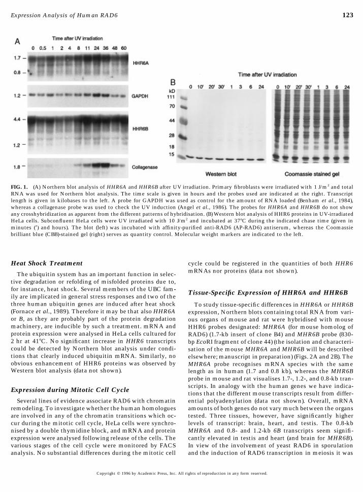
UV-Induced Expressionwith an equal volume of lysis buffer (0.5 M urea, 2% TritonX-100, 0.1% DTT, 1.6% Bio-lyte 3/5, 0.4% Bio-lyte 5/7 in In yeast, RAD6 transcription is induced upon UV irradia-
tion (§sixfold) within 30 min after exposure (Madura etdistilled water). One hundred microliters containing 50 mgprotein was loaded under 40 ml of 1:1 diluted overlay buffer al., 1990). To investigate whether this feature of RAD6 is
evolutionarily conserved, Northern blot analysis forand electrophoresis was performed for 3.5 hr at 600 V.The gels were gently removed from the glass tubes and HHR6A and HHR6B was performed on RNA from exponen-
tially growing cells, UV irradiated with 1, 5, or 10 J/m2, andequilibrated against sample buffer for about 90 min untilthe pH indicator in the acid part of the tube gel became harvested after different incubation periods. The blot was
subsequently probed with the cDNA probes for HHR6Ablue. The tubes were directly loaded on a 2.25-mm-thick15% SDS–polyacrylamide minigel (Bio-Rad) and electro- (1.7-kb Sal I fragment) and HHR6B (0.8-kb EcoRI fragment)
(Koken et al., 1991b), the UV-inducible collagenase or meth-phoresis was performed with 0.1 M sodium acetate in theanode buffer (Christy et al., 1989) at 100 V (stacking) and allotheionin IIa genes (Angel et al., 1986) (positive control
for UV induction), and GAPDH (Benham et al., 1984) (in-subsequently 150 V until the bromophenol blue reached thebottom of the gel. Blotting was done onto 0.45-mm polyvi- cluded as an internal standard for quantitation). HeLa cells,
primary human fibroblasts, and primary foreskin-derivednylidenedifluoride (PVDF) membranes (Millipore), ac-cording to the manufacturer’s description, using electro- keratinocytes were investigated. The latter cell types are
also subjected to UV irradiation in the body and are there-transfer. Blots were blocked in nonfat milk for 1 hr andincubated with the primary antibody diluted in nonfat milk fore the most relevant targets to study UV induction. Figure
1A shows the result of a ‘‘physiological’’ dose of 1 J/m2 UV(anti-yeast RAD6, 1:2000 or AP-RAD6 antiserum, 1:100)at 47C, overnight. After extensive washing with PBS/0.5% irradiation on primary fibroblasts. When corrected for the
slight variation in RNA amounts per lane (see GAPDH hy-Tween 20, the second antibody (goat anti-rabbit antibodies,alkaline phosphatase labeled (TAGO, Inc.)) was incubated bridisation) it appears that irradiation did not result in a
significant increase of human HHR6A (0.8 and 1.7 kb) andfor 1 hr at 47C, in a 1:1000 dilution in PBS/Tween. Afterseveral additional washings with PBS/Tween the antigen– HHR6B (1.2 and 4.4 kb) mRNAs, under conditions where
collagenase (Fig. 1A) and metallotheionin (not shown) dis-antibody complexes were visualised with the stainingmethod described by Blake (Blake et al., 1984). Carbamylate played clear induction. Also experiments using other UV
doses, time courses, and cells did not result in detectablecarbonic anhydrase (CA) (Pharmacia lab.) and 2-D SDS–PAGE standards (Bio-Rad) were used as standards for iso- induction (data not shown). To examine the possibility that
HHR6A and 6B may be induced at the protein (translational)electrofocusing. Prestained protein molecular weight mark-ers (Gibco"BRL) were used in the second dimension. level instead of the mRNA level like in yeast, protein ex-
pression was studied using affinity-purified anti-yeastFor normal SDS–polyacrylamide gel electrophoresis 1.5-mm-thick 11 or 15% gels were used with a 4% stacking RAD6 antiserum. Western analysis after 10 J/m2 UV irradia-
tion of HeLa cells (Fig. 1B) failed to reveal a significantgel. About 20 mg protein extract was loaded, and electropho-resis was performed with 0.1 M sodium acetate in the anode increase in the amount of the 17-kDa HHR6 proteins (for
details on the antiserum see below, and under Materialsbuffer (Christy et al., 1989) at 50 V (stacking) and 150 V(running). Blotting, antibody incubations, and staining were and Methods). We conclude that in contrast to S. cerevisiae
neither of the human genes is inducible by UV.done as described above.
Copyright q 1996 by Academic Press, Inc. All rights of reproduction in any form reserved.
/ m4450f8065 12-09-95 00:25:48 dba Dev Bio
123Expression Analysis of Human RAD6
FIG. 1. (A) Northern blot analysis of HHR6A and HHR6B after UV irradiation. Primary fibroblasts were irradiated with 1 J/m2 and totalRNA was used for Northern blot analysis. The time scale is given in hours and the probes used are indicated at the right. Transcriptlength is given in kilobases to the left. A probe for GAPDH was used as control for the amount of RNA loaded (Benham et al., 1984),whereas a collagenase probe was used to check the UV induction (Angel et al., 1986). The probes for HHR6A and HHR6B do not showany crosshybridization as apparent from the different patterns of hybridisation. (B) Western blot analysis of HHR6 proteins in UV-irradiatedHeLa cells. Subconfluent HeLa cells were UV irradiated with 10 J/m2 and incubated at 377C during the indicated chase time (given inminutes (*) and hours). The blot (left) was incubated with affinity-purified anti-RAD6 (AP-RAD6) antiserum, whereas the Coomassiebrilliant blue (CBB)-stained gel (right) serves as quantity control. Molecular weight markers are indicated to the left.
Heat Shock Treatment cycle could be registered in the quantities of both HHR6mRNAs nor proteins (data not shown).The ubiquitin system has an important function in selec-
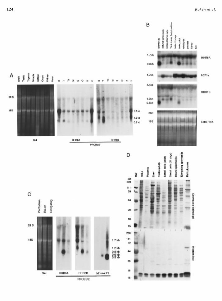
tive degradation or refolding of misfolded proteins due to,for instance, heat shock. Several members of the UBC fam- Tissue-Specific Expression of HHR6A and HHR6Bily are implicated in general stress responses and two of thethree human ubiquitin genes are induced after heat shock To study tissue-specific differences in HHR6A or HHR6B(Fornace et al., 1989). Therefore it may be that also HHR6A expression, Northern blots containing total RNA from vari-or B, as they are probably part of the protein degradation ous organs of mouse and rat were hybridised with mousemachinery, are inducible by such a treatment. mRNA and HHR6 probes designated: MHR6A (for mouse homolog ofprotein expression were analysed in HeLa cells cultured for RAD6) (1.7-kb insert of clone B4) and MHR6B probe (830-2 hr at 417C. No significant increase in HHR6 transcripts bp EcoRI fragment of clone 44) (the isolation and characteri-could be detected by Northern blot analysis under condi- sation of the mouse MHR6A and MHR6B will be describedtions that clearly induced ubiquitin mRNA. Similarly, no elsewhere; manuscript in preparation) (Figs. 2A and 2B). Theobvious enhancement of HHR6 proteins was observed by MHR6A probe recognises mRNA species with the sameWestern blot analysis (data not shown). length as in human (1.7 and 0.8 kb), whereas the MHR6B
probe in mouse and rat visualises 1.7-, 1.2-, and 0.8-kb tran-scripts. In analogy with the human genes we have indica-
Expression during Mitotic Cell Cycle tions that the different mouse transcripts result from differ-ential polyadenylation (data not shown). Overall, mRNASeveral lines of evidence associate RAD6 with chromatin
remodeling. To investigate whether the human homologues amounts of both genes do not vary much between the organstested. Three tissues, however, have significantly higherare involved in any of the chromatin transitions which oc-
cur during the mitotic cell cycle, HeLa cells were synchro- levels of transcript: brain, heart, and testis. The 0.8-kbMHR6A and 0.8- and 1.2-kb 6B transcripts seem signifi-nised by a double thymidine block, and mRNA and protein
expression were analysed following release of the cells. The cantly elevated in testis and heart (and brain for MHR6B).In view of the involvement of yeast RAD6 in sporulationvarious stages of the cell cycle were monitored by FACS
analysis. No substantial differences during the mitotic cell and the induction of RAD6 transcription in meiosis it was
Copyright q 1996 by Academic Press, Inc. All rights of reproduction in any form reserved.
/ m4450f8065 12-09-95 00:25:48 dba Dev Bio
124 Koken et al.
/ m4450f8065 12-09-95 00:25:48 dba Dev Bio
125Expression Analysis of Human RAD6
of interest to investigate the HHR6 expression in testis in cells (Fig. 2B), and both the 1.2- and the 1.7-kb mRNA spe-cies in pachytene spermatocytes (Fig. 2C), whereas they aremore detail.absent in the haploid cell types.
For several other genes, the observation has been madeTestis-Specific Expression of HHR6 mRNAs that although significant amounts of mRNA are present in
the testis no corresponding protein can be detected. It isThe mammalian testis is organised in tubular structuressurrounded by peritubular myoid cells, hormone producing speculated that these transcripts result from dysregulated
gene expression due to the extensive chromatin remodelingLeydig cells, and blood vessels (for a general review seeJohnson and Everitt, 1984). In the tubules Sertoli cells sup- taking place in spermatogenesis (reviewed by Ivell, 1992).
To examine whether both HHR6 mRNAs in testis are trans-port the developing germ cells. Complicated processes ofdifferentiation, growth, and mitotic and meiotic divisions lated into protein, Western blot analysis was performed.
Total cell extracts of different tissues and of various celltake place when spermatogonia develop into spermatozoa.After several mitotic divisions the spermatogonia differenti- types were incubated with affinity-purified anti-yeast
RAD6 antibody (AP-RAD6 antibody). This antiserum de-ate and give rise to primary spermatocytes. These cells un-dergo meiotic divisions which convert them first into sec- tects exclusively a major protein band of 17 kDa, the calcu-
lated molecular weight of HHR6A/B (Koken et al., 1991b),ondary spermatocytes and then into round spermatids. Sub-sequent differentiation results in elongated spermatids and in addition to a faint band of 25 kDa, which may represent
a ubiquitinated form of HHR6 (Figs. 2D and 3). HHR6 quan-finally spermatozoa, which have lost most of their cyto-plasm and possess highly compacted DNA. Dramatic tities vary from tissue to tissue; reticulocytes and adult
Sertoli cells contain high amounts, whereas immature Ser-changes in chromatin composition occur throughout sper-matogenesis, starting with synthesis of testis-specific his- toli cells and liver harbour relatively small quantities.
(Compare also the amounts of protein loaded in each lane,tones in spermatocytes and culminating in the total replace-ment of histones by transition proteins and ultimately by Fig. 5D, upper panel.) HHR6 proteins are also clearly de-
tected in round and elongating spermatids.protamines (Bucci et al., 1982; Smith et al., 1992).To examine HHR6 expression during this process, germ
cells and Sertoli cells of rat testis were isolated and tested Identification of HHR6A and HHR6B in Totalfor HHR6 mRNA and protein expression (Figs. 2B–2D).Protein Extracts and Determination of RelativeRNA from testes of 21-day-old rats, in which spermatogene-Amountssis is not yet complete and only a small number of round
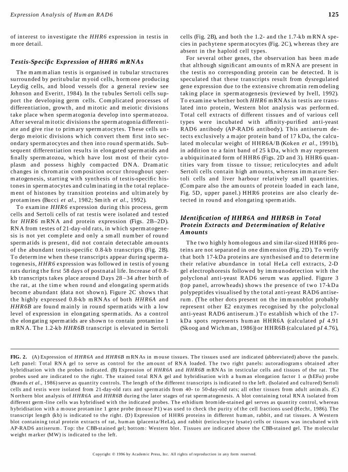
spermatids is present, did not contain detectable amounts The two highly homologous and similar-sized HHR6 pro-teins are not separated in one dimension (Fig. 2D). To verifyof the abundant testis-specific 0.8-kb transcripts (Fig. 2B).
To determine when these transcripts appear during sperma- that both 17-kDa proteins are synthesised and to determinetheir relative abundance in total HeLa cell extracts, 2-Dtogenesis, HHR6 expression was followed in testis of young
rats during the first 58 days of postnatal life. Increase of 0.8- gel electrophoresis followed by immunodetection with thepolyclonal anti-yeast RAD6 serum was applied. Figure 3kb transcripts takes place around Days 28–34 after birth of
the rat, at the time when round and elongating spermatids (top panel, arrowheads) shows the presence of two 17-kDapolypeptides visualised by the total anti-yeast RAD6 antise-become abundant (data not shown). Figure 2C shows that
the highly expressed 0.8-kb mRNAs of both HHR6A and rum. (The other dots present on the immunoblot probablyrepresent other E2 enzymes recognised by the polyclonalHHR6B are found mainly in round spermatids with a low
level of expression in elongating spermatids. As a control anti-yeast RAD6 antiserum.) To establish which of the 17-kDa spots represents human HHR6A (calculated pI 4.91the elongating spermatids are shown to contain protamine 1
mRNA. The 1.2-kb HHR6B transcript is elevated in Sertoli (Skoog and Wichman, 1986)) or HHR6B (calculated pI 4.76),
FIG. 2. (A) Expression of HHR6A and HHR6B mRNAs in mouse tissues. The tissues used are indicated (abbreviated) above the panels.Left panel: Total RNA gel to serve as control for the amount of RNA loaded. The two right panels: autoradiograms obtained afterhybridisation with the probes indicated. (B) Expression of HHR6A and HHR6B mRNAs in testicular cells and tissues of the rat. Theprobes used are indicated to the right. The stained total RNA gel and hybridisation with a human elongation factor 1 a (hEFa) probe(Brands et al., 1986) serve as quantity controls. The length of the different transcripts is indicated to the left. (Isolated and cultured) Sertolicells and testis were isolated from 21-day-old rats and spermatids from 40- to 50-day-old rats; all other tissues from adult animals. (C)Northern blot analysis of HHR6A and HHR6B during the later stages of rat spermatogenesis. A blot containing total RNA isolated fromdifferent germ-line cells was hybridised with the indicated probes. The ethidium bromide-stained gel serves as quantity control, whereashybridisation with a mouse protamine 1 gene probe (mouse P1) was used to check the purity of the cell fractions used (Hecht, 1986). Thetranscript length (kb) is indicated to the right. (D) Expression of HHR6 proteins in different human, rabbit, and rat tissues. A Westernblot containing total protein extracts of rat, human (placenta/HeLa), and rabbit (reticulocyte lysate) cells or tissues was incubated withAP-RAD6 antiserum. Top: the CBB-stained gel; bottom: Western blot. Tissues are indicated above the CBB-stained gel. The molecularweight marker (MW) is indicated to the left.
Copyright q 1996 by Academic Press, Inc. All rights of reproduction in any form reserved.
/ m4450f8065 12-09-95 00:25:48 dba Dev Bio
126 Koken et al.
the HeLa cell extract was mixed with [35S]methionine-la-beled HHR6A or HHR6B obtained through in vitro transla-tion (Fig. 3, bottom four panels). The left spot comigrateswith the in vitro-translated 35S-labeled HHR6A, whereasthe protein at the right coincides with the position of 35S-labeled HHR6B. In support of this identification is our ob-servation that the right dot (HHR6B) migrates at a slightlyhigher molecular weight than the left dot (HHR6A). Thisis consistent with the calculated molecular weight for bothproteins (HHR6A 17.243, HHR6B 17.312). The two proteins
FIG. 4. HHR6A and HHR6B protein expression in different tis-sues. 2-Dimensional gel electrophoresis was performed to separatethe two human HHR6 proteins in different human (HeLa/placenta),rabbit (reticulocyte lysate), and rat cells or tissues (young Sertolicells, adult liver, testis, round spermatids, elongating spermatids,and spermatozoa). (A minor contamination with some elongatingspermatids (or cytoplasmic fragments thereof) cannot be com-pletely excluded in the spermatozoa fraction.) The cell or tissuesource is indicated to the right and left. The direction of the pHgradient is given above the figure.
FIG. 3. Identification of HHR6A and HHR6B in total cell extractsby 2-dimensional gel electrophoresis. Top: Total HeLa extract was
seem to behave on gel according to their calculated isoelec-fractionated by 2-dimensional gel electrophoresis. From left to righttric points as judged by comparison with 2-D SDS–PAGEthe first dimension: isoelectrofocusing over a gradient from pH 6
to 4, and from top to bottom: size-fractionation by SDS–PAGE. standards (Bio-Rad) and carbamylated carbonic anhydraseThe boxed ‘‘2nd’’ indicates the molecular weight marker for the (Pharmacia). Minor modifications, such as phosphorylationsecond dimension given in kDa. C.A. marks the position of the or acetylation, which shift proteins in a pH gradient, arecarbamylated marker (Carbonic anhydrase, Bio-Rad), used as stan- not entirely excluded. Preliminary in vivo phosphorylationdard in IEF. The small 17-kDa band to the left of HHR6A and experiments, however, do not provide indications that aHHR6B (arrows) is a gel artefact which is sometimes encountered.
significant fraction of one of the HHR6 proteins is phos-Bottom 4 panels: Mixing experiment to prove the HHR6A andphorylated.HHR6B identity. Total HeLa extract was mixed with [35S]-
Two-dimensional gels were also used to examine the tis-methionine-labeled in vitro translated HHR6A (left) or HHR6Bsue-specific expression of both HHR6 proteins. Figure 4RNA (right) and separated in 2-dimensions. The gels were blottedshows that the ratios between HHR6A and HHR6B proteinsonto PVDF membranes, and HHR6A and HHR6B were visualised
with anti-yeast RAD6 antibodies (not affinity purified) (Western may vary significantly between different cells and tissues.blot). The blots were subsequently exposed to Kodak XAR5 film, HeLa cells have more HHR6A than HHR6B, total testisand the resulting autoradiogram is shown in the lower panels. The harbours equal amounts, whereas placenta contains morearrows indicate HHR6A (left two panels) and HHR6B (right two of the B protein. Both HHR6 proteins are present in roundpanels). The arrowhead indicates a form of HHR6A or HHR6B pro- and elongating spermatids, and trace amounts of proteintein which was formed in the reticulocyte lysate, and which may
may be detected even in rat epididymal spermatozoa.represent a ubiquitinated form of HHR6: calculated pI 5.26(HHR6A/ubiquitin) and pI 5.10 (HHR6B/ubiquitin) (Arnold and (Subcellular) Localisation of HHR6A and HHR6BGevers, 1990). The lower molecular weight spots on the autoradio-
The affinity-purified antibodies were also used for tissuegram probably represent breakdown products or are derived fromincomplete synthesis of the HHR6 proteins. sections of different mouse organs. Cells with a clear,
Copyright q 1996 by Academic Press, Inc. All rights of reproduction in any form reserved.
/ m4450f8065 12-09-95 00:25:48 dba Dev Bio
127Expression Analysis of Human RAD6
mainly nuclear, staining reaction could be detected with toxic stresses have gained increasing interest. Exposure ofcells to DNA injury triggers a cascade of reactions, includ-this antiserum in all tissues examined (data not shown). In
view of the RNA expression (Fig. 2) and the induction of ing intricate signal transduction pathways resulting in al-tered expression of numerous genes. A universal responseyeast RAD6 during meiotic recombination, mouse testis
was studied in more detail. In testis sections from 21-day- to inflicted gene damage in normal cells is arrest of cellcycle progression. This gives DNA repair mechanisms timeold mice only a weak reaction with the antibody was visible
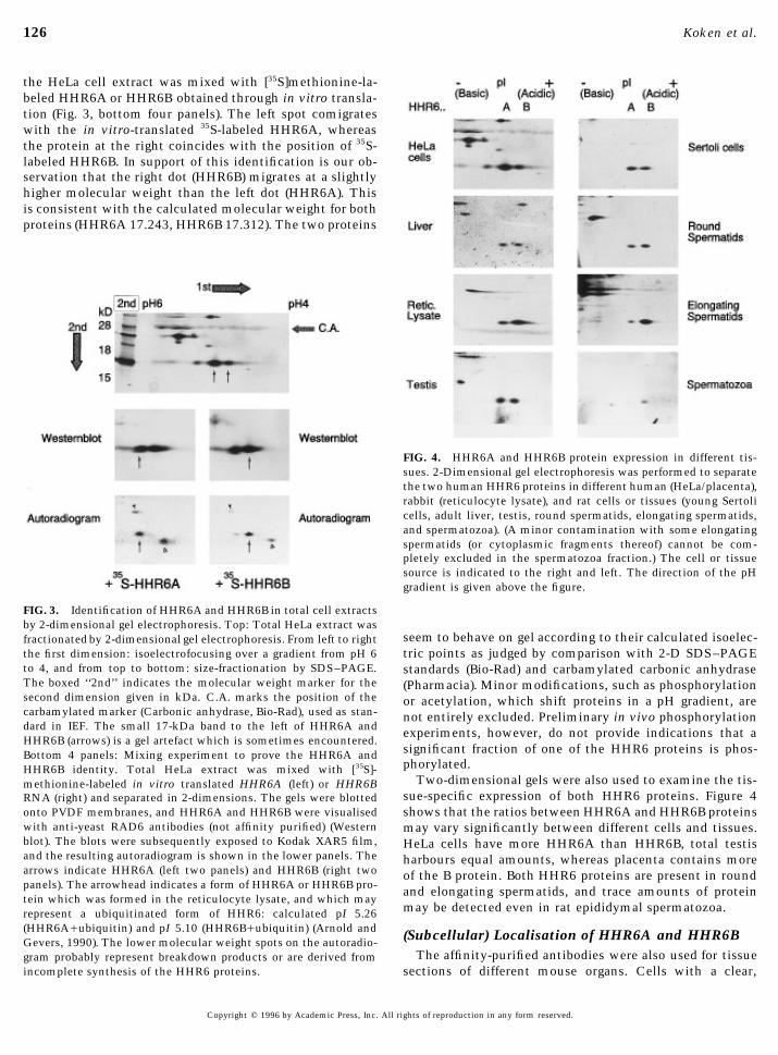
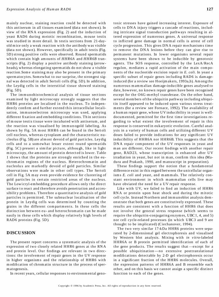
(data not shown). However, specifically in adult testis (Fig. to remove the DNA lesions before they can give rise topermanent mutations. In lower organisms several repair5D) strong positive cells were observed. Round spermatids
which contain high amounts of HHR6A and HHR6B tran- systems have been shown to be inducible by genotoxicagents. The SOS response, controlled by the LexA/RecAscripts (Fig. 2) display a positive antibody staining (arrow-
heads), whereas elongating spermatids do not show a visible regulon, mediates a rapid activation of the main compo-nents of the nucleotide excision repair in E. coli. In yeast areaction.Some staining may also be present in the primary
spermatocytes. Somewhat to our surprise, the strongest sig- specific subset of repair genes including RAD6 is damage-induced (for a review see Hoeijmakers, 1993a,b). Among thenal was detected in adult Sertoli cells (Fig. 5D). In addition,
the Leydig cells in the interstitial tissue showed staining numerous mammalian damage-inducible genes analysed todate, however, no known repair genes have been recognised(Fig. 5D).
The immunohistochemical analysis of tissue sections except for the O6G-methyl transferase. On the other hand,several members of the ubiquitin pathway including ubiqui-examined by light microscopy strongly suggests that the
HHR6 proteins are localised in the nucleus. To indepen- tin itself appeared to be induced upon various stress treat-ments (for a review see Fornace, 1992). The availability ofdently confirm and further extend this intracellular locali-
sation, immunoelectron microscopy was used, applying a human repair gene, whose UV inducibility in yeast is well-documented, permitted for the first time investigations re-different fixation and embedding conditions. Thin sections
of mouse testis tissue were incubated with antiserum, and garding to what extent the involvement of repair in thisresponse is conserved in eukaryotic evolution. Careful anal-the reaction was visualised by immunogold labeling. As
shown by Fig. 5A most HHR6 can be found in the Sertoli ysis in a variety of human cells and utilizing different UVdoses failed to provide indications for any significant UVcell nucleus, whereas cytoplasm and the characteristic nu-
cleolus (Fig. 5B) are almost devoid of gold particles. Leydig inducibility of HHR6A nor HHR6B. This suggests that theDNA repair component of the UV responses in yeast andcells and to a somewhat lesser extent round spermatids
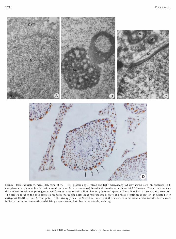
(Fig. 5C) present a similar picture, although, like in light man are different. Our recent findings with another repairgene, RAD23, whose transcription is enhanced after UVmicroscopy, the amount of protein detected is lower. Table
1 shows that the proteins are strongly enriched in the eu- irradiation in yeast, but not in man, confirm this idea (Ma-dura and Prakash, 1990, and manuscript in preparation).chromatin regions of the nucleus. Heterochromatin and
nucleolus are markedly devoid of gold particles. Similar These findings support the notion that a fundamentaldifference exist in this regard between the unicellular organ-observations were made in other cell types. The Sertoli
cell in Fig. 5A may even provide evidence for clustering of isms E. coli and yeast, and mammals. The relatively con-stant environment in which mammalian cells live maygold particles in certain restricted areas of the nucleus.
The Lowicryl-embedding procedure allows only the direct have obviated the need for a UV repair response.Like with UV, we failed to find an induction of HHR6surface to react and therefore avoids penetration and acces-
sibility problems. Therefore a quantitative analysis of gold RNA or protein upon heat shock and during the mitoticcell cycle. Instead Northern and immunoblot analysis dem-particles is permitted. The subnuclear localisation of the
protein in Leydig cells was determined by counting the onstrate that both genes are constitutively expressed. Theseresults are consistent with a function of HHR6 that doesgrains in the different compartments. In these cells the
distinction between eu- and heterochromatin can be made not involve the general stress response (which seems torequire the ubiquitin-conjugating enzymes, UBC1, 4, and 5)easily in these cells which display relatively high levels of
RAD6 proteins (Fig. 5D). nor cell cycle-related processes (in which UBC3 and 9 arethought to be implicated) (Ciechanover, 1994).
The two very similar 17-kDa HHR6 proteins were sepa-rated by 2-dimensional gel electrophoresis and visualisedDISCUSSIONby Western blot analysis. Mixing with in vitro-labeledHHR6A or B protein permitted identification of each ofThe present report concerns a systematic analysis of the
expression of two closely related HHR6 genes at the RNA the gene products. The results suggest that—except for apossible ubiquitination—no extensive posttranslationaland protein level. Our findings are relevant in two direc-
tions: the involvement of repair genes in the UV response modifications detectable by 2-D gel electrophoresis occurin a significant fraction of the HHR6 molecules. Overall,in higher organisms and the relationship of HHR6 with
modification of chromatin structure in the process of sper- the expression patterns of HHR6A and B ressemble eachother, and on this basis we cannot assign a specific distinctmatogenesis.
In recent years, cellular responses to environmental geno- function to each of the genes.
Copyright q 1996 by Academic Press, Inc. All rights of reproduction in any form reserved.
/ m4450f8065 12-09-95 00:25:48 dba Dev Bio
128 Koken et al.
FIG. 5. Immunohistochemical detection of the HHR6 proteins by electron and light microscopy. Abbreviations used: N, nucleus; CYT,cytoplasma; Nu, nucleolus; M, mitochondrion; and Ac, acrosome. (A) Sertoli cell incubated with anti-RAD6 serum. The arrows indicatethe nuclear membrane. (B) Higher magnification of A: Sertoli cell nucleolus. (C) Round spermatid incubated with anti-RAD6 antiserum.The arrows point to the gold particles found in the nucleus. (D) Light microscopic picture of a mouse testis cross section, incubated withanti-yeast RAD6 serum. Arrows point to the strongly positive Sertoli cell nuclei at the basement membrane of the tubule. Arrowheadsindicate the round spermatids exhibiting a more weak, but clearly detectable, staining.
Copyright q 1996 by Academic Press, Inc. All rights of reproduction in any form reserved.
/ m4450f8065 12-09-95 00:25:48 dba Dev Bio
129Expression Analysis of Human RAD6
TABLE 1 drastic chromosomal changes. In this respect spermatogene-Quantitation of HHR6 Protein in Leydig Cells sis may represent an advanced phenocopy of the process of
sporulation.Total no. The intracellular location of the HHR6 proteins in theof gold Surface No. of gold euchromatic part of the nucleus as indicated in this study
particles counted particles Factor ofby immunoelectron microscopy is consistent with a func-counted mm2 per mm2 enrichmenttion in chromatin transactions. In the immunogold labeling
Euchromatin 1820 87.1 20.9 21 experiments we have used the Lowicryl method whichHeterochromatin 43 14.0 3.1 3 allows only the direct surface to react. This eliminates pos-Nucleolus 8 7.86 1.0 1 sible artefacts inherent to some immunohistochemical pro-
cedures that are caused by unequal accessibility of antigenicdeterminants in different locations (Posthuma et al., 1987).Biochemical cell fractionation studies (see Materials andMethods for details on the procedure) suggest that a sub-However, clear quantitative differences exist for the
mammalian RAD6 proteins and mRNAs between the cells stantial fraction of the HHR6 proteins is only weakly associ-ated with the euchromatin.and tissues examined. Brain, heart, and testis show elevated
levels of specific mRNAs when compared to the other tis- High transcript levels of both HHR6 genes are found inround spermatids at a stage more than 2 weeks after thesues tested. Yeast RAD6 has been shown to be meiotically
induced, coinciding with the time when meiotic recombi- formation of the heterochromatic sex vesicle which be-comes visible in early pachytene spermatocytes (Stefaninination takes place (Madura et al., 1990). However, we find
high amounts of shorter mammalian RAD6 transcripts spe- et al., 1974). Since the human and the mouse HHR6A geneare located on the X chromosome (Koken et al., 1992), it iscifically in postmeiotic cells, i.e., round and elongating sper-
matids, whereas pachytene spermatocytes have levels be- remarkable that the total amount of HHR6A transcriptsseems to even increase during the transition of pachytenelow our detection. This renders a specific involvement in
meiotic recombination less likely. The analysis presented spermatocytes into round spermatids. This finding has beendescribed elsewhere in more detail (Hendriksen et al., 1995)here shows that the increase in mRNA and protein quanti-
ties of both RAD6 homologues during spermatogenesis co- and is explained by postmeiotic transcription. Hence,HHR6A is likely to represent one of the few recently discov-incides with the developmental stage when somatic and
testis-specific histones are removed from the chromatin and ered examples of X/Y-chromosomal genes whose gene prod-ucts may play an important role in postmeiotic stages ofreplaced by transition proteins and subsequently prot-
amines (Kistler et al., 1987). The induction of the two HHR6 spermatogenesis (reviewed in Hendriksen et al., 1995).Western blot analysis confirmed that both HHR6 proteinsgenes as representatives of the ubiquitin pathway at this
time of spermatogenesis is not without precedent. The are indeed present in the germ cells. This was important toverify, since it has been found for other genes that signifi-chicken ubiquitin II gene is induced at approximately the
same stage of spermatogenesis (Mezquita and Mezquita, cant quantities of testis-specific, shorter transcripts accu-mulate in spermatids, but that no corresponding proteins1991; Rocamora and Agell, 1990) and increased ubiquitina-
tion of histone H2A is observed (Oliva and Dixon, 1991). A can be detected (Capel et al., 1993; Ivell, 1992). It has beenspeculated that these transcripts could result from dysregu-ubiquitin-activating enzyme E1 encoded by the Y-chromo-
somal Sby gene exhibits testis-specific expression (Mitchell lated gene expression due to the extensive chromatin re-modeling events that take place in this stage of spermato-et al., 1991). This gene and the X-chromosomal homolog
Sbx show postmeiotic transcription in the mouse (Hendrik- genesis (Ivell, 1992). It cannot entirely be ruled out that thepresence of HHR6A protein in spermatids results from ansen et al., 1995). Interestingly, recently another E2 enzyme
(E217kD) was also found to be highly expressed in rat testis extremely long protein half-life following synthesis in ear-lier stages of spermatogenesis. The HHR6B mRNA is trans-(Wing and Jain, 1995). These observations together with the
in vitro demonstration that RAD6 is able to ubiquitinate lated more efficiently or the protein is more stable thanHHR6A as may be concluded from the larger amount ofhistones (Jentsch et al., 1987; Sung et al., 1988), provide
indirect evidence for involvement of ubiquitin and HHR6 HHR6B in elongating spermatids (Fig. 4). The transcriptionof the HHR6A gene in spermatids, together with the notionin the chromatin remodeling processes during spermatogen-
esis. The idea that HHR6 plays a role in restructuring chro- that different ratios of the A and B proteins are present indifferent cells and tissues, suggests that both proteins havematin may also further pertain to the present finding of
very high amounts of HHR6 in reticulocyte lysates (see Fig. a specific task which cannot be completely taken over bythe very homologous counterpart. Selective inactivation of4), since these cells have undergone extensive chromatin
modification prior to nuclear elimination. Finally, a con- either one or both of these genes, e.g., by gene targeting intotipotent mouse ES cells from which mutant mice strainsserved role of RAD6 in gross structural alterations of chro-
matin is consistent with the observation that RAD6 as well can be obtained, should reveal what is the specific role ofeach of the proteins and to which extent their functionsas rhp6/ are essential for sporulation (Morrison et al., 1988;
Reynolds et al., 1990) again a process known to involve overlap. Very recently, we have succeeded in generating
Copyright q 1996 by Academic Press, Inc. All rights of reproduction in any form reserved.
/ m4450f8065 12-09-95 00:25:48 dba Dev Bio
130 Koken et al.
P., Goodfellow, P., and Lovell-Badge, R. (1993). Circular tran-HHR6B knockout mouse mutants (H.P.R., unpublished re-scripts of the testis-determining gene sry in adult mouse testis.sults). The phenotype of these mice comprises specific de-Cell 73, 1019–1030.fects in spermatogenesis that are completely consistent
Christy, K. G., LaTart, D. B., and Osterhoudt, H. W. (1989). Modifi-with the above ideas. This finding underlines the signifi-cations for SDS–PAGE of proteins. Biotechniques 7, 692–693.cance of the observations described here. Thus the role of
Ciechanover, A. (1994). The ubiquitin-proteasome proteolytic path-this ubiquitin-conjugating enzyme in spermatogenesis in way. Cell 79, 13 –21.mammals may be an advanced phenocopy of the involve- Dignam, J. D., Lebovitz, R. M., and Roeder, R. G. (1983). Accuratement of yeast RAD6 in sporulation. transcription initiation by RNA polymerase II in a soluble extract
from isolated mammalian nuclei. Nucleic Acids Res. 11, 1475–1489.
Dohmen, R. J., Madura, K., Bartel, B., and Varshavsky, A. (1991).ACKNOWLEDGMENTSThe N-end rule is mediated by the UBC2(RAD6) ubiquitin-conju-gating enzyme. Proc. Natl. Acad. Sci. USA 88, 7351–7355.We thank Drs. L. and S. Prakash for the generous gift of the
Finley, D., and Chau, V. (1991). Ubiquitination. Annu. Rev. Cell.RAD6 polyclonal antiserum, Drs. F. J. Benham for the GAPDHBiol. 7, 25 –69.probe, P. Herrlich for the probes for collagenase and metallotheio-
Fornace, A. J. (1992). Mammalian genes induced by radiation: Acti-nin, T. Butt for ubiquitin probes, I. Laird for the human elongationvation of genes associated with growth control. Annu. Rev.factor 1a gene, and D. Meijer for the mouse protamine 1 probe. WeGenet. 26, 507–526.are very thankful for the help of the following colleagues: Dr. Theo
Fornace, A. J., Alamo, I., Hollander, M. C., and Lamoreaux, E.Luider for help with the 2-dimensional gel electrophoresis, Ton(1989). Ubiquitin mRNA is a major stress-induced transcript inVerkerk for FACS analysis, Dr. Claude Backendorf and Li Bin Mamammalian cells. Nucleic Acids Res. 17, 1215–1230.for some of RNA samples of UV-irradiated cells, Dr. Andre Hooge-
Fourney, R. M., Miyakoshi, J., Day, R. S., and Paterson, M. C.veen for preparation of synthetic oligopeptides, Wim Vermeulen(1988). Northern Blotting: Efficient RNA staining and Transfer.for microinjection experiments, Maarten Fornerod for the proteinFOCUS 10, 5–7.samples of cell cycle synchronisation experiments, Drs. Carel Meij-
Galavazi, G., Schenk, H., and Bootsma, D. (1966). Synchronizationers, Axel Themmen, and Leen Blok for helpful discussions, andof mammalian cells in vitro by inhibition of the DNA synthesisJan van Klaveren for helpful discussions and excellent technicalI: Optimal conditions. Exp. Cell Res. 41, 428–437.assistance. Dr. D. Bootsma is acknowledged for critical reading of
Gibbs, S., Lohmann, F., Teubel, W. J., Van der Putte, P., and Backen-the manuscript and continuous support. We are grateful to Mirkodorf, C. (1990). Characterization of the human spr2 promoter:Kuit and Tom de Vries Lentsch for photography, and Sjozef vanInduction after UV irradiation of TPA treatment and regulationBaal for computer assistance. This work was supported by theduring differentiation of cultured primary keratinocytes. NucleicDutch Cancer Society (Project IKR 88-2, 90-20 and 92-118) and theAcids Res. 18, 4401–4407.European Community (Contract B16-141-NL).
Grootegoed, J. A., Jansen, R., and Van der Molen, H. J. (1986). Effectof glucose on ATP dephosphorylation in rat spermatids. J. Re-prod. Fertil. 77, 99 –107.REFERENCES
Hecht, N. B. (1986). Molecular and cellular endocrinology of thetestis. In ‘‘Molecular and Cellular Endocrinology of the Testis’’Angel, P., Poting, A., Mallick, U., Ramsdorf, H. J., Schorp, M.,(M. Stefanini, M. Conti, R. Geremia, and E. Ziparo, Eds.), pp.and Herrlich, P. (1986). Induction of metallotheionein and other199–213. Elsevier, Amsterdam.mRNA species by carcinogens and tumor promoters in primary
Hendriksen, P. J. M., Hoogerbrugge, J. W., Themmen, A. P. N.,human skin fibroblasts. Mol. Cell. Biol. 6, 1760–1766.Koken, M. H. M., Hoeijmakers, J. H. J., Oostra, B. A., Van derArnold, J. E., and Gevers, W. (1990). Auto-ubiquitination of ubiqui-Lende, T., and Grootegoed, J. A. (1995). Postmeiotic transcriptiontin-activating enzymes from chicken breast muscle. Biochem. J.of X and Y chromosome genes during spermatogenesis in the267, 751–757.mouse. Dev. Biol. 170, 730–733.Bachmair, A., Finley, D., and Varshavsky, A. (1986). In vivo half-
Hershko, A., and Ciechanover, A. (1992). The ubiquitin system forlife of a protein is a function of its amino-terminal residue. Sci-protein degradation. Annu. Rev. Biochem. 761 –807.ence 234, 179–186.
Hoeijmakers, J. H. J. (1993a). Nucleotide excision repair I; from E.Benham, F. J., Hodgkinson, S., and Davies, K. E. (1984). A glyceral-coli to yeast. Trends Genet. 9, 173–177.dehyde-3-phosphate dehydrogenase pseudogene on the short arm
Hoeijmakers, J. H. J. (1993b). Nucleotide excision repair II; fromof the human X chromosome defines a multigene family. EMBOyeast to mammals. Trends Genet. 9, 211–217.J. 3, 2635–2640.
Ivell, R. (1992). ‘‘All that glisters is not gold’’ Common testis geneBlake, M. S., Johnston, K. H., Russell-Jones, G. J., and Gotschlich,transcripts are not always what they seem. Int. J. Androl. 15,E. C. (1984). A rapid, sensitive method for detection of alkaline85–92.phosphatase-conjugated anti-antibody on western blots. Anal.
Jentsch, S. (1992). The ubiquitin-conjugation system. Annu. Rev.Biochem. 136, 175–179.Genet. 26, 179–207.Brands, J. H. G. M., Maassen, J. A., Van Hemert, F. J., Amons, R.,
Jentsch, S., McGrath, J. P., and Varshavsky, A. (1987). The yeastand Moller, W. (1986). The primary structure of the a-subunit ofDNA repair gene RAD6 encodes a ubiquitin-conjugating enzyme.human elongation factor 1. Eur. J. Biochem. 155, 167–171.Nature 329, 131–134.Bucci, L. R., Brock, W. A., and Meistrich, M. L. (1982). Distribution
Jentsch, S., Seufert, W., Sommer, T., and Reins, H. A. (1990). Ubi-and synthesis of histone 1 subfractions during spermatogenesisquitin-conjugating enzymes: Novel regulators of eukaryotic cells.in the rat. Exp. Cell. Res. 140, 111–118.
Capel, B., Swain, A., Nicolis, S., Hacker, A., Walter, M., Koopman, Trends Biochem. Sci. 15, 195–198.
Copyright q 1996 by Academic Press, Inc. All rights of reproduction in any form reserved.
/ m4450f8065 12-09-95 00:25:48 dba Dev Bio
131Expression Analysis of Human RAD6
Johnson, M. H., and Everitt, B. J. (1984). ‘‘Essential Reproduction.’’ Posthuma, G., Slot, J. W., and Geuze, H. J. (1987). Usefulness ofthe immunogold technique in quantitation of a soluble proteinBlackwell Sci., Oxford, London, Edinborough, Boston.in ultra-thin sections. J. Histochem. Cytochem. 35, 405.Kistler, W. P., Heidaran, M. A., Cole, K. D., Kandala, J. C., and
Showman, R. M. (1987). In ‘‘Cell Biology of the Testis and Epidid- Promega (1991). Promega protocols and applications guide. Madi-son, WI.ymis’’ (B. J. D. M. C. OrgebinCrist, Ed.), pp. 102 –111. The New
York Academy of Sciences, New York. Radke, K., Carter, V. C., Moss, P., Dehazya, P., Schliwa, M., andMartin, G. S. (1983). Membrane association of a 36,000-daltonKoken, M. H. M., Reynolds, P., Bootsma, D., Hoeijmakers, J. H. J.,
Prakash, S., and Prakash, L. (1991a). Dhr6, a Drosophila homolog substrate for tyrosine phosphorylation in chicken embryo fibro-blasts transformed by avian sarcoma viruses. J. Cell Biol. 97,of the yeast DNA-repair gene RAD6. Proc. Natl. Acad. Sci. USA
88, 3832–3836. 1601–1611.Rechsteiner, M. (1988). ‘‘Ubiquitin.’’ Plenum, New York.Koken, M. H. M., Reynolds, P., Jaspers-Dekker, I., Prakash, L., Pra-
kash, S., Bootsma, D., and Hoeijmakers, J. H. J. (1991b). Structural Reynolds, P., Koken, M. H. M., Hoeijmakers, J. H. J., Prakash, S.,and functional conservation of two human homologs of the yeast and Prakash, L. (1990). The rhp6/ gene of SchizosaccharomycesDNA repair gene RAD6. Proc. Natl. Acad. Sci. USA 88, 8865– pombe: A structural and functional homolog of the RAD6 gene8869. from the distantly related yeast Saccharomyces cerevisiae.
EMBO J. 9, 1423–1430.Koken, M. H. M., Smit, E. M., Jaspers-Dekker, I., Oostra, B. A.,Hagemeijer, A., Bootsma, D., and Hoeijmakers, J. H. J. (1992). Reynolds, P., Weber, S., and Prakash, L. (1985). RAD6 gene of Sac-Localization of two human homologs, HHR6A and HHR6B, of charomyces cerevisiae encodes a protein containing a tract of 13the yeast DNA repair gene RAD6 to chromosomes Xq24-q25 and consecutive aspartates. Proc. Natl. Acad. Sci. USA 82, 168–172.5q23-q31. Genomics 12, 447–453. Rocamora, N., and Agell, N. (1990). Methylation of chick UbI and
Lawrence, C. (1994). The RAD6 DNA repair pathway in Saccharo- UbII polyubiquitin genes and their differential expression duringmyces cerevisiae: What does it do, and how does it do it? BioEs- spermatogenesis. Biochem. J. 267, 821 –829.says 16, 253–258. Roth, J., Bendayan, M., Carlemalm, E., Villiger, W., and Garavito,
Lee, K. A. W., Bindereif, A., and Green, M. R. (1988). A small- M. (1981). Enhancement of structural preservation and immuno-scale procedure for preparation of nuclear extracts that support cytochemical staining in low temperature embedded pancreatic
tissue. J. Histochem. Cytochem. 29, 663–671.efficient transcription and pre-mRNA splicing. Gene Anal. Tech.5, 22–31. Sambrook, J., Fritsch, E. F., and Maniatis, T. (1989). ‘‘Molecular
Cloning: A Laboratory Manual.’’ Cold Spring Harbor LaboratoryLue, N. F., and Kornberg, R. D. (1987). Accurate initiation at RNAPress, Cold Spring Harbor, NY.polymerase II promoters in extracts from Saccharomyces cerevis-
iae. Proc. Natl. Acad. Sci. USA 84, 8839–8843. Skoog, B., and Wichman, A. (1986). Calculation of the isoelectricpoints of polypeptides from the amino acid composition. TrendsLuider, T. M., Peters-van der Sanden, M. J. H., Molenaar, J. C.,Anal. Chem. 5, 82 –83.Tibboel, D., van der Kamp, A. W. M., and Meijers, J. H. C. (1992).
Characterization of HNK-1 antigens during formation of the Smith, F. F., Tres, L. L., and Kierszenbaum, A. L. (1992). Expressionof testis-specific histone genes during the development of ratavian enteric nervous system. Development 115, 561–572.spermatogenic cells in vitro. Dev. Dynam. 193, 49 –57.Madura, K., and Prakash, S. (1990). Transcript levels of S. cerevisiae
DNA repair gene RAD23 increase in response to UV light and Stefanini, M., De Martino, C., D’Agostino, A., Agrestini, A., andMonesi, V. (1974). Nucleolar activity of rat primary spermato-in meiosis but remain constant during the mitotic cell cycle.
Nucleic Acids Res. 18, 4737–4742. cytes. Exp. Cell Res. 86, 166–170.Sung, P., Berleth, E., Pickart, C., Prakash, S., and Prakash, L. (1991).Madura, K., Prakash, S., and Prakash, L. (1990). Expression of the
Saccharomyces cerevisiae DNA repair gene RAD6 that encodes Yeast RAD6 encoded ubiquitin conjugating enzyme mediatesprotein degradation dependent on the N-end-recognizing E3 en-a ubiquitin conjugating enzyme, increases in response to DNA
damage and in meiosis but remains constant during the mitotic zyme. EMBO J. 10, 2187–2193.cell cycle. Nucleic Acids Res. 18, 771–778. Sung, P., Prakash, S., and Prakash, L. (1988). The RAD6 protein
of Saccharomyces cerevisiae polyubiquitinates histones, and itsMather, J. P. (1980). Establishment and characterization of two dis-tinct mouse testicular epithelial cell lines. Biol. Reprod. 23, 243– acidic domain mediates this activity. Genes Dev. 2, 1476–1485.252. Themmen, A. P. N., Blok, L. J., Post, M., Baarends, W. M., J.W.,
H., Parmentier, M., Vassart, G., and Grootegoed, J. A. (1991).Merrifield, R. B. (1963). Solid phase peptide synthesis. The synthe-sis of a tetrapeptide. J. Am. Chem. Soc. 85, 2149–2154. Follitropin receptor down-regulation involves a cAMP-dependent
post-transcriptional decrease of receptor mRNA expression. Mol.Mezquita, J., and Mezquita, C. (1991). Characterization of a chickenCell. Endocrinol. 78, R7–R13.polyubiquitin gene preferentially expressed during spermatogen-
esis. FEBS Lett. 279, 69 –72. Thorne, A. W., Sautiere, P., Briand, G., and Crane-Robinson, C.(1987). The structure of ubiquitinated histone H2B. EMBO J. 6,Mitchell, M. J., Woods, D. R., Tucker, P. K., Opp, J. S., and Bishop,1005–1010.C. E. (1991). Homology of a candidate spermatogenic gene from
the mouse Y chromosome to the ubiquitin-activating enzyme Toebosch, A. M. W., Brusse, R., Verkerk, A., and Grootegoed, J. A.(1989). Quantitative evaluation of the maintenance and develop-E1. Nature 354, 483–486.ment of spermatocytes and round spermatids in cultured tubuleMorrison, A., Miller, E. J., and Prakash, L. (1988). Domain structurefragments from immature rat testis. Intern. J. Androl. 12, 360–and functional analysis of the carboxyl-terminal polyacidic se-374.quence of the RAD6 protein of Saccharomyces cerevisiae. Mol.
Cell. Biol. 8, 1179–1185. Varshavsky, A. (1992). The N-end rule. Cell 725–735.Vindelov, L. L., Christensen, I. J., and Nissen, N. J. (1983). A deter-Oliva, R., and Dixon, G. H. (1991). Vertebrate protamine genes and
the histone-to-protamine replacement reaction. Progr. Nucleic gent-trypsin method for the preparation of nuclei for flow cyto-metric DNA analysis. Cytometry 3, 323 –327.Acid Res. Mol. Biol. 40, 25–94.
Copyright q 1996 by Academic Press, Inc. All rights of reproduction in any form reserved.
/ m4450f8065 12-09-95 00:25:48 dba Dev Bio
132 Koken et al.
Watkins, J. F., Sung, P., Prakash, S., and Prakash, L. (1993). The Wing, S. S., and Jain, P. (1995). Molecular cloning, expression andcharacterization of a ubiquitin-conjugating enzyme (E217kD)extremely conserved amino terminus of RAD6 ubiquitin-conju-highly expressed in rat testis. Biochem. J. 305, 125 –132.gating enzyme is essential for amino-end rule-dependent protein
Zeller, R., Bloch, K. D., Williams, B. S., Arceci, R. J., and Seidman,degradation. Genes Dev. 7, 250–261.C. E. (1987). Localized expression of the atrial natriuretic factorWillemsen, R., Brucken, R., Sorber, C. W. J., Hoogeveen, A. T.,gene during cardiac embryogenesis. Genes Dev. 1, 693–698.Wisselaar, H. A., Van Dongen, J. M., and Reuser, A. J. J. (1991).
Zeller, R., Rogers, M., and Watkins, S. (1991). Current protocols inA quantitative immunoelectronmicroscopic study on soluble,molecular biology. In ‘‘Current Protocols in Molecular Biology’’membrane-associated and membrane-bound lysosomal enzymes(F. M. Ausubel, R. Brent, R. E. Kingston, D. D. Moore, J. G.in human intestinal epithelial cells. Histochem. J. 23, 467 –473.Seidman, J. A. Smith, and K. Struhl, Eds.), chapter 14. GreeneWillemsen, R., Van Dongen, J. M., Aerts, J. M. F. G., Schram,Publishing Associates & Wiley-Interscience, New York.A. W., Tager, J. M., Goudsmit, R., and Reuser, A. J. J. (1988). An
immunoelectron microscopic study of glucocerebrosidase in type Received for publication March 21, 1995Accepted September 16, 1995I Gauscher’s disease spleen. Ultrastruct. Pathol. 12, 471–478.
Copyright q 1996 by Academic Press, Inc. All rights of reproduction in any form reserved.
/ m4450f8065 12-09-95 00:25:48 dba Dev Bio