Bells and Trumpets, Jesters and Musici: Sounds and Musical ...
Upload
independentCategory
view
1download
0
protective effect of AbhrakaBhasma on spermatogenesis
This study was conducted to evaluate the protective effect of Abhraka Bhasma onspermatogenesis in heat-damaged testis. A histological analysis overthe sukravaha srotomula (testes) of male albino Wistar rat was carried out in orderto examine the potency of the test drug in preventing the organ from heatdamage. The current experiment was carried out on 32 healthy adult male albinoWistar rats divided into four groups. Sahastraputi Abhraka Bhasma, subjected to1000 putas, was used as the test drug. On sacrificing the animals after 30 days,it was observed that control animals (G1) had normal spermatogenesis and drug-induced animals (G2) showed hyperactive tubules. Testicular hyperthermiaoccurred in few (G3) animals, who were subjected to 43°C for 1 h daily for fourconsecutive weeks, resulting in degeneration of tubules with inspissatedspermatozoa (25%) leading to atrophy of the organ. 3% tubules showeddisintegration, 23% were in the recovery stage while 71% tubules exhibitedenhanced proliferation of germinal epithelium leading to hypertrophy andhyperplasia. The present study reveals that the test drug can correct heat-induced male infertility and provides us with the possibility of treatment ofhuman heat-induced oligozoospermia and azoospermia. Hence, thisayurvedic maharasa (primary mineral) can be a promising formulation as an anti-impotency fecundity drug.
Keywords: Sukravaha srotomula (testis), hyperthermia, hypertrophy, hyperplasia,fecundity
How to cite this article:Bhatia BS, Kale PG, Daoo JV, Panchal PP. Abhraka Bhasma treatment ameliorates proliferation of germinal epithelium after heat exposure in rats. Ancient Sci Life 2012;31:171-80
How to cite this URL:Bhatia BS, Kale PG, Daoo JV, Panchal PP. Abhraka Bhasma treatment ameliorates proliferation of germinal epithelium after heat exposure in rats. Ancient Sci Life [serial online] 2012 [cited 2013 Dec 13];31:171-80. Available from: http://www.ancientscienceoflife.org/text.asp?2012/31/4/171/107350
Introduction
Herbo-mineral formulations of Ayurveda, constituting Bhasma as an ingredient, are the superior forms of administration of nano-medicine. Abhraka Bhasma, a herbo-mineral product of Ayurvedic pharmaceutical, acts on both the doshas (bodily humors) and the disease to arrest the pathogenesis. It has

held a tremendous sway on the rasvadins (alchemists) and practitioners for centuries. Abhraka Bhasma is like a supreme ambrosia; it destroys vata (air), pitta (fire), and disease ksaya (phthisis).
(Rasaratnasamuccaya). [1] It acts as a synergistic agent, restoring the libido ofmen. Being a life-promoting drug, it helps in procreation and synthesize the sukra (sperms) (Rasa-jala-nidhi). [2] It has a property of oleation and is unctuous when used with appropriate vehicles (Rasaratnasamuccaya). It has been described that the supremacy of the drug may be due to their fast action in small doses with good palatability. [3]
Physiological scrotal hypothermia is necessary for normal spermatogenesis and fertility in mammals. Heat disrupts and destructs the natural rhythm of the malegonad, sukravaha srotomula (testes), which is the source of spermatozoa and androgens. Single exposure to heat induces germ cell apoptosis in a stage-specific and cell-specific fashion. [4] Primary lesion of heat inactivation of the testes is hypoxia in the germinal epithelium. [5] Dada et al. (2010), [6] and many other researchers have substantially reported spermatogenic arrest due to heat stress leading to low sperm counts. To evaluate the spermatogenic enhancingproperty of the test drug, this investigation was done such that the drug can bea promising formulation for alleviation of spermatogenesis in a heat-damaged organ.
Materials and Methods
Animals
The current experiment was carried out on 32 healthy adult male albino Wistar rats (150-200 g live body weight) obtained from Haffkine Institute, Parel, Mumbai, India. Animals were housed in polypropylene cages. They had free access to standard laboratory pellet diet (Amrut rat feed) and water ad libitum. The animal ethical clearance was obtained from Institutional Animal Ethics Committee(IAEC) approved by Committee for the Purpose of Control and Supervision of Experiment on Animals (CPCSEA).
Incubator (Heating apparatus)
A specially self-designed 8 inch × 8 inch × 24 inch heating apparatus based on modern technology was fabricated and manufactured by Hindustan Apparatus Mfg. Co., Kurla, Mumbai. This three compartmentalized heating apparatus had an inner chamber being made of stainless steel and outer of mild steel with powder coating [Figure 1]. Inside the outer chamber a safety thermostat, that cuts off if the incubator overheats to maintain a constant temperature of 43°C, was strategically placed along with a blower and an air ventilator at the top. A state-of-the-art digital microprocess-based controller was also installed to maintain different temperatures for varying intervals. The inner chamber had an electrical U-shaped rod heater mounted behind its wall.
Figure 1: Incubator
Click here to view
Abhraka Bhasma (Test drug)
Sahastraputi Abhraka Bhasma (subjected to 1000 putas) was procured from a renowned organization, Shree Dhootpapeshwar Ltd, Khetwadi, Mumbai, India, to study its regenerative potential on heat-damaged testes. The dose was calculatedby extrapolating the therapeutic dose of humans to rat on the basis of BSA ratio(conversion factor 0.018 for rats) by referring to the table of "Paget and Barnes" (Paget and Barnes, 1964). [7]
Therapeutic dose of Sahastraputi Abhraka Bhasma: 15-60 mg
For the experiment, selected human dose: 60 mg/kg b.w.
Dose for rats = Human dose × 0.018 = Xg/200 g of rat or, X × 5 = Y g/kg of rat
Therefore, Dose given = 60 × 0.018 = 1.08 mg/200 g of rat or, 1.08 × 5 = 5.4 mg/kg of rat
The criteria for effective human dose selection for this study
Dose range finding study
Dose range finding study was conducted before the start of the main experiment. The study was conducted by giving four doses-5, 25, 45, 60 mg-to a set of three animals in four groups. Dose range 15-60 mg was considered as that dose is the human therapeutic dose range mentioned in ayurvedic (Rasa shastra) classical texts which are considered to be authentic. For any further study, doses mentioned in these texts needs to be taken as these are time tested books. Doseshigher than 60 mg were avoided as it would have been of no use. There would havebeen unnecessary accumulation of the drug in the tissues interfering with the main aim of the study. As a result, the objective of the study "To evaluate the spermatogenic enhancing property of the test drug, this investigation was done such that the drug can be a promising formulation for alleviation of spermatogenesis in a heat-damaged organ" would not have been met. The histology of testis revealed that the lower doses did not produce any pronounced effect asdid the dose 60 mg. Hence, this maximum threshold effective therapeutic human dose was considered for the study.
In order to achieve and to establish a rational basis for this dose selection, many points were kept in mind. Exposures in rodents greatly in excess of the intended human exposures might not be relevant to human risk because they so greatly alter the physiology of the test species, the findings would not reflectwhat would occur following human exposure. Ideally, the dose selected for
bioassay provided an exposure of the test animal to Abhraka that
Allowed an adequate margin of safety over the human therapeutic exposure, Was tolerated without significant chronic physiological dysfunction and
was compatible with good survival, Provided comprehensive set of animal data that focus broadly on the
properties of Abhraka Bhasma and the suitability of the animal, and Permitted data interpretation in the context of clinical use.
The selection of the maximum threshold value of dose for this germinal epithelium study should provide information to aid in assessing the relevance ofstudy findings to humans. Volumes no greater and/or lower than those described in Rasa shastra was used so that no adverse effects interfered with the objective of the study.
Experimental design
Rats were divided into four groups of eight rats each. After markings, the following division into groups was done:
Group G1: Served as normal control
Group G2: Treated with Abhraka Bhasma
Group G3: Subjected to heat only
Group G4: Simultaneously given heat and Abhraka Bhasma
G3 and G4 were subjected to heat stress keeping the animals in a self-designed incubator at 43°C for 1 h daily (in the early morning after overnight fast) and then returned to their normal cages (at 33° C) for four successive weeks. The humidity was not less than 50% during this period of time. After heat treatment,the animals of G1 and G3 were fed orally with 0.5 ml of honey while animals of G2 and G4 were administered Abhraka Bhasma once daily (by oral gavage) using 0.5ml honey as a vehicle for 30 days. The rats were fed with basal diet 4 h after dosing to get a maximum effect of the test drug (OECD guidelines).
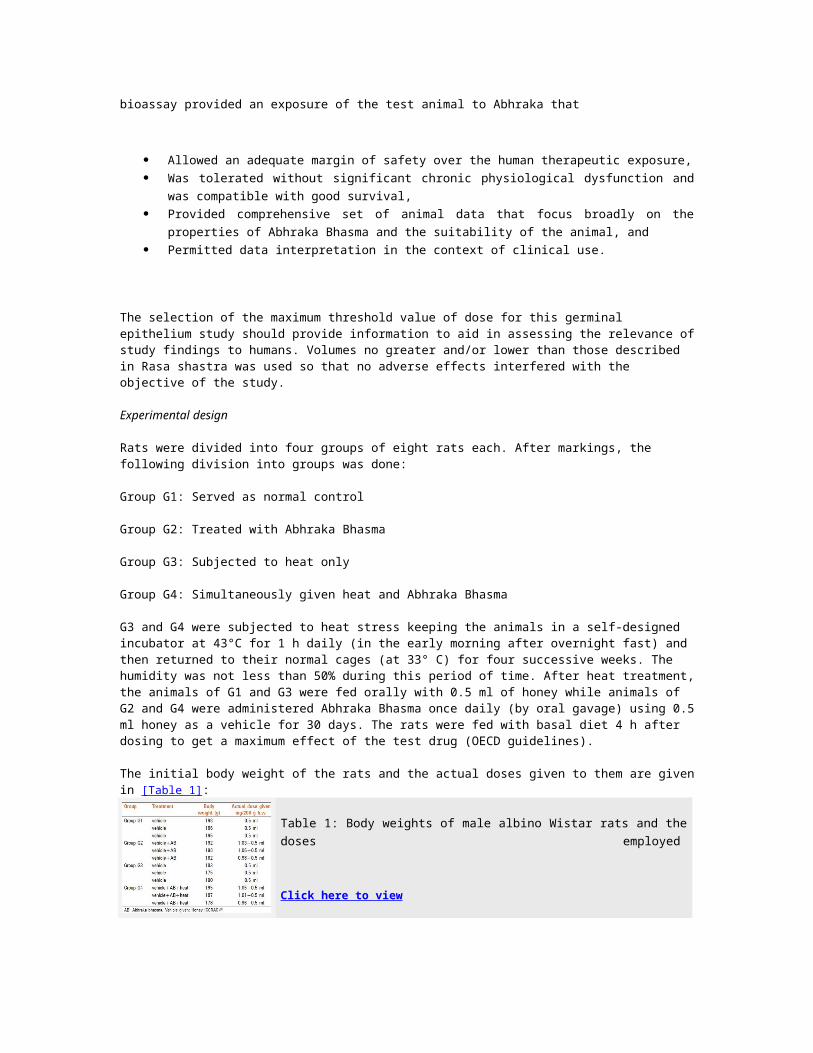
The initial body weight of the rats and the actual doses given to them are givenin [Table 1]:
Table 1: Body weights of male albino Wistar rats and thedoses employed
Click here to view
Sampling and analysis
The method reported by Saksena and Dixit (1987) as modified by Thakur and Dixit (2006) [9] was used. In brief, after 30 days of treatment, three animals from each group were sacrificed by a rapid decapitation method to dissect out testes for histopathological studies. Standard laboratory techniques [10] were followed for the same. Testis were removed and cut into small pieces, fixed in Bouin's fixative, and dehydrated with varying percentage of ethanol for histological studies. Sections were cut (6 μm), stained with eosin, and analyzed microscopically.
Results
In the rat, evaporate cooling was achieved by spreading of saliva onto the skin and fur. A decrease in testicular volume was observed due to reduced parenchymalvolume. Loss of weight of testis, atrophy of tubules, and degeneration of germinal epithelium were the primary effects of heat stress. The only observed effect of the treatment on the exposed scrotal area was a transient hyperemia which did not result in blisters or scar formation. Daily examination showed both the treated and control testes to be in a normal scrotal position.
Initially it was observed that heat treatment at temperature 43°C for 30 days showed testicular degeneration with no spermatids in the lumen in albino rats. Single dose of the test drug daily for 30 days showed 71% hyperactive tubules and high sperm count as compared to the negative control group. Hence, it is proved that the drug could protect heat-damaged testis from impairment induced by heat stress.
Testes findings of dissected rats at 30 days:-
Group G1 (Control + vehicle)
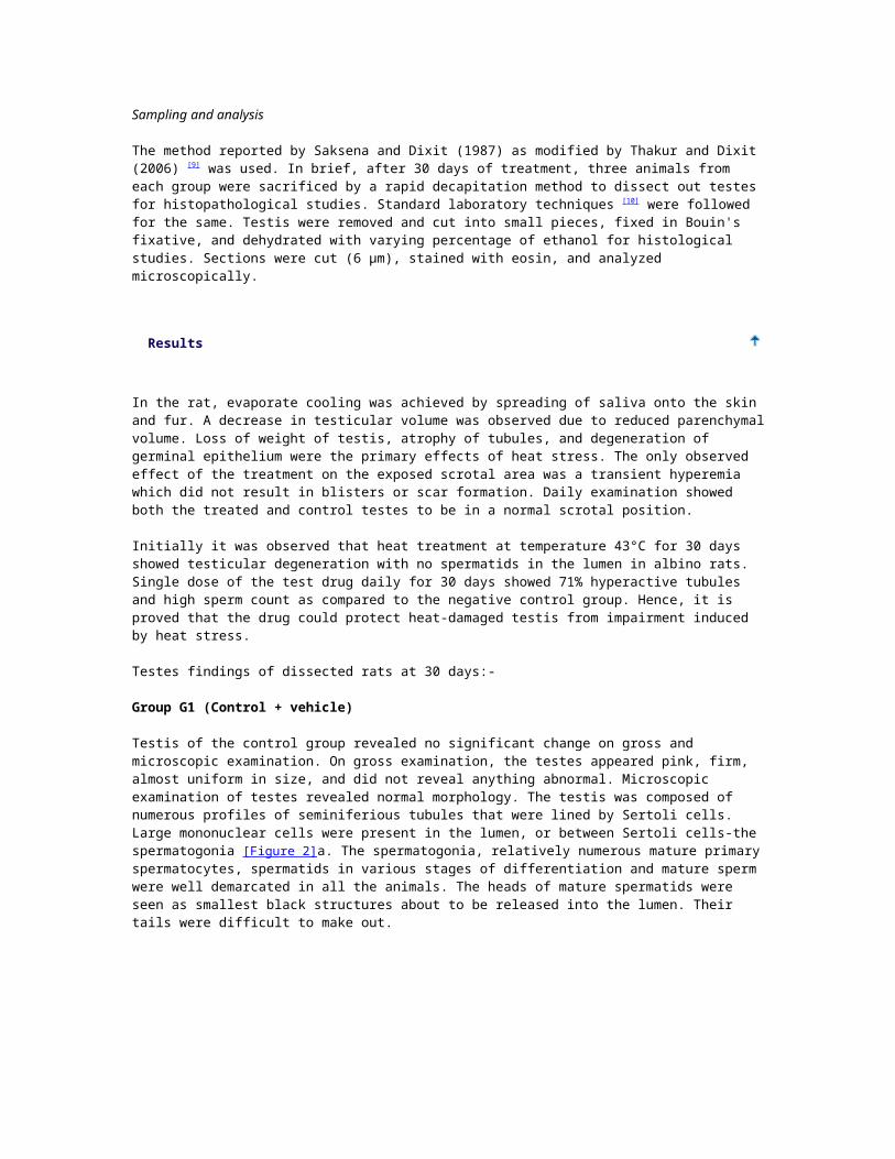
Testis of the control group revealed no significant change on gross and microscopic examination. On gross examination, the testes appeared pink, firm, almost uniform in size, and did not reveal anything abnormal. Microscopic examination of testes revealed normal morphology. The testis was composed of numerous profiles of seminiferious tubules that were lined by Sertoli cells. Large mononuclear cells were present in the lumen, or between Sertoli cells-the spermatogonia [Figure 2]a. The spermatogonia, relatively numerous mature primaryspermatocytes, spermatids in various stages of differentiation and mature sperm were well demarcated in all the animals. The heads of mature spermatids were seen as smallest black structures about to be released into the lumen. Their tails were difficult to make out.
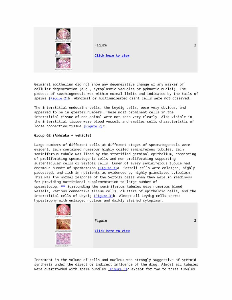
Figure 2
Click here to view
Germinal epithelium did not show any degenerative change or any marker of cellular degeneration (e.g., cytoplasmic vacuoles or pyknotic nuclei). The process of spermiogenesis was within normal limits and indicated by the tails ofsperms [Figure 2]b. Abnormal or multinucleated giant cells were not observed.
The interstitial endocrine cells, the Leydig cells, were very obvious, and appeared to be in greater numbers. These most prominent cells in the interstitial tissue of one animal were not seen very clearly. Also visible in the interstitial tissue were blood vessels and smaller cells characteristic of loose connective tissue [Figure 2]c.
Group G2 (Abhraka + vehicle)
Large numbers of different cells at different stages of spermatogenesis were evident. Each contained numerous highly coiled seminiferous tubules. Each seminiferous tubule was lined by the stratified germinal epithelium, consisting of proliferating spermatogenic cells and non-proliferating supporting sustentacular cells or Sertoli cells. Lumen of every seminiferous tubule had enormous number of spermatozoa [Figure 3]a. Sertoli cells were enlarged, highly processed, and rich in nutrients as evidenced by highly granulated cytoplasm. This was the normal response of the Sertoli cells when they were in readiness for providing nutritional supplementation to large number of spermatozoa. [11] Surrounding the seminiferous tubules were numerous blood vessels, various connective tissue cells, clusters of epitheloid cells, and the interstitial cells of Leydig [Figure 3]b. Almost all Leydig cells showed hypertrophy with enlarged nucleus and darkly stained cytoplasm.
Figure 3
Click here to view
Increment in the volume of cells and nucleus was strongly suggestive of steroid synthesis under the direct or indirect influence of the drug. Almost all tubuleswere overcrowded with sperm bundles [Figure 3]c except for two to three tubules
showing normal spermatogenesis [Figure 3]c. In some tubules, spermatids were found scattered amidst spermatozoa [Figure 3]a. The blood vessels of testis wereslightly dilated. Increased spermatogenesis was evident by the presence of high number of spermatozoa in seminiferous tubules and also by increase in spermatogenic elements as compared to control [Figure 3]c.
Group G3 (Heat + vehicle)
Gross examination of the testes revealed normal appearance. The histo-architecture and the overall spermatogenic profile in some tubules (50%) were considerably prevented in heat stroke rats. On cut section, in the atrophic testis, testicular hyperthermia resulted in degenerated tubules containing inspissated spermatozoa in about 25% of the tubules on an average in all the three animals. It lead to atrophy of the organ and mitoses were very rare in these atrophied tubules. The percentage of damaged tubules in each rat testis was determined by the evaluation of at least 50 widely separated, randomly selected tubules, and then average taken of the three readings. It was estimatedto be 15%, 41%, and 42% for the three heat-treated rats, respectively.
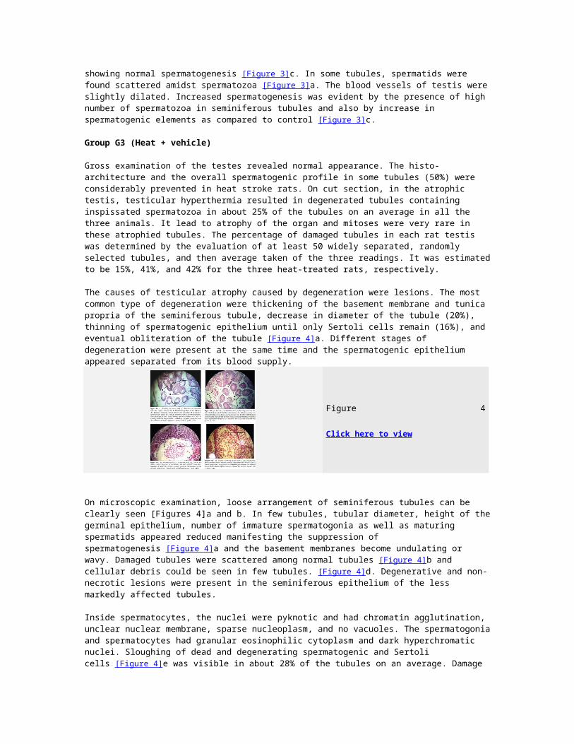
The causes of testicular atrophy caused by degeneration were lesions. The most common type of degeneration were thickening of the basement membrane and tunica propria of the seminiferous tubule, decrease in diameter of the tubule (20%), thinning of spermatogenic epithelium until only Sertoli cells remain (16%), and eventual obliteration of the tubule [Figure 4]a. Different stages of degeneration were present at the same time and the spermatogenic epithelium appeared separated from its blood supply.
Figure 4
Click here to view
On microscopic examination, loose arrangement of seminiferous tubules can be clearly seen [Figures 4]a and b. In few tubules, tubular diameter, height of thegerminal epithelium, number of immature spermatogonia as well as maturing spermatids appeared reduced manifesting the suppression of spermatogenesis [Figure 4]a and the basement membranes become undulating or wavy. Damaged tubules were scattered among normal tubules [Figure 4]b and cellular debris could be seen in few tubules. [Figure 4]d. Degenerative and non-necrotic lesions were present in the seminiferous epithelium of the less markedly affected tubules.
Inside spermatocytes, the nuclei were pyknotic and had chromatin agglutination, unclear nuclear membrane, sparse nucleoplasm, and no vacuoles. The spermatogoniaand spermatocytes had granular eosinophilic cytoplasm and dark hyperchromatic nuclei. Sloughing of dead and degenerating spermatogenic and Sertoli cells [Figure 4]e was visible in about 28% of the tubules on an average. Damage
to Sertoli cells was indicated by the presence of morphological changes and by adecrease in their numbers in few tubules (16%). The Sertoli cells contained swollen nuclei and cytoplasmic vacuoles. Spermatid desquamation marked degeneration of the affected tubules and cellular debris occupied in few of the tubules [Figure 4]c.
Sertoli cells are known to be more resistant to ischemia, heat injury, radiation, and other toxic agents that injure more sensitive spermatogenic cells. Histological damage to the germinal epithelium occurs in pachytene primary spermatocytes. Cytoplasmic membranes of the late meiotic prophase of pachytene spermatocytes are damaged causing mitochondrial damage and activation of lysosomes leading to autolysis of the cell. [12]
In some of these cells, especially the spermatids, the nuclei takes "bead-like appearance where chromatin had lost its granular structure, migrated to the periphery of the nucleus, and had coagulated into a homogeneous mass, [13] givingthe nucleus a vesiculated appearance (13%), lesions were not observed in the Leydig cells and there was absence of vacuoles and multinucleated masses-the giant cells. Some tubules showed partial recovery (21%), while heat stress resulted in marked suppression of spermatogenesis to complete azoospermia due toincrease germ cell apoptosis and their eventual phagocytosis by Sertoli cells [Figure 4]e.
Interstices between the tubules were devoid of Leydig cells except for interstices around few compact tubules here and there in all the animals. Large blood vessel can be easily marked out in all the three tubules [Figure 4]e.
Group G4 (Simul. heat and Abhraka)
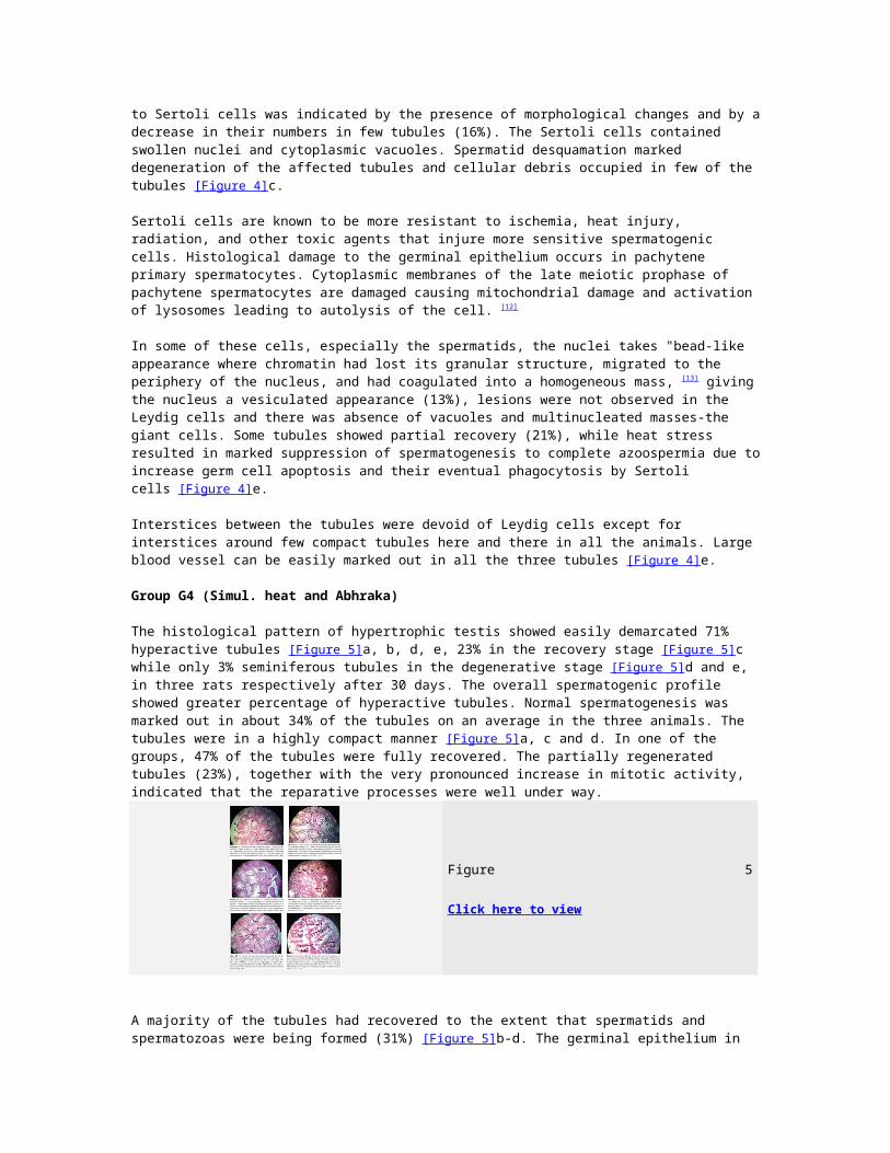
The histological pattern of hypertrophic testis showed easily demarcated 71% hyperactive tubules [Figure 5]a, b, d, e, 23% in the recovery stage [Figure 5]c while only 3% seminiferous tubules in the degenerative stage [Figure 5]d and e, in three rats respectively after 30 days. The overall spermatogenic profile showed greater percentage of hyperactive tubules. Normal spermatogenesis was marked out in about 34% of the tubules on an average in the three animals. The tubules were in a highly compact manner [Figure 5]a, c and d. In one of the groups, 47% of the tubules were fully recovered. The partially regenerated tubules (23%), together with the very pronounced increase in mitotic activity, indicated that the reparative processes were well under way.
Figure 5
Click here to view
A majority of the tubules had recovered to the extent that spermatids and spermatozoas were being formed (31%) [Figure 5]b-d. The germinal epithelium in
such tubules was thin but otherwise normal. The mitotic figures of the germinal cells compared favorably with that of the control testis, and the divisions werenormally distributed among all of the spermatogenic elements. Sloughing of newlyformed cells had decreased (0.9%) and no disorganization of nuclear chromatin was observed. Only 1% of the tubule showed distended lumen. In 15% of the tubules, spermatogenesis had progressed till spermatid formation. 34% of the tubules showed complete spermatogenesis.
Cytoplasmic membranes of the late meiotic prophase of pachytene spermatocytes were normal showing no mitochondrial damage. No vesiculation was seen in nuclei of spermatids. Mitosis was evident and there were no lesions to be seen. The basement membrane appeared to be thin and the spermatogenic epithelium was thickand the diameter of majority of the tubules was more than the animals in the heat and control animals.
Discussion
The deleterious effects of local heating on the testis of scrotal mammals is well established. As per the reports available in the literature, heat at 43°C causes testicular hyperthermia (confirmed by present study) and its degenerationby 7-8 days. [14] Histological examination of the Group G3 rats revealed suppression of spermatogenesis. Atrophy due to the traumatic effect of heat is reflected in the epithelium of seminiferous tubules of rats indicating either a direct action of heat on Sertoli cell-germ cell interaction, or an indirect action affecting the hormonal milieu at the level of the pituitary-testicular axis. Damaged Sertoli cells produce decreased amount of inhibin which decreases negative feedback on pituitary leading to an increase in FSH level. [15] FSH and LH levels have been reported to be elevated in azoospermia (Dada R et al., 2010). [5] This suggests a negative functional relationship between secretion of FSH and this step of germ cell maturation.
There is a possibility that heat causes detachment of spermatids by disturbing the blood testis barrier and prevents them from further maturation. Lastly, decreased serum testosterone levels have been reported with heat treatment (Lue Y et al., 1999). [3] The testosterone deficiency could be the cause of germ cells depletion. Intratesticular T and/or FSH clearly play a pivotal role in protecting germ cells at androgen-dependent stages VII-VIII against heat-inducedprogrammed germ cell death (Leu et al., 1998). The action of these somatic cells may be exercised by the production of soluble factors that can act as paracrine regulators of the multiplication and/or survival of germ cells. [16]
The current study established that treatment of heat-stroke rats with the drug Abhraka Bhasma causes hyperplasia and hypertrophy of the organ. Abhraka Bhasma is believed to increase sperm count but whether they protect testes against injury was not identified. The experimental animals in the present study showed a prominent enhancing effect of the drug on the histological structures of the testis- the seminiferous tubules, germinal epithelium, Sertoli cells, Leydig cells, luminal part of tubule, nucleus, and congestion in blood vessels. The comparison of germinal epithelium between experimental and control groups
revealed a significant difference. 71% of hyperactive tubules, 34% complete spermatogenic tubules, and 3% disintegrated tubules in the Abhraka Bhasma-cum-heat treated rats, as compared to none, 20% and 25% respectively in the heat-stressed rats, indicates that Abhraka Bhasma ameliorates spermatogenesis in degenerated tubules of heat-stroke rats. The lumen of the seminiferous tubules was found to contain enormous number of spermatozoa in the experimental rats. Itmight be due to higher androgen level in blood (probably due to hypertrophy of Leydig cells which is suggestive of increase in steroidogenesis), and/or an antioxidant mechanism which protects against oxidative damage to gonadal cells and mature spermatozoa. [17]
Pathological or accidental cell death resulting from extrinsic insults (osmotic,thermal, toxic, and traumatic effects) to the cell [18] is prevented by Abhraka Bhasma maintaining structural and functional integrity of the membranes. The test drug prevents sloughing of the germinal epithelium from the basement membrane and reduction in the population of the germ cells. The implication of this is that there is an elevation of viable sperms leading to increased fertility. Recently, it has been shown that abdominal heat stress induces germ cell loss through two apoptotic pathways: p53 is responsible for the initial phase of germ cell apoptosis and Fas is responsible for the second phase of apoptosis starting on day 10 of cryptorchidism. [19] It can be hypothesized that Abhraka Bhasma suppresses these two functions thus correcting the negative effects of heat stress.
To define cellular and molecular mechanisms of effects of Abhraka Bhasma, further investigation of the protective effect on germ cell death is required. The present test drug provides us with the possibility of treatment of human heat-induced oligozoospermia and azoospermia. There are several theories proposed about decreases in sperm counts in young, seemingly healthy people worldwide, and some people attribute the decrease to the presence of endocrine disrupters. Male infertility accompanied by decreased sperm counts is a serious problem for some people. Therapeutic approaches for male infertility have been developed including spermatogonial stem cell. These methods, however, are highlyinvasive and expensive treatments.
Our results suggest that Abhrak Bhasma drug therapy can be used to treat people with oligozoospermia, indicating that it could be a potential male anti-infertility agent. The concept of regenerative potential of Abhraka Bhasma may have potential application for sterility drug development for people living in equatorial and tropical regions. In these times of global warming, these effectsmay have more serious consequences for both the human population and domestic and wild animals. We, therefore, recommend that further studies be carried out in humans to corroborate these findings as well as medications involving AbhrakaBhasma to be brought into use as an anti-impotency fecundity drug.
Conclusion
In nutshell it could be concluded that an overall significant increase in the spermatogenic process and histometry of the testis is due to spermatogenic
enhancing properties of the Abhraka Bhasma. We demonstrated that Abhraka Bhasma reduces testicular damage induced by heat stress in heat-treated rats. These findings may generate a novel therapeutic strategy in heat stroke cases. This study confirms that Abhraka Bhasma is an important tool in solving the problem of male infertility as it provides some light on the spermatogenic enhancing properties of the mentioned drug.
References
1. Dole V. Vegbhatta Acharya's Rasa Ratna Samucchaya. Chowkhambha Sanskritseries Varanasi 2000;33:25-45.
2. Mookerji. B. Rasa- jala -nidhi. Parimal Publications 2001;2:29.
3. Maheswar T, Venkateshwarlu G. Standardization of Abhraka bhasma w.r.tchemical, pharmaceutical and metallographic studies. Ayu Renaissance2010;8:7-8.
4. Lue Y, Hikim A, Swerdloff R, Im P. Single Exposure to Heat Induces Stage-Specific Germ Cell Apoptosis in Rats: Role of Intratesticular Testosteroneon Stage Specificity. End 1999;140:1709-17.
5. Gladwell R. The effect of temperature on the potential difference And InputResistance Of Rat Seminiferous Tubules. J Physiol 1977;268:111-2.
7. Meena MK, Khushwah HK, Rajagopala M, Ravishankar B. An Experimentalevaluation on Nephroprotective activity of Nagaradi Kashaya. AYU Int Res JAyurveda 2009;30:55-61.
8. Central Council for Research in Ayurvedic sciences [homepage on theinternet] Guidelines for toxicity/safety profile evaluation ofBhasma/raskalpas; [Last Updated on 2012 Apr 11] Available from:www.ccras.nic.in/research_actvities/guidelinesfortoxicitybhasma.doc [Lastcited on 2012 Aug 2]
9. Dixit VK, Chauhan NS. Spermatogenic activity of rhizomes of Curculigoorchioides Gaertn in male rats. Int J App Res Nat Products 2008;1:26-31.
10. Singh M, Joshi D, Aryan C. Studies on testicular regenerative potential ofNaga Bhasma. Anc Sci Life 1989;9:95-8.
11. Majumdar SS, Tsuruta J, Griswold MD. Isolation and Culture of Sertoli Cellsfrom the Testes of Adult Siberian Hamsters: Analysis of ProteinsSynthesized and Secreted by Sertoli Cells Cultured from Hamsters Raised ina Long or a Short Photoperiod. Biol Reprod 1995;52:658-66. [PUBMED]
12. Blackshaw, AW, Hamilton D. The effect of heat on hydrolytic enzymes andspermatogenesis in the rat testis. J Rep Fertil 1970;22:569-71.
13. Williams WW, Cunningham B. Histological changes in the rat testis following
heat treatment. Yale J Bio Med 2010;12:309-16.
14. Kheradmand A, Dezfoulian O, Tarrahi MJ. Ghrelin attenuates heat-induceddegenerative effects in the rat testis. Regul Peptides 2011;167:97-104. [PUBMED]
15. Al-Rekab, Al-Wayelli D, Abed Khadim A. Evaluation of serum FSH, LH andTestosterone levels in infertile patients affected with different maleinfertility factors after IUI technique. Thi-Qar Med J 2010;4:112-22.
16. Jégou B, Velez de la Calle JF, Bauche F. Protective effect of medroxy-progesterone acetate plus testosterone against radiation-induced damage tothe reproductive function of male rats and their offspring. Proc Natl AcadSci U S A 1991;88:8710-4.
17. Sikka SC. Relative impact of Oxidative stress on Male reproductivefunction. Curr Med Chem 2001;8:851-62.
18. Farber JL, Chein KR, Mittnacht S. The pathogenesis of irreversible cellinjury in ischemia. Amer J Pathol 1981;102:271-81.
19. Zheng X, Yang Z, Zhang L. Overexpression of CIRP may reduce testiculardamage induced by cryptorchidism. Clin Invest Med 2009;32:103-11.
Copyright © 2022 FDOKUMEN