Demyelination in Mild Cognitive Impairment Suggests Progression Path to Alzheimer’s Disease
Upload
independentCategory
view
0download
0
Characterization of the Human NEK7 Interactome Suggests Catalyticand Regulatory Properties Distinct from Those of NEK6Edmarcia Elisa de Souza,†,‡ Gabriela Vaz Meirelles,† Barbara Biatriz Godoy,† Arina Marina Perez,†
Juliana Helena Costa Smetana,† Stephen J. Doxsey,§ Mark E. McComb,∥ Catherine E. Costello,∥
Stephen A. Whelan,∥ and Jorg Kobarg*,†,‡
†Laboratorio Nacional de Biociencias, Centro Nacional de Pesquisa em Energia e Materiais, Campinas, Sao Paulo, Brazil‡Departamento de Bioquímica-Programa de Pos-graduacao em Biologia Funcional e Molecular, Instituto de Biologia, UniversidadeEstadual de Campinas, Campinas, Sao Paulo, Brazil§Program in Molecular Genetics and Microbiology, University of Massachusetts Medical School, Worcester, Massachusetts 01605,United States∥Center for Biomedical Mass Spectrometry, Boston University School of Medicine, Boston, Massachusetts 02118, United States
*S Supporting Information
ABSTRACT: Human NEK7 is a regulator of cell division andplays an important role in growth and survival of mammaliancells. Human NEK6 and NEK7 are closely related, consisting ofa conserved C-terminal catalytic domain and a nonconservedand disordered N-terminal regulatory domain, crucial tomediate the interactions with their respective proteins. Here,in order to better understand NEK7 cellular functions, wecharacterize the NEK7 interactome by two screeningapproaches: one using a yeast two-hybrid system and theother based on immunoprecipitation followed by massspectrometry analysis. These approaches led to the identi-fication of 61 NEK7 interactors that contribute to a variety ofbiological processes, including cell division. Combining addi-tional interaction and phosphorylation assays from yeast two-hybrid screens, we validated CC2D1A, TUBB2B, MNAT1, and NEK9 proteins as potential NEK7 interactors and substrates.Notably, endogenous RGS2, TUBB, MNAT1, NEK9, and PLEKHA8 localized with NEK7 at key sites throughout the cell cycle,especially during mitosis and cytokinesis. Furthermore, we obtained evidence that the closely related kinases NEK6 and NEK7do not share common interactors, with the exception of NEK9, and display different modes of protein interaction, depending ontheir N- and C-terminal regions, in distinct fashions. In summary, our work shows for the first time a comprehensive NEK7interactome that, combined with functional in vitro and in vivo assays, suggests that NEK7 is a multifunctional kinase acting indifferent cellular processes in concert with cell division signaling and independently of NEK6.
KEYWORDS: Nek7, Nek6, proteomics, N and C terminal domain, cell cycle, cancer
■ INTRODUCTIONThe fidelity of the cell cycle is maintained, in part, by keyregulatory proteins, such as kinases. Protein kinases trigger andregulate cell division events such as centrosome duplication,spindle assembly, microtubule-kinetochore attachment, as wellas cytokinesis.1 The master regulators of the eukaryotic cellcycle are Cyclin-dependent kinases (CDK), members of theAurora and Polo-like kinases (PLKs) family and the morerecently discovered NimA related kinases (NEKs) family.2−4
The NEKs represent a large family of 11 serine/threonineprotein kinases in mammals, named NEK1 to NEK11, thatshare 40−45% sequence identity with the mitotic regulatorNIMA (Never in mitosis gene A) identified in the filamentousfungus Aspergillus nidulans.2,5−7 The deregulation of theseproteins directly affects the cell division and has been correlated
strongly with the uncontrolled cell proliferation and theappearance of tumors.8
The human protein NEK7 (NEK7) as well as the NEKs 1, 2,6, and 9 have been importantly shown to function in mitosiscontributing to the separation of centrosomes and establish-ment of the microtubule-based mitotic spindle.9 In fact, thedepletion of NEK7 via RNAi and inactive mutants disruptedlevels of γ-tubulin in interphase cells and caused an arrest inprometaphase associated with a fragile mitotic spindle,10−13
whereas its overexpression resulted in multinucleated cells anda high proportion of apoptotic cells.14 Allied to these studies,the centrosomal pericentriolar material (PCM) proteins do not
Received: May 3, 2014Published: August 5, 2014
Article
pubs.acs.org/jpr
© 2014 American Chemical Society 4074 dx.doi.org/10.1021/pr500437x | J. Proteome Res. 2014, 13, 4074−4090
Open Access on 08/05/2015

accumulate at the centrosomes in NEK7-depleted cells,indicating that NEK7 is involved in the recruitment of PCMproteins, which are necessary for both centriole duplication andspindle pole formation.15 Importantly, NEK7 along with NEK6and NEK9 constitute a mitotic signaling module in whichNEK6 and NEK7 are phosphorylated and activated by NEK9,contributing to mitosis progression. Furthermore, studies haveshown that the NEK7 absence leads to lethality duringembryogenesis and growth retardation, indicating an indis-pensable role for NEK7 in development and survival16 and thatthe closely related protein NEK6 can not compensate for theloss of NEK7 in the organism.Structurally, the human NEK6 and NEK7 share ∼86%
identity in their C-terminal domains7,17 and only ∼20% identityin their disordered N-terminal extensions.18,19 Studies indicatethat the two proteins have different roles and biologicalfunctions12 pointing to their disordered N-terminal extensionsas a possible component for the differential functions of thesekinases.18−21 Furthermore, NEK6 and NEK7 show differentialspatiotemporal tissue distribution20 and enzymatic control,21 aswell as distinct subcellular localization.13
Although all of these data suggest nonredundant roles forNEK6 and NEK7, the molecular basis for their specific roles isunknown, mainly because the protein interaction partners ofNEK7 in contrast to NEK622 are so far unknown.Therefore, we aimed to characterize the NEK7 protein
interactome, shedding light on the NEK7 functions and thusproviding additional information regarding the mechanismsthat orchestrate its differential function and regulation fromNEK6. We performed two interaction screening approaches forNEK7: a yeast two-hybrid (Y2H) system and an immunopre-cipitation followed by online liquid chromatography massspectrometry analysis (IP-LC−MS/MS). We have identified anumber of novel NEK7 protein interactors belonging to avariety of biological processes not previously described to beregulated by NEK7, as well as its already reported role in celldivision. Combining additional interaction and phosphorylationassays, we validated CC2D1A, TUBB2B, MNAT1, and NEK9proteins as possible NEK7 interactors and substrates.Interestingly, RGS2, TUBB, MNAT1, NEK9, and PLEKHA8localize with NEK7 in key sites during mitosis and cytokinesis.These results shed light on NEK7 functions in multiplemolecular pathways and denote a potential involvement for itsinteractors in cell division. Furthermore, we obtained evidencethat the closely related human kinases NEK6 and NEK7 do notshare the majority of interactors and show distinct mechanismsof specific interaction, suggesting their distinct, independent,and nonredundant signaling functions in the cell. We suggestthat via distinct mechanisms the N- and C-terminal domains ofNEK6 and NEK7 can contribute, remarkably, to bothregulation and catalysis.
■ EXPERIMENTAL PROCEDURES
Plasmid Constructs
The coding sequence of full-length human NEK7 was amplifiedby PCR from a leukocyte cDNA library (Clontech) using tefollowing primers: 5′-CGCGGATCCATGGATGAGCA-ATCACAAGG-3′ (forward) containing BamHI restrictionsite; 5′-CGGAATTCCATATGGATGAGCAATCACAAGG-3′(forward) containing EcoRI and NdeI restriction sites; 5′-GGGGAAGCTTTTAGCTGCTTGCAGTGCATGCATG-3′(reverse) containing HindIII restriction site; and 5′- ACGC-
GTCGACTTAGCTGCTTGCAGTGCAT-3′ (reverse) con-taining SalI restriction site. The amplified fragments corre-sponding to human NEK7 full-length sequence were clonedinto pGEM-T Easy (Promega) (pGEMT-NEK7 construct) andwere used as template to amplify the human NEK7 truncatedregion corresponding to its kinase domain only (NEK7Δ(1−44): the N-terminal region was deleted) with the specificprimers set 5′-CGGAATTCCATATGTTTCGAATAGAAAA-GAAAAT-3′ (forward) containing EcoRI and NdeI restrictionsites and 5′-ACGCGTCGACTTAGCTGCTTGCAGTGCAT-3′ (reverse) containing SalI restriction site. The amplifiedfragments were cloned into pGEM-T Easy (Promega)(pGEMT- NEK7Δ(1−44) construct). The chimeric constructsconsisting of NEK6 N-terminal-NEK7 C-terminal (N6C7) andNEK7 N-terminal-NEK6 C-terminal (N7C6) were generatedby gene synthesis (GenScript Corporation, see Figure 6B forthe constructs). The KpnI, EcoRI, NdeI, SalI, and ApaIrestriction sites were added to the synthetic genes.For the yeast two-hybrid screens we subcloned the full-length
NEK7 (NEK7-pBTMK) and the chimeric constructs N6C7(N6C7-pBTMK) and N7C6 (N7C6-pBTMK) into EcoRI/SalIrestriction sites of modified yeast expression vectorpBTM116K,23 in frame with the LexA DNA binding domain(Clontech). The recovered nucleotide sequences encoding theinteracting proteins identified to interact with human NEK7 inyeast two-hybrid screenings were subcloned from the vectorpACT2 (Clontech) into bacterial expression vector pGEX (GEHealthcare), which allows the expression of proteins in theform of a Glutathione S-transferase (GST) fusion.To obtain higher expression levels of kinases in bacteria, we
subcloned the full-length human NEK7 (6×His-NEK7construct) into the NdeI/HindIII restriction sites;NEK7Δ(1−44) and the chimeric constructs N6C7 andN7C6 were subcloned into NdeI/SalI restriction sites in themodified vector pET28a-His-TEV (Novagen/EMD Bioscien-ces), in fusion with a 6×His tag (constructs 6×His-NEK7Δ(1−44)), 6×His-N6C7, and 6×His-N7C6, respectively). Toexpress NEK7 in human cells, full-length NEK7 was insertedinto BamHI/SalI restriction sites in pCDNAFLAG (Invitro-gen) in fusion with a FLAG tag. The sequences of all vectorconstructs were confirmed by restriction endonuclease analysisand DNA sequencing.The full-length NEK6 and NEK6 kinase domain cloned into
pET28a-His-TEV in fusion with a His tag (6×His-NEK6 and6×His-NEK6Δ(1−33), respectively); full-length NEK6 clonedinto pBTM116KQ in fusion with binding domain DNA LexA(pBTMK-NEK6); the interacting proteins NEK9 (NEK9),Sorting nexin 26 (SNX26), Thyroid hormone receptorinteractor 4 (TRIP4), Pleiotrophin (PTN), and Peroxiredoxin3 (PRDX3) recovered in NEK6 Y2H into pACT2 in fusionwith GAL4 transcriptional activation domain (constructs:pACT-NEK9(806−979), pACT-SNX26, pACT-TRIP4,pACT-PTN and pACT-PRDX3, respectively) or subclonedinto pGEX in fusion with GST (constructs: GST-SNX26, GST-PTN, GST-PDX, GST-TRIP4) were obtained as described byMeirelles et al.22
Yeast Two-Hybrid Screening (Y2H)
The pBTM116K vector was used to express the full-lengthhuman NEK7, full-length human NEK6, N6C7 and N7C6linked to the C-terminus of LexA DNA-binding domainpeptide (constructs: pBTMK-NEK7, pBTMK-NEK6,pBTMK-N6C7 and pBTMK-N7C6, respectively) or as a
Journal of Proteome Research Article
dx.doi.org/10.1021/pr500437x | J. Proteome Res. 2014, 13, 4074−40904075

control (pBTM116K-control) in Saccharomyces cerevisiae strainL40 (trp1-901, his3Δ200, leu2-3, ade2 LYS2::(lexAop)4-HIS3URA3::(lexAop)8-lac GAL4), which contains the heterologousreporter genes HIS3 and LacZ.The yeast two-hybrid screenings were performed against the
three cDNA libraries fetal brain, bone marrow, and leukocyte,cloned in pACT2 vector expressing GAL4 activation domain(AD) fusion proteins (Matchmaker System, Clontech). Theautonomous activation of HIS3 gene was tested by co-transformation of yeast cells with pBTMK-NEK7 and pACT2as a control (pACT2-control), grown in minimal mediumplates without tryptophan (-W), leucine (-L), and histidine(-H) and containing 0, 5, 10, 20, 30, 50, 70, or 100 mM 3-amino-1,2,4-triazole (3-AT), a competitive inhibitor of His3pprotein (Imidazoleglycerol-phosphate dehydratase).24 Althoughthe autoactivation of HIS3 was not observed, the screeningswere performed by separate co-transformation of three cDNAlibraries with the pBTMK-NEK7 construct in minimal mediumplates without -W, -L, and -H, containing 5 mM 3-AT. The co-transformants showing the best growth conditions had theirrecombinant pACT2 plasmids isolated and sequenced.To test the growth capacity under interaction-selective
conditions, the recovered clones from the yeast two-hybridscreenings for NEK7 and for NEK622 were co-transformed inthe yeast strain L40 with pBTMK-NEK7, pBTMK-NEK6,pBTMK-N6C7, and pBTMK-N7C6 constructs or with thepBTM116K-control. The co-transformation products wereplated in minimal medium without -W, -L, and -H, containing3-AT gradient (1−100 mM). We considered as positiveinteractions the co-transformations showing a higher colonygrowth when compared to pBTM116K-control.
Antibodies
The following antibodies were used for either immunofluor-escence staining or Western blotting purposes: goat anti-NEK7(sc50756), goat (sc50763) or mouse (sc1004) anti-NEK9, goatanti-α-tubulin (TUBA) (sc8035) (all from Santa CruzBiotechnology); mouse anti-NEK7 (ab68060), rabbit anti-NEK7 (ab96538), goat anti-PLEKHA8 (ab38748), rabbit anti-β-tubulin (TUBB) (ab15568), rabbit anti-RGS2 (ab36561),rabbit anti-MNAT1 (ab65125), rabbit anti-ANKS1B(ab116083), mouse anti-CC2D1A (ab68302), and mouseanti-pericentrin (PCNT) (ab28144) (all from Abcam).Mouse anti-His5 (34660, QIAGEN), mouse anti-GST (madein house), mouse anti-FLAG (F1804, Sigma-Aldrich), rabbitanti-phosphothreonine (71-8200, Invitrogen), mouse anti-PLK1 (ab17057), goat anti-SMC1 (sc-21078), and goat anti-SMC3 (sc-8198) antibodies were used for Western blotting.Secondary antibodies for immunofluorescence staining wereobtained from the following sources: chicken anti-goat, anti-mouse, or anti-rabbit Alexa Fluor 488 or donkey anti-goat, anti-mouse, or anti-rabbit Alexa Fluor 546 (all from LifeTechnologies, Inc.); horseradish peroxidase (HRP)-conjugatedanti-mouse was obtained from (Calbiochem) and HRP-linkedanti-goat or anti-rabbit secondary antibodies for Westernblotting (WB) purposes were obtained from Sigma-Aldrich.
Immunoprecipitation Followed by Mass SpectrometryAnalysis (IP-LC−MS/MS)
To identify NEK7-associated proteins, FLAG-tagged wild-typeNEK7 (FLAG-NEK7) or pCDNAFLAG as a control (FLAG-control) were used to transfect HeLa cells in exponentialgrowth using X-tremeGENE 9 DNA Transfection Reagent(Roche).
Cells were harvested by centrifugation 24 h after transfection,and the pellets were incubated in lysis buffer (1% Triton X-100,5 mM EDTA, 0.1 mM sodium orthovanadate, 1 μg/μL DNaseand protease inhibitors cocktail diluted in PBS 1X) at 4 °C for30 min. The lysates were sequentially cleared by centrifugationand incubated overnight with Mouse anti-FLAG M2 AffinityGel (Sigma). The beads from samples transfected with FLAG-NEK7 or FLAG-control, in a combination of three experiments,were gently washed three times in Tris pH 8.0 buffer and weresubmitted for liquid chromatography-tandem MS (LC−MS/MS).The LC−MS/MS analyses were performed on tryptic
peptide samples using a nanoAcquity UPLC nanocapillaryhigh-performance LC system (Waters Corp., Milford, MA,USA) coupled to a Q Exactive hybrid quadrupole-Orbitrapmass spectrometer (Thermo Fisher Scientific) equipped with aTriVersa NanoMate ion source (Advion, Ithaca, NY, USA).Sample concentration and desalting were performed onlineusing a nanoAcquity UPLC trapping column (180 μm × 20mm, packed with 5-μm, 100-Å Symmetry C18 material;Waters) at a flow rate of 15 μL/min for 1 min. Separationwas accomplished on a nanoAcquity UPLC capillary column(150 μm × 100 mm, packed with 1.7-μm, 130-Å BEH C18material; Waters). A linear gradient of A and B buffers (bufferA: 1.5% ACN and 0.1% FA; buffer B: 98.5% ACN and 0.1%FA) from 2% to 40% buffer B over 80 min was used at a flowrate of 0.5 μL/min to elute peptides into the massspectrometer. Columns were washed and re-equilibratedbetween LC−MS/MS experiments. Electrospray ionizationwas carried out at 1.65 kV using the NanoMate, with the QExactive heated transfer capillary set to 250 °C. Mass spectrawere acquired in the positive-ion mode over the range m/z400−2000 at a resolution of 70,000 (full width at half-maximum at m/z 400; ∼1 spectrum/s) and AGC target >1 × e6
(Method 1) or >3 × e6 (Method 2). Mass accuracy afterinternal calibration was within 1 ppm. MS/MS spectra wereacquired at a rate of ∼10 MS/MS/s for the 10 most abundant,multiply charged species in each mass spectrum with aresolution of 17,500 and signal intensities >5 × e5 NL(Method1) or >2 × e5 NL (Method 2), using HCD withMS/MS collision energies set at either 24 V (Method 1) or 30V (Method 2), nitrogen as the collision gas, and MS/MSspectra acquisition over a range of m/z values dependent on theprecursor ion. Dynamic exclusion was set such that MS/MS foreach precursor ion species was excluded for either 4 s (Method1) or 20 s (Method 2) postacquisition. All spectra wererecorded in profile mode for further processing and analysis.MS and MS/MS data analysis was carried out using both
Proteome Discoverer (PD) 1.4 software (Thermo FisherScientific) and an in-house Mascot 2.4 server (Matrix Science,London, U.K.). The MS/MS data were searched against the IPIhuman 3.80 amino acid sequence database for protein/peptideidentification. The PD 1.4 search was set up with precursorintensity node and full tryptic peptides with a maximum of 2missed cleavage sites with carbamidomethyl cysteine andoxidized methionine included as variable modifications. Mascotsearch was set up for full tryptic peptides with a maximum of 4missed cleavage sites with carbamidomethyl cysteine andoxidized methionine included as variable modifications. Theprecursor mass tolerance was set to 10 ppm for Q Exactiveorbitrap data, and the maximum fragment mass error was 0.8Da. The PD1.4 searches for each Q Exactive method wereuploaded into Scaffold 4.2.1 (Proteome Software, Inc.) software
Journal of Proteome Research Article
dx.doi.org/10.1021/pr500437x | J. Proteome Res. 2014, 13, 4074−40904076

program while searching data via X! Tandem as described byWhelan et al.25 The quantitative data were performed withfilters set at 95% minimum protein ID probability (calculatedprobability of correct protein identification), a minimumnumber of five unique peptides for one protein (five uniquepeptides were used to reduce the NEK7 protein interacting listto strong interacting proteins at high abundance) and a peptidethreshold at 0.8% FDR (False Discovery Rate) algorithm.In order to avoid the selection of nonspecific interactors (for
example, proteins that interact with the solid-phase support,affinity reagent, or epitope tag) from affinity purification usinganti-FLAG, we compared the generated list of interactingproteins from quantitative analysis with the ContaminantRepository for Affinity Purification, CRAPome (http://www.crapome.org/). Those proteins that were present in thenegative controls of anti-FLAG affinity purifications underour experimental conditions were excluded from the list.
In Silico Protein−Protein Interaction (PPI) Analysis
To generate NEK7 PPI maps at proteome scale, the retrievedNEK7 interacting proteins from IP-LC−MS/MS and Y2H wereintegrated in interaction networks using the IntegratedInteractome System (IIS) platform, developed at NationalLaboratory of Biosciences, Brazil (http://www.lge.ibi.unicamp.br/lnbio/IIS/).26 The enriched biological processes from theGene Ontology database (GO, http://www.geneontology.org/) were calculated in each network using the hypergeometricdistribution.26 The interaction network was visualized usingCytoscape 2.8.3 software (http://www.cytoscape.org/).27 Theprediction of disordered amino acid residues in the Y2Hretrieved protein sequences was calculated by PONDR VL-XTpredictor (http://www.pondr.com/), and the domain compo-sition of these sequences was obtained by Pfam (http://pfam.sanger.ac.uk/), PROSITE (http://www.expasy.ch/prosite/), orInterPro (http://www.ebi.ac.uk/interpro/) databases.
Protein Expression and Purification
The proteins identified in the NEK7 yeast two-hybrid screens,β-tubulin-2B chain (TUBB2B), Coiled-coil and C2 domain-containing protein 1A (CC2D1A), and Regulator of G-proteinsignaling 2 (RGS2) in fusion with GST (GST-TUBB2B, GST-CC2D1A and GST-RGS2, respectively), were expressed in E.coli BL21 (DE3/pRARE) using 0.5 mM IPTG at 37 °C for 4 h.The GST-tagged proteins NEK9, comprising its regulatoryregion from 764 to 976 amino acid residues (GST-NEK9(764−976)) and CDK-activating kinase assembly factor MAT1(GST-MNAT1) were expressed in E. coli BL21 (DE3) cellsusing 1 mM IPTG at 25 and 30 °C, respectively, all for 4 h. Thefull-length human NEK7 protein (6×His-NEK7) or kinasedomain NEK7 (6×His-NEK7Δ(1−44)) and chimeric con-structs N6C7 (6×His-N6C7) and N7C6 (6×His-N7C6) wereexpressed in E. coli BL21 (DE3/pRARE) or BL21 (DE3) cells,respectively. Expression was induced for 4 h using 1 mMisopropyl-β-D-thio-galactoside (IPTG) at 28 °C to 6×His-NEK7 and 6×His-N7C6 constructs and 0.5 mM and 1 mMIPTG at 25 °C to 6×His-NEK7Δ(1−44) and 6×His-N6C7constructs, respectively. Induced cells were harvested and lysedby sonication in extraction buffer (50 mM HEPES pH 7.5, 5mM sodium phosphate, 300 mM NaCl, 5% glycerol)supplemented with 1 mM PMSF and 625 μg/mL lysozyme.The proteins GST-SNX26, GST-TRIP4, GST-PTN, and GST-PRDX3 were expressed as described by Meirelles et al.22 Thecell lysates were separated by centrifugation at 16,000 × g for10 min at 4 °C in order to obtain the supernatant. Cleared
fractions from protein expression in fusion with GST as well asGST as a control (control-GST) were purified by GST affinitychromatography medium binding to Glutathione Sepharose 4Fast Flow followed by elution in buffer containing 50 mM Tris-HCl and 50 mM reduced glutathione pH 8.0, according to theprotocol described for Batch Purification (GE Healthcare).Cleared fractions of 6×His-NEK7, 6×His-NEK7Δ(1−44),6×His-N6C7, and 6×His-N7C6 obtained by lysis were purifiedby affinity liquid chromatography using a HiTrap ChelatingAffinity Chromatography Column (GE Healthcare) followedby elution with a linear concentration gradient of imidazole (1to 100 mM) in extraction buffer. All of the eluted recombinantproteins were dialyzed against kinase buffer containing 50 mMMOPS pH 7.4, 300 mM NaCl, 10 mM MgCl2, and 0.1 mMPMSF. The purified proteins 6×His-NEK6 and 6×His-NEK6Δ(1−33) were obtained as described by Meirelles etal.22 The expression of all proteins was confirmed by WB usingmonoclonal mouse anti-GST clone 5.3.3 (produced in house)or mouse anti-5×His primary antibodies and Horseradishperoxidase (HRP)-conjugated goat anti-mouse secondaryantibodies, and the bands were detected by a luminolchemiluminescence detection system (Santa Cruz Biotech) orby SDS-PAGE stained with Coomassie blue (SDS-PAGE).
Pull-Down Assay
To validate some of the candidate NEK7 interacting proteinsrecovered by yeast two-hybrid and IP-LC−MS/MS, weperformed affinity purification (pull-down assay) using6×His-NEK7 or 6×His-RARA as bait. 6×His-RARA was usedas a control since the protein RARA (Retinoic acid receptoralpha) is not described to interact with NEK7 or its potentialinteractor ANKS1B and NEK9. For this purpose, humanembryonic kidney (HEK) 293T cells (HEK293T) werecollected by gently shaking off the flask and centrifuged.Next, HEK293T extracts produced by homogenization in lysisbuffer (50 mM Tris-HCl pH 7.5, 150 mM NaCl, 1 mM EDTA,0.5% Triton X-100, and protease inhibitor cocktail) wereincubated overnight at 4 °C with 6×His-NEK7, 6×His-RARAbound to Ni-NTA agarose resin (Qiagen), or Ni-NTA agaroseresin, previously equilibrated in wash buffer (50 mM Tris-HClpH 8.0, 1% NP40, and protease inhibitor cocktail). The resinscontaining the complexes were washed three times in washbuffer, and bound proteins were analyzed by WB using specificantibodies, as previously described.28
In Vitro Protein Phosphorylation
To test the ability of NEK7 to catalyze 32P or Pi transfer toNEK7 itself or exogenous substrates, GST-tagged proteins assubstrates or GST-control were previously equilibrated inkinase buffer and incubated at 30 °C in 100 μM ATP and 185kBq [γ-32P]ATP (222 TBq/mmol; NEN). Then 6×His-taggedNEK6Δ(1−33), NEK7Δ(1−44), N6C7, or N7C6 proteinswere added to this reaction for 30 min. The autophosphor-ylation was performed by 6×His-tagged proteins incubation in100 μM ATP and 185 kBq [γ-32P]ATP (222 TBq/mmol;NEN) without being added to the GST-tagged proteins, for 30min. Assays were carried out in a final volume of 50 μL.Electrophoresis sample buffer was added to stop the reaction,and the proteins were resolved by SDS-PAGE. 32P incorpo-ration was detected by autoradiography. In the case of thechemiluminescence assay, GST-NEK9(764−976) was incu-bated with or without 200 μM ATP, in the presence or absenceof 1 μg of 6×His-NEK7 in kinase buffer for 1 h in a reactionvolume of 50 μL. The reaction was stopped by addition of
Journal of Proteome Research Article
dx.doi.org/10.1021/pr500437x | J. Proteome Res. 2014, 13, 4074−40904077

electrophoresis sample buffer, and the proteins were resolvedby SDS-PAGE. Phosphoamino acids were detected by Westernblotting using polyclonal rabbit anti-phosphothreonine primaryantibody diluted 1/250 in 3% bovine serum albumin (BSA) andgoat anti-rabbit secondary antibodies conjugated to Horse-radish Peroxidase (Sigma). The protein bands were detected byCoomassie blue staining or luminol chemiluminescencedetection system (Santa Cruz Biotech) according to theinstructions supplied by the manufacturer.
In Vitro Kinase Activity Assay
The in vitro kinase activity assay was performed using LANCEUltra Kinase Activity Assay (Product N° TRF0126-D/TR0126-M, PerkinElmer) containing ULight-p70 S6K (Thr389)Peptide (phosphorylation motif LGFTYVAP). The assay wasoptimized with 80 nM 6×His-NEK7, 6×His-NEK7Δ(1−44),6×His-N6C7, 6×His-N7C6 6×His-NEK6, or 6×His-NEK6Δ(1−33) purified proteins, 50 nM ULight-p70 S6K(Thr389) Peptide, and 100 μM ATP, prediluted in kinasebuffer (50 mM HEPES pH 7.5, 10 mM MgCl2, 1 mM EGTA, 2mM DTT, and 0.01% Tween-20). Ten microliters total volumeof kinase reaction was added to the wells of a 384-wellOptiPlate. The kinase reactions were incubated for 60 min at23 °C and stopped by the addition of 10 mM EDTA. For thedetection of the phospho-substrate, the Eu-anti-phospho-p70
S6K (Thr389) antibody diluted in Detection Buffer was addedto a final concentration of 2 nM, and the reactions were thenincubated for 16 h at 4 °C. A reaction without addition ofULight-p70 S6K (Thr389) Peptide was perfomed and used as acontrol. The signal was measured on a 2104 EnVisionMultilabel Microplate Reader. Excitation wavelength was setto 320 nm, and emission was recorded at 665 nm.
Immunocytochemistry
HeLa cells were grown and evaluated for cell viability by trypanblue exclusion (Invitrogen). The cell count was performedusing Countess Automated Cell Counter (Invitrogen). A totalof 20−30,000 cells per well were seeded in 384-cell Carrierplates (PerkinElmer) to a final volume of 50 μL.The cells in a confluence of 70% were fixed and
permeabilized at room temperature for 20 min in a solutioncontaining 3.7% formaldehyde, 0.2% Triton X-100 in PBS 1Xsupplemented with 50 mM EGTA and 30 μg/mL taxol. Thecells were washed twice with PBS 1X to remove the fixingsolution. After removing the fixing solution, cells wereincubated for 30 min with blocking solution containing 3%BSA and 0.8% Triton X-100 diluted in PBS 1X. The fixationand permeabilization steps were made using the JANUSModular Dispense Technology (MDT) Automated Work-station (PerkinElmer). Then, the cells were washed twice with
Table 1. Human NEK7 Interacting Proteins Identified by the Yeast Two-Hybrid System Screensa
redundancy inlibraryc
protein interacting with Nek7b geneUniprotaccession
coded protein residues (retrieved/complete sequence) FB BM L
growthscored
Beta-2-microglobulin B2M P61769 1−119/119 1 0 0 1/0Serine/threonine-protein kinase Nek9 NEK9 Q8TD19 764−976/979 3 3 2 4/0Regulator of G-protein signaling 2 RGS2 P41220 1−211/211 0 0 1 1/1CDK-activating kinase assembly factor MAT1 MNAT1 P51948 1−309/309 1 2 0 1/1Secreted frizzled-related protein 4 SFRP4 Q6FHJ7 1−346/346 0 4 0 3/0Cytochrome b-c1 complex subunit 9 UQCR10 Q9UDW1 1−63/63 0 1 0 1/0NADH-ubiquinone oxidoreductase 75 kDa subunit,mitochondria
NDUFS1 P28331 451−727/727 0 2 0 1/0
5-Aminolevulinate synthase, nonspecific, mitochondrial ALAS1 P13196 94−640/640 0 1 0 1/0NFU1 iron-sulfur cluster scaffold homologue,mitochondrial
NFU1 Q9UMS0 22−208/254 1 1 4 1/0
Tubulin beta-2B chain TUBB2B Q9BVA1 247−430/445 0 0 1 1/1Hemoglobin subunit alpha HBA1 P69905 1−142/142 0 2 0 1/0Hemoglobin subunit beta HBB P68871 1−147/147 0 2 0 1/0Elongation factor 1-alpha 1 EEF1A1 P68104 340−432/462 0 1 0 1/0Elongation factor Tu, mitochondrial TUFM P49411 13−455/455 1 0 0 1/1Pleckstrin homology domain-containing family Amember 8 isoform 2
PLEKHA8 Q96JA3 103−418/459 0 0 2 1/0
Actin, cytoplasmic 2 ACTG1 P63261 187−375/375 0 0 3 1/140S ribosomal protein S6 RPS6 P62753 1−228/249 0 1 0 1/1Coiled-coil and C2 domain-containing protein 1A CC2D1A Q6P1N0 501−940/951 0 1 0 1/140S ribosomal protein S4, X isoform RPS4X P62701 5−263/263 0 2 0 1/1Ubiquitin-40S ribosomal protein S27a RPS27A P62979 1−156/156 0 2 0 1/0Ankyrin repeat and sterile alpha motif domain-containingprotein 1B
ANKS1B Q7Z6G8 6−426/426 0 1 0 3/0
FCH and double SH3 domains protein 2 FCHSD2 O94868 627−740/740 1 0 0 1/1MAP7 domain-containing protein 1 MAP7D1 Q3KQU3 715−841/841 1 0 0 1/1Transmembrane and TPR repeat-containing protein 4 TMTC4 Q5T4D3 467−741/741 0 0 9 4/0Transmembrane 9 superfamily member 3 TM9SF3 Q9HD45 398−589/589 0 0 2 1/0aThe proteins were selected from yeast cells under growth conditions in minimal medium without tryptophan, leucine, and histidine and containing3-AT. bResults from BLASTX (GenBank). cAbsolute number of sequences retrieved of each human library (FB: fetal brain, BM: bone marrow, L:leukocyte). dGrowth test in yeast cells transfected with recovered sequences in the vector pACT2 and pBTM116K-NEK7/empty pBTM116KQ(concentration of 3AT = 5 mM).
Journal of Proteome Research Article
dx.doi.org/10.1021/pr500437x | J. Proteome Res. 2014, 13, 4074−40904078

PBS 1X, and proteins were detected by incubating the cells for1 h at room temperature with goat anti-NEK7, goat or mouseanti-NEK9, goat anti-TUBA, mouse anti-NEK7, rabbit anti-NEK7, goat anti-PLEKHA8, rabbit anti-TUBB, rabbit anti-RGS2, rabbit anti-MNAT1, rabbit anti-ANKS1B, mouse anti-CC2D1A, or mouse anti-PCNT, diluted 1:100 in blockingsolution. Primary antibodies were removed by washing threetimes with a solution containing PBS 1X and 0.05% Tween 20,and the proteins were revealed by incubating for 2 h at roomtemperature with chicken anti-goat, anti-mouse, or anti-rabbitAlexa Fluor 488 or donkey anti-goat, anti-mouse, or anti-rabbitAlexa Fluor 546 diluted 1:300 in blocking solution. Thelabeling was stopped by removing the secondary antibody bywashing three times with a solution containing PBS 1X and0.05% Tween 20 (PBS-T), and the nucleus was stained for 5min with DAPI 0.5 mM diluted 1:100 in PBS-T. The cells wereobserved by fluorescence microscopy using Operetta High
Content Imaging System (PerkinElmer). The images editionwas performed using Volocity Demo 6.1.1 software.
■ RESULTS
Identification of Human NEK7 Interacting Proteins byYeast Two-Hybrid and Mass Spectrometry
To clarify NEK7 biological functions, we report here twoscreening approaches to identify proteins that interact withNEK7: the first was based on direct interactions using aY2H29,30 system, and the second was based on IP-LC−MS/MS.In order to follow the functional distribution pattern of
NEK7 across tissues, the Y2H was employed using full-lengthwild-type NEK7-LexA fusion protein as bait against threecDNA libraries: human fetal brain, bone marrow, andleukocyte. A total of 8.5 × 10³ screened co-transformants forfetal brain, 4.1 × 10⁴ for bone marrow, and 1.66 × 10⁴ forleukocyte yielded 88 positive clones for the HIS3 reporter gene
Table 2. NEK7 Interacting Proteins Identified by IP-LC−MS/MSa
proteinb geneUniprotaccession
fold changeTICc
FLAG-control totalspectra
FLAG-NEK7 totalspectra
Serine/threonine-protein kinase Nek7 NEK7 Q8TDX7 9.10 × 10⁹ 1291cDNA FLJ60124, highly similar to Mitochondrial dicarboxylatecarrier
SLC25A10 Q9UBX3 2.60 × 10⁸ 68
Importin-9 IPO9 Q96P70 1.00 × 10⁸ 88ATPase family AAA domain-containing protein 3A isoform 3 ATAD3A Q9NVI7 9.90 × 10⁷ 94Protein RCC2 RCC2 Q9P258 4.90 × 10⁷ 80Isoform 3 of Apoptosis-inducing factor 1, mitochondrial AIFM1 O95831 3.80 × 10⁷ 43Ubiquitin-40S ribosomal protein S27a RPS27A P62979 3.50 × 10⁷ 36Isoform 1 of AP-2 complex subunit beta AP2B1 P63010 3.30 × 10⁷ 22Isoform 1 of Structural maintenance of chromosomes protein 2 SMC2 O95347 2.80 × 10⁷ 35Isoform 1 of Caprin-1 CAPRIN1 Q14444 2.80 × 10⁷ 37Isoform 1 of Structural maintenance of chromosomes protein 4 SMC4 Q9NTJ3 2.70 × 10⁷ 69Isoform 1 of CLIP-associating protein 1 CLASP1 Q7Z460 2.50 × 10⁷ 89Isoform 1 of CLIP-associating protein 2 CLASP2 O75122 1.60 × 10⁷ 20Ras-related protein Rab-32 RAB32 Q13637 1.50 × 10⁷ 25Isoform 1 of Centrosomal protein of 170 kDa CEP170 Q5SW79 1.20 × 10⁷ 33Isoform 1 of Rho guanine nucleotide exchange factor 2 ARHGEF2 Q92974 1.20 × 10⁷ 24Structural maintenance of chromosomes protein 1A SMC1A Q14683 1.20 × 10⁷ 18Isoform 1 of Microtubule-associated protein 2 MAP2 P11137 1.10 × 10⁷ 17Zinc finger protein 622 ZNF622 Q969S3 6600000 32Isoform 1 of Cyclin-dependent kinase 13 CDK13 Q14004 6300000 17Isoform 2 of Nuclear mitotic apparatus protein 1 NUMA1 Q14980 5600000 23Structural maintenance of chromosomes protein 3 SMC3 Q9UQE7 5600000 24Isoform 1 of Dedicator of cytokinesis protein 4 DOCK4 Q8N1I0 5400000 11Isoform 1 of MAP7 domain-containing protein 1 MAP7D1 Q3KQU3 5000000 17Condensin complex subunit 1 NCAPD2 Q15021 4700000 28Condensin-2 complex subunit D3 NCAPD3 P42695 3300000 31Serine/threonine-protein kinase PLK1 PLK1 P53350 3000000 13Kinesin-like protein KIF20A KIF20A O95235 2900000 9Isoform 1 of Cyclin-dependent kinase 12 CDK12 Q9NYV4 2700000 92Isoform 1 of DNA repair protein RAD50 RAD50 Q92878 2100000 10Isoform B of AP-2 complex subunit alpha-1 AP2A1 O95782 2000000 15cDNA FLJ54776, highly similar to Cell division control protein 42homologue
CDC42 P60953 1700000 17
Condensin complex subunit 3 NCAPG Q9BPX3 830000 640S ribosomal protein S4, X isoform RPS4X P62701 1,9 115 23640S ribosomal protein S6 RPS6 P62753 1,4 71 149Tu translation elongation factor, mitochondrial precursor TUFM P49411 1,1 54 114Similar to Elongation factor 1-alpha 1 EEF1A1 P68104 0,8 44 63aThe proteins that co-immunoprecipitated with NEK7-FLAG but not FLAG-control and that were not deposited in the CRAPome or the proteinsthat overlapped with Y2H are listed. bThe overlapping proteins found by mass spectrometry (this table) and by yeast two-hybrid (Table 1) areunderlined. cFold change, as determined by the Scaffold program;25 TIC = total ion count.
Journal of Proteome Research Article
dx.doi.org/10.1021/pr500437x | J. Proteome Res. 2014, 13, 4074−40904079

that were amplified in E. coli and sequenced. To test if humanNEK7 could, in fact, interact with the proteins recovered in theY2H and reduce false-positive clones, the 88 retrievedinteracting proteins were tested in yeast cells for their growthcapacity in minimal medium plates without tryptophan, leucine,and histidine but containing 1−100 mM 3-AT gradient. Thescreen resulted in the identification of 25 positive interactingproteins for NEK7 (Table 1). Interestingly, we recovered theregulatory domain of NEK9 (amino acids 764−976) previouslydescribed to interact with NEK7,7 in a way validating ourapproach.The Y2H may fail to detect bona f ide interactors that are
unable to associate with NEK7 in yeast or may also yield false-positives that only associate in the context of this type of assay,and likewise, not all NEK7 interacting proteins may have beendetected using this approach. We therefore decided to use asecond assay based on IP-LC−MS/MS. We reasoned that thiscomplementary approach would generate a second list ofcandidates that could be compared and supplemented to theY2H screens to identify other potential candidate NEK7interactors. To better achieve the NEK7 interaction/functionalprofile, FLAG-NEK7 or FLAG-control were immunoprecipi-tated from HeLa cells in triplicate experiments and analyzed by
mass spectrometry. The HeLa cervical cancer-derived cell linewas used since the interference with NEK7 function inducesgrowth inhibition, mitotic arrest, and apoptosis,12 immediatelysuggesting potential NEK7 functions in this cell system. Whileuploading IP-LC−MS/MS data in Scaffold software using astringent filter setting (see Experimental Procedures), proteinsthat co-immunoprecipitated with NEK7-FLAG but not FLAG-control in the triplicate experiments and that were notdeposited in the Contaminants Repository for AffinityPurification (CRAPome) or those that overlapped with Y2Hresults were considered potential NEK7 interactors, yielding 36proteins (Table 2). Notably, six Y2H and IP-LC−MS/MSoverlapping proteins were identified (Isoform 1 of MAP7domain-containing protein 1 (MAP7D1), 40S ribosomalprotein S4, X isoform (RPS4X), 40S ribosomal protein S6(RPS6), Tu translation elongation factor, mitochondrialprecursor (TUFM), and Similar to Elongation factor 1-alpha1 (EEF1A1)), indicating them as potential NEK7-specificinteractors (Table 2, Figure 1). To further explore the NEK7signaling pathways and its interacting proteins, we performedProtein−Protein Interaction (PPI) functional network analysisusing both Y2H and IP-LC−MS/MS proteins, totaling 61interactors. The analysis resulted in the enrichment of diverse
Figure 1. Interaction network of human NEK7 protein partners identified by IP-LC−MS/MS and Y2H system screens. Two circles represent theinteractions retrieved by both methods. Y2H interactors also retrieved by IP-MS/MS are depicted in the intersection between the two circles.Thicker edges correspond to direct interactions retrieved by the Y2H screens. The proteins’ color code refers to their biological function given by thetop enriched Gene Ontology biological processes (p ≤ 0.05). The proteins included in these interaction analyses were selected from the IP-LC−MS/MS by the following criteria: exclusive peptides (no peptides in replicated FLAG-control), overlapping proteins with Y2H and proteins not depositedin the Contaminant Repository for Affinity Purification (CRAPome). NEK9 (also identified by the Y2H screening) and NEK6 were alreadydescribed to interact with NEK7.7,39 The protein−protein interaction network was built using the Integrated Interactome System (IIS) platform26
and visualized using the Cytoscape software.27
Journal of Proteome Research Article
dx.doi.org/10.1021/pr500437x | J. Proteome Res. 2014, 13, 4074−40904080

biological processes and cellular components based on theGene Ontology database (Figure 1 and Table S1, SupportingInformation). Furthermore, we analyzed the retrieved Y2Hsequences of NEK7 interacting proteins for their secondarystructure concerning the domain composition and presence ofdisordered amino acid residues. Consistent with the bio-informatic prediction, NEK7 protein partners were shown to be
organized in a variety of domain families and disorderedsegments (Table S2, Supporting Information). Disorderedprotein regions control the degree of motion between domainsand, interestingly, may enable the binding of different partnersas well as be targets of post-translational modifications such asphosphorylation.31,32 Together, our results indicate that NEK7may potentially interact with and phosphorylate distinct
Figure 2. In vitro interaction and phosphorylation assay of candidate interactors of human NEK7. (A) Western blotting (WB) analysis from pull-down (PD) of recombinant NEK7 binding to HEK293T (293T) endogenous CC2D1A, TUBB (β-tubulin), MNAT1, NEK9, ANKS1B, SMC1,SMC3, and PLK1 and not to the RARA or Ni-NTA agarose resin (Ni-NTA). ANKS1B and NEK9 do not bind to 6×His-RARA (right panel), and allproteins show no interaction with Ni-NTA agarose resin (Ni-NTA), therefore demonstrating the assay specificity. Molecular weight (kDa) of theproteins is indicated. The pull-down assay results are based on two independent experiments, and phosphorylation assay results are based on threeindependent experiments. (B) Human Nek7 in vitro autophosphorylation and phosphorylation of proteins NEK9, MNAT1, CC2D1A, andTUBB2B, retrieved in the yeast two-hybrid screens. The arrowheads indicate the positions of the GST-tagged proteins or GST-control, whereas thearrows indicate the position of the 6×His-NEK7 detected in the autoradiography (32P Autorad), WB, or SDS-PAGE. Molecular weight (kDa) of theproteins is indicated. (C) Western blotting analyses shows the NEK7 requirement for the NEK9 majority phosphorylation. Recombinant NEK9 wasincubated in the presence (+) or absence (−) of 6×His-NEK7 or 200 μM ATP. The black arrowheads indicate the positions of GST-NEK9(764−976), gray arrowheads indicate the positions of 6×His-NEK7 in the WB, and the asterisk points to the recombinant NEK9 upper mobility bands(∼51 kDa) in the SDS-PAGE (right panel).
Journal of Proteome Research Article
dx.doi.org/10.1021/pr500437x | J. Proteome Res. 2014, 13, 4074−40904081

proteins, composed of varied domains and disordered regions,therefore pointing to an involvement of NEK7 in a broad rangeof signaling pathways and hence highlighting it as amultifunctional kinase.
Human NEK7 Binds to CC2D1A, TUBB, MNAT1, NEK9,ANKS1B, SMC1, SMC3, and PLK1 and PhosphorylatesCC2D1A, TUBB2B, MNAT1, and NEK9 Proteins in Vitro
To confirm the specificity of NEK7 novel interacting proteinsand to test if they could also behave as substrates, someproteins of diverse functions recovered in the Y2H systemdirect interaction screens were selected, and additional assays ofpull-down and in vitro phosphorylation were carried out. For
pull-down assays, HEK293T cell extracts were incubated withvarious 6×His-NEK7 or 6×His-RARA constructs immobilizedon Ni-NTA agarose beads, and the bound protein complexeswere subjected to Western blotting using specific antibodies.Instead of using cell lines related to the tissues used toconstruct the cDNA libraries of the Y2H screens, we usedHEK293T cells since the NEK7 interactions may varyaccording to cell type and in the course of development anddifferentiation. The results revealed that NEK7 could interactwith all proteins tested (CC2D1A, TUBB, MNAT1, NEK9,ANKS1B, SMC1, SMC3, and PLK1) but not with RARA or Ni-NTA agarose resin (Figure 2A). Moreover, 6×His-RARA didnot pull-down the NEK7 interactor ANKS1B and NEK9
Figure 3. Subcelullar localization in HeLa cells of NEK7, NEK9, TUBB, and PCNT (Pericentrin) throughout the cell cycle. Endogenous proteinswere detected with primary antibodies against indicated proteins and revealed with Alexa Fluor 488 or Alexa Fluor 546 (Molecular Probes Inc.)secondary antibodies. The images were visualized by confocal fluorescence microscopy using an Operetta High Content Imaging System(PerkinElmer). PCNT was used to stain the centrosome. The nucleus was stained by Hoechst. The images were edited using Volocity Demo version6.1.1 software (PerkinElmer). The images represent analyses of at least 25 cells in each cell cycle phase from three independent experiments, and allcells showed the localization pattern represented in the images. Magnitude: 40X. The scale bars are indicated. Short arrows denote spindle pole, longarrows indicate cytoplasmic bridge, and arrowheads denote centrosome staining.
Journal of Proteome Research Article
dx.doi.org/10.1021/pr500437x | J. Proteome Res. 2014, 13, 4074−40904082

(Figure 2A, right panel), and all interactors showed nointeraction with Ni-NTA agarose resin, thereby showing theeffectiveness of the assay. Specifically, we confirmed thatTUBB, MNAT1, NEK9, and ANKS1B co-precipitated verystrongly with 6×His-NEK7 whereas CC2D1A co-precipitatedweakly (Figure 2A).For the proposed in vitro kinase assays, 6×His-NEK7 was
incubated with kinase buffer supplemented with ATP and[γ-32P]ATP in the presence or absence of their possiblesubstrates, NEK9, MNAT1, CC2D1A, and TUBB2B, in fusion
with GST or GST-control. The 6×His-NEK7 was able tophosphorylate recombinant NEK9 at ∼50 kDa, MNAT1 at∼62 kDa, CC2D1A at ∼76 kDa, and TUBB2B at ∼50 kDa.Furthermore, 6×His-NEK7 could autophosphorylate in theform of a monomer (∼36 kDa) (Figure 2B, 32P Autorad). Noactivity of 6×His-NEK7 was detected on GST-control (Figure2B, 32P Autorad, GST-control lane), thus allowing theconclusion that the phosphorylation was specific for the testedsubstrates.
Figure 4. Subcelullar localization in HeLa cells of NEK7, RGS2, MNAT1, PLEKH8, TUBA (α-tubulin), and PCNT (Pericentrin) throughout thecell cycle. Endogenous proteins were detected with primary antibodies (Santa Cruz Biotechnology or Abcam), revealed with Alexa Fluor 488 orAlexa Fluor 546 (Molecular Probes Inc.) secondary antibodies, and visualized by confocal fluorescence microscopy using an Operetta High ContentImaging System (PerkinElmer). PCNT (Pericentrin) was used to stain the centrosome and TUBA (α-tubulin) to stain the microtubules and/orcytoskeleton. The nucleus was stained by Hoechst. The images were edited using Volocity Demo version 6.1.1 software (PerkinElmer). The specificcell cycle phases and scale bars are indicated. At least 25 cells were analyzed in each cell cycle phase from three independent experiments, and all cellsshowed the localization pattern represented in the images. Magnitude: 60X in panels D and G; 40X in the other images. Short arrows denote spindlemidzone and pole, white long arrows denote cytoplasmic bridge, red long arrows indicate possible midbody, white arrowheads denote centrosome,and red arrowheads denote contractile ring localization.
Journal of Proteome Research Article
dx.doi.org/10.1021/pr500437x | J. Proteome Res. 2014, 13, 4074−40904083

In addition, to further assess the NEK9 phosphorylation byNEK7, we performed an in vitro phosphorylation assay usingchemiluminescence in which recombinant GST-NEK9(764−976) was incubated with or without ATP, in the presence orabsence of 6×His-NEK7. Interestingly, we observed that GST-NEK9(764−976) presented minimal phosphorylation whenpreincubated with or without ATP or with 6×His-NEK7without ATP. Importantly, we detected phosphorylated GST-NEK9(764−976), using the anti-phosphothreonine antibody
and GST-NEK9(764−976) as a double band detected by SDS-
PAGE in the presence of 6×His-NEK7 and ATP (Figure 2C,
anti-P-threonine and SDS-PAGE, respectively). Therefore,
these results suggest a likely requirement of NEK7 for NEK9
phosphorylation and point to a possible NEK7 positioning as
upstream NEK9 regulator, in a regulatory feedback mecha-
nism.7
Figure 5. Comparison of the interaction profile of human NEK6 and NEK7 and the chimeric constructs N6C7 and N7C6 using proteins identifiedby the yeast two-hybrid system. (A) The upper panel shows the interaction profile of NEK6, NEK7, N6C7, and N7C6 with NEK6 interactingproteins SNX26 (Sorting nexin 26), TRIP4 (Thyroid hormone receptor interactor 4), PTN (Pleiotrophin isoform CRAc), PRDX3 (Peroxiredoxin3), and NEK9, as described by Meirelles et al.22 The lower panel shows the interaction profile of NEK6, NEK7, N6C7, and N7C6 with NEK7interacting proteins SFRP4, TMTC4, PLEKHA8, ANKS1B, and NEK9 obtained by our yeast two-hybrid screen. The tests were performed intriplicate using growth selection by 3-amino-1,2,4-triazole (3-AT) gradient in minimal medium without tryptophan, leucine, and histidine. (B) Aschematic representation of relative positions of NEK6 and NEK7 domains (as described by O’ Regan et al.12 and Meirelles et al.19) and N6C7 andN7C6 chimeric constructs is shown. The results of the interaction profiles are indicated as follows: + = strong growth; (+) = reduced growth; − = nogrowth. aa: amino acid residues.
Journal of Proteome Research Article
dx.doi.org/10.1021/pr500437x | J. Proteome Res. 2014, 13, 4074−40904084

Subcellular Localization of Human NEK7 and Its InteractingProteins Throughout the Cell Cycle
Immunocytochemistry staining of endogenous NEK7 and α-tubulin in cells after depolymerization of the microtubuledemonstrated a centrosomal localization of NEK7, independentof the microtubule network and mitotic spindle integrity14,15
pointing to a NEK7 centrosomal function. To further explorethis and better understand the functional correlation of NEK7and its interacting proteins identified in the Y2H, we examinedby confocal immunofluorescence microscopy the endogenoussubcellular localization of these proteins with microtubuleproteins and with the known centrosome componentpericentrin (PCNT),33 throughout the different stages of thecell cycle.We verified that NEK7 and TUBB localized at the spindle
midzone in anaphase and at the cytoplasmic bridge incytokinesis (Figure 3A). TUBB mostly localized with PCNTin interphase and prophase (Figure 3B) and with NEK9 at thecytoplasmic bridge in cytokinesis (Figure 3C), while NEK9localized with PCNT in prophase, prometaphase, anaphase, andtelophase (Figure 3D). RGS2 was notably localized at theperinuclear region in interphase (presumably the microtubule-organizing center (MTOC)), at nucleating microtubules inprophase, at the spindle midzone and asters in anaphase, and atthe cytoplasmic bridge during cytokinesis (Figure 4A and B). Inaddition, RGS2 showed staining with PCNT during interphase,anaphase, and cytokinesis (Figure 4B). We observed evidentstaining of RGS2 with NEK7 in prophase (Figure 4C). Duringcytokinesis, a staining of RGS2 with NEK7 (Figure 4B) andwith α-tubulin (TUBA) (Figure 4D) was specifically observedat the cytoplasmic bridge, probably midbody. These observa-tions indicate a probable involvement of RGS2 and NEK7 inkey events during cell cycle, especially in mitosis andcytokinesis. MNAT1 localized with NEK7 and TUBA at thespindle pole during prometaphase (Figure 4E and F) and at thespindle midzone in anaphase but decreased at the site ofcleavage furrow ingression in telophase and disappeared in theabscission point in cytokinesis (Figure 4F). In addition, weobserved a staining of MNAT1 with PCNT in prophase(Figure 4G). We verified that NEK7 was distributed withPLEKHA8 in a diffuse manner in the cytoplasm untilprometaphase but was accumulated presumably at thecontractile ring with TUBB during telophase (Figure 4H).Taken together, these data denote a potential involvement ofNEK7 and its interacting proteins in molecular and functionalmechanisms throughout the cell cycle, especially in mitosis andcytokinesis. Table S3, Supporting Information, summarizes thelocalization of NEK7 and its interacting proteins in the differentsubcellular components throughout the cell cycle.
Differential Human NEK7 and NEK6 Interaction Profilesand the Role of N- and C-Terminal Domains in ProteinRecognition
In the search for a better understanding of the structuralunderpinnings that determine the regulation and catalysis, aswell as in the possible independent roles of human NEK6 andNEK7, we compared the interaction profile of five NEK6interacting proteins (NEK9, SNX26, TRIP4, PTN, andPRDX3), described by Meirelles et al.22 with five NEK7interacting proteins (NEK9, SFRP4, TMTC4, PLEKHA8, andANKS1B) in a 3-AT gradient growth selection assay using theyeast two-hybrid system. Interaction profile comparisonrevealed that aside from NEK9, which is common to both
NEK6 and NEK7, all other interactors were specific for eachNEK (Figure 5A), indicating that NEK6 and NEK7 do notshare common interactors, with the exception of NEK9,thereby pointing to independent and nonredundant cellularfunctions for NEK6 and NEK7.Previously, our group reported that the specific interactions
and activity of human NEK6 and NEK7 could be regulated bytheir disordered N-terminal domain.19 However, the partic-ipation of the N- and C-terminal domains of NEK6 and NEK7in regulation and catalysis is still unclear. This prompted us totest the interaction capacity of chimeric constructs consisting ofNEK6 N-terminus/NEK7 C-terminus (N6C7) and NEK7 N-terminus/NEK6 C-terminus (N7C6) (Figure 5B) withcommon NEK9 and four specific human NEK6 and NEK7interacting proteins, each. As shown in Figure 5A (upperpanel), the N7C6 chimera was able to interact strongly withthree (SNX26, PTN, and TRIP4) and weakly with one(PRDX3) of the NEK6 specific interacting proteins and, inaddition, with two (TMTC4 and PLEKHA8) NEK7 specificinteracting proteins (Figure 5A, lower panel). These findingsdemonstrate that the C-terminal catalytic domain of NEK6 mayhave an important role in recognition of the specific NEK6interacting proteins. Furthermore, the presence of the NEK7N-terminal could transplant some capacity to the chimera tointeract with two specific NEK7 partners, suggesting that forNEK7 both N- and C-terminal domains have importantcontributions in the interacting proteins recognition.Conversely, the N6C7 chimera did not interact with any of
the four NEK6 specific interactors (Figure 5A, upper panel)and showed no interaction with SFRP4 and TMTC4 out offour NEK7 specific interactors (Figure 5A, lower panel). Theseresults may suggest that in contrast to the NEK7 N-terminal,the NEK6 N-terminal cannot graft interacting capacity withNEK6 specific interactors to the NEK7 kinase domain.Accordingly, NEK7 may depend more on the N-terminalregion, which is only ∼20% conserved with NEK6, and less onthe conserved C-terminal catalytic region to promote itsinteractions. On the other hand, NEK6 N-terminal extensionseems to be essential to promote its interactions but dependsmostly on the ∼20% region that is similar to NEK7 and seemsto depend also on its C-terminal, particularly on its exclusiveregion. Together, our results suggest that NEK6 and NEK7display distinct interaction mechanisms in which the NEK7 N-terminus and the NEK6 C-terminus seem to play specific rolesin contributing to binding specificity of each kinase.Noteworthy, the NEK6 interacting partner NEK9 (amino
acid residues 806−979)22 presented a reduced interaction withthe N6C7 chimera (Figure 5A, upper panel), while NEK7interacting partner NEK9 (amino acid residues 764−976)presented similar interaction affinity with both chimeras(Figure 5A, lower panel). These data further suggest that theNEK9 amino acid residues 764−806 comprise the mostimportant region for ligation affinity with both NEK6 andNEK7.
N- and C-Terminal Domains of Human NEK6 and NEK7 AreImportant for Phosphorylation
On the basis of our interaction analyses results, we proposed toinvestigate the N- and C-terminal structural role of NEK6 andNEK7 for phosphorylation. For this purpose, we compared therelative activities of 6×His-NEK6, 6×His-NEK7, and mutantsconstructs 6×His-NEK6Δ(1−33), 6×His-NEK7Δ(1−44),6×His-N6C7, and 6×His-N7C6 on some NEK7 interacting
Journal of Proteome Research Article
dx.doi.org/10.1021/pr500437x | J. Proteome Res. 2014, 13, 4074−40904085
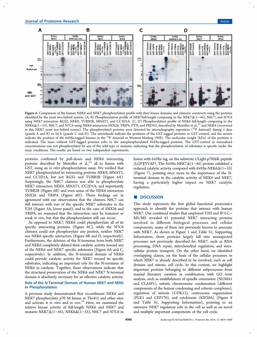
proteins confirmed by pull-down and NEK6 interactingproteins described by Meirelles et al.,22 all in fusion withGST, using an in vitro phosphorylation assay. We verified thatNEK7 phosphorylated its interacting proteins NEK9, MNAT1,and CC2D1A, but not RGS2 and TUBB2B (Figure 6A).Surprisingly, the N6C7 chimera was able to phosphorylateNEK7 interactors NEK9, MNAT1, CC2D1A, and importantlyTUBB2B (Figure 6B) and even some of the NEK6 interactorsSNX26 and TRIP4 (Figure 6D). These findings are inagreement with our observations that the chimera N6C7 canstill interact with two of the specific NEK7 substrates in theY2H (Figure 5A, lower panel), and in the case of SNX26 andTRIP4, we reasoned that the interaction may be transient orweak in vivo, but that the phosphorylation still can occur.As opposed to NEK7, NEK6 could phosphorylate all of its
specific interacting proteins (Figure 6C), while the N7C6chimera could not phosphorylate any protein, neither NEK7nor NEK6 specific interactors (Figure 6B and D, respectively).Furthermore, the deletion of the N-terminus from both NEK7and NEK6 completely ablated their catalytic activity toward anyof the NEK6 and NEK7 specific substrates (Figure 6A and C,respectively). In addition, the N-terminal domain of NEK6could provide catalytic activity for NEK7 toward its specificsubstrates, indicating an important role for the N-terminus ofNEK6 in catalysis. Together, these observations indicate thatthe structural preservation of the NEK6 and NEK7 N-terminaldomain is absolutely necessary for an effective catalytic activity.
Role of the N-Terminal Domain of Human NEK7 and NEK6in Phosphorylation
A previous study demonstrated that recombinant NEK6 andNEK7 phosphorylate p70 S6 kinase at Thr412 and other sitesand activate it in vitro and in vivo.34 Here, we examined therelative kinase activity of full-length NEK6 and NEK7 andmutants NEK7Δ(1−44), NEK6Δ(1−33), N6C7 and N7C6 in
fusion with 6×His tag, on the substrate ULight-p70S6K peptide(LGFTYVAP). The 6×His-NEK7Δ(1−44) protein exhibited areduced catalytic activity compared with 6×His-NEK6Δ(1−33)(Figure 7), pointing once more to the importance of the N-terminal domain in the catalytic activity of NEK6 and NEK7,having a particularly higher impact on NEK7 catalyticregulation.
■ DISCUSSION
This study represents the first global functional proteomicsapproach to identify the proteins that interact with humanNEK7. Our combined studies that employed Y2H and IP-LC−MS/MS revealed 61 potential NEK7 interacting proteinsinvolved in different biological processes and cellularcomponents, many of them not previously known to associatewith NEK7. As shown in Figure 1 and Table S1, SupportingInformation, these proteins largely fall into unsuspectedprocesses not previously described for NEK7, such as RNAprocessing, DNA repair, mitochondrial regulation, and intra-cellular protein transport. On the other hand, we identifiedoverlapping classes, on the basis of the cellular processes inwhich NEK7 is already described to be involved, such as celldivision and mitotic cell cycle. In this context, we highlightimportant proteins belonging to different subprocesses frommanual literature curation in combination with GO termanalysis, such as establishment of spindle orientation (NUMA1and CLASP1), mitotic chromosome condensation (differentcomponents of the human condensing and cohesin complexes),regulation of mitosis (CDK13), centrosome organization(PLK1 and CEP170), and cytokinesis (KIF20A) (Figure 8and Table S1, Supporting Information), pointing to anextensive NEK7 regulatory role in the cell as well as on noveland multiple important components of the cell cycle.
Figure 6. Comparison of the human NEK6 and NEK7 phosphorylation profile with their kinase domains and chimeric constructs using the proteinsidentified by the yeast two-hybrid system. (A, B) Phosphorylation profile of NEK7full-length comparing to the NEK7Δ(1−44), N6C7, and N7C6using NEK7 interactors RGS2, NEK9, TUBB2B, MNAT1, and CC2D1A. (C, D) Phosphorylation profile of NEK6 full-length comparing to theNEK6Δ(1−33), N6C7, and N7C6 using NEK6 interactors SNX26, TRIP4, PTN, and PRDX3, described by Meirelles et al.,22 and NEK9 (recoveredin this NEK7 yeast two-hybrid screen). The phosphorylated proteins were detected by autoradiography exposition (32P Autorad) during 5 days(panels A and B) or 24 h (panels C and D). The arrowheads indicate the positions of the GST-tagged proteins or GST-control, and the arrowsindicate the position of the 6×His-tagged kinases in the 32P Autorad or Western blotting (WB). The molecular weight (kDa) of the proteins isindicated. The lanes without GST-tagged proteins refer to the autophosphorylated 6×His-tagged proteins. The GST-control in normalizedconcentrations was not phosphorylated by any of the wild-type or mutants, indicating that the phosphorylation of substrates is specific under theassay conditions. The results are based on two independent experiments.
Journal of Proteome Research Article
dx.doi.org/10.1021/pr500437x | J. Proteome Res. 2014, 13, 4074−40904086

This notion is supported by the fact that we were able toshow that several of the NEK7 interacting proteins serve indeedas its in vitro substrates, including TUBB2B, MNAT1, RGS2(not shown here), CC2D1A, and NEK9, and by theobservation that several of these interacting proteins, includingNEK9, TUBB, MNAT1, RGS2, and PLEKH8, localized alongNEK7 in specific subcellular regions through the cell cycle(Figures 3, 4, and 8 and Table S3, Supporting Information).The major specific sites of localization include the spindle andspindle pole during mitosis, the contractile ring duringtelophase, and the cytoplasmic bridge (presumably midbody)during cytokinesis (Figures 3 and 4 and Table S3, SupportingInformation). In this scenario, the important potential NEK7targets such as TUBB2B, PLK1, SMC1A, and SMC3 werevalidated for interaction with NEK7 by in vitro pull-down assay(Figure 2A). It is relevant to emphasize that PLK1 waspreviously demonstrated to control the mitotic centrosomeseparation through phosphorylation/activation of NEK9 andNEK6/7-dependent sequential phosphorylation of kinesinKIF11.35 Furthermore, the potential NEK7 interactor andsubstrate, TUBB2B, is an isoform of β-tubulin class II thatpromotes the regulation of microtubule dynamics in humancells when phosphorylated,36,37 indicating a direct NEK7 role inregulating microtubules themselves. Together, these datasupport the idea that NEK7 may potentially act as amultifunctional kinase that regulates proteins categorized intodistinct or shared biological processes, specifically in the celldivision signaling.The evolutionarily conserved NEK6 and NEK7 are the result
of a duplication event during early chordate evolution, and their
high similarity to each other could suggest highly equivalentfunctions.16 Nevertheless, structural and functional studies haveindicated possible differential functions for thesekinases.13,17,18,20,21,35 The defining feature of NEKs is aconserved kinase domain in the N-terminal region and a C-terminal region highly divergent in composition and length butthat contains all the motifs that are typical of a regulatorydomain such as coiled coil motifs.38 NEK6 and NEK7 breakthis rule having a nonconserved short N-terminal extensionfollowed immediately by a C-terminal domain that is the kinasedomain itself. Studies performed by our group suggest that theinteractions of human NEK6 and NEK7 with their specific/different partners could be dependently regulated by theirdistinct and disordered N-terminal domains.19 Then, it isplausible that the N- and C-terminal domains of NEK6 andNEK7 likely act in concert to recognize specific interactors/substrates, via distinct mechanisms.To investigate the role of N- and C-terminal domains of
NEK6 and NEK7 in specific protein recognition andphosphorylation as well as to differentiate their functions, wedeveloped chimeric constructs of NEK6 and NEK7 andconducted an interaction comparative experiment by Y2H.We observed that, aside from NEK9, all other proteinsinteracted specifically with each NEK, providing evidence thatNEK6 and NEK7 could play independent and nonredundantroles in the cell. In this respect, we suggest that NEK7 couldphysically interact with various proteins within disparate cellularmicroenvironments to regulate cellular functions differentlyfrom NEK6. We also demonstrate that N7C6 chimerapresented the broad binding capacity with both NEK6 andNEK7 interactors, indicating a role of NEK6 C-terminalcatalytic domain and NEK7 N-terminal in protein recognition,in light of the fact that deletion of the N-terminus from NEK6had abrogated all interactions with its interactors.22 Thesestudies delineate NEK6 and NEK7 N-terminal domains asimportant regulatory structural components, corroboratingprevious data,18,19 and highlight a surprising role for their C-terminal catalytic domain in binding affinity and/or specificity.In addition, the evidence that NEK6 and NEK7 N- and C-terminal domains present different binding properties indicatesdistinct mechanisms for NEK6 and NEK7 interaction andregulation. The relatively distinct N-terminal structures ofNEK6 and NEK7 flanking the kinase domains may cause themajority of the differential response.Regarding phosphorylation, our results demonstrate a higher
activity of NEK6 on its specific substrates compared to NEK7.Importantly, N6C7 chimera showed a greater phosphorylationspectrum compared to wild-type NEK7, whereas N7C6chimera together with NEK6Δ(1−33) and NEK7Δ(1−44)mutants did not exhibit catalytic activity on different proteinssubstrates. These observations suggest that the NEK6 N-terminal domain is important for its catalytic function andprovides gain-of-function phenotypes for NEK7 kinase domainover nonspecific substrates. These findings are in line with ourobservations of the reduced catalytic activity on specific peptidesubstrates presented by NEK7Δ(1−44). In this regard, thecatalytic domain alone could be intrinsically autoinhibited,regulated in a protein interaction-specific manner or con-ceivably through interactions with the regulatory N-terminaldomain. Indeed, NEK7 structural studies showed that the N-terminal extension of NEK7 (residues 20−33) adopts a highlyunusual structure sitting on the core catalytic domain,18 whichmay contribute to stabilizing the active conformation of the
Figure 7. In vitro kinase activity assay of full-length NEK7 (6×His-NEK7), NEK7 kinase domain (6×His-NEK7Δ(1−44)), chimerasN6C7 (6×His-N6C7) and N7C6 (6×His-N7C6), full-length NEK6(6×His-NEK6), and NEK6 kinase domain (NEK6Δ(1−33)). Theenzymes were incubated at a concentration of 80 nM with 50 nMULight-p70 S6K Peptide, and the phosphorylation was scored in thepresence of 100 μM ATP. Kinase reactions were terminated after 60min by the addition of EDTA. A reaction without addition of ULight-p70 S6K Peptide was used as a control (Control). Data represent themean and standard deviation of three independent experiments. Thesignal was measured on a 2104 EnVision Multilabel MicroplateReader. Excitation wavelength was set to 320 nm, and fluorescenceemission was recorded at 665 nm.
Journal of Proteome Research Article
dx.doi.org/10.1021/pr500437x | J. Proteome Res. 2014, 13, 4074−40904087

kinase. Therefore, we hypothesized that NEK6 and NEK7 N-and C-terminal domains can contribute to both regulation andcatalysis, via distinct mechanisms. Structural and functionalstudies using truncated constructions and site-directed muta-genesis of NEK7 and NEK6 would further dissect the region(s)of their N- and C-terminal domains, involved in determiningthe regulation and catalysis mechanisms. This would help us tofurther understand the independent signaling functions ofNEK6 and NEK7 in mammalian cells.In summary, in the present study we have characterized a
comprehensive protein interactome for human NEK7 andidentified 61 proteins in diverse sets of pathways and processes;this result, combined with functional in vitro and in vivo assays,reinforces NEK7 participation in the regulation of keycomponents of cell division and reveals novel possible NEK7interactors and functions. We showed that the closely relatedhuman kinases NEK6 and NEK7 do not share commoninteractors, with the exception of NEK9, and display distinctmodes of protein interaction. In this regard, given thepreviously reported importance of NEK6 and NEK7 N-terminal domains in protein regulation, we provided evidencethat, via distinct mechanisms for NEK6 and NEK7, the N- andC-terminal domains are both important structural components
for regulation and catalysis of these kinases. Our data suggestdistinct, independent, and nonredundant signaling functions forNEK6 and NEK7 in the cell and provide a novel conceptualframework for future investigation of the structural andbiochemical basis of a distinct NEK6 and NEK7 regulationand catalysis.
■ ASSOCIATED CONTENT*S Supporting Information
Tables of node attributes from NEK7 first neighbors network,in silico analysis of protein interaction with NEK7 retrieved bythe yeast two-hybrid system screens, and proposed cellularlocalization and colocalization of NEK7 with interactingpartners throughout the cell cycle. This material is availablefree of charge via the Internet at http://pubs.acs.org.
■ AUTHOR INFORMATIONCorresponding Author
*Tel: (+55)19-3512-1125. Fax: (+55)19-3512-1006. E-mail:[email protected]
The authors declare no competing financial interest.
Figure 8. Schematic representation of human NEK7 interactors throughout the cell cycle. The scheme depicts the components of NEK7 and itsinteractors throughout the cell cycle according to biological processes (filled line) and subcellular localization (dashed arrows). The subprocesseswere manually classified according to the literature in combination with GO term analysis. See detailed legend for symbols at the bottom of thefigure. ER: endoplasmic reticulum.
Journal of Proteome Research Article
dx.doi.org/10.1021/pr500437x | J. Proteome Res. 2014, 13, 4074−40904088

■ ACKNOWLEDGMENTS
The authors thank Maria Eugenia Camargo for technicalassistance. We thank Dr. Artur T. Cordeiro and Gustavo F.Mercaldi for assistance with the assays performed using theEnVision Multilabel Microplate Reader, Tereza C. L and Silvafor assistance with the Operetta High Content Imaging System(PerkinElmer) and Mariana Bertini Teixeira by kindlyproviding the 6×His-RARA protein. This work was supportedby Fundacao de Amparo a Pesquisa do Estado Sao Paulo(FAPESP), Conselho Nacional de Pesquisa e Desenvolvimento(CNPq) and Centro Nacional de Pesquisa em Energia eMateriais (CNPEM). We also acknowledge the U.S. NationalInstitutes of Health contract HHSN268201000031C fromNHLBI and grant P41 GM104603 from NIGMS
■ REFERENCES(1) Ma, H. T.; Poon, R. Y. How protein kinases co-ordinate mitosisin animal cells. Biochem. J. 2011, 435, 17−31.(2) Fry, A. M.; Meraldi, P.; Nigg, E. A. A centrosomal function forthe human NEK2 protein kinase, a member of the NIMA family of cellcycle regulators. EMBO J. 1998, 17, 470−481.(3) Moniz, L.; Dutt, P.; Haider, N.; Stambolic, V. NEK family ofkinases in cell cycle, checkpoint control and cancer. Cell Div. 2011, 6,18.(4) Dodson, C. A.; Haq, T.; Yeoh, S.; Fry, A. M.; Bayliss, R. Thestructural mechanisms that underpin mitotic kinase activation.Biochem. Soc. Trans. 2013, 41, 1037−1041.(5) Morris, N. R. Mitotic mutants of Aspergillus nidulans. Genet. Res.1975, 26, 237−254.(6) Oakley, B. R.; Morris, N. R. A mutation in Aspergillus nidulansthat blocks the transition from interphase to prophase. J. Cell Biol.1983, 96, 1155−1158.(7) Belham, C.; Roig, J.; Caldwell, J. A.; Aoyama, Y.; Kemp, B. E.;Comb, M.; Avruch, J. A mitotic cascade of NIMA family kinases.Nercc1/NEK9 activates the NEK6 and NEK7 kinases. J. Biol. Chem.2003, 278, 34897−34909.(8) Li, J. J.; Li, S. A. Mitotic kinases: the key to duplication,segregation, and cytokinesis errors, chromosomal instability, andoncogenesis. Pharmacol Ther. 2006, 111, 974−984.(9) Upadhya, P.; Birkenmeier, E. H.; Birkenmeier, C. S.; Barker, J. E.Mutations in a NIMA-related kinase gene, NEK1, cause pleiotropiceffects including a progressive polycystic kidney disease in mice. Proc.Natl. Acad. Sci. U.S.A. 2000, 97, 217−221.(10) Quarmby, L. M.; Mahjoub, M. R. Caught NEK-ing: cilia andcentrioles. J. Cell Sci. 2005, 118, 5161−5169.(11) Kim, S.; Lee, K.; Rhee, K. NEK7 is a centrosomal kinase criticalfor microtubule nucleation. Biochem. Biophys. Res. Commun. 2007, 360,56−62.(12) O’Regan, L.; Blot, J.; Fry, A. M. Mitotic regulation by NIMA-related kinases. Cell Div. 2007, 2, 25.(13) O’Regan, L.; Fry, A. M. The NEK6 and NEK7 protein kinasesare required for robust mitotic spindle formation and cytokinesis. Mol.Cell. Biol. 2009, 29, 3975−3990.(14) Yissachar, N.; Salem, H.; Tennenbaum, T.; Motro, B. NEK7kinase is enriched at the centrosome, and is required for proper spindleassembly and mitotic progression. FEBS Lett. 2006, 580, 6489−6495.(15) Kim, S.; Kim, S.; Rhee, K. NEK7 is essential for centrioleduplication and centrosomal accumulation of pericentriolar materialproteins in interphase cells. J. Cell Sci. 2011, 124, 3760−3770.(16) Salem, H.; Rachmin, I.; Yissachar, N.; Cohen, S.; Amiel, A.;Haffner, R.; Lavi, L.; Motro, B. NEK7 kinase targeting leads to earlymortality, cytokinesis disturbance and polyploidy. Oncogene 2010, 29,4046−4057.(17) Kandli, M.; Feige, E.; Chen, A.; Kilfin, G.; Motro, B. Isolationand characterization of two evolutionarily conserved murin kinases(NEK6 and NEK7) related to the fungal mitotic regulator, NIMA.Genomics 2000, 68, 187−196.
(18) Richards, M. W.; O’Regan, L.; Mas-Droux, C.; Blot, J. M.;Cheung, J.; Hoelder, S.; Fry, A. M.; Bayliss, R. An autoinhibitorytyrosine motif in the cell-cycle-regulated NEK7 kinase is releasedthrough binding of NEK9. Mol. Cell 2009, 36, 560−570.(19) Meirelles, G. V.; Silva, J. C.; Mendonca Yde, A.; Ramos, C. H.;Torriani, I. L.; Kobarg, J. Human NEK6 is a monomeric mostlyglobular kinase with an unfolded short N-terminal domain. BMCStruct. Biol. 2011, 11, 12.(20) Feige, E.; Motro, B. The related murine kinases, NEK6 andNEK7, display distinct patterns of expression. Mech. Dev. 2002, 110,219−223.(21) Minoguchi, S.; Minoguchi, M.; Yoshimura, A. Differentialcontrol of the NIMA related kinases, NEK6 and NEK7, by serumstimulation. Biochem. Biophys. Res. Commun. 2003, 301, 899−906.(22) Meirelles, G. V.; Lanza, D. C. F.; Silva, J. C.; Bernachi, J. S.;Leme, A. F. P.; Kobarg, J. Characterization of NEK6 interactomereveals an important role for its short n-terminal domain andcolocalization with proteins at the centrosome. J. Proteome Res. 2010,9, 6298−6316.(23) Bartel, P. L.; Fields, S. Analyzing protein-protein interactionsusing two-hybrid system. Methods Enzymol. 1995, 254, 241−263.(24) Devault, A.; Martinez, A. M.; Fesquet, D.; Labbe, J. C.; Morin,N.; Tassano, J. P.; Nigg, E. A.; Digger, J. C.; Doree, M. MNAT1(‘menage a trois’) a new RING finger protein subunit stabilizing ciclinH-cdk7 complexes in starfish and Xenopus CAK. EMBO J. 1995, 14,5027−5036.(25) Whelan, S. A.; He, J.; Lu, M.; Souda, P.; Saxton, R. E.; Faull, K.F.; Whitelegge, J. P.; Chang, H. R. Mass spectrometry (LC−MS/MS)identified proteomic biosignatures of breast cancer in proximal fluid. J.Proteome Res. 2012, 11, 5034−5045.(26) Carazzolle, M. F.; de Carvalho, L. M.; Slepicka, H. H.; Vidal, R.O.; Pereira, G. A.; Kobarg, J.; Meirelles, G. V. IIS - IntegratedInteractome System: A Web-based platform for the annotation,analysis and visualization of protein-metabolite-gene-drug interactionsby integrating a variety of data sources and tools. PLoS One 2014, 9,e100385.(27) Shannon, P.; Markiel, A.; Ozier, O.; Baliga, N. S.; Wang, J. T.;Ramage, D.; Amin, N.; Schwikowski, B.; Ideker, T. Cytoscape: asoftware environment for integrated models of biomolecularinteraction networks. Genome Res. 2003, 13, 2498−2504.(28) Surpili, M. J.; Delben, T. M.; Kobarg, J. Identification of proteinsthat interact with the central coiled-Ccoil region of the human proteinkinase NEK1. Biochemistry 2003, 42, 15369−15376.(29) Bartel, P, Chien, C, Sternglanz, R, Fields, S. Cellular Interactionsin Development: A Practical Approach; Hartley, D.A., Ed.; OxfordUniversity Press: Oxford, 1993; pp 153−179.(30) Park, S.; Lim, B. B.; Perez-Terzic, C.; Mer, G.; Terzic, A.Interaction of asymmetric ABCC9-encoded nucleotide bindingdomains determines KATP channel SUR2A catalytic activity. J.Proteome Res. 2008, 7, 1721−1728.(31) Hegde, M. L.; Hazra, T. K.; Mitra, S. Functions of disorderedregions in mammalian early base excision repair proteins. Cell. Mol. LifeSci. 2010, 67, 3573−3587.(32) Mittag, T.; Kay, L. E.; Forman-Kay, J. D. Protein dynamics andconformational disorder in molecular recognition. J. Mol. Recognit.2010, 23, 105−116.(33) Doxsey, S. J.; Stein, P.; Evans, L.; Calarco, P. D.; Kirschner, M.Pericentrin highly conserved centrosome protein involved in micro-tubule organization. Cell 1994, 76, 639−650.(34) Belham, C.; Comb, M. J.; Avruch, J. Identification of the NIMAfamily kinases NEK6/7 as regulators of the p70 ribosomal S6 kinase.Curr. Biol. 2011, 11, 1155−1167.(35) Rapley, J.; Nicolas, M.; Groen, A.; Regue, L.; Bertran, M. T.;Caelles, C.; Avruch, J.; Roig, J. The NIMA-family kinase NEK6phosphorylates the kinesin Eg5 at a novel site necessary for mitoticspindle formation. J. Cell Sci. 2008, 121, 3912−3921.(36) Fourest-Lieuvin, A.; Peris, L.; Gache, V.; Garcia-Saez, I.; Juillan-Binard, C.; Lantez, V.; Job, D. Microtubule regulation in mitosis:
Journal of Proteome Research Article
dx.doi.org/10.1021/pr500437x | J. Proteome Res. 2014, 13, 4074−40904089

tubulin phosphorylation by the cyclin-dependent kinase Cdk1. Mol.Biol. Cell 2006, 17, 1041−1050.(37) Xu, W.; Xi, B.; Wu, J.; An, H.; Zhu, J.; Abassi, Y.; Feinstein, S.C.; Gaylord, M.; Geng, B.; Yan, H.; Fan, W.; Sui, M.; Wang, X.; Xu, X.Natural product derivative bis(4-fluorobenzyl) trisulfide inhibits tumorgrowth by modification of beta-tubulin at Cys 12 and suppression ofmicrotubule dynamics. Mol. Cancer Ther. 2009, 8, 3318−3330.(38) Meirelles, G. V.; Perez, A. M.; Souza, E. E.; Basei, F. L.; Papa, P.F.; Hanchuk, T. D. M.; Cardoso, V. B.; Kobarg, J. “Stop Ne(c)kingaround”: How systems biology can help to characterize the functionsof NEK family kinases from cell cycle regulation to DNA damageresponse. World J. Biol. Chem. 2014, 5, 141−160.(39) Ewing, R. M.; Chu, P.; Elisma, F.; Li, H.; Taylor, P.; Climie, S.;McBroom-Cerajewski, L.; Robinson, M. D.; O’Connor, L.; Li, M.;Taylor, R.; Dharsee, M.; Ho, Y.; Heilbut, A.; Moore, L.; Zhang, S.;Ornatsky, O.; Bukhman, Y. V.; Ethier, M.; Sheng, Y.; Vasilescu, J.;Abu-Farha, M.; Lambert, J. P.; Duewel, H. S.; Stewart, I. I.; Kuehl, B.;Hogue, K.; Colwill, K.; Gladwish, K.; Muskat, B.; Kinach, R.; Adams, S.L.; Moran, M. F.; Morin, G. B.; Topaloglou, T.; Figeys, D. Large-scalemapping of human protein-protein interactions by mass spectrometry.Mol. Syst. Biol. 2007, 3, 89.
Journal of Proteome Research Article
dx.doi.org/10.1021/pr500437x | J. Proteome Res. 2014, 13, 4074−40904090
Copyright © 2022 FDOKUMEN