Expression of the preoptic regulatory factor-1 and −2 genes in rat testis
8
Endocrine, vol.6, no. 1, 65-72, February 1997 0969-71 lX/97/6:65-72/$10.00 1997 byHumana Press Inc. All rights of anynature whatsoever reserved. Expression of the Preoptic Regulatory Factor-1 and -2 Genes in Rat Testis Developmental and Hormonal Regulation Felicia V. Nowak, 1,2 Gonzalo Torres, 2 Jennifer Golden? and Shuang-Bao Hu ~ 1Division of Endocrinology, Department of Internal Medicine and 2Department of Pharmacological and Physiological Sciences, St. Louis University Health Sciences Center, MO; 3Department of Molecular and Cell Biology, The University of Connecticut, Storrs, CT Hormone-responsive peptides play a vital role in development and regulation of testicular function. The preoptic regulatory factors, porf-1 and porf-2, were originally discovered in the rat brain, but are also expressed in the rat and human testis. In the brain expression is age-related, hormone-responsive, region- specific, and gender-related, suggesting that porf-1 and porf-2 are involved in gender-specific brain develop- ment and function. Tissue-specific porf-1 and porf-2 mRNAs are also found in the testis and hypophysec- tomy may alter testicular porf-2 expression. It was thus of interest to further examine porf-1 and porf-2 expres- sion in the testis to evaluate their potential as hor- mone-responsive peptides that regulate testicular development and function. Testicular expression of both porf-1 and -2 was analyzed as a function of matu- rational stage, aging and hypophysectomy by the solu- tion hybridization/nuclease protection assay, and cellular location determined by in situ hybridization histochemistry. Expression was quantitatively com- pared in normal male rats at 15, 30, and 60 d (n = 4) and at 2, 6, 12, and 24 mo of age (n -- 5). During develop- ment porf-1 is expressed at a constant level at 15, 30, and 60 d, then declines significantly with advancing age; levels at 24 mo are only 20% of those seen at 2 mo (p < 0.05). In contrast, porf-2 expression is highest at 15 d of age and steadily declines at 30 and 60 d, pla- teaus in the mature adult (6 and 12 too), then exhibits an additional significant decline in the aged 24 mo animals (6 vs 24 too, p < 0.05). Hypophysectomy of young adult rats at day 42 results in increased testicu- lar expression 12 d later of both porf-1 (p < 0.05) and porf-2 (p < 0.005) compared to intact 54-d-old rats (n = 5). In situ hybridization histochemistry confirms Received October 7,1996; Revised November 13,1996; Accepted November 13, 1996. Author to whom all correspondence and reprint requests should be addressed: Felicia V. Nowak, Division of Endocrinology, SLUHSC, 1402 S. Grand Blvd., St. Louis, MO 63104. E-maih [email protected] that both porf-1 and porf-2 are expressed in the mature testis at 60 d of age. Porf-2 mRNA is localized to immature germ cells including spermatogonia and primary spermatocytes. Porf-1 mRNA is associated with mature sperm and at low levels in the Sertoli cell cyto- plasm surrounding spermatocytes. These data suggest that porf-2 is a pituitary hormone-responsive factor in the developing testis and that both porf-1 and porf-2 have cell-type specific functions in the germ cell com- partment of the mature testis. Key Words: Preoptic regulatory factor genes; testis; development; puberty; pituitary; Sertoli cells; germ cells; aging; reproduction. 65 Introduction The preoptic regulatory factor genes, porf- 1 and porf-2, are widely expressed in the mammalian central nervous system (CNS) (1-3). Tissue-specific transcripts are also found in rat and human testis (1,4). In the CNS there are brain region-specific, gender-related, modifications of expression of both porfgenes during development and with aging (2, 3). Hypophysectomy and castration alter brain-specific transcripts in a region-dependentmanner indicating expres- sion may be sex-hormone responsive (4; Nowak, unpub- lished data). In addition, in the female rat, gonadal steroids affect expression ofporf-1 and porf-2 in the preoptic ante- rior hypothalamus and hippocampus. It is well-established that neuropeptides can have dis- tinct specific functions outside of the CNS often as autocrine or paracrine regulators of cell function. Testis- specific porf-1 and porf-2 mRNA transcripts are found in both rat and human testis (1,4) and hypophysectomy may alter testicular porf-2 expression. The authors, therefore, wished to examine the hypothesis that porf-1 and porf-2 might function as autocrine and paracrine factors in the testis.
-
Upload
independent -
Category
Documents
-
view
2 -
download
0
Transcript of Expression of the preoptic regulatory factor-1 and −2 genes in rat testis

Endocrine, vol. 6, no. 1, 65-72, February 1997 0969-71 lX/97/6:65-72/$10.00 �9 1997 by Humana Press Inc. All rights of any nature whatsoever reserved.
Expression of the Preoptic Regulatory Factor-1 and -2 Genes in Rat Testis Developmental and Hormonal Regulation
Felicia V. Nowak, 1,2 Gonzalo Torres, 2 Jennifer Golden? and Shuang-Bao Hu ~
1Division of Endocrinology, Department of Internal Medicine and 2Department of Pharmacological and Physiological Sciences, St. Louis University Health Sciences Center, MO; 3Department of Molecular and Cell Biology, The University of Connecticut, Storrs, CT
Hormone-responsive peptides play a vital role in development and regulation of testicular function. The preoptic regulatory factors, porf-1 and porf-2, were originally discovered in the rat brain, but are also expressed in the rat and human testis. In the brain expression is age-related, hormone-responsive, region- specific, and gender-related, suggesting that porf-1 and porf-2 are involved in gender-specific brain develop- ment and function. Tissue-specific porf-1 and porf-2 mRNAs are also found in the testis and hypophysec- tomy may alter testicular porf-2 expression. It was thus of interest to further examine porf-1 and porf-2 expres- sion in the testis to evaluate their potential as hor- mone-responsive peptides that regulate testicular development and function. Testicular expression of both porf-1 and -2 was analyzed as a function of matu- rational stage, aging and hypophysectomy by the solu- tion hybridization/nuclease protection assay, and cellular location determined by in situ hybridization histochemistry. Expression was quantitatively com- pared in normal male rats at 15, 30, and 60 d (n = 4) and at 2, 6, 12, and 24 mo of age (n -- 5). During develop- ment porf-1 is expressed at a constant level at 15, 30, and 60 d, then declines significantly with advancing age; levels at 24 mo are only 20% of those seen at 2 mo (p < 0.05). In contrast, porf-2 expression is highest at 15 d of age and steadily declines at 30 and 60 d, pla- teaus in the mature adult (6 and 12 too), then exhibits an additional significant decline in the aged 24 mo animals (6 vs 24 too, p < 0.05). Hypophysectomy of young adult rats at day 42 results in increased testicu- lar expression 12 d later of both porf-1 (p < 0.05) and porf-2 (p < 0.005) compared to intact 54-d-old rats (n = 5). In situ hybridization histochemistry confirms
Received October 7,1996; Revised November 13,1996; Accepted November 13, 1996. Author to whom all correspondence and reprint requests should be addressed: Felicia V. Nowak, Division of Endocrinology, SLUHSC, 1402 S. Grand Blvd., St. Louis, MO 63104. E-maih [email protected]
that both porf-1 and porf-2 are expressed in the mature testis at 60 d of age. Porf-2 mRNA is localized to immature germ cells including spermatogonia and primary spermatocytes. Porf-1 mRNA is associated with mature sperm and at low levels in the Sertoli cell cyto- plasm surrounding spermatocytes. These data suggest that porf-2 is a pituitary hormone-responsive factor in the developing testis and that both porf-1 and porf-2 have cell-type specific functions in the germ cell com- partment of the mature testis.
Key Words: Preoptic regulatory factor genes; testis; development; puberty; pituitary; Sertoli cells; germ cells; aging; reproduction.
65
Introduction
The preoptic regulatory factor genes, porf- 1 and porf-2, are widely expressed in the mammalian central nervous system (CNS) (1-3). Tissue-specific transcripts are also found in rat and human testis (1,4). In the CNS there are brain region-specific, gender-related, modifications of expression of both porfgenes during development and with aging (2, 3).
Hypophysectomy and castration alter brain-specific transcripts in a region-dependent manner indicating expres- sion may be sex-hormone responsive (4; Nowak, unpub- lished data). In addition, in the female rat, gonadal steroids affect expression ofporf-1 and porf-2 in the preoptic ante- rior hypothalamus and hippocampus.
It is well-established that neuropeptides can have dis- tinct specific functions outside of the CNS often as autocrine or paracrine regulators of cell function. Testis- specific porf-1 and porf-2 mRNA transcripts are found in both rat and human testis (1,4) and hypophysectomy may alter testicular porf-2 expression. The authors, therefore, wished to examine the hypothesis that porf-1 and porf-2 might function as autocrine and paracrine factors in the testis.

66 Preoptic Regulatory Factors-1 and -2 in Testis/Nowak et al. Endocrine
As a first step, studies were designed to investigate porf-1 and porf-2 mRNA expression in rat testis during sexual development, reproductive aging, and in response to hypophysectomy. The authors also wished to localize the cell types expressing these two genes in the testis. Finally, they asked the question whether brain and testis transcripts are derived from one or several porf-1 and porf-2 genes.
M a t e r i a l s a n d M e t h o d s
Animals
Sprague-Dawley outbred rats were obtained from Harlan Sprague-Dawley, Indianapolis, IN, for the 15, 30, and 60 d intact rat comparisons and from Charles River Laborato- ries, Boston, MA, for the hypophysectomy studies, includ- ing the control rats, and for in situ hybridization analysis. Fischer 344 rats, a commonly used rat model for aging, were used for the aging study (2, 6, 12, and 24 too). All rats were housed with food and water ad libitum. Hypophy- sectomies were done at 42 d of age and rats were sacrificed 12 d later along with intact 54-d-old rats. Hypophysectomized animals were maintained on 5% glucose in their drinking water. Lights were on for 12 h daily (6 Arcr-6 PM). Rats were anesthetized and sacrificed between 10 AM and 3 PM.
Brains and pituitary fossae were examined for absence of pituitary tissue in the hypophysectomized rats. Testes were weighed then quick frozen on dry ice and stored at -80~ for RNA quantitation by nuclease protection assay, or imbedded in mounting media, frozen in blocks and stored at-70~ until sectioned for in situ hybridization analysis.
Source of Materials
The T7 RNA polymerase was obtained from Promega (Madison, WI). Proteinase K, tRNA, and formamide were from Boehringer Mannheim (Indianapolis, IN). [32p]-CTP and [32p]-UTP were from Amersham (Arlington Heights, IL). [35S]-dATP was from New England Nuclear (Boston, MA); BSA was Pentex Fraction V (Miles Laboratories, Kankakee, IL); nitrocellulose was from Schleicher & Schuell (Keene, NH); SDS was from Fluka (Ronkonkoma, NY). Restriction enzymes were obtained as follows: BamHI from IBI (New Haven, CT); HindlII from New England Biolabs (Beverly, MA); SstI from Gibco-BRL (Grand Island, NY). All other dry chemicals were obtained from Sigma (St. Louis, MO) and wet chemicals from Mallinkrodt (Paris, KY).
RNA Extraction
Testes were cut while frozen into approx 100 mg blocks. RNA was extracted by a guanidine thiocyanate/phenol/ chloroform/isoamyl alcohol extraction technique (5). Indi- vidual RNA samples were quantitated by spectrophotom- etry at 260 nm and the yield was expressed as total RNA/ mg tissue. There was <15% variation in yield within a group. Isolated RNA was used immediately for RNase pro- tection assays or stored at -80~
Nuclease Protection Assays
Sense RNAs and riboprobes for porf-1 and porf-2 were prepared from pGEM plasmids as described previously (1), according to the protocol provided by the supplier (Pro- mega, Madison, WI). [32p]-CTP and T7 polymerase were used for probe synthesis. Transcription of the porf-1 insert yields a 188 nt probe that is complementary to the 5' end of the brain porf-1 eDNA. The porf-2 insert gives a 375 nt probe that is complementary to the 5' end of the porf-2 brain eDNA. The assays were carried out with 20 ~g sample RNA or 0, 0.3, 0.6, 1.25, 2.5, 5, and 10 pg synthetic RNA for the standard curves, as described previously (2). The protected RNA fragments were separated by 6% polyacrylamide gel electrophoresis, and the resulting autoradiographs were quantified by densitometry. The results are expressed in OD units and reflect the absolute specific RNA level with respect to the total extractable RNA. Readings can only be accurately compared within an assay as variables such as probe specific activity and X-ray film exposure time can affect unit readings. A wide range of synthetic standard RNAs was measured to insure that all samples fell within the linear range of the assay curve. The number of animals per group is indicated in the figure legends. All tissues assayed within a group were from the same set of four or five individual animals.
In Situ Hybridization Analysis
Testis frozen sections 8 jzaa in thickness were cut, thaw- mounted onto glass slides which had been treated with 0.25% chromium potassium sulfate/0.25% gelatin and allowed to dry thoroughly, then stored desiccated at-80~ Sections were postfixed in 4% formaldehyde in phosphate- buffered saline (PBS), treated with 0.25% acetic anhydride in 0.1M triethanolamine HC1, pH 8, and dehydrated by transfer through graded alcohol and chloroform (6). Hybridization buffer (45 p.L per section), followed by parafilm coverslips, were applied and the slides were incu- bated for 22 h at 37~ in humidified chambers. Hybridiza- tion buffer was 50% formamide, 4 x SSC (0.6M NaCI, 0.06M sodium citrate), 500 pg/mL sheared single stranded DNA, 250/.tg/mL yeast tRNA, 0.02% ficoll, 0.02% poly- vinylpyrrolidone, 0.02% bovine serum albumin (BSA), 50 mM dithiothrietol (DTT), 2% sodium polyacrylate, and 40,000 cpm 35S-oligonucleotide porf-1 or porf-2 probe. Synthetic 48 nucleotide DNA probes were end-labeled with [35S]- dATP using 2 U/pL terminal deoxynucleotidyl transferase in 500 mM potassium cacodylate pH 7.2, 10 mM COC12, 1.0 mMDTT for 5 min at 37~ The probe-specific activi- ties were between 2 and 8 x 10⁸ cprn/p.g. The sequence of the porf- 1 48 nucleotide probe, 5'CACAGTGGGAAGCCT GGTACATCAGCAAATCCCTCTGGCTGTGGTTGG3', is complementary to bases 41 through 88 of the rat porf-1 eDNA sequence (1) and the purified porf-2 48 nucleotide probe, 5'GCTGCACGAACACCTGGAGGAAGCGAA TGAGGTAGCACAGCACCATTC3', is complementary
Vol. 6, No. 1 Preoptic Regulatory Factors-1 and -2 in Testis/Nowak et al. 67
to bases 75 through 122 of the rat porf-2 cDNA sequence (1). Specificity of binding is indicated by the fact that under identical conditions and in paired specimens the two probes hybridize to different cell populations and areas.
Slides were washed in 1 x SSC at 55~ (4 x 15 min) then 1 x SSC at room temperature (2 x 30 min). Dried slides were dipped into Amersham LM-1 nuclear emulsion and exposed for 3 wk at 4~
Southern Blot Hybridization Analysis
Genomic DNA was prepared from brain and liver of male Sprague-Dawley rats. Brain DNA was isolated from pulverized frozen tissue (7). Liver DNA was purified from fresh nuclei isolated by homogenizing tissues in 5% citric acid, filtration through sterile gauze and centrifugation through a 0.88M sucrose cushion (7). Samples were treated with 1% SDS and proteinase K, 1 mg/mL, for 4 h at 37~ then extracted with phenol/chloroform followed by diethyl ether. The DNA was recovered by spooling with a glass rod after ethanol precipitation. The DNA was resolubilized in 1 x SSC, treated with DNase-free RNase and extracted with phenol/chloroform.
Plasmids containing full-length porf- 1 and porf-2 cDNA inserts and corresponding to graded numbers of diploid gene copies were digested with HindlII endonuclease and quantified by fluorometric assay using bisbenzimidazole (Hoechst Dye No. 33258) and a Hoeffer fluorometer (8).
Genomic DNA (20 pg) was digested with each of three restriction enzymes, BamHI, HindlII, and SstI, then elec- trophoresed through 0.9% agarose gels in parallel with the plasmid copy number standards. Two micrograms tomato DNA was used as carrier for the plasmids. DNA was trans- ferred to Nytran membranes (Schliecher & Schuell, Keene, NH) by the method of Southern (9) and fixed by UV crosslinking. RNA probes were prepared by transcription from porf- 1 and porf-2 subclones as described above under Nuclease Protection Assays, except that [32p]-UTP was used. Additional RNA probes corresponding to the 3' por- tions ofporf-1 and porf-2 cDNAs were also prepared. The 3' porf-1 probe is complementary to nucleotides 189 through 522 of the brain porf-1 cDNA (1). The 3' porf-2 probe is complementary to nucleotides 709 through 1352 of the brain porf-2 cDNA (1). The probes thus were of defined length and devoid of vector sequence, which makes them suitable for gene dosage analysis.
Prehybridization and the hybridization were carried out at 65~ for 2 and 16 h, respectively. Filters were pre- hybridized in 6 x SSC, 10 x Denhardt's (1% ficoll, 1% polyvinylpyrrolidone, 1% BSA), I mMEDTA, 100 jag/mL yeast tRNA, 1% SDS and hybridized in 6 x SSC, 5 x Denhardt's, 100 gg/mL yeast tRNA, 0.05% SDS, and between 5 x 10⁶ and 1 x 10⁷ cpm radiolabeled probe/mL. Filters were washed to a stringency of 0.1 x SSC at 65~ three times for 15 min each and exposed to X-ray film for 1-2 d a t -80oc with an intensifying screen.
Statistics Statistical comparisons were done using ANOVA fol-
lowed by Duncan's multiple range test. Significance was accepted at p < 0.05. All comparisons were within assay comparisons. Readings can only be validly compared within an assay as variables such as probe-specific activity and X-ray film exposure time can affect densitometry read- ings. Therefore, all samples to be compared were assayed within the same assay.
R e s u l t s
Expression of porf-1 and porf-2 mRNA in Testis During Sexual Development
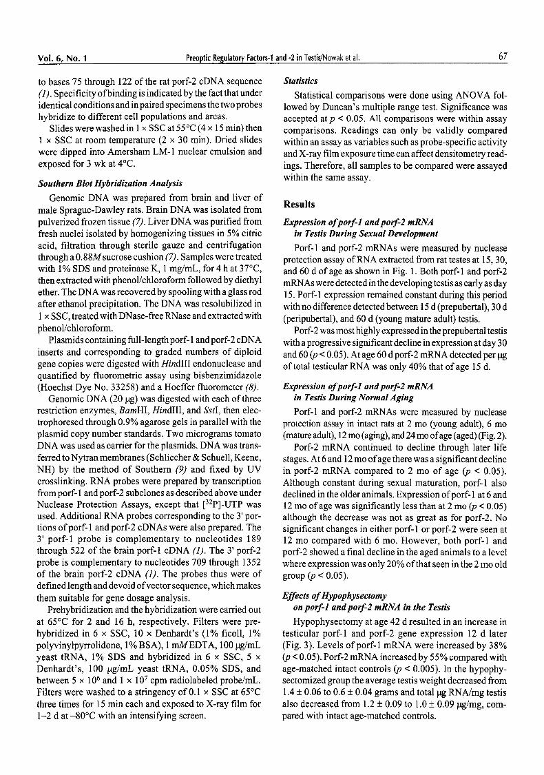
Porf-1 and porf-2 mRNAs were measured by nuclease protection assay of RNA extracted from rat testes at 15, 30, and 60 d of age as shown in Fig. 1. Both porf-1 and porf-2 mRNAs were detected in the developing testis as early as day 15. Porf-1 expression remained constant during this period with no difference detected between 15 d (prepubertal), 30 d (peripubertal), and 60 d (young mature adult) testis.
Porf-2 was most highly expressed in the prepubertal testis with a progressive significant decline in expression at day 30 and 60 (p < 0.05). At age 60 d porf-2 mRNA detected per tag of total testicular RNA was only 40% that of age 15 d.
Expression of porf-1 and porf-2 mRNA in Testis During Normal Aging
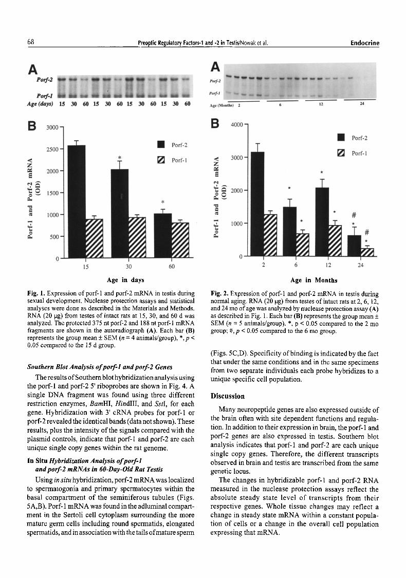
Porf-1 and porf-2 mRNAs were measured by nuclease protection assay in intact rats at 2 mo (young adult), 6 mo (mature adult), 12 mo (aging), and 24 mo of age (aged) (Fig. 2).
Porf-2 mRNA continued to decline through later life stages. At 6 and 12 mo of age there was a significant decline in porf-2 mRNA compared to 2 mo of age (p < 0.05). Although constant during sexual maturation, porf-1 also declined in the older animals. Expression ofporf- 1 at 6 and 12 mo of age was significantly less than at 2 mo (p < 0.05) although the decrease was not as great as for porf-2. No significant changes in either porf- 1 or porf-2 were seen at 12 mo compared with 6 mo. However, both porf-1 and porf-2 showed a final decline in the aged animals to a level where expression was only 20% of that seen in the 2 mo old group (p < 0.05).
Effects of Hypophysectomy on porf-1 and porf-2 mRNA in the Testis
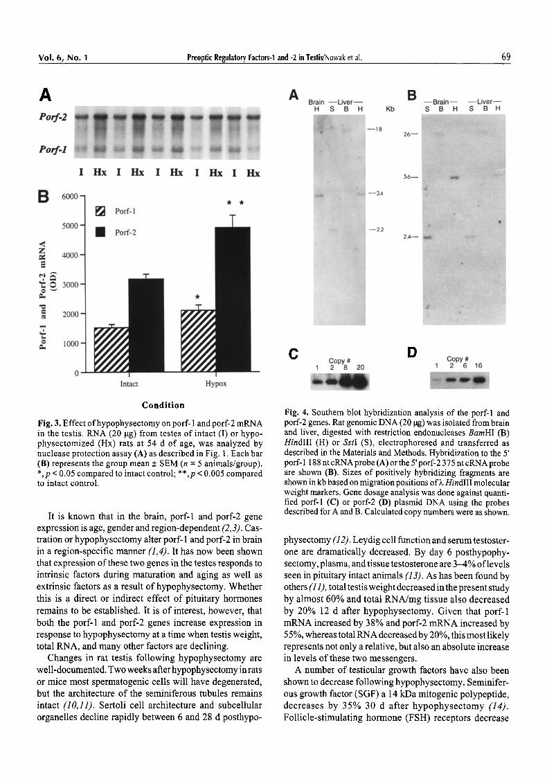
Hypophysectomy at age 42 d resulted in an increase in testicular porf-1 and porf-2 gene expression 12 d later (Fig. 3). Levels ofporf-1 mRNA were increased by 38% (p < 0.05). Porf-2 mRNA increased by 55% compared with age-matched intact controls (p < 0.005). In the hypophy- sectomized group the average testis weight decreased from 1.4 + 0.06 to 0.6 + 0.04 grams and total pg RNA/mg testis also decreased from 1.2 + 0.09 to 1.0 + 0.09/.tg/mg, com- pared with intact age-matched controls.
68 Preoptic Regulatory Factors-1 and -2 in Testis/Nowak et al. Endocrine
A P o r f . 2
P o r f - I
Age (days)
B ~
< z
0 ~
"o
t - O
15 30 60 15 30 60 15 30 60 15
A ~ ~ Porf-2 ~ ~ r
�9 ,;; 5
Porf.l ~'~ ~ ~ 0
3 0 60 Age(Months) 2
B 4000 -1
~ Q D w w .... ~
5"
~ ~ flD : ~
< Z
e,;
o
o
3000
2000
6 12 24
�9 Porf-2
[ ] Porf-1
* #
~r
15 30 60
Age in days
Fig. 1. Expression of porf-1 and porf-2 mRNA in testis during sexual development. Nuclease protection assays and statistical analyses were done as described in the Materials and Methods. RNA (20 lag) from testes of intact rats at 15, 30, and 60 d was analyzed. The protected 375 nt porf-2 and 188 nt porf-I mRNA fragments are shown in the autoradiograph (A). Each bar (B) represents the group mean + SEM (n = 4 animals/group), *, p < 0.05 compared to the 15 d group.
Southern Blot Analysis of porf-1 and porf-2 Genes
The results of Southern blot hybridization analysis using the porf-1 and porf-2 5' riboprobes are shown in Fig. 4. A single DNA fragment was found using three different restriction enzymes, BamHI, HindlII, and SstI, for each gene. Hybridization with 3' cRNA probes for porf-1 or porf-2 revealed the identical bands (data not shown). These results, plus the intensity of the signals compared with the plasmid controls, indicate that porf-1 and porf-2 are each unique single copy genes within the rat genome.
In Situ Hybridization Analysis of porf-1 and porf-2 mRNAs in 60-Day-Old Rat Testis
Using in situ hybridization, porf-2 mRNA was localized to spermatogonia and primary spermatocytes within the basal compartment of the seminiferous tubules (Figs. 5A,B). Porf-1 mRNA was found in the adluminal compart- ment in the Sertoli cell cytoplasm surrounding the more mature germ cells including round spermatids, elongated spermatids, and in association with the tails of mature sperm
2 6 12 24
Age in Months
Fig. 2. Expression of porf-1 and porf-2 mRNA in testis during normal aging. RNA (20 pg) from testes of intact rats at 2, 6, 12, and 24 mo of age was analyzed by nuclease protection assay (A) as described in Fig. 1. Each bar (B) represents the group mean + SEM (n = 5 animals/group). *, p < 0.05 compared to the 2 mo group; #, p < 0.05 compared to the 6 mo group.
(Figs. 5C,D). Specificity of binding is indicated by the fact that under the same conditions and in the same specimens from two separate individuals each probe hybridizes to a unique specific cell population.
Discuss ion
Many neuropeptide genes are also expressed outside of the brain often with site dependent functions and regula- tion. In addition to their expression in brain, the porf- 1 and porf-2 genes are also expressed in testis. Southern blot analysis indicates that porf-1 and porf-2 are each unique single copy genes. Therefore, the different transcripts observed in brain and testis are transcribed from the same genetic locus.
The changes in hybridizable porf-1 and porf-2 RNA measured in the nuclease protection assays reflect the absolute steady state level of transcripts from their respective genes. Whole tissue changes may reflect a change in steady state mRNA within a constant popula- tion of cells or a change in the overall cell population expressing that mRNA.
Vol. 6, No. 1 Preoptic Regulatory Factors-1 and -2 in Testis/Nowak et al. 69
A Porf.2
Porf-I
I Hx I Hx I Hx I Hx I Hx
B 6000-1 ~1 Porf-1
z == E
0 ~ ==
t . .
o
Intact Hypox
C o n d i t i o n
Fig. 3. Effect ofhypophysectomy on porf- 1 and porf-2 mRNA in the testis. RNA (20 ~tg) from testes of intact (I) or hypo- physectomized (Hx) rats at 54 d of age, was analyzed by nuclease protection assay (A) as described in Fig. 1. Each bar (B) represents the group mean + SEM (n = 5 animals/group). *,p < 0.05 compared to intact control; **,p < 0.005 compared to intact control.
It is known that in the brain, porf-1 and porf-2 gene expression is age, gender and region-dependent (2, 3). Cas- tration or hypophysectomy alter porf- 1 and porf-2 in brain in a region-specific manner (1,4). It has now been shown that expression of these two genes in the testes responds to intrinsic factors during maturation and aging as well as extrinsic factors as a result of hypophysectomy. Whether this is a direct or indirect effect of pituitary hormones remains to be established. It is of interest, however, that both the porf-1 and porf-2 genes increase expression in response to hypophysectomy at a time when testis weight, total RNA, and many other factors are declining.
Changes in rat testis following hypophysectomy are well-documented. Two weeks after hypophysectomy in rats or mice most spermatogenic cells will have degenerated, but the architecture of the seminiferous tubules remains intact (10,11). Sertoli cell architecture and subcellular organelles decline rapidly between 6 and 28 d posthypo-
A B Brain - - L i v e r - - - - B r a i n - - - - L i v e r - -
H S B H Kb S B H S B H :3
- - 18 26- -
~ , --3.4
- -22
5.6--
2.,d--
C D Copy # Copy # 1 2 8 20 1 2 6 16
Fig. 4. Southern blot hybridization analysis of the port'-1 and porf-2 genes. Rat genomic DNA (20 p.g) was isolated from brain and liver, digested with restriction endonucleases BamHI (B) HindIII (H) or SstI (S), electrophoresed and transferred as described in the Materials and Methods. Hybridization to the 5' porf- 1 188 nt cRNA probe (A) or the 5' porf-2 375 nt cRNA probe are shown (B). Sizes of positively hybridizing fragments are shown in kb based on migration positions of~. HindIII molecular weight markers. Gene dosage analysis was done against quanti- fied porf-1 (C) or porf-2 (D) plasmid DNA using the probes described for A and B. Calculated copy numbers were as shown.
physectomy (12). Leydig cell function and serum testoster- one are dramatically decreased. By day 6 posthypophy- sectomy, plasma, and tissue testosterone are 3-4% of levels seen in pituitary intact animals (13). As has been found by others (11), total testis weight decreased in the present study by almost 60% and total RNA/mg tissue also decreased by 20% 12 d after hypophysectomy. Given that porf-1 mRNA increased by 38% and porf-2 mRNA increased by 55 %, whereas total RNA decreased by 20%, this most likely represents not only a relative, but also an absolute increase in levels of these two messengers.
A number of testicular growth factors have also been shown to decrease following hypophysectomy. Seminifer- ous growth factor (SGF) a 14 kDa mitogenic polypeptide, decreases by 35% 30 d after hypophysectomy (14). Follicle-stimulating hormone (FSH) receptors decrease

70 Preoptic Regulatory Factors-1 and -2 in Testis/Nowak et al. Endocrine
Fig. 5. In situ hybridization ofporf- 1 and porf-2 mRNAs in 60-d-old rat testis. A and B show hybridization of35S-labeled porf-2 48 base oligonucleotide probe to immature germ cells including spermatogonia and primary spermatocytes in the basal compartment of the seminiferous tubules. Sections A and B are from two different rats. Sections were counterstained with cresyl violet (original magnifi- cation x320).
10-fold (15) and androgen-binding protein decreases 1000-fold (16). Inhibin, which is secreted by Sertoli cells in response to FSH, declines (17). These factors are all normally stimulated by pituitary hormones. In contrast TGF-fl, which stops cell growth and promotes differentia- tion, is negatively regulated by the pituitary (18); FSH inhibits both TGF-13 mRNA production and protein secre- tion from pig Sertoli cells. The decrease in porf-2 with puberty and increase after hypophysectomy suggests that it is normally under negative control by the pituitary. The changes in porf-1 are less dramatic, but it also increased posthypophysectomy, implying removal of a suppressive effect. This could be a direct effect of pituitary hormones or a result of the more generalized metabolic changes seen in hypophysectomized animals.
Substantial maturation and differentiation take place in the testis after birth and porf- 1 and porf-2 are expressed in the testis as early as 15 d of age. In the maturing testis
porf-2 expression is highest prepubertally, then declines rapidly, whereas porf-1 expression is constant during the peripubertal period, then also declines later in life. Thus, porf- 1 and porf-2 expression appear to correlate with func- tional activity of the testis. The differences in absolute lev- els ofporf-1 and porf-2 mRNAs at 60 d of age in Figs. 1 and 2 most likely reflect interassay variability in probe specific activities and autoradiographic exposure. However, a true difference between the two strains of rat cannot be excluded without a single-assay comparison.
Expression ofpeptide factors in the testes is often cell- type specific as well as tubule compartment and stage dependent (10,19-23). Many cell-type specific factors in the mammalian testes are expressed during development and spermatogenesis in an age and stage dependent fash- ion. These include inhibin, activin, TGF-131, TGF-133, TGF-ct, and IGF-I. Expression of the porf-1 and porf-2 genes in the mature testis is also cell-type specific, and

Vol. 6, No. 1 Preoptic Regulatory Factors-1 and -2 in Testis/Nowak et al. 71
i , 2 : ' : : : ' : : i r ~
Fig. 5. C and D show hybridization of 3sS-labeled porf- 1 48 base oligonucleotide probe within the Sertoli cell cytoplasm surround- ing maturing germ cells including round spermatids, elongated spermatids, and in association with the tails of mature spermatozoa. C and D are from the same two individual rats shown in A and B. Sections were counterstained with cresyl violet (original magnification x320).
compartment-specific with porf-1 being expressed in Ser- toli cells in the adluminal compartment, whereas porf-2 is expressed in germ cells in the basal compartment.
Porf-2 is expressed early in development, decreases with puberty, and hypophysectomy increases porf-2 mRNA back to levels seen in prepubertal rats. In situ hybridization analy- sis shows porf-2 to be present in mitotically active prelepto. tene germ cells, in the basal compartment within the blood-testes barrier. Given its proposed structure, a 75 amino acid peptide with a hydrophobic secretory leader sequence (1), porf-2 may be acting as a germ cell-derived regulatory factor on the pituitary or as a paracrine factor in the testis.
Porf-1 is moderately and steadily expressed during development. Expression is stimulated by removal of the pituitary gland. It is found in Sertoli cells surrounding maturing germ cells within the adluminal compartment, outside the blood-testes boundary. Porf-1 is a small hydrophilic peptide that does not appear to have a leader
sequence (1). It may, therefore, play a local regulatory role in the testicular microenvironment related to germ cell meiosis and differentiation as a cryptocrine or intracrine agent (18).
Although the results reported here support a role for porf-1 and porf-2 in testicular maturation and function, further studies are required to directly establish the roles of these two genes and their peptide products in the testes, as well as the mechanism of the effects of pituitary removal on porf-1 and porf-2 gene expression.
A c k n o w l e d g m e n t s
The authors thank Stewart Albert for helpful discussions; Sharon Shrader, Department of Pathology, St. Louis Uni- versity Health Sciences Center, for cutting the cryostat sections; and Marge Smith and Laura Carpenter for help with manuscript preparation.

72 Preoptic Regulatory Factors-1 and -2 in Testis/Nowak et al. Endocrine
References
1. Nowak, F. V. (1990). Mol. Endocrinol. 4, 1205-1210. 2. Hu, S.-B. and Nowak, F. V. (1994). Mol. Cell. Neurosci. 5,
376-381. 3. Hu, S.-B. and Nowak, F. V. (1995). Endocrine 3, 421-424. 4. Nowak, F. V. (1991). Mol. Cell. Neurosci. 2, 395-401. 5. Chomczynsky, P. and Sacchi N. (1987). Anal. Biochem. 162,
156-159. 6. Young, SW III. (1990). In: Handbook of chemical neu-
roanatomy: analysis of neuronal microcircuits and synaptic interactions, vol. 8. Bjorklund, A., Hokfelt, T., Wouterlood, F. G., and van den Pol, A. N. (eds). Elsevier: Amsterdam.
7. Davis, L. G., Dibner, M. D., and Battey, J. F. (1986). Basic meth- ods in molecular biology. Elsevier: Amsterdam, pp. 47-50.
8. Labarca, C. and Paigin, K. (1980). Anal. Biochem. 102, 344-352.
9. Southern, E. M. (1975). J. Mol. Biol. 99, 503-517. 10. Lian, G. and Enders, G. C. (1994). J. Androl. 15, 52-60. 11. Sun, Y.-T., Wreford, N. G., Robertson, D. M., and DeKretser,
D. M. (1990). Endocrinology 127, 1215-1223. 12. Ghosh, S., Bartke, A., Grasso, P., Reichert, L. E. Jr., and Russell,
L. D. (1992). Endocrinology 131,485-497.
13. Russell, L. D., Corbin, T. J., Ren, H. P., Amador, A., Bartke, A., and Ghosh, S. (1992). Endocrinology 131, 498-508.
14. Feig, L. A., Klagsbrun, M., and Bellve, A. R. (1983). J. Cell. Biol. 97, 1435-1443.
15. Thanki, K. H. and Steinberger, A. (1978). Andrologia 20, 195-202.
16. Sanborn, B. M., Steinberger, A., Tcholakian, K., and Stein- berger, E. (1977). Steroids 29, 493-502.
17. Krummen, L. A., Toppari, J., Kim, W. H., Morelos, B. S., Ahmad, N., Swerdloff, R. S., Ling, N., Shimasaki, S., Esch, F., and Bhasin, S. (1989). Endocrinology 125, 1630-1637.
18. Saez, J. M., Avallet, O., Lejeune, H., and Chatelain, P. G. (1991). Hormone Res. 36, 104-115.
19. Morales, C., Hugly, S., and Griswold, M. D. (1987). Biol. Reprod. 36, 1035-1046.
20. Toppari, J., Kangasniemi, M., Kaipia, A., Mall, P., Huhtaniemi, I., and Parvinen, M. (1991). J.. Electron. Microsc. Tech. 19, 203--214.
21. Garza, M. M, Schwarz, L. K., Bonner, J. M., and Boockfor, F. R. (1991). Endocrinology 128, 1869-1874.
22. Kleisch, S., Penttila, T.-L., Gromoll, J., Saunders, P. T. K., Nieschlag, E., and Parvinen, M. (1992). Mol. Cell. Endocrinol. 84, R45-R49.
23. Kierszenbaum, A. L. (1994). Endocr. Rev. 15, 116-134.