Expression of Herpes Virus Entry Mediator (HVEM) in the Cornea and Trigeminal Ganglia of Normal and...
10
896 Current Eye Research, 34(10), 896–904, 2009 Copyright © 2009 Informa Healthcare USA, Inc. ISSN: 0271-3683 print/ 1460-2202 online DOI: 10.3109/02713680903184250 ORIGINAL ARTICLE Expression of Herpes Virus Entry Mediator (HVEM) in the Cornea and Trigeminal Ganglia of Normal and HSV-1 Infected Mice S. Krisztian Kovacs 1 , Vaibhav Tiwari 2 , Emese Prandovszky 1 , Sandor Dosa 1 , Sarolta Bacsa 2 , Klara Valyi-Nagy 1 , Deepak Shukla 2 , and Tibor Valyi-Nagy 1 1 Department of Pathology, University of Illinois at Chicago, College of Medicine, Chicago, Illinois, USA 2 Departments of Ophthalmology and Visual Sciences and Microbiology and Immunology, University of Illinois at Chicago, College of Medicine, Chicago, Illinois, USA INTRODUCTION Infection of the human cornea by herpes simplex viruses (HSV) is primarily due to herpes sim- plex virus type 1 (HSV-1) in adults and herpes Received 13 April 2009; revised 11 July 2009; accepted 14 July 2009 Correspondence: Tibor Valyi-Nagy, M.D., Ph.D., University of Illinois at Chicago, College of Medicine, Department of Pathology, Room 130, 840 South Wood St., M/C 847, Chicago, IL 60612. E-mail: [email protected] ABSTRACT Purpose: Herpes virus entry mediator (HVEM) plays a critical role in the regulation of inflammation through interaction with its natural ligands LIGHT and lymphotoxin alpha and also serves as one of the entry receptors of herpes simplex virus (HSV). The purpose of this study was to better understand the expression of HVEM in the cornea and trigeminal ganglia (TG), which are important targets of HSV infection. Materials and Methods: Immunohistochemistry was used to define HVEM expression in the cornea and TG of normal and HSV-1 infected mice euthanized 2 to 5 days or 7 months following corneal inoculation of virus. Results: We found that HVEM is widely expressed in the normal corneal epithelium and endothelium, is weakly and focally expressed in the corneal stroma, and is expressed in a portion of neurons and non-neuronal cells in the TG. Acute HSV-1 keratitis and ganglionitis were associated with increased HVEM expression in the corneal epithelium and stroma and in neurons and non-neuronal cells of TG, and many inflammatory cells in these tissues also expressed HVEM. TG derived from mice 7 months after virus inoculation demonstrated latent HSV-1 infection that was associated with increased HVEM expression in neurons and non-neuronal cells relative to uninfected control tissues. Latent TG also contained focal infiltrates of mononuclear inflammatory cells, many of which expressed HVEM. Corneas derived from latently infected mice demonstrated chronic keratitis, with no evidence of virus replication or increased HVEM expression in the corneal epithelium, and inflammatory cells present showed only weak HVEM expression. Conclusions: HVEM is expressed in the cornea and TG and therefore may serve as an HSV entry receptor in these tissues. Furthermore, these findings raise the possibility that changes in HVEM expression following ocular HSV-1 infection can modulate HSV spread and infection-induced inflammation in the cornea and TG. KEYWORDS: Cornea; Herpes simplex virus; Herpes virus entry mediator; HveA; HVEM; TNFRS14; Trigeminal ganglia
-
Upload
midwestern -
Category
Documents
-
view
2 -
download
0
Transcript of Expression of Herpes Virus Entry Mediator (HVEM) in the Cornea and Trigeminal Ganglia of Normal and...
896
Current Eye Research, 34(10), 896–904, 2009Copyright © 2009 Informa Healthcare USA, Inc.ISSN: 0271-3683 print/ 1460-2202 onlineDOI: 10.3109/02713680903184250
ORIGINAL ARTICLE
Expression of Herpes Virus Entry Mediator (HVEM) in the Cornea and Trigeminal Ganglia
of Normal and HSV-1 Infected Mice
S. Krisztian Kovacs1, Vaibhav Tiwari2, Emese Prandovszky1, Sandor Dosa1, Sarolta Bacsa2, Klara Valyi-Nagy1, Deepak Shukla2, and Tibor Valyi-Nagy1
1Department of Pathology, University of Illinois at Chicago, College of Medicine, Chicago, Illinois, USA2Departments of Ophthalmology and Visual Sciences and Microbiology and Immunology, University of Illinois at
Chicago, College of Medicine, Chicago, Illinois, USA
INTRODUCTION
Infection of the human cornea by herpes simplex viruses (HSV) is primarily due to herpes sim-plex virus type 1 (HSV-1) in adults and herpes
Received 13 April 2009; revised 11 July 2009; accepted 14 July 2009
Correspondence: Tibor Valyi-Nagy, M.D., Ph.D., University of Illinois at Chicago, College of Medicine, Department of Pathology, Room 130, 840 South Wood St., M/C 847, Chicago, IL 60612. E-mail: [email protected]
ABSTRACT
Purpose: Herpes virus entry mediator (HVEM) plays a critical role in the regulation of inflammation through interaction with its natural ligands LIGHT and lymphotoxin alpha and also serves as one of the entry receptors of herpes simplex virus (HSV). The purpose of this study was to better understand the expression of HVEM in the cornea and trigeminal ganglia (TG), which are important targets of HSV infection. Materials and Methods: Immunohistochemistry was used to define HVEM expression in the cornea and TG of normal and HSV-1 infected mice euthanized 2 to 5 days or 7 months following corneal inoculation of virus. Results: We found that HVEM is widely expressed in the normal corneal epithelium and endothelium, is weakly and focally expressed in the corneal stroma, and is expressed in a portion of neurons and non-neuronal cells in the TG. Acute HSV-1 keratitis and ganglionitis were associated with increased HVEM expression in the corneal epithelium and stroma and in neurons and non-neuronal cells of TG, and many inflammatory cells in these tissues also expressed HVEM. TG derived from mice 7 months after virus inoculation demonstrated latent HSV-1 infection that was associated with increased HVEM expression in neurons and non-neuronal cells relative to uninfected control tissues. Latent TG also contained focal infiltrates of mononuclear inflammatory cells, many of which expressed HVEM. Corneas derived from latently infected mice demonstrated chronic keratitis, with no evidence of virus replication or increased HVEM expression in the corneal epithelium, and inflammatory cells present showed only weak HVEM expression. Conclusions: HVEM is expressed in the cornea and TG and therefore may serve as an HSV entry receptor in these tissues. Furthermore, these findings raise the possibility that changes in HVEM expression following ocular HSV-1 infection can modulate HSV spread and infection-induced inflammation in the cornea and TG.
KEYWORDS: Cornea; Herpes simplex virus; Herpes virus entry mediator; HveA; HVEM; TNFRS14; Trigeminal ganglia
Cur
r E
ye R
es D
ownl
oade
d fr
om in
form
ahea
lthca
re.c
om b
y E
BSC
O o
n 02
/14/
13Fo
r pe
rson
al u
se o
nly.

HVEM Expression in Cornea and Trigeminal Ganglia 897
© 2009 Informa Healthcare USA, Inc.
simplex virus type 2 (HSV-2) in neonates, and is an important cause of ocular disease.1–4 Latent HSV infection of the trigeminal ganglia (TG) plays an important role in the pathogenesis of recurrent HSV keratitis as a source of reactivated virus.3 Following corneal infection, HSV infects nerve endings, moves by retrograde axonal transport to nerve cell bodies in the TG, and establishes latent infection in neurons.3,5–7 During latent HSV-1 infec-tion of neurons, viral genomic DNA is present in neuronal nuclei, abundant viral gene expression is limited to the latency-associated transcripts (LAT), and viral proteins or infectious virus are usually not detectable. The virus periodically reactivates with production of infectious virus in the ganglia and consequently spread to peripheral tissues, including the cornea. In HSV keratitis, visual morbidity usu-ally results from recurrent disease through corneal scarring, thinning, and neovascularisation, with the immune response playing a key role in the develop-ment of these pathologic changes.4,8
Expression of cellular receptors of HSV entry is a critically important determinant of viral pathogen-esis, as these receptors will determine which tissues and cells become infected during primary and recur-rent infections. Although significant progress has been made in studies of the molecular basis of virus-host interactions leading to HSV entry into cells,9–12 the expression of HSV entry receptors in ocular and peripheral nervous system tissues is not completely understood.
Successful HSV infection depends upon inter-action between the viral particle and cell surface receptors required for viral entry. Viral glycopro-teins B and C (gB and gC) provide the initial binding interaction between the virus and cell-surface gly-cosaminoglycans, preferentially heparan sulphate.9–11 Following this attachment, HSV glycoprotein D (gD) interacts with one of three known cell-surface receptors that are necessary for viral entry.10–11 A spe-cific modification in heparan sulfate generated by certain 3-O-sulfotransferases generates a family of gD receptors that are specific for HSV-1.13 Two cell-surface proteins are known to mediate the entry of HSV-1 and HSV-2 into cultured cells by interacting with gD: nectin-1 and herpes virus entry mediator (HVEM).
Among HSV entry receptors, the expression of nectin-1 in the cornea and TG is relatively well characterized, while the expression of HVEM and 3-O-sulfated heparan sulfate receptors is less well understood. Nectin-1 is a member of the immun-globulin (Ig) superfamily closely related to the poliovirus receptor.14–17 Nectin-1 and the related proteins nectin-2 and nectin-3 are cell adhesion
molecules found at Ca2+-dependent cadherin-based cell adhesion junctions in epithelial cells, synapses, and other types of cells. Nectin-1 is widely expressed in the corneal epithelium and endothelium and in TG neurons.18–20 This widespread expression and the observation that nectin-1 is the primary entry receptor of HSV-1 into sensory neurons21–22 suggest that nectin-1 plays an important role in the patho-genesis of HSV keratitis. However, there is grow-ing evidence that HSV entry receptors other than nectin-1 also have a role in the pathogenesis of HSV ocular infections. For instance, 3-O-sulfated heparan sulfate has been shown to mediate HSV-1 entry into human corneal fibroblasts.23 HVEM in turn mediates HSV-2 entry into corneal fibroblasts24 and HSV-1 entry into the trabecular meshwork25 and conjuncti-val epithelial cells.26
HVEM, also called herpes virus entry protein A (HveA) and tumor necrosis factor receptor 14 (TNFRS14), is a member of the tumor necrosis factor receptor superfamily (TNFR) of proteins.27 HVEM plays a critical role in the regulation of inflammation through interaction with its natural ligands which are LIGHT and lymphotoxin alpha (Lt).28–30 Both ligands are structurally related to tumor necrosis fac-tor (TNF), exist as trimers,31–32 and presumably signal by inducing or altering receptor aggregation on the cell surface.28,33–35 The expression of HVEM in various tissues and cell types is not well understood. Studies using human tissue RNA blots found relatively high HVEM mRNA levels in the lung, spleen, and thymus, but no significant HVEM expression was detected in the brain, liver, or skeletal muscle.36 HVEM is expressed in T-cells, B-cells, natural killer cells (NK), dendritic cells (DCs), neutrophils, and myeloid cells.36 HVEM is expressed in ocular trabecular meshwork cells, cultured corneal fibroblasts, and conjuncti-val epithelial cells,23–26 and HVEM expression was detected in primary cultures of rat and mouse sen-sory neurons.21 HVEM expression was also detected in epithelial cells, fibroblasts, and endothelial cells in gingival tissue.37
To better understand the expression of HVEM in ocular and peripheral nervous system tissues, in the current study, we have used a well-established murine model of HSV-1 pathogenesis and immuno-histochemistry to determine HVEM expression in the cornea and TG. In addition to normal tissues, mouse eye and TG tissues derived from mice during the acute and latent stages of HSV-1 infection were also studied to determine if HSV-1 infection had an effect of HVEM expression. We show here that HVEM is widely expressed in murine cornea and TG, and that HSV-1 infection leads to changes in HVEM expression in these tissues.
Cur
r E
ye R
es D
ownl
oade
d fr
om in
form
ahea
lthca
re.c
om b
y E
BSC
O o
n 02
/14/
13Fo
r pe
rson
al u
se o
nly.
898 S. Krisztian Kovacs et al.
Current Eye Research
MATERIAL AND METHODS
Virus Stocks
HSV-1 strain 17+ was propagated and titered by plaque assay using baby hamster kidney (BHK) 21 clone 13 cells.
Infection of Mice, Recovery of Tissues, and Tissue Processing
To establish a collection of tissues for studies of HVEM expression in normal and HSV-1 infected eyes and trigeminal ganglia, 4- to 6-week-old female BALB/c mice were inoculated on their corneas with 105 PFU of HSV-1 strain 17+ after corneal scarification, as previ-ous described.38 Control mice were either not treated or were inoculated with an equivalent volume (5 µl) of sterile tissue-culture medium using the same inocu-lation technique (mock infection). Groups of at least three mice were euthanized 2–5 days (expected times of acute, productive HSV-1 infection) or at 7 months (expected time of latent HSV-1 infection) after HSV-1 inoculation. The eye bulbs and TG were aseptically removed, embedded in paraffin, and sectioned. Tissue sections were either stained with hematoxylin and eosin or were examined by immunohistochemistry for HSV-1 protein and HVEM expression. TG tissues derived from mice 7 months post infection (p.i.) were also examined by in situ hybridization for the expres-sion of HSV-1 LAT. All animal procedures followed an IAUCUC-approved protocol and the NIH guidelines for laboratory animal care as outlined in NIH publica-tion No. 86-23 (revised 1985).
Immunohistochemical Detection of HSV-1 Proteins
Tissue sections for immunohistochemistry were deparaffinized with xylene and rehydrated through a series of graded ethanols. Endogenous peroxidase activity was quenched using a 0.3% H2O2-methanol bath followed by several washes with phosphate-buffered saline. HSV-1 antigens were detected using a 1:1000 dilution of an HSV-1-specific antiserum raised in a rabbit (DAKO, Carpinteria, CA, USA) as previ-ously described.18,39 Tissue sections were incubated with primary antibody at 43°C for 32 min prior to the addition of biotinylated anti-rabbit immunoglobu-lin secondary antibody, avidin-horseradish peroxi-dase, and 3,3′-diaminobenzidine tetrahydrochloride (0.04%) in 0.05 M Tris-HCL (pH 7.4) and 0.025% H2O2 as a chromogen (Ventana Medical Systems, Tucson,
AZ, USA). Before staining, binding of secondary antibodies and conjugates was blocked by appropri-ate reagents provided by the manufacturer.
Immunohistochemical Detection of HVEM
Tissue sections for HVEM immunohistochemistry were deparaffinized with xylene and rehydrated through a series of graded ethanols. Endogenous peroxidase activity was quenched using a 0.3% H2O2-methanol bath followed by several washes with phosphate-buffered saline. Tissue sections were hydrated with distilled water, and antigen retrieval was performed using the DAKO Target Retrieval Solution 10X Concentrate. Nonspecific staining was blocked using an H2O2 solution for 10 min followed by a protein block for 10 min. Sections were incubated with a rabbit polyclonal antibody against HVEM (courtesy of Professor Patricia Spear, Northwestern University, Chicago, IL, USA) at 1:200 at room tem-perature for 1 hr, as previously described.26 HVEM staining was detected using the DAKO Envision+ kit. To characterize HVEM expression in tissues, a semi-quantitative scoring system was designed and used that placed HVEM expression in a given cell type into one of three categories: 1+ = focal weak HVEM expres-sion, 2+ = widespread moderate HVEM expression, 3+ = widespread strong HVEM expression.
In situ Hybridization for HSV-1-LAT Gene Expression
In situ hybridization was performed using a nick-translated 35S-labeled DNA probe specific for HSV-1 LAT (Bst2 ± Bst2 fragment) according to previously described techniques.40 The probe used in these experiments had a specific activity of .108 cpm/mg.
RESULTS
Establishment of a Collection of Tissues for Studies of HVEM Expression in Normal and HSV-1 Infected Murine Cornea and TG
To establish a collection of tissues for studies of HVEM expression in normal and HSV-1 infected eyes and TG, 4- to 6-week-old female BALB/c mice were inoculated on their corneas with 105 PFU of HSV-1. Control mice were either not treated or were inoculated with sterile tissue-culture medium (mock infection). HSV-1 inoculated mice euthanized at 2 or 5 days after HSV-1 inoculation demonstrated keratitis
Cur
r E
ye R
es D
ownl
oade
d fr
om in
form
ahea
lthca
re.c
om b
y E
BSC
O o
n 02
/14/
13Fo
r pe
rson
al u
se o
nly.
HVEM Expression in Cornea and Trigeminal Ganglia 899
© 2009 Informa Healthcare USA, Inc.
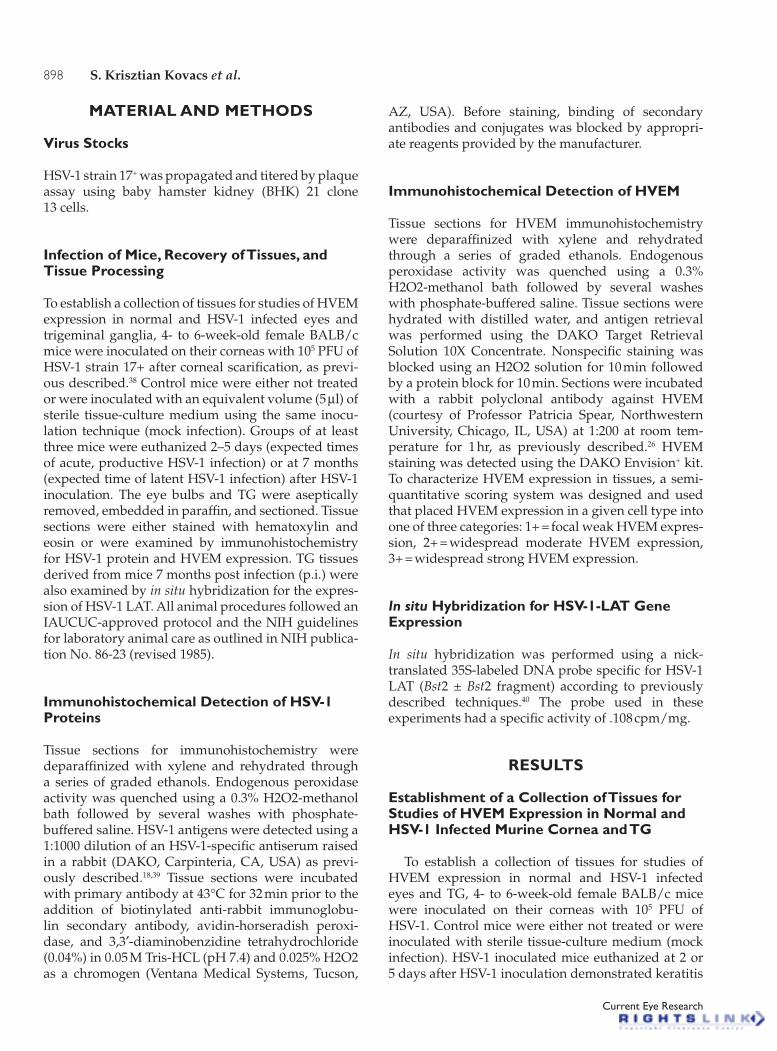
(focal ulceration of the corneal epithelium associ-ated with moderate to severe inflammatory infiltrates composed of neutrophils and mononuclear inflamma-tory cells primarily involving the corneal stroma) and HSV-1 protein expression in the corneal epithelium and stroma (Figure 1A). Uninfected mice showed no evidence of significant corneal inflammation, and no HSV-1 protein expression was detected in the cornea by immunohistochemistry (data not shown). Similar to uninfected mice, no HSV-1 protein expression was detected in the corneas of mock infected mice, and, probably due to the gentle and superficial nature of the corneal scarification procedure, no significant inflammation was detected in the the corneas of mock infected mice (Figure 1B).
The TG of mice euthanized 5 days after virus inoculation demonstrated focal inflammation and ex-pression of HSV-1 proteins in some neurons and non-neuronal cells (Figure 1C), consistent with HSV-1 repli-cation. No evidence of significant inflammation and no HSV-1 protein expression were detected in the TG of un-infected and mock infected mice (Figure 1D) or in mice that were euthanized 2 days after virus inoculation.
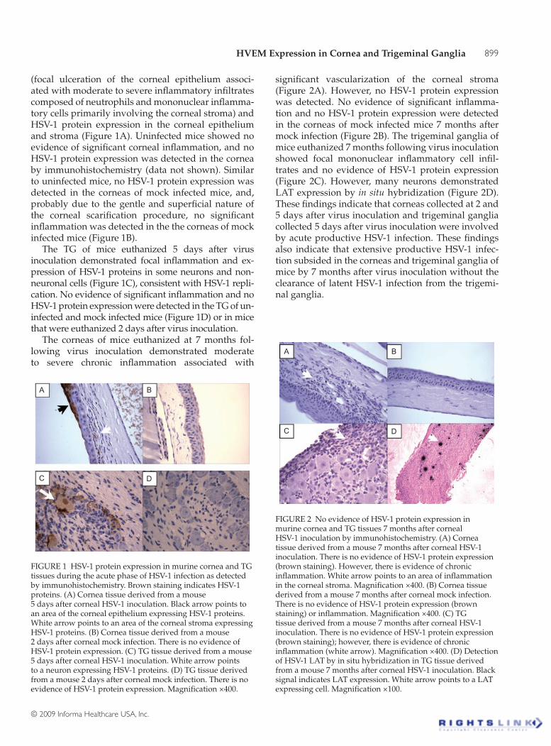
The corneas of mice euthanized at 7 months fol-lowing virus inoculation demonstrated moderate to severe chronic inflammation associated with
significant vascularization of the corneal stroma ( Figure 2A). However, no HSV-1 protein expression was detected. No evidence of significant inflamma-tion and no HSV-1 protein expression were detected in the corneas of mock infected mice 7 months after mock infection (Figure 2B). The trigeminal ganglia of mice euthanized 7 months following virus inoculation showed focal mononuclear inflammatory cell infil-trates and no evidence of HSV-1 protein expression (Figure 2C). However, many neurons demonstrated LAT expression by in situ hybridization (Figure 2D). These findings indicate that corneas collected at 2 and 5 days after virus inoculation and trigeminal ganglia collected 5 days after virus inoculation were involved by acute productive HSV-1 infection. These findings also indicate that extensive productive HSV-1 infec-tion subsided in the corneas and trigeminal ganglia of mice by 7 months after virus inoculation without the clearance of latent HSV-1 infection from the trigemi-nal ganglia.
A B
C D
FIGURE 1 HSV-1 protein expression in murine cornea and TG tissues during the acute phase of HSV-1 infection as detected by immunohistochemistry. Brown staining indicates HSV-1 proteins. (A) Cornea tissue derived from a mouse 5 days after corneal HSV-1 inoculation. Black arrow points to an area of the corneal epithelium expressing HSV-1 proteins. White arrow points to an area of the corneal stroma expressing HSV-1 proteins. (B) Cornea tissue derived from a mouse 2 days after corneal mock infection. There is no evidence of HSV-1 protein expression. (C) TG tissue derived from a mouse 5 days after corneal HSV-1 inoculation. White arrow points to a neuron expressing HSV-1 proteins. (D) TG tissue derived from a mouse 2 days after corneal mock infection. There is no evidence of HSV-1 protein expression. Magnification ×400.
A B
C D
FIGURE 2 No evidence of HSV-1 protein expression in murine cornea and TG tissues 7 months after corneal HSV-1 inoculation by immunohistochemistry. (A) Cornea tissue derived from a mouse 7 months after corneal HSV-1 inoculation. There is no evidence of HSV-1 protein expression (brown staining). However, there is evidence of chronic inflammation. White arrow points to an area of inflammation in the corneal stroma. Magnification ×400. (B) Cornea tissue derived from a mouse 7 months after corneal mock infection. There is no evidence of HSV-1 protein expression (brown staining) or inflammation. Magnification ×400. (C) TG tissue derived from a mouse 7 months after corneal HSV-1 inoculation. There is no evidence of HSV-1 protein expression (brown staining); however, there is evidence of chronic inflammation (white arrow). Magnification ×400. (D) Detection of HSV-1 LAT by in situ hybridization in TG tissue derived from a mouse 7 months after corneal HSV-1 inoculation. Black signal indicates LAT expression. White arrow points to a LAT expressing cell. Magnification ×100.
Cur
r E
ye R
es D
ownl
oade
d fr
om in
form
ahea
lthca
re.c
om b
y E
BSC
O o
n 02
/14/
13Fo
r pe
rson
al u
se o
nly.
900 S. Krisztian Kovacs et al.
Current Eye Research
HVEM Expression in Normal Murine Cornea and TG
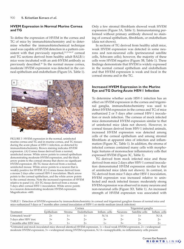
To define the expression of HVEM in the cornea and TG of mice by immunohistochemistry and to deter-mine whether the immunohistochemical technique used was capable of HVEM detection in a pattern con-sistent with that previously reported,21,23–26,37 corneal and TG sections derived from healthy adult BALB/c mice were incubated with an anti-HVEM antibody as previously described.26 In the normal mouse cornea, moderate HVEM expression was detected in the cor-neal epithelium and endothelium (Figure 3A. Table 1).
Only a few stromal fibroblasts showed weak HVEM expression (Figure 3A, Table 1). Immunostaining per-formed without primary antibody showed no stain-ing of corneal epithelium, fibroblasts, or endothelium (data not shown).
In sections of TG derived from healthy adult mice, weak HVEM expression was detected in some neu-rons and non-neuronal cells (perineuronal satellite cells, Schwann cells); however, the majority of these cells were HVEM negative (Figure 3B, Table 1). These findings demonstrate that HVEM is widely expressed in the normal corneal epithelium and endothelium and that HVEM expression is weak and focal in the corneal stroma and in the TG.
Increased HVEM Expression in the Murine Eye and TG During Acute HSV-1 Infection
To determine whether acute HSV-1 infection has an effect on HVEM expression in the cornea and trigemi-nal ganglia, immunohistochemistry was used to detect HVEM expression in the cornea and TG of mice euthanized 2 or 5 days after corneal HSV-1 inocula-tion or mock infection. The corneas of mock infected mice demonstrated HVEM expression similar to that of uninfected mice (data not shown). However, in corneal tissues derived from HSV-1 infected animals, increased HVEM expression was detected among cells of the corneal epithelium and among stromal fibroblasts at apparent sites of infection and inflam-mation (Figure 3C, Table 1). In addition, the stroma of infected corneas contained many cells with morpho-logic features of mononuclear inflammatory cells that expressed HVEM (Figure 3C, Table 1).
TG derived from mock infected mice and those derived from mice 2 days after HSV-1 corneal inocula-tion demonstrated HVEM expression similar to that of uninfected mice (data not shown). In contrast, in TG derived from mice 5 days after HSV-1 inoculation, HVEM expression was increased relative to unin-fected and mock infected tissues: moderately strong HVEM expression was observed in many neurons and non-neuronal cells (Figure 3D, Table 1). An increased strength of HVEM expression was associated with
A B
C D
FIGURE 3 HVEM expression in the normal, uninfected murine cornea and TG and in murine cornea, and TG tissues during the acute phase of HSV-1 infection, as detected by immunohistochemistry. Brown staining indicates HVEM expression. (A) Cornea tissue derived from a normal, uninfected mouse. White arrow points to corneal epithelium demonstrating moderate HVEM expression, and the black arrow points to the corneal stroma that shows no significant HVEM expression. (B) TG tissue derived from a normal, uninfected mouse. White arrow points to a neuron that is weakly positive for HVEM. (C) Cornea tissue derived from a mouse 2 days after corneal HSV-1 inoculation. Black arrow points to the corneal epithelium, and the white arrow points to the corneal stroma. Note the increased expression of HVEM relative to panel (A). (D) TG tissue derived from a mouse 5 days after corneal HSV-1 inoculation. White arrow points to a neuron demonstrating moderate HVEM expression. Magnification ×400.
TABLE 1 Detection of HVEM expression by immunohistochemistry in corneal and trigeminal ganglion tissues of normal mice and mice euthanized 5 days or 7 months after corneal inoculation of HSV-1 or sterile medium (mock infection)
TreatmentCornea Trigeminal ganglia
Epithelium Stroma Endothelium Inflam. cells Neurons Satellite cells Inflam. cellsUntreated/mock* 2+ 1+ 1+ N/A 1+ 1+ N/A5 days after HSV inoc 3+ 3+ 2+ 3+ 2+ 2+ 2+7 months after HSV inoc 2+ 1+ 2+ 1+ 3+ 2+ 2+*Untreated and mock inoculated mice showed identical HVEM expression. 1+ = focal weak HVEM expression, 2+ = widespread moderate HVEM expression, 3+ = widespread strong HVEM expression, N/A = nonapplicable, no inflammatory cells present.
Cur
r E
ye R
es D
ownl
oade
d fr
om in
form
ahea
lthca
re.c
om b
y E
BSC
O o
n 02
/14/
13Fo
r pe
rson
al u
se o
nly.

HVEM Expression in Cornea and Trigeminal Ganglia 901
© 2009 Informa Healthcare USA, Inc.
an increased number of HVEM-positive neurons in infected tissues as the majority of TG neurons were HVEM positive. In inflamed foci in the trigeminal gan-glia, many mononuclear inflammatory cells showed HVEM expression (Figure 3D, Table 1). These findings indicate that acute HSV-1 infection leads to enhanced HVEM expression in the cornea and TG.
HVEM Expression in the Murine Cornea and TG 7 Months after Corneal HSV-1 Inoculation
To determine whether latent HSV-1 infection in the TG modifies the expression of HVEM in the cornea and trigeminal ganglia, tissues derived from mice 7 months following HSV-1 inoculation were examined by immunohistochemistry using an HVEM-specific antiserum. As demonstrated above, corneal and TG tissues derived from these mice showed no evidence of HSV-1 protein expression but HSV-1 LAT expres-sion was readily detectable by in situ hybridization in the TG, consistent with latent HSV-1 infection in the TG. Consistent with previous reports,40–42 both latently infected TG and corneal tissues derived from these mice frequently demonstrated focal chronic inflam-mation (Figures 4A and 4B). Corneal tissues of latently infected mice demonstrated weaker HVEM immuno-reactivity in the corneal epithelium and stroma than those derived from acutely infected mice, and HVEM signal strength and distribution were similar to that detected in uninfected and mock infected control corneas. HVEM-positive cells could be observed pri-marily in the upper layer of corneal epithelium and in the endothelium and in occasional cells in the stroma (Figure 4A, Table 1). In inflamed foci in the cornea, moderate to severe chronic inflammation and signifi-cant vascularization was associated with only weak and focal HVEM expression in mononuclear inflam-matory cells (Figure 4A, Table 1).
In latently infected TG, widespread and strong HVEM expression was detected among neurons (Figure 4B, Table 1). Relative to uninfected and mock infected tissues, increased HVEM expression was also detected among non-neuronal cells. In inflamed foci in the trigeminal ganglia, many mononuclear inflam-matory cells showed HVEM expression, and neuronal HVEM immunoreactivity was stronger and denser in areas of chronic inflammation. The vast majority of neurons stained more intensively positive for HVEM than those in uninfected trigeminal ganglia.
These findings indicate that, similar to acute HSV-1 ganglionitis, latent HSV-1 infection is associated with in-creased neuronal and non-neuronal HVEM expression in the TG. However, in the cornea of latently infected mice, HVEM expression was reduced relative to acutely HSV-1 infected tissue and was similar to that detected in mock infected and uninfected cornea. Interestingly, HVEM expression was weak and infrequent in inflam-matory cells in the corneas of latently infected mice.
DISCUSSION
We show here that HVEM is expressed widely in the normal corneal epithelium and endothelium and is weakly and focally expressed in the corneal stroma. HVEM is also expressed in some neurons and non-neuronal cells in the normal murine TG. Acute HSV-1 keratitis and ganglionitis are associated with increased HVEM expression in the corneal epithelium and stroma and in TG neurons and non-neuronal cells, and many inflammatory cells in these tissues also express HVEM. Furthermore, we also show here that HSV-1 latency in TG is associated with increased HVEM expression in neurons and non-neuronal cells relative to uninfected control tissues and many mononuclear inflammatory cells in latently infected TG express HVEM. Corneas derived from latently infected mice demonstrated chronic keratitis with no evidence of virus replication or increased HVEM expression in the corneal epithe-lium and endothelium, and inflammatory cells present showed only weak HVEM expression.
These findings indicate that several different cell types in the murine cornea and trigeminal ganglia express HVEM and thus suggest that HVEM may serve as an entry receptor for HSV within these tis-sues. The finding that HVEM expression is modified by the HSV-1 infection in the cornea and trigeminal ganglia is novel and may be of clinical significance. Increased expression of HVEM in the corneal epithe-lium and stroma and in TG neurons and non-neuronal cells during acute infection may increase the number of cells susceptible to HSV entry. This may be par-ticularly important in the case of corneal stromal
A B
FIGURE 4 HVEM expression in murine cornea and TG tissues 7 months after corneal HSV-1 inoculation. Brown staining indicates HVEM expression. (A) Cornea tissue derived from a mouse 7 months after corneal mock infection. Black arrow points to HVEM expressing corneal epithelium. (B) TG tissue derived from a mouse 7 months after corneal HSV-1 inoculation. White arrow points to a neuron strongly positive for HVEM. Magnification ×400.
Cur
r E
ye R
es D
ownl
oade
d fr
om in
form
ahea
lthca
re.c
om b
y E
BSC
O o
n 02
/14/
13Fo
r pe
rson
al u
se o
nly.

902 S. Krisztian Kovacs et al.
Current Eye Research
fibroblasts that showed little or no HVEM expres-sion in the absence of HSV-1 infection, but demon-strated increased HVEM expression following HSV-1 inoculation. Similarly, an increased number of HVEM-expressing neurons and non-neuronal cells in the TG during HSV-1 infection may increase the number of cells susceptible for virus entry.
The mechanism by which HSV-1 infection modi-fies HVEM infection in the cornea and TG is unclear from our studies. Corneas and TG of mock infected (scarified, but not virus exposed) mice showed no evidence of increased HVEM expression, thus the increased HVEM expression detected in the HSV-1 inoculated mice could not be due to the scarification process. HVEM expression was increased in a much larger number of cells in the cornea and TG than the number of cells that expressed HSV-1 proteins during acute HSV-1 infection. Furthermore, HVEM expression was increased in a large number of neu-rons and non-neuronal cells in latently infected TG in the absence of HSV-1 protein expression. These find-ings suggest that HSV-1 replication was not directly responsible for the induction of HVEM expression in the cornea and TG. It is possible that HSV-1 infection-induced host inflammatory responses and associated changes in cellular activation may have contributed to the observed increased HVEM expression. It is well known that HVEM expression can be dependent on cell differentiation and activation. For instance, naive T-cells express high levels of HVEM, but expression decreases during activation, with high levels being re-expressed at the end of the activation phase.43–44 Interestingly, our studies indicated that HVEM expres-sion was not increased in the cornea of mice 7 months after HSV-1 inoculation, although these tissues har-bored prominent chronic inflammation. These find-ings suggest that the inflammation associated with chronic herpetic keratitis differs from acute keratitis in its capacity to induce HVEM expression in cells of the corneal epithelium and stroma.
HVEM plays a critical role in the regulation of inflammation through interaction with its natural ligands LIGHT and lymphotoxin alpha (Lt).28 Con-sistent with previous reports, we found evidence of HVEM expression among inflammatory cells in HSV-1 infected corneal and trigeminal ganglion tissues. It was very interesting to note that HVEM expression was widespread among inflammatory cells in the acutely infected cornea and ganglia and in the latently infected ganglia, but was quite weak in inflammatory cells present in the cornea 7 months after virus inocu-lation. The significance of these findings is not clear at this point; however, it is possible that differential expression of HVEM in nervous system tissues and in
the cornea has a role in a site-specific regulation of the immune response in these tissues.
In summary, observations reported here indicate that HVEM is widely expressed in the cornea and TG, and therefore may serve as an HSV entry receptor in these tissues. Furthermore, our findings raise the pos-sibility that special and temporal changes in HVEM expression following ocular HSV-1 infection play a role in the regulation of HSV spread in the cornea and TG and in the regulation of HSV-1 infection-induced inflammation in the cornea and TG.
ACKNOWLEDGEMENTS
This work was supported by fellowship grants to S.K.K., E.M., S.D. and S.B. by the Rosztoczy Foundation.
Declaration of interest: The authors report no conflict of interest. The authors alone are responsible for the content and writing of the paper.
REFERENCES
[1] Easty DL, Carter C, Funk A. Systemic immunity in herpetic keratitis. Br J Ophtalmol. 1981;65:82–88.
[2] Liesegang TJ. Epidemiology of ocular herpes simplex. Natural history in Rochester, Minnesota, 1950 through 1982. Arch Ophtalmol. 1989;107:1160–1165.
[3] Whitley RJ. Herpes simplex viruses. In: Fields BN, Knipe DM, Howley PM, eds. Fields Virology. 4th ed. Philadelphia: Lippincott-Raven; 2001:2461–2509.
[4] Kaye S, Choudhari A. Herpes simplex keratitis. Prog Retin Eye Res. 2006;25:355–380.
[5] Mott KR, Bresee CJ, Allen SJ, BenMohamed L, Wechsler SL, Ghiasi H. Level of herpes simplex virus type 1 latency correlates with severity of corneal scarring and exhaustion of CD8+ T cells in trigeminal ganglia of latently infected mice. J Virol. 2009;83:2246–2254.
[6] Toma HS, Murina AT, Areaux RG Jr, Neumann DM, Bhattacharjee PS, Foster TP, Kaufman HE, Hill JM. Ocular HSV-1 latency, reactivation, and recurrent disease. Semin Ophthalmol. 2008;23:249–273.
[7] Valyi-Nagy T, Shukla D, Engelhard HH, Kavouras J, Scanlan P. Latency strategies of alphaherpesviruses: Herpes simplex virus and varicella-zoster virus latency in neurons. In: Minarovits J, Gonczol E, Valyi-Nagy T, eds. Chapter 1. Latency Strategies of Herpesviruses. New York: Springer; 2007:1–36.
[8] Doymaz, MZ, Rouse, BT. Immunopathology of herpes simplex virus infections. In: Rouse BT, ed. Herpes Simplex Virus. Pathogenesis, Immunobiology, and Control. Berlin: Springer; 1992:121–136.
[9] Spear PG. Entry of alphaherpesviruses into cells. Sem Virol. 1993;4:167–180.
[10] Spear PG, Eisenberg RJ, Cohen GH. Three classes of cell surface receptors for entry. Virology. 2000;275:1–8.
[11] Shukla D, Spear PG. Herpesviruses and heparan sulfate: An intimate relationship in aid of viral entry. J Clin Invest. 2001;108:503–510.
Cur
r E
ye R
es D
ownl
oade
d fr
om in
form
ahea
lthca
re.c
om b
y E
BSC
O o
n 02
/14/
13Fo
r pe
rson
al u
se o
nly.
HVEM Expression in Cornea and Trigeminal Ganglia 903
© 2009 Informa Healthcare USA, Inc.
[12] Taylor JM, Lin E, Susmarski N, Yoon M, Ware AF, Pfeffer K, Miyoshi J, Takai Y, Spear PG. Alternative entry receptors for herpes simplex virus and their roles in disease. Cell Host Microbe. 2007;2:19–28.
[13] Shukla D, Liu J, Blaiklock P, Shworak NW, Bai X, Esko JD, Cohen GH., Eisenberg RJ, Rosenberg RD, Spear PG. A novel role for 3-O-sulfated heparan sulfate in herpes simplex virus 1 entry. Cell. 1999;99:13–22.
[14] Eberle F, Dubreuil P, Mattei MG, Devilard E, Lopez M. The human PRR2 gene, related to the human poliovirus receptor gene (PVR), is the true homolog of the murine MPH gene. Gene. 1995;159:267–272.
[15] Lopez M, Eberle F, Mattei MG, Gabert J, Birg F, Bardin F, Maroc C, Dubreuil P. Complementary DNA characterization and chromosomal localization of a human gene related to the poliovirus receptor-encoding gene. Gene. 1995;155:261–265.
[16] Geraghty RJ, Krummenacher C, Cohen GH, Eisenberg RJ, Spear PG. Entry of alphaherpesviruses mediated poliovirus receptor-related protein 1 and poliovirus receptor. Science. 1998;280:1618–1620.
[17] Warner MS, Geraghty RJ, Martinez WM, Montgomery RI, Whitbeck JC, Xu R, Eisenberg RJ, Cohen GH, Spear PG. A cell surface protein with herpesvirus entry activity (HveB) confers susceptibility to infection by mutants of herpes simplex virus type 1, herpes simplex virus type 2, and pseudorabies virus. Virology. 1998;246:179–189.
[18] Valyi-Nagy T, Sheth V, Clement C, Tiwari V, Scanlan P, Kavouras JH, Leach L, Guzman-Hartman G, Dermody TS, Shukla D. Herpes simplex virus entry receptor nectin-1 is widely expressed in the murine eye. Curr Eye Res. 2004;29:303–309.
[19] Shukla D, Scanlan PM, Tiwari V, Sheth V, Clement C, Guzman-Hartman G, Dermody TS, Valyi-Nagy T. Expression of nectin-1 in normal and herpes simplex virus type 1-infected murine brain. Appl Immunhistochem Mol Morphol. 2006;14:341–347.
[20] Guzman G, Oh SD, Shukla D, Engelhard HH, Valyi-Nagy T. Expression of entry receptor nectin-1 of Herpes simplex virus 1 and/or Herpes simplex virus 2 in normal and neoplastic human nervous system tissues. Acta Virol. 2006;50:59–66.
[21] Richart SM, Simpson SA, Krummenacher C, Whitbeck JC, Pizer LI, Cohen GH, Eisenberg RJ, Wilcox CL. Entry of herpes simplex virus type 1 into primary sensory neurons in vitro is mediated by nectin-1/HveC. J Virol. 2003;77: 3307–3311.
[22] Simpson SA, Manchak MD, Hager EJ, Krummenacher C, Whitbeck JC, Levin MJ, Freed CR, Wilcox CL, Cohen GH, Eisenberg RJ, Pizer LI. Nectin-1/HveC mediates herpes simplex virus type 1 entry into primary human sensory neurons and fibroblasts. J Neurovirol. 2005;11:208–218.
[23] Tiwari V, Clement C, Xu D, Valyi-Nagy T, Yue BY, Liu J, Shukla D. A role for 3-O sulfated heparan sulfate as the receptor for herpes simplex virus type-1 entry into primary human corneal fibroblasts. J Virol. 2006;80: 8970–8980.
[24] Tiwari V, Shukla SY, Yue BY, Shukla D. Herpes simplex virus type 2 entry into cultured human corneal fibroblasts is mediated by herpesvirus entry mediator. J Gen Virol. 2007;88:2106–2110.
[25] Tiwari V, Clement C, Scanlan PM, Kowlessur D, Yue BY, Shukla D. A role for herpesvirus entry mediator for herpes simplex virus 1 entry into primary human trabecular meshwork cells. J Virol. 2005;79:13173–13179.
[26] Akhtar J, Tiwari V, Oh M, Kovacs M, Jani A, Kovacs SK, Valyi-Nagy T, Shukla D. HVEM and nectin-1 are the major mediators of herpes simplex virus 1 (HSV-1) entry into human conjunctival epithelium. Invest Ophthal Vis Sci. 2008;49:4026–4035.
[27] Montgomery RI, Warner MS, Lum BJ, Spear PG. Herpes simplex virus-1 entry into cells mediated by a novel member of the TNF/NGF receptor family. Cell. 1996;87:427–436.
[28] Murphy KM, Nelson CA, Sedy JR. Balancing co-stimulation and inhibition with BTLA and HVEM. Nat Rev Immunol. 2006;6:671–681.
[29] Wang Y, Subudhi SK, Anders RA, Lo J, Sun Y, Blink S, Wang Y, Wang J, Liu X, Mink K, Degrandi D, Pfeffer K, Fu YX. The role of herpesvirus entry mediator as a negative regulator of T-cell mediated responses. J Clin Invest. 2005;115: 711–717.
[30] Watts TH. TNF/TNFR family members in costimulation of T cell responses. Annu Rev Immunol. 2005;23:23–68.
[31] Gray PV, Aggarwal BB, Benton CV, Bringmann TS, Henzel WJ, Jarrett JA, Leung DW, Moffat B, Ng P, Svedersky LP, et al. Cloning and expression of cDNA for human lymphotoxin, a lymphokin with tumor necrosis activity. Nature. 1984;312:721–724.
[32] Rooney IA, Butrivich KD, Glass AA, Borboroglu S, Benedict CA, Whitbeck JC, Cohen GH, Eisenberg RJ, Ware CF. The lymphotoxin-b receptor is necessary and sufficient for LIGHT-mediated apoptosis of tumor cells. J Biol Chem. 2000;275:14307–14315.
[33] Loetscher H, Gentz R, Zulauf M, Lustig A, Tabuchi H, Schlager E-J, Brockhurst M, Gallati H, Manneberg M, Lesslauer W. Recombinant 55-kDa tumor necrosis factor (TNF) receptor. J Biol Chem. 1991;266:18324–18329.
[34] Siegel RM, Frederiksen JK, Zacharias DA, Chan FK, Johnson M, Lynch D, Tsien RY, Lenardo MJ. Fas preassociation required for apoptosis signaling and dominant inhibition by pathogenic mutations. Science. 2000;288:2354–2357.
[35] Smith CA, Farrah T, Goodwin RG. The TNF receptor superfamily of cellular and viral proteins: Activation, costimulation, and death. Cell. 1994;76:959–962.
[36] Kwon BS, Tan KB, Ni J, Oh KO, Lee ZH, Kim KK, Kim YJ, Wang S, Gentz R, Yu GL, Harrop J, Lyn SD, Silverman C, Porter TG. A newly identified member of the tumor necrosis factor receptor superfamily with a wide tissue distribution and involvement in lymphocyte activation. J Biol Chem. 1997;272:14272–14276.
[37] Hung SL, Cheng YY, Wang YH, Chang KW, Chen YT. Expression and roles of herpesvirus entry mediators A and C in cells of oral origin. Oral Microbiol Immunol. 2002;17: 215–223.
[38] Valyi-Nagy T, Gesser RM, Raengsakulrach B, Deshmane SL, Randazzo BP, Dillner AJ, Fraser NW. A thymidine kinase-negative HSV-1 strain establishes a persistent infection in SCID mice that features uncontrolled peripheral replication but only marginal nervous system involvement. Virology. 1994;199:484–490.
[39] Milatovic D, Zhang Y, Olson SJ, Montine KS, Roberts LJ, Morrow JD, Dermody TS, Montine TJ, Valyi-Nagy T. Herpes simplex virus type 1 encephalitis is associated with elevated levels of F2-isoprostanes and F4 neuroprostanes. J Neurovirol. 2002;8:297–307.
[40] Valyi-Nagy T, Olson SJ, Valyi-Nagy K, Montine TJ, Dermody TS. Herpes simplex virus type 1 latency in the murine nervous system is associated with oxidative damage to neurons. Virology. 2000;278:309–321.
Cur
r E
ye R
es D
ownl
oade
d fr
om in
form
ahea
lthca
re.c
om b
y E
BSC
O o
n 02
/14/
13Fo
r pe
rson
al u
se o
nly.

904 S. Krisztian Kovacs et al.
Current Eye Research
[41] Carr HS, Halford WP, Veress LA, Noisakran S, Perng GC, Wechsler SL. The persistent elevated cytokine mRNA levels in trigeminal of mice latently infected with HSV-1 are not due to the presence of latency associated transcript (LAT) RNAs. Virus Res. 1998;54:1–8.
[42] Liu T, Tang Q, Hendricks RL. Inflammatory infiltration of the trigeminal ganglion after herpes simplex virus type 1 corneal infection. J Virol. 1996;70:264–271.
[43] Cheung TC, Humphreys IR, Potter KG, Norris PS, Shumway HM, Tran BR, Patterson G, Jean-Jaques R, Yoon M, Spear PG, Murphy KM, Lurain NS, Benedict CA,
Ware CF. Evolutionarily divergent herpesviruses modulate T-cell activation by targeting the herpesvirus entry mediator cosignaling pathway. Proc Natl Acad Sci USA. 2005;102:13218–13223.
[44] Morel Y, Schiano de Colella JM, Harrop J, Deen KC, Holmes SD, Wattam TA, Khandekar SS, Truneh A, Sweet RW, Gestaut JA, Olive D, Costello RT. Reciprocal expression of the TNF family receptor herpes virus entry mediator and its ligand LIGHT on activated T cells: LIGHT down-regulates its own receptor. J Immunol. 2000;165: 4397–4404.
Cur
r E
ye R
es D
ownl
oade
d fr
om in
form
ahea
lthca
re.c
om b
y E
BSC
O o
n 02
/14/
13Fo
r pe
rson
al u
se o
nly.

Copyright of Current Eye Research is the property of Taylor & Francis Ltd and its content may not be copied or
emailed to multiple sites or posted to a listserv without the copyright holder's express written permission.
However, users may print, download, or email articles for individual use.