Voltage-gated Potassium Channel (Kv) Subunits Expressed in the Rat Cochlear Nucleus
Upload
independentCategory
view
1download
0
Cellular/Molecular
Hyperpolarization-Activated Cyclic Nucleotide-GatedChannels and cAMP-Dependent Modulation of Exocytosis inCultured Rat Lactotrophs
Ana I. Calejo,1,2* Jernej Jorgacevski,2,4* X Bostjan Rituper,2 X Alenka Gucek,2 X Patrícia M. Pereira,1
Manuel A.S. Santos,1 Maja Potokar,2,4 Nina Vardjan,2,4 Marko Kreft,2,3,4 Paula P. Goncalves,1 and Robert Zorec2,4
1Centre for Environmental and Marine Studies & Departamento de Biologia, Universidade de Aveiro, 3810-193 Aveiro, Portugal, 2Laboratory ofNeuroendocrinology-Molecular Cell Physiology, Institute of Pathophysiology, Faculty of Medicine, and 3Biotechnical Faculty, University of Ljubljana, 1000Ljubljana, Slovenia, and 4Celica Biomedical Center, 1000 Ljubljana, Slovenia
Hormone and neurotransmitter release from vesicles is mediated by regulated exocytosis, where an aqueous channel-like structure,termed a fusion pore, is formed. It was recently shown that second messenger cAMP modulates the fusion pore, but the detailedmechanisms remain elusive. In this study, we asked whether the hyperpolarization-activated cyclic nucleotide-gated (HCN) channels,which are activated by cAMP, are involved in the regulation of unitary exocytic events. By using the Western blot technique, a real-timePCR, immunocytochemistry in combination with confocal microscopy, and voltage-clamp measurements of hyperpolarizing currents,we show that HCN channels are present in the plasma membrane and in the membrane of secretory vesicles of isolated rat lactotrophs.Single vesicle membrane capacitance measurements of lactotrophs, where HCN channels were either augmented by transfection orblocked with an HCN channel blocker (ZD7288), show modulated fusion pore properties. We suggest that the changes in localcation concentration, mediated through HCN channels, which are located on or near secretory vesicles, have an important role inmodulating exocytosis.
Key words: cAMP; capacitance measurements; exocytosis; HCN channels; lactotrophs; RT-PCR
IntroductionCommunication between cells is of key importance for multicel-lular organisms, and secretory vesicles containing signaling mol-ecules play a vital role in this process (Jorgacevski et al., 2012).Secretory vesicles are trafficked to the plasma membrane (Po-tokar et al., 2011), where they dock, get primed, and finally fusewith the plasma membrane. The transition from nonfused tofully fused vesicle proceeds through a hemifused stage, then afusion pore is formed, and finally, the expansion of the fusionpore results in full fusion of the vesicle membrane with theplasma membrane (Chernomordik and Kozlov, 2008). However,not all vesicles complete all these transitions at once. Some vesi-
cles are engaged in transient exocytosis for minutes before fullfusion (Jorgacevski et al., 2010). Fusion pore conductance (Gp), aparameter proportional to the fusion pore diameter of the samevesicle, is relatively constant and has been shown to depend onthe vesicle diameter (Jorgacevski et al., 2010).
Our previous results indicate that cAMP has a biphasic mod-ulatory role in the release of prolactin (PRL) from lactotrophs,likely through modulating unitary exocytic events, where lowercAMP levels augment exocytosis and higher cAMP values inhibitexocytosis (Calejo et al., 2013). It has been proposed that cAMPcan affect exocytosis by acting (1) through a protein kinase A(PKA)-dependent mechanism (Kasai et al., 2002), (2) via theexchange protein directly activated by cAMP (Epac; de Rooij etal., 1998), and (3) directly on the hyperpolarization-activated andcyclic nucleotide-gated (HCN) channels (Seino and Shibasaki,2005). cAMP facilitates the opening of HCN channels by shiftingthe activation curve to more positive voltages, which results in aflux of Na�, K�, and Ca 2� (Gauss et al., 1998; Yu et al., 2004;Michels et al., 2008). These specific cation currents are present inmost neuroendocrine cells, including lactotrophs (Gonzalez-Iglesias et al., 2006a; Stojilkovic et al., 2012). HCN channels arearranged in a homotetrameric or heterotetrameric configuration,depending on the combination of the four known isoforms(HCN1–HCN4). All HCN channel isoforms are expressed in thepituitary gland, where the HCN2 mRNA transcript is most abun-dant (Kretschmannova et al., 2012).
Received Dec. 18, 2013; revised Sept. 30, 2014; accepted Oct. 6, 2014.Author contributions: A.I.C., J.J., B.R., A.G., P.M.P., M.A.S.S., P.P.G., and R.Z. designed research; A.I.C., J.J., B.R.,
A.G., and P.M.P. performed research; M.K. contributed unpublished reagents/analytic tools; A.I.C., J.J., B.R., P.M.P.,and M.P. analyzed data; A.I.C., J.J., B.R., A.G., P.M.P., M.A.S.S., M.P., N.V., P.P.G., and R.Z. wrote the paper.
This work was supported by the Ministry of Higher Education, Sciences, and Technology of the Republic ofSlovenia (P3 310; J3 3654; J3 4051; J3 4146); the Portuguese Foundation of Science and Technology of the Portu-guese Ministry of Sciences, Technology, and High Education (Bilateral Agreement between Portugal and Slovenia;Project 441.00 Slovenia, SFRH/BD/41217/2007 to A.I.C.). The funders had no role in study design, data collection andanalysis, decision to publish, or preparation of the manuscript. We kindly acknowledge Dr. Ana Moroni (Milan, Italy)and Dr. S. Stojilkovic (Bethesda, MD, USA) for their help with HCN2 cloning and discussions.
*A.I.C. and J.J. contributed equally to this work.The authors declare no competing financial interests.Correspondence should be addressed to Professor Robert Zorec, Institute of Pathophysiology, Faculty of Medi-
cine, University of Ljubljana, Zaloska 4, 1000 Ljubljana, Slovenia. E-mail: [email protected]:10.1523/JNEUROSCI.5290-13.2014
Copyright © 2014 the authors 0270-6474/14/3315638-10$15.00/0
15638 • The Journal of Neuroscience, November 19, 2014 • 34(47):15638 –15647

To address the question of whether HCN channels regulateexocytosis in neuroendocrine cells, we used rat pituitary lac-totrophs, which are, due to the robust secretory activity of rela-tively large vesicles, an excellent model for studying exocytosis(Vardjan et al., 2007; Jorgacevski et al., 2008, 2010). We reportthat the HCN2 isoform is localized in the plasma membrane andlikely in a fraction of PRL-containing vesicles in single isolatedpituitary lactotrophs. By using the cell-attached patch-clamptechnique to monitor unitary exocytic events, we demonstratethat HCN2 channels modulate exocytosis by presumably chang-ing the local concentration of cations.
Materials and MethodsEthics statement. Male Wistar rats were killed according to the Interna-tional Guiding Principles for Biomedical Research Involving Animalsdeveloped by the Council for International Organizations of MedicalSciences and Animal Protection Act (Official Gazette of the Republic ofSlovenia (RS), no. 40/85, 22/87, 43/07). The procedures using animalswere approved by the Veterinary Administration of the RS. The protocolfor the killing of the animals used in our study was approved by theVeterinary Administration of the Ministry for Agriculture and the Envi-ronment of the RS (permit no. 34401-29/2009/2), issued on April 22,2009. We have followed the Rule of Three R’s to reduce the impact ofresearch on animals.
Cell cultures. Lactotrophs were isolated according to the previouslydescribed method (Ben-Tabou et al., 1994; Rituper et al., 2013). Afterisolation, lactotrophs were plated onto poly-L-lysine-coated glass cover-slips and kept in high-glucose DMEM (Invitrogen) with 10% newborncalf serum and L-glutamine at 37°C with 95% humidity and 5% CO2.
Western blot. Total lysates of pituitary gland were prepared using RIPAbuffer supplemented with protease inhibitor mixture (Roche). Proteinswere loaded in 4 –15% SDS-PAGE gel and transferred to a polyvinylidenefluoride membrane (GE Healthcare) for 1 h at 100 V and 4°C. Mem-branes were incubated with polyclonal rabbit anti-HCN2 antibodies (1:200; Alamone) in 5% w/v nonfat dry milk, 1 � Tris-buffered saline(TBS), 0.1% Tween 20 (TBS-T), overnight at 4°C with constant agitation.Afterward, they were washed out and incubated with secondary antibod-ies (Irdye680 Red anti-rabbit IgG, 1:10,000; Li-Cor Biosciences) in thesame buffer and washed again in TBS. The monoclonal rabbit anti-tubulin antibodies (1:500; Abcam) were applied under the same condi-tions to label tubulin as the loading control. Using an Odyssey infraredimaging system (Li-Cor Biosciences), membranes were scanned for flu-orescence emission at 700 nm and analyzed with the system’s software.
Real-time PCR. Freshly isolated lactotrophs were incubated at 37°C for12 h, frozen in liquid N2, and stored at �80°C. Total RNA and genomicDNA (gDNA) were extracted from lactotroph cultures using an AllPrepDNA/RNA/Protein Mini Kit (Qiagen), according to the manufacturer’sprotocol. A total of 2 �g of total RNA was reverse transcribed by theSuperScript II Reverse transcriptase enzyme (Invitrogen) with oli-go(dT)12–18 primers at 42°C for 1 h. A real-time PCR was performed ona 7500 Real-Time PCR System (Applied Biosystems), using PlatinumSYBR Green qPCR SuperMix-UDG (Invitrogen), according to the man-ufacturer’s instructions. Primer sets were chosen according to previouslypublished data (El-Kholy et al., 2007): the HCN2 forward primer wasGGG AAT CGA CTC CGA GGT CTA C and the reverse primer was AGACTG AGG ATC TTG GTG AAA CG; for �-actin, the forward primer wasTAG CCA TCC AGG CTG TGT TG and the reverse primer was GGAGCG CGT AAC CCT CAT AG. �-Actin was used as an endogenous controlfor normalization. Determination of PCR efficiency and specificity was doneby observing the standard and melting curves, respectively, for all primersets. Reverse transcriptase-minus and nontemplate controls were performedfor all samples. All data were performed in triplicate and are representative ofthree independent experiments. The quantity of HCN2 and �-actin wascalculated based on standard curves produced from gDNA, and relativeHCN2 channel transcript abundance was determined for a specific amountof total RNA using �-actin as control.
Immunocytochemistry and confocal microscopy. For immunocyto-chemistry, cells were fixed (4% formaldehyde, 15 min, room tempera-
ture (RT)) and permeabilized (0.1% Triton X-100, 10 min, RT). Toreduce nonspecific binding of antibodies, cells were incubated in 3%bovine serum albumin and 10% goat serum in PBS (1 h, 37°C). Cells werethen labeled first with primary mouse polyclonal anti-HCN2 antibodies(1:50; Abcam) or rabbit polyclonal anti-PRL antibodies (1:40; MilliporeBioscience Research Reagents) and then with secondary antibodies con-jugated to Alexa Fluor 488 or Alexa Fluor 546 (1:600, 45 min, 37°C;Invitrogen). The plasma membrane was labeled before fixation (2%formaldehyde, 10 min, RT) with 5% Vybrant DiD (DiD; 4 min, 37°C;Invitrogen). Immunolabeled cells were recorded with an inverted confo-cal microscope (Zeiss LSM 510) equipped with an oil-immersion Plan-Apochromatic objective (63�; numerical aperture, 1.4). For excitationof Alexa Fluor 488, an Ar-ion laser was used in combination with abandpass emission filter (505–530 nm). For excitation of Alexa Fluor543, a He-Ne laser was used in combination with a long-pass emissionfilter (cutoff, �560 nm). A He-Ne laser at 633 nm was used for excitationof DiD in combination with a long-pass emission filter, with the cutoff of�650 nm. We analyzed the colocalization of respective fluorescentmarkers by using ColocAna software (Celica Biomedical) that countsred, green, and colocalized pixels. The colocalization was calculated forall pixels above the threshold (25% of maximum green and red fluores-cence intensity) and was expressed as the percentage of colocalizedmarker pixels. The mean fluorescence was determined for the whole celland for the plasma membrane and cell interior separately, using customsoftware written for Matlab (MathWorks). Images of cells were separatedinto the cell interior and the cell periphery (plasma membrane) by man-ually delimiting the perimeter of the cells according to the DiD signal.The mean fluorescence intensity was then automatically calculated forthe rim (plasma membrane � 0.35 �m, which is the mean diameter ofPRL-containing vesicles; Zorec et al., 1991; Angleson et al., 1999) and inthe region positioned centrally to the rim (see Fig. 2).
Transfection. Twenty-four hours after isolation, the lactotrophs werecotransfected with plasmid DNA (pDNA) encoding the HCN2 channelisoform (Vaccari et al., 1999) and pDNA encoding-enhanced green flo-rescence protein (EGFP). Lipofectamine (Invitrogen) transfectionreagent was applied according to the manufacturer’s guidelines. Electro-physiologic recordings of membrane capacitance (Cm) and hyperpolar-izing currents (Ih) were performed on EGFP-positive cells only.
Electrophysiologic measurements of Cm. Cell-attached capacitance mea-surements were performed with a dual-phase lock-in patch-clamp am-plifier (1591 Hz; SWAM IIC, Celica Biomedical). All experiments wereperformed at RT. We used fire-polished pipettes heavily coated withSylgard and with resistance of 3– 6 M�. The bath and pipettes containedextracellular solution, which consisted of 10 mM HEPES/NaOH, pH 7.2,10 mM D-glucose, 130 mM NaCl, 8 mM CaCl2, 1 mM MgCl2, and5 mM KCl with or without 100 �M 4-ethylphenylamino-1,2-dimethyl-6-methylaminopyrimidinium chloride (ZD7288, Tocris Bioscience). Asine wave (111 mV rms) was applied to the pipette and the pipette po-tential was held at 0 mV. After recording for several minutes, the cellswere stimulated with 10 mM N6,2�-O-dibutyryl adenosine-3�,5�-cyclicmonophosphate (dbcAMP). During the experiments, the phase anglewas adjusted to nullify the changes in the real (Re) trace in response to themanually generated 10 femtofarad (fF) calibration pulses. Vesicle capac-itance (Cv) and Gp were calculated from the imaginary (Im) and Re partof the admittance traces as reported previously (Lollike and Lindau,1999): Cv [(Re 2 � Im 2)/Im]/�, where � is the angular frequency(� 2�f; f is the sine-wave frequency, 1591 Hz) and Gp (Re 2 �Im 2)/Re. The fusion pore radius was estimated using the equation Gp (�r 2)/(��), where r represents the fusion pore radius, � is the estimatedresistivity of saline (100 � cm), and � the estimated length of a gapjunction channel (15 nm; Spruce et al., 1990). Vesicle diameter was cal-culated using a specific Cm of 8 fF/�m 2. We used custom-made software(CellAn, Celica Biomedical) written for Matlab (Math Works) to analyzeunitary exocytic events. Unless stated otherwise, chemicals were pur-chased from Sigma-Aldrich.
Electrophysiologic measurements of Ih. Ih were measured in whole-cellvoltage-clamp configuration. All experiments were performed at RT.Cells were placed in extracellular bath solution that contained the follow-ing: 113 mM NaCl, 20 mM KCl, 20 mM tetraethylammonium chloride, 2
Calejo, Jorgacevski et al. • HCN Channels, cAMP, and Fusion Pore Regulation J. Neurosci., November 19, 2014 • 34(47):15638 –15647 • 15639

mM CaCl2, 1 mM MgCl2, 10 mM 4-(2-hydrixyethyl)piperazine-1-ethane-sulfonic acid, 10 mM D-glucose, 1 mM 4-amino-pyridine, 1 mM BaCl2, 1mM CdCl2, and 0.001 mM tetrodotoxin with or without 100 �M ZD7288.The pH was adjusted to 7.3 with NaOH. Patch pipettes were pulledfrom borosilicate glass capillaries (Harvard Apparatus) and heat pol-ished to a tip resistance of 4 –5 M�. Pipette solution contained thefollowing: 150 mM KCl, 2 mM MgCl2, 10 mM 4-(2-hydroxyethyl)-1-piperazineethanesulfonic acid, 2 mM Na2ATP, and 2 mM EGTA. The pHwas adjusted to 7.3 with KOH. Voltage-clamp measurements were per-formed using a SWAM IIC amplifier (Celica Biomedical). The amplitudeof Ih was calculated by subtracting the instantaneous current amplitudefrom the steady-state current at the end of the test voltage pulse to �120mV, and the density of Ih was calculated by dividing the current ampli-tude by the Cm for each given cell as explained by Kretschmannova et al.(2012). The voltage pulse duration was 30 s.
Intracellular free Ca2� measurements. Controls, HCN2-transfectedcells, and ZD7288-treated cells were loaded with 2 �M Fluo-4 AM (Invit-rogen) for 30 min at 37°C and washed with the extracellular solution for30 min at 37°C in the presence or in the absence of ZD7288. Imaging wasperformed with a confocal microscope (Zeiss LSM 780) with a 40�, 1.3numerical aperture objective (Zeiss Plan-Apochromat). An Ar-ion laserwas used to excite Fluo-4 in combination with a bandpass emission filter(505–530 nm). Fluo-4 fluorescence, reporting changes in [Ca 2�]i, wasmeasured in a region of interest covering the entire cell image. All imageswere taken with the identical laser power and detector gain settings. Thefluorescence intensity change was defined as F/F0 (F(t) � F0)/F0,expressed as a percentage. F0 is the average nonstimulated fluorescenceintensity of 10 frames with the lowest intensity of fluorescence. F(t) is thefluorescence intensity at given time.
Data analysis. All statistics were performed using Sigma Plot. All re-sults are presented as means � SEM. Statistical significance was evaluatedusing the Student’s t test or ANOVA with Holm-Sidak post hoc test fornormally distributed data and the Mann–Whitney test or ANOVA onranks with Dunn’s post hoc test for non-normally distributed data (*p �0.05, **p � 0.01, and ***p � 0.001.
ResultsOur previous results show that an increase in intracellularcAMP significantly affects secretory activity in cultured pitu-itary lactotrophs (Calejo et al., 2013). Here we investigatedwhether HCN channels mediate the mechanism by whichcAMP affects exocytosis.
The HCN2 isoform is present in lactotrophsWe used the Western blot technique to test whether HCN2 ispresent in the pituitary (Fig. 1a). In the total lysate, we detected aweak 95 kDa band. Due to the apparent low expression of HCN2at the protein level, we next performed quantitative real-timePCR specifically from lactotroph lysates (Fig. 1b). For this, wecollected cell lysates from �95% pure primary lactotroph cul-tures, as determined by labeling the cells with antibodies againstPRL (Fig. 1c) and counting the fluorescent cells in cell culturesobtained from three independent isolations. In pituitary lac-totrophs, the quantity of HCN2 mRNA transcripts was 0.5%,compared with the mRNA of the housekeeping gene, �-actin(Fig. 1b).
We then used immunocytochemical labeling of cultured lac-totrophs with anti-PRL and anti-HCN2 antibodies in combina-tion with confocal microscopy (Fig. 1c). We first assessed theproportion of PRL-containing vesicles near the plasma mem-brane (Fig. 1ci). The colocalization of PRL and of the plasmamembrane dye DiD fluorescent signals was 61 � 5%, indicatingthat a large proportion of PRL-containing vesicles are locatednear the plasma membrane (Fig. 1civ), as reported previously(Jorgacevski et al., 2010). Similarly, we determined the propor-tion of HCN2 in the plasma membrane. Here, the colocalization
Figure 1. Augmentation of HCN2 channel isoform, which is present in the plasma mem-brane and in vesicles of lactotrophs, affects membrane conductance. a, Western blot of thelysate of the whole pituitary gland showing a weak band corresponding to the HCN2 isoformand a stronger (double) band corresponding to tubulin. b, In isolated lactotrophs, quantitativereal-time PCR revealed that the relative expression of HCN2 mRNA in comparison with �-actinmRNA is 0.54 � 0.02%. ci, Representative confocal image of a lactotroph that was exposed tothe plasma membrane marker DiD, fixed and labeled with anti-PRL antibodies (PRL). The maskshows the colocalization between PRL and DiD. cii, Representative confocal image of a lac-totroph that was exposed to DiD, fixed, and labeled with anti-HCN2 antibodies (HCN2). Themask shows the colocalization between HCN2 and DiD. ciii, Representative confocal image of afixed lactotroph, labeled with anti-HCN2 antibodies (HCN2) and with anti-PRL antibodies (PRL).The mask shows the colocalization between HCN2 and PRL. Scale bar, 3 �m. civ, The meancolocalization of DiD and PRL was 64 � 7% (colocalized pixels vs PRL fluorescence pixels), thecolocalization of DiD and HCN2 fluorescence was 55 � 5% (colocalized pixels vs HCN2 fluores-cence pixels), and the colocalization of PRL and HCN2 fluorescence was 22 � 3% (colocalizedpixels vs HCN2 fluorescence pixels). Numbers above error bars denote the number of analyzedcells from two different isolations. Values are means � SEM.
15640 • J. Neurosci., November 19, 2014 • 34(47):15638 –15647 Calejo, Jorgacevski et al. • HCN Channels, cAMP, and Fusion Pore Regulation

Figure 2. Overexpression of the HCN2 isoform augments the presence of HCN2 in lactotrophs. a, Representative confocal images of a nontransfected lactotroph and a lactotroph 48 h aftertransfection with pDNA encoding HCN2. Both lactotrophs were fixed and immunolabeled with anti-HCN2 antibodies. Scale bars, 3 �m. We analyzed the intensity of fluorescence, as depicted on thediagram in the lower part of the panel. The fluorescence intensity was separated into two areas: the rim representing the plasma membrane (depicted in gray) and the cell interior, which is locatedcentrally to the rim. The thickness of the rim representing the plasma membrane was 0.7 �m, which corresponds to the mean diameter of two lactotroph vesicles (Zorec et al., 1991; Angleson et al.,1999). b, The mean fluorescence, measured in confocal micrographs of fixed HCN2-labeled lactotrophs. The mean fluorescence of HCN2-labeled lactotrophs transfected with HCN2 plasmidsincreased 24 h after transfection ( p � 0.001) and remained elevated 48 h after transfection ( p 0.009). However, 72 h after transfection, the mean fluorescence of HCN2-transfected cellsdecreased to basal levels ( p 0.1 vs control). c, Separate analysis of the mean fluorescence in the interior region of the cell and in the plasma membrane region showed that both regions followa similar trend to the mean fluorescence of whole cells, analyzed in b. d, Voltage-clamp measurements of Ih in control, ZD7288-treated, and HCN2-overexpressed lactotrophs. di, The representativecurrent Ih density recordings in response to a 30 s hyperpolarizing voltage pulse (to �120 mV). The dotted line depicts baseline current density and the dashed line depicts instantaneous currentdensity amplitude elicited by a voltage step of �80 mV. dii, Representative HCN currents for ZD7288-treated, control, and HCN2-transfected cells. diii, ZD7288 treatment significantly reduced Ih
current density, while transfection with HCN2 pDNA strongly enhanced Ih current density. The numbers above the error bars indicate the number of cells analyzed. Values are means � SEM. *p �0.05, **p � 0.01, ***p � 0.001.
Calejo, Jorgacevski et al. • HCN Channels, cAMP, and Fusion Pore Regulation J. Neurosci., November 19, 2014 • 34(47):15638 –15647 • 15641

of HCN2 with the plasma membrane DiD fluorescent signals was55 � 5%, indicating that approximately half of the HCN2-containing channels are located in the plasma membrane ornearby, considering the optical limits of the confocal microscope(Fig. 1cii). In neurons, HCN channels are also located in thecytoplasmic vesicular structures (Noam et al., 2010). To checkwhether HCN2-containing channels are colocalized with PRL-containing vesicles, we immunolabeled lactotrophs with antibodiesagainst HCN2 and PRL (Fig. 1ciii). The mean colocalization of thefluorescence signals of these two markers was 22 � 3% (Fig. 1civ),indicating that HCN2 channels are located either on or near approx-imately one-fifth of PRL-containing vesicles.
The expression of HCN2-containing channels in lactotrophscan be augmentedWe next augmented the expression of HCN2 in lactotrophs bytransfection with pDNA encoding HCN2. Cotransfection wasperformed using equal amounts of pDNA encoding HCN2 and
pEGFP (see Materials and Methods). The latter was used to iden-tify transfected fluorescent green cells. To determine the extent ofamplification of HCN2 expression, we used antibodies againstHCN2 to immunolabel nontransfected (control) lactotrophs andlactotrophs 24, 48, and 72 h after transfection (Fig. 2a).
The mean fluorescence intensity of HCN2, determined in theregion of the whole-cell image, was 2.6 � 0.3 a.u. (n 11 cells) incontrols and significantly increased 24 h after the transfection to4.3 � 0.3 a.u. (n 13 cells; p � 0.001; Fig. 2b). This increase wasstill observed 48 h after transfection, 4.3 � 0.4 a.u. (n 11 cells,p 0.009; Fig. 2b), whereas 72 h after transfection the meanfluorescence decreased below the control values to 1.6 � 0.4 a.u.(n 5 cells, p 0.1). To determine whether the observed in-crease in fluorescent signals (after 24 and 48 h) occurred propor-tionally in the plasma membrane and in the cell interior, weperformed a separate analysis of the fluorescence intensity of theplasma membrane region and of the cell interior region, as shownin the scheme in Figure 2a. The results show that the increase inHCN2 fluorescence was similar in both regions, at the plasmamembrane and in the cell interior (p � 0.001, compared withcontrols; Fig. 2c), and indicate that the expression of HCN2 isaugmented in localities close to the fusing vesicles.
Augmentation of HCN2 expression and inhibition of HCNchannels with ZD7288 affect membrane conductanceTo quantify the augmentation of HCN2 expression and translo-cation of HCN2 channels to the plasma membrane, we per-formed whole-cell voltage-clamp measurements to record Ih,which are mediated by HCN channels (Gauss et al., 1998; Kret-schmannova et al., 2012; Fig. 2d). The mean current density incontrols was 2.6 � 0.5 pA/pF (n 49 cells) and was stronglyattenuated in cells that were treated by a selective HCN channelblocker ZD7288 (0.6 � 0.4 pA/pF, n 15 cells, p � 0.05; Fig.2diii). On the other hand, transfection of lactotrophs with HCN2pDNA enhanced Ih density to 9.9 � 2.3 pA/pF (n 25 cells, p �0.01; Fig. 2diii). It can be concluded that the augmentation ofHCN2 expression significantly increases the conductance uponhyperpolarization of the plasma membrane (�4�) and thatblockade of HCN channels with ZD7288 practically abolishes Ih.Furthermore, these results confirm that, after transfection,HCN2 pDNA is successfully translated and that fully functionalHCN channels with HCN2 isoform are inserted into the plasmamembrane. Assuming there are no changes in HCN channel gat-ing and conductance, the increase in current density suggests afourfold increase in functional HCN channels in transfected cells(twice the increase estimated by immunocytochemistry).
Transient and full-fusion exocytic events are present inlactotrophs overexpressing HCN2We have previously shown that the increase in intracellularcAMP affects unitary exocytic events by stabilizing transient fu-sion pores with relatively wide fusion pore diameters and withprolonged fusion pore dwell times (Calejo et al., 2013). Here, weused a cell-attached patch-clamp technique to monitor discretechanges in Cm, with sufficient resolution to detect unitary fusionsof vesicles with the plasma membrane in lactotrophs overex-pressing HCN2, as described by Rituper et al. (2013). Cm mea-surements were performed on EGFP-positive (fluorescent)lactotrophs as shown in Figure 3a. We observed in Cm discretereversible steps that depict transient fusion pore openings oftransient exocytosis (Fig. 3b, 1, 2) and irreversible upward stepsthat represent full-fusion unitary exocytic events (Fig. 3d, 3;Vardjan et al., 2007; Jorgacevski et al., 2008). Transient exocytic
Figure 3. Cell-attached Cm measurements on transfected lactotrophs confirm the presenceof transient and full-fusion exocytic events. a, Lactotrophs cotransfected with HCN2 pDNA andEGFP pDNA were used for the Cm measurements in the cell-attached configuration. ai–aiii, Allmeasurements were performed only on lactotrophs exhibiting EGFP fluorescence, as shown forthe same cell under transmitted light (ai), under epifluorescence (aii), and during the measure-ment (aiii). Scale bar, 3 �m. b, An epoch from the representative recording showing reversiblesteps in the Im part of the admittance trace. Reversible events in Im, which likely representtransient exocytic events, either exhibited projections (1) to the Re part of the admittance traceor not (2). The arrow points to the calibration pulse in Im, which was triggered manually toensure the correct phase angle settings. c, We calculated Cv and Gp for all reversible events withprojections (see Materials and Methods). d, A representative example of irreversible upwardsteps in the Im trace and the corresponding Re trace (3), which likely denotes a full-fusionexocytic event. e, A scheme of the stages that a vesicle must pass to completely fuse with theplasma membrane; from narrow fusion pore formation (1), to widening of the fusion pore (2),and finally to full fusion of the vesicle membrane and the plasma membrane (3).
15642 • J. Neurosci., November 19, 2014 • 34(47):15638 –15647 Calejo, Jorgacevski et al. • HCN Channels, cAMP, and Fusion Pore Regulation
events, observed in the Im trace of the admittance signal, wereoften projected to the Re part of the admittance signal. Theseevents indicate the presence of a narrow fusion pore (Fig. 3b, 1;Lollike and Lindau, 1999) and were used to calculate the Gp,which is proportional to the fusion pore diameter (Fig. 3c; seeMaterials and Methods). We calculated the fraction of narrowfusion pore events as the percentage of transient exocytic eventswith projections versus all transient exocytic events. In addition,we calculated the Cv, which was used to estimate the surface areaof vesicles and, by considering that vesicles are spheres, theirdiameter (see Materials and Methods).
HCN2 modulates fusion pore properties in lactotrophsPreviously, we observed that the Gp in vesicles that are engaged intransient exocytosis depends on the Cv (Jorgacevski et al., 2010).In transient exocytic events the Gp–Cv relationship represents ameasure of efficient vesicular release. If we consider that otherlimiting factors (e.g., vesicular matrix in large dense core vesicles)are unchanged, a higher Gp–Cv relationship indicates a higherrate of vesicular release. To assess whether HCN channels affectthe Gp–Cv relationship, we analyzed transient exocytic events,which appear in the Im trace and exhibit a projection to the Retrace, to calculate Gp and Cv (Fig. 3c). We first compared how theoverexpression of HCN2 and the inhibition of HCN channels (byZD7288) under cAMP-stimulated conditions affect the Gp–Cv
relationship (Fig. 4a). The respective datasets were best fittedwith linear regression lines with the following slopes: 20 pS/fF forHCN2-transfected lactotrophs and 6 pS/fF for lactotrophstreated with ZD7288 (Fig. 4a). These results indicate that HCNchannels regulate the transitions from narrow to wider fusionpore states. However, the Gp–Cv relationship can only be deter-mined for the fusion events that exhibit relatively narrow fusionpores (with a projection between the Im and the Re trace), whichcorresponds to �25% of all events in controls (Fig. 4b). Thetreatment with ZD7288 increased the proportion of narrow fu-sion pores to �38% and the addition of cAMP further increasedit to �55%. On the other hand, the overexpression of HCN2decreased the proportion of narrow fusion pores to �17%, whichdecreased further to �8% after the addition of cAMP.
These results support the conclusion,that HCN channels are involved in themodulation of the fusion pore diameter.Our next aim was to analyze the occur-rence of transient and full-fusion exocyticevents, to see whether HCN channels alsoaffect exocytic activity.
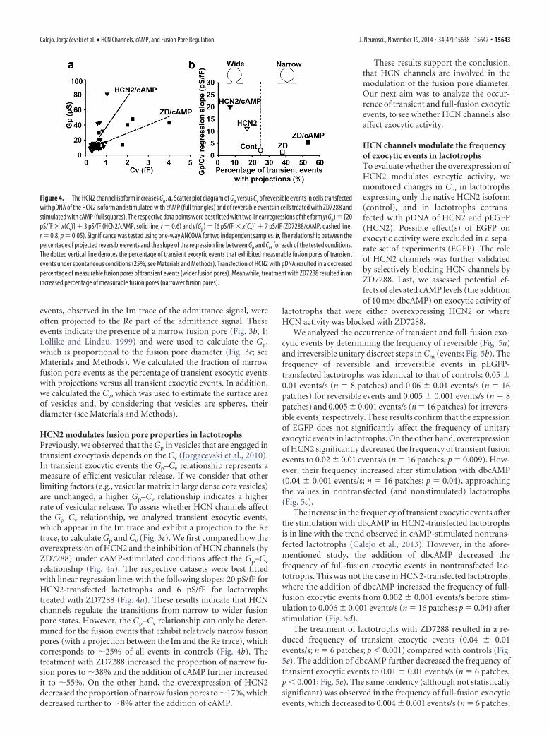
HCN channels modulate the frequencyof exocytic events in lactotrophsTo evaluate whether the overexpression ofHCN2 modulates exocytic activity, wemonitored changes in Cm in lactotrophsexpressing only the native HCN2 isoform(control), and in lactotrophs cotrans-fected with pDNA of HCN2 and pEGFP(HCN2). Possible effect(s) of EGFP onexocytic activity were excluded in a sepa-rate set of experiments (EGFP). The roleof HCN2 channels was further validatedby selectively blocking HCN channels byZD7288. Last, we assessed potential ef-fects of elevated cAMP levels (the additionof 10 mM dbcAMP) on exocytic activity of
lactotrophs that were either overexpressing HCN2 or whereHCN activity was blocked with ZD7288.
We analyzed the occurrence of transient and full-fusion exo-cytic events by determining the frequency of reversible (Fig. 5a)and irreversible unitary discreet steps in Cm (events; Fig. 5b). Thefrequency of reversible and irreversible events in pEGFP-transfected lactotrophs was identical to that of controls: 0.05 �0.01 events/s (n 8 patches) and 0.06 � 0.01 events/s (n 16patches) for reversible events and 0.005 � 0.001 events/s (n 8patches) and 0.005 � 0.001 events/s (n 16 patches) for irrevers-ible events, respectively. These results confirm that the expressionof EGFP does not significantly affect the frequency of unitaryexocytic events in lactotrophs. On the other hand, overexpressionof HCN2 significantly decreased the frequency of transient fusionevents to 0.02 � 0.01 events/s (n 16 patches; p 0.009). How-ever, their frequency increased after stimulation with dbcAMP(0.04 � 0.001 events/s; n 16 patches; p 0.04), approachingthe values in nontransfected (and nonstimulated) lactotrophs(Fig. 5c).
The increase in the frequency of transient exocytic events afterthe stimulation with dbcAMP in HCN2-transfected lactotrophsis in line with the trend observed in cAMP-stimulated nontrans-fected lactotrophs (Calejo et al., 2013). However, in the afore-mentioned study, the addition of dbcAMP decreased thefrequency of full-fusion exocytic events in nontransfected lac-totrophs. This was not the case in HCN2-transfected lactotrophs,where the addition of dbcAMP increased the frequency of full-fusion exocytic events from 0.002 � 0.001 events/s before stim-ulation to 0.006 � 0.001 events/s (n 16 patches; p 0.04) afterstimulation (Fig. 5d).
The treatment of lactotrophs with ZD7288 resulted in a re-duced frequency of transient exocytic events (0.04 � 0.01events/s; n 6 patches; p � 0.001) compared with controls (Fig.5e). The addition of dbcAMP further decreased the frequency oftransient exocytic events to 0.01 � 0.01 events/s (n 6 patches;p � 0.001; Fig. 5e). The same tendency (although not statisticallysignificant) was observed in the frequency of full-fusion exocyticevents, which decreased to 0.004 � 0.001 events/s (n 6 patches;
Figure 4. The HCN2 channel isoform increases Gp. a, Scatter plot diagram of Gp versus Cv of reversible events in cells transfectedwith pDNA of the HCN2 isoform and stimulated with cAMP (full triangles) and of reversible events in cells treated with ZD7288 andstimulated with cAMP (full squares). The respective data points were best fitted with two linear regressions of the form y(Gp) [20pS/fF � x(Cv)] � 3 pS/fF (HCN2/cAMP, solid line, r 0.6) and y(Gp) [6 pS/fF � x(Cv)] � 7 pS/fF (ZD7288/cAMP, dashed line,r 0.8, p 0.05). Significance was tested using one-way ANCOVA for two independent samples. b, The relationship between thepercentage of projected reversible events and the slope of the regression line between Gp and Cv, for each of the tested conditions.The dotted vertical line denotes the percentage of transient exocytic events that exhibited measurable fusion pores of transientevents under spontaneous conditions (25%; see Materials and Methods). Transfection of HCN2 with pDNA resulted in a decreasedpercentage of measurable fusion pores of transient events (wider fusion pores). Meanwhile, treatment with ZD7288 resulted in anincreased percentage of measurable fusion pores (narrower fusion pores).
Calejo, Jorgacevski et al. • HCN Channels, cAMP, and Fusion Pore Regulation J. Neurosci., November 19, 2014 • 34(47):15638 –15647 • 15643

ZD) and to 0.002 � 0.001 events/s (n 6patches; after the addition of cAMP;Fig. 5f).
These data show that HCN channelscan influence exocytic activity.
HCN channels affect [Ca 2�]i levelsin lactotrophsTo test whether the observed changes inexocytic activity in HCN2-transfected andZD7288-treated cells are related to thechanges in [Ca 2�]i, we performed cal-cium measurements with a calcium indi-cator, Fluo-4 AM (Fig. 6). The additionof dbcAMP did not significantly affect[Ca 2�]i levels in cells overexpressingHCN2 (Fig. 6a,c). In contrast, the addi-tion of dbcAMP to ZD7288-treated cellssignificantly reduced [Ca 2�]i (Fig. 6b,c).Moreover, the mean Fluo-4 fluorescencewas measured before the addition of db-cAMP in controls, in HCN2-transfectedcells, and in ZD7288-treated cells. Themean basal [Ca 2�]i, which was signifi-cantly higher in controls compared withHCN2-transfected cells and ZD7288-treated cells, respectively (Fig. 6d), wasthen related to the frequency of reversibleand irreversible exocytic events in all threeexperimental groups. Data pairs were fit-ted with linear regression lines that hadPearson’s coefficient (r) � 0.99 for bothfits, indicating a strong correlation be-tween these two parameters (Fig. 6e).
These results indicate the involvementof HCN channels in the regulation of[Ca 2�]i and consequently indirectly alsoin exocytic activity in pituitary lac-totrophs. However, some of the HCN-mediated effects, such as those involvingcAMP-mediated fusion pore widening,are believed to directly affect the fusionpore, since cAMP did not affect [Ca 2�]i in HCN2-transfectedcells, while an increase in [Ca 2�]i is required for fusion porewidening (Vardjan et al., 2007).
DiscussionThe results of this study show that regulation of exocytic fusionpore in lactotrophs involves HCN channels, which are gated bycAMP. HCN channels are located either in the vesicular mem-brane or in the plasma membrane next to the fused vesicle, whichaffects local membrane curvature and consequently dilates fusionpore as described by Kabaso and coworkers (Kabaso et al., 2012).Based on this model and on the results showing that electrostaticinteractions determine fusion pore properties (Calejo et al.,2012), we suggest that fusion pore geometry is affected by localchanges in cation concentration. Changes in cAMP levels affectHCN channel conductance and thus can alter local cation con-centration during hyperpolarization and during early stages of de-polarization (Biel et al., 2002). This mechanism, regulating transient(kiss-and-run) exocytosis, may play a more general role in the ner-vous system.
Our results unambiguously confirm that HCN channels, andthe HCN2 isoform specifically, are present in isolated rat lac-totrophs (Fig. 1). We focused on the HCN2 isoform for threereasons: (1) this isoform is the most ubiquitously distributedisoform in the brain (Shi et al., 1999); (2) mRNA transcripts ofthis isoform are the most abundant in the pituitary gland (Kret-schmannova et al., 2012); and (3) HCN2 is highly sensitive tocAMP (Kaupp and Seifert, 2001). Using Western blotting, weconfirmed the native expression of HCN2 in the pituitary gland (Fig.1c). The overall amount of HCN2 mRNA transcript in lactotrophs,which was �1% of the mRNA transcript of �-actin (Fig. 1d), is inagreement with previous studies on other secretory cells, where verylow amounts of the transcripts were detected (El-Kholy et al., 2007).Similarly, the subcellular localization of HCN channels in lac-totrophs is consistent with previous findings (Noam et al., 2010; Fig.1). It is therefore not surprising that, in our experiments, �55% ofHCN2 is located in the plasma membrane of lactotrophs and �22%are either on or near PRL-containing vesicles (Fig. 1civ). The remain-ing �23% that are not accounted for are most likely localized inother subcellular organelles, such as constitutive vesicles, endo-somes, endoplasmic reticulum, and the Golgi apparatus.
Figure 5. The HCN2 channel isoform modulates the frequency of exocytic events in lactotrophs. a, b, Two epochs of therepresentative electrophysiologic recording, which was made on a lactotroph transfected with pDNA of HCN2 and stimulated withdbcAMP, show reversible (a) and irreversible (b) events in the Im part of the admittance trace. The arrow marks the calibrationpulse in the Im trace, used to adjust the phase of the lock-in amplifier. c, The mean frequency of reversible events was 0.05 � 0.01events/s in EGFP-transfected cells, significantly higher than in HCN2-transfected cells (0.02 � 0.01 events/s). After stimulation ofHCN2-transfected cells with dbcAMP, the mean frequency of reversible events increased to 0.04 � 0.01 events/s. d, The meanfrequency of irreversible upward events in EGFP-transfected cells was 0.005 � 0.001 events/s, significantly more than in HCN2-transfected cells (0.002 � 0.001 events/s). However, the addition of dbcAMP to the HCN2-transfected cells increased the fre-quency of irreversible upward events to 0.006 � 0.001 events/s. e, The addition of ZD7288 (blocker of HCN2 channels) decreasedthe mean frequency of reversible events from 0.06 � 0.01 events/s (controls) to 0.04 � 0.01 events/s (ZD7288) and to 0.01 �0.01 events/s (with ZD7288 and cAMP). f, A similar trend was observed in the frequencies of irreversible upward events: 0.005 �0.001 events/s (controls), 0.004 � 0.001 events/s (ZD7288-treated cells), and 0.002 � 0.001 events/s (with ZD7288 and cAMP).Values are means � SEM. The numbers above the error bars indicate the number of patches. *p � 0.05, **p � 0.01, ***p �0.001.
15644 • J. Neurosci., November 19, 2014 • 34(47):15638 –15647 Calejo, Jorgacevski et al. • HCN Channels, cAMP, and Fusion Pore Regulation

Figure 6. HCN channels affect [Ca 2�]i levels in lactotrophs. a, b, Fluo-4 fluorescence intensity (FFluo-4) changes in the representative HCN2-transfected cell (a) and in the representativeZD7288-treated cell (b) before and after the addition of 10 mM dbcAMP (�cAMP). Changes in the Fluo-4 fluorescence are proportional to changes in [Ca 2�]i. The horizontal dashed line representsF0 (see Materials and Methods). The vertical dashed line denotes the time point of dbcAMP addition. c, The addition of dbcAMP did not affect [Ca 2�]i levels in HCN2-transfected cells. On the otherhand, the addition of dbcAMP significantly decreased [Ca 2�]i levels in ZD7288-treated cells. The significance was tested with paired Student’s t test. d, The mean Fluo-4 fluorescence before theaddition of dbcAMP was significantly higher in controls compared with HCN2-transfected cells and ZD7288-treated cells, respectively. e, The correlation between the resting mean Fluo-4fluorescence and the mean frequency of reversible (filled circles) and irreversible (empty circles) exocytic events. Fitted to the data are two regression lines of the form y (frequency of irreversibleevents) [2.7 � 10 �5 � x (mean Ffluo)] � [3.0 � 10 �4 (r � 0.99)] and y (frequency of reversible events) [3.0 � 10 �4 � x (mean Ffluo)] � [4.4 � 10 �3] (r � 0.99). Values are means �SEM. The numbers above the error bars indicate the number of cells analyzed. *p � 0.05, **p � 0.01, ***p � 0.001.
Calejo, Jorgacevski et al. • HCN Channels, cAMP, and Fusion Pore Regulation J. Neurosci., November 19, 2014 • 34(47):15638 –15647 • 15645

The surface expression of HCN2 and HCN4 isoforms is main-tained by recycling endosomes, which store HCN channels(Hardel et al., 2008). The abundance of HCN channels on the cellsurface therefore depends on secretory and endocytic pathways.Is the opposite true as well? Do HCN channels affect the stages ofvesicular fusion with the plasma membrane that involve the fu-sion pore? The fact that, following the transfection with theplasmid-encoding HCN2 isoform, we observed an equivalent in-crease of HCN2 isoform in the plasma membrane and in the cellinterior, is an indication that the 24 – 48 h time window was suf-ficient for the synthesis, the traffic to the plasma membrane, andinternalization in the plasma membrane to reach a balance (Fig.2c). This observation was confirmed by voltage-clamp measure-ments of HCN-specific Ih (Fig. 2d). Measurements of Ih duringthe 24 – 48 h window after transfection with HCN2 pDNA re-vealed �4-fold increase in Ih density, indicating increased num-bers of functional HCN2 channels in the plasma membrane. Thisexperiment also confirms that the increase in the HCN2 immuno-fluorescence signal reflects the increase in the quantity of HCNchannels.
cAMP has been shown to affect the release of PRL from lac-totrophs, facilitating pace-making activity and voltage-gatedcalcium influx by predominantly PKA-independent and Epac-independent mechanisms (Gonzalez-Iglesias et al., 2006b). Thismakes HCN channels very likely candidates to be involved di-rectly in the regulation of exocytosis. Our results support thisnotion, since both the overexpression of HCN2 and the inhibi-tion of HCN channels by ZD7288 had pronounced effects on theproperties and occurrence of unitary exocytic events (Figs. 4, 5).The latter effect is correlated to altered [Ca 2�]i levels (Fig. 6) andmay indicate that HCN channels affect exocytotic activity indi-rectly via the modulation of [Ca 2�]i. In contrast, the cAMP-mediated fusion pore widening (Calejo et al., 2013; Fig. 4) is morelikely due to a direct effect via HCN channels at the exocytoticapparatus, since cAMP did not affect [Ca 2�]i in HCN2-transfected cells, while an increase in [Ca 2�]i is required for fu-sion pore widening (Vardjan et al., 2007).
Overexpression of HCN2 increased the inward cation currentduring hyperpolarization (Fig. 2d) and resulted in fusion poreopenings with relatively wide fusion pores (Fig. 4). This effect wasstrengthened by the addition of cAMP, an activator of HCNchannels. On the other hand, near complete blockage of HCNcurrents by ZD7288 resulted in the opening of a very narrowfusion pores (Fig. 2d). Fusion pores were even narrower ifZD7288-treated cells were stimulated with cAMP (Fig. 2b). Thisis likely due to the altered local cation concentrations, whichaffect fusion pore geometry via electrostatic interactions, as pro-posed by Kabaso et al. (2012). Moreover, cAMP induced mark-edly different effects in the presence or in the absence of ZD7288.While the addition of cAMP dilated fusion pores in the presenceof normally conducting HCN channels, likely by stimulating addi-tional influx of cations (although the mean [Ca2�]i remained un-changed; Fig. 6c), it further narrowed fusion pores when the HCNchannels were blocked. This effect is likely caused by the activation ofsarco/endoplasmic reticulum Ca2�-ATPase (SERCA) in responseto the elevated cAMP (Schmidt et al., 1999), which subsequentlydecreases [Ca2�]i, as demonstrated in Figure 6c.
It has recently been shown that vesicular calcium channelsregulate exocytosis by facilitating calcium entry through the fu-sion pore. This generates microdomains of increased [Ca 2�]i
around the fused vesicles, which leads to the expansion of thefusion pore (Miklavc et al., 2011). Previous studies on lactotrophsshowed a strong correlation between an increase in [Ca 2�]i and
an increase exocytic activity (Vardjan et al., 2007; Jorgacevski etal., 2008). Although major physiologic implications of Ca 2� sig-naling through these channels remain unclear, overexpression ofHCN2 should result in a local increase in [Ca 2�]i, which would inturn increase the Ca 2�-dependent fusion of vesicles. Interest-ingly, the mean [Ca 2�]i was �3-fold lower in the HCN2-transfected cells (compared with controls; Fig. 6d), whichcorrelates well with the observed �2.5-fold reduction in the rateof exocytic activity (Fig. 5c,d). It is known that adenylyl cyclases(ACs) are intimately associated with sites of Ca 2� entry into thecell (Cooper et al., 1995). The local increase in [Ca 2�]i, due to theoverexpression of HCN2, may stimulate ACs and elevate cAMP.Then elevated cAMP may activate SERCA and potentially reduce[Ca 2�]i below control levels (Mons et al., 1998; Fig. 6), therebyinhibiting exocytic activity (Fig. 5). An increase in cAMP via thealternate route (dbcAMP) should then rescue exocytic activity,either by increasing local [Ca 2�]i (Yu et al., 2004; Zhong et al.,2004) or via activation of PKA-dependent mechanisms. Whileglobal [Ca 2�]i levels appeared unchanged (Fig. 6c), Ca 2� maystill be increased locally, in exocytosis-relevant microdomains,resulting in the rescue of the rate of exocytic activity (Fig. 5c,d).
Kabaso et al. (2012) have proposed that rhythmic opening andclosing of the fusion pore is related to the influx of cations into thevesicles. Cation influx takes place through HCN channels, whichare near fusing vesicles (through the fusion pore and vesicularHCN channels and/or through the channels in the plasma mem-brane). Therefore, cation influx increases local cation concentra-tion at the fusion pore. Inhibition of cation current with ZD7288should then have the opposite effect. Indeed, ZD7288 significantlyreduced the frequency of reversible exocytic events in our experi-ments (Fig. 5e,f). Basal [Ca2�]i in HCN2-transfected cells andZD7288-treated cells correlate well with the reduction in the rate ofexocytic activity (Fig. 6e), which is an indication of a Ca2�-dependent mechanism. Nonetheless, impeded inflow of other cat-ions and putative direct blocking effect by ZD7288, likely viainteractions with the exocytic machinery (El-Kholy et al., 2007), mayalso play a role. Surprisingly, the addition of dbcAMP to ZD7288-treated cells further inhibited the rate of exocytic activity, aneffect presumably caused by the activation of SERCA pumps,which further reduced [Ca 2�]i in the absence of Ih (Fig. 6c).
In summary, our results demonstrate the role of HCN channelsin regulated exocytosis in cultured rat lactotrophs. HCN channelsmodulate the rate of exocytic activity, likely via their role in theregulation of [Ca2�]i. Moreover, they appear to be involved in theregulation of fusion pore geometry, probably via electrostatic inter-actions due to changes in local cation concentration.
ReferencesAngleson JK, Cochilla AJ, Kilic G, Nussinovitch I, Betz WJ (1999) Regula-
tion of dense core release from neuroendocrine cells revealed by imagingsingle exocytic events. Nat Neurosci 2:440 – 446. CrossRef Medline
Ben-Tabou S, Keller E, Nussinovitch I (1994) Mechanosensitivity ofvoltage-gated calcium currents in rat anterior pituitary cells. J Physiol476:29 –39. Medline
Biel M, Schneider A, Wahl C (2002) Cardiac HCN channels: structure,function, and modulation. Trends Cardiovasc Med 12:206 –212. CrossRefMedline
Calejo AI, Jorgacevski J, Silva VS, Stenovec M, Kreft M, Goncalves PP, ZorecR (2012) Aluminium-induced changes of fusion pore properties atten-uate prolactin secretion in rat pituitary lactotrophs. Neuroscience 201:57– 66. CrossRef Medline
Calejo AI, Jorgacevski J, Kucka M, Kreft M, Goncalves PP, Stojilkovic SS,Zorec R (2013) cAMP-mediated stabilization of fusion pores in culturedrat pituitary lactotrophs. J Neurosci 33:8068 – 8078. CrossRef Medline
15646 • J. Neurosci., November 19, 2014 • 34(47):15638 –15647 Calejo, Jorgacevski et al. • HCN Channels, cAMP, and Fusion Pore Regulation

Chernomordik LV, Kozlov MM (2008) Mechanics of membrane fusion.Nat Struct Mol Biol 15:675– 683. CrossRef Medline
Cooper DM, Mons N, Karpen JW (1995) Adenylyl cyclases and the interac-tion between calcium and cAMP Signaling. Nature 374:421– 424.CrossRef Medline
de Rooij J, Zwartkruis FJ, Verheijen MH, Cool RH, Nijman SM, WittinghoferA, Bos JL (1998) Epac is a Rap1 guanine-nucleotide-exchange factordirectly activated by cAMP. Nature 396:474 – 477. CrossRef Medline
El-Kholy W, MacDonald PE, Fox JM, Bhattacharjee A, Xue T, Gao X, Zhang Y,Stieber J, Li RA, Tsushima RG, Wheeler MB (2007) Hyperpolarization-activated cyclic nucleotide-gated channels in pancreatic beta-cells. Mol Endocri-nol 21:753–764. CrossRef Medline
Gauss R, Seifert R, Kaupp UB (1998) Molecular identification of ahyperpolarization-activated channel in sea urchin sperm. Nature 393:583–587. CrossRef Medline
Gonzalez-Iglesias AE, Kretschmannova K, Tomic M, Stojilkovic SS (2006a)ZD7288 inhibits exocytosis in an HCN-independent manner and down-stream of voltage-gated calcium influx in pituitary lactotrophs. BiochemBiophys Res Commun 346:845– 850. CrossRef Medline
Gonzalez-Iglesias AE, Jiang Y, Tomic M, Kretschmannova K, Andric SA,Zemkova H, Stojilkovic SS (2006b) Dependence of electrical activityand calcium influx-controlled prolactin release on adenylyl cyclase signal-ing pathway in pituitary lactotrophs. Mol Endocrinol 20:2231–2246.CrossRef Medline
Hardel N, Harmel N, Zolles G, Fakler B, Klocker N (2008) Recycling endo-somes supply cardiac pacemaker channels for regulated surface expres-sion. Cardiovasc Res 79:52– 60. CrossRef Medline
Jorgacevski J, Stenovec M, Kreft M, Bajic A, Rituper B, Vardjan N, StojilkovicS, Zorec R (2008) Hypotonicity and peptide discharge from a singlevesicle. Am J Physiol Cell Physiol 295:C624 – 631. CrossRef Medline
Jorgacevski J, Fosnaric M, Vardjan N, Stenovec M, Potokar M, Kreft M,Kralj-Iglic V, Iglic A, Zorec R (2010) Fusion pore stability of peptidergicvesicles. Mol Membr Biol 27:65– 80. CrossRef Medline
Jorgacevski J, Kreft M, Vardjan N, Zorec R (2012) Fusion pore regulation inpeptidergic vesicles. Cell Calcium 52:270 –276. CrossRef Medline
Kabaso D, Calejo AI, Jorgacevski J, Kreft M, Zorec R, Iglic A (2012) Fusionpore diameter regulation by cations modulating local membrane anisot-ropy. ScientificWorldJournal 2012:983138. Medline
Kasai H, Suzuki T, Liu TT, Kishimoto T, Takahashi N (2002) Fast andcAMP-sensitive mode of Ca 2�-dependent exocytosis in pancreatic beta-cells. Diabetes 51:S19 –S24. CrossRef Medline
Kaupp UB, Seifert R (2001) Molecular diversity of pacemaker ion channels.Annu Rev Physiol 63:235–257. CrossRef Medline
Kretschmannova K, Kucka M, Gonzalez-Iglesias AE, Stojilkovic SS (2012)The expression and role of hyperpolarization-activated and cyclicnucleotide-gated channels in endocrine anterior pituitary cells. Mol En-docrinol 26:153–164. CrossRef Medline
Lollike K, Lindau M (1999) Membrane capacitance techniques to monitorgranule exocytosis in neutrophils. J Immunol Methods 232:111–120.CrossRef Medline
Michels G, Brandt MC, Zagidullin N, Khan IF, Larbig R, van Aaken S, Wip-permann J, Hoppe UC (2008) Direct evidence for calcium conductanceof hyperpolarization-activated cyclic nucleotide-gated channels and hu-
man native If at physiological calcium concentrations. Cardiovasc Res78:466 – 475. CrossRef Medline
Miklavc P, Mair N, Wittekindt OH, Haller T, Dietl P, Felder E, Timmler M,Frick M (2011) Fusion-activated Ca2� entry via vesicular P2X4 recep-tors promotes fusion pore opening and exocytotic content release inpneumocytes. Proc Natl Acad Sci U S A 108:14503–14508. CrossRefMedline
Mons N, Decorte L, Jaffard R, Cooper DM (1998) Ca2�-sensitive adenylylcyclases, key integrators of cellular signalling. Life Sciences 62:1647–1652.CrossRef Medline
Noam Y, Zha Q, Phan L, Wu RL, Chetkovich DM, Wadman WJ, Baram TZ(2010) Trafficking and surface expression of hyperpolarization-activatedcyclic nucleotide-gated channels in hippocampal neurons. J Biol Chem285:14724 –14736. CrossRef Medline
Potokar M, Stenovec M, Kreft M, Gabrijel M, Zorec R (2011) Physiopatho-logic dynamics of vesicle traffic in astrocytes. Histol Histopathol 26:277–284. Medline
Rituper B, Gucek A, Jorgacevski J, Flasker A, Kreft M, Zorec R (2013) High-resolution membrane capacitance measurements for the study of exocy-tosis and endocytosis. Nat Protoc 8:1169 –1183. CrossRef Medline
Schmidt U, Hajjar RJ, Kim CS, Lebeche D, Doye AA, Gwathmey JK (1999)Human heart failure: cAMP stimulation of SR Ca2�-ATPase activity andphosphorylation level of phospholamban. Am J Physiol 277:H474 –H480.Medline
Seino S, Shibasaki T (2005) PKA-dependent and PKA-independent path-ways for cAMP-regulated exocytosis. Physiol Rev 85:1303–1342. CrossRefMedline
Shi W, Wymore R, Yu H, Wu J, Wymore RT, Pan Z, Robinson RB, Dixon JE, McK-innon D, Cohen IS (1999) Distribution and prevalence of hyperpolarization-activated cation channel (HCN) mRNA expression in cardiac tissues. Circ Res85:e1–e6. CrossRef Medline
Spruce AE, Breckenridge LJ, Lee AK, Almers W (1990) Properties of thefusion pore that forms during exocytosis of a mast cell secretory vesicle.Neuron 4:643– 654. CrossRef Medline
Stojilkovic SS, Kretschmannova K, Tomic M, Stratakis CA (2012) Depen-dence of the excitability of pituitary cells on cyclic nucleotides. J Neuroen-docrinol 24:1183–1200. CrossRef Medline
Vaccari T, Moroni A, Rocchi M, Gorza L, Bianchi ME, Beltrame M, DiFran-cesco D (1999) The human gene coding for HCN2, a pacemaker channelof the heart. Biochim Biophys Acta 1446:419 – 425. CrossRef Medline
Vardjan N, Stenovec M, Jorgacevski J, Kreft M, Zorec R (2007) Subnano-meter fusion pores in spontaneous exocytosis of peptidergic vesicles.J Neurosci 27:4737– 4746. CrossRef Medline
Yu X, Duan KL, Shang CF, Yu HG, Zhou Z (2004) Calcium influx throughhyperpolarization-activated cation channels (I(h) channels) contributesto activity-evoked neuronal secretion. Proc Natl Acad Sci U S A 101:1051–1056. CrossRef Medline
Zhong N, Beaumont V, Zucker RS (2004) Calcium influx through HCNchannels does not contribute to cAMP-enhanced transmission. J Neuro-physiol 92:644 – 647. CrossRef Medline
Zorec R, Sikdar SK, Mason WT (1991) Increased cytosolic calcium stimu-lates exocytosis in bovine lactotrophs. Direct evidence from changes inmembrane capacitance. J Gen Physiol 97:473– 497. CrossRef Medline
Calejo, Jorgacevski et al. • HCN Channels, cAMP, and Fusion Pore Regulation J. Neurosci., November 19, 2014 • 34(47):15638 –15647 • 15647
Copyright © 2022 FDOKUMEN