Exogenous endothelial cells as accelerators of hematopoietic reconstitution
Upload
independentCategory
view
3download
0
O
EtAE
Baf
Mdha
Raawa
Clu
Ka
Ileti
esa(tmttp((hh
F
A
R
0d
RIGINAL ARTICLES
xogenous Testosterone Enhances Responsivenesso Social Threat in the Neural Circuitry of Socialggression in Humans
rno J. Hermans, Nick F. Ramsey, and Jack van Honk
ackground: In a range of species, the androgen steroid testosterone is known to potentiate neural circuits involved in intraspecificggression. Disorders of impulsive aggression in humans have likewise been associated with high testosterone levels, but human evidenceor the link between testosterone and aggression remains correlational and inconclusive.
ethods: Twelve female participants underwent functional magnetic resonance imaging during three sessions while viewing stimuliiffering in social threat value: angry and happy facial expressions. The first session served to establish associations between baselineormone levels and neural activation. Participants were retested in a second and third session after placebo-controlled sublingualdministration of .5 mg testosterone.
esults: Findings demonstrate consistent activation to angry versus happy faces in areas known to be involved in vertebrate reactiveggression, such as the amygdala and hypothalamus. Suprathreshold clusters were also found in the orbitofrontal cortex (Brodmannrea 47), a region implicated in impulse control in humans. Baseline endocrine profiles of high testosterone and low cortisol were associatedith stronger activation in subcortical structures. Neural responses in most activated regions were more persistent after testosterone
dministration than after placebo.
onclusions: These data demonstrate that testosterone enhances responsiveness in neural circuits of social aggression. Based on animaliterature, it is argued that actions of testosterone on subcortical reactive aggression circuits give rise to this effect. Implications for our
nderstanding of the pathophysiology of disorders of impulsive aggression are discussed.ey Words: Aggression, amygdala, fMRI, glucocorticoid, hypothal-mus, testosterone
ntraspecific aggression in vertebrates, including humans, isthought to be controlled by an integrated molecular, neuro-anatomic, and behavioral substrate that exhibits strong evo-
utionary stability (1). This system appears to underlie thestablishment of hierarchies in social animals, often merelyhrough ritualized, species-specific behavioral displays of hostilentent, such as angry facial expressions (2,3).
The hypothalamic–pituitary– gonadal (HPG) axis, through itsnd product testosterone, is an important agent in regulating thisystem. Across vertebrates, males are generally more physicallyggressive than females, and gonadectomy reduces aggression4,5). Moreover, animal research has unequivocally shown thatestosterone elevation increases aggressiveness (6 – 8). In hu-ans, there is correlational evidence for a link between testos-
erone and aggression within both sexes (9). Disorders charac-erized by impulsive, reactive aggression, such as antisocialersonality disorder (APD) and borderline personality disorderBPD), have likewise been associated with high testosterone10 –13). Pending conclusive causal evidence (see, e.g., 9),owever, the mechanism through which testosterone may affectuman aggression remains obscure.
rom Helmholtz Institute (EJH, JvH), Utrecht University; F.C. Donders Centrefor Cognitive Neuroimaging (EJH), Radboud University Nijmegen,Rudolf Magnus Institute of Neuroscience (NFR), University Medical Cen-ter Utrecht, the Netherlands.
ddress reprint requests to Erno J. Hermans, M.A., F.C. Donders Centre forCognitive Neuroimaging, P.O. Box 9101, 6500 HB Nijmegen, the Nether-lands; E-mail: [email protected].
eceived April 26, 2006; revised April 4, 2007; accepted May 3, 2007.
006-3223/08/$34.00oi:10.1016/j.biopsych.2007.05.013
Pathways controlling reactive aggression converge in amyg-dalar and hypothalamic regions. Animal research has shown thattestosterone exerts its influence partly through interactions witharginine vasopressin (AVP) in the amygdala (14,15) and the(anterior) hypothalamus (16). Also, testosterone downregulatesthe hypothalamic–pituitary–adrenal (HPA) axis, resulting inchronically depressed cortisol (17). Hypocortisolemia in turn hasalso been associated with heightened aggression and social rank(18,19). In agreement, a recent study has demonstrated a signif-icant positive relation between testosterone and overt aggressionin delinquent adolescent males with low cortisol exclusively (20).
The capacity to inhibit reactive aggression, or impulse control,is generally attributed to the orbitofrontal cortex (OFC). Lesionsto the OFC are known to result in socially aberrant behavior(21,22). Likewise, marked OFC hypometabolism has been ob-served in patients with personality disorders (23,24). As indicatedby low cerebrospinal fluid levels of 5-hydroxytryptamine (5-HT)catabolites, this hypometabolism appears to originate from re-duced functioning of the serotonergic system (4), and testoster-one has been suggested to play a role in the etiology of thisabnormality (5).
Human neuroimaging findings have most consistently identi-fied the amygdala and (lateral) OFC in responding to angry facialexpressions (25–29). Behavioral experiments have furthermoreshown that affective responding to angry faces is positivelyrelated to testosterone levels (30,31) but negatively to cortisollevels (32).
In this study, 12 healthy female volunteers were tested usingfunctional magnetic resonance imaging (fMRI) during threesessions. The reason for including only women was that womenhave lower endogenous testosterone levels and therefore pre-sumably require a smaller dose to attain measurable effects (33).Moreover, there are no indications that effects of testosterone on
affective responding to angry facial expressions may exhibitBIOL PSYCHIATRY 2008;63:263–270© 2008 Society of Biological Psychiatry

qtepcausim(hPpsorta
M
P
1cspp
cctt
rew
M
rEeg
aSfhs
D
matm(l
P
n
264 BIOL PSYCHIATRY 2008;63:263–270 E.J. Hermans et al.
w
ualitative gender differences (30). The first session examinedhe pattern of brain responses to angry versus happy facialxpressions in relation to levels of testosterone and cortisol. As inrevious studies (33), happy facial expressions were chosen asontrol stimuli because these provide for a comparison betweensocial threat and a social safety signal while controlling for
nspecific activity due to face and expression perception. Wecrutinized a hierarchical model (34) consisting of cortical inhib-tory control, implemented in the lateral OFC (especially Brod-ann area 47 [BA47]) and subcortical reactive aggression circuits
i.e., amygdaloid region [medial/central nuclei, stria terminalis],ypothalamus, and brainstem subnuclei [periaqueductal grey, orAG]). In a second and third session, the same group ofarticipants was retested in a placebo-controlled crossover de-ign to test effects of testosterone administration on functioningf these circuits. We predicted that responding in subcorticaleactive aggression circuits would be associated with high tes-osterone and low cortisol and that testosterone elevation wouldffect responding in a similar region-specific manner.
ethods and Materials
articipantsParticipants were 12 healthy, adult female volunteers (aged
8–28; mean age 22.6). Exclusion criteria were history of endo-rine or psychiatric disorder, left-hand dominance, habitualmoking, current pregnancy, history of closed-head injury, andresence of metal objects in the body. Use of (recreational)sychotropic drugs within 2 weeks of testing was not allowed.
Ten women used standard estrogen/progestagen oral contra-eptives. For the other two women who did not use oralontraceptives, testing was restricted to the preovulatory phaseo control for (minor) variations of androgen levels throughouthe menstrual cycle.
All procedures were approved by the local institutionaleview board in accordance with the declaration of Helsinki, andach participant provided written informed consent. Participantsere debriefed and received payment.
aterials and ApparatusStimuli were selected from two photosets: the Karolinska Di-
ected Emotional Faces (35) and the Pictures of Facial Affect (36).ight actors were chosen, and oval cutouts of two photographs ofach, happy (H) and angry (A), were included in the set. All wereray-scaled and equalized in luminance and contrast.
Stimulus presentation was controlled by an x86 notebook PCnd E-Prime (Psychology Software Tools, Pittsburgh, Pennsylvania).timuli were back-projected onto a screen near the participants’eet, which they viewed through a 45° angle mirror attached to theead coil. Participants were scanned in an ACS-NT 1.5-Tesla MRIcanner (Philips Medical Systems, Best, the Netherlands).
rug SamplesTestosterone solutions consisted of .5 mg of testosterone, 5
g of hydroxypropyl-beta-cyclodextrin (carrier), 5 mg ethanol,nd .5 mL water. Placebo solutions differed only in absence ofestosterone. Details concerning sublingual administration, phar-acokinetics, and efficacy time-course are available elsewhere
37). Identical procedures have been applied repeatedly in ouraboratory (38).
rocedureParticipants were tested using fMRI on three separate after-
oons (after 1:30 PM), the first of which did not involve drug
ww.sobp.org/journal
administration. They were instructed not to eat within 1.5 hoursbefore the appointment and to drink only water. Upon arrival,saliva samples (9 mL) were obtained.
On the second and third testing day, participants arrived at thelaboratory 3.5 hours before scanning. After providing salivasamples, participants received the .5 mL drug sample and wereinstructed to keep it under the tongue without swallowing for afull minute. They were asked to return 3.5 hours later and avoidphysically or psychologically straining activities in the meantime.Further procedures were equal during all testing sessions.
After standard MRI safety screening, participants were es-corted into the scanner room. Scans with the following charac-teristics were obtained:
1. T2*-weighted blood oxygen level–dependent (BOLD) im-ages (segmented three-dimensional echo planar imaging(EPI) with navigator echo, flip angle 9.5°, echo time/repetition time (TE/TR): 19.1/28.6 ms, filed of view (FOV):256 � 256 � 120 mm, matrix 64 � 64 � 30, voxel size 4 mmisotropic, acquisition time 3.26 sec), with oblique angula-tion. This segmented short TE sequence was designed tominimize signal dropout and image distortion due to mag-netic field inhomogeneity around the air-tissue interfaces.Details on 3D-EPI are described elsewhere (39).
2. Reference image: identical to functional images except flipangle: 30°, resulting in more T1-weighting. This image wasused to facilitate registration of structural and functionalimages.
3. T1-weighted structural images (TE/TR 4.6/30 msec, flipangle 30°, FOV 256 � 180 � 208 mm, matrix: 256 � 256 �150 mm, slice thickness 1.2 mm, voxel size 1 � 1 � 1.2 mm,duration: 8 min).
During the functional scan series, participants watched alter-nating 26.08-sec epochs. In each epoch, all eight stimuli werepresented in random order and repeated seven times (total 56stimuli). Each was presented for a duration of 200 msec with astimulus onset asynchrony of 467 msec. Resting fixation epochswere also included (�). Epochs were presented in the followingorder: �AH�AH�AHHA�HA�HA� (or A and H reversed,counterbalanced across participants), which precludes task co-variation with linear drifts of BOLD signal (see Figure 1). Afterscanning, another saliva sample was collected.
Functional MRI Image AnalysisPreprocessing of fMRI data was performed with SPM99 (UCL
London, United Kingdom). All functional scans were motion-corrected using transformation parameters, yielding a minimalsum of squared differences with the reference image. Registra-tion of the reference and structural scans was achieved byestimating parameters resulting in maximum mutual information.All images were normalized to standard (Montreal NeurologicalInstitute [MNI], Montreal, Quebec, Canada) space using affinetransformations and nonlinear deformations and then smoothedwith an 8-mm full-width at half maximum gaussian kernel.Normalized anatomic images were averaged over participants toserve as background for anatomic localization.
Statistical analysis steps started with fitting session-specificgeneral linear models containing box-car functions with hemo-dynamic delay for both task conditions (angry vs. happy),movement correction parameters, and a discrete cosine trans-form high-pass filter with a cutoff of 1.17e-2 Hz. Proportionalscaling was applied to each volume to remove unspecific global
(drug) effects. Contrast images (three sessions � 12 participants)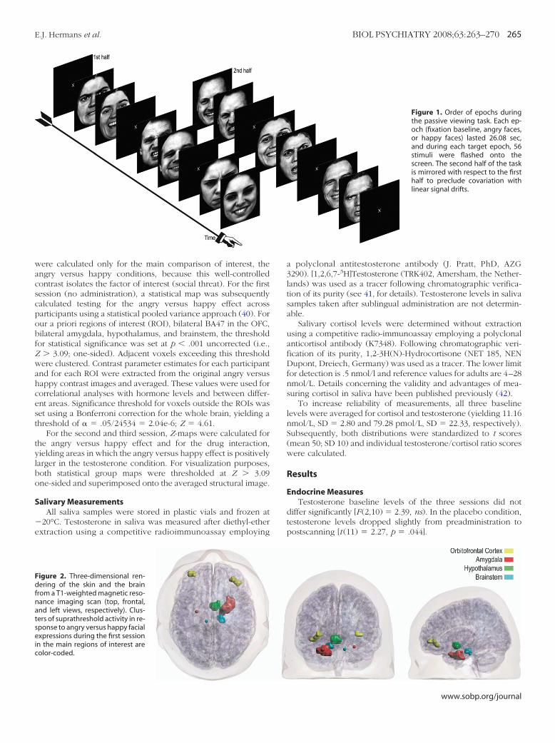
wacscpobfZwahcest
tylbo
S
�e
Fdfnatseic
E.J. Hermans et al. BIOL PSYCHIATRY 2008;63:263–270 265
ere calculated only for the main comparison of interest, thengry versus happy conditions, because this well-controlledontrast isolates the factor of interest (social threat). For the firstession (no administration), a statistical map was subsequentlyalculated testing for the angry versus happy effect acrossarticipants using a statistical pooled variance approach (40). Forur a priori regions of interest (ROI), bilateral BA47 in the OFC,ilateral amygdala, hypothalamus, and brainstem, the thresholdor statistical significance was set at p � .001 uncorrected (i.e.,
� 3.09; one-sided). Adjacent voxels exceeding this thresholdere clustered. Contrast parameter estimates for each participantnd for each ROI were extracted from the original angry versusappy contrast images and averaged. These values were used fororrelational analyses with hormone levels and between differ-nt areas. Significance threshold for voxels outside the ROIs waset using a Bonferroni correction for the whole brain, yielding ahreshold of � � .05/24534 � 2.04e-6; Z � 4.61.
For the second and third session, Z-maps were calculated forhe angry versus happy effect and for the drug interaction,ielding areas in which the angry versus happy effect is positivelyarger in the testosterone condition. For visualization purposes,oth statistical group maps were thresholded at Z � 3.09ne-sided and superimposed onto the averaged structural image.
alivary MeasurementsAll saliva samples were stored in plastic vials and frozen at
20°C. Testosterone in saliva was measured after diethyl-etherxtraction using a competitive radioimmunoassay employing
igure 2. Three-dimensional ren-ering of the skin and the brain
rom a T1-weighted magnetic reso-ance imaging scan (top, frontal,nd left views, respectively). Clus-ers of suprathreshold activity in re-ponse to angry versus happy facialxpressions during the first session
n the main regions of interest areolor-coded.
a polyclonal antitestosterone antibody (J. Pratt, PhD, AZG3290). [1,2,6,7-3H]Testosterone (TRK402, Amersham, the Nether-lands) was used as a tracer following chromatographic verifica-tion of its purity (see 41, for details). Testosterone levels in salivasamples taken after sublingual administration are not determin-able.
Salivary cortisol levels were determined without extractionusing a competitive radio-immunoassay employing a polyclonalanticortisol antibody (K7348). Following chromatographic veri-fication of its purity, 1,2-3H(N)-Hydrocortisone (NET 185, NENDupont, Dreiech, Germany) was used as a tracer. The lower limitfor detection is .5 nmol/l and reference values for adults are 4–28nmol/L. Details concerning the validity and advantages of mea-suring cortisol in saliva have been published previously (42).
To increase reliability of measurements, all three baselinelevels were averaged for cortisol and testosterone (yielding 11.16nmol/L, SD � 2.80 and 79.28 pmol/L, SD � 22.33, respectively).Subsequently, both distributions were standardized to t scores(mean 50; SD 10) and individual testosterone/cortisol ratio scoreswere calculated.
Results
Endocrine MeasuresTestosterone baseline levels of the three sessions did not
differ significantly [F (2,10) � 2.39, ns). In the placebo condition,testosterone levels dropped slightly from preadministration topostscanning [t (11) � 2.27, p � .044].
Figure 1. Order of epochs duringthe passive viewing task. Each ep-och (fixation baseline, angry faces,or happy faces) lasted 26.08 sec,and during each target epoch, 56stimuli were flashed onto thescreen. The second half of the taskis mirrored with respect to the firsthalf to preclude covariation withlinear signal drifts.
www.sobp.org/journal

isrwCp3fol
F
sesroccarFo
C
hcssH
Fasa
266 BIOL PSYCHIATRY 2008;63:263–270 E.J. Hermans et al.
w
Cortisol baseline levels were lowest on the first day of testingn comparison with the testosterone and placebo administrationessions [F (1,11) � 11.35, p � .006 and F (1,11) � 9.01, p � .012,espectively], which is explained by the fact that saliva samplesere collected at a later time of day during the first session.ortisol levels also dropped from the preadministration toostscanning samples for the second and third sessions [F(1,11) �9.26, p � .001], but no interaction with drug administration wasound [F (1,11) � 1.45, ns), indicating that exogenous testoster-ne elevation did not significantly decrease endogenous cortisolevels.
unctional MRI Results, First SessionResults for the main contrast of interest (angry vs. happy) are
ummarized in Table 1. The main regions of interest all showedvidence of suprathreshold activity. Amygdala responding wastronger in the right hemisphere. An additional focus of activityeaching a more conservative whole-brain corrected Z thresholdf 4.61 was found in the inferior temporal gyrus. Moreover, twolusters of activity, one in the brainstem and one in the insularortex, were more activated during the happy than during thengry face conditions. Figure 2 shows a three-dimensionalendering of clusters of activity in the main regions of interest. Inigure 3, all voxels exceeding the Z � 3.09 ROI threshold areverlaid onto axial slices of the averaged structural image.
orrelational AnalysesAveraged contrast parameter estimates for the angry versus
appy comparison per suprathreshold cluster were used fororrelational analyses with endocrine measures. Nonparametrictatistics (Spearman’s rho) were applied because of the smallample size. Results of these analyses are summarized in Table 2.ypothalamic activity to angry versus happy facial expressions
igure 4. Scatterplots of the correlation between testosterone/cortisol rationd the average magnitude of the blood oxygen level– dependent re-ponse to angry versus happy facial expressions in the hypothalamus (left)
nd bilateral amygdala (right).ww.sobp.org/journal
exhibited a negative correlation with baseline cortisol. Baselinetestosterone levels correlated positively with activity in theinferior temporal gyrus only. The testosterone–cortisol ratio,however, proved more predictive: significant positive correla-tions were found with activity in the (predominantly right)amygdala, hypothalamus, brainstem, and inferior temporal gy-rus. Scatterplots of the first two of these are shown in Figure 4.
Subsequently, interregional nonparametric correlations acrossparticipants were calculated between activated clusters (seeTable 3). Activation during angry versus happy face conditionsin the bilateral amygdala, hypothalamus, and combined brain-stem clusters proved strongly interrelated. There was noevidence for a correlation between the lateral OFC and any ofthe other ROIs, however. Moreover, combined brainstemclusters responses were positively correlated with inferiortemporal gyrus activity.
Functional MRI Results, Second and Third SessionResults of the drug administration sessions are summarized in
Table 4. The angry versus happy contrast over these two sessionsyielded a pattern of activated areas similar to the first session (i.e.,bilateral BA47 in the orbitofrontal cortex, and the right amyg-dala), although not all regions reached significance. There wasalso evidence of stronger activity in the happy condition than inthe angry condition in the brainstem (pons) and parahippocam-pal gyrus.
Drug interaction effects are shown in the lower half of Table 4and Figure 5. As predicted, there were significant interactioneffects in the greater part of the network specified in the firstsession with greater angry versus happy activity in the testoster-one versus placebo condition. These effects were most pro-nounced in the amygdala and hypothalamus, but suprathresholdclusters were also found in the brainstem and lateral OFC (BA47).The peak location of the interaction effect in the amygdalaappears to lie somewhat more medial than the main effects.Figure 6 shows the averaged activity in the angry (vs. happy)conditions in five clusters that exhibit the interaction effect.Further separate tests were performed on the contrast parameterestimates for angry versus happy conditions for the testosteroneand placebo conditions separately to determine whether effectswere carried mainly by activations in the testosterone conditionsor by deactivations in the placebo condition. For these tests,contrast parameter estimates from all supratheshold voxels wereaveraged and tested using t tests. These analyses show that thetestosterone effect in the left OFC is carried by a significantresponse to angry faces relative to happy faces. Drug interaction
Figure 3. Ten axial slices at Z � �30to Z � 6 in MNI space from the av-eraged, normalized, anatomicscans from all 12 participants (lefthemisphere is left). All voxels ex-ceeding the region of interestthreshold of Z � 3.09 (p � .001)from the contrast angry versushappy from the first scan sessionare overlaid onto these slices. SeeTable 1 for additional data for eachcluster.
effects in the hypothalamus and brainstem appear to be carried

mtps
D
sgw(cofaphamsahIh
n
E.J. Hermans et al. BIOL PSYCHIATRY 2008;63:263–270 267
ainly by deactivations to angry faces relative to happy faces inhe placebo condition [t (11) � 2.72, p � .02, and t (11) � 2.45,
� .032, respectively]. Other separate t tests did not reachignificance.
iscussion
The purpose of this study was to gather insight into the neuralubstrates of human social aggression, in particular by investi-ating the regulatory role of steroid hormones. The main findingsere, first, that cortical (the lateral OFC), as well as subcortical
the amygdala and its efferents), circuits implicated in aggressionan be identified functionally using fMRI in healthy volunteersbserving social threat stimuli: angry contrasted with happyacial expressions. Second, the degree to which these subcorticalreas respond to social threat is associated with endocrinearameters, with strongest effects in participants with a profile ofigh testosterone and low cortisol. Third, in the two drugdministration sessions, interaction effects were observed inost areas within the network that was activated in the first
ession, which suggest that overall the differential responses tongry and happy expressions are stronger, or more resistant toabituation, after testosterone versus placebo administration.nteraction effects were most pronounced in the amygdala andypothalamus.
The medial and central nuclei of the amygdala, the beducleus of the stria terminalis, and efferent structures such as the
Table 1. Summary of Suprathreshold Clusters of ActivaSession
Region Side
Expression Main Effect: ActivationsHypothalamus RAmygdala RInferior temporal gyrus (BA20) RSuperior brainstem/posterior hippocampus ROrbitofrontal cortex (BA47) ROrbitofrontal cortex (BA47) LAmygdala LBrainstem (Pons) L
Expression Main Effect: DeactivationsBrainstem RInsular cortex R
Coordinates are defined in Montreal Neurological InaActivation significant at a p � .001 uncorrected thrbActivation significant at a p � .05 whole brain Bonfe
adjacent voxels with p � .001, uncorrected.
hypothalamus and brainstem areas (e.g., PAG) have traditionallybeen characterized as a defensive circuit that choreographsautonomic, endocrine, and behavioral fight–flight responses toimpending threat (43). Although this notion implies partly over-lapping circuits for flight and fight, or fear and anger, contem-porary human neuroimaging research has placed much moreemphasis on the role of these pathways in fear than in anger.Neural responses to angry facial expressions are likewise some-times interpreted in terms of fear. This study replicated theexisting data by showing responses to angry faces in the bilateralamygdala (26 –29), as well as in some of the expected efferentpathways of the amygdala, the hypothalamus and brainstemsubnuclei. However, the main findings of this study place theseresponses in a different perspective.
Facial expressions of anger are a constituent part of sociallyaggressive behavior. Evolutionarily inspired theories emphasizethat, over the course of evolution, selection pressures may havetended to moderate intraspecific conflicts over resources forsurvival and procreation, channeling them into ritualized dyadicexchanges of social signals of angry defiance (2). Nonverbalsocial behavior is therefore thought to be regulated by relativelyclosed, prewired, genetic programs (44) that integrate dedicatedneural and molecular pathways with species-specific nonverbalbehavior, the main vehicle of which is facial expression (45).Hence, angry facial expressions are presumed to play an impor-tant role in establishing and structuring social hierarchies (34),
Figure 5. Ten axial slices at z � �30to z � 6 in MNI space from the aver-aged, normalized, anatomic scansfrom all 12 participants (left hemi-sphere is left). All voxels exceedingthe region of interest threshold ofZ � 3.09 (p � .001) for the druginteraction (i.e., areas with a stron-ger angry vs. happy face effect inthe testosterone compared withplacebo session) from the secondand third sessions are overlaid ontothese slices. See Table 4 for addi-tional data.
o Angry versus Happy Facial Expressions in the First
X Y Z Extent Max z
8 0 �8 23 4.91b
24 0 �24 55 4.80b
60 �16 �20 9 4.80b
20 �24 �20 11 4.21a
40 40 �8 11 4.00a
�32 52 0 6 3.90a
�24 �8 �20 1 3.36a
�8 �24 �24 1 3.14a
16 �20 �4 25 4.71b
36 4 �4 51 4.70b
e (MNI) space. BA, Brodmann’s area.d (one-sided and for regions of interest only).-corrected threshold. Extent indicates the cluster size of
tion t
stitutesholrroni
www.sobp.org/journal

af
ha(Apclprptaiaslwawtftsat
TF
R
ALROLRHBSPI
T
R
12345
268 BIOL PSYCHIATRY 2008;63:263–270 E.J. Hermans et al.
w
nd evoke affective responses in the observer that vary as aunction of social status (30,33,46).
Individual differences in functioning of the endocrine systemsave proven to be reliably associated with social rank. The HPAnd HPG axes exhibit mutually inhibitory functional interactions17) with apparently opposite effects upon social dominance.lthough aggressive episodes are accompanied by HPA-initiatedhasic cortisol increases (47), a profile of low testosterone andhronically high cortisol is related to social submissiveness andow aggression in a range of species (19,48). In agreement,revious research has shown that various measures of affectiveesponses to angry facial expressions in human volunteers areredicted by high levels of anger, dominance, drive, and testos-erone levels (30,31,46,49), and are oppositely related to socialnxiety and cortisol levels (32,46). Moreover, recent evidencendicates that cortisol mediates the relation between testosteronend aggression: a positive linear relation between overt aggres-ion and testosterone was found in adolescent delinquents withow cortisol levels only (20). Hence, in our study correlationsere calculated between BOLD responses within the activatedreas and the (standardized) testosterone:cortisol ratio. In lineith predictions, results show that individuals with a high
estosterone:cortisol ratio respond more to angry (vs. happy)aces in the amygdala, hypothalamus, and brainstem areas. Notehat this finding is at odds with an interpretation of activity inubcortical defense circuits solely in terms of fear because bothnimal (50) and human research (38,51) has shown that testos-erone has fear-reducing properties.
able 2. Summary of Rank Order Correlations between Endocrine Parametacial Expressions in Activated Clusters during the First Session
egion
Cort
rho p
mygdala �.32 .308eft �.31 .319ight �.28 .379rbitofrontal Cortex (BA47) �.20 .527
eft �.10 .746ight �.26 .417ypothalamus �.58a .048rainstem �.30 .342uperior (/post- hippocampus) �.32 .308ons �.21 .513
nferior Temporal Gyrus (BA20) .08 .795
BA, Brodmann’s area; Cort, cortisol; T, testosterone.aP � .05.bP � .01
able 3. Summary of Nonparametric Cross-Correlations between Suprathr
egion
1)
rho p
) Bil. amygdala) Bil. OFC (BA47) �.05 .88) Hypothalamus .60a .04) Brainstem .76b .004) Inferior Temporal Gyrus (BA20) .36 .26
BA, Brodmann’s area; Bil, bilateral; OFC, orbitofrontal cortex.ap � .05.
bp � .01.ww.sobp.org/journal
Results from the second and third sessions of this studysupport this interpretation by showing that responses in amyg-dalar–hypothalamic regions persist more strongly after testoster-one administration. This finding is consistent with research onrodents demonstrating that testosterone interacts with AVP inthese regions to regulate aggression (15,16,52). As suggested byFigure 6, responses in these regions appear to habituate morestrongly in the placebo condition, which is consistent withwell-known habituation effects of amygdalar responding (53).Note that although the peak location of the drug interactioneffect in the amygdala appears to lie medial with respect to themain effect, the underlying resolution of the statistical maps doesnot warrant inferences about amygdalar subnuclei.
In addition to interaction effects in subcortical areas, smallclusters of increased activity to angry (vs. happy) facial expres-sions were found in the lateral OFC (BA47). Other than subcor-tical reactive aggression circuits, the role of the OFC in humansocial aggression appears to be inhibitory (34). Our data replicateearlier findings of responses in this area to angry facial expres-sions (25). Because the angry facial expression signals conspe-cifics to amend current behaviors, it has been argued thatresponding to angry faces recruits processes that are also impli-cated in response reversal and behavioral extinction, functionsthat have been ascribed to the OFC (34,54). These notions aresupported by neuropsychologic observations of patients withOFC lesions. Often these result in impulsively aggressive, aber-rant behavior (21,55). In agreement, neuroimaging studies haveshown that impulsively aggressive individuals exhibit OFC hy-
d Blood Oxygen Level–Dependent Responses to Angry versus Happy
T T/Cort Ratio
rho p rho p
.12 .721 .68a .015�.09 .787 .51 .090
.22 .498 .69a .013�.13 .688 .03 .914�.34 .285 �.17 .587
.14 .664 .27 .391
.27 .397 .83b .001
.27 .397 .70a .011
.22 .498 .56 .059
.09 .787 .50 .101
.80b .002 .67a .017
d Clusters in the First Session
2) 3) 4)
p rho p rho p
3 .918 .57 .57a .053 .30 .21 .51 �.07 .83
ers an
eshol
rho
.0�.1�.3

perfiwastobhsTimspi
stpsohl
Fvmfi
T
R
E
E
D
thresu
E.J. Hermans et al. BIOL PSYCHIATRY 2008;63:263–270 269
oactivity (56,57). Moreover, studies that used procedures tonhance emotional states in BPD found augmented amygdalaresponding (58) in addition to reduced OFC activity (59), whichits into a picture of subcortical preeminence due to malfunction-ng OFC impulse control. This OFC malfunctioning is associatedith reduced serotonergic (5HT) neurotransmission (4). Ingreement, selective serotonin reuptake inhibitors reduce impul-iveness in personality disorders (60), and reduce OFC hypome-abolism (24). Consistent with findings of heightened testoster-ne in personality-disordered patients (10 –13), testosterone haseen shown to suppress 5HT systems (61– 63). These effects,owever, likely take place on a longer timescale. In our study, aubtle acute effect of testosterone on OFC reactivity was found.his effect may be taken to reflect an increased effort for
nhibitory control over increased subcortical activation. In agree-ent, it has been argued that testosterone interferes with cortico–
ubcortical communication, thus reducing efficacy of OFC im-ulse control (64). Further research is needed to resolve this
ssue.In conclusion, by showing a more persistent response to
ocial threat conveyed through facial expressions after adminis-ration of testosterone, our findings shed light on the neuralathways through which gonadal steroids regulate social aggres-ion in humans. Our findings are consistent with rodent modelsf reactive aggression but also with neuroanatomic models ofuman aggression regulation that imply an important role for theateral OFC in impulse control. Moreover, these data suggest that
igure 6. Bar graphs showing averaged contrast estimates for the angryersus happy facial expression comparison (with standard errors of theean) in the testosterone and placebo sessions. Separate graphs depict the
able 4. Summary of Suprathreshold Clusters for Expression Main Effects a
egion Side
xpression Main Effect: ActivationsOrbitofrontal Cortex (BA47/10) LOrbitofrontal Cortex (BA47) RAmygdala R
xpression Main Effect: DeactivationsParahippocampal gyrus (BA28) RBrainstem (Pons) L
rug by Expression Interaction: Testosterone ActivationsAmygdala RHypothalamus RBrainstem (Pons) LOrbitofrontal Cortex (BA47/46) LOrbitofrontal Cortex (BA47) R
Coordinates are defined in MNI space. BA, Brodmann’s area.aActivation significant at a p � .001 uncorrected threshold (one-sided abActivation significant at a p � .05 whole brain Bonferroni-corrected
ncorrected.
ve regions that exhibit a significant drug interaction effect.
testosterone plays a causal role in disorders of impulsive aggres-sion. More research on this topic is warranted.
This research was supported by grants from the NetherlandsOrganization for Scientific Research (Grant No. 016-005-060)and the Research Institute for Psychology and Health. This workwas performed while Erno J. Hermans was employed at UtrechtUniversity. Erno J. Hermans, Nick F. Ramsey, and Jack van Honkreported no biomedical financial interests or potential conflictsof interest.
1. Nelson RJ, Chiavegatto S (2001): Molecular basis of aggression. TrendsNeurosci 24:713–719.
2. Öhman A (1986): Face the beast and fear the face: Animal and socialfears as prototypes for evolutionary analyses of emotion. Psychophysi-ology 23:123–145.
3. Blair RJ (2003): Facial expressions, their communicatory functions andneuro-cognitive substrates. Philos Trans R Soc Lond B Biol Sci 358:561–572.
4. Lee R, Coccaro E (2001): The neuropsychopharmacology of criminalityand aggression. Can J Psychiatry 46:35– 44.
5. Giammanco M, Tabacchi G, Giammanco S, Di Majo D, La Guardia M(2005): Testosterone and aggressiveness. Med Sci Monit 11:RA136 –145.
6. Lumia AR, Thorner KM, McGinnis MY (1994): Effects of chronicallyhigh doses of the anabolic androgenic steroid, testosterone, onintermale aggression and sexual behavior in male rats. Physiol Behav55:331–335.
7. Melloni RH Jr, Connor DF, Hang PT, Harrison RJ, Ferris CF (1997): Anabolic-androgenic steroid exposure during adolescence and aggressive behav-ior in golden hamsters. Physiol Behav 61:359 –364.
8. Rejeski WJ, Brubaker PH, Herb RA, Kaplan JR, Koritnik D (1988): Anabolicsteroids and aggressive behavior in cynomolgus monkeys. J Behav Med11:95–105.
9. Archer J (2006): Testosterone and human aggression: An evaluation ofthe challenge hypothesis. Neurosci Biobehav Rev 30:273– 436.
10. Virkkunen M, Rawlings R, Tokola R, Poland RE, Guidotti A, Nemeroff C, etal. (1994): CSF biochemistries, glucose metabolism, and diurnal activityrhythms in alcoholic, violent offenders, fire setters, and healthy volun-teers. Arch Gen Psychiatry 51:20 –27.
11. Stalenheim EG, Eriksson E, von Knorring L, Wide L (1998): Testosteroneas a biological marker in psychopathy and alcoholism. Psychiatry Res77:79 – 88.
12. Aromäki AS, Lindman RE, Eriksson CJ (2002): Testosterone, sexuality andantisocial personality in rapists and child molesters: A pilot study. Psy-
rug by Expression Interactions in the Second and Third Sessions
X Y Z Extent Max Z
�32 60 �4 1 3.59a
40 48 �8 1 3.42a
20 4 �20 3 3.39a
20 �24 �9 24 6.51b
�4 �17 �19 16 5.29b
12 �4 �24 15 4.94b
8 0 0 17 4.45a
�8 �20 �28 7 4.05a
�48 44 �8 6 3.99a
44 52 �12 2 3.88a
r regions of interest only).hold. Extent indicates the cluster size of adjacent voxels with p � .001,
nd D
nd fo
chiatry Res 110:239 –247.
www.sobp.org/journal

1
1
1
1
1
1
1
2
2
2
2
2
2
2
2
2
2
3
3
3
3
3
3
3
3
3
270 BIOL PSYCHIATRY 2008;63:263–270 E.J. Hermans et al.
w
3. Rasanen P, Hakko H, Visuri S, Paanila J, Kapanen P, Suomela T, TiihonenJ (1999): Serum testosterone levels, mental disorders and criminal be-haviour. Acta Psychiatr Scand 99:348 –352.
4. Ferris CF, Delville Y (1994): Vasopressin and serotonin interactions in thecontrol of agonistic behavior. Psychoneuroendocrinology 19:593– 601.
5. de Vries GJ, Miller MA (1998): Anatomy and function of extrahypotha-lamic vasopressin systems in the brain. Prog Brain Res 119:3–20.
6. Delville Y, Mansour KM, Ferris CF (1996): Testosterone facilitates aggres-sion by modulating vasopressin receptors in the hypothalamus. PhysiolBehav 60:25–29.
7. Viau V (2002): Functional cross-talk between the hypothalamic–pituitary–gonadal and –adrenal axes. J Neuroendocrinol 14:506 –513.
8. Sapolsky RM (1990): Adrenocortical function, social rank, and personal-ity among wild baboons. Biol Psychiatry 28:862– 878.
9. Kalin NH (1999): Primate models to understand human aggression. J ClinPsychiatry 60 Suppl 15:29 –32.
0. Popma A, Vermeiren R, Geluk CA, Rinne T, van den Brink W, Knol DL, et al.(2006): Cortisol moderates the relationship between testosterone andaggression in delinquent male adolescents. Biol Psychiatry 61:405–11.
1. Blair RJ, Cipolotti L (2000): Impaired social response reversal. A case of“acquired sociopathy.” Brain 123 (Pt 6):1122–1141.
2. Eslinger PJ, Damasio AR (1985): Severe disturbance of higher cognitionafter bilateral frontal lobe ablation: patient EVR. Neurology 35:1731–1741.
3. Goyer PF, Andreason PJ, Semple WE, Clayton AH, King AC, Compton-Toth BA, et al. (1994): Positron-emission tomography and personalitydisorders. Neuropsychopharmacology 10:21–28.
4. New AS, Buchsbaum MS, Hazlett EA, Goodman M, Koenigsberg HW, Lo J,et al. (2004): Fluoxetine increases relative metabolic rate in prefrontalcortex in impulsive aggression. Psychopharmacology (Berl) 176:451–458.
5. Blair RJ, Morris JS, Frith CD, Perrett DI, Dolan RJ (1999): Dissociable neuralresponses to facial expressions of sadness and anger. Brain 122 (Pt 5):883– 893.
6. Whalen PJ, Shin LM, McInerney SC, Fischer H, Wright CI, Rauch SL (2001):A functional MRI study of human amygdala responses to facial expres-sions of fear versus anger. Emotion 1:70 – 83.
7. Fischer H, Sandblom J, Gavazzeni J, Fransson P, Wright CI, Backman L(2005): Age-differential patterns of brain activation during perceptionof angry faces. Neurosci Lett 386:99 –104.
8. Hariri AR, Tessitore A, Mattay VS, Fera F, Weinberger DR (2002): Theamygdala response to emotional stimuli: a comparison of faces andscenes. Neuroimage 17:317–323.
9. Sprengelmeyer R, Rausch M, Eysel UT, Przuntek H (1998): Neural struc-tures associated with recognition of facial expressions of basic emo-tions. Proc Biol Sci 265:1927–1931.
0. Van Honk J, Tuiten A, Verbaten R, van den Hout M, Koppeschaar H,Thijssen J, de Haan E (1999): Correlations among salivary testosterone,mood, and selective attention to threat in humans. Horm Behav 36:17–24.
1. Wirth MM, Schultheiss OC (2007): Basal testosterone moderates re-sponses to anger faces in humans. Physiol Behav 90:496 –505.
2. Van Honk J, Tuiten A, van den Hout M, Koppeschaar H, Thijssen J, deHaan E, Verbaten R (1998): Baseline salivary cortisol levels and precon-scious selective attention for threat. A pilot study. Psychoneuroendocri-nology 23:741–747.
3. Van Honk J, Tuiten A, Hermans E, Putman P, Koppeschaar H, Thijssen J, etal. (2001): A single administration of testosterone induces cardiac accel-erative responses to angry faces in healthy young women. Behav Neu-rosci 115:238 –242.
4. Blair RJ (2004): The roles of orbital frontal cortex in the modulation ofantisocial behavior. Brain Cogn 55:198 –208.
5. Lundqvist D, Flykt A, Öhman A (1998): The Karolinska Directed EmotionalFaces. Stockholm: Karolinska Institute.
6. Ekman P, Friesen WV (1976): Pictures of Facial Affect. Palo Alto, CA: Con-sulting Psychologists Press.
7. Tuiten A, Van Honk J, Koppeschaar H, Bernaards C, Thijssen J, Verbaten R(2000): Time course of effects of testosterone administration on sexualarousal in women. Arch Gen Psychiatry 57:149 –153.
8. Hermans EJ, Putman P, Baas JMP, Koppeschaar H, Van Honk J (2006): Asingle administration of testosterone reduces fear potentiated startle in
humans. Biological Psychiatry 59:872– 874.ww.sobp.org/journal
39. van Gelderen P, Ramsey NF, Liu G, Duyn JH, Frank JA, Weinberger DR,Moonen CT (1995): Three-dimensional functional magnetic resonanceimaging of human brain on a clinical 1.5-T scanner. Proc Natl Acad SciU S A 92:6906 – 6910.
40. Ramsey NF, Tallent K, Van Gelderen P, Frank JA, Moonen CTW, Wein-berger DR (1996): Reproducibility of human 3D fMRI brain maps ac-quired during a motor task. Hum Brain Mapp 4:113–121.
41. Dabbs JM, Jr. (1990): Salivary testosterone measurements: Reliabilityacross hours, days, and weeks. Physiol Behav 48:83– 86.
42. Kirschbaum C, Hellhammer DH (1994): Salivary cortisol in psychoneu-roendocrine research: recent developments and applications. Psycho-neuroendocrinology 19:313–333.
43. Kling AS, Brothers LA (1992): The amygdala and social behavior. In: J.P.Aggleton (ed.). The Amygdala. New York: Wiley-Liss, pp 353–377.
44. Mayr E (1974): Behavior programs and evolutionary strategies. Am Sci62:650 – 659.
45. Fridlund AJ (1991): Evolution and facial action in reflex, social motive,and paralanguage. Biol Psychol 32:3–100.
46. Putman P, Hermans EJ, Van Honk J (2004): Emotional Stroop perfor-mance for masked angry faces: It’s BAS, not BIS. Emotion 4:305–311.
47. Summers CH, Watt MJ, Ling TL, Forster GL, Carpenter RE, Korzan WJ, et al.(2005): Glucocorticoid interaction with aggression in non-mammalianvertebrates: Reciprocal action. Eur J Pharmacol 526:21–35.
48. Blanchard DC, Sakai RR, McEwen B, Weiss SM, Blanchard RJ (1993):Subordination stress: Behavioral, brain, and neuroendocrine correlates.Behav Brain Res 58:113–121.
49. Van Honk J, Tuiten A, de Haan E, van den Hout M, Stam H (2001):Attentional biases for angry faces: Relationships to trait anger and anx-iety. Cogn Emotion 15:279 –297.
50. Aikey JL, Nyby JG, Anmuth DM, James PJ (2002): Testosterone rapidlyreduces anxiety in male house mice (Mus musculus). Horm Behav 42:448 – 460.
51. Van Honk J, Peper JS, Schutter DJLG (2005): Testosterone reduces un-conscious fear but not consciously experienced anxiety: Implications forthe disorders of fear and anxiety. Biol Psychiatry 58:218 –225.
52. Koolhaas JM, Van den Brink TH, Roozendaal B, Boorsma F (1990): Medialamygdala and aggressive behavior: Interaction between testosteroneand vasopressin. Aggressive Behav 16:223–229.
53. Fischer H, Wright CI, Whalen PJ, McInerney SC, Shin LM, Rauch SL (2003):Brain habituation during repeated exposure to fearful and neutral faces:A functional MRI study. Brain Res Bull 59:387–392.
54. Dias R, Robbins TW, Roberts AC (1996): Dissociation in prefrontal cortexof affective and attentional shifts. Nature 380:69 –72.
55. Damasio H, Grabowski T, Frank R, Galaburda AM, Damasio AR (1994):The return of Phineas Gage: Clues about the brain from the skull of afamous patient. Science 264:1102–1105.
56. De La Fuente JM, Goldman S, Stanus E, Vizuete C, Morlan I, Bobes J,Mendlewicz J (1997): Brain glucose metabolism in borderline personal-ity disorder. J Psychiatr Res 31:531–541.
57. Soloff PH, Meltzer CC, Becker C, Greer PJ, Kelly TM, Constantine D (2003):Impulsivity and prefrontal hypometabolism in borderline personalitydisorder. Psychiatry Res 123:153–163.
58. Herpertz SC, Dietrich TM, Wenning B, Krings T, Erberich SG, Willmes K, etal. (2001): Evidence of abnormal amygdala functioning in borderlinepersonality disorder: A functional MRI study. Biol Psychiatry 50:292–298.
59. Schmahl CG, Vermetten E, Elzinga BM, Bremner JD (2004): A positronemission tomography study of memories of childhood abuse in border-line personality disorder. Biol Psychiatry 55:759 –765.
60. Coccaro EF, Kavoussi RJ (1997): Fluoxetine and impulsive aggressivebehavior in personality-disordered subjects. Arch Gen Psychiatry54:1081–1088.
61. Sundblad C, Eriksson E (1997): Reduced extracellular levels of serotoninin the amygdala of androgenized female rats. Eur Neuropsychopharma-col 7:253–259.
62. Martinez-Conde E, Leret ML, Diaz S (1985): The influence of testosteronein the brain of the male rat on levels of serotonin (5-HT) and hydroxyin-dole-acetic acid (5-HIAA). Comp Biochem Physiol C 80:411– 414.
63. van de Kar L, Levine J, Van Orden LS, 3rd (1978): Serotonin in hypotha-lamic nuclei: increased content after castration of male rats. Neuroendo-crinology 27:186 –192.
64. Schutter DJLG, Van Honk J (2004): Decoupling of midfrontal delta-beta
oscillations after testosterone administration. Int J Psychophysiol 53:71–73.Copyright © 2022 FDOKUMEN