The ultrastructure of transplanted rabbit retinal epithelium
Review of Palaeobotany and Palynology 213 (2015) 27–41
Contents lists available at ScienceDirect
Review of Palaeobotany and Palynology
j ourna l homepage: www.e lsev ie r .com/ locate / revpa lbo
Exine ultrastructure of in situ Protohaploxypinus from a Permianpeltasperm pollen organ, Russian Platform
Natalia Zavialova ⁎, Eugeny KarasevA.A.Borissiak Paleontological Institute, Russian Academy of Sciences, Profsoyuznaya 123, Moscow 117647, Russia
⁎ Corresponding author. Tel.: +7 495 339 60 22; fax: +E-mail address: [email protected] (N. Zavialova).
http://dx.doi.org/10.1016/j.revpalbo.2014.11.0030034-6667/© 2014 Elsevier B.V. All rights reserved.
a b s t r a c t
a r t i c l e i n f oArticle history:Received 8 September 2014Received in revised form 7 November 2014Accepted 15 November 2014Available online 22 November 2014
Keywords:PermianPeltasperm pteridospermPollen organPollen grainExine ultrastructure
The finemorphology and exine ultrastructure are studied in pollen grains of Protohaploxypinus-type,whichwereextracted from a pollen organ of Permotheca striatifera from the upper Permian (Lopingian) Isady locality(Vologda Region, Russia). The pollen grains are bisaccate and striate, with up to ten proximal ribs. The ectexineand endexine differ in ultrastructure and electron density. The ectexine is alveolate; the endexine is moreelectron-dense and appears homogeneous, though some indices of layering were observed under highermagni-fications. The sacci appear protosaccate. Areas that flank the body are a diminished and more regular version ofthe sacci. In ribs, the ectexine includes an outer continuous layer, a thinner underlying alveolate layer, and aninner layer. Grooves between the ribs either retain the inner homogeneous ectexinal layer resting on the endex-ine or are lined by the endexine alone. The distal face of the body is covered by the endexine alone. The obtaineddata are compared with available ultrastructural information on pollen grains of the Protohaploxypinus-type ofdifferent origins and with that on other peltasperm pollen types such as Vittatina, Vesicaspora and Cycadopites.The diversity of species of Permotheca is outlined.
© 2014 Elsevier B.V. All rights reserved.
1. Introduction
Peltasperms are a fascinating gymnosperm group with regard totheir pollen diversity. Pollen types which have been found in situ inpeltasperm pollen organs from deposits of different geological agesand geography are Vesicaspora Schemel, 1951, Falcisporites Leschikemend. Klaus 1963, Protohaploxypinus (Samoilovich) Hart 1964,Striatopodocarpidites Sedova, 1956, Vittatina Luber ex Jansonius, 1962,and Cycadopites Wodehouse, 1933 (Townrow, 1960; Gomankov,1986; Gomankov and Meyen, 1986; Bomfleur et al., 2011; Zavialovaand Van Konijnenburg-van Cittert, 2011). These bisaccate non-striate,bisaccate striate, non-saccate striate and nonsaccate, monosulcatepollen are morphologically very different from each other. We haverecently reviewed the diversity of pollen types ascribed to peltasperms(Zavialova and Van Konijnenburg-van Cittert, 2011). Most of thisdiversity is observed in older members of the group; Falcisporites andCycadopites are found in pollen organs of Triassic peltasperms. Wehope that ultrastructural data will help in understanding the relation-ships between peltasperm taxa and in discoveringmorphological trans-formations between their dissimilar pollen types.
The pollen types found in peltasperm pollen organs are not confinedto peltasperms alone; they were also found in pollen organs of gymno-sperms unrelated to peltasperms (Balme, 1995). A detailed comparisonat the ultrastructural level between peltasperm pollen types and pollen
7 495 339 12 66.
of the same morphological types that belong to other plant groups isimportant for determination of botanical affinities of such pollen typesin palynological assemblages. Chaloner (2013) discussed the occurrenceof very similar striate bisaccate pollen among disparate taxonomicgroups of gymnosperms in the Permian–Triassic and considered thisphenomenon as a palynological puzzle.
To elucidate the problem, detailed information on well-preservedpollen grains of unequivocal peltaspermous affinity is needed. In thispaper, we document the fine morphology and exine ultrastructure ofpollen grains of Protohaploxypinus-type, which were extracted from apollen organ of Permotheca striatifera Meyen et Gomankov, 1986,found in the late Permian Lagerstädt Isady, Russian Platform (Aristovet al., 2013). As far as we are aware, the present study is the first thatshows the ultrastructure of such pollen in situ from a peltasperm pollenorgan.
2. Material and methods
2.1. Isady locality
Specimen PIN, no. 5339/3 of Permotheca striatiferaused for this studywas collected on the left bank of the Sukhona River opposite the villageof Purtovino (Russia, Vologda Region, Velikoustyugskii District(60°36′56″ N, 45°36′55″ E)). The locality is the uppermost part of theUpper Severodvinian Substage (of the Putyatinian Horizon), near itsboundary with the Vyatkian Stage (Bykovian Horizon). The boundarybetween the Severodvinian and Vyatkian regional stages corresponds
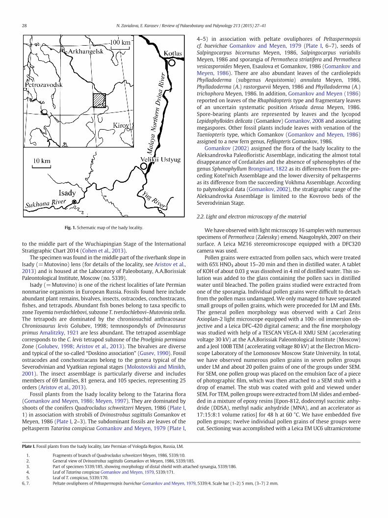
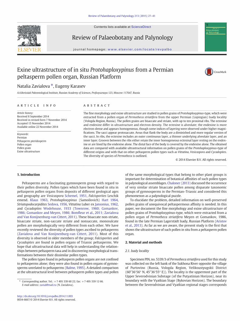
Fig. 1. Schematic map of the Isady locality.
28 N. Zavialova, E. Karasev / Review of Palaeobotany and Palynology 213 (2015) 27–41
to the middle part of the Wuchiapingian Stage of the InternationalStratigraphic Chart 2014 (Cohen et al., 2013).
The specimenwas found in themiddle part of the riverbank slope inIsady (=Mutovino) lens (for details of the locality, see Aristov et al.,2013) and is housed at the Laboratory of Paleobotany, A.A.BorissiakPaleontological Institute, Moscow (no. 5339).
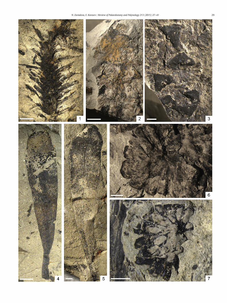
Isady (=Mutovino) is one of the richest localities of late Permiannonmarine organisms in European Russia. Fossils found here includeabundant plant remains, bivalves, insects, ostracodes, conchostracans,fishes, and tetrapods. Abundant fish bones belong to taxa specific tozone Toyemia tverdochlebovi, subzone T. tverdochlebovi–Mutovinia stella.The tetrapods are dominated by the chroniosuchid anthracosaurChroniosaurus levis Golubev, 1998; temnospondyls of Dvinosaurusprimus Amalitzky, 1921 are less abundant. The tetrapod assemblagecorresponds to the C. levis tetrapod subzone of the Proelginia permianaZone (Golubev, 1998; Aristov et al., 2013). The bivalves are diverseand typical of the so-called “Doskino association” (Gusev, 1990). Fossilostracodes and conchostracans belong to the genera typical of theSeverodvinian and Vyatkian regional stages (Molostovskii and Minikh,2001). The insect assemblage is particularly diverse and includesmembers of 69 families, 81 genera, and 105 species, representing 25orders (Aristov et al., 2013).
Fossil plants from the Isady locality belong to the Tatarina flora(Gomankov and Meyen, 1986; Meyen, 1997). They are dominated byshoots of the conifers Quadrocladus schweitzeri Meyen, 1986 (Plate I,1) in association with strobili of Dvinostrobus sagittalis Gomankov etMeyen, 1986 (Plate I, 2–3). The subdominant fossils are leaves of thepeltasperm Tatarina conspicua Gomankov and Meyen, 1979 (Plate I,
Plate I. Fossil plants from the Isady locality, late Permian of Vologda Region, Russia, LM.
1. Fragments of branch of Quadrocladus schweitzeri Meyen, 1986, 5339/10.2. General view of Dvinostrobus sagittalis Gomankov et Meyen, 1986, 5339/185.3. Part of specimen 5339/185, showing morphology of distal shield with attache4. Leaf of Tatarina conspicua Gomankov and Meyen, 1979, 5339/171.5. Leaf of T. conspicua, 5339/170.
6, 7. Peltate ovuliphores of Peltaspermopsis buevichae Gomankov and Meyen, 1979
4–5) in association with peltate ovuliphores of Peltaspermopsiscf. buevichae Gomankov and Meyen, 1979 (Plate I, 6–7), seeds ofSalpingocarpus bicornutus Meyen, 1986, Salpingocarpus variabilisMeyen, 1986 and sporangia of Permotheca striatifera and Permothecavesicasporoides Meyen, Esaulova et Gomankov, 1986 (Gomankov andMeyen, 1986). There are also abundant leaves of the cardiolepidsPhylladoderma (subgenus Aequistomia) annulata Meyen, 1986,Phylladoderma (A.) rastorguevii Meyen, 1986 and Phylladoderma (A.)trichophora Meyen, 1986. In addition, Gomankov and Meyen (1986)reported on leaves of the Rhaphidopteris type and fragmentary leavesof an uncertain systematic position Arisada densa Meyen, 1986.Spore-bearing plants are represented by leaves and the lycopodLepidophylloides delicata (Gomankov) Gomankov, 2008 and associatingmegaspores. Other fossil plants include leaves with venation of theTaeniopteris type, which Gomankov (Gomankov and Meyen, 1986)assigned to a new fern genus, Fefilopteris Gomankov, 1986.
Gomankov (2002) assigned the flora of the Isady locality to theAleksandrovka Paleofloristic Assemblage, indicating the almost totaldisappearance of Cordaitales and the absence of sphenophytes of thegenus Sphenophyllum Brongniart, 1822 as its differences from the pre-ceding Kotel'nich Assemblage and the lower diversity of peltaspermsas its difference from the succeeding Vokhma Assemblage. Accordingto palynological data (Gomankov, 2002), the stratigraphic range of theAleksandrovka Assemblage is limited to the Kovrovo beds of theSeverodvinian Stage.
2.2. Light and electron microscopy of the material
Wehave observedwith lightmicroscopy 16 sampleswith numerousspecimens of Permotheca (Zalessky) emend. Naugolnykh, 2007 on theirsurface. A Leica MZ16 stereomicroscope equipped with a DFC320camera was used.
Pollen grains were extracted from pollen sacs, which were treatedwith 65% HNO3 about 15–20 min and then in distilled water. A tabletof KOH of about 0.03 g was dissolved in 4 ml of distilled water. This so-lution was added to the glass containing the pollen sacs in distilledwater until bleached. The pollen grains studied were extracted fromone of the sporangia. Individual pollen grains were difficult to detachfrom the pollen mass undamaged. We only managed to have separatedsmall groups of pollen grains, which were proceeded for LM and EMs.The general pollen morphology was observed with a Carl ZeissAxioplan-2 light microscope equipped with a 100× oil immersion ob-jective and a Leica DFC-420 digital camera; and the fine morphologywas studied with help of a TESCAN VEGA-II XMU SEM (acceleratingvoltage 30 kV) at the A.A.Borissiak Paleontological Institute (Moscow)and a Jeol 100B TEM (accelerating voltage 80 kV) at the Electron Micro-scope Laboratory of the Lomonosov Moscow State University. In total,we have observed numerous pollen grains in seven pollen groupsunder LM and about 20 pollen grains of one of the groups under SEM.For SEM, one pollen group was placed on the emulsion face of a pieceof photographic film, which was then attached to a SEM stub with adrop of enamel. The stub was coated with gold and viewed underSEM. For TEM, pollen groupswere extracted from LM slides and embed-ded in a mixture of epoxy resins [Epon-812, dodecenyl succinic anhy-dride (DDSA), methyl nadic anhydride (MNA), and an accelerator as17:15:8:1 volume ratios] for 48 h at 60 °C. We have embedded fivepollen groups; twelve individual pollen grains of these groups werecut. Sectioning was accomplished with a Leica EM UC6 ultramicrotome
d synangia, 5339/186.
, 5339/4. Scale bar (1–2) 5 mm, (3–7) 2 mm.
29N. Zavialova, E. Karasev / Review of Palaeobotany and Palynology 213 (2015) 27–41
30 N. Zavialova, E. Karasev / Review of Palaeobotany and Palynology 213 (2015) 27–41
at the A.A.Borissiak Paleontological Institute. The sections wereobserved unstained.
We tried to orientate the pollen grains transversely and longitudi-nally. Since we dealt with groups of pollen grains, variously stuck toeach other, we got oblique sections as well. We made a series of ultra-thin sections of 60 nm thick, which we collected on a grid, then severalthick sections, which were discarded, with a total thickness of about4 μm, then the next series of ultrathin sections, etc. This way, we cutout the specimen throughout and hoped to observe all morphological
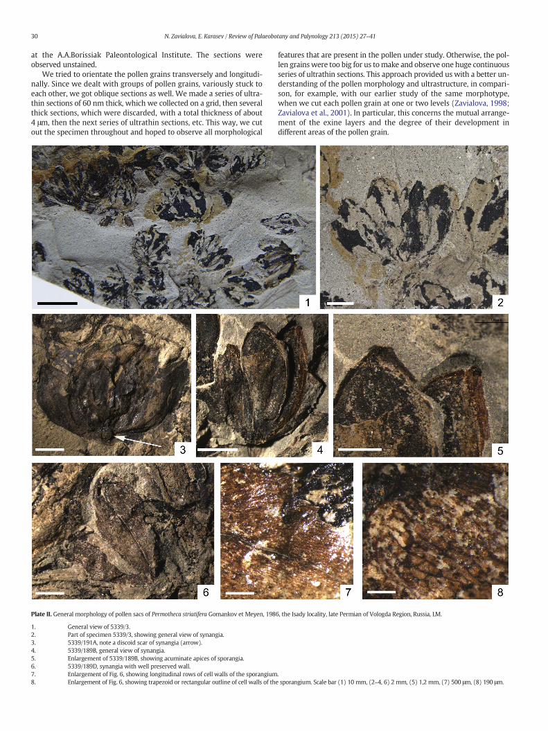
Plate II. General morphology of pollen sacs of Permotheca striatifera Gomankov et Meyen, 198
1. General view of 5339/3.2. Part of specimen 5339/3, showing general view of synangia.3. 5339/191A, note a discoid scar of synangia (arrow).4. 5339/189B, general view of synangia.5. Enlargement of 5339/189B, showing acuminate apices of sporangia.6. 5339/189D, synangia with well preserved wall.7. Enlargement of Fig. 6, showing longitudinal rows of cell walls of the sporangium8. Enlargement of Fig. 6, showing trapezoid or rectangular outline of cell walls of the
features that are present in the pollen under study. Otherwise, the pol-len grainswere too big for us tomake and observe one huge continuousseries of ultrathin sections. This approach provided us with a better un-derstanding of the pollen morphology and ultrastructure, in compari-son, for example, with our earlier study of the same morphotype,when we cut each pollen grain at one or two levels (Zavialova, 1998;Zavialova et al., 2001). In particular, this concerns the mutual arrange-ment of the exine layers and the degree of their development indifferent areas of the pollen grain.
6, the Isady locality, late Permian of Vologda Region, Russia, LM.
.sporangium. Scale bar (1) 10 mm, (2–4, 6) 2 mm, (5) 1,2 mm, (7) 500 μm, (8) 190 μm.

31N. Zavialova, E. Karasev / Review of Palaeobotany and Palynology 213 (2015) 27–41
3. Results
3.1. Pollen organs
Genus Permotheca Zalessky, 1929 emend. Naugolnykh, 2007Permotheca striatifera S. Meyen et Gomankov, 1986
1986 Permotheca striatifera S. Meyen et Gomankov, pp. 120–122, pl. XIII,figs. 11–13, pl. XIV, figs. 1–3; text-fig. 63 (a–c, k)
Isolated synangia are racemose, with 5 to 9 sporangia (Plate II, 1–2).The adaxial surface of the synangium shows an abscission scar (Plate II,3). The sporangia are elliptic or with rounded or acuminate apices,about 10 mm long and 5 mm wide. The sporangia are fused at theirbases, whereas their apices remain unfused (Plate II, 4–5). Themaximalwidth of the sporangium is near itsmiddle part. The sporangium surfaceis weakly longitudinally striated. The sporangium wall consists oflong trapezoid or rectangular cells, which are longitudinally arranged(Plate II, 6–8).
3.2. Pollen grains
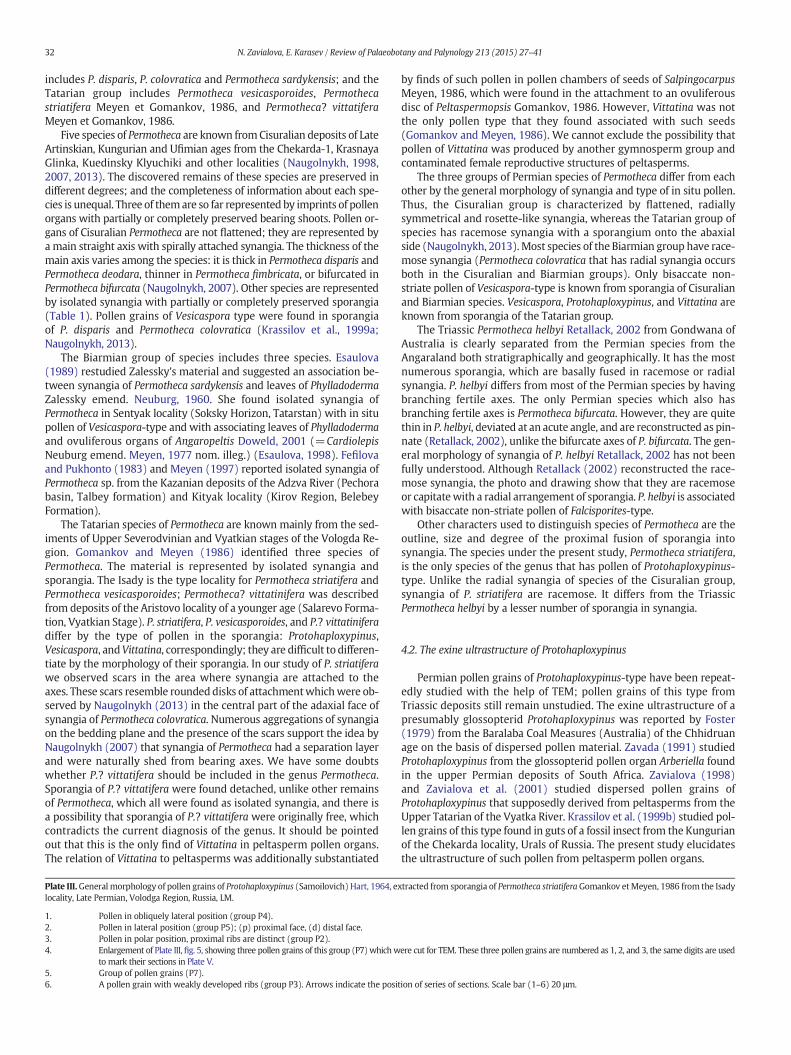
Pollen grains superimpose over each other in pollen masses, so thatthe margins of individual pollen grains were not always visible: onlyone of the sacci was clearly seen in some specimens, or only ribs of thebody in others (Plate III, 5; Plate IV, 1). Thoughwe have numerous pollengrains at hand, we managed to measure only 18 pollen grains (in someonly the length or only the width), which range from 81.0 to 101.0 μmin length and from50.0 μmto68.4 μminwidth. Pollen grains are bisaccateand striate (Plate III, 1–6).Most pollen grains bear 6 to 10 ribs at the prox-imal face of the body (Plate III, 4), but there were a few specimens, inwhich we observed only three indistinct ribs (Plate III, 6). The ribs varyin width and are situated at different angles to each other, in a way thatsome of them stretch from one saccus to the other, whereas other ribsdo not reach the other saccus (Plate III, 3). The maximal width of a rib isabout 5.3–6.8 μm. Pollen grains more commonly are flattened in thepolar position (Plate III, 5). Pollen grains in the equatorial position showthat the distal face of the body do not bear ribs and is lighter in transmit-ted light (=thinner) than the proximal face. The length of the striateproximal face of the body (=cappa) is about two thirds of the total lengthof the pollen and the length of the psilate distal face of the body is aboutone quarter of the total length of the pollen (Plate III, 2). The sacci areslightly inclined toward the distal side (Plate III, 2).
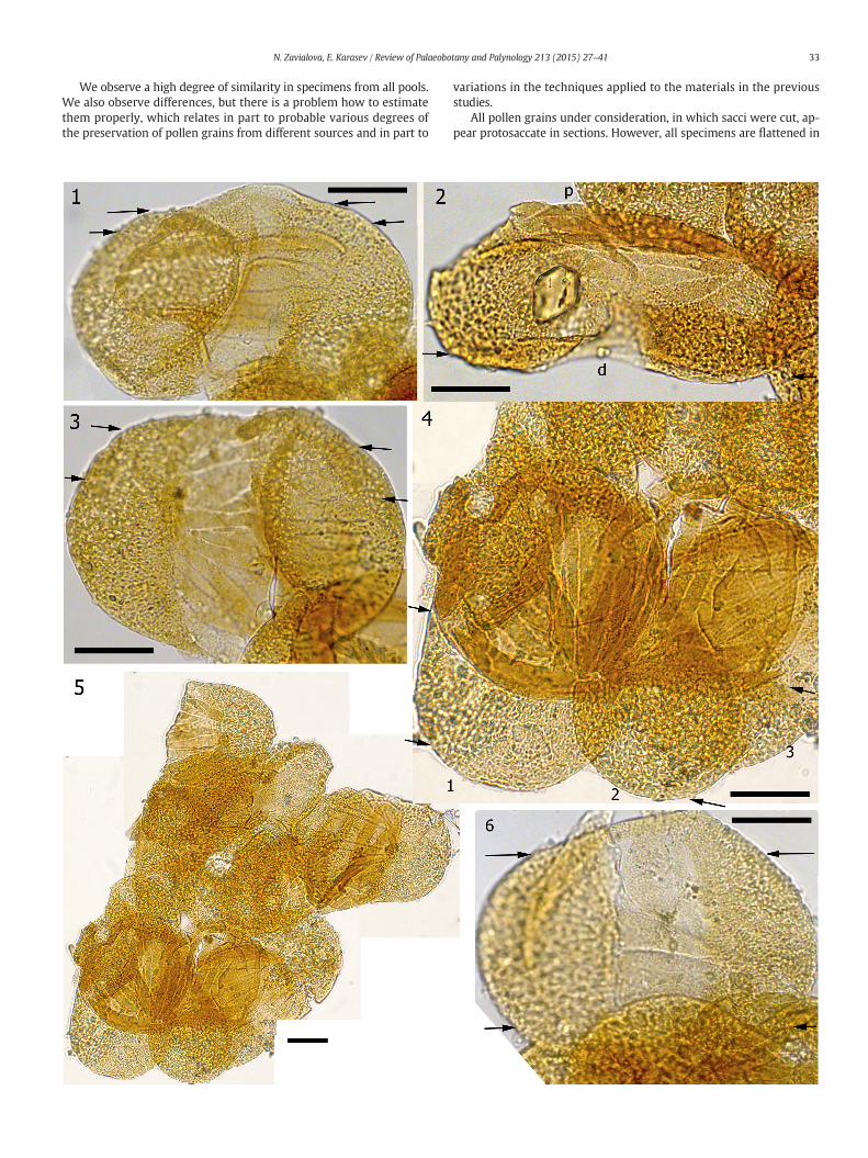
The surface of thepollen grains is almost psilate (Plate IV, 1–3). Prox-imally, the ribs are clearly visible (Plate IV, 3). The sacci are somewhatpitted (Plate IV, 2, 3). It appears from the SEM images that the outer-most layer of the exine is continuous, covering the underlying layers(if any) completely. Two sacci are connected (Plate IV, 2).
TEM shows that the exine consists of an ectexine and endexine,whichdiffer in ultrastructure and electron density (Plate V, 2). The ectexine is al-veolate; the endexine is more electron dense and appears homogeneous.
The sacci are covered by a continuous outer ectexinal layer (Plate V,1). The pits whichwere observed under SEM are due to the undulationsof this layer;we donot see any perforations in sections in the outer layerof the exine (Plate V, 1, 2). Under this layer, the sacci are completelyfilled with ectexinal partitions (Plate VI, 4). However, since the pollengrains are strongly flattened and bear quite thin sacci (varying from1.4 to 2.0 μm in thickness), they may merely appear protosaccate(Scheuring, 1976) at this stage of preservation. The partitions are vari-ously directed that in sections they appear as rods of changing width,if cut longitudinally or obliquely, or from irregular to regular granules,if transversely (Plate VI, 7).
Some extremely thin threads of the endexine enter the saccus al-ready in peripheral sections (Plate V, 1, 8). The endexine demarcatingthe body of the pollen is thick, constant in thickness over the perimeterof the body, and appears homogeneous in the overwhelming majority
of sections (Plate V, 2–4) even under high magnifications (Plate VI, 6).We observed indices of layered organization in few sections undermagnifications of 40000× and higher in the inner (Plate V, 5, 6) and inthe outer (Plate VII, 4, 5) areas of the endexine. The thin threads of theendexine, which were detected in saccate areas, belong to outer areasof the endexine and peel off them (Plate VI, 5, 8). This phenomenonadditionally counts for the layered nature of the endexine, in spite ofits homogeneous appearance in most sections. Though being consider-ably thick, the endexine is easily foldable (Plate V, 4, 7).
Ultrastructurally, the areas that flank the body are a diminished andmore regular version of the sacci: a continuous outer ectexinal layer isunderlined by partitions (Plate V, 3, lower part of this figure).
The proximal and distal faces of the body are very different. The ribsare present only proximally (Plate V, 4, 6, 7; Plate VI, 1). Their ectexineincludes an outer continuous layer, a thinner underlying alveolate layer,and an inner layer. Grooves between the ribs either retain the inner ho-mogeneous ectexinal layer resting on the endexine or are lined by theendexine alone. The grooves vary in width. Those that are relativelywide are constituted by the endexine alone (Plate VI, 2). Distally, thebody is covered by the endexine alone (Plate VI, 3, 6): it is the outersurface of the endexine that we observed in distal views of the pollengrains under SEM (Plate IV, 2).
4. Discussion
4.1. Permotheca and other pollen organs of peltasperms
Zalessky (1929) introduced the genus Permotheca with the type spe-cies Permotheca sardykensis Zallesky, 1929 for isolated synangia on thematerial from the Upper Kazanian deposits of the Kullarovo locality,Sardyk river, Tatarstan. However, the holotype was not designated andthe diagnosis was not given. Fefilova and Pukhonto (1983) provided avery brief diagnosis of the genus. Gomankov andMeyen (1986)publisheda more detailed diagnosis and were the first who demonstrated relation-ships between Permotheca and other Peltaspermales. Naugolnykh (2007)revised the genus and emended the diagnosis; he added to the diagnosisthe information about the structure of microstrobiles and treatedAnthicocladus Zalessky, 1937 and Asterodiscus Zalessky, 1937 as youngersynonyms of Permotheca based on the similar general morphology.Some Late Paleozoic and Mesozoic pteridosperms show similarities toPermotheca. Thus, Euromerian peltasperms Callipterianthus Roselt, 1962and Pterispermostrobus Stopes, 1914 from the Late Carboniferous–EarlyPermian deposits have similar synangiate (fused) agglomerations ofsporangia, but differ by flattened microsporophylls and pinnate fertilebranches (Taylor et al., 2006). The glossopterid Arberiella Pant etNautiyal, 1960 has similar elliptic to falcate pollen sacs and fine longitu-dinal striation of sporangia (Gomankov and Meyen, 1986). In addition,in situ Protohaploxypinus is known from sporangia of Arberiella(Zavada, 1991). Unlike Permotheca, pollen sacs of Arberiella are unfusedand attached to modified scale-like leaves of Eretmonia Du Toit 1932emend. Lacey et al. 1975 or Glossotheca Surange et Maheshwari, 1970.Pollen sacs of Arberiella are 0.5–1 mm long in comparison to those ofPermotheca, which are 1.5–10 mm long. Microsporophylls of Triassicpeltasperms Antevsia Harris, 1937 and Townrovia Retallack, 1981resemble Permotheca, but differ in having flattened and pinnate fertileaxes and free sporangia (Taylor et al., 2006; Taylor and Taylor, 2009;Bomfleur et al., 2011).
Currently, the genus Permotheca includes nine species from thePermian (Russian Platform) and one from Triassic (Australia). Wehave grouped the Permian species in three pools by their age, geogra-phy of the locality, and in part, the general morphology of the sporangia(Table 1). Some of the species occur in more than one group. TheCisuralian group includes Permotheca fimbriata (Zalessky) Naugolnykh,2007, Permotheca bifurcata Naugolnykh, 2007, Permotheca deodaraNaugolnykh, 2007, Permotheca disparis (Zalessky) Naugolnykh, 1999,and Permotheca colovratica Naugolnykh, 2013; the Biarmian group
32 N. Zavialova, E. Karasev / Review of Palaeobotany and Palynology 213 (2015) 27–41
includes P. disparis, P. colovratica and Permotheca sardykensis; and theTatarian group includes Permotheca vesicasporoides, Permothecastriatifera Meyen et Gomankov, 1986, and Permotheca? vittatiferaMeyen et Gomankov, 1986.
Five species of Permotheca are known fromCisuralian deposits of LateArtinskian, Kungurian and Ufimian ages from the Chekarda-1, KrasnayaGlinka, Kuedinsky Klyuchiki and other localities (Naugolnykh, 1998,2007, 2013). The discovered remains of these species are preserved indifferent degrees; and the completeness of information about each spe-cies is unequal. Three of themare so far represented by imprints of pollenorgans with partially or completely preserved bearing shoots. Pollen or-gans of Cisuralian Permotheca are not flattened; they are represented byamain straight axis with spirally attached synangia. The thickness of themain axis varies among the species: it is thick in Permotheca disparis andPermotheca deodara, thinner in Permotheca fimbricata, or bifurcated inPermotheca bifurcata (Naugolnykh, 2007). Other species are representedby isolated synangia with partially or completely preserved sporangia(Table 1). Pollen grains of Vesicaspora type were found in sporangiaof P. disparis and Permotheca colovratica (Krassilov et al., 1999a;Naugolnykh, 2013).
The Biarmian group of species includes three species. Esaulova(1989) restudied Zalessky's material and suggested an association be-tween synangia of Permotheca sardykensis and leaves of PhylladodermaZalessky emend. Neuburg, 1960. She found isolated synangia ofPermotheca in Sentyak locality (Soksky Horizon, Tatarstan) with in situpollen of Vesicaspora-type and with associating leaves of Phylladodermaand ovuliferous organs of Angaropeltis Doweld, 2001 (=CardiolepisNeuburg emend. Meyen, 1977 nom. illeg.) (Esaulova, 1998). Fefilovaand Pukhonto (1983) and Meyen (1997) reported isolated synangia ofPermotheca sp. from the Kazanian deposits of the Adzva River (Pechorabasin, Talbey formation) and Kityak locality (Kirov Region, BelebeyFormation).
The Tatarian species of Permotheca are known mainly from the sed-iments of Upper Severodvinian and Vyatkian stages of the Vologda Re-gion. Gomankov and Meyen (1986) identified three species ofPermotheca. The material is represented by isolated synangia andsporangia. The Isady is the type locality for Permotheca striatifera andPermotheca vesicasporoides; Permotheca? vittatinifera was describedfrom deposits of the Aristovo locality of a younger age (Salarevo Forma-tion, Vyatkian Stage). P. striatifera, P. vesicasporoides, and P.? vittatiniferadiffer by the type of pollen in the sporangia: Protohaploxypinus,Vesicaspora, andVittatina, correspondingly; they are difficult to differen-tiate by the morphology of their sporangia. In our study of P. striatiferawe observed scars in the area where synangia are attached to theaxes. These scars resemble roundeddisks of attachmentwhichwere ob-served by Naugolnykh (2013) in the central part of the adaxial face ofsynangia of Permotheca colovratica. Numerous aggregations of synangiaon the bedding plane and the presence of the scars support the idea byNaugolnykh (2007) that synangia of Permotheca had a separation layerand were naturally shed from bearing axes. We have some doubtswhether P.? vittatifera should be included in the genus Permotheca.Sporangia of P.? vittatifera were found detached, unlike other remainsof Permotheca, which all were found as isolated synangia, and there isa possibility that sporangia of P.? vittatifera were originally free, whichcontradicts the current diagnosis of the genus. It should be pointedout that this is the only find of Vittatina in peltasperm pollen organs.The relation of Vittatina to peltasperms was additionally substantiated
Plate III.General morphology of pollen grains of Protohaploxypinus (Samoilovich) Hart, 1964, exlocality, Late Permian, Volodga Region, Russia, LM.
1. Pollen in obliquely lateral position (group P4).2. Pollen in lateral position (group P5); (p) proximal face, (d) distal face.3. Pollen in polar position, proximal ribs are distinct (group P2).4. Enlargement of Plate III, fig. 5, showing three pollen grains of this group (P7)whichw
to mark their sections in Plate V.5. Group of pollen grains (P7).6. A pollen grain with weakly developed ribs (group P3). Arrows indicate the posit
by finds of such pollen in pollen chambers of seeds of SalpingocarpusMeyen, 1986, which were found in the attachment to an ovuliferousdisc of Peltaspermopsis Gomankov, 1986. However, Vittatina was notthe only pollen type that they found associated with such seeds(Gomankov and Meyen, 1986). We cannot exclude the possibility thatpollen of Vittatina was produced by another gymnosperm group andcontaminated female reproductive structures of peltasperms.
The three groups of Permian species of Permotheca differ from eachother by the general morphology of synangia and type of in situ pollen.Thus, the Cisuralian group is characterized by flattened, radiallysymmetrical and rosette-like synangia, whereas the Tatarian group ofspecies has racemose synangia with a sporangium onto the abaxialside (Naugolnykh, 2013). Most species of the Biarmian group have race-mose synangia (Permotheca colovratica that has radial synangia occursboth in the Cisuralian and Biarmian groups). Only bisaccate non-striate pollen of Vesicaspora-type is known from sporangia of Cisuralianand Biarmian species. Vesicaspora, Protohaploxypinus, and Vittatina areknown from sporangia of the Tatarian group.
The Triassic Permotheca helbyi Retallack, 2002 from Gondwana ofAustralia is clearly separated from the Permian species from theAngaraland both stratigraphically and geographically. It has the mostnumerous sporangia, which are basally fused in racemose or radialsynangia. P. helbyi differs from most of the Permian species by havingbranching fertile axes. The only Permian species which also hasbranching fertile axes is Permotheca bifurcata. However, they are quitethin in P. helbyi, deviated at an acute angle, and are reconstructed as pin-nate (Retallack, 2002), unlike the bifurcate axes of P. bifurcata. The gen-eral morphology of synangia of P. helbyi Retallack, 2002 has not beenfully understood. Although Retallack (2002) reconstructed the race-mose synangia, the photo and drawing show that they are racemoseor capitatewith a radial arrangement of sporangia. P. helbyi is associatedwith bisaccate non-striate pollen of Falcisporites-type.
Other characters used to distinguish species of Permotheca are theoutline, size and degree of the proximal fusion of sporangia intosynangia. The species under the present study, Permotheca striatifera,is the only species of the genus that has pollen of Protohaploxypinus-type. Unlike the radial synangia of species of the Cisuralian group,synangia of P. striatifera are racemose. It differs from the TriassicPermotheca helbyi by a lesser number of sporangia in synangia.
4.2. The exine ultrastructure of Protohaploxypinus
Permian pollen grains of Protohaploxypinus-type have been repeat-edly studied with the help of TEM; pollen grains of this type fromTriassic deposits still remain unstudied. The exine ultrastructure of apresumably glossopterid Protohaploxypinus was reported by Foster(1979) from the Baralaba Coal Measures (Australia) of the Chhidruanage on the basis of dispersed pollen material. Zavada (1991) studiedProtohaploxypinus from the glossopterid pollen organ Arberiella foundin the upper Permian deposits of South Africa. Zavialova (1998)and Zavialova et al. (2001) studied dispersed pollen grains ofProtohaploxypinus that supposedly derived from peltasperms from theUpper Tatarian of the Vyatka River. Krassilov et al. (1999b) studied pol-len grains of this type found in guts of a fossil insect from the Kungurianof the Chekarda locality, Urals of Russia. The present study elucidatesthe ultrastructure of such pollen from peltasperm pollen organs.
tracted from sporangia of Permotheca striatiferaGomankov etMeyen, 1986 from the Isady
ere cut for TEM. These three pollen grains are numbered as 1, 2, and 3, the same digits are used
ion of series of sections. Scale bar (1–6) 20 μm.
33N. Zavialova, E. Karasev / Review of Palaeobotany and Palynology 213 (2015) 27–41
We observe a high degree of similarity in specimens from all pools.We also observe differences, but there is a problem how to estimatethem properly, which relates in part to probable various degrees ofthe preservation of pollen grains from different sources and in part to
variations in the techniques applied to the materials in the previousstudies.
All pollen grains under consideration, in which sacci were cut, ap-pear protosaccate in sections. However, all specimens are flattened in
Plate IV. Surface of pollen grains of Protohaploxypinus (Samoilovich) Hart, 1964, extracted from sporangia of Permotheca striatiferaGomankov et Meyen, 1986 from the Isady locality, LatePermian, Volodga Region, Russia, SEM.
1. Pollen mass extracted from the sporangium.2. Blowing up of Fig. 1 shows the distal face of one of the pollen grain.3. Blowing up of Fig. 1 shows a pollen grain compressed in an obliquely-proximal position. Sacci and striate proximal face of the body are visible. Scale bar (1) 100 μm,
(2, 3) 20 μm.
34 N. Zavialova, E. Karasev / Review of Palaeobotany and Palynology 213 (2015) 27–41
various degrees, and the sacci are very thin in sections. Thus, a section ofa dispersed pollen of Protohaploxypinus show a cavity in one part of thesaccus (Zavialova et al., 2001, pl. 1, fig. 10), but no cavity in the other(Zavialova et al., 2001, pl. 1, fig. 9). Therefore, the protosaccate appear-ance could be a preservational feature. Three-dimensionally preservedspecimens, similar to those studied by Osborn and Taylor (1993), areneeded to resolve this question and there is a possibility that theinterpretation will be changed.
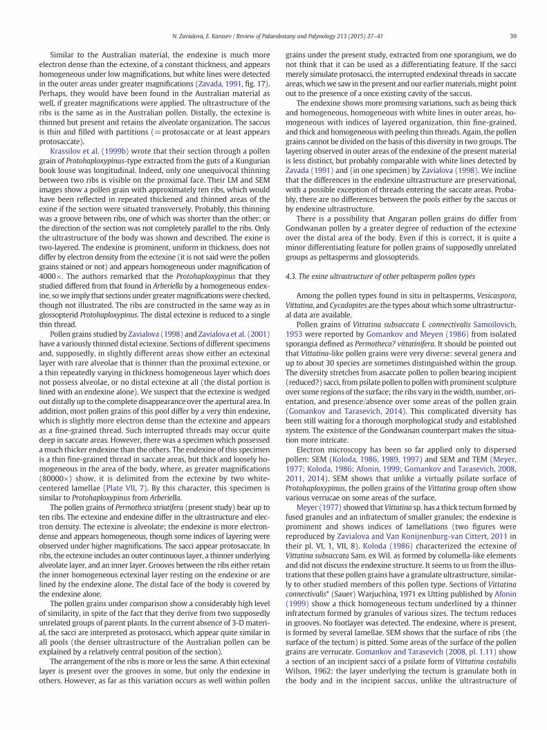
A transverse section published by Foster (1979) shows a saccus andproximal ribs. The sacci occupy a greater area distally than proximally,and the distal portion of the body remained unstudied. The exine
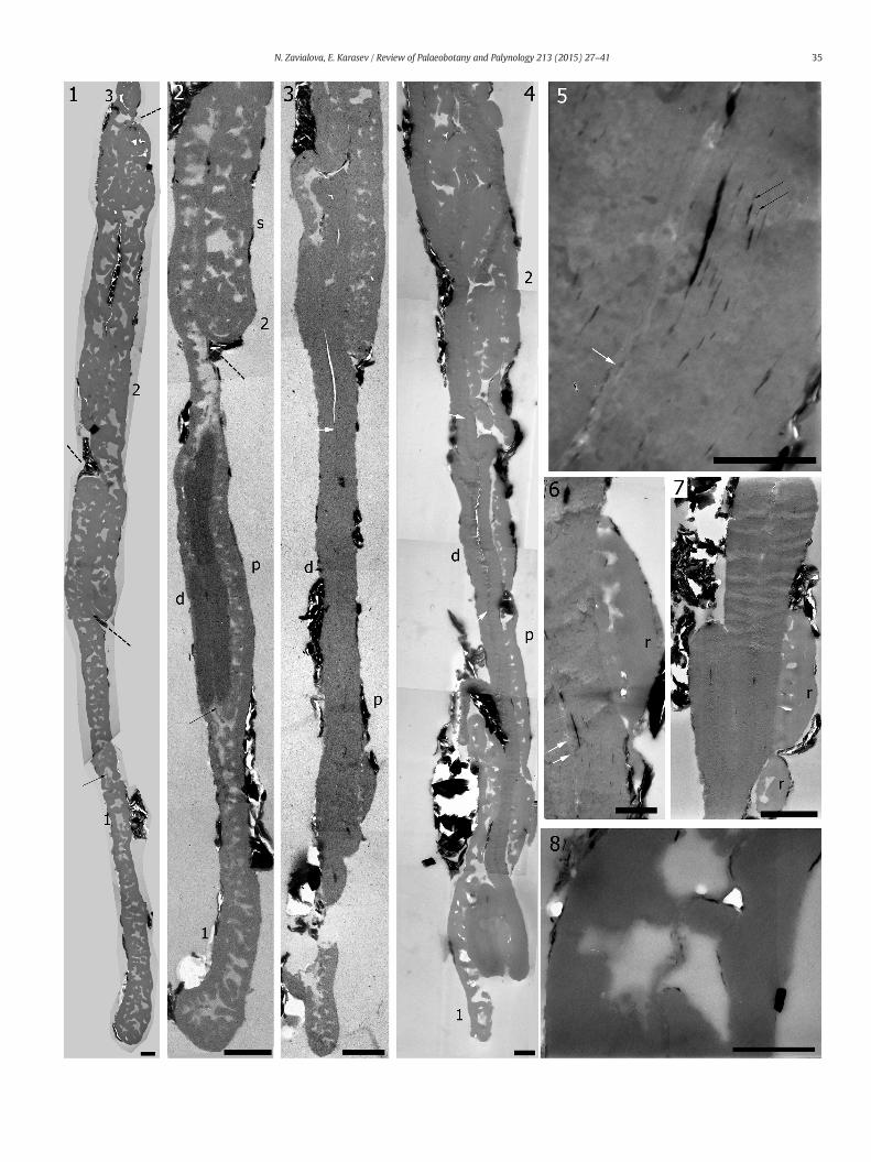
Plate V. Exine ultrastructure of pollen grains of Protohaploxypinus (Samoilovich) Hart, 1964, exlocality, Late Permian, Volodga Region, Russia, TEM. Three pollen grains of Group P7 were cut
1. Section through sacci of the pollen grains, the deepest section is through pollen grthrough a saccus of pollen grain 2 is more superficial; and section through pollenboundaries between the pollen grains.
2. Deeper section: a thick, homogeneous and electron-dense endexine is present in pthat shed out of themain portion of the endexine. Distally, merely a very thin ectof the sacci appear as granules.
3. Deeper section: distally, the endexine is exposed; proximaly, first ribs appear, th4. Deeper section: the endexine is still the only layer of the exine that is present in th
areas. The pollen grains are tightly posed over each other, one can trace individual po5. Endexine, layered nature can be observed (black arrows). Enlargement of Plate6. Proximal rib, enlargement from a section close to that shown in Plate V, fig. 3.7. Two proximal ribs, enlargement from a section close to that shown in Plate V, fi8. A thread of endexine in the saccus region, enlargement from Fig. 1 (arrow). (s) s
position of the gametophyte cavity; (r) rib. Scale bar (1, 4) 1 μm, (2, 3) 2 μm, (5
Plate VI. Exine ultrastructure of pollen grains of Protohaploxypinus (Samoilovich) Hart, 1964, exlocality, Late Permian, Volodga Region, Russia, TEM, group P7 (1, 2, 7) and group P2 (3–6, 8). (
1. Pollen 3 of group P7, a portion of a section showing proximal ribs (r); the endex2. Pollen 3 of group P7, a deeper section in the same area as in Fig. 1, note a consid3. Longitudinal section of a pollen grain from group P2. Distal face is covered by th4. Group P2. Saccus area. A thin thread is shed out from a thick endexine toward t5. Group P2. Shedding threads of the endexine.6. Group P2. Longitudinally cut rib. An enlarged area of a section adjacent to that s7. Group P7 (pollen 3), saccus area. Most partitions are cut transversely and appea8. Group P2. Endexinal thread enters the saccus area. Enlargement of Plate VI, fig. 4
Plate VII. Exine ultrastructure of pollen grains of Protohaploxypinus (Samoilovich) Hart, 1964, exlocality, Late Permian, Volodga Region, Russia, TEM, groups P5 and P3. (see on page 11)
1. A portion of a longitudinal section through a pollen grain of group P5. The pollen2. Enlargement of a section adjacent to that shown in Plate VII, 1. Many partitions
3, 6. Portion of a section of group P4. Ectexine and endexine remarkably differ in elec4, 5. Areas of longitudinal section of a pollen grain of group P3 (Plate III, 6). Arrows p
7. Area of a longitudinal section of a dispersed Protohaploxypinus studied by Zavial(d) distal face. Scale bar (1, 3) 0.67 μm, (2, 5) 1 μm, (4, 6) 0.5 μm, (7) 0.25 μm. *Gobe more suitably incorporated in Vittatina costabilisWilson, 1962.
includes an ectexine and endexine. The former is densely alveolatewith rare and small alveolae both in the saccus and ribs. The endexineis prominent, muchmore electron dense than the ectexine, and appearshomogeneous under amagnification of 8000×. Proximally, the ectexinevaries in thickness considerably: it becomes very thin and homoge-neous between the ribs. The endexine is uniform in thickness.
Oblique sections made by Zavada (1991) show a saccus and bothproximal and distal areas of the body. The exine is two-layered. Theectexine is alveolate and seems less dense (=alveolae aremore volumi-nous and partitions are thinner) than that in the Australian material,though it can be related to the different position of the sections.
tracted from sporangia of Permotheca striatifera Gomankov et Meyen, 1986 from the Isadymore or less transversely (Plate III, 4).
ain 1 (some thin threads of the endexine are already present in the saccus, arrow); sectiongrain 3 is through the most peripheral region of one of its saccus. Interrupted lines mark
ollen 1. The endexine is easily folded and cut twice. The arrowpoints on endexinal threadsexinal layer retains. A portion of the saccus of pollen 2 is visible; transversely cut partitions
e endexine also is exposed over a considerable distance.e distal area of the body; proximal ribs are distinct. The endexine shows folds in severalllen via endexinal contour, but it is difficult to decidewhich rib belongs towhich pollen grain.V, fig. 6.
g. 3.accus; (p) proximal face of the body; (d) distal face of the body; white arrow points on the, 8) 0.5 μm, (6) 0.67 μm, (7) 1.25 μm.
tracted from sporangia of Permotheca striatifera Gomankov etMeyen, 1986 from the Isadysee on page 10)
ine is much thinner than in previous sections and very variable in thickness.erable groove (g) between two of the ribs (r).e endexine alone.he saccus area.
hown in Fig. 3.r as granules; the outer layer is continuous (without perforations).. Scale bar (1–4, 7) 1 μm, (6, 8) 0.67 μm.
tracted from sporangia of Permotheca striatiferaGomankov et Meyen, 1986 from the Isady
was pressed in equatorial position and the section passed through the distal area and sacci.of the sacci are cut transversely.tron density.oint white lines in outer portions of the endexine.ova (1998). Arrows point white lines in outer portions of the endexine. (p) proximal face,mankov and Tarasevich (2011) argued that the specimen shown by Afonin (1999) should
35N. Zavialova, E. Karasev / Review of Palaeobotany and Palynology 213 (2015) 27–41
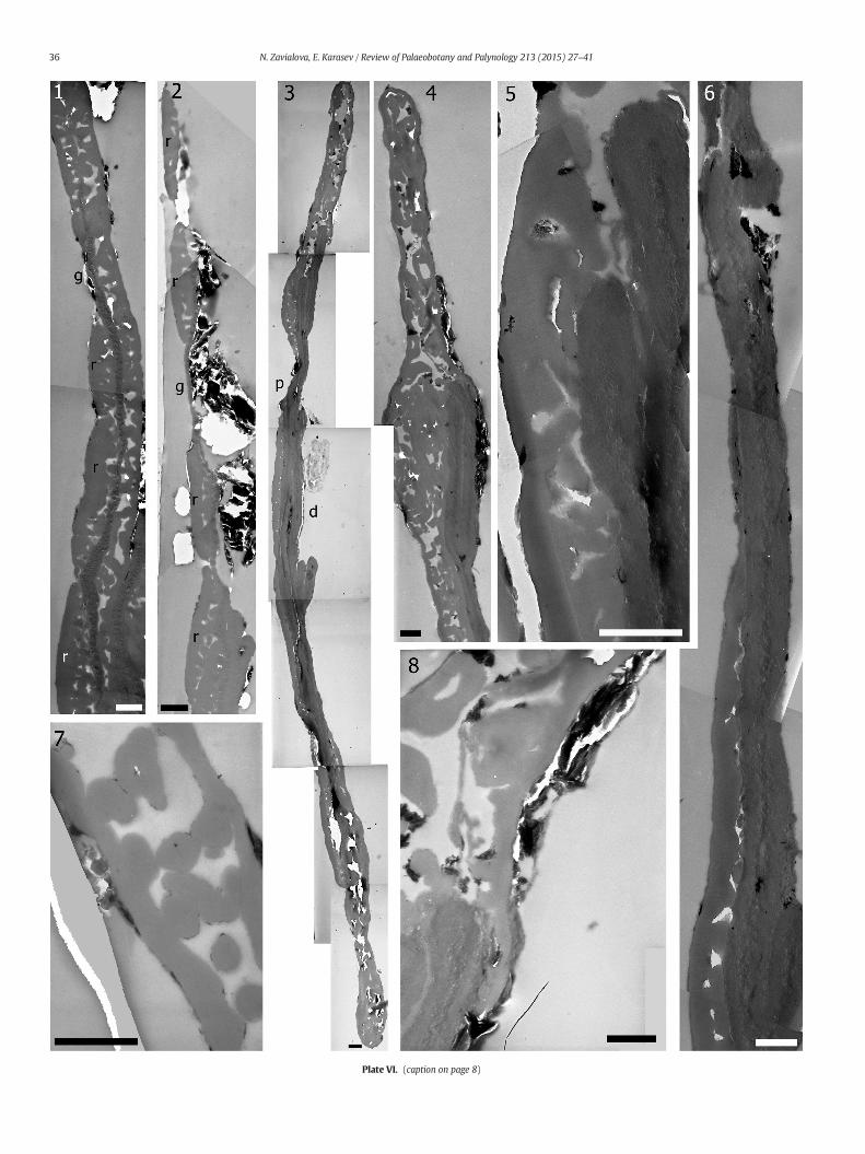
Plate VI. (caption on page 8)
36 N. Zavialova, E. Karasev / Review of Palaeobotany and Palynology 213 (2015) 27–41
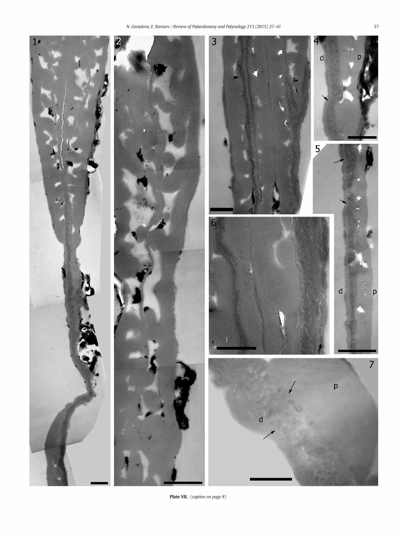
Plate VII. (caption on page 8)
37N. Zavialova, E. Karasev / Review of Palaeobotany and Palynology 213 (2015) 27–41
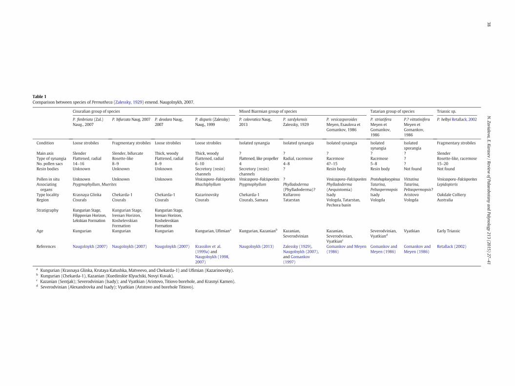
Table 1Comparison between species of Permotheca (Zalessky, 1929) emend. Naugolnykh, 2007.
Cisuralian group of species Mixed Biarmian group of species Tatarian group of species Triassic sp.
P. fimbriata (Zal.)Naug., 2007
P. bifurcata Naug, 2007 P. deodara Naug.,2007
P. disparis (Zalessky)Naug., 1999
P. colovratica Naug.,2013
P. sardykensisZalessky, 1929
P. vesicasporoidesMeyen, Esaulova etGomankov, 1986
P. striatiferaMeyen etGomankov,1986
P.? vittatiniferaMeyen etGomankov,1986
P. helbyi Retallack, 2002
Condition Loose strobiles Fragmentary strobiles Loose strobiles Loose strobiles Isolated synangia Isolated synangia Isolated synangia Isolatedsynangia
Isolatedsporangia
Fragmentary strobiles
Main axis Slender Slender, bifurcate Thick, woody Thick, woody ? ? ? ? ? SlenderType of synangia Flattened, radial Rosette-like Flattened, radial Flattened, radial Flattened, like propeller Radial, racemose Racemose Racemose ? Rosette-like, racemoseNo. pollen sacs 14–16 8–9 8–9 6–10 4 4–8 4?–15 5–8 ? 15–20Resin bodies Unknown Unknown Unknown Secretory (resin)
channelsSecretory (resin)channels
? Resin body Resin body Not found Not found
Pollen in situ Unknown Unknown Unknown Vesicaspora–Falcisporites Vesicaspora–Falcisporites ? Vesicaspora–Falcisporites Protohaploxypinus Vittatina Vesicaspora–FalcisporitesAssociatingorgans
Psygmophyllum, Muerites Rhachiphyllum Psygmophyllum Phylladoderma(Phylladoderma)?
Phylladoderma(Aequistomia)
Tatarina,Peltaspermopsis
Tatarina,Peltaspermopsis?
Lepidopteris
Type locality Krasnaya Glinka Chekarda-1 Chekarda-1 Kazarinovsky Chekarda-1 Kullarovo Isady Isady Aristovo Oakdale CollieryRegion Cisurals Cisurals Cisurals Cisurals Cisurals, Samara Tatarstan Vologda, Tatarstan,
Pechora basinVologda Vologda Australia
Stratigraphy Kungurian Stage,Filippovian Horizon,Lekskian Formation
Kungurian Stage,Irenian Horizon,KoshelevskianFormation
Kungurian Stage,Irenian Horizon,KoshelevskianFormation
Age Kungurian Kungurian Kungurian Kungurian, Ufimiana Kungurian, Kazanianb Kazanian,Severodvinian
Kazanian,Severodvinian,Vyatkianc
Severodvinian,Vyatkiand
Vyatkian Early Triassic
References Naugolnykh (2007) Naugolnykh (2007) Naugolnykh (2007) Krassilov et al.(1999a) andNaugolnykh (1998,2007)
Naugolnykh (2013) Zalessky (1929),Naugolnykh (2007),and Gomankov(1997)
Gomankov and Meyen(1986)
Gomankov andMeyen (1986)
Gomankov andMeyen (1986)
Retallack (2002)
a Kungurian (Krasnaya Glinka, Krutaya Katushka, Matveevo, and Chekarda-1) and Ufimian (Kazarinovsky).b Kungurian (Chekarda-1), Kazanian (Kuedinskie Klyuchiki, Novyi Kuvak).c Kazanian (Sentjak); Severodvinian (Isady); and Vyatkian (Aristovo, Titiovo borehole, and Krasnyi Kamen).d Severodvinian (Alexandrovka and Isady); Vyatkian (Aristovo and borehole Titiovo).
38N.Zavialova,E.Karasev
/ReviewofPalaeobotany
andPalynology
213(2015)
27–41
39N. Zavialova, E. Karasev / Review of Palaeobotany and Palynology 213 (2015) 27–41
Similar to the Australian material, the endexine is much moreelectron dense than the ectexine, of a constant thickness, and appearshomogeneous under low magnifications, but white lines were detectedin the outer areas under greater magnifications (Zavada, 1991, fig. 17).Perhaps, they would have been found in the Australian material aswell, if greater magnifications were applied. The ultrastructure of theribs is the same as in the Australian pollen. Distally, the ectexine isthinned but present and retains the alveolate organization. The saccusis thin and filled with partitions (=protosaccate or at least appearsprotosaccate).
Krassilov et al. (1999b) wrote that their section through a pollengrain of Protohaploxypinus-type extracted from the guts of a Kungurianbook louse was longitudinal. Indeed, only one unequivocal thinningbetween two ribs is visible on the proximal face. Their LM and SEMimages show a pollen grain with approximately ten ribs, which wouldhave been reflected in repeated thickened and thinned areas of theexine if the section were situated transversely. Probably, this thinningwas a groove between ribs, one of which was shorter than the other; orthe direction of the section was not completely parallel to the ribs. Onlythe ultrastructure of the body was shown and described. The exine istwo-layered. The endexine is prominent, uniform in thickness, does notdiffer by electron density from the ectexine (it is not said were the pollengrains stained or not) and appears homogeneous under magnification of4000×. The authors remarked that the Protohaploxypinus that theystudied differed from that found in Arberiella by a homogeneous endex-ine, sowe imply that sections under greatermagnificationswere checked,though not illustrated. The ribs are constructed in the same way as inglossopterid Protohaploxypinus. The distal ectexine is reduced to a singlethin thread.
Pollen grains studied by Zavialova (1998) and Zavialova et al. (2001)have a variously thinned distal ectexine. Sections of different specimensand, supposedly, in slightly different areas show either an ectexinallayer with rare alveolae that is thinner than the proximal ectexine, ora thin repeatedly varying in thickness homogeneous layer which doesnot possess alveolae, or no distal ectexine at all (the distal portion islined with an endexine alone). We suspect that the ectexine is wedgedout distally up to the complete disappearance over the apertural area. Inaddition, most pollen grains of this pool differ by a very thin endexine,which is slightly more electron dense than the ectexine and appearsas a fine-grained thread. Such interrupted threads may occur quitedeep in saccate areas. However, there was a specimen which possessedamuch thicker endexine than the others. The endexine of this specimenis a thin fine-grained thread in saccate areas, but thick and loosely ho-mogeneous in the area of the body, where, as greater magnifications(80000×) show, it is delimited from the ectexine by two white-centered lamellae (Plate VII, 7). By this character, this specimen issimilar to Protohaploxypinus from Arberiella.
The pollen grains of Permotheca striatifera (present study) bear up toten ribs. The ectexine and endexine differ in the ultrastructure and elec-tron density. The ectexine is alveolate; the endexine is more electron-dense and appears homogeneous, though some indices of layering wereobserved under higher magnifications. The sacci appear protosaccate. Inribs, the ectexine includes an outer continuous layer, a thinner underlyingalveolate layer, and an inner layer. Grooves between the ribs either retainthe inner homogeneous ectexinal layer resting on the endexine or arelined by the endexine alone. The distal face of the body is covered bythe endexine alone.
The pollen grains under comparison show a considerably high levelof similarity, in spite of the fact that they derive from two supposedlyunrelated groups of parent plants. In the current absence of 3-Dmateri-al, the sacci are interpreted as protosacci, which appear quite similar inall pools (the denser ultrastructure of the Australian pollen can beexplained by a relatively central position of the section).
The arrangement of the ribs is more or less the same. A thin ectexinallayer is present over the grooves in some, but only the endexine inothers. However, as far as this variation occurs as well within pollen
grains under the present study, extracted from one sporangium, we donot think that it can be used as a differentiating feature. If the saccimerely simulate protosacci, the interrupted endexinal threads in saccateareas,whichwe saw in the present and our earliermaterials,might pointout to the presence of a once existing cavity of the saccus.
The endexine shows more promising variations, such as being thickand homogeneous, homogeneous with white lines in outer areas, ho-mogeneous with indices of layered organization, thin fine-grained,and thick and homogeneouswith peeling thin threads. Again, the pollengrains cannot be divided on the basis of this diversity in two groups. Thelayering observed in outer areas of the endexine of the present materialis less distinct, but probably comparable with white lines detected byZavada (1991) and (in one specimen) by Zavialova (1998). We inclinethat the differences in the endexine ultrastructure are preservational,with a possible exception of threads entering the saccate areas. Proba-bly, there are no differences between the pools either by the saccus orby endexine ultrastructure.
There is a possibility that Angaran pollen grains do differ fromGondwanan pollen by a greater degree of reduction of the ectexineover the distal area of the body. Even if this is correct, it is quite aminor differentiating feature for pollen grains of supposedly unrelatedgroups as peltasperms and glossopterids.
4.3. The exine ultrastructure of other peltasperm pollen types
Among the pollen types found in situ in peltasperms, Vesicaspora,Vittatina, and Cycadopites are the types aboutwhich some ultrastructur-al data are available.
Pollen grains of Vittatina subsaccata f. connectivalis Samoilovich,1953 were reported by Gomankov and Meyen (1986) from isolatedsporangia defined as Permotheca? vittatinifera. It should be pointed outthat Vittatina-like pollen grains were very diverse: several genera andup to about 30 species are sometimes distinguished within the group.The diversity stretches from asaccate pollen to pollen bearing incipient(reduced?) sacci, frompsilate pollen topollenwithprominent sculptureover some regions of the surface; the ribs vary in thewidth, number, ori-entation, and presence/absence over some areas of the pollen grain(Gomankov and Tarasevich, 2014). This complicated diversity hasbeen still waiting for a thorough morphological study and establishedsystem. The existence of the Gondwanan counterpart makes the situa-tion more intricate.
Electron microscopy has been so far applied only to dispersedpollen: SEM (Koloda, 1986, 1989, 1997) and SEM and TEM (Meyer,1977; Koloda, 1986; Afonin, 1999; Gomankov and Tarasevich, 2008,2011, 2014). SEM shows that unlike a virtually psilate surface ofProtohaploxypinus, the pollen grains of the Vittatina group often showvarious verrucae on some areas of the surface.
Meyer (1977) showed thatVittatina sp. has a thick tectum formedbyfused granules and an infratectum of smaller granules; the endexine isprominent and shows indices of lamellations (two figures werereproduced by Zavialova and Van Konijnenburg-van Cittert, 2011 intheir pl. VI, 1, VII, 8). Koloda (1986) characterized the ectexine ofVittatina subsaccata Sam. ex Wil. as formed by columella-like elementsand did not discuss the endexine structure. It seems to us from the illus-trations that these pollen grains have a granulate ultrastructure, similar-ly to other studied members of this pollen type. Sections of Vittatinaconnectivalis* (Sauer) Warjuchina, 1971 ex Utting published by Afonin(1999) show a thick homogeneous tectum underlined by a thinnerinfratectum formed by granules of various sizes. The tectum reducesin grooves. No footlayer was detected. The endexine, where is present,is formed by several lamellae. SEM shows that the surface of ribs (thesurface of the tectum) is pitted. Some areas of the surface of the pollengrains are verrucate. Gomankov and Tarasevich (2008, pl. 1.11) showa section of an incipient sacci of a psilate form of Vittatina costabilisWilson, 1962: the layer underlying the tectum is granulate both inthe body and in the incipient saccus, unlike the ultrastructure of
40 N. Zavialova, E. Karasev / Review of Palaeobotany and Palynology 213 (2015) 27–41
Protohaploxypinus. Later, they studied a verrucate form of the same spe-cies (Gomankov and Tarasevich, 2011). Each of numerous ribs is a rowof verrucae. The verrucae are composed of a tectum of fused granulesof various sizes and an underlying layer of smaller granules, at placesaligned into columellate-like structures. The tectum diminishes be-tween the verrucae. This organization is characteristic of the proximalface, which is covered by these verrucate ribs. Distally, merely theunderlying granulate layer remains. The endexine is thick and appearshomogeneous (under magnification of 8000×), and of constant thick-ness around the perimeter of the pollen. Gomankov and Tarasevich(2014) studied members of Vittatina from deposits ranging in agefrom the Kungurian to Tatarian and concluded that all of them sharedthe same ultrastructural organization, which was similar in ribs and inincipient sacci.
In their light-microscopical study of the morphology of dispersedpollen grains of Protohaploxypinus and Striatopodocarpidites from theupper Tatarian of the Vyatka River, Foster and Gomankov (1994) hy-pothesized that striate bisaccate pollen grains of these types couldhave lost sacci in the course of fossilization and, as a result, can beidentified in palynological assemblages as separate striate entities.Such specimens of Protohaploxypinus and Striatopodocarpidites, whichare devoid of sacci, resemble pollen grains of Vittatina-type. However,available TEM data show that the exine ultrastructure of pollen grainsof Protohaploxypinus and Striatopodocarpidites in areas of the bodydiffers from the exine ultrastructure of pollen grains of Vittatina.
All ultrastructurally studied pollen grains of Vittatina show variantsof a granulate ultrastructure, that is in contrast to the ultrastructure ofProtohaploxypinus. SEM studies show significant dissimilarities betweenVittatina and Protohaploxypinus. The evolutionary transformation of thegranulate wall structure to the alveolate structure (or vise versa) is notapparent in these taxa nor do any intermediatemorphologies exist. Thissupports our doubts about assigning Permotheca (?) vittatinifera to thegenus Permotheca. We are looking forward to finding better preservedspecimens of this species allowing a more confident morphologicalcomparison with other species of Permotheca and an ultrastructuralstudy of in situ Vittatina which will elucidate the relation withpeltasperms and to understand the nature of the diversity of Vittatina.
Krassilov et al. (1999a) published ultrathin sections of in situVesicaspora from early Permian Permotheca. Dispersed pollen grains ofVesicaspora which supposedly derived from peltasperm were ultra-structurally studied by Zavialova (1998) and Zavialova et al. (2001)from the upper Permian of the Russian Platform. Early Permian pollenwere interpreted as protosaccate, and the Late Permian pollen also ap-pear protosaccate or intermediate proto/eusaccate, although it can bea preservational feature, since the dispersed pollen grainswere stronglyflattened. Lamellae were found in the endexine of the early Permianpollen grains, but were not detected in the Late Permian dispersed pol-len. Both Early and Late Permian pollen have a thicker proximalectexine, with outer and inner homogeneous layers, and a row ofalveolae between them. The distal aperture is formed by a considerablethinning of the ectexine. The morphology of the proximal area of thebody in Protohaploxypinus has something in common with that ofVesicaspora; the outer and inner homogeneous layers and intermediatealveolate layer are present in both pollen types. The main featuresdistinguishing Protohaploxypinus from Vesicaspora at the level of exineultrastructure are strong variations in thickness and presence of theouter and intermediate layers due to the striations.
The pollen grains of Antevsia zeilleri (Nathorst) Harris, 1937 from theUpper Triassic of Germany are of the Cycadopites-type (Zavialova andVan Konijnenburg-van Cittert, 2011). The proximal exine includes arow of lacunae covered by a solid, thick tectum and underlined by afoot layer. Pillars are hanging from the tectum between the lacunae.The exine is thinning to a homogeneous layer in the apertural region.The latter is bordered by thicker alveolate areas of the exine, at placesresembling a saccus-like ultrastructure. The endexine includes white-line-centred lamellae. Basing on ultrastructural data, Zavialova and
Van Konijnenburg-van Cittert (2011) hypothesized a transformationfrom Permian Vesicaspora into Triassic Cycadopites extracted from pol-len sacs of Antevsia. Proximal areas in the exine of pollen of Antevsiaare usually more homogeneous than in the cappa of PermianVesicaspora; however, some sections of Antevsiapollen appear very sim-ilar. One can imagine the following transformation which would allowthe dissimilar pollen of Permian and Triassic peltasperms to be related:the proximal exine of Vesicaspora became less alveolate; sacci disap-peared, leaving as remnants lateral extensions that were observed inAntevsia pollen; and the ultrastructure of the distal area did not changeappreciably.
5. Conclusions
The exine ultrastructure of Protohaploxypinus pollen grains extractedfrom a peltaspermpollen organwas investigated. Our present and previ-ous (Zavialova and Van Konijnenburg-van Cittert, 2011) results contrib-ute to the understanding of the diversity of pollen types occurring inPermian and Triassic peltasperms. Several similarities are observedbetween the ultrastructure of Protohaploxypinus and Vesicaspora. Theexine ultrastructure of Vittatina differs principally from other peltaspermpollen types (Protohaploxypinus, Cycadopites and Vesicaspora). No ultra-structural information is still available about Falcisporites. Once morewe stress the necessity of TEM studies of in situ pollen grains. Scantydata so far available for many of the fossil plant groups cannot providea solid basis for analysis, and the palynological puzzles are doomed toremain puzzles.
Acknowledgments
We thankDr. RomanRakitov (A.A.Borissiak Paleontological Institute,Moscow) for the assistance with SEM, the head of the Laboratoryof Electron Microscopy of Lomonosov Moscow State University(Moscow), Mr. Georgii Davidovich for the assistance with TEM, Dr.Maria Tekleva for the discussion of the preliminary version of the man-uscript, the reviewers and the editor for their valuable suggestions, andthe Russian Foundation for Basic Research, project no. 13-04-00624-afor the financial support.
References
Afonin, S.A., 1999. Ultrastructure of Permian pollen grains of Vittatina. Paleontol. J. 33 (4),137–142 (In Russian).
Amalitzky, V.P., 1921. Severodvinian Excavations of Prof. V.P. Amalitskii: 1. Akad. NaukPress, Petrograd, 16 pp. (In Russian).
Aristov, D.S., Bashkuev, A.S., Golubev, V.K., Gorochov, A.V., Karasev, E.V., Kopylov, D.S.,Ponomarenko, A.G., Rasnitsyn, A.P., Rasnitsyn, D.A., Sinitshenkova, N.D., Sukatsheva,I.D., Vassilenko, D.V., 2013. Fossil insects of the Middle and Upper Permian ofEuropean Russia. Paleontol. J. 47 (7), 641–832.
Balme, B.E., 1995. Fossil in situ spores and pollen grains: an annotated catalogue. Rev.Palaeobot. Palynol. 87, 81–323.
Bomfleur, B., Taylor, E.L., Taylor, T.N., Serbet, R., Krings, M., Kerp, H., 2011. Systematics andpaleoecology of a new peltaspermalean seed fern from the Triassic Polar vegetationof Gondwana. Int. J. Plant Sci. 172 (6), 807–835.
Brongniart, A., 1822. Historie des végétaux fossiles, ou recherches botaniques etgéologiques sur les végétaux renfermés dans les diverses couches du globe, 1. Dufouret d'Ocagne, Paris (448 pp.).
Chaloner, W.G., 2013. Three palynological puzzles. Int. J. Plant Sci. 174 (3), 602–607.Cohen, K.M., Finney, S.C., Gibbard, P.L., Fan, J.X., 2013. The ICS International Chrono-
stratigraphic Chart. Episodes 2013 (updated), 199–204.Doweld, A.B., 2001. Prosyllabus tracheophytorum: Tentamen systematis plantarum
vascularium (Tracheophyta). GEOS, Moscow, 33–110 pp. (In Russian).Esaulova, N.K., 1989. Materials for the Revision of Taxa of Flora of the Upper Kama Region
From the Collection of M.D. Zalessky. Manuscript deposited at VINITI No. 4465-В89,39 pp. (In Russian).
Esaulova, N.K., 1998. Macroflora. In: Esaulova, N.K., Lozovsky, V.R., Rozanov, A.Y. (Eds.),Stratotypes and Reference Sections of the Upper Permian in the Regions of theVolga and Kama Rivers. GEOS, Moscow, pp. 303–333 (In Russian).
Fefilova, L.A., Pukhonto, S.K., 1983. Macroflora. In: Meyen, S.V., Molin, V.A. (Eds.),Palaeontological Atlas of the Permian Deposits of the Pechora Coal Basin. Nauka,Leningrad, pp. 28–92 (In Russian).
Foster, C.B., 1979. Permian plant microfossils of the Blair Athol coal measures, Baralabacoal measures, and basal Rewan Formation of Queensland. Geological Survey ofQueensland, Publication 372Paleontological paper 45 (244 pp.).
41N. Zavialova, E. Karasev / Review of Palaeobotany and Palynology 213 (2015) 27–41
Foster, C.B., Gomankov, A.V., 1994. A new structure in pollen assigned toStriatopodocarpites Sedova 1956 and Protohaploxypinus Samolovich emend. Morbey1975, from the Late Permian (Tatarian) of the Russian Platform. J. Aust. Geol.Geophys. 15 (2), 235–238.
Golubev, V.K., 1998. Revision of the Late Permian Chroniosuchians (Amphibia,Anthracosauromorpha) from Eastern Europe. Paleontol. J. 32 (4), 390–401 (In Russian).
Gomankov, A.V., 1986. Systematic affinities of the Permian miospores. The Theory andPractice of the Palynological Studies of the Permian and Triassic of the USSR.Manuscript deposited at VINITI, Syktyvkar 4839-B86, pp. 4–9 (In Russian).
Gomankov, A.V., 1997. The Permian (Tatarian) flora from the Kotel'nich vertebratelocality (Kirov oblast). Stratigr. Geol. Correl. 5 (4), 3–12 (In Russian).
Gomankov, A.V., 2002. Flora and stratigraphy of the Tatarian stage of the East-EuropeanPlatform. Author's Abstract of the Thesis on Professor Degree, Geological Institute ofRAS, Moscow. 48 p. (In Russian).
Gomankov, A.V., 2008. The Tatarian Peltasperms of the Russian Platform: morphology,ecology, and evolution. In: Budantsev, L.Y. (Ed.), Questions of Paleofloristics and Sys-tematics of Fossil PlantsLectures in Commemoration of A.N. Kryshtofovich. vol. 6. BINRAS, St. Petersburg, pp. 42–60 (In Russian).
Gomankov, A.V., Meyen, S.V., 1979. On members of the family Peltaspermaceae from thePermian of the Russian Platfrom. Paleontol. J. 2, 124–138 (In Russian).
Gomankov, A.V., Meyen, S.V., 1986. Tatarina flora (composition and distribution in LatePermian of Eurasia). Tr. Geol. Inst. Akad. Nauk SSSR. no. 401. Nauka, Moscow, 175pp. (In Russian).
Gomankov, A.V., Tarasevich, V.F., 2008. Ultrastructure of some Vittatina-like pollen grainsfrom the Permian of the Russian Platform. Palynology: Stratigraphy and Geoecology,Vol. 1, Collection of Scientific Papers to XII All-Russian Palynological Conference.VNIGRI, St. Petersburg, pp. 48–53 (In Russian).
Gomankov, A.V., Tarasevich, V.F., 2011. Verrucate Vittatina-like pollen grains from thePermian of the East European Platform and the ultrastructure. Problems of ModernPalynology. XIII Russian palynological conference vol. I, pp. 20–22 (In Russian).
Gomankov, A.V., Tarasevich, V.F., 2014. Ultrastructure of Vittatina-like pollen grains fromthe Permian of the Russian Platform. 9th European Palaeobotany — PalynologyConference, Padova, 26–31 August 2014, pp. 82–83.
Gusev, A.K., 1990. Nonmarine Bivalves From the Upper Permian of the European USSR.Kazan Gos. Univ, Kazan (In Russian).
Harris, T., 1937. The fossil flora of Scoresby Sound, East Greenland. Part. 5: stratigraphicrelations of the plant beds. Medd. Groenl. Kobenhavn 112, 1–114.
Hart, G.F., 1964. A review of the classification and distribution of the Permian miospores:disaccate striatiti. Compte Rendu, 5th International Congress de Stratigraphie etGeologie de Carboniferous, pp. 1171–1199.
Jansonius, J., 1962. Palynology of Permian and Triassic sediments, Peace River Area, West-ern Canada. Palaeontogr. Abt. B 110 (1–4), 35–98.
Klaus, W., 1963. Spores aus dem süd-Alpinen Perm. Jahrb. Geol. Bundesanst. 106,229–361.
Koloda, N.A., 1986. On the classification of Vittatina. Theory and Practice ofPalynological Studies of the Permian and Triassic in USSRVINITI, no. 4839-V86.Syktyvkar, pp. 12–17 (In Russian).
Koloda, N.A., 1989. Ventralvittatina is a new genus of Permian striate pollen. PalynologicalSubdivision and Correlation of Phanerozoic Deposits of the European North of USSR.Syktyvkar. Proceedings of Institute of Geology of Komi Branch of Ural Department ofthe Academy of Sciences of USSR 71, pp. 60–71 (In Russian).
Koloda, N.A., 1997. On the morphology and taxonomy of Permian striate pollen.SyktyvkarProceedings of Institute of Geology of Komi Branch of Ural Department ofthe Academy of Sciences of USSR 91, pp. 74–81 (In Russian).
Krassilov, V.A., Afonin, S.A., Naugolnykh, S.V., 1999a. Permothecawith in situ pollen grainsfrom the Lower Permian of the Urals. Palaeobotanist 48, 19–25.
Krassilov, V.A., Rasnitsyn, A.P., Afonin, S.A., 1999b. Pollen morphotypes from the intestineof a Permian booklouse. Rev. Palaeobot. Palynol. 106, 89–96.
Lacey, W., van Dijk, D.E., Gordon-Gray, K.D., 1975. Fossil plants from the Upper Permian inthe Mooi River district of Natal, South Africa. Ann. Natal. Mus. 22, 324–420.
Meyen, S.V., 1977. Cardiolepidaceae— a New Permian conifer family of Northern Eurasia.Paleontol. J. 3, 128–138 (in Russian).
Meyen, S.V., 1997. Permian conifers of Western Angaraland. Rev. Palaeobot. Palynol. 96(3–4), 351–447.
Meyer, N.R., 1977. Comparativemorphological studies of the development and ultrastruc-ture in the sporoderm of gymnosperms and primitive angiosperms. (Professor thesis)Komarov Botanical Institute, Academy of Sciences of USSR, Leningrad, 360 pp.,179 plates. (In Russian).
Molostovskii, E.A., Minikh, A.V., 2001. Tatarian Beds From the Sukhona River. NauchnayaKniga, Saratov, 204 pp., (In Russian).
Naugolnykh, S.V., 1998. Kungurian flora of the Middle Cis-Urals (transactions of GIN RAS509). GEOS, Moscow, 201 pp. (In Russian).
Naugolnykh, S.V., 2007. Permian floras of the Ural Mountains. Tr. Geol. Inst. Ross. Akad.Nauk, 322 pp. (In Russian).
Naugolnykh, S.V., 2013. New male reproductive organs of gymnosperms Permothecacolovratica sp. nov. from the Lower Permian of the Ural Mountains. Paleontol. J. 47(1), 114–126.
Neuburg, M.F., 1960. Permian Flora of the Pechora basin. Part 1: Lycopodiales andGinkgoales (In Russian)Tr. Geol. Inst. Akad. Nauk SSSR. vol. 43. Nauka, Moscow,95 pp. (In Russian).
Osborn, J.M., Taylor, T.N., 1993. Pollen morphology and ultrastructure of theCorystospermales: permineralized in situ pollen from the Triassic of Antarctica. Rev.Palaeobot. Palynol. 79, 205–219.
Pant, D.D., Nautiyal, D.D., 1960. Some seeds and sporangia of Glossopteris flora fromRaniganj Coalfield, India. Palaeontogr. Abt. B 107 (1–3), 41–64.
Retallack, G.J., 1981. Middle Triassic megafossil plants from Long Gully, near Otematata,north Otago, New Zealand. J. Roy. Soc. N. Z. 11 (3), 167–200.
Retallack, G.J., 2002. Lepidopteris callipteroides, the earliest Triassic seed fern in the Syd-ney Basin, southeastern Australia. Alcheringa 26, 475–599.
Roselt, G., 1962. Untersuchungen zur Gattung Callipteris: I. Callipteris scheibei Gothan,Aufbau und Habitus des ganzen Gewächses sowie stratigraphisches undgeographisches Vorkommen. II. Callipterianthus arnhardtii n.g., n. sp., die erstedurch Zusammenhang erwiesene Callipetris-Fruktifikation. Akademie-Verlag, Berlin(80 pp.).
Samoilovich, S.R., 1953. Pollen and spores from Permian deposits of Cherdynskii andAktyubinskii regions of Fore-Urals. Proceedings of VNIGRI vol. 75, pp. 5–91 (InRussian).
Schemel, M.P., 1951. Small spores of the Mystic coal of Iowa. Am. Midl. Nat. 46, 743–750.Scheuring, B., 1976. Proximal exine filaments, a widespread feature among Triassic
Protosaccites and Circumpolles to secure dispersal of entire tetrads. Pollen Spores18, 611–639.
Sedova, M.A., 1956. Order coniferales. In: Kiparisova, L.D., Markovsky, B.P., Radchenko,G.P. (Eds.), Materials of Palaeontology, New Families and Genera. PaleontologiyaNovaya Seriya 12 (VSEGEI), p. 246, 351 (In Russian).
Stopes, M.C., 1914. The “Fern Ledges” Carboniferous Flora of St. John, New Brunswick.Canada Dept. Mines Mem.. 41. Government Printing Bureau, Ottawa (167 pp.).
Surange, K.R., Maheshwari, H.K., 1970. Some male and female fructifications ofGlossopteridales from India. Palaeontogr. Abt. B 129 (4–6), 178–192.
Taylor, E.L., Taylor, T.N., 2009. Seed ferns from the late Paleozoic and Mesozoic: Anyangiosperm ancestors lurking there? Am. J. Bot. 96 (1), 237–251.
Taylor, E.L., Taylor, T.N., Kerp, H., Hermsen, E.J., 2006. The Mesozoic seed ferns: oldparadigms, new discoveries. J. Torrey Bot. Soc. 133 (1), 62–82.
Townrow, J.A., 1960. The Peltaspermaceae, a pteridosperm family of Permian and Triassicage. Palaeontology 3, 333–361.
Wodehouse, R.P., 1933. Tertiary pollen II. The oil shales of the Eocene Green River Forma-tion. Bull. Torrey Bot. Club 60, 479–524.
Zalessky, M.D., 1929. Sur des debris de nouvelles plantes permiennes. Bull. Acad. Sci. URSSCl. Sci. Phys. Math. Ser. 7 (6), 677–689.
Zalessky, M.D., 1937. Sur la distinction de l'Étage Bardien dans le Permien de l'Oural et sursa flore fossile. Probl. Paleontol. 2–3, 37–101.
Zavada, M.S., 1991. The ultrastructure of pollen found in dispersed sporangia of Arberiella(Glossopteridaceae). Bot. Gaz. 152 (2), 248–255.
Zavialova, N.E., 1998. Morphology and ultrastructure of fossil pollen grains (from theUpper Permian deposits of the Vyatka River and the Lower Jurassic deposits of west-ern Siberia). Thesis for the degree of Doctor Philosophy submitted to the RussianAcademy of Sciences. 280 pp (in Russian, unpublished).
Zavialova, N., Van Konijnenburg-van Cittert, J.H.A., 2011. Exine ultrastructure of in situpeltasperm pollen from the Rhaetian of Germany and its implications. Rev. Palaeobot.Palynol. 168, 7–20.
Zavialova, N., Meyer-Melikian, N.R., Gomankov, A.V., 2001. Ultrastructure of some Perm-ian pollen grains from the Russian Platform. In: Goodman, D.K., Clarke, R.T. (Eds.),Proceedings of the IX International Palynological Congress, Houston, Texas, U.S.A.,1996. American Association of Stratigraphic Palynologists Foundation, pp. 99–114.
Copyright © 2022 FDOKUMEN