Excited States of Water-Soluble Metal Porphyrins as Microenvironmental Probes for DNA and DNA-Model...
11
Excited States of Water-Soluble Metal Porphyrins as Microenvironmental Probes for DNA and DNA-Model Compounds: Time-Resolved Transient Absorption and Resonance Raman Studies of Ni(TMpy-P4) in [Poly(dG-dC)] 2 and [Poly(dA-dT)] 2 Victor A. Galievsky and Vladimir S. Chirvony Institute of Molecular and Atomic Physics, Academy of Sciences of Belarus, 70 F. Skaryna AVenue, Minsk 220072, Belarus Sergei G. Kruglik, Vladimir V. Ermolenkov, and Valentine A. Orlovich B. I. StepanoV Institute of Physics, Academy of Sciences of Belarus, 70 F. Skaryna AVenue, Minsk 220072, Belarus Cees Otto UniVersity of Twente, Department of Applied Physics, P.O. Box 217, 7500 AE Enschede, The Netherlands Peter Mojzes ² and Pierre-Yves Turpin* UniVersite Pierre et Marie Curie, LPBC (CNRS URA 2056), B. 138, 4 place Jussieu, 75252 Paris Cedex 05, France ReceiVed: February 27, 1996; In Final Form: April 30, 1996 X The dynamics and mechanisms of photoexcitation relaxation of the water-soluble cationic metalloporphyrin nickel(II) 5,10,15,20-tetrakis[4-(N-methylpyridyl)]porphyrin (Ni(TMpy-P4)) bound to DNA-model polynucle- otides, i.e. poly(dG-dC) 2 and poly(dA-dT) 2 , and free in a mere phosphate buffer, have been studied in detail by using time-resolved picosecond transient absorption (TA) and nanosecond resonance Raman (RR) spectroscopies. For the Ni(TMpy-P4)-poly(dG-dC) 2 complex, double-exponential kinetics of relaxation has been found, with time constants of e10 and 350 ( 20 ps, and absolute absorption spectra have been reconstructed from experimentally measured difference spectra. The long-lived transient species has been assigned to the excited intramolecular metal-centered (d,d) state 3 B 1g of the 4-coordinate Ni porphyrin intercalated between G-C base pairs. Transient RR spectra originating from this state have also been obtained and discussed. A much more complicated process of excitation relaxation has been found for the Ni(TMpy- P4)-poly(dA-dT) 2 complex, where at least four relaxation components can be separated with time constants of e10, ∼100, ∼450 ps, and .1 ns. Our studies support the existence of at least two types of Ni(TMpy-P4) interaction with poly(dA-dT) 2 , each having its own kinetics of TA decay and transient RR spectra. Both TA and RR sets of data show that a major part of Ni porphyrin molecules yields a photophysical behavior typical for a 4-coordinate species, the excited (d,d) state 3 B 1g playing the key role in relaxation processes, while a minor part of Ni(TMpy-P4) also participates in axial ligand binding/release photoprocesses. Comparative analysis of transient RR spectra of Ni(TMpy-P4) bound to the A-T sequence and free in a phosphate buffer shows that no 6-coordinate 3 B 1g (L) 2 transient species is photogenerated in the complex with poly(dA-dT) 2 , and therefore, axial coordination of only one extra-ligand molecule (most probably from the surrounding water solution) to the porphyrin central Ni ion is proposed to explain the experimental results. Possible processes of Ni(TMpy-P4) binding to poly(dA-dT) 2 are discussed on the basis of the current photophysical data. Introduction Since the pioneering work of Fiel et al., 1 interactions of water- soluble cationic porphyrins with DNA attracted a growing interest, 2-8 in the perspective of their possible biomedical applications. Indeed, these molecules, being intercalated in or outside bound to DNA, are known as models for anticancer drugs 5,9,10 and as photosensitizers in photodynamic therapy of cancer. 11-14 Furthermore it has been recently found that porphyrins can inhibit HIV-1, the virus responsible for AIDS. 15-17 Until recently, excited-state properties of porphyrins bound to DNA have been only related to their ability to bring about DNA photocleavage, most likely through singlet oxygen genera- tion. 5,12,13,18 For biomedical applications, porphyrins which exhibit efficiently populated and long-lived excited (π,π*) triplet states, e.g. H 2 (TMpy-P4) and its Zn derivative, are of highest interest. However, from the point of view of specific interac- tions with nucleic acids, porphyrins substituted by transition metals can present even greater interest, in spite of their eventually short (subnanosecond) excited state lifetimes. 19-22 This interest lies in their ability to promote photoinduced axial ligand(s) attachment and/or release via metal-centered (d,d) and charge transfer (CT) (d,π*) or (π,d) electronic excited states. * Author to whom correspondence should be sent. e-mail: [email protected]. ² Permanent address: Institute of Physics, Charles University, Ke Karlovu 5, CZ-12116, Prague 2, Czech Republic. X Abstract published in AdVance ACS Abstracts, June 15, 1996. 12649 J. Phys. Chem. 1996, 100, 12649-12659 S0022-3654(96)00604-1 CCC: $12.00 © 1996 American Chemical Society
-
Upload
sorbonne-fr -
Category
Documents
-
view
1 -
download
0
Transcript of Excited States of Water-Soluble Metal Porphyrins as Microenvironmental Probes for DNA and DNA-Model...

Excited States of Water-Soluble Metal Porphyrins as Microenvironmental Probes for DNAand DNA-Model Compounds: Time-Resolved Transient Absorption and Resonance RamanStudies of Ni(TMpy-P4) in [Poly(dG-dC)]2 and [Poly(dA-dT)]2
Victor A. Galievsky and Vladimir S. ChirvonyInstitute of Molecular and Atomic Physics, Academy of Sciences of Belarus, 70 F. Skaryna AVenue,Minsk 220072, Belarus
Sergei G. Kruglik, Vladimir V. Ermolenkov, and Valentine A. OrlovichB. I. StepanoV Institute of Physics, Academy of Sciences of Belarus, 70 F. Skaryna AVenue,Minsk 220072, Belarus
Cees OttoUniVersity of Twente, Department of Applied Physics, P.O. Box 217, 7500 AE Enschede, The Netherlands
Peter Mojzes† and Pierre-Yves Turpin*UniVersite Pierre et Marie Curie, LPBC (CNRS URA 2056), B. 138, 4 place Jussieu,75252 Paris Cedex 05, France
ReceiVed: February 27, 1996; In Final Form: April 30, 1996X
The dynamics and mechanisms of photoexcitation relaxation of the water-soluble cationic metalloporphyrinnickel(II) 5,10,15,20-tetrakis[4-(N-methylpyridyl)]porphyrin (Ni(TMpy-P4)) bound to DNA-model polynucle-otides, i.e. poly(dG-dC)2 and poly(dA-dT)2, and free in a mere phosphate buffer, have been studied in detailby using time-resolved picosecond transient absorption (TA) and nanosecond resonance Raman (RR)spectroscopies. For the Ni(TMpy-P4)-poly(dG-dC)2 complex, double-exponential kinetics of relaxation hasbeen found, with time constants ofe10 and 350( 20 ps, and absolute absorption spectra have beenreconstructed from experimentally measured difference spectra. The long-lived transient species has beenassigned to the excited intramolecular metal-centered (d,d) state3B1g of the 4-coordinate Ni porphyrinintercalated between G-C base pairs. Transient RR spectra originating from this state have also been obtainedand discussed. A much more complicated process of excitation relaxation has been found for the Ni(TMpy-P4)-poly(dA-dT)2 complex, where at least four relaxation components can be separated with time constantsof e10,∼100,∼450 ps, and.1 ns. Our studies support the existence of at least two types of Ni(TMpy-P4)interaction with poly(dA-dT)2, each having its own kinetics of TA decay and transient RR spectra. Both TAand RR sets of data show that a major part of Ni porphyrin molecules yields a photophysical behavior typicalfor a 4-coordinate species, the excited (d,d) state3B1g playing the key role in relaxation processes, while aminor part of Ni(TMpy-P4) also participates in axial ligand binding/release photoprocesses. Comparativeanalysis of transient RR spectra of Ni(TMpy-P4) bound to the A-T sequence and free in a phosphate buffershows that no 6-coordinate3B1g(L)2 transient species is photogenerated in the complex with poly(dA-dT)2,and therefore, axial coordination of only one extra-ligand molecule (most probably from the surroundingwater solution) to the porphyrin central Ni ion is proposed to explain the experimental results. Possibleprocesses of Ni(TMpy-P4) binding to poly(dA-dT)2 are discussed on the basis of the current photophysicaldata.
IntroductionSince the pioneering work of Fiel et al.,1 interactions of water-
soluble cationic porphyrins with DNA attracted a growinginterest,2-8 in the perspective of their possible biomedicalapplications. Indeed, these molecules, being intercalated in oroutside bound to DNA, are known as models for anticancerdrugs5,9,10and as photosensitizers in photodynamic therapy ofcancer.11-14 Furthermore it has been recently found thatporphyrins can inhibit HIV-1, the virus responsible for AIDS.15-17
Until recently, excited-state properties of porphyrins boundto DNA have been only related to their ability to bring aboutDNA photocleavage, most likely through singlet oxygen genera-tion.5,12,13,18 For biomedical applications, porphyrins whichexhibit efficiently populated and long-lived excited (π,π*) tripletstates, e.g. H2(TMpy-P4) and its Zn derivative, are of highestinterest. However, from the point of view of specific interac-tions with nucleic acids, porphyrins substituted by transitionmetals can present even greater interest, in spite of theireventually short (subnanosecond) excited state lifetimes.19-22
This interest lies in their ability to promote photoinduced axialligand(s) attachment and/or release via metal-centered (d,d)and charge transfer (CT) (d,π*) or (π,d) electronic excitedstates.
* Author to whom correspondence should be sent. e-mail:[email protected].
† Permanent address: Institute of Physics, Charles University, Ke Karlovu5, CZ-12116, Prague 2, Czech Republic.
X Abstract published inAdVance ACS Abstracts,June 15, 1996.
12649J. Phys. Chem.1996,100,12649-12659
S0022-3654(96)00604-1 CCC: $12.00 © 1996 American Chemical Society
+ +
+ +

Besides these photochemical reactions, it is reasonable tosuggest that pure intramolecular photophysical processes, oc-curring via these states, can also depend on properties of thesurrounding medium, e.g. local polarity, because energies ofthe metal-centered CT and (d,d) excited states depend on thelocal intensity of the electric field. Therefore, studies oftransition-metal porphyrins specifically incorporated into nucleicacids and model polynucleotides can give, in principle, importantinformation about the porphyrin local environment.A first observation of such a specific photoreaction has been
made in 1990 for the Cu(TMpy-P4)-DNA complex.23 In thiswork, besides porphyrin ground-state vibrational lines, new lineshave been found in RR spectra obtained at high excitationintensities by using 10-ns laser pulses: these additional lineshave been assigned to an exciplex formed between photoexcitedCu(TMpy-P4) and DNA. Further RR studies, carried out withvarious DNA and DNA-model compounds,24-27 have proventhe crucial role of DNA AT sites and of the flexibility of Pu-Py alternating double-helical structures for the exciplex forma-tion. In addition, the involvement of a carbonyl CO group fromthymine or uracil residues as an axial ligand for Cu(TMpy-P4)in exciplex formation has been suggested, on the basis of RRstudies of various DNA-model oligo- and polynucleotides.26,27
An electronic origin of the exciplex has been proposed, in termsof an excited transient (d,d) state of a 5-coordinate Cuporphyrin,26 on the basis of comparison with a similar processof exciplex formation between photoexcited non-water-solubleCu(II) porphyrins (CuOEP and CuTPP) and oxygen-containingσ-donor solvent molecules, such as tetrahydrofuran and 1,4-dioxane.21,22
Finally, time-resolved picosecond TA and RR measurementsof Cu(TMpy-P4)-poly(dA-dT)2 complexes28 supported thissuggestion and showed that binding of a CO group of thymineas an axial ligand to the 4-coordinate Cu(TMpy-P4) occurs inthe porphyrin triplet (π,π*) excited state. Quenching of thelatter occurs with a time constant of 35 ps to populate adownward shifted excited (d,d) state, this last being the keyexciplex state and having a lifetime of 3.2 ns. It is worth notethat, for such an exciplex to be created, specific geometricalpositions of Cu(TMpy-P4) versus T (or U) residues are required,as well as proper secondary structure and flexibility of thepolymer. Therefore, the study of such an exciplex providesimportant structural information about the porphyrin-poly-nucleotide interactions.Furthermore, additional information about the excited-state
properties of metalloporphyrin can be expected in consideringthat, under the conditions of Cu(TMpy-P4)-poly(dA-dT)2exciplex formation, photoexcited Cu(TMpy-P4) is not accessible(at least the major part of it) to the quenching influence ofsurrounding water molecules. This contrasts with the case offree Cu(TMpy-P4) in a mere phosphate buffer, where themetalloporphyrin excited states are efficiently quenched directlyto the ground state by water molecules, with a time-constant oftens of picosecond.28
The purpose of the present study is to investigate thephotoinduced behavior of a cationic metalloporphyrin containinganother transition metal, i.e. Ni(TMpy-P4), bound to DNA-model polynucleotides poly(dG-dC)2 and poly(dA-dT)2. Ni-(II) porphyrins are considered for a long time as exemplarysystems for elucidating both ground-state properties and excited-state features involved in the relaxation processes of metal-loporphyrin complexes. Structure of the excited states, relax-ation pathways, and rate constants of non-water-soluble Ni(II)porphyrins have been comprehensively investigated in numerousstudies using time-resolved transient absorption19,20,29-35 and
resonance Raman/CARS21,36-41 spectroscopies. The nickelexcited d-electron configuration (dz2,dx2-y2) arising from thepromotion of an electron from the inner filled dz2 to the upperunfilled dx2-y2 metal orbitals42 gives rise to low-lying singlet1B1g and triplet3B1g metal-centered excited states, which arethe key states in the photophysics of 4-coordinate Ni porphyrins(Figure 1A). Theoretical calculations42 have shown that, forall 4-coordinate (diamagnetic) Ni porphyrins and whatever theporphyrin ligand, (d,d) excited states lie considerably below thelowest excited (π,π*) singlet 1Q and triplet3T electronic states.Although the exact energy positions of the1B1g and3B1g statesare unknown, triplet-triplet energy transfer experiments haveindicated43 that the3B1g state lies lower than 9000 cm-1.A lack of emission from the1Q (π,π*) state of Ni porphyrins44
results from its ultrafast radiationless deactivation via the (d,d)electronic level subsystem. It has been found that for all of the4-coordinate Ni porphyrins, excited (d,d) state is populated fromupper-lying (πfπ*) excited states within less than 50 fs, asindirectly estimated from helium temperature absorption band-widths by using the uncertainty relationship.45 Subsequentexcitation relaxation proceeds with a biphasic kinetics. Theproperties of the lowest and long-lived (d,d) excited state(hundreds of ps), presumably a (3B1g) triplet state, have alreadybeen settled for non-water-soluble Ni(II) porphyrins.19-21,29-41
The nature of the short-lived transient species (lifetimee10ps) is still currently under debate. Earlier studies proposed thatthis transient species may originate from different excitedelectronic states,1Q (π,π*),29 3T1(π,π*),30,31 or 1B1g (d,d).32,33
More recently, Rodriguez et al.34,35explained this fast relaxationprocess by the cooling of vibrationally hot (d,d) excited states,on the basis of femtosecond transient absorption studies.Meanwhile, Courtney et al.39 proposed a mechanism of fastconformational relaxation, on the basis of narrowing of theporphyrin marker lines in transient RR spectra. Recent time-resolved RR studies of NiOEP by Sato and Kitagawa40
confirmed the existence of vibrationally hot (d,d) excited states;however, possible contribution from the conformational relax-ation was not completely ruled out in this study. At last, a recentsaturation nanosecond resonance CARS study of NiOEP41
showed that two excited electronic states, presumably1B1g and3B1g, participate in the relaxation processes, since two prominentsets of excited-state Raman lines were found in transient RCARSspectra recorded at high excitation power. Consequently, all theprocesses mentioned above, i.e.1B1g and 3T1 excited-state
Figure 1. Schematic diagram of the main electronic states and possiblerelaxation pathways for Ni(II) porphyrins (NiP): (A) Ni is 4-coordinatein the NiP ground state, and (B) Ni is 6-coordinate in the NiP groundstate, having two ligands (L) attached. Broken line arrows indicaterelaxation pathways of excited electronic states. Waved lines indicaterelaxation ofphotoinducedthermodynamically nonequilibrated 6-co-ordinate (for scheme A) and 4-coordinate (for scheme B) ground statespecies. For some states, lifetimes are indicated, if known from theliterature. For more details see refs 30-35, 42, 44.
12650 J. Phys. Chem., Vol. 100, No. 30, 1996 Galievsky et al.
+ +
+ +

population, vibrational heating/cooling, and conformationalchanges, likely occur at the early steps of relaxation for Niporphyrins. The extent to which each physical mechanismcontributes remains to be determined in each particular case.The photophysical relaxation scheme for 6-coordinate NiP,
i.e. NiP(L)2 (Figure 1B), is much less elaborated. It is onlyknown that effective photodissociation of axial ligands occurswithin a time interval shorter than 10 ps, and the1A1g(L)2 stateis likely a dissociative state, which results in the formation ofthe nonequilibrated 4-coordinate1A1g ground electronic state,followed by backliganding and formation of the initial (equili-brated)3B1g(L)2 ground state.31-33
From what precedes it is clear that Ni porphyrins can beconsidered as well-investigated molecules. However, theexcited-state properties of the water-soluble derivative Ni(TMpy-P4) and its complexes with DNA and DNA-model compoundshave not been investigated yet. Concerning the ground-stateproperties of these complexes it is well established that, inaqueous solution, free Ni(TMpy-P4) exists as an equilibriummixture of 4- (1A1g) and 6-coordinate (3B1g(L)2) ground-statespecies, two water molecules being axially ligated to the centralatom in the latter case.46,47 In the presence of an excess of G-Cbase pairs, e.g. in poly(dG-dC)2, the 6-coordinate species losesits axial water ligands and is converted to the 4-coordinatespecies, which is subsequently intercalated at the G-C sites.47-49
Much more puzzling is the question concerning the type oflocation for Ni(TMpy-P4) within poly(dA-dT)2. Unusualcircular dichroism47,51,52 and RR49,50 features found for thiscomplex supports the existence of at least two different typesof interaction: one is a groove-binding mode whereas the secondhas been assigned to partial intercalation (or stacked aggregation)along isolated regions of the A-T sequence.In our study, we have used two spectroscopic techniques
which well complement each other, i.e. time-resolved picosec-ond TA and nanosecond saturation RR spectroscopies. Thesemethods have proven their efficiency for investigating excitedstates of complex organic molecules21,22,28 and provide bothtemporal and structural information about the objects underinvestigation.
Experimental Section
Materials. The Ni(II) derivative of 5,10,15,20-tetrakis(4-(N-methylpyridyl))porphyrin is a generous gift of K. Nakamoto(Marquette University, Milwaukee WI). Poly(dA-dT)2 andpoly(dG-dC)2 were purchased from Pharmacia Biochemicals.All compounds have been used as they came. Sample solutionshave been prepared by mixing an aqueous solution of Ni(TMpy-P4) with a phosphate buffer solution of the nucleic acids. Thefinal solution thus obtained (pH) 6.8, ionic strengthµ ) 0.2)contained porphyrin and nucleic acids (concentration in basepairs for double-stranded polynucleotides) at ca. 3.0× 10⁻⁵
and 1.0× 10⁻³ M, respectively, as determined spectrophoto-metrically. Thus, the molar ratio of base pairs (residues)/NiPwas ca. 30.Transient Absorption Spectroscopy. Transient absorption
spectra and kinetics of absorption changes were measured on ahomemade tunable pump-probe absorption spectrometer havinga 10-ps time resolution. Pumping was provided by the secondharmonic of a YAlO3:Nd solid-state master laser (λpump) 540nm), while the output beam of an optical parametric oscillatorwas used as a probing beam tunable over the entire visible region(360-1000 nm). An intracavity negative-feedback electroop-tical system, in combination with the intracavity pulse selection,provided a high laser intensity stability (intensity dispersion<2%) and resulted in sufficiently high sensivity and accuracy ofthe spectrometer (3× 10⁻⁴ OD units).
Resonance Raman Spectroscopy. RR spectra were recordedseveral times, independently at three different nanosecondRaman spectrometers available in our laboratories in Minsk,22
Paris,26 and Enschede.53 All systems were based on Q-switchedNd:YAG lasers, used as pump source for other laser devicessuch as dye lasers, Raman shifters, and frequency mixers. AllRaman spectra were collected in using nearly backscatteringgeometries. The samples were contained either in a rotatingcell with 1 mm thickness or in a stirring standard spectropho-tometric cell. Raman scattering signal was dispersed either bya double diffraction DFS-5222 and Jobin-Yvon HG2S26 mono-chromator (in the case of single-channel detection system) orby the Spex Triplemate 1877 polychromator in combination witha PAR-1421 multichannel array detector.53 Spectral resolutionin all experiments was typically ca. 5-7 cm-1. Laser radiationwas focused by a spherical lens off ) 25 cm focal length.Variation of excitation intensity was obtained by changing boththe diameter of the illuminated spot on the sample cell and theenergy of the laser beam by using neutral density filters andapertures.All experiments were performed at ambient temperature or
4 °C. All photoinduced spectral changes reported in the presentwork were completely reversible, and no accumulation of stablenew forms was found. The absence of sample degradation waschecked before and after laser experiments by recordingstationary absorption spectra.
Results
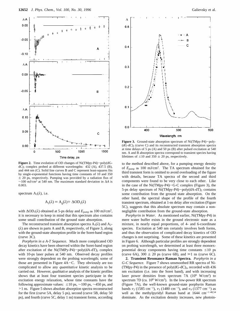
1. Transient Absorption and Kinetics. Porphyrin in a G-CSequence.Excitation of the Ni(TMpy-P4)-poly(dG-dC)2 com-plex with 10-ps laser pulses at 540 nm resulted in fully reversibleoptical density (OD) changes observable in the 390-470 nmspectral region corresponding to the porphyrin Soret band.Typical kinetics of OD changes are biphasic and can be wellfitted by double-exponential functions, with time constants ofe10 ps and 350( 20 ps. An example of such a typical two-component kinetics is shown in Figure 2A, recorded atλprobe)432 nm. According to the properties of isosbestic points,selected wavelengths can also be found where only the first orsecond decay component are separately detected (curves 2B and2C recorded at 437.5 and 444 nm, respectively).To reconstruct the absolute absorption spectra of both short-
lived and long-lived transient species from their experimentaldifference spectra (not shown), we estimated the percentage ofprobed molecules which is pumped from the ground to thecorresponding transient states. This has been done by measuringthe dependence of the observed OD changes for both compo-nents (i.e. at different time delays) as a function of the pumpingenergyEpump.(i) For the long-lived transient species (OD changes measured
at 50-ps delay) a saturation curve of OD changes as a functionof Epumphas been obtained, which shows a plateau for pumpingenergy densitiesEpump g 50 mJ/cm2; about 100% of themolecules are thus pumped into the second (long-lived) transientstate for pumping densities greater than this threshold. Sincewe routinely usedEpump= 100 mJ/cm2, we can simply add themeasured OD changes for the second transient species, i.e.∆OD2(λ), to the ground state absorption spectrum Ag(λ) toobtain the absolute absorption spectrum A2(λ) of the second(long-lived) transient species.(ii) As for the first (short-lived) transient species, only
near-saturation conditions have been achieved for thisspecies (OD changes measured at 5-ps delay) even at pumpingdensityEpump= 200 mJ/cm2. Consequently, since we have useda similar expression to reconstruct its absolute absorption
Excited States of Water-Soluble Metal Porphyrins J. Phys. Chem., Vol. 100, No. 30, 199612651
+ +
+ +
spectrum A1(λ), i.e.
with ∆OD1(λ) obtained at 5-ps delay andEpump= 100 mJ/cm2,it is necessary to keep in mind that this spectrum also containssome small contribution of the ground state absorption.The reconstructed transient absorption spectra A1(λ) and A2-
(λ) are shown in parts A and B, respectively, of Figure 3, alongwith the ground-state absorption profile in the Soret-band region(curve 3C).Porphyrin in a A-T Sequence.Much more complicated OD
decay kinetics have been observed within the Soret-band regionafter excitation of the Ni(TMPy-P4)-poly(dA-dT)2 complexwith 10-ps laser pulses at 540 nm. Observed decay profileswere strongly dependent on the probing wavelength; some ofthose are presented in Figure 4A-C. They obviously are toocomplicated to allow anyquantitatiVe kinetic analysis to becarried out. However,qualitatiVeanalysis of the kinetic profilesshows that at least four transient species participate in theexcitation energy relaxation, whose time constants have thefollowing approximate values:e10 ps,∼100 ps,∼450 ps, and>1 ns. Figure 5 shows absolute absorption spectra reconstructedfor the first (curve 5A, delay 5 ps), second (curve 5B, delay 50ps), and fourth (curve 5C, delay 1 ns) transient forms, according
to the method described above, for a pumping energy densityof Epump = 100 mJ/cm2. The TA spectrum obtained for thethird transient form is omitted to avoid overloading of the figurewith details, because TA spectra of the second and thirdcomponents were found to be very close to each other. Likein the case of the Ni(TMpy-P4)-G-C complex (Figure 3), the5-ps delay spectrum of Ni(TMpy-P4)-poly(dA-dT)2 containssome contribution from the ground state absorption. On theother hand, the spectral shape of the profile of the fourthtransient spectrum, obtained at 1-ns delay after excitation (Figure5C), suggests that this absolute spectrum may contain a non-negligible contribution from the ground-state absorption.Porphyrin in Water.As mentioned earlier, Ni(TMpy-P4) in
mere water buffer exists in the ground electronic state as amixture, in nearly equal proportions, of 4- and 6-coordinatespecies. Excitation at 540 nm certainly involves both forms,and thus the observation of complicated decay kinetics of ODchanges is not surprising. Some of these kinetics are presentedin Figure 6. Although particular profiles are strongly dependenton probing wavelength, we determined at least three monoex-ponential decay components having time constantse10 ps(curve 6A), 300( 20 ps (curve 6B), and.1 ns (curve 6C).2. Transient Resonance Raman Spectra.Porphyrin in a
G-C Sequence.Figure 7 shows unsmoothed RR spectra of Ni-(TMpy-P4) in the presence of poly(dG-dC)2, recorded with 436nm excitation (i.e. into the Soret band), and with increasinglaser power densities from spectrum 7A (106 W/cm2) tospectrum 7D (ca. 108 W/cm2). In the low-power RR spectrum(Figure 7A), the well-known ground-state porphyrin Ramanbandsν2 (1585 cm-1), ν3 (1480 cm-1), andν4 (1377 cm-1) aswell as the methylpyridyl Raman band at 1644 cm-1 48,54
dominate. As the excitation density increases, new photoin-
Figure 2. Time evolution of OD changes of Ni(TMpy-P4)-poly(dG-dC)2 complex probed at different wavelengths: 432 (A), 437.5 (B),and 444 nm (C). Solid line curves B and C represent least-squares fitsby single-exponential functions having time constants of 10 and 350( 20 ps, respectively. Pumping was provided by a radiation flux of∼100 mJ/cm2 at 540 nm. The maximum standard deviation in∆A is0.003.
A1(λ) ) Ag(λ)+ ∆OD1(λ)
Figure 3. Ground-state absorption spectrum of Ni(TMpy-P4)-poly-(dG-dC)2 (curve C) and its reconstructed transient absorption spectraat time delays of 5 ps (A) and 50 ps (B) after pulsed excitation at 540nm. A and B absorption spectra correspond to transient species havinglifetimes ofe10 and 350( 20 ps, respectively.
12652 J. Phys. Chem., Vol. 100, No. 30, 1996 Galievsky et al.
+ +
+ +

duced transient Raman bands also increase, appearing as low-frequency satellites of the ground-state bands which in turnbecome much weaker, i.e. 1585T 1563 cm-1, 1480T 1459cm-1, and 1376T 1363 cm-1, while the methylpyridyl Ramanband remains practically unaltered.Porphyrin in a A-T Sequence.Transient RR spectra of Ni-
(TMpy-P4) bound to poly(dA-dT)2 (Figure 8), recorded underthe same experimental conditions as for Figure 7, reveal verysimilar photoinduced changes, with pairs of porphyrin markerbands originating from both the ground and photoinducedtransient excited states. However, a more careful examinationallows slight differences between spectra of Ni porphyrin-poly-(dG-dC)2 and-poly(dA-dT)2 complexes to be found, the mostpronounced one being the presence of a 1468 cm-1 secondsatellite of theν3 band in the latter case (see Discussion fordetails).Transient RR spectra of Ni(TMpy-P4)-poly(dG-dC)2 com-
plex recorded with excitations at 427 and 416 nm and of Ni-(TMpy-P4)-poly(dA-dT)2 complex recorded with 416 nmexcitation (spectra not shown) reveal the same peculiarities asthose of Figures 7 and 8. It should be noted that in one of ourprevious papers,23 apart from a noticeable intensity decrease ofthe ground-state lines corresponding to some ground statedepletion at “high” power density, any photoinduced spectralchanges in RR spectra of Ni(TMpy-P4) bound to A-T sequencewere reported. However, our present results unambiguouslyprove the existence of a new transient species in this complexcreated at sufficiently high excitation density: Raman spectra
were recorded independently several times at three differentlaboratories, and in all cases the results were as presented inFigures 7 and 8. Presumably, the failure of previous attemptsto observe such photoinduced changes was due to unconvenientexcitation power density.To test the dynamics of the complexes in intense nanosecond
laser field, a special experiment was carried out in whichporphyrin Raman marker lineν2 at ca. 1585 cm-1, its excited-state counterpartν2* at ca. 1561 cm-1, and methylpyridyl lineat ca. 1644 cm-1 were normalized on the water band at ca. 3450cm-1 in RR spectra recorded at low (Figure 9A,C) and high(Figure 9B,D) excitation densities. For this purpose only twospectral ranges of interest, i.e. 1500-1700 and 2500-4000cm-1, were scanned in a standard acquisition mode with small(1 cm-1) and large (5 cm-1) steps, respectively. It is clearlyseen in Figure 9A-D that, in increasing excitation power,ground-state Raman bandν2 is transformed into its excited-state low frequency counterpartν2*, both lines having compa-rable intensities, eventually. This supports the assumption thatalmost full conversion of Ni(TMpy-P4) molecules occurs, somekind of “switching” between the ground and transient excitedstates reflected in theν2 andν2* lines, respectively, and that noother transient state measurably contributes to this process. Notealso that both pairs of RR spectra, i.e. 9A and 9C on one hand,9B and 9D on the other, have been recorded under the sameintensity levels of excitation.Porphyrin in Water.Since TA experiments have proven the
presence of a long-lived (.1 ns) relaxation component in theNi(TMpy-P4)-poly(dA-dT)2 complex, which supports theexistence of some binding/release photoprocesses, comparativeRR studies have been performed for free Ni(TMpy-P4) in amere phosphate buffer, where it exists in its ground electronicstate as a nearly equal mixture of 4- and 6-coordinate species.Figure 9 also shows RR spectra of this free Ni(TMpy-P4) within
Figure 4. Time evolution of OD changes of Ni(TMpy-P4)-poly(dA-dT)2 complex probed at different wavelengths: 423 (A), 426 (B), and446 nm (C). Solid line curve C represents a least-squares fit to a single-exponential function with 450( 30 ps time constant.
Figure 5. Ground-state absorption spectrum of Ni(TMpy-P4)-poly-(dA-dT)2 (curve D) and its reconstructed transient absorption spectraat time delays of 5 ps (A), 50 ps (B), and 1000 ps (C) after pulsedexcitation at 540 nm.
Excited States of Water-Soluble Metal Porphyrins J. Phys. Chem., Vol. 100, No. 30, 199612653
+ +
+ +

the ν2 line region, recorded at 418 nm excitation, i.e. close tothe absorption maximum of the 4-coordinate species, and at lowand high excitation powers (Figure 9E,F, respectively). On theother hand, Figure 10 shows the RR spectra within theν4 lineregion, obtained with excitation at 436 nm, i.e. close to theabsorption maximum of the 6-coordinate form of free Ni(TMpy-P4). Again, all experimental conditions (geometry, focusingconditions, and excitation density) were maintained the samefor the pairs of spectra of Figure 10A and 10C (low density)and 10B and 10D (high density), respectively.Summarized frequency data of Ni(TMpy-P4) Raman marker
lines in different molecular environments, recorded with excita-tions at 416, 418, 427, and 436 nm, are presented in Table 1.
Discussion
1. Ni(TMpy-P4) in Poly(dG-dC)2. Ni(TMpy-P4) is knownto be intercalated between base pairs in poly(dG-dC)2, as a4-coordinate species.47-49 Double-exponential kinetics of ab-sorption changes (Figure 2) observed for this complex can bereliably interpreted in terms of formation (first step of decay,τe10 ps) and decay (second step of decay,τ ) 350( 20 ps) ofthe metal-centered3B1g excited (d,d) state, according to thegeneral scheme of excitation relaxation for diamagnetic 4-co-ordinate Ni porphyrins19,20,30-33 (see Figure 1A). Since thistransient species is the only one prominent long-lived photo-induced species detected in TA studies, we can also reliably
link it to the transient Raman bandsνi* arising in RR spectraat high excitation power (Figure 7, Table 1).Indeed, the (d,d) nature of this transient species is confirmed
by both TA and RR data. First, its absorption profile (Figure3B) is typical for the3B1g excited state of 4-coordinate Niporphyrins,30-34 which exhibits a clear red shift from thecorresponding ground-state absorption profile, in keeping ap-proximately the same shape. Second, the lifetime of the long-lived transient species is also typical for the3B1g excited (d,d)state of 4-coordinate Ni porphyrins. At last, transient RR spectrarecorded at high excitation power (Figure 7) contain prominentadditional Raman linesν2*, ν3*, and ν4* pairing their ground-state counterparts with a downward frequency shift of 12-22cm-1. Such peculiarities in RR spectra of Ni(TMpy-P4)-poly-(dG-dC)2 complex are consistent with those observed in RRspectra of the excited (d,d) state forâ-alkyl substituted Niporphyrins which were comprehensively investigated in the lastdecade.21,36-41 However, although RR data on photoinducedligation-state changes have been published for NiTPP innitrogen-containing organic bases,55-58 we could not find in theliterature any transient RR spectrum from the excited (d,d) stateof 4-coordinate meso-substituted Ni tetraarylporphyrins.Using the well-known correlation between the frequencies
of the Raman marker bands and the porphyrin core size of TPPmetallocomplexes,59 we estimated the value of the macrocycle
Figure 6. Time evolution of OD changes for free Ni(TMpy-P4) in aphosphate buffer observed at probing wavelengths 480 (A), 430 (B),and 443 nm (C). Solid line curves A and B represent least-squares fitsto single-exponential functions having time constants of 10 and 300(20 ps, respectively.
Figure 7. Transient RR spectra of Ni(TMpy-P4) in poly(dG-dC)2,recorded with excitation at 436 nm. Incident power density of laserflux successively increases from spectrum A (Iex ≈ 106 W/cm2) tospectrum D (Iex ≈ 108 W/cm2).
12654 J. Phys. Chem., Vol. 100, No. 30, 1996 Galievsky et al.
+ +
+ +

diameter in the excited (d,d) state. The center-to-pyrroledistance was found to be 1.98 Å (on the basis of theν2* modefrequency), 1.99 Å (ν3*), and 2.00 Å (ν4*), which gives anaverage value of 1.99 Å. However, this value should only beconsidered as approximative since, if we apply the sameprocedure to the ground-state wavenumber valuesνi, the corediameter would be estimated as 1.91 Å (ν2), 1.93 Å (ν3), and1.90 Å (ν4), while the core size real value of Ni(TMpy-P4) isknown to be 1.955 Å from crystallographic data.60 In any case,the wavenumbers of the downward shifted Raman marker bandsreveal quite large porphyrin core expansion of 0.06-0.1 Å inthe excited (d,d) state of the porphyrin when intercalated in poly-(dG-dC)2, this being necessary to accommodate the increasedelectron density in the dx2-y2 metal orbital.Concerning the nature of the first transient species, generated
within a femtosecond time scale after photoexcitation, our dataare not sufficient to state a definite conclusion. (Note also thatwe cannot observe the femtosecond kinetics of creation eitherbecause of insufficient time resolution, and its decay time isestimated to bee10 ps.) Owing to the spectral similarities ofabsorption bands from the ground and both transient states(Figure 3), the first transient species may be associated withthe1B1g excited (d,d) state. This assignment is quite plausiblesince it allows the ultrafast radiationless transition1Q f 1B1g
from the (π,π*) to the (d,d) electronic state subsystems to beexplained in a consistent way in terms of an allowed transitionbetween excited states of same multiplicity. However, on the
basis of our experimental data we cannot rule out the existenceof other possible physical processes, namely processes ofvibrational heating/cooling,34,35,40conformational changes,39 andformation/decay of T1 state, as already stated in the Introduction.The absence of any long-lived component having decay time
.1 ns in the relaxation kinetics allows one to assume thatphotoinitiated processes of axial ligand binding/release (forma-tion and decay of3B1g(L)2 state in Figure 1A) do not occur inNi(TMpy-P4)-poly(dG-dC)2 complex, so that no transient 5-or 6-coordinate Ni porphyrin species is generated after photo-excitation. This implies that, during the excited-state lifetime,intercalated Ni(TMpy-P4) is not accessible to water moleculeswhich could in principle be possible extra axial ligands, as itcan occur for free Ni(TMpy-P4) in a water buffer.2. Ni(TMpy-P4) in Poly(dA-dT) 2. The photoinduced
behavior of Ni(TMpy-P4)-poly(dA-dT)2 complex is much morecomplicated than that of the previous complex. Complicatedrelaxation kinetics observed in TA, in which at least 4 decaycomponents can be separated (Figure 4), supports the existence
Figure 8. Same as in Figure 7 but for the Ni(TMpy-P4)-poly(dA-dT)2 complex.
Figure 9. RR spectra of Ni(TMpy-P4) in the region of theν2 line,normalized on the water band at ca. 3430 cm-1. Spectra A and Bconcerns the complex with poly(dA-dT)2, (A, low Iex; B, high Iex; λex) 427 nm), spectra C and D concerns the complex with poly(dG-dC)2
(C, low Iex; D, high Iex; λex ) 427 nm), and spectra E and F concernsthe free molecule in a phosphate buffer (E, lowIex; F, high Iex; λex )418 nm).
Excited States of Water-Soluble Metal Porphyrins J. Phys. Chem., Vol. 100, No. 30, 199612655
+ +
+ +

of more than one type of localization for Ni(TMpy-P4) in A-Tsequences, as suggested earlier on the basis of unusual CD andRR features of this complex.47-52 Indeed, literature data29-33
indicate that 4-coordinate Ni porphyrins in a noncomplexingenvironment exhibit two-phase excited-state decay kineticshaving considerably different time constants, i.e.e10 ps and afew hundreds of picoseconds. Let us remember that the samekinetic picture was observed for the Ni(TMpy-P4)-poly(dG-dC)2 complex discussed above, where only one type ofporphyrin localization (intercalation) was unambiguously es-tablished. On the other hand, the existence of at least two typesof Ni(TMpy-P4) binding modes to poly(dA-dT)2 could seriouslycomplicate the OD decay kinetics, if these two types of Ni-(TMpy-P4) complexes have different lifetimes for the transientstates populated during excitation relaxation. This suggestionis quite plausible since the lifetime of the3B1g state of Niporphyrins in organic solvents is solvent- (i.e. environment-)dependent.61 In this case, even simple summation of two pairsof different decay kinetics may result in a very complicatedwavelength-dependent kinetic picture, and this is indeed the casefor Ni(TMpy-P4)-poly(dA-dT)2 complexes (Figure 4).
Long-lived components (τ .1 ns) in the relaxation kineticsfor this complex (long-decay “tails” of curves in Figure 4A,B)are well-known photophysical markers of the existence of axialligand binding/release photoprocesses30-33 (formation and decayof 3B1g(L)2 state in Figure 1A). This implies that photoexcitedNi(TMpy-P4), when complexed with A-T, is at least partiallyaccessible for other ligand molecules and that some amount ofnonequilibrated and axially ligated 5- or 6-coordinate Ni(TMpy-P4) species is photogenerated. However, as already mentionedin the Results, the shape of the absorption profile originatingfrom the long-lived fourth transient species (Figure 5C) alsocontains a considerable contribution from the ground stateabsorption; this indicates that the yield of formation of an axiallyligated species is low.Most Parts of A-T Interacting Porphyrins Remain 4-Coor-
dinate. RR studies of Ni(TMpy-P4)-poly(dA-dT)2 complexfully support the conclusions derived from TA data. As alreadymentioned in the Results, RR spectra of Ni(TMpy-P4) com-plexed with G-C (Figure 7) and A-T sequences (Figure 8)exhibit very similar power-dependent features. In both casesprominent counterparts of theν2, ν3, andν4 porphyrin ground-state marker lines arise at high excitation power, the wavenum-bers of the excited-stateνi* lines being very close for bothpolynucleotides (Table 1). The dynamic behavior of bothsystems in a saturated nanosecond laser field is also quite similar(Figure 9). All of this supports the major part of Ni(TMpy-P4) complexed with poly(dA-dT)2 being 4-coordinated, theintramolecular3B1g excited (d,d) state playing a dominant rolein the relaxation processes, like in the case of the G-C complex.However, a more careful examination shows that the RR spectraof the complex with A-T contains additional features to thoseof the complex with G-C, the most evident being a secondsatellite for theν3 line at 1468 cm-1 in the former (Figure 8)which is absent in the latter (Figure 7). In addition, in the high-power spectrum of the A-T complex (Figure 8D), bothν2-ν2*andν4-ν4* pairs of lines are less resolved than in the spectrumof the G-C complex (Figure 7D), although experimentalconditions were the same. All of this can reasonably beexplained by the occurrence of some additional (presumablyligated) species contributing to the spectrum of Ni(TMpy-P4)-poly(dA-dT)2.Comparative RR study of Ni(TMpy-P4) complexed with
polynucleotides and free in a phosphate buffer permitted spectraprovided by the3B1g excited state of the 4-coordinate porphyrinand by the3B1g(L)2 ground state of the 6-coordinate complexto be reliably distinguished. Indeed, theν4 porphyrin mode,known as an oxidation state andπ-electron density marker line,59
is not only sensitive to the porphyrin core expansion whichoccurs to accommodate the d-electron promotion from the dz2
to the dx2-y2 orbitals but also very sensitive to the donation ofelectron density from axial ligands to the central metal ion and,through this, to the interaction between the metal dp and theporphyrin eg* orbitals. This can account for the 19-20 cm-1
frequency shift between theν4 mode of 4-coordinate species(electronic state1Α1g, Figure 7A, Table 1) and itsν4′ counterpartof the 6-coordinate species in water (electronic state3B1g(L)2,Figure 10C, Table 1, see also ref 49), where both core expansionand electron density donation take place, in comparison to the13-15 cm-1 shift observed in the v4-ν4* pair for the4-coordinate species (only core expansion, Figure 7, Table 1).Taking this into account, the bandshape of theν4* mode (Figure10A,B) reveals that, for Ni(TMpy-P4) in A-T sequence, notransient 6-coordinate species is photogenerated because thereis no evidence of any shoulder at the position of the 6-coordinateline at ca.1357 cm-1.
Figure 10. Comparative RR spectra of Ni(TMpy-P4) in poly(dA-dT)2
(A, “low” Iex≈ 106 W/cm2; B, “high” Iex≈ 2 × 10⁷ W/cm2) and in aphosphate buffer (C and D, excitation intensities were equal to thoseof A and B, respectively) in the region of theν4 line (excitationwavelength, 436 nm).
12656 J. Phys. Chem., Vol. 100, No. 30, 1996 Galievsky et al.
+ +
+ +

Minor Part of A-T Bound Porphyrins Participates in Pho-toinduced Binding/Release Processes.However, since a pho-toinduced ligation process was reliably detected in TA mea-surements, we now assume that a 5-coordinate species with oneaxial ligand attached to the central Ni ion can account for thewhole set of data obtained for Ni(TMpy-P4)-poly(dA-dT)2complex. Indeed, for nonequilibrated 5-coordinate species, thedownward frequency shifts of the porphyrin marker bands areexpected to be much less pronounced than in the case of6-coordinate species and, therefore, these bands can be partiallyhindered by theνi* bands from the excited (d,d) state of4-coordinate species.Power-induced transformations of Raman spectra in a satu-
rated nanosecond laser field support the assumption that theyield of formation of axially ligated Ni(TMpy-P4) is low.Indeed, transient RR spectra of both complexes with G-C(Figure 9D) and A-T sequences (Figure 9B) were recorded atapproximately the same excitation power. The relative intensi-ties of theν2* Raman lines at ca. 1563 cm-1, normalized onthe water band, do not significantly differ for both complexes(note that the difference in the resonance enhancement of Ramanbands has to contribute to this intensity difference), whereasthe lifetimes of the most long-lived transient species are verydifferent: 350 ps for Ni(TMpy-P4)-poly(dG-dC)2 and.1 nsfor Ni(TMpy-P4)-poly(dA-dT)2 complexes. This can beconsistently explained only if we assume that the contributionfrom the axially ligated species, with nanosecond lifetime, tothe spectrum presented in Figure 9B is small.At this step of the discussion, partial conclusions can be
settled: the major part of Ni porphyrin molecules exists as a4-coordinate species, having the “normal” route of excitationrelaxation via the long-lived (hundreds of ps)3B1g excited state.On the other hand, a minor part of the molecules participatesin photoinduced binding/release processes, thus involving anonstationary 5-coordinate Ni porphyrin species having alifetime.1 ns. Let us consider now in more detail the possiblegeometrical structures which can be proposed on the basis ofthese photophysical data.Geometrical Considerations.Three types of Ni(TMpy-P4)
localization in A-T sequences can be considered in the case ofa dominant 4-coordinate porphyrin species inaccessible tosurrounding water molecules: (i) intercalation, (ii) outsidestacked aggregation, and (iii) edge-on binding in the minorgroove of the double helix.(i) Although intercalation is fully consistent with our pho-
tophysical data, it can hardly be proposed (but not completelyruled out) as a major type of localization after consideration ofstationary absorption spectra. Intercalated Ni(TMpy-P4) com-plex (for instance in G-C sequences) is characterized by a largehypochromicity (%H) 46) and a substantial red shift (∆λ )14 nm) of the Soret band (Figure 3C49). However, this is notthe case here (%H) 20,∆λ ) 6 nm, Figure 5D49), and thereis no peak or shoulder at ca. 435 nm in the absorption spectrumof the A-T complex, which may support intercalation.(ii) Outside stacked aggregation along the polynucleotide is
also very unlikely as a major type of localization, since theporphyrin concentration is low enough in our experiments torule out this possibility.
(iii) Consequently the edge-on binding of Ni(TMpy-P4) inthe minor groove of the A-T sequence, where the porphyrinprotrudes into the polynucleotide interior far enough to bescreened from the surrounding water molecules, is consistentwith all experimental data shown either in the present work orin the literature for this complex.Concerning the minor part of Ni(TMpy-P4) molecules
participating in photoinduced ligand binding/release processes,it is plausible to assume that Ni(TMpy-P4) is located in a face-on manner in the major groove of poly(dA-dT)2 and can adoptone water molecule as an extra axial ligand, thus involving a5-coordinate species. Coordination of Ni porphyrins was thesubject of numerous studies by TA,29-35 RR,55-58,62 and CDtechniques.63 It was found that free Ni porphyrin can exist insolution only as a 4- or 6-coordinate species. However, whenthe environment causes geometrical restrictions, as it occurs inNi-reconstituted hemoglobin and myoglobin, the central Ni ioncan attach only one ligand molecule, thus yielding a 5-coordinatespecies.62,63 Indeed, the same situation would occur for Ni-(TMpy-P4) externally bound to poly(dA-dT)2 in the majorgroove: a water molecule can be ligated to the central Ni iononly at one side, since the other is screened by the polynucle-otide. It should also be noted that a carbonyl group of thyminemay be considered as a possible candidate for axial liganding,by analogy with the case of Cu(TMpy-P4)-poly(dA-dT)2exciplex which was comprehensively studied earlier.23-28
However, the affinity for water is much higher for Ni than forCu porphyrins, as it is clearly seen in stationary Raman spectraof free metal complexes in buffer solution: Ni(TMpy-P4) existsas a nearly equal mixture of 4- and 6-coordinate species whileCu(TMpy-P4) is strictly 4-coordinate.48,49 Moreover, interactionbetween excited Ni(TMpy-P4) and CO group of thymine impliesgeometrical changes for both metalloporphyrin and polynucle-otide,26 while interaction with water does not because there arealways a lot of mobile “small” water molecules nearby theporphyrin Ni ion. Therefore, although the affinity of Ni(TMpy-P4) for CO group of thymine is not known, it is more likelythat water molecules are involved as axial ligands in the5-coordinate Ni(TMpy-P4)-L complex.Another question can arise: why cannot a water molecule
coordinate to Ni(TMpy-P4) in its electronic ground state if,according to the proposed type of localization, Ni ion ispermanently accessible to water from one side? We speculatethat fixation of Ni(TMpy-P4) by the four positively chargedmethylpyridyl groups to the double helix decreases its affinityfor water in the electronic ground state, so that the building ofa stable 5-coordinate complex is energetically unfavorable.Indeed, the 5-coordinate structure seems to be domed to someextent, and extra energy is required to keep such a structurestationary. In addition, photoexcited Ni(TMpy-P4) is supposedto have an increased affinity for axial ligands and thus can moreeasily attach a water molecule, producing a nonstationary5-coordinate complex.3. Ni(TMpy-P4) in a Phosphate Buffer Solution.At last
we briefly discuss our results concerning free Ni(TMpy-P4) ina phosphate buffer. Complicated TA kinetics obtained for thiscomplex (Figure 6) certainly has its origin in a simultaneousoccurrence of the following two opposite photoprocesses: (a)
TABLE 1: Frequencies (cm-1) of Raman Marker Bands in the Ground Electronic State (νi), Their 4-Coordinate Excited-State(Rows 1, 2) or 6-Coordinate Ground-State (row 3) Counterparts (νi′) for Ni(TMpy-P4) in Various Molecular Environments
environment ν2 ν2* (ν2′) ν3 ν3* (ν3′) ν4 ν4* (ν4′)poly(dG-dC)2 1585-1586 1563 1480-1481 1459-1460 1376-1377 1363-1364poly(dA-dT)2 1582-1583 1560-1563 1477-1478 1455-1456a 1377-1378 1362-1364water buffer 1580-1584 (1560-1561) 1479b (1456b) 1377-1378 (1356-1358)a There exists a second satellite at 1468 cm-1. bData from ref 49.
Excited States of Water-Soluble Metal Porphyrins J. Phys. Chem., Vol. 100, No. 30, 199612657
+ +
+ +

photoattachement of surrounding water molecules to initially4-coordinate species, and (b) photodissociation of the extrali-gands from initially 6-coordinate species (see Figure 1). Onthe basis of these TA kinetics, absorption spectra have beenreconstructed at various delays after photoexcitation (not shown).However, those absorption spectra obtained at delays shorterthan ca. 1 ns are difficult to analyze since they containcontributions from excited- and ground-state transient species.At larger delays, e.g.>1 ns, all of the electronic excited statesfrom both 4- and 6-coordinate species have decayed becausetheir lifetimes are not longer than a few hundreds of picoseconds(refs 30-33 and this work). Therefore, OD changes remainingat 1 ns can essentially be assigned to nonequilibrated 4- and6-coordinate species resulting from photoexcitation. Experi-mental observations support this suggestion: the TA spectrumobtained at 1 ns delay (not shown) resembles that of the initialground state, but shows an increased contribution from the6-coordinate species (Soret band maximum at ca. 440 nm) and,correspondingly, a decreased contribution from the 4-coordinatespecies (Soret band maximum at ca. 420 nm). This off-balancemixed state then relaxes in a nanosecond time scale (at least)(see long-lived tails in Figure 6C), this being in agreement withliterature data obtained for various Ni porphyrins in organicsolvents.30-33
It is noteworthy that transient RR spectra of Ni(TMpy-P4)obtained in a water buffer at various excitation powers showfeatures different from those described for the complexes withboth polynucleotides. Figure 9 presents RR spectra of free Ni-(TMpy-P4) within theν2 line region, recorded at low and highexcitation powers (Figure 9E,F, respectively). Although theseunsmoothed spectra look noisy, power-induced changes can benoted, consisting in relative intensity redistribution between theRR lines of the initially 4- and 6-coordinate species. However,these photoinduced changes in RR spectra of free Ni(TMpy-P4) are much less pronounced than in the case of complexeswith polynucleotides (Figure 9A-D). It is also worthwhile tonote that the3B1g(L)2 6-coordinate line at ca. 1561 cm-1 isdominant in both low- and high-power spectra (Figure 9E,F,respectively), in spite of using 418 nm excitation close to theabsorption maximum of the1A1g 4-coordinate species. Herethe term “low-excitation power” means a power density of 5×10⁵ to 106 W/cm2; such an intensity is high enough to producea considerable population of the nonequilibrated3B1g(L)2 statehaving a lifetime.1 ns. This well explains the differencesbetween free Ni(TMpy-P4) RR spectra recorded by us at low-excitation power using 10-ns laser pulses and those availablein the literature obtained with cw laser excitation.48,49 Onexcitation at 436 nm, i.e. close to the absorption maximum ofthe 3B1g(L)2 6-coordinate species, no observable populationincrease of1A1g 4-coordinate species has been detected inincreasing the power density (Figure 10C, D).Consequently, although the detailed interpretation of the
intermediate steps of excitation relaxation for Ni(TMpy-P4) ina phosphate buffer remains difficult at the moment, our datasupport the photogeneration of the most long-lived nonequili-brated3B1g(L)2 state of the 6-coordinate species, which playsthe dominant role in the relaxation processes. Further studiesof free Ni(TMpy-P4) are now in progress and will be submittedfor future publication.
Conclusion
In the current spectroscopic studies, we have obtained thefollowing main results:(i) For Ni(TMpy-P4)-poly(dG-dC)2 complexes, a double-
exponential kinetics of photoexcitation relaxation was found,
with time constants ofe10 ps and 350( 20 ps. The long-lived transient species was assigned to the excited intramolecularmetal-centered (d,d) state (3B1g) of the 4-coordinate Ni porphyrinintercalated between G-C base pairs.(ii) For the Ni(TMpy-P4)-poly(dA-dT)2 complexes, four
relaxation components can be separated with time constants ofe10 ps,∼100 ps,∼450 ps, and.1 ns. This indicates differentNi(TMpy-P4) localizations within A-T sequence, each specieshaving its own double-exponential kinetics of TA decay andtransient RR spectra. Indeed, both TA and RR data show thatthe major part of Ni porphyrins exhibits photophysical behaviorcharacteristic for a 4-coordinate species, while a minor partparticipates in axial binding/release photoprocesses. Possibletypes of binding of Ni(TMpy-P4) to poly(dA-dT)2 are discussed.(iii) From a comparison between transient RR spectra of free
Ni(TMpy-P4) (in a mere phosphate buffer) and complexed withpoly(dA-dT)2, it can be concluded that no 6-coordinate3B1g-(L)2 species is photogenerated in the Ni(TMpy-P4)-poly(dA-dT)2 complex. Therefore, possible axial coordination of onlyone extra ligand molecule (from the surrounding water buffer)to the porphyrin central Ni ion is proposed.(iv) in a phosphate buffer, where Ni(TMpy-P4) exists as a
ground state equilibrium of both 4- and 6-coordinate species,photoinduced transformations in RR spectra were found to bedifferent from those observed in polynucleotide complexes.Relaxation kinetics provides very fast (e10 ps), intermediate(hundreds of ps) and slow (.1ns) components. Although aprecise interpretation of the intermediate steps of relaxation isdifficult at the moment, both TA and RR data support thehypothesis that the long-lived nonequilibrated3B1g(L)2 state ofthe 6-coordinate Ni(TMpy-P4) plays the dominant role in therelaxation processes.
Acknowledgment. The authors would like to thank Dr. S.M. Bachilo for assistance in software used for picosecondspectrometer control and in simulating kinetic absorption data,and Mr. A. Voroshilov for technical assistance in Ramanmeasurements. This work has been supported in part by theFundamental Research Foundation of the Republic of Belarus(Grant F-157). For performing this study, V.S.C. and S.G.K.have been personally partly supported by the French Ministryof Foreign Affairs, through the French Embassy in Minsk,Republic of Belarus. Part if this work was supported by a grantfrom the Netherlands Organization for Scientific Research(Grant NB 70-256) and the “Stichting UniversiteitsfondsTwente” to S.G.K. for which S.G.K. and C.O. express theirgratitude.
References and Notes
(1) Fiel, R. J.; Howard, J. C.; Mark, E. N.; Datta-Gupta, N.Nucl. AcidsRes.1979, 6, 3093.
(2) Fiel, R.J. Biomol. Struct. Dyn.1989, 6, 1259.(3) Pasternack, R. F.; Gibbs, E. J.Metal-DNA Chemistry; Tullius, T.
D., Ed.; ACS Symposium Series No. 402; American Chemical Society:Washington, DC, 1989; p 59.
(4) Raner, G.; Goodisman, J.; Dabrowiak, J. C.Metal-DNA Chemistry;Tullius, T. D., Ed.; ACS Symposium Series No. 402; American ChemicalSociety: Washington, DC, 1989; p 74.
(5) Marzilli, L. G. New J. Chem.1990, 14, 409.(6) Sari, M. A.; Battoni, J. P.; Dupre´, D.; Mansuy, D.; Le Pecq, J. B.
Biochemistry1990, 29, 4205.(7) Groves, J. T.; Farrell, T. P.J. Am. Chem. Soc.1989, 11, 4998.(8) Slama-Schwok, A.; Lehn, J.-M.Biochemistry1990, 29, 7895.(9) Waring, M. J.Annu. ReV. Biochem.1981, 50, 159.(10) Wilson, W. D.; Jones, R. L.Intercalation Chemistry; Whittingham,
M. S., Jacobson, A. J., Eds.; Academic Press: New York, 1982; p 445.(11) Musser, D. A.; Datta-Gupta, N.; Fiel, R. J.Biochem. Biophys. Res.
Commun.1980, 97, 918.(12) Fiel, R. J.; Datta-Gupta, N.; Mark, E. H.; Howard, J. C.Cancer
Res.1981, 41, 3543.
12658 J. Phys. Chem., Vol. 100, No. 30, 1996 Galievsky et al.
+ +
+ +
(13) Praseuth, D.; Gaudemer, A.; Verlhac, J.-B.; Kraljic, I.; Sissoeff,I.; Guille, E.Photochem. Photobiol.1986, 44, 717.
(14) Villanueva, A.; Hazen, M. J.; Stockert, J.-C.Experientia1986, 42,1269.
(15) Dixon, D. W.; Marzilli, L. G.; Schinazi, R.Ann. N.Y. Acad.Sci.1990, 611, 511.
(16) Sessler, J. L.; Cyr, M. J.; Lynch, V.J. Am. Chem. Soc.1990, 112,2810.
(17) Mastruzzo, L.; Woisard, A.; Ma, D. D. F.; Rizzarzlli, E.; Favre,A.; Le Doan, T.Photochem. Photobiol.1994, 60, 316.
(18) Verlhac, J. B.; Gaudemer, A.; Kraljic, I.NouV. J. Chim.1984., 8,401.
(19) Holten, D.; Gouterman, M.Optical Properties and Structure ofTetrapyrroles; Blauer, G., Sund, H., Eds.; de Gruyter: Berlin, 1985; p 64.
(20) Dzhagarov, B. M.; Chirvony, V. S.; Gurinovich, G. P.LaserPicosecond Spectroscopy and Photochemistry Biomolecules;Letokhov, V.S., Ed.; Adam Hilger: Bristol, 1987; Chapter 3.
(21) Apanasevich, P. A.; Chirvony, V. S.; Kruglik, S. G.; Kvach, V.V.; Orlovich, V. A. Laser Applications in Life Sciences; Akhmanov, S. A.,Poroshina, M. Yu., Koroteev, N. I., Touletaev, B. N., Eds.; SPIE:Bellingham, WA, 1991; Proc. SPIE 1403, Part I, p 195.
(22) Kruglik, S. G.; Apanasevich, P. A.; Chirvony, V. S.; Kvach, V.V.; Orlovich, V. A. J. Phys. Chem.1995, 99, 2978.
(23) Turpin, P.-Y.; Chinsky, L.; Laigle, A.; Tsuboi, M.; Kincaid, J. R.;Nakamoto, K.Photochem. Photobiol.1990, 51, 519.
(24) Chinsky, L.; Turpin, P.-Y.; Al-Obaidi, A. H. R.; Bell, S.; Hester,R. E. J. Phys. Chem.1991, 95, 5754.
(25) Strahan, G. D.; Lu, D.; Tsuboi, M.; Nakamoto, K.J. Phys. Chem.1992, 96, 6450.
(26) Mojzes, P.; Chinsky, L.; Turpin, P.-Y.J. Phys. Chem.1993, 97,4841.
(27) Turpin, P.-Y.; Chinsky, L.; Mojzes, P.Laser Spectroscopy ofBiomolecules; Korppi-Tommola, J. E. I., Ed.; SPIE: Bellingham, WA, 1993;Proc. SPIE 1921, p 361.
(28) Kruglik, S. G.; Galievsky, V. A.; Chirvony, V. S.; Apanasevich,P. A.; Ermolenkov, V. V.; Orlovich, V. A.; Chinsky, L.; Turpin, P.-Y.J.Phys. Chem.1995, 99, 5732.
(29) Kobayashi, T.; Straub, K. D.; Rentzepis, P. M.Photochem.Photobiol.1979, 29, 925.
(30) Chirvony, V. S.; Dzhagarov, B. M.; Timinskii, Yu.V.; Gurinovich,G. P.Chem. Phys. Lett. 1980, 70, 79.
(31) Chirvony, V. S.; Dzhagarov, B. M.; Shul'ga, A. M.; Gurinovich,G. P.Dokl. Akad. Nauk SSSR1981, 259, 144.
(32) Kim, D.-H.; Kirmaier, C.; Holten, D.Chem. Phys.1983, 75, 305.(33) Kim, D.-H.; Holten, D.Chem. Phys. Lett.1983, 98, 584.(34) Rodriguez, J.; Holten, D.J. Chem. Phys.1989, 91, 3525.(35) Rodriguez, J.; Kirmaier, C.; Holten, D.J. Chem. Phys.1991, 94,
6020.(36) Findsen, E. W.; Shelnutt, J. A.; Ondrias, M. R.J. Phys. Chem.
1988, 92, 307.(37) Chikishev, A. Yu.; Kamalov, V. F.; Koroteev, N. I.; Kvach, V. V.;
Shkurinov, A. P.; Toleutaev, B. N.Chem. Phys. Lett.1988, 144, 90.
(38) Apanasevich, P. A.; Kvach, V. V.; Orlovich, V. A.J. RamanSpectrosc.1989, 20, 125.
(39) Courtney, S. H.; Jedju, T. M.; Friedman, J. M.; Alden, R. G.;Ondrias, M. R.Chem. Phys. Lett.1989, 164, 39.
(40) Sato, S.; Kitagawa, T.Appl. Phys.1994, B59, 415.(41) Kruglik, S. G.; Apanasevich, P. A.; Kvach, V. V.; Orlovich, V. A.
5th International Conference on Laser Applications in Life Sciences;Apanasevich, P. A., Koroteev, N. I., Kruglik, S. G., Zadkov, V. N., Eds.;SPIE: Bellingham, WA, Proc. SPIE, Vol. 2370, p 196.
(42) Ake, R. L.; Gouterman, M.Theor. Chim. Acta1970, 17, 408.(43) Tsvirko, M. P.; Solov’ev, K. N.; Sapunov, V. V.Optika I
Spektroskopija1974, 36, 335.(44) Antipas, A.; Gouterman, M.J. Am. Chem. Soc.1983, 105, 4896.(45) Aronowitz, Y. J.; Gouterman, M.J. Mol. Spectrosc.1977, 64, 267.(46) Pasternack, R. F.; Spiro, E. G.; Teach, M.J. Inorg. Nucl. Chem.
1974, 36, 599.(47) Pasternack, R. F.; Gibbs, E. J.; Villafranca, J. J.Biochemistry1983,
22, 2406.(48) Blom, N.; Odo, J.; Nakamoto, K.; Strommen, D. P.J. Phys. Chem.
1986, 90, 2847.(49) Schneider, J. H.; Odo, J.; Nakamoto, K.Nucl. Acids Res.1988,
16, 10323.(50) Butje, K.; Schneider, J. H.; Kim, J.-J. P.; Wang, Y.; Ikuta, S.;
Nakamoto, K.J. Inorg. Biochem.1989, 37, 119.(51) Carvlin, M. J.; Fiel, R. J.Nucl. Acids Res.1983, 11, 6121.(52) Ford, K.; Fox, K. R.; Neidle, S.; Waring, M. J.Nucl. Acids Res.
1987, 15, 2221.(53) Kruglik, S. G.; Otto, C.; Ermolenkov, V. V.; Orlovich, V. A.;
Galievsky, V. A.; Chirvony, V. S.; Turpin, P.-Y.Chem. Phys. Lett.,submitted.
(54) Li, X.-Y.; Czernuszewicz, R. S.; Kincaid, J. R.; Su, Y. O.; Spiro,T. G. J. Phys. Chem.1990, 94, 31.
(55) Kim, D.; Su, Y. O.; Spiro, T. G.Inorg. Chem.1986, 25, 3988.(56) Kim, D.; Spiro, T. G.J. Am. Chem. Soc.1986, 108, 2099.(57) Findsen E. W., Shelnutt J. A., Friedman J. M., Ondrias M. R.Chem.
Phys. Lett.1986, 126, 465.(58) Findsen E. W., Alston K., Shelnutt J. A., Ondrias M. R.J. Am.
Chem. Soc.1986, 108, 4009.(59) Spiro, T. G.; Li, X.-Y.Biological Applications of Raman Spec-
troscopy; Wiley: New York, 1988; Vol. III, Chapter I, pp 2-37.(60) Kirner, J. F.; Garofollow, J.; Scheidt, W. R.Inorg. Nucl. Chem.
Lett. 1975, 11, 107.(61) Chirvony, V. S. Unpublished results.(62) Shelnutt, J. A.; Alston, K.; Ho, J.-Y.; Yu, N.-T.; Yamamoto, T.;
Rifkind, J. M.Biochemistry1986, 25, 620.(63) Choi, S.; Phillips, J. A.; Ware, W., Jr.; Wittschieben, C.; Medforth,
C. J.; Smith, K. M.Inorg. Chem.1994, 33, 3873.
JP960604I
Excited States of Water-Soluble Metal Porphyrins J. Phys. Chem., Vol. 100, No. 30, 199612659
+ +
+ +