Excitation energy trapping and dissipation by Ni-substituted bacteriochlorophyll a in reconstituted...
12
Excitation Energy Trapping and Dissipation by Ni-Substituted Bacteriochlorophyll a in Reconstituted LH1 Complexes from Rhodospirillum rubrum Petar H. Lambrev, † Yuliya Miloslavina, † Ivo H. M. van Stokkum, ‡ Andreas D. Stahl, ‡ Maciej Michalik, § Anna Susz, § Jędrzej Tworzydlo, § Joanna Fiedor, ∥ Gabriella Huhn, ⊥ Marie-Louise Groot, ‡ Rienk van Grondelle, ‡ Győ ző Garab,* ,† and Leszek Fiedor § † Biological Research Centre, Hungarian Academy of Sciences, Temesva ́ ri krt. 62, 6726 Szeged, Hungary ‡ Institute for Lasers, Life and Biophotonics, Faculty of Sciences, VU University Amsterdam, De Boelelaan 1081, 1081 HV Amsterdam, The Netherlands § Faculty of Biochemistry, Biophysics and Biotechnology, Jagiellonian University, ul. Gronostajowa 7, 30-387 Krakó w, Poland ∥ Department of Medical Physics and Biophysics, Faculty of Physics and Applied Computer Science, AGH University of Science and Technology, Reymonta 19, 30-059 Krakó w, Poland ⊥ Faculty of Science, Eö tvö s Lora ́ nd University, Pa ́ zma ́ ny Pe ́ ter Sé ta ́ ny 1/A, 1117 Budapest, Hungary * S Supporting Information ABSTRACT: Bacteriochlorophyll a with Ni 2+ replacing the central Mg 2+ ion was used as an ultrafast excitation energy dissipation center in reconstituted bacterial LH1 complexes. B870, a carotenoid-less LH1 complex, and B880, an LH1 complex containing spheroidene, were obtained via reconstitu- tion from the subunits isolated from chromatophores of Rhodospirillum rubrum. Ni-substituted bacteriochlorophyll a added to the reconstitution mixture partially substituted the native pigment in both forms of LH1. The excited-state dynamics of the reconstituted LH1 complexes were probed by femtosecond pump−probe transient absorption spectroscopy in the visible and near-infrared spectral region. Spheroidene-binding B880 containing no excitation dissipation centers displayed complex dynamics in the time range of 0.1−10 ps, reflecting internal conversion and intersystem crossing in the carotenoid, exciton relaxation in BChl complement, and energy transfer from carotenoid to the latter. In B870, some aggregation-induced excitation energy quenching was present. The binding of Ni-BChl a to both B870 and B880 resulted in strong quenching of the excited states with main deexcitation lifetime of ca. 2 ps. The LH1 excited-state lifetime could be modeled with an intrinsic decay time constant in Ni-substituted bacteriochlorophyll a of 160 fs. The presence of carotenoid in LH1 did not influence the kinetics of energy trapping by Ni-BChl unless the carotenoid was directly excited, in which case the kinetics was limited by a slower carotenoid S 1 to bacteriochlorophyll energy transfer. ■ INTRODUCTION Several fundamental processes occur in the light-harvesting antennas of all photosynthetic systems, including absorption of photons, storage of the excitation energy, then transfer of excitation energy to the reaction centers, and energy losses via radiative or radiationless decays. Understanding the function of the photosynthetic apparatus cannot be complete without a thorough mechanistic treatment of these processes and their effects on the entire system. Light-harvesting antennas, on the one hand, are designed for minimal quantum losses in order to allow for efficient energy transfer. On the other hand, most photosynthetic organisms actively regulate and make use of the thermal dissipation in the antenna for protection against harmful excess radiation. 1,2 In light-harvesting pigment−protein complexes, the energy of the electronically excited pigments, chlorophylls or bacteriochlorophylls (BChls), is dissipated via internal conversion and intra- and intermolecular vibrational relaxation 3 involving vibrational modes of the pigment and its proteinous and lipidic environment. This creates vibrationally hot molecules, which can be intermediates in various secondary reactions affecting the structure and function of the antennas via the so-called hot-molecule 4 or thermo-optic 5 mechanism. In this work, we explore the use of purple bacterial LH1 complexes reconstituted with Ni-substituted BChl a (Ni- Special Issue: Rienk van Grondelle Festschrift Received: February 28, 2013 Revised: June 28, 2013 Published: July 9, 2013 Article pubs.acs.org/JPCB © 2013 American Chemical Society 11260 dx.doi.org/10.1021/jp4020977 | J. Phys. Chem. B 2013, 117, 11260−11271
-
Upload
independent -
Category
Documents
-
view
0 -
download
0
Transcript of Excitation energy trapping and dissipation by Ni-substituted bacteriochlorophyll a in reconstituted...

Excitation Energy Trapping and Dissipation by Ni-SubstitutedBacteriochlorophyll a in Reconstituted LH1 Complexes fromRhodospirillum rubrumPetar H. Lambrev,† Yuliya Miloslavina,† Ivo H. M. van Stokkum,‡ Andreas D. Stahl,‡ Maciej Michalik,§
Anna Susz,§ Jędrzej Tworzydło,§ Joanna Fiedor,∥ Gabriella Huhn,⊥ Marie-Louise Groot,‡
Rienk van Grondelle,‡ Gyozo Garab,*,† and Leszek Fiedor§
†Biological Research Centre, Hungarian Academy of Sciences, Temesvari krt. 62, 6726 Szeged, Hungary‡Institute for Lasers, Life and Biophotonics, Faculty of Sciences, VU University Amsterdam, De Boelelaan 1081, 1081 HVAmsterdam, The Netherlands§Faculty of Biochemistry, Biophysics and Biotechnology, Jagiellonian University, ul. Gronostajowa 7, 30-387 Krakow, Poland∥Department of Medical Physics and Biophysics, Faculty of Physics and Applied Computer Science, AGH University of Science andTechnology, Reymonta 19, 30-059 Krakow, Poland⊥Faculty of Science, Eotvos Lorand University, Pazmany Peter Setany 1/A, 1117 Budapest, Hungary
*S Supporting Information
ABSTRACT: Bacteriochlorophyll a with Ni2+ replacing thecentral Mg2+ ion was used as an ultrafast excitation energydissipation center in reconstituted bacterial LH1 complexes.B870, a carotenoid-less LH1 complex, and B880, an LH1complex containing spheroidene, were obtained via reconstitu-tion from the subunits isolated from chromatophores ofRhodospirillum rubrum. Ni-substituted bacteriochlorophyll aadded to the reconstitution mixture partially substituted thenative pigment in both forms of LH1. The excited-statedynamics of the reconstituted LH1 complexes were probed byfemtosecond pump−probe transient absorption spectroscopy inthe visible and near-infrared spectral region. Spheroidene-binding B880 containing no excitation dissipation centers displayedcomplex dynamics in the time range of 0.1−10 ps, reflecting internal conversion and intersystem crossing in the carotenoid,exciton relaxation in BChl complement, and energy transfer from carotenoid to the latter. In B870, some aggregation-inducedexcitation energy quenching was present. The binding of Ni-BChl a to both B870 and B880 resulted in strong quenching of theexcited states with main deexcitation lifetime of ca. 2 ps. The LH1 excited-state lifetime could be modeled with an intrinsic decaytime constant in Ni-substituted bacteriochlorophyll a of 160 fs. The presence of carotenoid in LH1 did not influence the kineticsof energy trapping by Ni-BChl unless the carotenoid was directly excited, in which case the kinetics was limited by a slowercarotenoid S1 to bacteriochlorophyll energy transfer.
■ INTRODUCTION
Several fundamental processes occur in the light-harvestingantennas of all photosynthetic systems, including absorption ofphotons, storage of the excitation energy, then transfer ofexcitation energy to the reaction centers, and energy losses viaradiative or radiationless decays. Understanding the function ofthe photosynthetic apparatus cannot be complete without athorough mechanistic treatment of these processes and theireffects on the entire system. Light-harvesting antennas, on theone hand, are designed for minimal quantum losses in order toallow for efficient energy transfer. On the other hand, mostphotosynthetic organisms actively regulate and make use of thethermal dissipation in the antenna for protection againstharmful excess radiation.1,2 In light-harvesting pigment−proteincomplexes, the energy of the electronically excited pigments,
chlorophylls or bacteriochlorophylls (BChls), is dissipated viainternal conversion and intra- and intermolecular vibrationalrelaxation3 involving vibrational modes of the pigment and itsproteinous and lipidic environment. This creates vibrationallyhot molecules, which can be intermediates in various secondaryreactions affecting the structure and function of the antennasvia the so-called hot-molecule4 or thermo-optic5 mechanism. Inthis work, we explore the use of purple bacterial LH1complexes reconstituted with Ni-substituted BChl a (Ni-
Special Issue: Rienk van Grondelle Festschrift
Received: February 28, 2013Revised: June 28, 2013Published: July 9, 2013
Article
pubs.acs.org/JPCB
© 2013 American Chemical Society 11260 dx.doi.org/10.1021/jp4020977 | J. Phys. Chem. B 2013, 117, 11260−11271

BChl) as a model system for investigating the thermaldissipation processes in photosynthetic antennas. The sub-stitution of the central Mg2+ ion in BChl a with Ni2+ leads to adramatic shortening of the pigment’s excited-state lifetime,from 3 ns to less than 100 fs6,7 without significantly altering itsground state properties. Because of the rapid excitationredistribution in bacterial antenna complexes (see below), aNi-BChl molecule bound to the complex can then serve as anultrafast excitation energy trap8,9 that focuses the dissipationprocess in both space and time, thus potentially facilitatingunique spectroscopic investigation. Importantly, as shown inour recent study,10 the excitation reaching the Ni-analogue ofBChl a is very promptly and with 100% efficiency convertedinto heat and dissipated to the surrounding, due to the peculiarbonding between Ni2+ ion in the central pocket of the pigment.LH1, the core light-harvesting antenna of purple photo-
synthetic bacteria,11−15 is a membrane-integral pigment−protein complex noncovalently binding BChl a or b andcarotenoids (Crts) and is characterized by a strong near-infrared absorption band (usually 870−890 nm in BChl a-containing species, corresponding to BChl Qy transition). LH1surrounds the reaction center (RC) and collects light energyeither directly or via energy transfer from the peripheralantenna LH2. The basic building module of the LH1 complexis the α,β-heterodimer consisting of two polypeptides, twoBChl a and a single Crt molecule.16 B870, the Crt-less form ofLH1, can be reversibly dissociated into and reassembled fromheterodimeric subunits B820, termed after the position of theirmain absorption maximum.17,18 Isolated B820 subunits canbind Crts, which strongly promote their association, first to anintermediate iB873 in the presence of substoichiometricamounts of Crt, and then to B880.19,20 Analysis of two-dimensional (2D) crystals of reconstituted LH1 and nativeLH1-RC complexes from Rhodospirillum rubrum21,22 hasrevealed 16 heterodimer units containing 32 BChls, arrangedin a closed circle with an overall diameter of 116 Å. Severalfactors are responsible for the absorption shifts from 780 nm,through 820 nm and 870/880 nm, apparently related to theaggregation state of BChl.11,13 Most notably, it is the strongCoulombic interaction, or excitonic coupling, between thepigment molecules23 but also the dielectric properties of theenvironment, H-bonds and other interactions with theapoprotein and charge-transfer states.24,25 In addition to thealtered absorption wavelength, excitonic interactions in theantenna have far-reaching consequences on its light-harvestingfunction by broadening the absorption band, increasing thedipole strength (superradiance),26 and providing the physicalmechanism for ultrafast excitation energy transfer(EET),15,27−31 which occurs with a near unity efficiency inthe purple bacterial photosynthetic unit and delivers energy tothe RC within tens of picoseconds.32−34
According to the disordered exciton model,35 the Qyabsorption of LH1 is dominated by a few low-energy excitonstates. In each of those states the energy is delocalized overseveral (or all) BChls. There has been much controversy overthe delocalization size (also termed coherence length) in LH1and LH2. Most authors agree that, due to disorder breaking thering’s symmetry and site degeneracy, the excited state islocalized over 3−4 BChls.26,36−40 However, there are alsoexperimental evidences that the energy is delocalized over theentire BChl ensemble.41 In isolated LH1 the energyredistribution is complete within about 0.5 ps, and theequilibrated excited state has a lifetime of 0.7 ns. The Crts
binding to LH1 can also serve as light-harvesting pigments bytransferring absorbed energy to BChl.15,42,43 The opticallyexcited S2 state (1Bu
+) is energetically close to the Qx transitionof BChl and the transition dipole moments are nearly parallel,hence the main energy transfer pathway is typically S2→Qx,which in LH2 has been found to occur on a time scale of 50−250 fs28,43,44 and about 200 fs in LH1.45 Despite the rapid EET,the efficiency of this pathway can be low because it competeswith the fast internal conversion to the Crt S1 state. Asecondary EET pathway is S1→Qy, generally much slower sincethe S1 state has very low oscillator strength.42,46,47 The S1→Qyrate has been estimated for various Crts and LH complexes tolie between (1.5 ps)−1 and (30 ps)−1.47,48 The efficiency of thisEET pathway is determined by the lifetime of the given Crt’s S1state, typically several picoseconds. Additional pathways ofCrt→BChl EET may involve the dark Crt states lying betweenS2 and S1−1Bu
−, S*, etc.49 In their model of the transientabsorption data of reconstituted LH1, Akahane et al.45
proposed that 1Bu−→Qx EET is active in LH1 with
neurosporene and spheroidene, in addition to the other twoEET pathways. In spite of the abundancy of quantum chemicalcalculations and experimental data, the Crt and Crt−BChlexcited-state dynamics remain not fully understood.In this work we investigated the dynamics of EET and energy
trapping by Ni-BChl incorporated in LH1 reconstituted eitherwith and without Crts. As Crts are known to mediate theformation and stabilize the structure of the oligomericLH1,19,50,51 we have addressed a question of how the presenceof Crt in LH1 affects the energy trapping by Ni-BChl.
■ EXPERIMENTAL METHODS
Preparation of Crt-Less LH1. Rhodospirillum rubrum strainG9 (lacking Crts) was grown under aerobic conditions.Chromatophores were first isolated from the bacterial cellscollected from the growing chamber. B820 heterodimers wereisolated as described previously19 by a stepwise solubilization ofchromatophores with octyl-β-glucopyranoside (β-OG, analyt-ical grade, Anatrace, USA). Ni-BChl was prepared bytransmetalation method as described.7 Purified Ni-BChldissolved in acetone was added in small aliquots so that theabsorbance of Ni-BChl in the final solution was 20% of theabsorbance of B820. The final acetone concentration was 5%.The sample was further incubated for 4 h at room temperatureunder slow agitation. The Ni-substituted and nonsubstituted(control) B820 samples were purified by ion-exchangechromatography on a column of fast-flow DEAE Sepharose(Sigma, USA). After complete absorption of the sample, thecolumn was washed with 20 mM TRIS·HCl buffer (TB)containing 1% β-OG and increasing amounts of NaCl. Thepurified B820 complexes were used for the reconstitution ofLH1 (B870). Reconstitution was performed by centrifugationin a concentrating filter device with 10 kDa cutoff (AmiconUltra, Millipore, USA). Because the micelle molecular weight ofβ-OG is 8 kDa, during the centrifugation the detergentconcentration in the retentate gradually decreases. B870 isformed near the critical micellar concentration (CMC), washedrepeatedly with TB (pH 8.0), and finally resuspended in bufferwith 0.4% β-OG to obtain absorbance at 870 nm of ∼10/mm.Prior to spectroscopic measurements, the β-OG concen-
tration was increased to 0.7% to minimize large-scaleaggregation. The sample was thoroughly mixed and sonicatedfor one minute to achieve homogeneity.
The Journal of Physical Chemistry B Article
dx.doi.org/10.1021/jp4020977 | J. Phys. Chem. B 2013, 117, 11260−1127111261
Reconstitution of LH1 with Spheroidene. Isolation ofnative LH1, extraction of Crts, and reconstitution of LH1 withspheroidene was done according to the procedures describedpreviously.51 To obtain Sph-LH1 substituted with Ni-BChl,before adding Sph to the reconstitution mixture, a portion of19.8 μg Ni-BChl dissolved in 200 μL of acetone was added to50 mL of lauryl dimethylamine oxide (LDAO)-dissociated Crt-less chromatophores (containing 72 μg of BChl a) and thesolution was incubated for 1 h at ambient temperature. Thereconstitution mixture was loaded on a small DEAE-cellulose(DE52, Whatman, UK) column, and the fraction of purifiedLH1 was eluted with 20 mM TB (pH 7.8) containing 0.2 MNaCl and 0.045% LDAO (analytical grade, Fluka, Switzerland),and then concentrated (5−10-fold) using centrifugal concen-trating devices (Amicon Ultra, Millipore, USA). Thirtykilodalton cutoff filters were necessary in this case for emptydetergent micelles to pass through.Pigment Stoichiometry. For the analysis of pigment
composition, aliquots of the B870 and B880 complexes wereloaded onto a small column of DEAE-sepharose FastFlow(B870) or DEAE-cellulose DE52 (B880) equilibrated with therespective buffers. The column was rinsed with distilled waterto remove the detergent, and the pigments were extracted withacetone and dried under vacuum. Prior to HPLC analysis, theextracts were dissolved in a mixture of 99.85% toluene/0.1% 2-propanol/0.05% methanol. Pigment composition of the extractswas analyzed as described previously,9 using a Prostar HPLCsystem (Varian, USA), equipped with a 5 μm Si-60LiChrospher column (250 × 4.6 mm, Merck, Germany) anda TIDAS diode array detector (J&M Analytik, Germany).Pigment identification was based on absorption spectra andretention times of pure BChl a, Ni-BChl and Sph. The detailedanalytical procedure will be described elsewhere.Absorption and Fluorescence Spectroscopy. Absorp-
tion spectra were measured in a 1 mm quartz cell using Cary 50spectrophotometer (Varian). Steady-state fluorescence emis-sion spectra were recorded at 17 °C in 1 cm cells, on aFluoromax-P spectrofluorometer (Horiba, Japan), equippedwith a 0.22 m double monochromator and a cooled infrared-extended PMT detector.Femtosecond Transient Absorption Spectroscopy.
Room-temperature TA experiments in the visible and near-infrared (NIR) spectral ranges were performed with a setupdescribed in more detail in Pawlowicz et al.52 Briefly, the outputof a Ti:Sapphire regenerative-amplified laser system (Hurri-cane, SpectraPhysics, USA), operating at 1 kHz and producing85 fs, 800 nm pulses, was divided into two beams. One beamwas sent to a home-built noncollinear optical parametricamplifier (NOPA) system to generate the excitation pulse atthe desired center wavelengths of 509, 590−600 and 860 nm. Anarrow bandwidth and high conversion efficiency in the NOPAwere obtained by placing an appropriate interference filter inthe path of the white-light seed of the NOPA. The pump beamwas polarized at the magic angle orientation (54.7°) withrespect to the probe beam with a Berek polarizer. Theexcitation pulse from the NOPA was focused at the sample witha spot diameter of ∼120 μm and spatially overlapped with theprobe beam. A phase-locked chopper operating at 500 Hzensured that only every other excited shot and the change intransmission and hence optical density could be measured. Thetime delay between pump and probe beams was controlled bysending the pump beam over a moveable delay line.
The probe beam was focused on a 2-mm sapphire plate togenerate white light continuum. After passing through thesample, the probe beam was dispersed onto a 2068-channelCCD detector. The sample was placed in a cell consisting oftwo CaF2 windows separated by 50 μm Teflon spacer. Theabsorbance was measured to be 0.45−0.7 μm at the Qy region.The sample was moving during the measurement in a Lissajouspattern to ensure a fresh spot for every excitation pulse.The time-resolved data were subjected to global and target
analyses53 using the Glotaran software.54
Atomic Force Microscopy (AFM). Muscovite mica sheets(Grade V-5) purchased from SPI Supplies (USA) were chosenas a support for the samples. A few microliters of Crt-less LH1solution (in TB containing 0.4% β-OG) was pipetted directlyonto the surface of freshly cleaved mica. Following deposition,the mica sheet was placed in enclosed Petri dishes for severalhours in order to evaporate the excess of water. Analysis ofsamples was done using a Solver PRO scanning probemicroscope (NT-MDT Ltd., Russian Federation). The surfacescanning was performed in a semicontact (tapping) mode toavoid altering the sample in subtle ways. The images presentedwere processed by applying a low-pass filter.
■ RESULTS
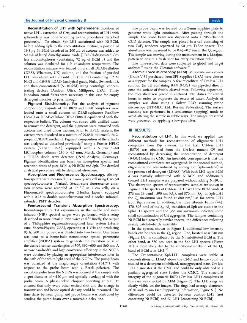
Reconstitution of LH1. In this work we applied twodifferent methods for reconstitution of oligomeric LH1complexes from Rsp. rubrum. In the first, Crt-less LH1(B870) was obtained from the Crt-less mutant G9 andreconstituted by decreasing the concentration of detergent(β-OG) below its CMC. An inevitable consequence is that thereconstituted complexes are aggregated. In the second method,oligomerization was induced by adding Crt (spheroidene) inthe presence of detergent (LDAO). With both LH1 types BChla was partially substituted with Ni-BChl and additionallycontrol LH1 samples were prepared without adding Ni-BChl.The absorption spectra of representative samples are shown inFigure 1. The spectra of Crt-less LH1 have three BChl bands at375 nm (B-band), 590 nm (Qx), and 870 nm (Qy). In Sph-LH1the Qy maximum was found at 880 nm,51 as for native LH1from Rsp. rubrum. In addition, the three vibronic bands (443,470, 503 nm) of the S0→S2 transition of Sph are visible in theSph-LH1 spectra and the 366 nm maximum indicates somesmall contamination of Crt aggregates. The samples containingNi-BChl had generally similar spectra, the differences reflectingmainly batch-to-batch variability.In the spectra shown in Figure 1, additional low intensity
bands can be seen in the Qx region. One, located near 540 nm(Figure 1A), is contributed by the Ni-substituted BChl a. Theother band, at 550 nm, seen in the Sph-LH1 spectra (Figure1B) is most likely due to the vibrational sideband of the Qxband of BChl a in LH1.55
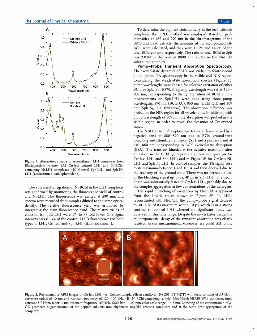
The Crt-containing Sph-LH1 complexes were stable atconcentrations of LDAO above the CMC and hence could bestudied in a detergent-solubilized, nonaggregated state. Crt-lessLH1 dissociates at the CMC and could be only obtained in apartially aggregated state (below the CMC). The structuralintegrity of the oligomeric B870 (Crt-less LH1) complexes inthis case was checked by AFM (Figure 2). The LH1 rings areclearly visible on the images. The rings had average diametersof 20 and 25 nm (see Supporting Information, Figure S1). Nodifferences could be detected between control LH1 (notcontaining Ni-BChl) and Ni-LH1 (containing Ni-BChl).
The Journal of Physical Chemistry B Article
dx.doi.org/10.1021/jp4020977 | J. Phys. Chem. B 2013, 117, 11260−1127111262
The successful integration of Ni-BChl in the LH1 complexeswas confirmed by monitoring the fluorescence yield of controland Ni-LH1. The fluorescence was excited at 590 nm, andspectra were recorded from samples diluted to the same opticaldensity. The relative fluorescence yield was estimated byintegrating the main fluorescence band. The relative yields ofemission from Ni-LH1 were 17- to 33-fold lower (the signalintensity was 6−3% of the control LH1’s fluorescence) in bothtypes of LH1, Crt-less and Sph-LH1 (data not shown).
To determine the pigment stoichiometry in the reconstitutedcomplexes, the HPLC method was employed. Based on peakintensities at 467 and 790 nm in the chromatograms of theB870 and B880 extracts, the amounts of the incorporated Ni-BChl were calculated, and they were 19.5% and 14.7% of thetotal BChl content, respectively. The ratio of total BChl to Sphwas 2:0.89 in the control B880 and 2:0.91 in the Ni-BChlsubstituted complex.
Pump−Probe Transient Absorption Spectroscopy.The excited-state dynamics of LH1 was studied by femtosecondpump−probe TA spectroscopy in the visible and NIR region.Considering the steady-state absorption spectra (Figure 1),pump wavelengths were chosen for selective excitation of eitherBChl or Sph. For B870, the pump wavelength was set at 590−600 nm, corresponding to the Qx transition of BChl a. Themeasurements on Sph-LH1 were done using three pumpwavelengths, 590 nm (BChl Qx), 860 nm (BChl Qy), and 509nm (Sph S2, 0−0 transition). The absorption difference wasprobed in the NIR region for all wavelengths. In addition, withpump wavelength of 509 nm, the absorption was probed in thevisible region, in order to reveal the dynamics of Crt excitedstates.The NIR transient absorption spectra were characterized by a
negative band at 880−890 nm due to BChl ground-statebleaching and stimulated emission (SE) and a positive band at840−860 nm, corresponding to BChl excited-state absorption(ESA). The transient kinetics at the negative maximum afterexcitation in the BChl Qx region are shown in Figure 3A forCrt-less LH1 and Sph-LH1, and in Figure 3B for Crt-less Ni-LH1 and Sph-Ni-LH1. In control samples, the TA signal roseto its maximum between 1 and 10 ps and then decayed due tothe recovery of the ground state. There was no detectable lossof the bleaching signal up to ca. 40 ps in Sph-LH1. The decayphase was substantially faster in Crt-less LH1, probably due tothe complex aggregation at low concentrations of the detergent.The rapid quenching of excitations by Ni-BChl is apparent
from the kinetic traces shown in Figure 3B. In LH1sreconstituted with Ni-BChl, the pump−probe signal decayedto 30−40% of its maximum within 10 ps, which is in a strongcontrast to control LH1 whereof no significant decay wasobserved in this time range. Despite the much faster decay, themultiexponential decay of the transient absorption was clearlyresolved in our measurement. Moreover, we could still follow
Figure 1. Absorption spectra of reconstituted LH1 complexes fromRhodospirillum rubrum. (A) Crt-less control LH1 and Ni-BChl-containing Ni-LH1 complexes. (B) Control Sph-LH1 and Sph-Ni-LH1 (reconstituted with spheroidene).
Figure 2. Representative AFM images of Crt-less LH1. (A) Control sample, silicon cantilever (NSG01 NT-MDT) with force constant of 5.5 N/m,curvature radius of 10 nm and resonant frequency of 120−190 kHz. (B) Ni-BChl-containing sample, MiroMasch Hi’RES-W14 cantilever, forceconstant 5.7 N/m, radius 1 nm, resonant frequency 160 kHz. Scale bar − 100 nm; color scale range − 4.5 nm. Lowering of the concentration of β-OG promotes oligomerization of the peptide subunits into oligomeric ring-like antenna complexes and at the same time aggregation of thecomplexes.
The Journal of Physical Chemistry B Article
dx.doi.org/10.1021/jp4020977 | J. Phys. Chem. B 2013, 117, 11260−1127111263
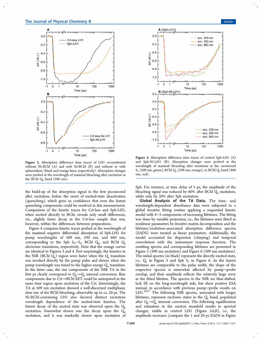
the build-up of the absorption signal in the first picosecondafter excitation, before the onset of excited-state deactivation(quenching), which gives us confidence that even the fastestquenching components could be resolved in this measurement.Comparison of the kinetic traces for Crt-less and Sph-LH1,when excited directly to BChl, reveals only small differences,viz., slightly faster decay in the Crt-less sample that was,however, within the differences between preparations.Figure 4 compares kinetic traces probed at the wavelength of
the maximal negative differential absorption of Sph-LH1 forpump wavelengths of 509 nm, 590 nm, and 860 nm,corresponding to the Sph S0−S2, BChl Qx, and BChl Qyelectronic transitions, respectively. Note that the orange curvesare identical in Figures 3 and 4. Not surprisingly, the kinetics inthe NIR (BChl Qy) region were faster when the Qy transitionwas invoked directly by the pump pulse and slower when thepump wavelength was tuned to the higher-energy Qx transition.In the latter case, the rise components of the NIR TA in thefirst ps clearly correspond to Qx→Qy internal conversion. Risecomponents due to Crt→BChl EET could be anticipated in thesame time region upon excitation of the Crt. Interestingly, theTA at 509 nm excitation showed a well-discerned multiphasicslow rise of the BChl bleaching, observable up to ca. 20 ps. TheNi-BChl-containing LH1 also showed distinct excitationwavelength dependence of the excited-state kinetics. Thefastest decay of the excited state was observed upon the Qyexcitation. Somewhat slower was the decay upon the Qxexcitation, and it was markedly slower upon excitation of
Sph. For instance, at time delay of 5 ps, the amplitude of thebleaching signal was reduced by 60% after BChl Qy excitation,while only by 20% after Sph excitation.
Global Analysis of the TA Data. The time- andwavelength-dependent absorbance data were subjected to aglobal iterative fitting routine applying a sequential kineticmodel with 4−5 components of increasing lifetimes. The fittingwas done by variable projection, i.e., the lifetimes were fitted asnonlinear parameters by iterative matrix decomposition and thelifetime/evolution-associated absorption difference spectra(EADS) were treated as linear parameters. Additionally, themodel accounted for dispersion (chirping) and temporalconvolution with the instrument response function. Theresulting spectra and corresponding lifetimes are presented inFigure 5 (590 nm excitation) and Figure 6 (509 nm excitation).The initial spectra (in black) represent the directly excited state,i.e., Qx in Figure 5 and Sph S2 in Figure 6. As the fastestlifetimes are comparable to the pulse width, the shape of therespective spectra is somewhat affected by pump−probeoverlap, and their amplitude reflects the relatively large errorin the fitted lifetime. The spectra in the NIR are blue-shifted,lack SE on the long-wavelength side, but show positive ESAinstead, in accordance with previous pump−probe results onLH1.56,57 The following NIR spectra, associated with longerlifetimes, represent excitonic states in the Qy band, populatedafter Qx→Qy internal conversion. The following equilibrationand relaxation in the exciton manifold results in spectralchanges, visible in control LH1 (Figure 5A,B), i.e., theamplitude increases (compare the 1 and 20 ps EADS in Figure
Figure 3. Absorption difference time traces of LH1 reconstitutedwithout Ni-BChl (A) and with Ni-BChl (B) and without or withspheroidene (black and orange lines, respectively). Absorption changeswere probed at the wavelength of maximal bleaching after excitation atthe BChl Qx band (590 nm).
Figure 4. Absorption difference time traces of control Sph-LH1 (A)and Sph-Ni-LH1 (B). Absorption changes were probed at thewavelength of maximal bleaching after excitation at the carotenoidS2 (509 nm, green), BChl Qx (590 nm, orange), or BChl Qy band (860nm, red).
The Journal of Physical Chemistry B Article
dx.doi.org/10.1021/jp4020977 | J. Phys. Chem. B 2013, 117, 11260−1127111264
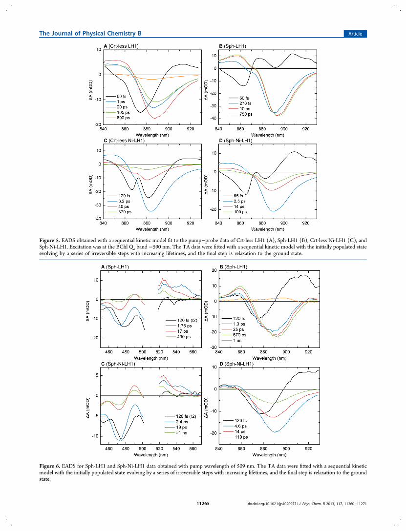
Figure 5. EADS obtained with a sequential kinetic model fit to the pump−probe data of Crt-less LH1 (A), Sph-LH1 (B), Crt-less Ni-LH1 (C), andSph-Ni-LH1. Excitation was at the BChl Qx band −590 nm. The TA data were fitted with a sequential kinetic model with the initially populated stateevolving by a series of irreversible steps with increasing lifetimes, and the final step is relaxation to the ground state.
Figure 6. EADS for Sph-LH1 and Sph-Ni-LH1 data obtained with pump wavelength of 509 nm. The TA data were fitted with a sequential kineticmodel with the initially populated state evolving by a series of irreversible steps with increasing lifetimes, and the final step is relaxation to the groundstate.
The Journal of Physical Chemistry B Article
dx.doi.org/10.1021/jp4020977 | J. Phys. Chem. B 2013, 117, 11260−1127111265
5A and the 0.27 ps and the 10 ps EADS in Figure 5B), thenegative maximum shifts to longer wavelengths, and theappearance of SE broadens the spectra on the long-wavelengthside.56 In Sph-LH1, the lifetime associated with excitonrelaxation was 270 fs, very close to previously reported values.An additional, much slower (10 ps) component was detected,which probably reflects vibrational relaxation. Note that thespectral evolution in Sph-LH1 (Figure 5B) proceeds withoutany loss of overall amplitude, which would signify excitationdecay. There’s only one decay component with 0.75 ns lifetime,which is in good agreement with the excited-state decay innative LH1 from Rhodobacter sphaeroides58 and native andreconstituted B875 from Rubrivivax gelatinosus.59 In contrast toSph-LH1, the TA of Crt-less LH1 did not decay mono-exponentially, but two additional lifetimes were present, of 20and 100 ps. The result is in accordance with the 0.39 nsfluorescence lifetime of Rsp. rubrum B873, as determined byChang et al.,60 and which, as mentioned above, can probably beexplained by aggregation-induced excited state quenching.On a subpicosecond time scale, Ni-LH1 showed similar
spectral evolution reflecting internal conversion as the controlLH1 (Figure 5C,D). The follow-up spectra also have verysimilar shape as the respective control LH1 spectra. Nosubpicosecond decay components could be resolved, but onlyexciton equilibration components, as with the control samples.These were omitted in the presented spectra since they did notsignificantly improve the fit. The excitation trapping by Ni-BChl is manifested in the picosecond decay lifetimes. Themajor decay lifetimes were 3.2 and 2.5 ps for Crt-less Ni-LH1and Sph-Ni-LH1, respectively. The amplitudes of the followingEADS (40 ps in 5C and 14 ps in Figure 5D) were lower by 60−70%, indicating that the 2−3 ps decay components wereresponsible for quenching of 60−70% of the excitations. Still,significant proportion of the excited states decayed on a muchslower time scale, mainly through the 40 ps component (Crt-less Ni-LH1) and the 100 ps component (Sph-Ni-LH1). Atcloser inspection, the spectra of these slower componentsappeared blue-shifted by ∼3 nm, compared to the spectra of themain quenching component. This may be a hint that theyoriginate from a structurally different population of complexesin the sample, or, alternatively, from different excited states.Perhaps this heterogeneity was present also in the controlsamples but was not resolved kinetically because of the uniformdecay lifetime.Upon Sph excitation (509 nm), the initial NIR spectra
(Figure 6A, B) were significantly blue-shifted and exhibitedpositive ESA around 920 nm. This ESA can be attributed to theshort-lived S2 and hot S1 states of Sph as well as BChl Qx statespopulated via EET from Sph. The subsequent evolution in thecontrol Sph-LH1 is spectrally very similar as with 590 nmexcitation, showing a gradual red shift, broadening andincreasing in overall amplitude due to the population ofemitting low-energy excitonic states in the Qy region. Theassociated lifetimes are, however, significantly longer, whichprobably reflects the slower Sph→Qy EET pathways. The decayof BChl excited states was again monoexponential with a 0.67ns lifetime, but the best fit was achieved by adding a long-lived(≫1 ns) ESA component that can be attributed to the Crt.As the above-described transients show, the decay kinetics of
Ni-LH1 was affected by excitation wavelength. The lifetime ofthe fastest quenching component was 4.6 ps with 509 nmexcitation, as compared to 2.5 ps with 590 nm. Moreover, asmaller proportion (36%) of the ground state bleaching was
recovered by this kinetic component. The slower lifetimes didnot differ between 509 and 590 nm excitation, and theassociated spectra were again blue-shifted by 3 nm.The transient absorption in the visible range, revealing
dynamics of the Crt excited states, was measured and analyzedindependently with a 4-component sequential model (Figure6A, C). The initial spectra, assigned to Sph S2 state (1Bu
+)clearly show bleaching of the S0→S2 transition band. The S2state decays with a 120 fs lifetime. The following spectrum,associated with a 2 ps lifetime, has ESA maxima at 525 and 550nm, corresponding to triplet state and S1 (2Ag
−) state,respectively. The 17−19 ps component has higher contributionof triplet state and also a shoulder at ∼540 nm, which could beattributed to either the S1 or S* state. The longer-livedcomponent spectra are unambiguously assigned to 3Sph. TheCrt lifetimes match closely the kinetics in the BChl region. Thisindicates that EET from Crt to BChl occurs via multiplepathways with time constants from ∼100 fs to ∼20 ps.Evidently, the Crt dynamics was not affected by the presence ofNi-BChl in Ni-LH1.
Global Target Analysis. The above presented analysis ofthe data provides an estimate of the lifetimes and the generaldynamics of the system under different excitation wavelengths,but it obviously does not accurately depict the multiplepathways of energy transfer between pigments and excitedstates and hence cannot give a detailed account on the specificEET rate constants and spectra. We have therefore performed atarget analysis of the pump−probe data of Sph-LH1 and Sph-Ni-LH1 with a branched model. The kinetic scheme used forfitting the Sph-Ni-LH1 data is shown in Figure 7. A very similar
scheme plus exciton relaxation components was used for fittingthe control Sph-LH1 data (Supporting Information, Figure S2).The model was fitted to the pump−probe data obtained withthe three pump wavelengths simultaneously with the same setof rate constants, only varying the initially populated excitedstates. The species-associated absorption difference spectra(SADS) were estimated independently for each excitation andprobe wavelength range and are plotted in Figure 8 for Sph-Ni-LH1. The respective control LH1 SADS are shown in Figure S3(Supporting Information). The quality of the fits isdemonstrated in Figures S4 and S5.
Figure 7. Kinetic scheme of the model used for simultaneous globaltarget analysis of the TA data obtained from Sph-Ni-LH1. The speciescompartments S2, S1, S*, and T1 represent Sph excited states and Qx,Qy
0, and Qy represent BChl excited states. The initially populatedcompartments were S2, Qx, and Qy
0 for the 509, 590, and 860 nmpump wavelengths, respectively. Compartments A1 and A2 arepopulated as Qy and represent sample heterogeneity. The fitted rateconstants (ps−1) are shown next to the arrows.
The Journal of Physical Chemistry B Article
dx.doi.org/10.1021/jp4020977 | J. Phys. Chem. B 2013, 117, 11260−1127111266

In the kinetic scheme we distinguish four Crt states: S2, S1,S*, and T1. There are two pathways for Crt→BChl EET: fromS2 to Qx and from S1 to Qy. Four BChl states were needed to fitthe kinetics of control LH1. One of them describes the directlyexcited BChl* (Qx or Qy
0), and three subsequent states todescribe the exciton relaxation (cf. Figure S2) and spectralevolution. The exciton relaxation was not explicitly modeled forthe Ni-LH1 data. The quenching by the Ni-BChl centers isquantified by the amplitudes of the BChl SADS and by the ratesdescribing their spectral evolution. In this way the data with theCrt, BChl Qx. and BChl Qy excitation were simultaneouslyfitted. The estimated rate constants are indicated in Figure 7.The control sample exhibited a subtle spectral evolution (red-shift), reflected in the SADS (Figure S3) and also evident fromthe selected traces and fits in Figure S4. The final SADS decaysin 0.7 ns. By contrast, the Qy compartment decayed with a 2 pslifetime in the Ni-LH1 sample, but two additional time scaleswere required to describe the quenching. This apparentheterogeneity is modeled by two compartments (populatedas Qy), with lifetimes of 12 and 108 ps. The fraction of thesemore slowly quenched complexes varied between 21−31% forthe different data sets (see Table 1). The dominant fractionquenched within 2 ps varied between 42 and 55%. The SADSof the more slowly quenched complexes, and especially the
longest-lived one (108 ps), were blue-shifted, compared to themain Qy SADS.
■ DISCUSSION
Reconstitution of LH1. In this work we employed tworeconstitution techniques to incorporate Ni-BChl into LH1from Rsp. rubrum. In the first, Crt-less LH1 (B870)19 areassembled mostly due to hydrophobic interactions between theN-terminal extensions of the α and β polypeptides at themembrane interface.61,62 In the second method, Crtsspecifically interact with the subunits and promote complexformation under conditions where Crt-less complexes disso-ciate.19,20,51 The B870 complexes are structurally similar to thenative, Crt-binding, LH1 antenna, as it can be seen from AFMimages (Figure 2), but there are also some differences, e.g., theNIR absorption maximum is blue-shifted by 10 nm relative toits 880 nm position characteristic for the strain.19,51,63 The shiftof the excitonic transition could be accompanied by alteredenergy transfer and hence energy trapping dynamics. Aconsequence of the Crt-less LH1 reconstitution method is ahigher level of aggregation. We strived to minimize the size andheterogeneity of the aggregates by carefully adjusting theconcentration of the detergent and applying mechanical andultrasonic homogenization before measurements. However,even small aggregates may have a profound effect on the energytransfer dynamics because of energy migration between B870s.The larger effective domain size raises the likelihood ofnonlinear (two-photon) reactions such as exciton−excitonannihilation.64 This can be one possible explanation for the fastexcited state decay lifetimes (20−100 ps) in our Crt-less LH1preparations. Since exciton−exciton annihilation stronglydepends on the excitation rate, we performed test measure-ments reducing the excitation power by up to an order of
Figure 8. SADS resulting from global target analysis of the TA data for Sph-Ni-LH1, performed using the kinetic model shown in Figure 7.
Table 1. Fractions of Quenched Lifetimes in Ni-LH1 atDifferent Excitation Wavelengths
λpump (nm) Qy (1.9 ps) A1 (12 ps) A2 (108 ps)
509 47% 28% 25%590 42% 27% 31%860 55% 21% 24%
The Journal of Physical Chemistry B Article
dx.doi.org/10.1021/jp4020977 | J. Phys. Chem. B 2013, 117, 11260−1127111267

magnitude, but did not observe any appreciable differences inthe kinetic traces, other than the reduction of signal-to-noiseratio (data not shown). It is possible that the excited statelifetime is shortened by dissipative states formed by interactionsbetween the aggregated complexes. Interestingly, shorterexcited-state lifetimes were found in carotenoid-less LH2compared to native complexes from the purple bacteriumAllochromatium minutissimum.65 The authors proposed that inthe absence of Crts BChl triplets could accumulate and quenchthe excited states via singlet-triple annihilation. Such amechanism cannot be completely ruled out but again shouldbe dependent on the excitation rate, which we could notconfirm. Lastly, a small amount of excited state quenchers couldbe present such as oxidized BChls, whose effect would be largerin the aggregated Crt-less LH1 where energy can migratebetween complexes.66
Excited State Dynamics of Control LH1. The LH1complexes reconstituted with Sph were stable in detergentsolution (0.045% LDAO), showing neither scattering noraggregation. Furthermore, the NIR absorption maximummatched that of the native antenna (880 nm) and so did theexcited-state lifetime (0.7 ns). The absence of any faster decaycomponents in the transient absorption confirmed that noexciton−exciton annihilation took place under the measure-ment conditions. Thus, we have obtained a detailed, high-resolution spectroscopic picture of the ultrafast dynamics inLH1, devoid of nonlinear processes and aggregation-relatedartifacts.The TA data reveal the exciton relaxation dynamics
occurring on a subpicosecond time scale. The position of thenegative band in the time-resolved spectra shifted to longerwavelengths, its overall amplitude increased, and the band wasasymmetrically enhanced on the long-wavelength side due toSE following the population of lower-energy emitting excitonicstates by internal conversion from the initially populated states.A similar dynamic red shift was found by Visser et al.56 in theTA kinetics of LH1 from Rsp. rubrum after excitation in theBChl Qx region. As in our results, it occurred with a ∼300 fstime constant and was attributed to relaxation in the excitonmanifold. Monshouwer et al.57 observed a similar spectralchange in LH1 from Rps. viridis. In addition to the earlierfindings, owing to the lack of annihilation, we could detect afurther 10-ps spectral equilibration component in Sph-LH1(Figure 5B), that could be related to vibrational relaxation andslower reorganization of the environment.67,68 Even slower (20ps) lifetimes were found in the spectral evolution of BChl uponexcitation of Sph. An obvious explanation for these lifetimes isthat they stem from a slow Sph→BChl EET.Analysis of the transient absorption in the NIR as well as the
visible range could provide clues about the dynamics of the Crtexcited states and EET pathways, which is apparently a complexand controversial topic of research.49 Even though there is asmall overlap between the optical transitions of Crts and BChl,several pathways of Crt→BChl EET are possible. The energy ofthe S2 state is in the range of the BChl Qx transition and S2→Qx EET occurs with a time constant of 60−240 fs.28,42,44 Ourdata revealed fast lifetimes (120−200 fs) in the NIR and in thevisible range that could be unequivocally assigned to S2 decayand concomitant excitation of BChl via the Qx state. Fitting theTA data in the visible range with a simple unbranched modelrevealed lifetimes of ∼2 and 20 ps that matched the NIRdynamics (1.3 and 25 ps). An inspection of the associatedspectra hinted toward the decay of Sph S1 state in the visible
wavelength range and the population of emitting BChlexcitonic states in the Qy range. The lifetimes and the shapeof the visible-range spectra are very similar to those obtained byGradinaru et al. from Rsp. rubrum chromatophores69 exceptthat the Crt absorption maxima are at different wavelengthsbecause the native LH1 contains spirilloxanthin instead of Sph.The authors found that the S1 lifetime of spirilloxanthin (1.4ps) was the same in solution and in the LH1 and concludedthat this state was not involved in EET. Considering that the S1lifetime of Sph in solution is 9 ps, the 2 ps lifetime in ourexperiments further corroborates the existence of EET from S1.The unbranched model fitting also resolved a long-lived (≫1ns) component in the visible range, which, based on the EADS,clearly belonged to the triplet state of Sph.Based on the above considerations, the target analysis was
performed with a branched kinetic model (Figure 7) fitting therate constants of internal conversion and intersystem crossingamong Sph and BChl states as well as of S2→Qx and S1→QyEET. The latter two are (230 fs)−1 and (3.7 ps)−1, respectively.Small differences notwithstanding, the control sample’smodeling results conform to literature data on the Crtdynamics in LH2 and LH1. The efficient singlet-to-tripletconversion found by Gradinaru et al.69 and Papagiannakis etal.70,71 is confirmed in our data. These papers also reported astate with SADS maximum at 525 nm, like the triplet state, butwith a broader long-wavelength shoulder. It decays with a timeconstant of 70 ps to the triplet and to the ground state. Ourdata are consistent with these findings, with respect to theSADS and lifetime of this state, which was ascribed to S*.Koyama’s group has published a TA investigation of LH1reconstituted with different Crts.45 They resolved anintermediate state between S2 and S1−1Bu
−, which is aprecursor to the T1 state. The evolution kinetics and theSADS differ from the S* state in the present model, hence theseare probably different intermediates. The remaining SADS arevery similar and so are the fitted rate constants of Sph internalconversion and Sph→BChl EET.
Dissipation of Excitations by Ni-BChl. The steady-statefluorescence and the TA results of LH1 complexes containingNi-BChl showed the expected strong quenching, in sampleswith and without Crts. Interestingly, the kinetics wereheterogeneous with lifetimes ranging from 2 to 100 ps. Themain dissipation channel, with about 50% fractional amplitude,appeared to be (2 ps)−1. Faster excited-state decay componentswere not resolved in the TA data, even upon direct excitation inthe Qy exciton band. These results are in contrast to thepreviously reported TA study by Fiedor et al.,8 in which Rb.sphaeroides B870 complexes containing 20% or more Ni-BChlshowed a 60 fs deactivation lifetime. This difference can bepartly explained by a smaller ring in this reconstituted LH1,estimated to be close to 20 BChl a molecules.8,9 Subpicosecondkinetic components were present in the data, but they wereassociated with internal conversion among excited states.Therefore it can be concluded that the excited states directlypopulated by the pump pulse, i.e., Qx and higher-energyexcitonic states, are not trapped by Ni-BChl, which is notsurprising, since the excitations must first migrate to thequencher molecules.To understand the origin of the 2 ps decay lifetime, one must
consider three factors determining the deactivation kinetics: theintrinsic deactivation rate constant of the Ni-BChl centers kq,the number of Ni-BChl molecules in the complex Nq, and themean effective rate constant of energy transfer (hopping)
The Journal of Physical Chemistry B Article
dx.doi.org/10.1021/jp4020977 | J. Phys. Chem. B 2013, 117, 11260−1127111268

between BChls, kh. In reality all four parameters N, Nq, kq, khshould be (broad) distributions rather than single values,considering the static and dynamic disorder in the system.72 Asa simple example, let all complexes consist of 32 total BChlsand one Ni-BChl with kq = 16 ps−1. Let us further assumeinstantaneous energy equilibration (kh =∞), i.e., the excitationsare delocalized over the whole complex. The excited-statelifetime of the complex will be
τ = × =k N N1/( / ) 2 psq q
The relative content of Ni-BChl in the samples was 12−15%,equivalent to about four Ni-BChls per complex, on average.Taking into account the Poisson statistical distribution, theestimated average lifetime is 0.66 ps. Thus, a model assumingfully delocalized excitations and 60 fs dissipation time constantof Ni-BChl cannot explain the experimental results. An intrinsicdissipation rate constant of (200 fs)−1 results in theexperimentally observed 2 ps lifetime. This result seemsreasonable considering that Musewald et al.6 performed TAmeasurements on Ni-BChl in toluene and pyridine and foundthat the Qy state mainly decays with lifetime of 450 and 200 fs,respectively. Next, we take into account that the delocalizationlength may not cover the whole ring, as has been proposed byseveral groups. We calculated the effective lifetime by treatingthe excitations as localized73 and migrating with an effectiverate constant of 100 fs.74 Simulation of the kinetics for 15%average Ni-BChl content resulted in a 2 ps lifetime if theintrinsic deactivation lifetime of Ni-BChl was fixed to 160 fs.It must be noted that in the above simulations, due to the
statistical distribution of Nq, there is also a distribution of decaylifetimes, which may be a reason for the lifetime heterogeneityobserved in the TA data. It could be then speculated that the∼10-ps lifetime originates from complexes with only one Ni-BChl and 2 ps is the mean lifetime of complexes with two ormore Ni-BChls. The longer, 100 ps, decay lifetimes, mostprobably reflect partially dissociated complexes, which contain asmaller number of subunits and are thus statistically less likelyto contain Ni-BChl. The blue-shifted spectra of thesecomponents corroborate this notion. As the measurementswere performed at high protein concentration, it is likely thatEET between complexes takes place and excitations incomplexes without Ni-BChl can be quenched by EET tothose containing Ni-BChl. Natural disorder could also be thereason for slower migration and relaxation rates.72
It is notable that the excitation trapping by Ni-BChl was notaffected by the presence or absence of Sph in the complexes. Itcan be thus inferred that also the Crts do not influence thedynamics of EET between BChls in LH1. Only direct excitationof Sph had an effect on the dissipation kinetics, since thekinetics was partially rate-limited by the slower S1→Qx EET.
■ CONCLUSIONS
The ultrafast spectroscopy of LH1 complexes reconstitutedwith Sph are generally in good agreement with literature dataregarding native LH1 and LH2 complexes69−71 as well as ofreconstituted Sph-LH1.45 We found that Crt-less LH1 hadshorter excited-state lifetime, probably caused by the lack ofdetergent and consequent aggregation of the B870 complexesor due to quenching by BChl triplets accumulated in theabsence of Crt.65 The bound Crt did not appear to have a directeffect on the EET among BChls.
The Ni-BChl molecules in LH1 complexes can effectivelydissipate the absorbed light energy within a few picoseconds.Only the lowest-energy BChl excited-states can be deactivatedby Ni-BChl. The dissipation kinetics is evidently limited byexcited state energy equilibration within the complex. Althoughthe dissipation rate is not as fast as previously determined,8 yetit is accelerated by more than two orders of magnitude incomparison to the native antenna, which makes this system agood model for the thermal energy dissipation in pigment−protein complexes. These results are the basis for futureinvestigation of the heat dissipation in pigment−proteincomplexes reconstituted with the Ni-analogues of (bacterio)-chlorophylls by ultrafast vibrational spectroscopy.
■ ASSOCIATED CONTENT*S Supporting InformationFigure S1: Distribution of ring diameters estimated from AFMimages. Figure S2: Target analysis scheme used for fitting theTA data of control Sph-LH1. Figures S3 and S4: Measured andfitted kinetic TA traces.This material is available free of chargevia the Internet at http://pubs.acs.org.
■ AUTHOR INFORMATIONCorresponding Author*E-mail: [email protected]; Tel.: +36 30 433 131; Fax:+36 30 433 434.
NotesThe authors declare no competing financial interest.
■ ACKNOWLEDGMENTSThis project was supported by grants from LaserLab Europe(Project LCVU-1606 to P.H.L. and Y.M.), the HungarianScientific Research Fund (OTKA-PD 104530 to P.H.L.,TAMOP-4.2.2.A-11/1/KONV-2012-0060 to G.G.), the Hun-garian National Innovation Office and A*STAR Singapore(NIH-A*STAR TET_10-1-2011-0279 to G.G.), the EuropeanCommission (EC-MC ITN 238017 HARVEST to G.G.), theEuropean Research Council (Advanced Grant Proposal 267333PHOTPROT to R.v.G.), and the Foundation for Polish Science(TEAM/2010-5/3 to L.F.).
■ REFERENCES(1) Niyogi, K. K. Safety Valves for Photosynthesis. Curr. Opin. PlantBiol. 2000, 3, 455−460.(2) Li, Z.; Wakao, S.; Fischer, B. B.; Niyogi, K. K. Sensing andResponding to Excess Light. Annu. Rev. Plant Biol. 2009, 60, 239−260.(3) Bixon, M.; Jortner, J. Intramolecular Radiationless Transitions. J.Chem. Phys. 1968, 48, 715−726.(4) Nakashima, N.; Yoshihara, K. Role of Hot Molecules Formed byInternal Conversion in UV Single-Photon and MultiphotonChemistry. J. Phys. Chem. 1989, 93, 7763−7771.(5) Cseh, Z.; Rajagopal, S.; Tsonev, T.; Busheva, M.; Papp, E.; Garab,G. Thermooptic Effect in Chloroplast Thylakoid Membranes. Thermaland Light Stability of Pigment Arrays with Different Levels ofStructural Complexity. Biochemistry 2000, 39, 15250−15257.(6) Musewald, C.; Hartwich, G.; Lossau, H.; Gilch, P.; Pollinger-Dammer, F.; Scheer, H.; Michel-Beyerle, M. E. Ultrafast Photophysicsand Photochemistry of [Ni]-Bacteriochlorophyll a. J. Phys. Chem. B1999, 103, 7055−7060.(7) Hartwich, G.; Fiedor, L.; Simonin, I.; Cmiel, E.; Schafer, W.; Noy,D.; Scherz, A.; Scheer, H. Metal-Substituted Bacteriochlorophylls. 1.Preparation and Influence of Metal and Coordination on Spectra. J.Am. Chem. Soc. 1998, 120, 3675−3683.
The Journal of Physical Chemistry B Article
dx.doi.org/10.1021/jp4020977 | J. Phys. Chem. B 2013, 117, 11260−1127111269
(8) Fiedor, L.; Scheer, H.; Hunter, C. N.; Tschirschwitz, F.; Voigt, B.;Ehlert, J.; Nibbering, E.; Leupold, D.; Elsaesser, T. Introduction of a 60fs Deactivation Channel in the Photosynthetic Antenna LH1 by Ni-Bacteriopheophytin A. Chem. Phys. Lett. 2000, 319, 145−152.(9) Fiedor, L.; Leupold, D.; Teuchner, K.; Voigt, B.; Hunter, C. N.;Scherz, A.; Scheer, H. Excitation Trap Approach to Analyze Size andPigment-Pigment Coupling: Reconstitution of LH1 Antenna ofRhodobacter sphaeroides with Ni-Substituted Bacteriochlorophyll.Biochemistry 2001, 40, 3737−3747.(10) Pilch, M.; Dudkowiak, A.; Jurzyk, B.; Lukasiewicz, J.; Susz, A.;Stochel, G.; Fiedor, L. Molecular Symmetry Determines theMechanism of a Very Efficient Ultrafast Excitation-to-Heat Conversionin Ni-Substituted Chlorophylls. Biochim. Biophys. Acta 2013, 1827,30−37.(11) Robert, B.; Cogdell, R. J.; van Grondelle, R. The Light-Harvesting System of Purple Bacteria. In Light-Harvesting Antennas inPhotosynthesis; Green, B. R., Parson, W. W., Eds.; Kluwer AcademicPublishers: Dordrecht, The Netherlands, 2003; pp 169−194.(12) Law, C. J.; Roszak, A. W.; Southall, J.; Gardiner, A. T.; Isaacs, N.W.; Cogdell, R. J. The Structure and Function of Bacterial Light-Harvesting Complexes (Review). Mol. Membr. Biol. 2004, 21, 183−191.(13) Cogdell, R. J.; Gall, A.; Kohler, J. The Architecture and Functionof the Light-Harvesting Apparatus of Purple Bacteria: From SingleMolecules to in vivo Membranes. Q. Rev. Biophys. 2006, 39, 227−324.(14) Law, C. J.; Cogdell, R. J. The Light Harvesting System of PurpleAnoxygenic Photsynthetic Bacteria. In Primary Processes of Photosyn-thesis: Basic Principles and Apparatus; Renger, G., Ed.; The RoyalSociety of Chemistry: Cambridge, U.K., 2008; pp 205−259.(15) Hu, X. C.; Ritz, T.; Damjanovic, A.; Autenrieth, F.; Schulten, K.Photosynthetic Apparatus of Purple Bacteria. Q. Rev. Biophys. 2002, 35,1−62.(16) Cogdell, R. J.; Lindsay, J. G.; Valentine, J.; Durant, I. A FurtherCharacterisation of the B890 Light-Harvesting Pigment−ProteinComplex from Rhodospirillum rubrum Strain S1. FEBS Lett. 1982,150, 151−154.(17) Miller, J. F.; Hinchigeri, S. B.; Parkes-Loach, P. S.; Callahan, P.M.; Sprinkle, J. R.; Riccobono, J. R.; Loach, P. A. Isolation andCharacterization of a Subunit Form of the Light-Harvesting Complexof Rhodospirillum rubrum. Biochemistry 1987, 26, 5055−5062.(18) Visschers, R. W.; Chang, M. C.; van Mourik, F.; Parkes-Loach,P. S.; Heller, B. A.; Loach, P. A.; van Grondelle, R. FluorescencePolarization and Low-Temperature Absorption Spectroscopy of aSubunit Form of Light-Harvesting Complex I from Purple Photo-synthetic Bacteria. Biochemistry 1991, 30, 5734−5742.(19) Fiedor, L.; Scheer, H. Trapping of an Assembly Intermediate ofPhotosynthetic LH1 Antenna Beyond B820 Subunit. J. Biol. Chem.2005, 280, 20921−20926.(20) Nakagawa, K.; Suzuki, S.; Fujii, R.; Gardiner, A. T.; Cogdell, R.J.; Nango, M.; Hashimoto, H. Probing the Effect of the Binding Site onthe Electrostatic Behavior of a Series of Carotenoids Reconstitutedinto the Light-Harvesting 1 Complex from Purple PhotosyntheticBacterium Rhodospirillum rubrum Detected by Stark Spectroscopy. J.Phys. Chem. B 2008, 112, 9467−9475.(21) Karrasch, S.; Bullough, P. A.; Ghosh, R. The 8.5 Å ProjectionMap of the Light-Harvesting Complex I from Rhodospirillum rubrumReveals a Ring Composed of 16 Subunits. EMBO J. 1995, 14, 631−638.(22) Walz, T.; Ghosh, R. Two-Dimensional Crystallization of theLight-Harvesting I Reaction Centre Photounit from Rhodospirillumrubrum. J. Mol. Biol. 1997, 265, 107−111.(23) Scherz, A.; Parson, W. W. Exciton Interactions in Dimers ofBacteriochlorophyll and Related Molecules. Biochim. Biophys. Acta1984, 766, 666−678.(24) Alden, R. G.; Johnson, E.; Nagarajan, V.; Parson, W. W.; Law, C.J.; Cogdell, R. J. Calculations of Spectroscopic Properties of the LH2Bacteriochlorophyll-Protein Antenna Complex from Rhodopseudomo-nas acidophila. J. Phys. Chem. B 1997, 101, 4667−4680.
(25) Beekman, L. M. P.; Steffen, M.; van Stokkum, I. H. M.; Olsen, J.D.; Hunter, C. N.; Boxer, S. G.; van Grondelle, R. Characterization ofthe Light-Harvesting Antennas of Photosynthetic Purple Bacteria byStark Spectroscopy. 1. LH1 Antenna Complex and the B820 Subunitfrom Rhodospirillum rubrum. J. Phys. Chem. B 1997, 101, 7284−7292.(26) Monshouwer, R.; Abrahamsson, M.; van Mourik, F.; vanGrondelle, R. Superradiance and Exciton Delocalization in BacterialPhotosynthetic Light-Harvesting Systems. J. Phys. Chem. B 1997, 101,7241−7248.(27) Hu, X.; Ritz, T.; Damjanovic, A.; Schulten, K. PigmentOrganization and Transfer of Electronic Excitation in the Photo-synthetic Unit of Purple Bacteria. J. Phys. Chem. B 1997, 101, 3854−3871.(28) Sundstrom, V.; Pullerits, T.; van Grondelle, R. PhotosyntheticLight-Harvesting: Reconciling Dynamics and Structure of PurpleBacterial LH2 Reveals Function of Photosynthetic Unit. J. Phys. Chem.B 1999, 103, 2327−2346.(29) van Grondelle, R.; Novoderezhkin, V. Dynamics of ExcitationEnergy Transfer in the LH1 and LH2 Light-Harvesting Complexes ofPhotosynthetic Bacteria. Biochemistry 2001, 40, 15057−15068.(30) Renger, T.; May, V.; Kuhn, O. Ultrafast Excitation EnergyTransfer Dynamics in Photosynthetic Pigment−Protein Complexes.Phys. Rep. Rev. Sect. Phys. Lett. 2001, 343, 137−254.(31) van Grondelle, R.; Novoderezhkin, V. I. Energy Transfer inPhotosynthesis: Experimental Insights and Quantitative Models. Phys.Chem. Chem. Phys. 2006, 8, 793−807.(32) Freiberg, A.; Timpmann, K.; Jackson, J. A.; Lin, S.; Woodbury,N. W. Exciton Relaxation and Transfer in the Antenna Network ofPhotosynthetic Bacteria. In Photosynthesis: Mechanism and Effects;Proceedings of the XI International Congress on PhotosynthesisBudapest, Hungary, August 17−22, 1998; Garab, G., Ed.; KluwerAcademic Publishers: Dordrecht, 1998; pp 771.(33) Cheng, Y. C.; Fleming, G. R. Dynamics of Light Harvesting inPhotosynthesis. Annu. Rev. Phys. Chem. 2009, 60, 241−262.(34) Scholes, G. D.; Fleming, G. R.; Olaya-Castro, A.; van Grondelle,R. Lessons from Nature About Solar Light Harvesting. Nat. Chem.2011, 23, 763−774.(35) Novoderezhkin, V.; Monshouwer, R.; van Grondelle, R.Disordered Exciton Model for the Core Light-Harvesting Antennaof Rhodopseudomonas viridis. Biophys. J. 1999, 77, 666−681.(36) Jimenez, R.; van Mourik, F.; Yu, J. Y.; Fleming, G. R. Three-Pulse Photon Echo Measurements on LH1 and LH2 Complexes ofRhodobacter sphaeroides: A Nonlinear Spectroscopic Probe of EnergyTransfer. J. Phys. Chem. B 1997, 101, 7350−7359.(37) Chachisvilis, M.; Kuhn, O.; Pullerits, T.; Sundstrom, V. Excitonsin Photosynthetic Purple Bacteria: Wavelike Motion or IncoherentHopping? J. Phys. Chem. B 1997, 101, 7275−7283.(38) Novoderezhkin, V.; Monshouwer, R.; van Grondelle, R. Exciton(De)Localization in the LH2 Antenna of Rhodobacter sphaeroides asRevealed by Relative Difference Absorption Measurements of the LH2Antenna and the B820 Subunit. J. Phys. Chem. B 1999, 103, 10540−10548.(39) Pullerits, T.; Chachisvilis, M.; Sundstrom, V. ExcitonDelocalization Length in the B850 Antenna of Rhodobacter sphaeroides.J. Phys. Chem. 1996, 100, 10787−10792.(40) Damjanovic, A.; Kosztin, I.; Kleinekathofer, U.; Schulten, K.Excitons in a Photosynthetic Light-Harvesting System: A CombinedMolecular Dynamics, Quantum Chemistry, and Polaron Model Study.Phys. Rev. E 2002, 65, 031919.(41) Leupold, D.; Stiel, H.; Teuchner, K.; Nowak, F.; Sandner, W.;Ucker, B.; Scheer, H. Size Enhancement of Transition Dipoles to One-and Two-Exciton Bands in a Photosynthetic Antenna. Phys. Rev. Lett.1996, 77, 4675−4678.(42) Ritz, T.; Damjanovic, A.; Schulten, K.; Zhang, J.-P.; Koyama, Y.Efficient Light Harvesting through Carotenoids. Photosynth. Res. 2000,66, 125−144.(43) Koyama, Y.; Kakitani, Y. Mechanisms of Carotenoid-to-Bacteriochlorophyll Energy Transfer in the Light Harvesting AntennaComplexes 1 and 2: Dependence on the Conjugation Length of
The Journal of Physical Chemistry B Article
dx.doi.org/10.1021/jp4020977 | J. Phys. Chem. B 2013, 117, 11260−1127111270

Carotenoids. In Chlorophylls and Bacteriochlorophylls: Biochemistry,Biophysics, Functions and Applications; Grimm, B., Porra, R. J., Rudiger,W., Scheer, H., Eds.; Springer: Dordrecht, The Netherlands, 2006; pp431−443.(44) Krueger, B. P.; Scholes, G. D.; Jimenez, R.; Fleming, G. R.Electronic Excitation Transfer from Carotenoid to Bacteriochlorophyllin the Purple Bacterium Rhodopseudomonas Acidophila. J. Phys.Chem. B 1998, 102, 2284−2292.(45) Akahane, J.; Rondonuwu, F. S.; Fiedor, L.; Watanabe, Y.;Koyama, Y. Dependence of Singlet-Energy Transfer on theConjugation Length of Carotenoids Reconstituted into the LH1Complex from Rhodospirillum rubrum G9. Chem. Phys. Lett. 2004, 393,184−191.(46) Scholes, G. D.; Harcourt, R. D.; Fleming, G. R. ElectronicInteractions in Photosynthetic Light-Harvesting Complexes: The Roleof Carotenoids. J. Phys. Chem. B 1997, 101, 7302−7312.(47) Zhang, J.-P.; Fujii, R.; Qian, P.; Inaba, T.; Mizoguchi, T.;Koyama, Y.; Onaka, K.; Watanabe, Y. Mechanism of the Carotenoid-to-Bacteriochlorophyll Energy Transfer Via the S1 State in LH2Complexes from Purple Bacteria. J. Phys. Chem. B 2000, 104, 3683−3691.(48) Krueger, B. P.; Scholes, G. D.; Fleming, G. R. Calculation ofCouplings and Energy-Transfer Pathways between the Pigments ofLH2 by the Ab Inito Transition Density Cube Method. J. Phys. Chem.B 1998, 102, 5378−5386.(49) Polivka, T.; Sundstrom, V. Dark Excited States of Carotenoids:Consensus and Controversy. Chem. Phys. Lett. 2009, 477, 1−11.(50) Fiedor, J.; Pilch, M.; Fiedor, L. Tuning the Thermodynamics ofAssociation of Transmembrane Helices. J. Phys. Chem. B 2009, 113,12831−12838.(51) Fiedor, L.; Akahane, J.; Koyama, Y. Carotenoid-InducedCooperative Formation of Bacterial Photosynthetic LH1 Complex.Biochemistry 2004, 43, 16487−16496.(52) Pawlowicz, N. P.; Groot, M. L.; van Stokkum, I. H. M.; Breton,J.; van Grondelle, R. Charge Separation and Energy Transfer in thePhotosystem II Core Complex Studied by Femtosecond MidinfraredSpectroscopy. Biophys. J. 2007, 93, 2732−2742.(53) van Stokkum, I. H. M.; Larsen, D. S.; van Grondelle, R. Globaland Target Analysis of Time-Resolved Spectra. Biochim. Biophys. Acta2004, 1657, 82−104.(54) Snellenburg, J.; Laptenok, S.; Seger, R.; Mullen, K. M.; vanStokkum, I. H. M. Glotaran: A Java-Based Graphical User Interface forthe R Package TIMP. J. Stat. Software 2012, 49, 1−22.(55) Fiedor, L. Hexacoordination of Bacteriochlorophyll in Photo-synthetic Antenna LH1. Biochemistry 2006, 45, 1910−1918.(56) Visser, H. M.; Somsen, O. J. G.; van Mourik, F.; Lin, S.; vanStokkum, I. H. M.; van Grondelle, R. Direct Observation of Sub-picosecond Equilibration of Excitation Energy in the Light-HarvestingAntenna of Rhodospirillum rubrum. Biophys. J. 1995, 69, 1083−1099.(57) Monshouwer, R.; Baltuska, A.; van Mourik, F.; van Grondelle, R.Time-Resolved Absorption Difference Spectroscopy of the LH-1Antenna of Rhodopseudomonas viridis. J. Phys. Chem. A 1998, 102,4360−4371.(58) Hunter, C. N.; Bergstroem, H.; Van Grondelle, R.; Sundstroem,V. Energy-Transfer Dynamics in Three Light-Harvesting Mutants ofRhodobacter sphaeroides: A Picosecond Spectroscopy Study. Biochem-istry 1990, 29, 3203−3207.(59) Jirsakova, V.; Reiss-Husson, F.; Ranck, J.-L.; Moya, I.Fluorescence Life-Times and Aggregation States of the Core LightHarvesting Complex B875 from Rubrivivax Gelatinosus. Photosynth.Res. 1997, 54, 35−43.(60) Chang, M. C.; Callahan, P. M.; Parkes-Loach, P. S.; Cotton, T.M.; Loach, P. A. Spectroscopic Characterization of the Light-Harvesting Complex of Rhodospirillum rubrum and Its StructuralSubunit. Biochemistry 1990, 29, 421−429.(61) Loach, P. A.; Parkes-Loach, P. S. Structure−FunctionRelationships in Core Light-Harvesting Complexes (LH1) asDetermined by Characterization of the Structural Subunit and byReconstitution Experiments. In Anoxygenic Photosynthetic Bacteria;
Blankenship, R. E., Madigan, M. T., Bauer, C. E., Eds.; KluwerAcademic Publishers: Dordrecht, The Netherelands, 1995; pp 437−471.(62) Arluison, V.; Seguin, J.; Robert, B. Biochemical Characterizationof the Dissociated Forms from the Core Antenna Proteins from PurpleBacteria. Biochemistry 2002, 41, 11812−11819.(63) Picorel, R.; Belanger, G.; Gingras, G. Antenna HolochromeB880 of Rhodospirillum rubrum S1. Pigment, Phospholipid, andPolypeptide Composition. Biochemistry 1983, 22, 2491−2497.(64) Valkunas, L.; Trinkunas, G.; Liuolia, V.; van Grondelle, R.Nonlinear Annihilation of Excitations in Photosynthestic Systems.Biophys. J. 1995, 69, 1117−1129.(65) Theiss, C.; Leupold, D.; Moskalenko, A. A.; Razjivin, A. P.;Eichler, H. J.; Lokstein, H. Femtosecond Spectroscopy of Native andCarotenoidless Purple-Bacterial LH2 Clarifies Functions of Carote-noids. Biophys. J. 2008, 94, 4808−4811.(66) Lambrev, P. H.; Schmitt, F. J.; Kussin, S.; Schoengen, M.;Varkonyi, Z.; Eichler, H. J.; Garab, G.; Renger, G. Functional DomainSize in Aggregates of Light-Harvesting Complex II and ThylakoidMembranes. Biochim. Biophys. Acta 2011, 1807, 1022−1031.(67) Polivka, T.; Pullerits, T.; Herek, J. L.; Sundstrom, V. ExcitonRelaxation and Polaron Formation in LH2 at Low Temperature. J.Phys. Chem. B 2000, 104, 1088−1096.(68) Novoderezhkin, V. I.; van Grondelle, R. Exciton-VibrationalRelaxation and Transient Absorption Dynamics in LH1 ofRhodopseudomonas Viridis: A Redfield Theory Approch. J. Phys.Chem. B 2002, 106, 6025−6037.(69) Gradinaru, C. C.; Kennis, J. T. M.; Papagiannakis, E.; vanStokkum, I. H. M.; Cogdell, R. J.; Fleming, G. R.; Niederman, R. A.;van Grondelle, R. An Unusual Pathway of Excitation EnergyDeactivation in Carotenoids: Singlet-to-Triplet Conversion on anUltrafast Timescale in a Photosynthetic Antenna. Proc. Natl. Acad. Sci.U.S.A. 2001, 98, 2364−2369.(70) Papagiannakis, E.; Das, S. K.; Gall, A.; van Stokkum, I. H. M.;Robert, B.; van Grondelle, R.; Frank, H. A.; Kennis, J. T. M. LightHarvesting by Carotenoids Incorporated into the B850 Light-Harvesting Complex from Rhodobacter sphaeroides R-26.1: Excited-State Relaxation, Ultrafast Triplet Formation, and Energy Transfer toBacteriochlorophyll. J. Phys. Chem. B 2003, 107, 5642−5649.(71) Papagiannakis, E.; Kennis, J. T. M.; van Stokkum, I. H. M.;Cogdell, R. J.; van Grondelle, R. An Alternative Carotenoid-to-Bacteriochlorophyll Energy Transfer Pathway in Photosynthetic LightHarvesting. Proc. Natl. Acad. Sci. U.S.A. 2002, 99, 6017−6022.(72) Novoderezhkin, V. I.; Cohen, S. T. A.; van Grondelle, R.Dynamics of Exciton Relaxation in LH2 Antenna Probed byMultipulse Nonlinear Spectroscopy. J. Phys. Chem. A 2011, 115,3834−3844.(73) Abramavicius, D.; Valkunas, L.; van Grondelle, R. ExcitonDynamics in Ring-Like Photosynthetic Light-Harvesting Complexes:A Hopping Model. Phys. Chem. Chem. Phys. 2004, 6, 3097−3105.(74) Bradforth, S. E.; Jimenez, R.; van Mourik, F.; van Grondelle, R.;Fleming, G. R. Excitation Transfer in the Core Light-HarvestingComplex (LH-1) of Rhodobacter Sphaeroides: An UltrafastFluorescence Depolarization and Annihilation Study. J. Phys. Chem.1995, 99, 16179−16191.
The Journal of Physical Chemistry B Article
dx.doi.org/10.1021/jp4020977 | J. Phys. Chem. B 2013, 117, 11260−1127111271