Preparation of reconstituted acetylcholine receptor membranes suitable for AFM imaging of...
10
Chemistry and Physics of Lipids 163 (2010) 117–126 Contents lists available at ScienceDirect Chemistry and Physics of Lipids journal homepage: www.elsevier.com/locate/chemphyslip Preparation of reconstituted acetylcholine receptor membranes suitable for AFM imaging of lipid–protein interactions Ngoc Vuong a,1 , John E. Baenziger a,∗ , Linda J. Johnston b,∗∗ a Department of Biochemistry, Microbiology, and Immunology, University of Ottawa, Ottawa, Ontario K1H 8M5, Canada b Steacie Institute for Molecular Sciences, National Research Council of Canada, 100 Sussex Drive, Ottawa, Ontario K1A 0R6, Canada article info Article history: Received 22 June 2009 Received in revised form 4 September 2009 Accepted 24 September 2009 Available online 2 October 2009 Keywords: Nicotinic acetylcholine receptor Atomic force microscopy Reconstitution Supported lipid bilayers Proteoliposomes Fluorescence abstract The nicotinic acetylcholine receptor (nAChR) has been reconstituted in POPC vesicles at high lipid–protein (L/P) ratios for the preparation of supported lipid bilayers with a low protein density for studies of protein–lipid interactions using atomic force microscopy (AFM). Initial reconstitutions using a standard dialysis method with bulk L/P ratios ranging from 20:1 to 100:1 (w/w) gave heterogeneous samples that contained both empty vesicles and proteoliposomes with a range of L/P ratios. This is problematic because empty vesicles adsorb and rupture to form bilayer patches more rapidly than do protein-rich vesicles, resulting in the loss of protein during sample washing. Although it was not possible to find reconstitution conditions that gave homogeneous populations of vesicles with high L/P ratios, an addi- tional freeze–thaw cycle immediately after dialysis did reproducibly yield a fraction of proteoliposomes with L/P ratios above 100:1. These proteoliposomes were separated by sucrose gradient centrifugation and used to prepare supported bilayers with well-separated individual receptors and minimal adsorbed proteoliposomes. AFM images of such samples showed many small features protruding from the bilayer surface. These features range in height from 1 to 5 nm, consistent with the smaller intracellular domain of the protein exposed, and have lateral dimensions consistent with an individual receptor. Some bilayers with reconstituted protein also had a small fraction of higher features that are assigned to nAChR with the larger extracellular domain exposed and showed evidence for aggregation to give dimers or small oligomers. This work demonstrates the importance of using highly purified reconstituted membranes with uniform lipid–protein ratios for AFM studies of integral membrane protein–lipid interactions. © 2009 Elsevier Ireland Ltd. All rights reserved. 1. Introduction Atomic force microscopy (AFM) has been widely used to char- acterize the structures of peptides and proteins in supported phospholipid bilayers formed from both reconstituted and native membranes (Frederix et al., 2009). Ordered two-dimensional arrays of membrane proteins including rhodopsin, porins, bacterial hexag- onally packed intermediate layers, photosynthetic core-complexes and connexons are particularly amenable to high resolution AFM imaging (Arechaga and Fotiadis, 2007; Buzhynskyy et al., 2007; Fotiadis et al., 2003; Muller et al., 1997; Scheuring, 2006; Stamouli et al., 2003; Thimm et al., 2005). These studies have provided unprecedented structural data for supramolecular assemblies of membrane proteins in aqueous buffer under close to physiolog- ∗ Corresponding author. Tel.: +1 613 562 5800x8222; fax: +1 613 562 5251. ∗∗ Corresponding author. Tel.: +1 613 990 0973; fax: +1 613 952 0068. E-mail addresses: [email protected] (J.E. Baenziger), [email protected] (L.J. Johnston). 1 Tel.: +1 613 562 5800x8222; fax: +1 613 562 5251. ical conditions, where conformational changes and the effects of factors such as pH and ligand binding can be readily studied. The ability to obtain high lateral resolution, on the order of ∼1 nm, is critically dependent on obtaining samples with a high density of protein over a relatively large and flat membrane area. Reconsti- tution at such high protein density is still largely empirical and generally depends on removing detergent from a fully solubilized lipid/protein/detergent mixture. Interestingly, a recent report has shown that integral membrane proteins such as the bacterial light harvesting complex can be incorporated into existing lipid bilayers that are destabilized by detergent, providing a potentially useful approach for preparing arrays of protein for structural analysis from minimal amounts of protein (Milhiet et al., 2006). On the other hand, AFM studies aimed at probing protein–lipid interactions require the use of reconstituted samples with much lower levels of protein. Supported bilayers with high lipid–protein ratios are required for characterizing the distribution of individ- ual membrane proteins within the membrane plane and thus for probing the preference of proteins for various lipid environments. Supported films with high lipid–protein ratios are necessary for interrogating individual molecules, as for example in correlated flu- 0009-3084/$ – see front matter © 2009 Elsevier Ireland Ltd. All rights reserved. doi:10.1016/j.chemphyslip.2009.09.003
-
Upload
independent -
Category
Documents
-
view
1 -
download
0
Transcript of Preparation of reconstituted acetylcholine receptor membranes suitable for AFM imaging of...
PA
Na
b
a
ARRAA
KNARSPF
1
apmooaiFeum
L
0d
Chemistry and Physics of Lipids 163 (2010) 117–126
Contents lists available at ScienceDirect
Chemistry and Physics of Lipids
journa l homepage: www.e lsev ier .com/ locate /chemphys l ip
reparation of reconstituted acetylcholine receptor membranes suitable forFM imaging of lipid–protein interactions
goc Vuonga,1, John E. Baenzigera,∗, Linda J. Johnstonb,∗∗
Department of Biochemistry, Microbiology, and Immunology, University of Ottawa, Ottawa, Ontario K1H 8M5, CanadaSteacie Institute for Molecular Sciences, National Research Council of Canada, 100 Sussex Drive, Ottawa, Ontario K1A 0R6, Canada
r t i c l e i n f o
rticle history:eceived 22 June 2009eceived in revised form 4 September 2009ccepted 24 September 2009vailable online 2 October 2009
eywords:icotinic acetylcholine receptortomic force microscopyeconstitutionupported lipid bilayersroteoliposomesluorescence
a b s t r a c t
The nicotinic acetylcholine receptor (nAChR) has been reconstituted in POPC vesicles at high lipid–protein(L/P) ratios for the preparation of supported lipid bilayers with a low protein density for studies ofprotein–lipid interactions using atomic force microscopy (AFM). Initial reconstitutions using a standarddialysis method with bulk L/P ratios ranging from 20:1 to 100:1 (w/w) gave heterogeneous samplesthat contained both empty vesicles and proteoliposomes with a range of L/P ratios. This is problematicbecause empty vesicles adsorb and rupture to form bilayer patches more rapidly than do protein-richvesicles, resulting in the loss of protein during sample washing. Although it was not possible to findreconstitution conditions that gave homogeneous populations of vesicles with high L/P ratios, an addi-tional freeze–thaw cycle immediately after dialysis did reproducibly yield a fraction of proteoliposomeswith L/P ratios above 100:1. These proteoliposomes were separated by sucrose gradient centrifugationand used to prepare supported bilayers with well-separated individual receptors and minimal adsorbed
proteoliposomes. AFM images of such samples showed many small features protruding from the bilayersurface. These features range in height from 1 to 5 nm, consistent with the smaller intracellular domain ofthe protein exposed, and have lateral dimensions consistent with an individual receptor. Some bilayerswith reconstituted protein also had a small fraction of higher features that are assigned to nAChR withthe larger extracellular domain exposed and showed evidence for aggregation to give dimers or smalloligomers. This work demonstrates the importance of using highly purified reconstituted membranesin rat
with uniform lipid–prote. Introduction
Atomic force microscopy (AFM) has been widely used to char-cterize the structures of peptides and proteins in supportedhospholipid bilayers formed from both reconstituted and nativeembranes (Frederix et al., 2009). Ordered two-dimensional arrays
f membrane proteins including rhodopsin, porins, bacterial hexag-nally packed intermediate layers, photosynthetic core-complexesnd connexons are particularly amenable to high resolution AFMmaging (Arechaga and Fotiadis, 2007; Buzhynskyy et al., 2007;
otiadis et al., 2003; Muller et al., 1997; Scheuring, 2006; Stamoulit al., 2003; Thimm et al., 2005). These studies have providednprecedented structural data for supramolecular assemblies ofembrane proteins in aqueous buffer under close to physiolog-∗ Corresponding author. Tel.: +1 613 562 5800x8222; fax: +1 613 562 5251.∗∗ Corresponding author. Tel.: +1 613 990 0973; fax: +1 613 952 0068.
E-mail addresses: [email protected] (J.E. Baenziger),[email protected] (L.J. Johnston).
1 Tel.: +1 613 562 5800x8222; fax: +1 613 562 5251.
009-3084/$ – see front matter © 2009 Elsevier Ireland Ltd. All rights reserved.oi:10.1016/j.chemphyslip.2009.09.003
ios for AFM studies of integral membrane protein–lipid interactions.© 2009 Elsevier Ireland Ltd. All rights reserved.
ical conditions, where conformational changes and the effects offactors such as pH and ligand binding can be readily studied. Theability to obtain high lateral resolution, on the order of ∼1 nm, iscritically dependent on obtaining samples with a high density ofprotein over a relatively large and flat membrane area. Reconsti-tution at such high protein density is still largely empirical andgenerally depends on removing detergent from a fully solubilizedlipid/protein/detergent mixture. Interestingly, a recent report hasshown that integral membrane proteins such as the bacterial lightharvesting complex can be incorporated into existing lipid bilayersthat are destabilized by detergent, providing a potentially usefulapproach for preparing arrays of protein for structural analysis fromminimal amounts of protein (Milhiet et al., 2006).
On the other hand, AFM studies aimed at probing protein–lipidinteractions require the use of reconstituted samples with muchlower levels of protein. Supported bilayers with high lipid–protein
ratios are required for characterizing the distribution of individ-ual membrane proteins within the membrane plane and thus forprobing the preference of proteins for various lipid environments.Supported films with high lipid–protein ratios are necessary forinterrogating individual molecules, as for example in correlated flu-
1 Physic
olbnitapmtdiihs(sfsotcR
aa(fntceRmafa(emrtle
itlosbleatssfla
ePpat
18 N. Vuong et al. / Chemistry and
rescence and AFM-based force spectroscopy measurements. Highipid–protein ratio bilayers are also advantageous for membraneiosensing, where protein availability may be an issue. Unfortu-ately, there are relatively few examples of the use of reconstituted
ntegral membrane receptors in supported bilayers at low pro-ein density for AFM imaging (Periasamy et al., 2009; Slade etl., 2002). Individual insulin receptors reconstituted into a phos-hatidylcholine (PC) membrane have been imaged, with heighteasurements confirming that the protein is oriented in two direc-
ions, with either the larger extracellular or shorter intracellularomains protruding out of the bilayer (Slade et al., 2002). This study
llustrates the difficulty of controlling the orientation of the proteinn supported membranes, although in some cases this challengeas been addressed by using specific labels (e.g., using histidine ortreptavidin tethers) to immobilize the protein in one orientationElie-Caille et al., 2005; Giess et al., 2004; Trepout et al., 2007). Aecond practical limitation is that the conditions necessary for theormation of supported membranes from proteoliposomes may beignificantly different than those for lipid vesicles, since the processf vesicle adsorption, rupture, and spreading is known to depend onhe nature of the support as well as vesicle composition, lipid con-entration, and temperature (Goksu et al., 2009; Johnston, 2007;ichter et al., 2006).
The nicotinic acetylcholine receptor (nAChR) is one member ofsuper-family of neurotransmitter-gated ion channels that playscentral role in inter-neuronal communication within the brain
Sine and Engel, 2006). Due to its natural abundance, the nAChRrom Torpedo has been used extensively as a model for probingeurotransmitter-gated ion channel–lipid interactions. The func-ional capabilities of the nAChR are highly dependent upon the lipidomposition of its surrounding membrane environment (Criadot al., 1984; Fong and McNamee, 1986; Hamouda et al., 2006;ankin et al., 1997; Ryan et al., 1996). nAChR–lipid interactionsay even play a role in nAChR function in humans (Shen et
l., 2006). Current models suggest that lipids influence nAChRunction by modulating the equilibria between activatable restingnd non-activatable desensitized and/or uncoupled conformationsBaenziger et al., 2000; daCosta and Baenziger, 2009). The pres-nce of both anionic lipids and cholesterol in a reconstituted PCembrane is required for optimal stabilization of the activatable
esting state. With a structural model approaching atomic resolu-ion now available (Unwin, 2005), potential mechanisms by whichipids influence nAChR conformational equilibria are beginning tomerge (Brannigan et al., 2008; daCosta and Baenziger, 2009).
One unique feature of lipid–protein interactions for the nAChRs that lipids not only influence nAChR structure (and thus func-ion), the nAChR also influences lipid structure, and does so in aipid-selective manner (daCosta et al., 2002, 2004). Reconstitutionf the nAChR into bilayers composed of PC has little effect on thetructure of the lipid bilayer. In contrast, incorporation into mem-ranes composed of PC and the anionic lipid, phosphatidic acid (PA)
eads to a substantial increase in the lateral packing density. Theffects of the nAChR on the lateral packing density of lipid bilayersppear to result, at least in part, from an nAChR-induced concentra-ion of cations at the membrane surface (Sturgeon and Baenziger,ubmitted for publication). The ability of the nAChR to influence theurrounding membrane environment could play a role in lipid raftormation. It has been suggested that the nAChR is associated withipid rafts during transport to synaptic membranes (Marchand etl., 2002).
Although previous studies have imaged dry native and het-
rogeneous reconstituted nAChR membranes (Lal and Yu, 1993;uu et al., 2000), these high nAChR density membranes (i.e. highrotein–lipid ratio) are not suitable for probing nAChR–lipid inter-ctions. As a first step towards studying the intriguing interactionshat occur between the nAChR and its lipid environment using AFM,s of Lipids 163 (2010) 117–126
we have developed a method for reconstituting the receptor at highlipid–protein ratios and have optimized conditions for preparingsupported lipid bilayers. Our results demonstrate that reconsti-tution protocols often give rise to a heterogeneous population ofproteoliposomes with a wide range of lipid–protein ratios. Recon-stitutions performed at high lipid–protein ratios generally lead tothe formation of empty vesicles, which are problematic in that theyrupture and spread to form supported bilayers more rapidly thandensely packed proteoliposomes. By separating vesicle fractionsusing sucrose density centrifugation, however, we obtained pro-teoliposomes with uniform lipid–protein ratios that reproduciblyform supported bilayers with low loadings of receptor. AFM anal-ysis of the resulting membranes reveals many individual receptorswith heights that are consistent with predominantly the smallercytoplasmic domain of the nAChR protruding out of the bilayer. Ourresults show the importance of using highly purified reconstitutedmembranes with uniform lipid–protein ratios for AFM studies ofintegral membrane protein–lipid interactions.
2. Materials and methods
2.1. Materials
Frozen Torpedo californica electroplax tissue was obtained fromAquatic Research Consultants (San Pedro, CA). 1-Palmitoyl-2-oleoyl phosphatidylcholine (POPC) was from Avanti Polar Lipids,Inc. (Alabaster, AL). Cholesterol, carbamylcholine, and sodiumcholate were from Sigma–Aldrich. Alpha-bungarotoxin conju-gated to Alexa488 (�BTx-A488) and Texas Red dihexadecanoyl-phosphatidylethanolamine (TR-DHPE) were from Invitrogen. TheLipofast mini extruder and 400 nm polycarbonate filters wereobtained from Avestin (Ottawa, ON).
2.2. Preparation of lipid vesicles
POPC alone in chloroform or in the presence of 0.2 mol% TexasRed DHPE solubilized in chloroform/methanol 1:1 (vol:vol) wasvortexed briefly, dried down under a stream of nitrogen gas andthen dried further under vacuum for at least 10 h to remove remain-ing solvent. The lipid film was hydrated with Tris dialysis buffer(TDB = 10 mM Tris, 100 mM NaCl, 0.01 mM EDTA, 0.01% NaN3, pH7.8) to a final lipid concentration of 1 mg/ml and vortexed for1 min to generate large vesicles. The vesicle solution was then soni-cated in a water bath at room temperature for ∼10 min to generatesmall unilamellar vesicles. Once the solution was transparent, thevesicles were diluted to 0.1 mg/ml and stored at 4 ◦C. All smallunilamellar vesicle solutions were used for AFM imaging within1 week.
2.3. Reconstitution of the nAChR
The nAChR was reconstituted into POPC membranes asdescribed in detail by daCosta et al. (2002). Briefly, Torpedo synapticmembranes were solubilized in TDB containing 1% cholate and thenapplied to a bromoacetylcholine-derivatized Affi-Gel 102 column.The column bound nAChR was washed with nine column volumesof TDB containing various concentrations of POPC solubilized in 1%sodium cholate. The nAChR was eluted from the column in threecolumn volumes of TDB containing 10 mM of carbamylcholine and0.13 mM POPC solubilized by 0.5% sodium cholate. The eluted frac-tions containing protein (A280 > 0.05 a.u.) were pooled and dialyzed
against 2 l of TDB (Spectrapo/Por type 2 tubing, 12,000–14,000 Dacut off, Spectrum Laboratories Inc.) with 5 buffer changes, onceevery ∼12 h. The dialyzed samples were centrifuged at 120,000 × gfor 2 h at 4 ◦C and the pellets resuspended in a small volume of TDBand stored at −80 ◦C. The protein was characterized by SDS-PAGE
Physic
tpl
m0lwl(taU
2
d(dTaSwlCcgwvolFAud
2
llc5(lw(aiviimins
2
twaol
N. Vuong et al. / Chemistry and
o verify sample purity. TLC analysis showed that the reconstitutedroteoliposomes contained only POPC, with no detectable native
ipid.Note that the lipid–protein ratios of reconstituted POPC–nAChR
embranes prepared as described above are typically in the.5–1.0/1.0 (w/w) range (daCosta et al., 2002). To obtain higher
ipid–protein ratios, the solubilized nAChR eluted from the columnas mixed with an appropriate volume of 5 mg/ml of POPC solubi-
ized in 1% cholate in TDB to make up the desired lipid–protein ratiosee Section 3). Final protein concentrations were determined usinghe BCA assay (Pierce Biotechnology Inc.). Lipid concentration wasssayed using the Phospholipid C/choline assay (Wako ChemicalsSA Inc.). 2% Triton X-100 was included in both assays.
.4. Sucrose gradient centrifugation
Reconstituted nAChR vesicles were purified further usingiscontinuous sucrose gradient centrifugation. Preparative scale10 ml) step gradients were prepared by layering solutions withecreasing concentrations of sucrose in TDB on top of one another.he nAChR in 1 ml of TDB was then layered on top of the sucrose,nd the gradients centrifuged at 41,000 rpm for 20 h at 4 ◦C in aW41 swinging bucket rotor (Beckmann). Fractions of 250 or 400 �lere drawn from the gradient (starting from the top) and the
ipid and protein content of each analyzed via the Phospholipidand BCA protein assays. For preparative purposes, those fractions
ontaining the vesicles of interest were isolated from the sucroseradient directly by inserting a sharp syringe needle through theall of the sucrose gradient tube at the fraction of interest. Isolated
esicles were then dialyzed against 2 l of TDB with 6 buffer changes,nce every ∼12 h. Following dialysis, each sample was diluted to aipid concentration of 0.1 mg/ml with TDB and stored at −80 ◦C.or fluorescence microscopy, the nAChR was labeled with �BTx-488 for 1 h after preparative sucrose gradient centrifugation. Thenbound toxin and sucrose in the sample were then removed byialysis.
.5. Atomic force microscopy (AFM)
The appropriate volume (between 70 and 125 �l) of either pureipid vesicles or nAChR proteoliposomes in TDB (both at 0.1 mg/mlipid) was mixed with distilled water containing the desired con-entration of CaCl2 (typically 10 mM) to give a final volume of00 �l. The lipid mixtures were deposited on freshly cleaved micahigh grade mica sheets, Ted Pella) and incubated for varyingengths of time (1–20 h) in an AFM fluid cell. Unbound material was
ashed away by flowing 100 ml of either water or imaging buffer2.5 mM HEPES, 50 mM NaCl) into the AFM cell. The sample wasllowed to equilibrate for 5 min after washing. Prior to AFM imag-ng, the AFM tip and holder were washed sequentially with 25 mlolumes of 70% ethanol, water, and imaging buffer. Bilayers weremaged using a PicoSPM atomic force microscope (Molecular Imag-ng/Agilent) in MAC (magnetic alternating current)-mode using
agnetic coated silicon tips (Type II, spring constants of ∼0.5 N/m)n aqueous solutions. Either a 30 �m × 30 �m or 5 �m × 5 �m scan-er was used with a scan rate between 0.7 and 1.4 Hz. All imageshown are flattened raw data.
.6. Total internal reflection fluorescence (TIRF) microscopy
Fluorescence images of bilayers were taken on an Olympus IX81
otal internal reflection fluorescence (TIRF) microscope equippedith a high resolution CCD camera (CoolSNAP, Photometrics, US)nd a 60×/1.45 NA Plan Apochromat objective. Supported bilayersf either POPC labeled with Texas Red DHPE or proteoliposomesabeled with �BTx-A488 were prepared as described above for
s of Lipids 163 (2010) 117–126 119
the AFM measurements, except that the volumes of sample wereincreased proportionally to account for the increased surface areaof the mica used in the larger TIRF cell. TR-DHPE and �BTx-A488were excited at 543 and 488 nm, respectively.
3. Results and discussion
3.1. Preparation of POPC bilayers
Supported POPC bilayers on mica were prepared by the incuba-tion of POPC vesicles in water in the presence of 10 mM CaCl2 for1 h, followed by extensive washing to remove unadsorbed vesicles.Fig. 1 shows AFM images of bilayer patches and a continuous bilayermembrane, prepared using two different initial lipid concentra-tions. The bilayer patches have a height of 4.5 nm (see cross-sectionin Fig. 1A) consistent with the expected thickness of a fluid POPCbilayer. Increasing either the incubation time (20 h) or the lipidconcentration gives a continuous bilayer that is featureless witha low surface roughness (Fig. 1B). By contrast, in the absence ofCa2+, only bilayer patches were formed, even at long incubationtimes of 20 h. Neither the use of HEPES buffer nor EDTA washingto remove excess Ca2+ (both of which were employed for bilay-ers with reconstituted protein) had any effect on the uniform andfeatureless POPC bilayers such as that shown in Fig. 1B. Extrac-tion of a POPC bilayer and quantification using enzymatic detectionof choline generated by phospholipid hydrolysis indicated that anindividual POPC bilayer prepared in our standard AFM fluid cellcontains 1–2 �g of lipid.
3.2. Reconstitution of nAChR
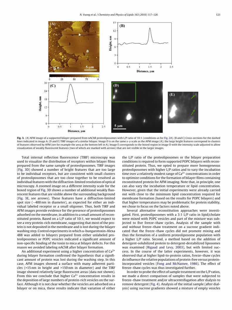
The receptor was initially reconstituted using a slight modifi-cation of the procedure typically employed for the preparation ofnAChR proteoliposomes with low L/P ratios (typically ∼1:2 (w:w))for FT-IR studies (daCosta et al., 2002; Ryan et al., 2002). Our goal,however, was to obtain samples with L/P ratios of 100:1 (w:w)or higher in order to facilitate imaging of individual receptors.Increasing the lipid concentration (from 0.13 to 1.6 mM) in thelipid-cholate detergent solution used to elute the purified recep-tor from the affinity column only increased the L/P ratio to ∼3:1(w:w). The solubilized receptor eluted from the column was there-fore further diluted with a lipid-cholate solution prior to dialysis,ultracentrifugation and storage at −80 ◦C. Several attempts to formsupported POPC bilayers with samples of reconstituted nAChR witha L/P ratio of 100:1 (w:w) using the same conditions as for POPCbilayers gave variable AFM results. In some cases, bilayer patchesthat showed little or no evidence of raised features expected forreconstituted protein were obtained (Fig. 2A). In other cases, sam-ples appeared to have an intact lipid membrane but with manysmall roughly spherical particles (typically 10–20 nm in height and>100 nm in diameter) adhering to the surface. This material rapidlycontaminated the AFM probe and was easily moved by the tip dur-ing scanning, making such samples very difficult to image.
One possible explanation for the variable results for supportedbilayers prepared as described above is that the vesicle populationhas a heterogeneous distribution of L/P ratios. We reasoned thatproteoliposomes might rupture to form a bilayer membrane moreslowly than empty vesicles, which could account for the observa-tion of patches that did not incorporate protein and large particlesadhering to the bilayer. In order to characterize the L/P ratio in the
initial proteoliposomes, the sample was separated on a discontinu-ous sucrose gradient and the protein and lipid content of individualfractions was analyzed. As illustrated in Fig. 2B, there are two vesi-cle populations in the sample; the band near the top at 0–5% sucrosecontains no protein and occurs at a similar density to POPC vesicles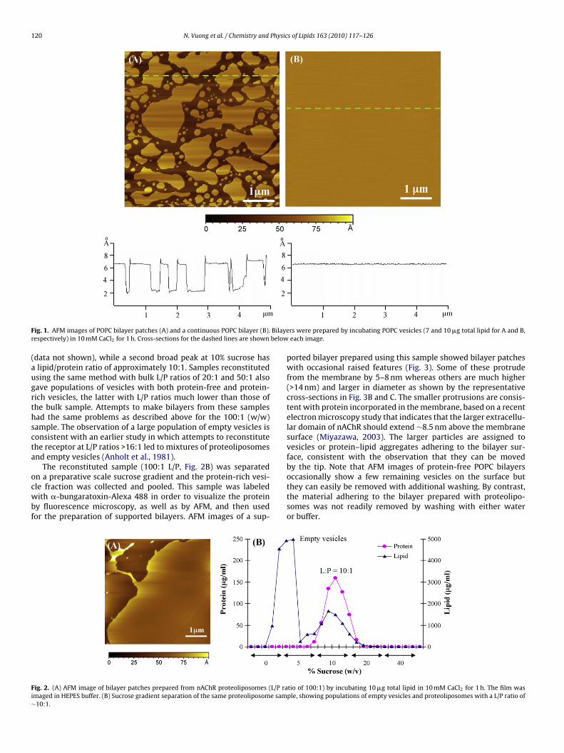
120 N. Vuong et al. / Chemistry and Physics of Lipids 163 (2010) 117–126
F Bilayer below
(augrthscta
ocwbf
Fi∼
ig. 1. AFM images of POPC bilayer patches (A) and a continuous POPC bilayer (B).espectively) in 10 mM CaCl2 for 1 h. Cross-sections for the dashed lines are shown
data not shown), while a second broad peak at 10% sucrose haslipid/protein ratio of approximately 10:1. Samples reconstitutedsing the same method with bulk L/P ratios of 20:1 and 50:1 alsoave populations of vesicles with both protein-free and protein-ich vesicles, the latter with L/P ratios much lower than those ofhe bulk sample. Attempts to make bilayers from these samplesad the same problems as described above for the 100:1 (w/w)ample. The observation of a large population of empty vesicles isonsistent with an earlier study in which attempts to reconstitutehe receptor at L/P ratios >16:1 led to mixtures of proteoliposomesnd empty vesicles (Anholt et al., 1981).
The reconstituted sample (100:1 L/P, Fig. 2B) was separated
n a preparative scale sucrose gradient and the protein-rich vesi-le fraction was collected and pooled. This sample was labeledith �-bungaratoxin-Alexa 488 in order to visualize the proteiny fluorescence microscopy, as well as by AFM, and then usedor the preparation of supported bilayers. AFM images of a sup-
ig. 2. (A) AFM image of bilayer patches prepared from nAChR proteoliposomes (L/P ramaged in HEPES buffer. (B) Sucrose gradient separation of the same proteoliposome sam10:1.
rs were prepared by incubating POPC vesicles (7 and 10 �g total lipid for A and B,each image.
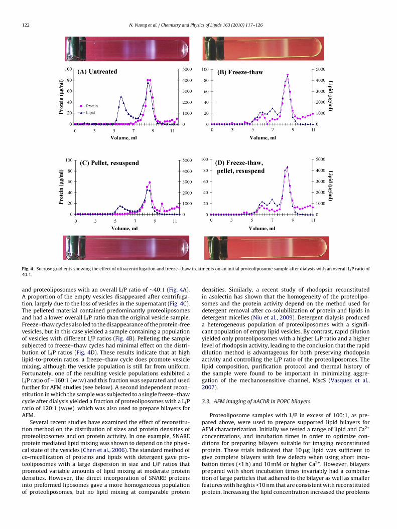
ported bilayer prepared using this sample showed bilayer patcheswith occasional raised features (Fig. 3). Some of these protrudefrom the membrane by 5–8 nm whereas others are much higher(>14 nm) and larger in diameter as shown by the representativecross-sections in Fig. 3B and C. The smaller protrusions are consis-tent with protein incorporated in the membrane, based on a recentelectron microscopy study that indicates that the larger extracellu-lar domain of nAChR should extend ∼8.5 nm above the membranesurface (Miyazawa, 2003). The larger particles are assigned tovesicles or protein–lipid aggregates adhering to the bilayer sur-face, consistent with the observation that they can be movedby the tip. Note that AFM images of protein-free POPC bilayers
occasionally show a few remaining vesicles on the surface butthey can easily be removed with additional washing. By contrast,the material adhering to the bilayer prepared with proteolipo-somes was not readily removed by washing with either wateror buffer.tio of 100:1) by incubating 10 �g total lipid in 10 mM CaCl2 for 1 h. The film wasple, showing populations of empty vesicles and proteoliposomes with a L/P ratio of
N. Vuong et al. / Chemistry and Physics of Lipids 163 (2010) 117–126 121
F s withl the sao e E cov ) that
up(toimbr(svAasstw4tnr
dccciFtfb
ig. 3. (A) AFM image of a supported bilayer prepared from nAChR proteoliposomeines indicated in image A. (D and E) TIRF images of a similar bilayer. Image D is onf features observed by AFM (see for example the area at the bottom left in A). Imagisualization of weakly fluorescent features (two of which are marked with arrows
Total internal reflection fluorescence (TIRF) microscopy wassed to visualize the distribution of receptors within bilayer filmsrepared from the same sample of proteoliposomes. TIRF imagesFig. 3D) showed a number of bright features that are too largeo be individual receptors, but are consistent with small clustersf proteoliposomes that are too close together to be resolved asndividual features with the diffraction-limited resolution of optical
icroscopy. A zoomed image on a different intensity scale for theoxed region of Fig. 3D shows a number of additional weakly fluo-escent features that are visible above the surrounding backgroundFig. 3E, see arrows). These features have a diffraction-limitedpot size (∼400 nm in diameter), as expected for either an indi-idual labeled receptor or a small oligomer. Thus, both TIRF andFM images provide evidence for the presence of proteoliposomesdsorbed on the membrane, in addition to a small amount of recon-tituted protein. Based on a L/P ratio of 10:1, we would expect toee a very protein-rich membrane, suggesting that most of the pro-ein is not deposited in the membrane and is lost during the bilayerashing step. Control experiments in which �-bungarotoxin-Alexa
88 was added to bilayers prepared from either unlabeled pro-eoliposomes or POPC vesicles indicated a significant amount ofon-specific binding of the toxin to mica at bilayer defects. For thiseason we avoided labeling nAChR after bilayer formation.
An additional experiment using a higher concentration of Ca2+
uring bilayer formation confirmed the hypothesis that a signifi-ant amount of protein was lost during the washing step. In thisase, AFM images showed a surface with many adherent parti-les (>15 nm in height and >150 nm in diameter) and the TIRF
mage showed relatively large fluorescent areas (data not shown).rom this we conclude that higher Ca2+ concentration results inhe deposition of large numbers of protein-rich vesicles on the sur-ace. Although it is not clear whether the vesicles are adsorbed on ailayer or on mica, these results indicate that variation of eitherL/P ratio of 10:1 (conditions as for Fig. 2A). (B and C) Cross-sections for the dashedme x–y scale as the AFM image (A); the large bright features correspond to clustersrresponds to the boxed region in image D with the intensity scale adjusted to alloware not visible in the larger images.
the L/P ratio of the proteoliposomes or the bilayer preparationconditions is required to form supported POPC bilayers with recon-stituted protein. Thus, we opted to prepare more homogeneousproteoliposomes with higher L/P ratios and to vary the incubationtime over a relatively modest range of Ca2+ concentrations in orderto optimize conditions for the formation of bilayer films containingreconstituted protein for AFM imaging. Note that, in principle, onecan also vary the incubation temperature or lipid concentration.However, given that the initial experiments were already carriedout with close to the minimum lipid concentration required formembrane formation (based on the results for POPC bilayers) andthat higher temperatures may be problematic for protein stability,we chose to focus on the factors noted above.
Several alternative reconstitution approaches were investi-gated. First, proteoliposomes with a 3:1 L/P ratio in lipid/cholatewere mixed with POPC vesicles and part of the mixture was sub-jected to five freeze–thaw cycles. Analysis of the sample withand without freeze–thaw treatment on a sucrose gradient indi-cated that the freeze–thaw cycles did not promote mixing andthus the formation of a uniform proteoliposome population witha higher L/P ratio. Second, a method based on the addition ofdetergent-solubilized protein to detergent-destabilized liposomeswas examined (Rigaud and Levy, 2003), but with limited suc-cess. In the course of the latter experiments, however, it wasobserved that at higher lipid-to-protein ratios, freeze–thaw cyclesdo influence the relative populations of protein-free versus protein-incorporated vesicles (Fong and McNamee, 1986). The effect offreeze–thaw cycles was thus investigated further.
In order to probe the effect of sample treatment on the L/P ratios,we made a direct comparison of samples that were subjected tofreeze–thaw treatment and/or ultracentrifugation after dialysis toremove detergent (Fig. 4). Analysis of the initial sample (after dial-ysis) using sucrose gradients showed a mixture of empty vesicles
122 N. Vuong et al. / Chemistry and Physics of Lipids 163 (2010) 117–126
F treatm4
aAtTaFvosblmFLfscrA
tppcctpdio
ig. 4. Sucrose gradients showing the effect of ultracentrifugation and freeze–thaw0:1.
nd proteoliposomes with an overall L/P ratio of ∼40:1 (Fig. 4A).proportion of the empty vesicles disappeared after centrifuga-
ion, largely due to the loss of vesicles in the supernatant (Fig. 4C).he pelleted material contained predominantly proteoliposomesnd had a lower overall L/P ratio than the original vesicle sample.reeze–thaw cycles also led to the disappearance of the protein-freeesicles, but in this case yielded a sample containing a populationf vesicles with different L/P ratios (Fig. 4B). Pelleting the sampleubjected to freeze–thaw cycles had minimal effect on the distri-ution of L/P ratios (Fig. 4D). These results indicate that at high
ipid-to-protein ratios, a freeze–thaw cycle does promote vesicleixing, although the vesicle population is still far from uniform.
ortunately, one of the resulting vesicle populations exhibited a/P ratio of ∼160:1 (w:w) and this fraction was separated and usedurther for AFM studies (see below). A second independent recon-titution in which the sample was subjected to a single freeze–thawycle after dialysis yielded a fraction of proteoliposomes with a L/Patio of 120:1 (w/w), which was also used to prepare bilayers forFM.
Several recent studies have examined the effect of reconstitu-ion method on the distribution of sizes and protein densities ofroteoliposomes and on protein activity. In one example, SNARErotein mediated lipid mixing was shown to depend on the physi-al state of the vesicles (Chen et al., 2006). The standard method ofo-micellization of proteins and lipids with detergent gave pro-
eoliposomes with a large dispersion in size and L/P ratios thatromoted variable amounts of lipid mixing at moderate proteinensities. However, the direct incorporation of SNARE proteinsnto preformed liposomes gave a more homogeneous populationf proteoliposomes, but no lipid mixing at comparable protein
ents on an initial proteoliposome sample after dialysis with an overall L/P ratio of
densities. Similarly, a recent study of rhodopsin reconstitutedin asolectin has shown that the homogeneity of the proteolipo-somes and the protein activity depend on the method used fordetergent removal after co-solubilization of protein and lipids indetergent micelles (Niu et al., 2009). Detergent dialysis produceda heterogeneous population of proteoliposomes with a signifi-cant population of empty lipid vesicles. By contrast, rapid dilutionyielded only proteoliposomes with a higher L/P ratio and a higherlevel of rhodopsin activity, leading to the conclusion that the rapiddilution method is advantageous for both preserving rhodopsinactivity and controlling the L/P ratio of the proteoliposomes. Thelipid composition, purification protocol and thermal history ofthe sample were found to be important in minimizing aggre-gation of the mechanosensitive channel, MscS (Vasquez et al.,2007).
3.3. AFM imaging of nAChR in POPC bilayers
Proteoliposome samples with L/P in excess of 100:1, as pre-pared above, were used to prepare supported lipid bilayers forAFM characterization. Initially we tested a range of lipid and Ca2+
concentrations, and incubation times in order to optimize con-ditions for preparing bilayers suitable for imaging reconstitutedprotein. These trials indicated that 10 �g lipid was sufficient togive complete bilayers with few defects when using short incu-
bation times (<1 h) and 10 mM or higher Ca2+. However, bilayersprepared with short incubation times invariably had a combina-tion of large particles that adhered to the bilayer as well as smallerfeatures with heights <10 nm that are consistent with reconstitutedprotein. Increasing the lipid concentration increased the problems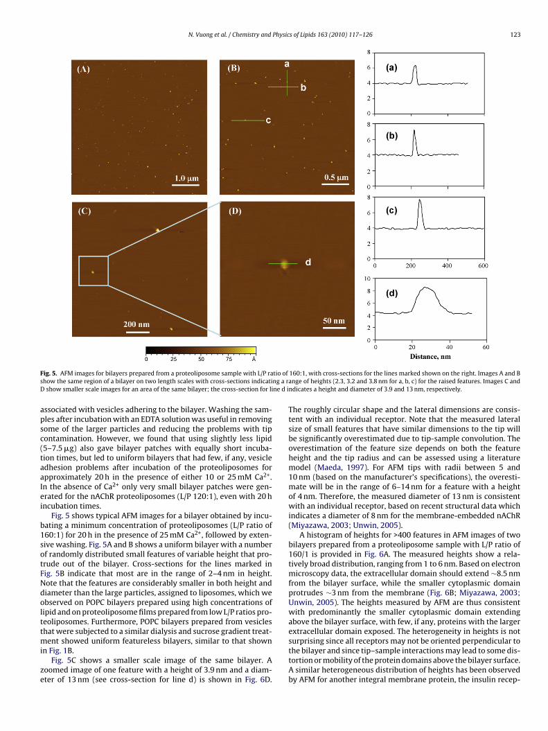
N. Vuong et al. / Chemistry and Physics of Lipids 163 (2010) 117–126 123
F tio ofs ng a rD ne d i
apsc(taaIei
b1sotFNdolttmi
ze
ig. 5. AFM images for bilayers prepared from a proteoliposome sample with L/P rahow the same region of a bilayer on two length scales with cross-sections indicatishow smaller scale images for an area of the same bilayer; the cross-section for li
ssociated with vesicles adhering to the bilayer. Washing the sam-les after incubation with an EDTA solution was useful in removingome of the larger particles and reducing the problems with tipontamination. However, we found that using slightly less lipid5–7.5 �g) also gave bilayer patches with equally short incuba-ion times, but led to uniform bilayers that had few, if any, vesicledhesion problems after incubation of the proteoliposomes forpproximately 20 h in the presence of either 10 or 25 mM Ca2+.n the absence of Ca2+ only very small bilayer patches were gen-rated for the nAChR proteoliposomes (L/P 120:1), even with 20 hncubation times.
Fig. 5 shows typical AFM images for a bilayer obtained by incu-ating a minimum concentration of proteoliposomes (L/P ratio of60:1) for 20 h in the presence of 25 mM Ca2+, followed by exten-ive washing. Fig. 5A and B shows a uniform bilayer with a numberf randomly distributed small features of variable height that pro-rude out of the bilayer. Cross-sections for the lines marked inig. 5B indicate that most are in the range of 2–4 nm in height.ote that the features are considerably smaller in both height andiameter than the large particles, assigned to liposomes, which webserved on POPC bilayers prepared using high concentrations ofipid and on proteoliposome films prepared from low L/P ratios pro-eoliposomes. Furthermore, POPC bilayers prepared from vesicleshat were subjected to a similar dialysis and sucrose gradient treat-
ent showed uniform featureless bilayers, similar to that shownn Fig. 1B.
Fig. 5C shows a smaller scale image of the same bilayer. Aoomed image of one feature with a height of 3.9 nm and a diam-ter of 13 nm (see cross-section for line d) is shown in Fig. 6D.
160:1, with cross-sections for the lines marked shown on the right. Images A and Bange of heights (2.3, 3.2 and 3.8 nm for a, b, c) for the raised features. Images C andndicates a height and diameter of 3.9 and 13 nm, respectively.
The roughly circular shape and the lateral dimensions are consis-tent with an individual receptor. Note that the measured lateralsize of small features that have similar dimensions to the tip willbe significantly overestimated due to tip-sample convolution. Theoverestimation of the feature size depends on both the featureheight and the tip radius and can be assessed using a literaturemodel (Maeda, 1997). For AFM tips with radii between 5 and10 nm (based on the manufacturer’s specifications), the overesti-mate will be in the range of 6–14 nm for a feature with a heightof 4 nm. Therefore, the measured diameter of 13 nm is consistentwith an individual receptor, based on recent structural data whichindicates a diameter of 8 nm for the membrane-embedded nAChR(Miyazawa, 2003; Unwin, 2005).
A histogram of heights for >400 features in AFM images of twobilayers prepared from a proteoliposome sample with L/P ratio of160/1 is provided in Fig. 6A. The measured heights show a rela-tively broad distribution, ranging from 1 to 6 nm. Based on electronmicroscopy data, the extracellular domain should extend ∼8.5 nmfrom the bilayer surface, while the smaller cytoplasmic domainprotrudes ∼3 nm from the membrane (Fig. 6B; Miyazawa, 2003;Unwin, 2005). The heights measured by AFM are thus consistentwith predominantly the smaller cytoplasmic domain extendingabove the bilayer surface, with few, if any, proteins with the largerextracellular domain exposed. The heterogeneity in heights is not
surprising since all receptors may not be oriented perpendicular tothe bilayer and since tip–sample interactions may lead to some dis-tortion or mobility of the protein domains above the bilayer surface.A similar heterogeneous distribution of heights has been observedby AFM for another integral membrane protein, the insulin recep-
124 N. Vuong et al. / Chemistry and Physics of Lipids 163 (2010) 117–126
F red fr( dues,p -secti
t2
piscb1seotN(btwocfns
pitR
F1c
ig. 6. (A) Histogram showing the distribution of heights for raised features measuProtein Data Bank code 2BG9). The model defines the location of ∼80% of the resiortion of the structure in this orientation). The nAChR is ∼16 nm long with a cross
or, reconstituted in phosphatidylcholine bilayers (Slade et al.,002).
An AFM image of a bilayer containing reconstituted nAChR pre-ared from a second proteoliposome sample (L/P 120:1) is shown
n Fig. 7A. The sample is qualitatively similar in that a number ofmall features of variable height are observed. However, in thisase, the features show a wider range of heights, as indicatedy the cross-sections (Fig. 7B and C) showing receptors that are0 and 2.8 nm in height. The raised features observed from thisample were frequently larger in the lateral dimension, providingvidence for receptor aggregation. A small scale image (Fig. 7D)f a region in the centre of Fig. 7A illustrates this clearly withwo small adjacent protrusions in the lower right of the image.ote that the nAChR is known to form disulfide-linked dimers
daCosta et al., 2005). The lower L/P ratios for this sample maye responsible for the increased evidence for protein aggrega-ion, although additional work under exactly controlled conditionsould be required to establish if this is in fact the case. A histogram
f heights obtained for multiple images and bilayers (Fig. 7E) indi-ates that this proteoliposome sample has a small but significantraction of features with heights in excess of 5 nm, as expected forAChR oriented with the larger extracellular domain on the bilayerurface.
The generally accepted mechanism for the formation of a sup-orted bilayer on hydrophilic substrates such as glass or mica
nvolves adsorption and flattening of vesicles followed by rup-ure and unfolding to give a planar membrane (Goksu et al., 2009;ichter et al., 2006). Previous studies have shown that the nAChR
ig. 7. (A) AFM image for a bilayer prepared from proteoliposomes with L/P of 120:1. (B0 and 2.8 nm. (D) A zoomed image shows two adjacent features (arrow). (E) The histogonsistent with the larger extracellular domain of the protein protruding from the bilaye
om multiple images for the bilayer shown in Fig. 6. (B) Atomic model of the nAChRwith undefined structures being located mainly in the cytoplasmic domain (loweronal diameter of roughly 8 nm. The bilayer position is indicated by the grey lines.
is reconstituted into vesicles with the large extracellular domainpreferentially oriented outwards (Anholt et al., 1981; Fong andMcNamee, 1986). Thus one would predict that the smaller cyto-plasmic domain will be exposed on the upper bilayer surface afterbilayer rupture and spreading. This is consistent with our AFMobservations since both reconstituted samples showed predomi-nantly the smaller cytoplasmic domain on the bilayer surface. Thepresence of a larger fraction of features consistent with exposedextracellular domains for one proteoliposome sample may indi-cate a larger fraction of receptor with the opposite orientation inthe initial vesicles. This could result from differences in either thesample preparation or the length of time the sample was storedprior to the preparation of supported bilayers. An alternate pos-sibility is that vesicle rupture and spreading to give a supportedbilayer leads to some scrambling of protein orientation. The vesiclefusion process may also lead to redistribution of proteins withinthe plane of the membrane.
Proteoliposomes with L/P ratios >100:1 give reproduciblesupported bilayers with low densities of reconstituted protein.However, the amount of protein is lower than would be expectedbased on the L/P ratios of the individual samples. For example,using estimated values for the area of POPC and nAChR moleculesat the bilayer surface, a bilayer with L/P of 100:1 would have
individual proteins separated by only ∼100 nm if we assume thatproteins are distributed homogeneously throughout the mem-brane. Although such a protein distribution is unlikely, it is clearthat there is a substantially larger average separation betweenindividual reconstituted proteins, based on the AFM images. Theand C) Cross-sections for the lines marked b and c in image A indicate heights ofram indicates that there is a small but significant fraction of features with heightsr surface.
Physic
oglt
dtsodtdrtZsuan
4
daovebiApspcowailecnsTtdtsbnldooeptrs
A
aw
N. Vuong et al. / Chemistry and
bservation of a much lower than expected protein density sug-ests that bilayer formation favors adsorption and rupture of theeast protein-rich liposomes within the proteoliposome popula-ion.
The AFM images obtained for the nAChR reconstituted at lowensity in a fluid POPC bilayer do not allow resolution of the pen-americ structure of the nAChR. This is not surprising as detailedtructural information is generally only obtained from relativelyrdered and rigid films formed from membranes at high proteinensity (Frederix et al., 2009). High resolution structural informa-ion has been obtained from AFM studies of the nAChR in highensity dry native membranes (Lal and Yu, 1993). These studiesevealed the pentameric structure of the nAChR, consistent withhe data obtained from electron microscopy (Jones et al., 1988;ingsheim et al., 1982). While the data obtained by AFM in thistudy is clearly of lower resolution, these membrane films areniquely amenable to studying other aspects of nAChR–lipid inter-ctions, such as protein aggregation and the distribution of theAChR in lipid raft versus non-raft bilayers.
. Conclusions
Samples of POPC–nAChR proteoliposomes prepared by the stan-ard dialysis method with bulk L/P ratios between 20:1 and 100:1re heterogeneous, containing both empty vesicles and prote-liposomes with a range of L/P ratios. The presence of emptyesicles is particularly problematic for preparing supported bilay-rs since empty vesicles adsorb and rupture to form supportedilayer patches more rapidly than do protein-rich vesicles, result-
ng in the loss of the majority of protein during sample washing.ttempts to vary conditions to favor adsorption and rupture ofrotein-rich vesicles were unsuccessful. Although it was not pos-ible to find reconstitution conditions that gave homogeneousopulations of vesicles with high L/P ratios, additional freeze–thawycles immediately after dialysis did reproducibly yield a fractionf proteoliposomes with L/P ratios in excess of 100:1. Fractionsith the desired L/P ratio were separated on a sucrose gradient
nd used for the preparation of supported bilayers for AFM imag-ng. Optimization of conditions indicated that the use of minimalipid concentrations with lengthy incubation times in the pres-nce of Ca2+ was optimal for the preparation of supported bilayersontaining well-separated individual receptors with a minimumumber of adsorbed proteoliposomes. AFM images of such sampleshowed many small features protruding from the bilayer surface.hese features ranged in height from 1 to 5 nm, consistent withhe smaller intracellular domain of the protein exposed. The lateralimensions were in the range expected for an individual recep-or after taking into account the convolution of probe and featureize. Some bilayers with reconstituted protein also had a smallut significant fraction of higher features that are assigned toAChR with the opposite orientation with the larger extracellu-
ar domain exposed and showed evidence for aggregation to giveimers or small oligomers. The predominance of a single proteinrientation for reconstituted nAChR is consistent with the knownrientation of the protein in reconstituted vesicles. In addition tostablishing suitable conditions for preparing reconstituted sam-les for studies of lipid–protein interactions, this work indicateshe importance of characterizing the lipid and protein content ofeconstituted proteoliposomes prior to functional or biophysicaltudies.
uthor contributions
This project was initiated by JEB and LJJ. NV, JEB, and LJJ designedll the experiments. NV performed all the experiments. JEB and LJJrote the manuscript.
s of Lipids 163 (2010) 117–126 125
Acknowledgements
LJJ and NV acknowledge partial support from a Natural Sci-ences and Engineering Research Council grant to LJJ. This work wasalso supported by a grant from the Canadian Institutes of HealthResearch to JEB.
References
Anholt, R., Lindstrom, J., Montal, M., 1981. Stabilization of acetylcholine receptorchannels by lipid in cholate solution and during reconstitution in vesicles. J.Biol. Chem. 256, 4377–4387.
Arechaga, I., Fotiadis, D., 2007. Reconstitution of mitochondrial ATP synthase intolipid bilayers for structural analysis. J. Struct. Biol. 160, 287–294.
Baenziger, J.E., Morris, M.L., Darsaut, T.E., Ryan, S.E., 2000. Effect of membrane lipidcomposition on the conformational equilibria of the nicotinic acetylcholinereceptor. J. Biol. Chem. 275, 777–784.
Brannigan, G., Hénin, J., Law, R., Eckenhoff, R., Klein, M.L., 2008. Embedded choles-terol in the nicotinic acetylcholine receptor. Proc. Natl. Acad. Sci. U.S.A. 105,14418–14423.
Buzhynskyy, N., Hite, R.K., Walz, T., Scheuring, S., 2007. The supramolecular archi-tecture of junctional microdomains in native lens membranes. EMBO Rep. 8,51–55.
Chen, X., Arac, D., Wang, T.-M., Gilpin, C.J., Zimmerberg, J., 2006. SNARE-mediatedlipid mixing depends on the physical state of vesicles. Biophys. J., 2062–2074.
Criado, M., Eibl, H., Barrantes, F.J., 1984. Functional properties of the acetylcholinereceptor incorporated in model lipid membranes. Differential effects of chainlength and head group of phospholipids on receptor affinity states and receptor-mediated ion translocation. J. Biol. Chem. 259, 9188–9198.
daCosta, C.J., Ogrel, A.A., Mccardy, E.A., Blanton, M.P., Baenziger, J.E., 2002.Lipid–protein interactions at the nicotinic acetylcholine receptor. A functionalcoupling between nicotinic receptors and phosphatidic acid-containing lipidbilayers. J. Biol. Chem. 277, 201–208.
daCosta, C.J.B., Baenziger, J.E., 2009. A lipid dependent uncoupled conformation ofthe acetylcholine receptor. J. Biol. Chem. 284, 17819–17825.
daCosta, C.J.B., Kaiser, D.E.E., Baenziger, J.E., 2005. Role of glycosylation and mem-brane environment in nicotinic acetycholine receptor stability. Biophys. J. 88,1755–1764.
daCosta, C.J.B., Wang, I.D., Mckay, M.E., Baenziger, J.E., 2004. Phosphatidic acid andphosphatidylserine have distinct structural and functional interactions with thenicotinic acetylcholine receptor. J. Biol. Chem. 279, 14967–14974.
Elie-Caille, C., Fliniaux, O., Pantigny, J., Maziere, J.-C., Bourdillon, C., 2005.Self-assembly of solid-supported membranes using a triggered fusion ofphospholipid-enriched proteoliposomes prepared from the inner mitochondrialmembrane. Langmuir 21, 4661–4668.
Fong, T.M., McNamee, M.G., 1986. Correlation between acetylcholine receptor func-tion and structural properties of membranes. Biochemistry 25, 830–840.
Fotiadis, D., Liang, Y., Filipek, S., Saperstein, D.A., Engel, A., Palczewski, K., 2003.Rhodopsin dimers in native disc membranes. Nature 421, 127–128.
Frederix, P.L.T.M., Bosshart, P.D., Engel, A., 2009. Atomic force microscopy of biolog-ical membranes. Biophys. J. 96, 329–338.
Giess, F., Friedrich, M.G., Heberle, J., Naumann, R.L., Knoll, W., 2004. The protein-tethered lipid bilayer: a novel mimic of the biological membrane. Biophys. J. 87,3213–3220.
Goksu, E.I., Vanegas, J.M., Blanchette, C.D., Lin, W.-C., Longo, M.L., 2009. AFMfor structure and dynamics of biomembranes. Biochim. Biophys. Acta 1788,254–266.
Hamouda, A.K., Sanghvi, M., Sauls, D., Machu, T.K., Blanton, M.P., 2006. Assessing thelipid requirements of the Torpedo californica nicotinic acetylcholine receptor.Biochemistry 45, 4327–4337.
Johnston, L.J., 2007. Nanoscale imaging of domains in supported lipid membranes.Langmuir 23, 5886–5895.
Jones, O.T., Eubanks, J.H., Earnest, J.P., McNamee, M.G., 1988. A minimum num-ber of lipids are required to support the functional properties of the nicotinicacetylcholine receptor. Biochemistry 27, 3733–3742.
Lal, R., Yu, L., 1993. Atomic force microscopy of cloned nicotinic acetylcholine recep-tor expressed in Xenopus oocytes. Proc. Natl. Acad. Sci. 90, 7280–7284.
Maeda, H., 1997. An atomic force microscopy study for the assembly structures oftobacco mosaic virus and their size evaluation. Langmuir 13, 4150–4161.
Marchand, S., Devillers-Thiery, A., Pons, S., Changeux, J.-P., Cartaud, J., 2002. Rap-syn escorts the nicotinic acetylcholine receptor along the exocytic pathway viaassociation with lipid rafts. J. Neurosci. 22, 8891–8901.
Milhiet, P.-E., Gubellini, F., Berquand, A., Dosset, P., Rigaud, J.-L., Le Grimellec, C., Levy,D., 2006. High-resolution AFM of membrane proteins directly incorporated athigh density in planar lipid bilayer. Biophys. J. 91, 3268–3275.
Miyazawa, A., 2003. Structure and gating mechanism of the acetylcholine receptor
pore. Nature 423, 949–955.Muller, D., Schoenenberger, C.-A., Schabert, F., Engel, A., 1997. Structural changesin native membrane proteins monitored at subnanometer resolution with theatomic force microscope: a review. J. Struct. Biol. 119, 149–157.
Niu, S.-L., Doctrow, B., Mitchell, D.C., 2009. Rhodopsin activity varies in proteolipo-somes prepared by different techniques. Biochemistry 48, 156–163.
1 Physic
P
P
R
R
R
R
R
S
S
Vasquez, V., Cortes, D.M., Furukawa, H., Perozo, E., 2007. An optimized purification
26 N. Vuong et al. / Chemistry and
eriasamy, N., Teichert, J.H., Wiese, K., Vogel, R.F., Winter, R., 2009. Effects of tem-perature and pressure on the lateral organization of model membranes withfunctionally reconstituted multidrug transporter LmrA. Biochim. Biophys. Acta1788, 390–401.
uu, G., Artursson, E., Gustafson, I., Lundstrom, M., Jass, J., 2000. Distribution andstability of membrane proteins in lipid membranes on solid supports. Biosens.Bioelectron. 15, 31–41.
ankin, S.E., Addona, G.H., Kloczewiak, M.A., Bugge, B., Miller, K.W., 1997. Thecholesterol dependence of activation and fast desensitization of the nicotinicacetylcholine receptor. Biophys. J. 73, 2446–2455.
ichter, R.P., Berat, R., Brisson, A.R., 2006. Formation of solid-supported lipid bilay-ers: an integrated view. Langmuir 22, 3497–3505.
igaud, J.-L., Levy, D., 2003. Reconstitution of membrane proteins into liposomes.Methods Enzymol. 372, 65–86.
yan, S.E., Demers, C.N., Chew, J.P., Baenziger, J.E., 1996. Structural effects of neutraland anionic lipids on the nicotinic acetylcholine receptor. An infrared differencespectroscopy study. J. Biol. Chem. 271, 24590–24597.
yan, S.E., Hill, D.G., Baenziger, J.E., 2002. Dissecting the chemistry of nicotinicreceptor–ligand interactions with infrared difference spectroscopy. J. Biol.
Chem. 277, 10420–10426.cheuring, S., 2006. AFM studies of the supramolecular assembly of bacterial pho-tosynthetic core-complexes. Curr. Opin. Chem. Biol. 10, 387–393.
hen, X.M., Deymeer, F., Sine, S.M., Engel, A.G., 2006. Slow-channel mutation inacetylcholine receptor alphaM4 domain and its efficient knockdown. Ann. Neu-rol. 60, 128–136.
s of Lipids 163 (2010) 117–126
Sine, S.M., Engel, A.G., 2006. Recent advances in Cys-loop receptor structure andfunction. Nature 440, 448–455.
Slade, A., Luh, J., Ho, S., Yip, C.M., 2002. Single molecule imaging of supported planarlipid bilayer-reconstituted human insulin receptors by in situ scanning probemicroscopy. J. Struct. Biol. 137, 283–291.
Stamouli, A., Kafi, S., Klein, D.C.G., Oosterkamp, T.H., Frenken, J.W.M., Cogdell, R.J.,Aartsma, T.J., 2003. The ring structure and organization of light harvesting 2complexes in a reconstituted lipid bilayer, resolved by atomic force microscopy.Biophys. J. 84, 2483–2491.
Sturgeon, R.M., Baenziger, J. E., submitted for publication.Thimm, J., Mechler, A., Lin, H., Rhee, S., Lal, R., 2005. Calcium-dependent open/closed
conformations and interfacial energy maps of reconstituted hemichannels. J.Biol. Chem. 280, 10646–10654.
Trepout, S., Mornet, S., Benabdelhak, H., Ducruix, A., Brisson, A.R., Lambert, O., 2007.Membrane protein selectively oriented on solid support and reconstituted intoa lipid membrane. Langmuir 23, 2647–2654.
Unwin, N., 2005. Refined structure of the nicotinic acetylcholine receptor at 4 Aresolution. J. Mol. Biol. 346, 967–989.
and reconstitution method for the MscS channel: strategies for spectroscopicalanalysis. Biochemistry 46, 6766–6773.
Zingsheim, H.-P., Neugebauer, D.-C., Frank, J., Hanicke, W., Barrantes, F.J., 1982.Dimeric arrangement and structure of the membrane-bound acetylcholinereceptor studied by electron microscopy. EMBO J. 1, 541–547.