Role and Mechanisms of Action of Acetylcholine in the ... - NCBI
14
Effect of Acetylcholine on Cholangiocyte Secretory Functions 1349 J. Clin. Invest. © The American Society for Clinical Investigation, Inc. 0021-9738/97/09/1349/14 $2.00 Volume 100, Number 6, September 1997, 1349–1362 http://www.jci.org Role and Mechanisms of Action of Acetylcholine in the Regulation of Rat Cholangiocyte Secretory Functions D. Alvaro,* G. Alpini, ‡ A.M. Jezequel, § C. Bassotti, i C. Francia,* F. Fraioli,* R. Romeo,* L. Marucci, i G. Le Sage, ‡ S.S. Glaser, ‡ and A. Benedetti i *Division of Gastroenterology, Department of Clinical Medicine, University of Rome, “La Sapienza,” Viale dell’Universita’ 37, 00185 Rome, Italy; ‡ Department of Internal Medicine, Scott & White Hospital and Texas A&M University, Health Science Center, College of Medicine, Temple, Texas 76504; § Institute of Experimental Pathology, and i Department of Gastroenterology, University of Ancona, 60020 Ancona, Italy Abstract We investigated, in isolated bile duct units (IBDU) and chol- angiocytes isolated from normal rat liver, the occurrence of acetylcholine (ACh) receptors, and the role and mecha- nisms of ACh in the regulation of the Cl 2 /HCO 3 2 exchanger activity. The Cl 2 /HCO 3 2 exchanger activity was evaluated measuring changes in intracellular pH induced by acute Cl 2 removal/readmission. M3 subtype ACh receptors were detected in IBDU and isolated cholangiocytes by immuno- fluorescence, immunoelectron microscopy, and reverse tran- scriptase PCR. M1 subtype ACh receptor mRNA was not detected by reverse transcriptase PCR and M2 subtype was negative by immunofluorescence. ACh (10 mM) showed no effect on the basal activity of the Cl 2 /HCO 3 2 exchanger. When IBDU were exposed to ACh plus secretin, ACh sig- nificantly (P , 0.03) increased the maximal rate of alkalin- ization after Cl 2 removal and the maximal rate of recovery after Cl 2 readmission compared with secretin alone (50 nM), indicating that ACh potentiates the stimulatory effect of se- cretin on the Cl 2 /HCO 3 2 exchanger activity. This effect of ACh was blocked by the M3 ACh receptor antagonist, 4-diphenyl-acetoxy-N-(2-chloroethyl)-piperidine (40 nM), by the intracellular Ca 21 chelator, 1,2-bis (2-Aminophenoxy)- ethane-N,N,N9,N9-tetraacetic acid acetoxymethylester (50 mM), but not by the protein kinase C antagonist, staurosporine (0.1 mM). Intracellular cAMP levels, in isolated rat cholan- giocytes, were unaffected by ACh alone, but were markedly higher after exposure to secretin plus ACh compared with secretin alone (P , 0.01). The ACh-induced potentiation of the secretin effect on both intracellular cAMP levels and the Cl 2 /HCO 3 2 exchanger activity was individually abolished by two calcineurin inhibitors, FK-506 and cyclosporin A (100 nM). Conclusions: M3 ACh receptors are markedly and diffu- sively represented in rat cholangiocytes. ACh did not influ- ence the basal activity of the Cl 2 /HCO 3 2 exchanger, but en- hanced the stimulation by secretin of this anion exchanger by a Ca 21 -dependent, protein kinase C–insensitive pathway that potentiates the secretin stimulation of adenylyl cyclase. Calcineurin most likely mediates the cross-talk between the calcium and adenylyl cyclase pathways. Since secretin tar- gets cholangiocytes during parasympathetic predomi- nance, coordinated regulation of Cl 2 /HCO 3 2 exchanger by secretin (cAMP) and ACh (Ca 21 ) could play a major role in the regulation of ductal bicarbonate excretion in bile just when the bicarbonate requirement in the intestine is maxi- mal. (J. Clin. Invest. 1997. 100:1349–1362.) Key words: ace- tylcholine • secretin • cAMP • Ca 21 • calcineurin • adenylyl cyclase Introduction Cholangiocytes lining the intrahepatic biliary tree play an im- portant role in modifying the composition of canalicular bile through secretory or reabsorptive processes (1–3). Bicarbon- ate excretion in bile is a major function of the biliary epithe- lium and is predominantly driven by an apical located Cl 2 / HCO 3 2 exchanger. Fluid and bicarbonate secretion from bile ducts is under hormonal regulation (1–3). Secretin (4), bombe- sin (5), and vasoactive intestinal peptide (6) induce ductal choleresis by enhancing, in cholangiocytes, bicarbonate excre- tion through the stimulation of the Cl 2 /HCO 3 2 exchanger. In contrast, somatostatin (7) and gastrin (8) inhibit ductal fluid secretion. The cholinergic system plays a fundamental role in controlling gastrointestinal physiology through the regulation of vascular, metabolic, and secretory events, as well as motility (9, 10). However, currently available data on the role of the cholinergic system in the regulation of bile secretion or ductal fluid secretion are scanty. In bile–fistula dogs with interrupted enterohepatic circulation, distal stimulation of the vagus nerve increases bile flow and bicarbonate biliary excretion, while vagotomy decreases basal bile flow and bicarbonate output (11, 12). In anaesthetized sheep (13), vagal electrical stimula- tion enhances bicarbonate biliary output without significant changes in bile flow. Insulin stimulation of bile flow is thought to be mediated by cholinergic mechanisms being inhibited by atropine (14). At the cellular level, the acetylcholine agonist, carbachol, induces bicarbonate rich fluid secretion in pancre- atic tissue (15), salivary glands (16), and eccrine sweat glands (17), and uses Ca 21 as its intracellular messenger. As far as liver cells are concerned, while showing no effect in isolated hepatocytes, acetylcholine elicits Ca 21 increase and oscillation Address correspondence to Domenico Alvaro, M.D., via VAL- SOLDA 45/i, 00141 Rome, Italy. Phone: 39-6-4997-2023; FAX: 39-6- 444-0806; E-mail: [email protected] Received for publication 21 March 1997 and accepted in revised form 24 July 1997.
-
Upload
khangminh22 -
Category
Documents
-
view
3 -
download
0
Transcript of Role and Mechanisms of Action of Acetylcholine in the ... - NCBI

Effect of Acetylcholine on Cholangiocyte Secretory Functions
1349
J. Clin. Invest.© The American Society for Clinical Investigation, Inc.0021-9738/97/09/1349/14 $2.00Volume 100, Number 6, September 1997, 1349–1362http://www.jci.org
Role and Mechanisms of Action of Acetylcholine in the Regulation of RatCholangiocyte Secretory Functions
D. Alvaro,* G. Alpini,
‡
A.M. Jezequel,
§
C. Bassotti,
i
C. Francia,* F. Fraioli,* R. Romeo,* L. Marucci,
i
G. Le Sage,
‡
S.S. Glaser,
‡
and A. Benedetti
i
*
Division of Gastroenterology, Department of Clinical Medicine, University of Rome, “La Sapienza,” Viale dell’Universita’ 37, 00185 Rome, Italy;
‡
Department of Internal Medicine, Scott & White Hospital and Texas A&M University, Health Science Center, College of Medicine, Temple, Texas 76504;
§
Institute of Experimental Pathology, and
i
Department of Gastroenterology, University of Ancona, 60020 Ancona, Italy
Abstract
We investigated, in isolated bile duct units (IBDU) and chol-angiocytes isolated from normal rat liver, the occurrence ofacetylcholine (ACh) receptors, and the role and mecha-nisms of ACh in the regulation of the Cl
2
/HCO
3
2
exchangeractivity. The Cl
2
/HCO
3
2
exchanger activity was evaluatedmeasuring changes in intracellular pH induced by acuteCl
2
removal/readmission. M3 subtype ACh receptors weredetected in IBDU and isolated cholangiocytes by immuno-fluorescence, immunoelectron microscopy, and reverse tran-scriptase PCR. M1 subtype ACh receptor mRNA was notdetected by reverse transcriptase PCR and M2 subtype wasnegative by immunofluorescence. ACh (10
m
M) showed noeffect on the basal activity of the Cl
2
/HCO
3
2
exchanger.When IBDU were exposed to ACh plus secretin, ACh sig-nificantly (
P
,
0.03) increased the maximal rate of alkalin-ization after Cl
2
removal and the maximal rate of recoveryafter Cl
2
readmission compared with secretin alone (50 nM),indicating that ACh potentiates the stimulatory effect of se-cretin on the Cl
2
/HCO
3
2
exchanger activity. This effectof ACh was blocked by the M3 ACh receptor antagonist,4-diphenyl-acetoxy-
N-
(2-chloroethyl)-piperidine (40 nM), bythe intracellular Ca
2
1
chelator, 1,2-bis (2-Aminophenoxy)-ethane-N,N,N
9
,N
9
-tetraacetic acid acetoxymethylester (50
m
M),but not by the protein kinase C antagonist, staurosporine(0.1
m
M). Intracellular cAMP levels, in isolated rat cholan-giocytes, were unaffected by ACh alone, but were markedlyhigher after exposure to secretin plus ACh compared withsecretin alone (
P
,
0.01). The ACh-induced potentiation ofthe secretin effect on both intracellular cAMP levels and theCl
2
/HCO
3
2
exchanger activity was individually abolishedby two calcineurin inhibitors, FK-506 and cyclosporin A(100 nM).
Conclusions: M3 ACh receptors are markedly and diffu-
sively represented in rat cholangiocytes. ACh did not influ-ence the basal activity of the Cl
2
/HCO
3
2
exchanger, but en-hanced the stimulation by secretin of this anion exchangerby a Ca
2
1
-dependent, protein kinase C–insensitive pathwaythat potentiates the secretin stimulation of adenylyl cyclase.Calcineurin most likely mediates the cross-talk between thecalcium and adenylyl cyclase pathways. Since secretin tar-gets cholangiocytes during parasympathetic predomi-nance, coordinated regulation of Cl
2
/HCO
3
2
exchanger bysecretin (cAMP) and ACh (Ca
2
1
) could play a major role inthe regulation of ductal bicarbonate excretion in bile justwhen the bicarbonate requirement in the intestine is maxi-mal. (
J. Clin. Invest.
1997. 100:1349–1362.) Key words: ace-tylcholine
•
secretin
•
cAMP
•
Ca
2
1
•
calcineurin
•
adenylylcyclase
Introduction
Cholangiocytes lining the intrahepatic biliary tree play an im-portant role in modifying the composition of canalicular bilethrough secretory or reabsorptive processes (1–3). Bicarbon-ate excretion in bile is a major function of the biliary epithe-lium and is predominantly driven by an apical located Cl
2
/HCO
3
2
exchanger. Fluid and bicarbonate secretion from bileducts is under hormonal regulation (1–3). Secretin (4), bombe-sin (5), and vasoactive intestinal peptide (6) induce ductalcholeresis by enhancing, in cholangiocytes, bicarbonate excre-tion through the stimulation of the Cl
2
/HCO
3
2
exchanger. Incontrast, somatostatin (7) and gastrin (8) inhibit ductal fluidsecretion. The cholinergic system plays a fundamental role incontrolling gastrointestinal physiology through the regulationof vascular, metabolic, and secretory events, as well as motility(9, 10). However, currently available data on the role of thecholinergic system in the regulation of bile secretion or ductalfluid secretion are scanty. In bile–fistula dogs with interruptedenterohepatic circulation, distal stimulation of the vagus nerveincreases bile flow and bicarbonate biliary excretion, whilevagotomy decreases basal bile flow and bicarbonate output(11, 12). In anaesthetized sheep (13), vagal electrical stimula-tion enhances bicarbonate biliary output without significantchanges in bile flow. Insulin stimulation of bile flow is thoughtto be mediated by cholinergic mechanisms being inhibited byatropine (14). At the cellular level, the acetylcholine agonist,carbachol, induces bicarbonate rich fluid secretion in pancre-atic tissue (15), salivary glands (16), and eccrine sweat glands(17), and uses Ca
2
1
as its intracellular messenger. As far asliver cells are concerned, while showing no effect in isolatedhepatocytes, acetylcholine elicits Ca
2
1
increase and oscillation
Address correspondence to Domenico Alvaro, M.D., via VAL-SOLDA 45/i, 00141 Rome, Italy. Phone: 39-6-4997-2023; FAX: 39-6-444-0806; E-mail: [email protected]
Received for publication 21 March 1997 and accepted in revisedform 24 July 1997.

1350
Alvaro et al.
in isolated bile duct units (IBDU),
1
due to both influx of extra-cellular Ca
2
1
and the mobilization of thapsigargin-sensitiveCa
2
1
stores (18). This indicates the presense of muscarinic re-ceptors in the biliary system but not in hepatocytes.
The aim of this study was to investigate the occurrence ofacetylcholine (ACh) receptors as well as the role and mecha-nisms of ACh in the regulation of Cl
2
/HCO
3
2
exchanger activ-ity in IBDU and in pure preparations of cholangiocytes iso-lated from rat liver.
Methods
Materials
ACh, secretin, carbachol, collagenase A, BSA (essential fatty acid free),EDTA, penicillin/streptomycin, heparin, Hepes, D(
1
)glucose, insu-lin, soybean trypsin inhibitor (type I-s), DMSO, deoxyribonuclease(DN-25), nigericin, Na
1
-gluconate, K
1
-gluconate, hemicalcium glu-conate, 1,2-bis (2-Aminophenoxy)ethane-N,N,N
9
,N
9
-tetraacetic acidacetoxymethylester (BAPTA/AM), and staurosporine were pur-chased from Sigma Chemical Co. (St. Louis, MO). 2,7,bis(carboxy-ethyl)-5(6)-carboxy-fluorescein-acetomethylester (BCECF-AM) wereobtained from Molecular Probes, Inc. (Eugene, OR). Liebowitz 15(L 15), MEM,
a
-MEM,
L
-glutamine, gentamicin, and FCS were ob-tained from GIBCO BRL (Gaithersburg, MD). Matrigel was fromCollaborative Research Inc. (Bedford, MA), Pronase and 4-diphenyl-acetoxy-N-(2-chloroethyl)piperidine (4-DAMP) were from Calbio-chem Corp. (La Jolla, CA). Cyclosporin A was kindly provided bySandoz Pharma Ltd. (Basel, Switzerland) and FK-506 by Fujisawa,GmbH (Munchen, Germany). The Gene Amp
r
RNA PCR core Kitwas obtained from Perkin-Elmer Corp. (Norwalk, CT). The microFast Track™ Kit for Poly A
1
mRNA extraction was purchased fromInvitrogen Corp. (San Diego, CA). The kit for determining intracel-lular cAMP levels was purchased from Amersham Corp. (ArlingtonHeights, IL).
Isolation of bile duct units
Male Sprague-Dawley rats (CD strain; Charles River Italia, Calco, It-aly) weighing 200–270 g, were housed in temperature and light con-trolled rooms, maintained on a GLP diet in pellets (Nossan, Correz-zana, Italy), and were given water ad libitum. Animals receivedhumane care and the study protocol was in compliance with our insti-tution’s ethical guidelines. IBDU were prepared as described previ-ously (19, 20). In brief, the animals were anesthetized intraperito-neally with pentobarbital (50 mg/kg), the portal vein was cannulatedby a 16 gauge cannula and the liver perfused in situ for 10 min withCa
2
1
, Mg
2
1
-free Hanks’ medium/0.019% EDTA, and then for 10–15min with Ca
2
1
and Mg
2
1
containing Hanks’ buffer supplemented with0.05% collagenase A. The portal tissue residue was separated me-chanically from parenchymal tissue and then finely minced, enzymat-ically digested in a solution containing collagenase (0.066%), pronase(0.033%), and DNase (3%), and sequentially filtered through 100-and 30-
m
m mesh Nitex Swiss nylon monofilament screens (Tetko Co.,Elmsford, NY). Fragments remaining on the filters were further di-gested in a solution containing pronase (0.033%) and DNase (3%).Fragments were then filtered again through 100- and 30-
m
m meshand those remaining on the 30-
m
m filter were collected in 4–10 ml
a
-MEM medium supplemented with 0.1-
m
M insulin, 3% FCS, 2-mM
L
-glutamine, gentamicin (550
m
g/ml), and penicillin/streptomycin(100,000 U, 100 mg/liter). Bile duct fragments were used for indirectimmunofluorescence, immunoelectron microscopy, and for func-tional study as described below. For indirect immunofluorescence,part of the material was embedded in Tissue Tek, snap-frozen in iso-pentane precooled in liquid nitrogen and stored at
2
70
8
C. 4-
m
m-thickcryostat sections were cut, placed on gelatin-coated slides, and fixedin absolute acetone for 10 min. For immunoelectron microscopy, smallfragments of IBDU were fixed in periodate-lysine-paraformaldehydefor 1 h at 4
8
C, rinsed in PBS, and exposed to the antibody as shownbelow. The functional study fragments were plated on small cover-slips, (4
3
2 mm), layered in 12-mm diameter tissue culture plasticwells (Corning Glass Works, Corning, NY), covered with a thin layerof Matrigel and incubated at 37
8
C in a 95% O
2
/5% CO
2
–equilibratedincubator. The medium was changed after 24 h and the experimentsperformed between 24–48 h after plating. Viability (trypan blue exclu-sion) was evaluated in plated IBDU at the beginning and end of thefunctional study and in the presence of both agonists and antagonists.
Light microscopy
Indirect immunofluorescence.
The sections were rinsed in PBS andincubated for 1 h at room temperature in normal sera to suppressnonspecific antibody binding. Thereafter, the sections were incubatedovernight at 4
8
C with mouse anti–mAChR mAb M35 monoclonal an-tibody (50
m
g/ml; Chemunex, Maisons-Alfort, France) (
n
5
25) inPBS. In paired experiments (
n
5
20), the sections were preincubatedwith 40 nM of 4-DAMP for 30 min at room temperature and then in-cubated with M35 mAb as described previously. The M35 antibodywas raised in mice against muscarinic receptor protein purified frombovine forebrain homogenates and was shown to recognize the M2and M3 subtypes (21–23). PBS was only used in negative controls.Binding of mAb M35 was visualized by exposure to goat anti–mouseIgG conjugated with FITC (Sigma Chemical Co.; F-5897). In differ-ent experiments (
n
5
15), the sections were incubated overnight at4
8
C with M2 muscarinic ACh receptor mAb (2
m
g/ml; Chemicon In-ternational Inc., Temecula, CA) in PBS. The M2 muscarinic ACh re-ceptor mAb is specific for the M2 receptor and shows no cross-reac-tivity with the other subtypes (M1, M2, or M4). PBS only was used innegative controls. The fluorescent signal was analyzed by a quantita-tive image analysis system (Olympus Cue-3 versus Image analyzer)connected to an Olympus inverted epifluorescence microscope andobserved through a
3
60 Nomarski optic. An excitation wavelength of440 nm was used. Emitted light (510-nm dichroic mirror, 510–560-nmband pass filter) was transmitted through an image intensifier in aCCD camera (Hamamatsu model C-5310, type 11; Hamamatsu Pho-totonics, Hamamatsu City, Japan). Finally, fluorescence emission in-tensity was quantified using a computerized system. Briefly, in everysingle cell of bile ductules incubated with M35 antibody, the fluores-cent signal was measured both at the basolateral and at the apicalarea. Values were corrected for the background fluorescence ob-tained from a region with no cells and secondly for the backgroundmeasured in the cellular space. Such background fluorescence wasevaluated by means of a thresholding procedure (24). These parame-ters were calculated in bile ducts of different diameters (
,
50
m
m and
.
50
m
m).
Immunoperoxidase
After rinsing in PBS, the fragments of IBDU were preincubated in0.1-M phosphate buffer, pH 7.4, containing 20% normal swine serumfor 1 h at room temperature. Subsequently, the sections were rinsedin PBS (3
3
10 min) and then incubated with the primary mAb M35for 2 h at room temperature. After incubation, sections were washedin PBS (3
3
10 min). A rabbit anti–mouse antibody (1:50; DAKO-PATTS, Copenhagen, Denmark) was used as the secondary antibodyfor 30 min at room temperature. Finally, the sections were incubatedwith a peroxidase-conjugated swine antibody (1:50; DAKOPATTS), astertiary antibody, for 30 min at room temperature. Sections were rinsedagain with PBS (3
3
10 min) and the reaction product was visualizedwith diaminobenzidine (DAB) for light and electron microscopy.
1.
Abbreviations used in this paper:
ACh, acetylcholine;
b
i
, intrinsicbuffering power; BAPTA/AM, 1,2-bis (a-Aminophenoxy)ethane-N,N,N
9
,N
9
-tetraacetic acid acetoxymethylester; BCECF-AM, 2,7,bis-(carboxyethyl)-5-(6)-carboxy-fluorescein-acetomethylester; 4-DAMP,4-diphenyl-acetoxy-
N
-(2-chloroethyl)-piperidine; GAPDH, glyceral-dehyde-3-phosphate dehydrogenase; IBDU, isolated bile duct units;PKC, protein kinase C; RT-PCR, reverse transcriptase PCR.
Effect of Acetylcholine on Cholangiocyte Secretory Functions
1351
For light microscopy, the developed sections were mounted onglass slides with clearium. Controls were performed by omission ofthe primary antibody. Microphotographs were taken using an Olym-pus Vanox photomicroscope equipped with an automatic image ana-lyzer.
Electron microscopy
For electron microscopy, IBDU were processed as described abovefor light microscope up to the exposure to DAB. The samples werethen fixed with 1% osmium tetroxide in 0.1-M phosphate buffer, pH7.4, followed by dehydration and embedding in Epon/Araldite. Elec-tron microscopy was performed on 60-nm ultrathin sections, with orwithout staining with lead citrate. The results were evaluated on frag-ments showing intact epithelium. This meant the presence of a con-tinuous cellular wall surrounding the lumen of bile ducts, intact junc-tional complexes, no evidence of blebs on the plasma membrane, theidentification of basement membrane components along the basolat-eral aspect of the cell, and intact microvilli on the luminal side.
Solutions
The composition of solutions used in the study have been describedpreviously in detail (4, 19, 25, 26). Secretin and ACh were made up asa concentrated solution in the appropriate perifusion buffer contain-ing 1% (wt/vol) BSA. These were infused (1:50 vol/vol dilution) intothe perifusion fluid, at a rate calculated to produce the required finalconcentration. Staurosporine, Cyclosporin A, and FK-506 were dis-solved in DMSO (stock solution) and then diluted (1:10,000, vol/vol)to the required final concentration.
Intracellular pH (pH
i
) determination
pH
i
was measured using a microfluorimetric single-cell method(SPEX-AR-CM-micro system; Spex Industries, Edison, NJ) andBCECF-AM as a fluorescent pH
i
indicator (4, 19, 25, 26). IBDU onglass coverslips were loaded with BCECF-AM (12
m
M) for 40 min,washed for 10 min in a BCECF-free medium and transferred into athermostated perfusion chamber placed on the stage of a Nikon Dia-phot inverted microscope. The perfusion media were kept at 37
8
Cand gassed both by direct bubbling and an artificial lung. The micro-scope was connected with a rotating chopper mirror, which rapidly al-ternated the light generated by a 150 W xenon lamp between two ex-citation beams (490 and 440 nm). Light was attenuated to 0.1% byheat reflectors and neutral density filters (Omega Optics, Eugene,Oregon). The emitted light was read at 530 nm, integrated by thecomputer with 500 ms acquisitions every 1500 ms and displayed inreal time. All measurements for each excitation wavelength were cor-rected for background fluorescence obtained from dye-free IBDU.Fluorescence intensity exceeded background autofluorescence atleast 45-fold. The 490/440 Fi ratio data were converted to pH
i
valuesby using the nigericin (12
m
M) calibration curve technique (4, 19).Over the pH range, 6.4–7.7, fluorescence varied in a linear fashionwith pH
out
.
Total and intrinsic intracellular buffering power
The intrinsic buffering power (
b
i
) was determined at different pH
i
asdescribed (4, 19, 25, 26), by exposing the cells to 30-mM NH
4
Cl inHepes, Na1-free buffered solutions, and then decreasing the NH4Clconcentration by 5 or 10 mM for each step to 0 mM (n 5 9) fromthree IBDU preparations. The bi was then calculated from the mid-point change in pHi at each step. bi values were then plotted versuspHi using a best fit program (Enzfitter). As in other studies (8, 14, 15,16), a single exponential function provides a better description of theknown bi/pHi relationships. bi changed from 8.9 mM per pH unit atpHi 7.7 to 26.9 mM per pH unit at pHi 7 and to 65.1 mM per pH unitat pHi 6.5. For each experimental protocol, bi was obtained from therelative bi/pHi curves as estimated above, for that pHi at which dpHi/dt was measured. H1 fluxes were then calculated by the formula: H1
fluxes 5 bi 3 dpHi/min. In this formula, btot substituted for bi whenexperiments were performed in the presence of bicarbonate. btot wascalculated from the formula btot 5 bi 1 2.303 3 [HCO3
2]i where in-
tracellular [HCO32] is derived from the Henderson-Hasselbach equa-
tion.
Cl2/HCO32 exchanger activity
The activity of the Cl2/HCO32 exchanger was evaluated (4, 19, 25, 26)
by measuring the net pHi increase and the maximal rate of alkaliniza-tion after acute Cl2 removal from the perfusing solution (equimolarsubstitution with Na1-gluconate), as well as the maximal rate of pHi
recovery after Cl2 readmission.
Purification and phenotypic characterization of cholangiocytes from rat liverAfter standard collagenase perfusion, a mixed nonparenchymal cellfraction (z 50% pure by g-glutamyltranspeptidase) was isolated fromportal tract residue as described previously (27–31). Briefly, after aseries of enzymatic digestions, virtually pure preparations of cholan-giocytes were purified by immunoaffinity separation (27–31) using anantibody raised against an antigen expressed by all intrahepatic chol-angiocytes (27). Cell number and viability was assessed by standardtrypan blue exclusion. Purity was assessed by histochemistry for g-glu-tamyltranspeptidase (32), a specific marker of cholangiocytes in ratliver (3, 27, 33). The degree of contamination of cholangiocyte prepa-rations was determined by positive identification for hepatocytes, en-dothelial, and mesenchymal cells as described previously (34).
In vitro molecular analysisThe genetic expression of selected messages, i.e., M3 muscarinic AChreceptor and glyceraldehyde-3-phosphate dehydrogenase (GAPDH),the housekeeping gene (30, 31), was measured by reverse tran-scriptase PCR (RT-PCR) using 5 ng of Poly A1 mRNA extractedfrom pure preparations of cholangiocytes by a commercially availablekit (Invitrogen Corp.) according to the instructions supplied by thevendor. For the molecular analysis of M3 muscarinic ACh receptorstandard RT-PCR conditions were used: 35 step cycles, 30 s at 948C,30 s at 558C, and 45 s at 728C. Specific oligonucleotide primers for theM3 muscarinic receptor gene (sense 59-GAAGGAGAGGCATAC-CGCTAAAC-39 and antisense 59-AGAACCAAGAGTCAAATC-CACGCTATAG-39, expected fragment length 375 bp) were basedon the rat M3 muscarinic ACh receptor sequence (35, 36). For themolecular analysis of the M3 muscarinic ACh receptor genes, weused brain and yeast transfer RNA as positive and negative controls,respectively.
For the molecular analysis of the M1 muscarinic ACh receptor,specific oligonucleotide primers were designed from the coding re-gion of the M1 rat muscarinic receptor (35). The 59 sequence of theprimers started at base pair 997 (59-GGAGTCCCTCACATCCTC-CGA-39) and the 39 sequence started at base pair 1471 (59-CGC-CAGCGCCTCTTGTCCCAG-39) with an expected fragment lengthof 474 bp. For the molecular analysis of the M1 muscarinic ACh re-ceptor in pure preparations of cholangiocytes, we used the same RT-PCR conditions as Elsing et al. (37): 30 cycles of denaturation at 958Cfor 1 min annealing at 658C for 1 min, and extension at 728C for 1 min.Rat brain and yeast transfer RNA were used as positive and negativecontrols, respectively.
Specific primers for the GAPDH gene (sense 59-GTGACT-TCAACAGCAACTCCCATTC-39 and antisense 59-GTTATGGG-GTCTGGGATGGAATTGTG-39, expected fragment length 294 bp)were based on the rat GAPDH sequence (38). Rat kidney and yeasttransfer RNA were the positive and negative controls for theGAPDH gene, respectively.
Intracellular cAMP levelsBasal- and agonist-induced intracellular cAMP levels in pure prepa-rations of isolated cholangiocytes were measured at 228C, as de-scribed previously by us (7, 29, 31) and others (39). After isolation,cholangiocytes were incubated for 2 h at 378C to restore secretin re-ceptors damaged by treatment with proteolytic enzymes (39) andstimulated for 5 min at 228C in the presence of 0.2% BSA with (a)secretin (50 nM); (b) ACh (10 mM); (c) secretin (50 nM) plus ACh

1352 Alvaro et al.
(10 mM); (d) carbachol (100 mM); (e) secretin 50 nM plus carbachol(100 mM); or (f) 0.2% BSA. In a separate set of experiments, aliquotsof the same cholangiocyte preparations were treated with 100-nM cy-closporin A or FK-506, two inhibitors of calcineurin (40, 41), for 60min at 378C before being exposed to secretin (50 nM) or secretin (50nM) plus ACh (10 mM) for 5 min at 228C. After extraction with etha-nol, cAMP levels were determined by a commercially available kit(Amersham Corp.) according to the vendor’s instructions. Intracellu-lar cAMP levels were expressed as femtomol per 105 cells.
Statistical analysisData are presented as arithmetic means6SEM unless otherwise indi-cated. Statistical analysis was conducted using the paired or unpairedStudent’s t test as appropriate or the ANOVA when more than twogroups were compared.
Results
Immunofluorescence and immunoelectron microscopy detec-tion of ACh receptors in IBDU. Exposure to the anti-mAChRmAb M35 yielded a specific reaction product on normal ratcholangiocytes, as shown by both indirect immunofluorescence(Fig. 1), light, and immunoelectron microscopy (Fig. 2).
Under the light microscope, M35 immunoreactivity stainedthe membrane of the cholangiocytes. The immunofluores-cence was distributed at the periphery of the cytoplasm with apreferential localization in the basolateral area of cholangio-cytes (Fig. 1) as shown by a basolateral immunofluorescencetotal value higher than the apical value (8.1661.2 versus3.6361.02 fluorescence units; P , 0.01). No differences werefound between ducts of diameter , 50 mm and ducts of diame-
ter . 50 mm (basolateral fluorescence 8.561.2 and 7.9961.1respectively, and apical fluorescence 3.161.1 and 2.6160.2fluorescence units). In sample preincubated with 40 nM of4-DAMP, for 20 min at 378C, and exposed to the anti-mAChRM35 antibody, no staining was evident at the level of cholan-giocytes. At the ultrastructural level there was evidence ofdeposition of a dense reaction product along the basolateralmembranes, with a preferential localization at the level of in-vaginations of the cell membrane (Fig. 2 a). The morphologi-cal features of these invaginations were often reminiscent ofcoated pits, suggesting receptor-mediated endocytosis (Fig. 2 a).
When different sections were exposed to the M2 musca-rinic ACh receptor monoclonal antibody, a specific stainingwas evident in endothelial cells of the vascular wall near to bileducts. In contrast, no staining appeared at the level of plasma-membrane of biliary epithelial cells.
Effect of ACh on basal and secretin-stimulated Cl2/HCO32
exchanger activity. ACh (10 mM) showed no measurable ef-fect on basal pHi of IBDU cultured in bicarbonate rich me-dium (aMEM) and perfused with KRB (n 5 7). To evaluatethe effect of ACh on the basal activity of the Cl2/HCO3
2 ex-changer, two sequential Cl2 removal/readmission maneuverswere performed, the second during superfusion with ACh (10mM, n 5 25; Table I, Fig. 3). The net pHi increase and the max-imal rate of alkalinization after Cl2 removal as well as themaximal rate of pHi recovery after Cl2 readmission did notshow any significant difference either in the presence (secondmaneuver) or absence (first maneuver) of ACh (Cl2 removal 50.15360.015 versus 0.14160.014 pHU/min, H1 flux 5 9.1360.932versus 8.4160.890 mM/min; Cl2 readmission 5 0.19860.016
Figure 1. Immunofluorescence of IBDU isolated from rat liver and exposed to muscarinic ACh receptor monoclonal antibody (M35). Fluores-cent signal was preferentially localized at the basolateral area of the biliary epithelium (arrowheads). Differences between small (3 to 10 cells) and large ducts (20 to 30 cells) were not evident. 360.

Effect of Acetylcholine on Cholangiocyte Secretory Functions 1353
versus 0.18460.017 pHU/min, H1 flux 5 12.2461.095 versus11.3261.25 mM/min). These experiments indicate that AChhas no effect on the basal activity of the Cl2/HCO3
2 ex-changer.
To evaluate whether ACh could influence the secretinstimulation of the Cl2/HCO3
2 exchanger activity, two sequen-tial Cl2 removal/readmission maneuvers were performed, thesecond during exposure to ACh (10 mM) 1 secretin (50 nM)(Fig. 3, Table I). Findings were compared with experimentswhere secretin alone was administered in correspondence withthe second Cl2 removal/readmission maneuver (Fig. 3, TableI). Secretin alone (second maneuver) significantly (P , 0.02)enhanced the maximal rate of alkalinization after Cl2 removalas well as the maximal rate of pHi recovery after Cl2 readmis-sion compared with correspondent parameters measured inthe absence of this hormone. This confirms (4, 19) a secretinstimulation of the Cl2/HCO3
2 exchanger activity. When IBDU
were exposed to secretin plus ACh, ACh induced an addi-tional stimulation of the Cl2/HCO3
2 exchanger activity com-pared to that induced by secretin alone. In fact, the maximalrate of alkalinization after Cl2 removal (0.29260.022 pHU/min, H1 flux 5 17.4261.692 mM/min) and the maximal rate ofpHi recovery after Cl2 readmission (0.32560.023 pHU/min,H1 flux 5 20.3361.490 mM/min) measured during exposure tosecretin plus ACh were significantly (P , 0.05) higher thancorrespondent parameters measured during exposure to secre-tin alone (Table I, Fig. 3). Parameters measured after Cl2 re-moval showed that the Cl2/HCO3
2 exchanger activity is stimu-lated 63% by secretin alone and 118% by secretin plus ACh.These findings indicate that ACh enhances the stimulatory ef-fect of secretin on Cl2/HCO3
2 exchanger activity in IBDU.Effect of 4-DAMP on ACh plus secretin–induced stimula-
tion of Cl2/HCO32 exchanger activity. IBDU were preincu-
bated (15–20 min) and perfused with 40-nM 4-DAMP, a mus-
Figure 2. Immunoelectron microscopy of freshly isolated bile ducts from rat liver. (a) Results with muscarinic ACh receptor monoclonal anti-body (M35) and peroxidase conjugated secondary antibody. There is evidence of localization of dense reaction product at the level of invagina-tions of the cell membrane, exhibiting, at times, the configuration of coated pits (arrows). (b) Results without primary antibody followed by per-oxidase-conjugated secondary antibody. No reaction product can be seen along the cell membrane. N, nucleus; bar 5 0.5 mm.
Table I. Effect of ACh, Secretin, and ACh plus Secretin on Cl2/HCO32 Exchanger Activity in IBDU
Control ACh Control Secretin Control ACh plus secretin
n 5 25 n 5 19 n 5 29
Basal pHi 7.1860.012 7.1960.012 7.2160.014 7.2060.015 7.1960.012 7.1960.013Cl2 removal
Delta pHi 0.2460.011 0.2560.010 0.2360.012 0.2860.016 0.2460.011 0.3060.017pH, max U/min 0.14160.014 0.15360.015 0.12960.011 0.21060.018* 0.13460.012 0.29260.022*§
H1 flux, mM/min 8.4160.890 9.1360.932 7.7060.738 12.5361.096* 8.1460.804 17.4261.692*‡
Cl2 readmissionpH, max U/min 0.18460.017 0.19860.016 0.17460.015 0.25660.020* 0.18260.014 0.32560.023*‡
H1 flux, mM/min 11.3261.25 12.2461.095 10.7561.025 15.9261.481* 11.2261.123 20.3361.490*‡
Two sequential Cl2 removal/readmission maneuvers were performed, the second during exposure to 10-mM ACh, 50-nM secretin, or ACh plus secre-tin. Data are mean6SEM. *P , 0.02 versus control values. ‡P , 0.05 versus secretin alone. §P , 0.03 versus secretin alone.

1354 Alvaro et al.
carinic receptor antagonist with a high affinity for M3 subtypereceptor (42–46). Two sequential Cl2 removal/readmissionmaneuvers were then performed, the second during exposureto 50-nM secretin or 50-nM secretin plus 10-mM ACh. Find-ings were compared with similar paired experiments per-formed in the absence of 4-DAMP (Fig. 4, Table II).
4-DAMP showed no effect on the secretin stimulation ofthe Cl2/HCO3
2 exchanger activity. The maximal rate of alka-linization after Cl2 removal and the maximal rate of pHi recov-ery after Cl2 readmission measured during exposure to secre-tin (second maneuver) were similar both in the absence andpresence of 4-DAMP (Fig. 4).
In contrast, in the presence of 4-DAMP, ACh failed to in-crease the secretin stimulatory effect on the activity of Cl2/HCO3
2 exchanger. In fact, the maximal rate of alkalinizationafter Cl2 removal and the maximal rate of pHi recovery afterCl2 readmission measured during exposure to secretin plusACh (second maneuver) were significantly (P , 0.03) lower inthe presence than in the absence of 4-DAMP. From the pa-rameters measured after Cl2 removal, the Cl2/ HCO3
2 ex-changer activity appears to be stimulated 98% by secretin plusACh in the absence of 4-DAMP and 55% in the presence of4-DAMP.
Effect of the intracellular Ca21 chelator, BAPTA/AM, onACh plus secretin–induced stimulation of Cl2/HCO3
2 ex-changer activity. IBDU were preincubated (45 min) and per-fused with 50 mM of the intracellular Ca21 chelator, BAPTA/AM. Thereafter, two sequential Cl2 removal/readmission ma-neuvers were performed, the second during exposure to 50-nMsecretin (n 5 12) or 50-nM secretin plus 10-mM ACh (n 5 19).Figure 3. Effect of ACh on basal- and secretin-stimulated Cl2/
HCO32 exchanger activity in IBDU. The Cl2/HCO3
2 exchanger ac-tivity was evaluated by measuring the maximal rate of alkalinization after Cl2 removal (equimolar substitution with gluconate) and the maximal rate of pHi recovery after Cl2 readmission. Two sequential Cl2 removal/readmission maneuvers were performed, the second during superfusion with 10-mM ACh (top, n 5 25), 50-nM secretin (middle, n 5 19) or ACh plus secretin (bottom, n 5 29). ACh alone showed no effect on the maximal rate of alkalinization after Cl2 re-moval nor on the maximal rate of pHi recovery after Cl2 readmission in comparison with correspondent control parameters (first Cl2 re-moval/readmission maneuver). Secretin alone increased both the rate
of intracellular alkalinization after Cl2 removal and the rate of pHi recovery after Cl2 readmission in comparison with correspondent control parameters (first Cl2 removal/readmission maneuver). Expo-sure to ACh plus secretin (bottom, second maneuver), induced an ad-ditional increase in both the rate of intracellular alkalinization after Cl2 removal and the rate of pHi recovery after Cl2 readmission, com-pared with correspondent parameters measured during exposure to secretin alone (middle, second Cl2 removal/readmission maneuver).
Table II. Effect of 4-DAMP on Secretin and ACh plus Secretin Stimulation of the Cl2/HCO32 Exchanger Activity in IBDU
Control Secretin ControlACh plussecretin 4-DAMP
4-DAMP 1secretin 1 ACh
n 5 10 n 5 16 n 5 16
Basal pHi 7.2260.018 7.2160.018 7.2060.013 7.2160.013 7.1860.011 7.1760.012Cl2 removal
Delta pHi 0.2460.020 0.2960.021 0.2260.012 0.3060.019 0.2560.011 0.3260.016pH, max U/min 0.12760.016 0.19260.022* 0.14360.013 0.28360.016*§ 0.13160.012 0.20360.018*‡
H1 flux, mM/min 7.5460.940 11.3361.402* 8.0460.790 16.2361.012*§ 7.8560.768 12.0861.283*‡
Cl2 readmissionpH, max U/min 0.17960.017 0.26160.020* 0.19960.014 0.33360.024*§ 0.16960.013 0.24960.019*‡
H1 flux, mM/min 10.5561.031 15.13161.499* 11.96061.011 20.6761.270*§ 10.2261.002 15.36061.131*‡
Two sequential Cl2 removal/readmission maneuvers were performed, the second during exposure to secretin (50 nM) or ACh (10 mM) plus secretinin controls or in IBDU pretreated and perfused with 4-DAMP, a muscarinic acetylcholine receptor antagonist with high affinity for M3 receptors.Data were compared with experiments performed in the absence of 4-DAMP. *P , 0.02 versus control values. ‡P , 0.03 versus ACh plus secre-tin. §P , 0.05 versus secretin alone. Data are mean6SEM.

Effect of Acetylcholine on Cholangiocyte Secretory Functions 1355
Findings were compared with similar paired experiments per-formed in the absence of BAPTA/AM (Table III).
BAPTA/AM showed no effect on the secretin stimulationof the Cl2/HCO3
2 exchanger. In fact, the maximal rate of alka-linization after Cl2 removal and the maximal rate of pHi recov-ery after Cl2 readmission measured during exposure to secre-tin (second maneuver) were similar either in the absence orpresence of BAPTA/AM. From the parameters measured af-ter Cl2 removal, the Cl2/HCO3
2 exchanger activity appears tobe stimulated 64% by secretin in the absence of BAPTA/AMand 65% in the presence of BAPTA/AM. In contrast, chela-tion of intracellular Ca21 with BAPTA/AM completely inhib-ited the effect of ACh on the secretin stimulation of Cl2/HCO3
2 exchanger activity. In fact, the maximal rate of alkalin-ization after Cl2 removal and the maximal rate of pHi recoveryafter Cl2 readmission measured during exposure to secretinplus ACh (second maneuver) were significantly lower (P ,0.03) in the presence than in the absence of BAPTA/AM. Thestimulation of the Cl2/HCO3
2 exchanger activity by secretinplus ACh decreased from 109% in control conditions to 62%in the presence of BAPTA/AM.
Effect of staurosporine on ACh plus secretin–induced stimu-lation of Cl2/ HCO3
2 exchanger activity. Since ACh increasescytosolic Ca21 in IBDU (18), we evaluated whether proteinkinase C (PKC) could be involved in the ACh plus secretin–induced stimulation of Cl2/HCO3
2 exchanger activity, by usingthe PKC inhibitor staurosporine. IBDU exposed to 0.1-mMstaurosporine were submitted to two sequential Cl2 removal/
readmission maneuvers, the second during administration ofsecretin plus ACh (Table IV). Findings were compared withsimilar paired experiments performed in the absence of stauro-sporine. The maximal rate of alkalinization after Cl2 removaland the maximal rate of pHi recovery after Cl2 readmissionmeasured during exposure to secretin plus ACh were similarboth in the absence and presence of staurosporine. From theparameters measured after Cl2 removal, the Cl2/HCO3
2 ex-changer activity appears to be stimulated 120% by secretinplus ACh in the absence of staurosporine and 123% in thepresence of staurosporine. Staurosporine alone (Table IV) didnot influence pHi changes induced by acute Cl2 removal/read-mission that were similar with respect to control values (firstmaneuver in untreated IBDU), indicating that staurosporinealone does not affect the activity of the Cl2/HCO3
2 exchanger.These experiments demonstrate that blocking PKC with stau-rosporine failed to influence the stimulatory effect of ACh plussecretin on the Cl2/HCO3
2 exchanger. Thus, ACh enhance-ment of the secretin stimulation of Cl2/HCO3
2 exchanger ac-tivity occurs by a PKC insensitive mechanism.
Effect of the calcineurin inhibitors, FK-506 and cyclosporinA on ACh plus secretin–induced stimulation of Cl2/HCO3
2 ex-changer activity. IBDU, preincubated (30–45 min) with 100-nMFK-506, were submitted to two sequential Cl2 removal/read-mission maneuvers, the second during administration of secre-tin (Fig. 5, Table V) or secretin plus ACh (Fig. 5, Table V).Findings were compared with similar paired experiments per-formed in the absence of FK-506. The maximal rate of alkalin-
Figure 4. Effect of 4-DAMP, a muscarinic receptor antagonist with high affinity for M3 subtype receptor, on secretin and secretin plus ACh in-duced stimulation of the Cl2/HCO3
2 exchanger activity in IBDU. Two sequential Cl2 removal/readmission maneuvers were performed, the sec-ond during superfusion with 50-nM secretin or 10-mM ACh plus 50-nM secretin. Findings were compared with similar paired experiments per-formed in IBDU, preincubated, and perfused with 40-nM 4-DAMP. The maximal rate of alkalinization after Cl2 removal and the maximal rate of pHi recovery after Cl2 readmission measured during exposure to secretin (second maneuver, top) were similar in the absence (left) or pres-ence of 4-DAMP (right), indicating that 4-DAMP has no effect on the secretin stimulation of the Cl2/HCO3
2 exchanger activity. In contrast, in the presence of 4-DAMP, ACh failed to increase the secretin stimulatory effect on the activity of Cl2/HCO3
2 exchanger.

1356 Alvaro et al.
ization after Cl2 removal and the maximal rate of pHi recoveryafter Cl2 readmission measured during exposure to secretinwere similar both in the absence and presence of FK-506.From the parameters measured after Cl2 removal, the Cl2/HCO3
2 exchanger activity appears to be similarly stimulatedby secretin both in the absence (163%) and presence (170%)of FK-506, indicating that the calcineurin inhibitor has no im-pact on the stimulatory effect of secretin on the anion ex-changer.
In contrast, in IBDU treated with FK-506, ACh failed toincrease the response rate to secretin. In fact, the activity of
the Cl2/HCO32 exchanger measured during exposure to secre-
tin plus ACh (second manuever) was significantly lower (P ,0.03) in IBDU treated with FK-506 in comparison to untreatedcontrols (Fig. 5, Table V). In IBDU incubated with FK-506,the activity of the Cl2/HCO3
2 exchanger measured during ex-posure to secretin plus ACh (second maneuver) was signifi-cantly (P , 0.02) higher in comparison with the basal values(first control maneuver) but similar to that measured in thepresence of secretin alone (Fig. 5, Table V). ACh plus secretinstimulated by 104% the Cl2/HCO3
2 exchanger activity in con-trol IBDU, but only 53% in FK-506 treated IBDU.
Table III. Effect of BAPTA/AM on Secretin and ACh plus Secretin Stimulation of the Cl2/HCO32 Exchanger Activity in IBDU
Control Secretin BAPTA/AM BAPTA/AM plus secretin
n 5 12 n 5 12
Basal pHi 7.1960.014 7.2160.015 7.2360.014 7.2060.015Cl2 removal
Delta pHi 0.2260.017 0.2860.018 0.2560.015 0.2960.016pH, max U/min 0.12160.014 0.19960.017* 0.12560.016 0.20660.018*H1 flux, mM/min 7.3460.780 11.9361.093* 7.4960.668 12.1861.069*
Cl2 readmissionpH, max U/min 0.17360.017 0.25260.021* 0.18060.019 0.28360.022*H1 flux, mM/min 11.0661.111 15.7461.334* 10.9961.114 17.1961.631*
Control ACh plus secretin BAPTA/AMBAPTA/AM 1secretin 1 ACh
n 5 19 n 5 19
Basal pHi 7.1860.011 7.1960.011 7.2260.011 7.2060.012Cl2 removal
Delta pHi 0.2460.014 0.2860.015 0.2560.016 0.3260.017pH, max U/min 0.12960.011 0.27060.016*§ 0.13360.013 0.21560.016*‡
H1 flux, mM/min 7.9960.740 16.5361.006*§ 7.8260.768 12.6361.051*‡
Cl2 readmissionpH, max U/min 0.17560.014 0.34560.021*§ 0.17860.013 0.24960.017*‡
H1 flux, mM/min 11.2961.001 22.5361.709*§ 10.6260.916 14.9661.131*‡
Two sequential Cl2 removal/readmission maneuvers were performed, the second during exposure to secretin (50 nM) or acetylcholine (10 mM) plussecretin in IBDU pretreated and perfused with BAPTA/AM, an intracellular Ca21 chelator. Data were compared with similar paired experimentsperformed in the absence of BAPTA/AM. *P , 0.02 versus control values. ‡P , 0.03 versus ACh plus secretin. §P , 0.05 versus secretin alone. Dataare mean6SEM.
Table IV. Effect of Staurosporine on ACh plus Secretin Stimulation of the Cl /HCO32 Exchanger Activity in IBDU
Stauro. Stauro. ControlACh plussecretin Stauro.
Stauro. 1ACh1 secretin
n 5 8 n 5 18 n 5 18
Basal pHi 7.2160.017 7.1960.015 7.1860.011 7.2060.012 7.2260.013 7.2160.013Cl2 removal
Delta pHi 0.2360.019 0.2560.019 0.2560.016 0.3260.017 0.2460.015 0.3160.017pH, max U/min 0.13360.018 0.14860.020 0.13560.013 0.29860.019* 0.12660.010 0.28160.017*H1 flux, mM/min 7.9461.140 8.8361.002 8.0561.268 17.7861.081* 7.5260.914 16.7861.302*
Cl2 readmissionpH, max U/min 0.19160.019 0.18360.021 0.16960.012 0.33460.021* 0.16360.011 0.31260.019*H1 flux, mM/min 11.8061.511 11.3161.409 10.4261.214 21.0661.331* 10.1161.223 19.6661.302*
Two sequential Cl2 removal/readmission maneuvers were performed, the second during exposure to ACh (10 mM) plus 50-nM secretin in IBDU per-fused with 0.1-mM staurosporine. Findings were compared with similar paired experiments performed in the absence of staurosporine. Control exper-iments with staurosporine alone were also performed. *P , 0.02 versus control values. Data are mean6SEM.

Effect of Acetylcholine on Cholangiocyte Secretory Functions 1357
Cyclosporin A, a different calcineurin inhibitor, showedthe same effects as those reported for FK-506. In fact, in IBDUpreincubated (30–45 min) with 100-nM cyclosporin A, AChplus secretin (n 5 12) failed to significantly increase the Cl2/HCO3
2 exchanger activity in comparison to that measured inthe presence of secretin alone (n 5 12) (Cl2 removal 50.22560.018 versus 0.21360.017 pHU/min; H1 flux 5 13.4961.055 versus 12.6961.033 mM/min; Cl2 readmission 5 0.26560.018 versus 0.27060.020 pHU/min; H1 flux 5 17.03261.122versus 17.56661.376 mM/min). In these latter experiments(i.e., cyclosporin A plus secretin), the Cl2/HCO3
2 exchangeractivity was similar to that measured during secretin exposurein control untreated IBDU (n 5 12) (Cl2 removal 50.20860.014 pHU/min; H1 flux 5 12.2361.126 mM/min; Cl2
readmission 5 0.26160.021 pHU/min; H1 flux 5 16.02661.296 mM/min) but significantly higher (P , 0.03) than thebasal Cl2/HCO3
2 exchanger activity (Cl2 removal 5 0.12860.012 pHU/min; H1 flux 5 7.5360.627 mM/min; Cl2 readmis-sion 5 0.17060.014 pHU/min; H1 flux 5 10.9360.976 mM/min). These findings indicate that cyclosporin A blocks the ef-fect of ACh but not that of secretin on the Cl2/HCO3
2 ex-changer activity in IBDU.
Studies in isolated cholangiocytes. Virtually pure prepara-tions of cholangiocytes were isolated from normal rat liver, apurity commonly achieved by immunoaffinity separation (27–31). Hepatocytes and mesenchymal cells were not present. Vi-ability, assessed by trypan blue exclusion, was always . 99%.
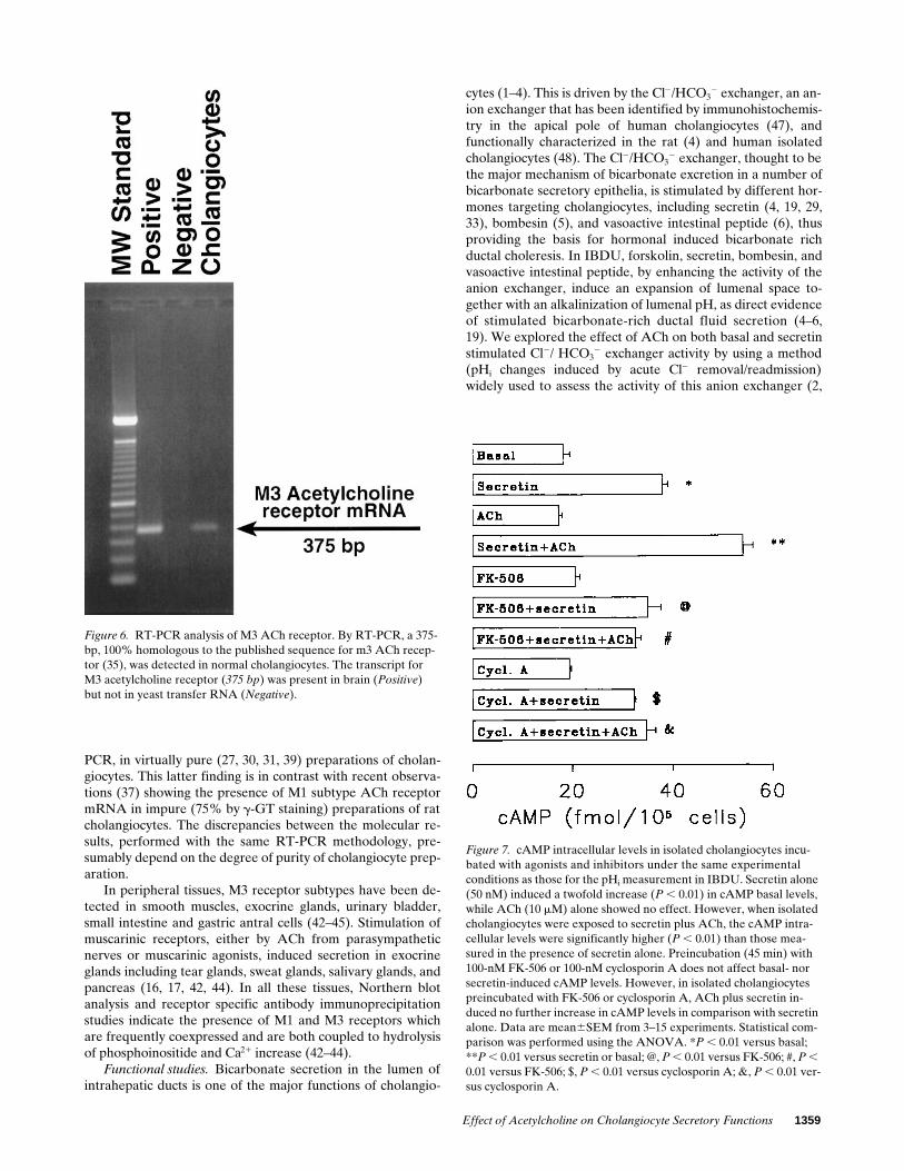
Molecular analysis by RT-PCR shows that cholangiocytesfrom normal rat liver express the transcript for CK-19, a spe-cific marker of cholangiocytes (3, 34). As shown in Fig. 6, the
transcript for the M3 ACh receptor (375 bp) was present inbrain (positive controls) but not in yeast transfer RNA (nega-tive control). By RT-PCR, a 375-bp, 100% homologous to thepublished sequence for the M3 ACh receptor (35), was de-tected in normal cholangiocytes.
On the contrary, we did not detect by RT-PCR the pres-ence of the transcript for the M1 muscarinic ACh receptor inpure preparations of cholangiocytes from normal rat liver(three cell preparations).
cAMP intracellular levels in isolated cholangiocytes. Fig. 7shows intracellular cAMP levels in isolated cholangiocytesincubated with agonists and inhibitors under experimentalconditions similar to those used for the pHi measurement inIBDU. Secretin alone induced a twofold increase in cAMPbasal levels (38.061.08 versus 18.1361.369 fmol/105 cells; P ,0.01), while ACh alone (17.2260.653 fmol/105 cells) showedno effect. However, when isolated cholangiocytes were ex-posed to secretin plus ACh, the cAMP intracellular levels(54.1762.017 fmol/105 cells), were significantly higher (P ,0.01) than those measured in the presence of secretin alone.Similar findings were obtained with the muscarinic agonist,carbachol (100 mM), which failed to significantly influence thebasal values of cAMP (20.9360.408 versus 17.65462.338 fmol/105 cells), but significantly increased the cAMP levels inducedby secretin alone (carbachol plus secretin 5 66.9562.88 versussecretin alone 5 38.7761.908 fmol/105 cells; P , 0.01).
Basal cAMP levels, in isolated cholangiocytes preincubated(45 min) with 100-nM FK-506 (20.5460.872 fmol/105 cells) or100-nM cyclosporin A (19.3660.41 fmol/105 cells), were simi-lar to the values in untreated cells (Fig. 7). In addition, se-
Figure 5. Effect of the calcineurin inhibitor, FK-506, on ACh plus secretin–induced stimulation of Cl2/HCO32 exchanger activity. IBDU, prein-
cubated (30–45 min) with 100-nM FK-506, were submitted to two sequential Cl2 removal/readmission maneuvers, the second during administra-tion of secretin (top right) or secretin plus ACh (bottom right). The findings were compared with similar paired experiments performed in the ab-sence of FK-506 (SECRETIN, top left; secretin plus ACh, bottom left). The maximal rate of alkalinization after Cl2 removal and the maximal rate of pHi recovery after Cl2 readmission measured during exposure to secretin were similar both in the absence and presence of FK-506. In contrast, in IBDU treated with FK-506, ACh failed to increase the response rate to secretin.

1358 Alvaro et al.
major subtypes (M1–M4) with different tissue distribution,pharmacology, and functions (42–46). At least five distinct butrelated muscarinic genes (m1–m5) have been demonstratedrecently by molecular cloning studies, but the expressed prod-uct of the m5 receptor gene does not yet have an equivalentfunctional pharmacological profile nor a defined tissue local-ization (42–46). From a functional point of view, M1, M3, andM5 are preferentially coupled to hydrolysis of phosphoinosi-tide, while M2 and M4 to the inhibition of the adenylyl cyclaseactivity (42–46). In this study we used a monoclonal antibodyto detect the occurrence of muscarin receptors in IBDU by im-munofluorescence and immunoelectron microscopy. The anti-body used (M35) recognizes both M2 and M3 subtype recep-tors (21–23), but the immunofluorescence study performed byusing another antibody specific for M2 subtypes yielded nega-tive findings, suggesting that those findings obtained with M35are related to the distribution of M3 receptor subtypes. Theseappeared extensively represented in cholangiocytes withoutdifferences relative to the size of the ducts, but with a signifi-cantly higher staining of the basolateral compared to the apicalmembranes. In addition, at the ultrastructural level there was apreferential localization at the level of invaginations of the cellmembrane with subcellular features reminiscent of coated pits,suggesting a process of receptor-mediated endocytosis. Theoccurrence of M3 receptor subtypes has also been confirmedby the RT-PCR analysis performed in pure preparations of ratcholangiocytes. On the contrary, M2 subtype receptors werenot found by indirect immunofluorescence in IBDU and, thetranscript for M1 subtype receptor, was not detected by RT-
Table V. Effect of the Calcineurin Inhibitor, FK-506, on Secretin and Secretin plus ACh Stimulation of the Cl2/HCO32 Exchanger
Activity in IBDU
Control Secretin FK-506FK-506 plus
secretin
n 5 13 n 5 13
Basal pHi 7.2060.013 7.2160.013 7.1860.014 7.1760.014Cl2 removal
Delta pHi 0.2360.016 0.2860.017 0.2260.015 0.2660.017pH, max U/min 0.12560.013 0.20460.017* 0.13060.014 0.22160.020*H1 flux, mM/min 7.4160.693 12.0361.102* 8.0260.995 13.1361.133*
Cl2 readmissionpH, max U/min 0.16760.015 0.27760.019* 0.17560.017 0.26960.019*H1 flux, mM/min 10.8161.067 16.73161.408* 11.05361.116 17.1661.243*
ControlACh plussecretin FK-506
FK-506 1secretin1 ACh
n 5 18 n 5 18
Basal pHi 7.2160.012 7.1960.013 7.2360.012 7.2260.013Cl2 removal
Delta pHi 0.2460.013 0.3260.016 0.2660.012 0.2960.013pH, max U/min 0.13560.010 0.27560.016*§ 0.14260.014 0.21760.017*‡
H1 flux, mM/min 8.1160.723 16.9361.143*§ 8.0560.786 11.9961.081*‡
Cl2 readmissionpH, max U/min 0.18760.015 0.35160.022*§ 0.18160.014 0.26060.017*‡
H1 flux, mM/min 11.46361.206 22.76661.699*§ 10.90261.021 15.63661.111*‡
Two sequential Cl2 removal/readmission maneuvers were performed, the second during exposure to secretin (50 nM) or ACh (10 mM) plus secretinin IBDU pretreated and perfused with FK-506, a calcineurin inhibitor. Data were compared with similar paired experiments performed in the absenceof FK-506. *P , 0.02 versus control values. ‡P , 0.03 versus ACh plus secretin. §P , 0.05 versus secretin alone. Data are mean6SEM.
cretin induced a cAMP increase similar to untreated cells(35.0362.70 fmol/105 cells with FK-506 plus secretin and32.2560.358 fmol/105 cells with cyclosporin A plus secretin;P , 0.01 versus FK-506 or cyclosporin A alone) (Fig. 7). Incontrast, in isolated cholangiocytes preincubated with FK-506or cyclosporin A, ACh plus secretin induced no further in-crease in cAMP levels (32.6061.08 and 34.6661.91 fmol/105
cells, respectively) in comparison with secretin alone. Theseexperiments indicate that ACh potentiation of secretin-stimu-lated cAMP accumulation is dependent on calcineurin activityin that it is blocked by two distinct inhibitors of calcineurin.
Discussion
The main findings emerging from this study are that: (a) M3ACh receptors are present in IBDU of different sizes, whileM1 and M2 subtypes are absent; (b) ACh has no effect on thebasal activity of the Cl2/HCO3
2 exchanger, but it significantlypotentiates the stimulatory effect of secretin on this anion ex-changer; (c) the effect of ACh is abolished by 4-DAMP, a mus-carinic receptor antagonist with high affinity for M3 subtype;(d) the ACh-induced potentiation of the secretin stimulatoryeffect on the Cl2/HCO3
2 exchanger appears to be mediated byintracellular Ca21 as it is sensitive to the intracellular Ca21 che-lator BAPTA/AM, but is not mediated by PKC as it is stauro-sporine insensitive; and (e) ACh acts by potentiating the secre-tin induced cAMP intracellular levels, through a calcineurinmediated regulation of adenylyl cyclase activity.
ACh muscarinic receptors are presently classified into four
Effect of Acetylcholine on Cholangiocyte Secretory Functions 1359
PCR, in virtually pure (27, 30, 31, 39) preparations of cholan-giocytes. This latter finding is in contrast with recent observa-tions (37) showing the presence of M1 subtype ACh receptormRNA in impure (75% by g-GT staining) preparations of ratcholangiocytes. The discrepancies between the molecular re-sults, performed with the same RT-PCR methodology, pre-sumably depend on the degree of purity of cholangiocyte prep-aration.
In peripheral tissues, M3 receptor subtypes have been de-tected in smooth muscles, exocrine glands, urinary bladder,small intestine and gastric antral cells (42–45). Stimulation ofmuscarinic receptors, either by ACh from parasympatheticnerves or muscarinic agonists, induced secretion in exocrineglands including tear glands, sweat glands, salivary glands, andpancreas (16, 17, 42, 44). In all these tissues, Northern blotanalysis and receptor specific antibody immunoprecipitationstudies indicate the presence of M1 and M3 receptors whichare frequently coexpressed and are both coupled to hydrolysisof phosphoinositide and Ca21 increase (42–44).
Functional studies. Bicarbonate secretion in the lumen ofintrahepatic ducts is one of the major functions of cholangio-
cytes (1–4). This is driven by the Cl2/HCO32 exchanger, an an-
ion exchanger that has been identified by immunohistochemis-try in the apical pole of human cholangiocytes (47), andfunctionally characterized in the rat (4) and human isolatedcholangiocytes (48). The Cl2/HCO3
2 exchanger, thought to bethe major mechanism of bicarbonate excretion in a number ofbicarbonate secretory epithelia, is stimulated by different hor-mones targeting cholangiocytes, including secretin (4, 19, 29,33), bombesin (5), and vasoactive intestinal peptide (6), thusproviding the basis for hormonal induced bicarbonate richductal choleresis. In IBDU, forskolin, secretin, bombesin, andvasoactive intestinal peptide, by enhancing the activity of theanion exchanger, induce an expansion of lumenal space to-gether with an alkalinization of lumenal pH, as direct evidenceof stimulated bicarbonate-rich ductal fluid secretion (4–6,19). We explored the effect of ACh on both basal and secretinstimulated Cl2/ HCO3
2 exchanger activity by using a method(pHi changes induced by acute Cl2 removal/readmission)widely used to assess the activity of this anion exchanger (2,
Figure 6. RT-PCR analysis of M3 ACh receptor. By RT-PCR, a 375-bp, 100% homologous to the published sequence for m3 ACh recep-tor (35), was detected in normal cholangiocytes. The transcript for M3 acetylcholine receptor (375 bp) was present in brain (Positive) but not in yeast transfer RNA (Negative).
Figure 7. cAMP intracellular levels in isolated cholangiocytes incu-bated with agonists and inhibitors under the same experimental conditions as those for the pHi measurement in IBDU. Secretin alone (50 nM) induced a twofold increase (P , 0.01) in cAMP basal levels, while ACh (10 mM) alone showed no effect. However, when isolated cholangiocytes were exposed to secretin plus ACh, the cAMP intra-cellular levels were significantly higher (P , 0.01) than those mea-sured in the presence of secretin alone. Preincubation (45 min) with 100-nM FK-506 or 100-nM cyclosporin A does not affect basal- nor secretin-induced cAMP levels. However, in isolated cholangiocytes preincubated with FK-506 or cyclosporin A, ACh plus secretin in-duced no further increase in cAMP levels in comparison with secretin alone. Data are mean6SEM from 3–15 experiments. Statistical com-parison was performed using the ANOVA. *P , 0.01 versus basal; **P , 0.01 versus secretin or basal; @, P , 0.01 versus FK-506; #, P , 0.01 versus FK-506; $, P , 0.01 versus cyclosporin A; &, P , 0.01 ver-sus cyclosporin A.

1360 Alvaro et al.
4–6, 19, 25, 26, 29). The rationale for studying the coordinatedeffect of secretin and ACh derives from the fact that secretintargets the intrahepatic biliary epithelium in a period (diges-tive phase) of parasympathetic predominance (9, 10). We havefound that ACh alone induced no significant stimulation of theCl2/HCO3
2 exchanger activity. This is in agreement with aprevious study (18) showing that ACh alone failed to stimulatefluid secretion in the lumenal space of rat IBDU in spite of asignificant increase of cytosolic Ca21 signaling. However, whenadministered alone, ACh failed to stimulate fluid secretion(18) and the activity of the anion exchanger, but potentiatedthe secretin stimulation of the Cl2/HCO3
2 exchanger activityby z 50%. The ACh-induced potentiation of the secretin ef-fect on Cl2/HCO3
2 exchanger activity was blocked by 4-DAMP,a muscarinic receptor antagonist which possesses 10–20-foldfunctional selectivity toward M3 over M2 receptors, but whichcannot functionally discriminate between M1 and M3 muscar-inic receptors (21–23, 42). Having excluded by RT-PCR theoccurrence of M1 receptor subtypes, the sensitivity to 4-DAMPindicates that the effects of ACh are mediated by the activationof M3 subtype muscarinic receptors. An aspecific effect of4-DAMP may be excluded since the stimulation of Cl2/HCO3
2
exchanger activity induced by secretin alone was not influenced.ACh signal transduction pathway. ACh is known to elicit
Ca21 signaling in IBDU involving both influx of extracellularCa21 and mobilization of thapsigargin-sensitive Ca21 stores(18). We have shown that the intracellular Ca21 chelatingagent, BAPTA/AM, blocks the effect of ACh on secretin stim-ulation of Cl2/HCO3
2 exchanger activity. Thus, Ca21 shouldbe considered the primary mediator of ACh-induced potentia-tion of secretin effect on the Cl2/HCO3
2 exchanger activity.ACh does not appear to act via a Ca21-activated PKC, sincethe PKC antagonist staurosporine failed to block potentiationof secretin-stimulated Cl2/HCO3
2 exchanger activity inducedby ACh. For both BAPTA/AM and staurosporine, an aspe-cific toxic effect could be excluded by the fact that these sub-stances did not influence the intracellular pHi, the basal activ-ity of the Cl2/HCO3
2 exchanger, nor the rate of response tosecretin alone. To further elucidate the ACh mechanism of ac-tion, we measured cAMP intracellular levels in isolated chol-angiocytes. While ACh alone failed to influence the intra-cellular levels of cAMP, when administered together withsecretin, ACh doubled the secretin-induced cAMP intracellu-lar accumulation indicating a sensitization of adenylyl cyclaseto secretin. An effect of ACh on cAMP breakdown may be ex-cluded since measurement of cAMP intracellular levels havebeen performed in the presence of IBMX concentration thatshould fully inhibit soluble phosphodiesterases. An increase inadenylyl cyclase activity has been reported in different celltypes after stimulation of M1, M3, and M5 receptor subtypesand this effect is thought to occur by activation, via IP3/Ca21,of Ca21-sensitive adenylyl cyclase isoforms (42, 49–53). On thecontrary, stimulation of M2/M4 subtype receptors generallyhas been coupled with an inhibition of a previously stimulatedadenylyl cyclase, via pertussis toxin-sensitive G proteins (42–46, 49). However, M2 receptor subtypes have been not de-tected by immunofluorescence in IBDU and M4 subtypeshave only been detected so far in the rat striatum and rabbitlung, but not in the pancreas, intestine, or salivary glands (42–46, 49).
Thus our findings indicate that ACh elicits a sensitizationof the adenylyl cyclase complex to cAMP agonists, such as se-
cretin, by a mechanism of interaction at the second messengerlevel. This mechanism is responsible of the ACh-induced po-tentiation of the secretin stimulatory effect on the Cl2/HCO3
2
exchanger. In secretory epithelia, including colonic cell lines(53), intestinal epithelia (54), pancreatic acini (51, 55), salivaryglands (16) and gastric chief cells (56), cross-talk between theCa21 and cAMP signaling pathways has been shown to be ofrelevance in the regulation of secretory processes. It is knownthat Ca21 may regulate cAMP production, but the mechanismsof the cross-talk between the two signaling pathways have onlyrecently begun to be investigated at a molecular level. PKC, Gproteins, Ca21–calmodulin complex, and calcineurin have beendifferently involved as regulatory components of the cascade(53–58). The cross-talk between Ca21 and cAMP signalingpathways may be responsible for either potentiating or termi-nating agonist-stimulated second messenger level, dependingon the cell system under investigation. In our cell system, thecross-talk between signaling pathways seems to be mediatedby calcineurin, since two distinct calcineurin inhibitors (cy-closporin A and FK-506), without influencing other cell func-tion parameters, blocked the stimulatory effect of ACh onsecretin-induced cAMP levels and Cl2/HCO3
2 exchanger ac-tivity. Moreover, the rabbit anti–protein phosphatase 2Bapolyclonal antibody (i.e., calcineurin) yielded a specific reac-tion product on normal IBDU, as shown by indirect immuno-fluorescence (personal observation). The inhibitory effect ofFK-506 and cyclosporin A on calcineurin activity has beenwidely documented in many different cell types where it de-pends on binding to the immunophillins FK-binding protein 12(FK506) and cyclophillin (cyclosporine) (40, 41). Calcineurin isa Ca21-calmodulin–dependent serine/threonine phosphatase2B, which plays a role in signal transduction in different secre-tory epithelia other than in nervous tissues (40, 41). Cal-cineurin activation, in fact, induces enzyme secretion in pan-creatic acini, zymogen granule fusion in parotid glands, andpepsinogen secretion in gastric chief cells (55, 56, 59). In anumber of different cell types (52, 55, 56, 59), Ca21 agonistshave been shown to regulate adenylyl cyclase activity by aCa21-calmodulin–dependent activation of calcineurin that inturn selectively acts on certain adenylyl cyclase isoforms. Atleast ten different isoforms of adenylyl cyclases have been de-scribed to date (60–65). Other than having distinct tissue distri-bution profiles, they largely differ with respect to regulation byG proteins, Ca21 calmodulin, calcineurin and PKC (60–65).Isoforms I, III, and VIII have been reported to be stimulatedby the Ca21-calmodulin complex and/or calcineurin (63–65). Indispersed gastric chief cells, Ca21 mobilizing agents, includingcholinergic agonists, lead to a potentiation of cholera toxin-and forskolin-induced pepsinogen secretion by an augmenta-tion of cAMP cellular levels and, recently, calcineurin has beenproposed as the mediator of the cross-talk between the Ca21 andadenylyl cyclase signaling pathways (56). In bovine adrenalglomerulosa cells (52), angiotensin II induced enhancement ofACTH-stimulated cAMP formation, an effect partially medi-ated by PKC (staurosporine sensitivity) and completely by cal-cineurin (inhibition by FK-506 and cyclosporine). In this study,a regulation of G proteins and/or adenylyl cyclase activities byphosphorylation/dephosphorylation steps (PKC/calcineurin)has been suggested as the underlying mechanism of the cross-talk between Ca21 and cAMP. On the contrary, in other cell sys-tems, including mouse pituitary tumor (AT10) cells (57), cal-cineurin exerts a feedback inhibition of agonist-evoked cAMP

Effect of Acetylcholine on Cholangiocyte Secretory Functions 1361
formation, an effect also documented in purified bovine brainadenynyl cyclase (66). Expression of different isoforms of ade-nylyl cyclase or G proteins could explain the divergent effectsof Ca21 documented in different cell types.
Since ATP, like ACh, elicits cytosolic Ca21 signaling inIBDU without a direct effect on fluid secretion (18), the inter-action of ATP with secretin or other cAMP agonists in modu-lating ductal fluid secretion should be investigated in light ofthe present findings.
In conclusion, this study provides morphological and func-tional evidence that ACh plays a role in the regulation ofbicarbonate secretion from biliary ducts. This regulation prob-ably takes place during the digestive phase when the parasym-pathetic system predominates and secretin targets the biliaryepithelium. In this phase the bicarbonate requirement in theintestine is maximal. By acting on M3 receptor subtypes, AChinduces a Ca21-calcineurin mediated potentiation of the secre-tin-induced adenylyl cyclase activity. The doubled cAMP in-tracellular levels generate an almost maximal stimulation ofCl2/HCO3
2 exchanges and bicarbonate excretion in the ductallumen. The modulating action of Ca21 on adenylyl cyclasecould serve as a means of amplifying the secretory responseand prevent the impairment of the overall secretory process.
Acknowledgments
This work was supported by Consiglio Nazionale olelle Ricerche (Pro-getto finalizzato FATMA, 94.00527PF41), by CNR (96.03444.CT04),by CNR (96.03445.CT04).
References
1. Roberts, S.K., and N.F. LaRusso. 1994. Pathobiology of biliary epithelia.Curr. Opin. Gastroenterol. 10:526–533.
2. Boyer, J.L. 1996. Bile duct epithelium: frontiers in transport physiology.Am. J. Physiol. 270:G1–G5.
3. Alpini, G., I.O. Phillips, and N.F. LaRusso. 1994. The biology of biliaryepithelia. In The Liver: Biology and Pathobiology. I.M. Arias, N. Fausto, W.B.Jacoby, D.A. Schachter, and D.A. Shafritz, editors. Raven Press, Ltd., NewYork. 623–653.
4. Alvaro, D., W.K. Cho, A. Mennone, and J.L. Boyer. 1993. Effect of se-cretin on intracellular pH regulation in isolated rat bile duct epithelial cells. J.Clin. Invest. 92:1314–1325.
5. Cho, W.K., A. Mennone, and J.L. Boyer. 1995. Effect of bombesin on se-cretion in isolated polarized intrahepatic bile ductular units (IBDU). Gastroen-terology. 108:A1049.
6. Cho, W.K., A. Mennone, S.A. Rydberg, and J.L. Boyer. 1995. VIP is apotent stimulus of bicarbonate and fluid secretion in bile ducts. Hepatology. 22:A294.
7. Tietz, P.S., G. Alpini, L.D. Pham, and N.F. LaRusso. 1995. Somatostatininhibits secretin-induced ductal hypercholeresis and exocytosis by cholangio-cytes. Am. J. Physiol. 269:G110–G118.
8. Glaser, S.S., R. Rodgers, J. Phinizy, W.E. Robertson, J. Lasater, G.LeSage, and G. Alpini. 1996. Gastrin inhibits secretin-induced hypercholeresisin the bile duct ligated rat liver through the cAMP system. Gastroenterology.110:1197.
9. Walsh, J.H., and E.A. Mayer. 1993. Gastrointestinal hormones. In Gas-trointestinal Disease, 5th Ed. M.H. Sleisenger and J.S. Fordtran, editors. W.B.Saunders Company Ltd., London. 18–44.
10. Walsh, J.H. Gastrointestinal hormones and peptides. 1981. In Physiol-ogy of the Gastrointestinal Tract. L.R. Johnson, editor. Raven Press, Ltd., NewYork. 59–144.
11. Tanturi, C.A., and A.C. Ivy. 1938. On the existence of secretory nervesin the vagi for and the reflex excitation and inhibition of bile secretion. Am. J.Physiol. 121:270–283.
12. Kaminski, D.L., J. Dorighi, and M. Jellinek. 1994. Effect of electrical va-gal stimulation on canine hepatic bile flow. Effect of electrical vagal stimulationon canine hepatic bile flow. Am. J. Physiol. 227:487–493.
13. Pass, M., and T. Heath. 1976. Effect of electrical stimulation of the vagusnerves on bile secretion in anaesthetized sheep. Aust. J. Biol. Sci. 29:351–355.
14. Nahrwold, D.L., D.L. Kaminski, and R.C. Rose. 1972. Cholinergic con-
trol of bile flow and composition. Physiologist. 15:224.15. Frizzel, R.A., and A.P. Morris. 1994. Chloride conductance of salt-
secreting epithelial cells. In Current Topics in Membranes. Academic Press,Inc., Orlando. 42:173–214.
16. Larsson, O., and L. Olgart, 1989. The enhancement of carbachol-inducedsalivary secretion by VIP and CGRP in rat parotid gland is mimicked by forsko-lin. Acta Physiol. Scand. 137:231–236.
17. Sato, K., and F. Sato. 1981. Role of calcium in cholinergic and adrener-gic mechanisms of eccrine sweat secretion. Am. J. Physiol. 241:C113–C120.
18. Nathanson, M.H., A.D. Burgstahler, A. Mennone, and J.L. Boyer. 1996.Characterization of cytosolic Ca21 signaling in rat bile duct epithelia. Am. J.Physiol. 271:G86–G96.
19. Mennone, A., D. Alvaro, W. Cho, and J.L. Boyer. 1995. Isolation ofsmall polarized bile duct units. Proc. Natl. Acad. Sci. USA. 92:6527–6531.
20. Benedetti, A., L. Marucci, C. Bassotti, R. Mancini, S. Contucci, A.M.Jezequel, and F. Orlandi. 1993. Tubulo-vesicular transcytotic pathway in rat bil-iary epithelium: a study in perfused liver and in isolated intrahepatic bile duct.Hepatology. 18:422–432.
21. Andre, C., J.B. DeBacker, J.P. Guillet, J.C. Venderheyden, P. Vauque-lin, and A.D. Strosberg. 1983. Purification of muscarinic acetylcholine receptorsby affinity chromatography. EMBO (Eur. Mol. Biol. Organ.) J. 2:499–504.
22. Andre, C., J.P. Guillet, J.P. DeBacker, J.G. Venderheyden, J. Hoebeke,and A.D. Strosberg. 1984. Monoclonal antibodies against the native or denatur-ated forms of muscarinic acetylcholine receptor. EMBO (Eur. Mol. Biol. Or-gan.) J. 3:17–21.
23. Andre, C., S. Marullo, J.G. Guillet, A. Convents, M. Lauwereys, S.Kaveri, J. Hoebeke, and A.D. Strosberg. 1987. Immunochemical studies of themuscarinic acetylcholine receptor. J. Recept. Res. 7:65–107.
24. Takahashi, T., J.M. Lasker, A.S. Rosman, and C.S. Lieber. 1993. Induc-tion of cytochrome P-4502E1 in the human liver by ethanol is caused by a corre-sponding increase in encoding messenger RNA. Hepatology. 17:236–245.
25. Alvaro, D., P. Della Guardia, A. Bini, A. Gigliozzi, S. Furfaro, T. LaRosa, C. Piat, and L. Capocaccia. 1995. Effect of glucagon on intracellular pHregulation in isolated rat hepatocyte couplets. J. Clin. Invest. 96:665–675.
26. Alvaro, D., A. Mennone, and J.L. Boyer. 1993. Effect of ursodeoxy-cholic acid on intracellular pH regulation in isolated rat bile duct epithelialcells. Am. J. Physiol. 265:G783–G791.
27. Ishii, M., B. Vroman, and N.F. LaRusso. 1989. Isolation and morpho-logic characterization of bile duct epithelial cells from normal rat liver. Gastro-enterology. 97:1236–1247.
28. Alpini, G., S. Roberts, J. Phinizy, W. Robertson, S. Gubba, O. Colegio,G. LeSage, and N.F. LaRusso. 1995. Heterogeneity of the proliferative capacityof rat cholangiocytes. Gastroenterology. 1098:A1025. (Abstr.)
29. LeSage, G., S.S. Glaser, S. Gubba, W.E. Robertson, J.L. Phinizy, J. La-sater, R.E. Rodgers, and G. Alpini. 1996. Regrowth of the rat biliary tree after70% partial hepatectomy is coupled to increased secretin-induced ductal secre-tion. Gastroenterology. 111:1633–1644.
30. Alpini, G., G.D. Ulrich, J.O. Phillips, L.D. Pham, L.J. Miller, and N.F.LaRusso. 1994. Upregulation of secretin receptor gene expression in rat cholan-giocytes after bile ligation. Am. J. Physiol. 266:G922–G928.
31. Alpini, G., S.K. Roberts, S.M. Kuntz, Y. Ueno, S. Gubba, P. Podila, G.LeSage, and N.F. LaRusso. 1996. Morphological, molecular and functional het-erogeneity of cholangiocytes from normal rat liver. Gastroenterology. 110:1636–1643.
32. Rutemburg, A.M., H. Kim, J.W. Fischbein, J.S. Hanker, H.L. Wasser-krug, and A.M. Seligman. 1969. Histochemical and ultrastructural demonstra-tion of gamma-glutamyl transpeptidase activity. J. Histochem. Cytochem. 17:517–526.
33. Alpini, G., R. Lenzi, L. Sarkozi, and N. Tavoloni. 1988. Biliary physiol-ogy in rats with bile ductular cell hyperplasia. Evidence for a secretory functionof proliferated bile ductules. J. Clin. Invest. 81:569–578.
34. Alpini, G., R. Lenzi, W.R. Zhai, M.H. Liu, P.A. Slott, F. Paronetto, andN. Tavoloni. 1989. Isolation of a nonparenchymal liver cell fraction enriched incells with biliary epithelial phenotypes. Gastroenterology. 97:1248–1260.
35. Bonner, T., N. Buckely, A. Young, and M. Brann. 1987. Identification ofa family of muscarinic acetylcholine genes. Science (Wash. DC). 237:527–532.
36. Peralta, E.G., J.W. Winslow, G.L. Peterson, D.H. Smith, A. Ashkenazi,J. Ramachandran, M.I. Schimerlik, and D.J. Capon. 1987. Primary structureand biochemical properties of an M2 muscarinic receptor. Science (Wash. DC).236:600–606.
37. Elsing, C., C. Hübner, B.A. Fitscher, A. Kassner, and W. Stremmel.1997. Muscarinic acetylcholine receptor stimulation of biliary epithelial cellsand its effect on bile secretion in the isolated perfused rat liver. Hepatology. 25:804–813.
38. Calnek, D., and A. Quaroni. 1993. Differential localization by in situ hy-bridization of distinct keratin mRNA species during intestinal epithelial cell de-velopment and differentiation. Differentiation. 53:95–104.
39. Kato, A., G.J. Gores, and N.F. LaRusso. 1992. Secretin stimulates exo-cytosis in isolated bile duct epithelial cells by a cyclic AMP-mediated mecha-nism. J. Biol. Chem. 267:15523–15529.
40. Morris, R. 1994. Model of action of FK506, cyclosporin A and rapamy-cin. Transplant. Proc. 26:3272–3275.
1362 Alvaro et al.
41. Schreiber, S.L., and G.R. Crabtree. 1992. The mechanism of action ofcyclosporin A and FK506. Immunol. Today. 13:136–142.
42. Caulfield, M.P. 1993. Muscarinic receptors—Characterization, couplingand function. Pharmacol. Ther. 58:319–379.
43. Dorje, F., A.I. Levey, and M.R. Brann. 1991. Immunological detectionof muscarinic receptor subtype proteins (m1-m5) in rabbit peripheral tissue.Mol. Pharmacol. 40:459–462.
44. Maeda, A., T. Kubo, M. Mishina, and S. Numa. 1988. Tissue distributionof mRNAs encoding muscarinic acetylcholine receptor subtypes. FEBS Lett.239:339–342.
45. Weigert, N., K. Schaffer, U. Wegner, V. Schusdziarra, M. Classen, andW. Schepp. 1994. Functional characterization of a muscarinic receptor stimulat-ing gastrin release from rabbit antral G-cells in primary culture. Eur. J. Phar-macol. 264:337–344.
46. Barlow, R.B., K.J. Berry, P.A.M. Glenton, N.M. Nikolaou, and K.S.Soh. 1976. A comparison of affinity constant for muscarinic-sensitive acetylcho-line receptors in guinea-pig atrial pacemaker cells at 298C and in ileum at 298Cand 378C. Br. J. Pharmacol. 58:613–620.
47. Martinez-Anso, E., J.E. Castillo, J. Diez, J.F. Medina, and J. Prieto.1994. Immunohistochemical detection of chloride/bicarbonate anion exchangerin human liver. Hepatology. 19:1400–1406.
48. Strazzabosco, M., R. Joplin, A. Zeembery, A, Granato, C. Spirli, C.Poci, and S. Crepaldi 1994. Bicarbonate transport in cultured human intrahe-patic bile duct epithelial cells (hBDE) from normal subjects and patients withprimary biliary cirrhosis (PBC). Hepatology. 2:404A.
49. Jones, S.V.P., A.I. Levey, D.M. Weiner, J. Ellis, E. Novotny, S.H. Yu, F.Dorje, J. Wess, and M.R. Brann. 1992. Muscarinic acetylcholine receptors. InMolecular Biology of G-protein Coupled Receptors. M. Brann, editor. Birk-hauser Boston, Cambridge, MA. 170–197.
50. Felder, C.C., R.Y. Kanterman, A.L. Ma, and J. Axelrod. 1989. A trans-fected m1 muscarinic acetylcholine receptor stimulates adenylate cyclase via phos-phatidylinositol hydrolysis. J. Biol. Chem. 264:20356–20362.
51. Pan, G.-Z., M.J. Collen, and J.D. Gardner. 1982. Action of cholera toxinon dispersed acini from rat pancreas. Post-receptor modulation involving cyclicAMP and calcium. Biochim. Biophys. Acta. 720:338–345.
52. Baukal, A.J., L. Hunyady, K.J. Catt, and T. Balla. 1994. Evidence forparticipation of calcineurin in potentiation of agonist-stimulated cyclic AMPformation by the calcium-mobilizing hormone, Angiotensin II. J. Biol. Chem.269:24546–24549.
53. Warhurst, G., K.E. Fogg, N.B. Higgs, A. Tonge, and J. Grundy. 1994.Ca21-mobilizing agonists potentiate forskolin- and VIP-stimulated cAMP pro-
duction in human colonic cell line, HT29-cl.19A: role of [Ca21]i and protein ki-nase C. Cell Calcium. 15:162–174.
54. Aminaroff, B.M., M.C. Laburthe, C.M. Rouyer-Fessard, J.G. Demaille,and G.E. Rossellin. 1983. Calmodulin stimulation of adenylate cyclase of intes-tinal epithelium. Eur. J. Biochem. 130:33–37.
55. Groblewski, G.E., C.C. Wagner, and J.A. Williams. 1994. Cyclosporin Ainhibits Ca21/calmodulin-dependent protein phosphatase and secretion in pan-creatic acinar cells. J. Biol. Chem. 269:15111–15117.
56. Raufman, J.P., J. Lin, and R.D. Raffaniello. 1996. Calcineurin mediatescalcium-induced potentiation of adenylyl cyclase activity in dispersed chief cellsfrom guinea pig stomach. J. Biol. Chem. 271:19877–19822.
57. Antoni, F.A., R.J.O. Barnard, M.J. Shipston, S.M. Smith, J. Simpson,and J.M. Paterson. 1995. Calcineurin feedback inhibition of agonist-evokedcAMP formation. J. Biol. Chem. 270:28055–28061.
58. Harrison, J.K., G.H.K. Hewlett, and M.E. Gnegy. 1989. Regulation ofcalmodulin-sensitive adenylate cyclase by the stimulatory G-protein, Gs. J.Biol. Chem. 264:15880–15885.
59. Cooperstein, S.J., and D.T. Watkins. 1995. Ca(11)-calmodulin-depen-dent phosphorylation and dephosphorylation of rat parotid secretion granules.Biochem. Biophys. Res. Comm. 215:75–81.
60. Caldwell, K.K., C.L. Boyajian, and D.M.F. Cooper. 1992. The effects ofCa11 and calmodulin on adenylyl cyclase activity in plasma membranes derivedfrom neural and non-neural cells. Cell Calcium. 13:107–121.
61. Cooper, D.M.F., N. Mons, and J.W. Kerpen. 1995. Adenylyl cyclase andthe interaction between calcium and cAMP signalling. Nature (Lond.). 374:421–424.
62. Paterson, J.M., S.M. Smith, A.J. Harmar, and F.A. Antoni. 1995. Con-trol of a novel adenylyl cyclase by calcineurin. Biochem. Biophys. Res. Comm.214:1000–1008.
63. Tang, W.J., J. Krupinski, and A.G. Gilman. 1991. Expression and char-acterization of calmodulin-activated (type I) adenylyl cyclase. J. Biol. Chem.266:8595–8603.
64. Choi, E.J., Z. Xia, and D.R. Storm. 1992. Stimulation of the type III ol-factory adenylyl cyclase by calcium and calmodulin. Biochemistry. 31:6492–6498.
65. Cali, J.J., J.C. Zwaagstra, N. Mons, D.M.F. Cooper, and J. Krupinski.1994. Type VIII adenylyl cyclase. A Ca21/calmodulin-stimulated enzyme ex-pressed in discrete regions of rat brain. J. Biol. Chem. 269:12190–12195.
66. Salter, R.S., M.H. Krinks, C.B. Klee, and E.J. Neer. 1981. Calmodulinactivates the isolated catalytic unit of brain adenylate cyclase. J. Biol. Chem.256:9830–9833.