Excimer laser treatment of copper-coated mild steel
6
JOURNAL OF MATERIALS SCIENCE 32 (1997) 1425 — 1430 Excimer laser treatment of copper-coated mild steel C. N. PANAGOPOULOS, A. MARKAKI Laboratory of Physical Metallurgy, National Technical University of Athens, Zografos, 15 780, Athens, Greece E. HONTZOPOULOS Institute of Electronic Structure and Laser, University of Crete, Iraklion, Greece Copper-coated mild steel specimens were irradiated with a high power excimer laser. Mild steel was first coated with a thin nickel coating. Laser-induced surface structures were observed by means of optical microscopy. The surface roughness was observed to be dependent on the number of pulses per step. Energy dispersive X-ray (EDX) analysis showed that nickel and iron atoms diffused into the copper coating. The microhardness of the copper coating was found to increase after laser treatment due to solid solution strengthening from iron and nickel atoms. 1. Introduction The use of laser beam technology to improve the properties of the near surface region, which can deter- mine the performance of a material, has been exten- sively studied. Laser surface alloying is a fast and efficient technique for producing surface layers with improved properties. The alloying elements can be added by pre- or codeposition methods. Surface treatments by means of laser beam radi- ation are performed today on an industrial and re- search scale mainly by using a continuous CO 2 laser. The most important problem of this laser arises from the high reflectivity of light at 10.6 lm. This problem can be overcome by using Nd—YAG or excimer lasers [radiation in the visible and ultraviolet (u.v.) wavelength ranges, respectively] that have shorter wavelengths and the capabilities of giving very high pulse energies, reducing the problems due to the reflec- tion. However, it should be noted that in comparison with continuous lasers, CO 2 , pulsed Nd—YAG and excimer lasers are not as flexible and emit lower power levels [1]. Some of the recent studies in the technological field of surface alloying with a pulsed laser are listed below: Wang and Spaepen [2] studied the formation of Nb—Si alloys by pulsed laser radiation, using an Nd—YAG laser, or multilayered films of Nb and Si on a copper substrate. The lattice parameters of these solid solutions suggest that the solute atoms can be interstitial or substitutional. Lin and Spaepen [3] using pulsed laser treatment with Nd—YAG created Ni —Nb alloys of multilayered films of Ni and Nb on a copper substrate, which was first coated by an evaporated Al film. This work pre- sented the effects of quenching on the extension of primary solubility and the occurrence of intermetallic compounds. Hirvonen et al. [4] have implanted Ti ions into Fe surfaces forming a thin amorphous surface alloy by excimer laser irradiation. This alloy improves the wear and friction properties of the surface as compared with the unimplanted surface. Jervis et al. [5] studied the formation of amorphous Ti alloy layers by excimer laser mixing of Ti on AISI 304 stainless steel surfaces. The laser mixing process, unlike Ti ion implantation, does not result in high incorporation of C in the processed layer. Michaelides and Panagopoulos [6] examined elec- trodeposited zinc on a copper substrate irradiated with an excimer laser. Zinc atoms were found to dif- fuse into the copper substrate. The surface morpho- logy and structure after laser treatment were exami- ned. Panagopoulos et al. [7] irradiated electroplated zinc on an aluminium substrate with a high power excimer laser. Zinc atoms were found to diffuse into the substrate. The microstructure of the surface after laser treatment was mainly dendritic. The surface roughness was a function of the lasing conditions. Das [8] studied pure aluminium after surface laser alloying with nickel using an Nd—YAG laser pulse. It was observed that the surface roughness of the alloyed surface decreased with an increase in the extent of defocusing of the laser beam. By using a Nd—YAG laser, Tayal and Mukherjee [9] achieved laser boriding of low carbon steel. High hardness values were obtained due to the variety of microstructure. The work described in this paper aimed to produce surface alloys on copper-coated mild steel after inter- action with very short pulses of a high intensity ex- cimer laser beam. The copper coating was elec- trodeposited on mild steel prior to laser treatment. An evaluation of the effect of the lasing conditions on the 0022—2461 ( 1997 Chapman & Hall 1425
Transcript of Excimer laser treatment of copper-coated mild steel
JOURNAL OF MATERIALS SCIENCE 32 (1997) 1425—1430
Excimer laser treatment of copper-coatedmild steel
C. N. PANAGOPOULOS, A. MARKAKILaboratory of Physical Metallurgy, National Technical University of Athens, Zografos,15780, Athens, Greece
E. HONTZOPOULOSInstitute of Electronic Structure and Laser, University of Crete, Iraklion, Greece
Copper-coated mild steel specimens were irradiated with a high power excimer laser. Mild
steel was first coated with a thin nickel coating. Laser-induced surface structures were
observed by means of optical microscopy. The surface roughness was observed to be
dependent on the number of pulses per step. Energy dispersive X-ray (EDX) analysis showed
that nickel and iron atoms diffused into the copper coating. The microhardness of the copper
coating was found to increase after laser treatment due to solid solution strengthening from
iron and nickel atoms.
1. IntroductionThe use of laser beam technology to improve theproperties of the near surface region, which can deter-mine the performance of a material, has been exten-sively studied. Laser surface alloying is a fast andefficient technique for producing surface layers withimproved properties. The alloying elements can beadded by pre- or codeposition methods.
Surface treatments by means of laser beam radi-ation are performed today on an industrial and re-search scale mainly by using a continuous CO
2laser.
The most important problem of this laser arises fromthe high reflectivity of light at 10.6 lm. This problemcan be overcome by using Nd—YAG or excimer lasers[radiation in the visible and ultraviolet (u.v.)wavelength ranges, respectively] that have shorterwavelengths and the capabilities of giving very highpulse energies, reducing the problems due to the reflec-tion. However, it should be noted that in comparisonwith continuous lasers, CO
2, pulsed Nd—YAG and
excimer lasers are not as flexible and emit lower powerlevels [1].
Some of the recent studies in the technological fieldof surface alloying with a pulsed laser are listed below:
Wang and Spaepen [2] studied the formation ofNb—Si alloys by pulsed laser radiation, using anNd—YAG laser, or multilayered films of Nb and Si ona copper substrate. The lattice parameters of thesesolid solutions suggest that the solute atoms can beinterstitial or substitutional.
Lin and Spaepen [3] using pulsed laser treatmentwith Nd—YAG created Ni—Nb alloys of multilayeredfilms of Ni and Nb on a copper substrate, which wasfirst coated by an evaporated Al film. This work pre-sented the effects of quenching on the extension ofprimary solubility and the occurrence of intermetallic
compounds.0022—2461 ( 1997 Chapman & Hall
Hirvonen et al. [4] have implanted Ti ions into Fesurfaces forming a thin amorphous surface alloy byexcimer laser irradiation. This alloy improves the wearand friction properties of the surface as compared withthe unimplanted surface.
Jervis et al. [5] studied the formation of amorphousTi alloy layers by excimer laser mixing of Ti on AISI304 stainless steel surfaces. The laser mixing process,unlike Ti ion implantation, does not result in highincorporation of C in the processed layer.
Michaelides and Panagopoulos [6] examined elec-trodeposited zinc on a copper substrate irradiatedwith an excimer laser. Zinc atoms were found to dif-fuse into the copper substrate. The surface morpho-logy and structure after laser treatment were exami-ned.
Panagopoulos et al. [7] irradiated electroplatedzinc on an aluminium substrate with a high powerexcimer laser. Zinc atoms were found to diffuse intothe substrate. The microstructure of the surface afterlaser treatment was mainly dendritic. The surfaceroughness was a function of the lasing conditions.
Das [8] studied pure aluminium after surface laseralloying with nickel using an Nd—YAG laser pulse. Itwas observed that the surface roughness of the alloyedsurface decreased with an increase in the extent ofdefocusing of the laser beam.
By using a Nd—YAG laser, Tayal and Mukherjee[9] achieved laser boriding of low carbon steel. Highhardness values were obtained due to the variety ofmicrostructure.
The work described in this paper aimed to producesurface alloys on copper-coated mild steel after inter-action with very short pulses of a high intensity ex-cimer laser beam. The copper coating was elec-trodeposited on mild steel prior to laser treatment. An
evaluation of the effect of the lasing conditions on the1425

surface of copper-coated mild steel is made, togetherwith a determination of the elemental concentration inthe alloyed surfaces and hardness measurements.
2. Experimental procedureThe substrate material was mild steel with a carboncontent of 0.07 wt%. Specimens with dimensions of3.0]1.0 cm2 and thicknesses of 0.5 mm were used inthis study.
The specimens were annealed at 550 °C for about2 h, to eliminate residual stresses resulting from ma-chining. The methods used to prepare the surface ofthe substrate for electrodeposition was alkaline clean-ing (in a solution of 0.2 M NaOH at 70 °C for 20 min)and acid pickling with an inhibitor (HCl acid for a fewminutes). Afterwards the specimens were mechanicallypolished with a series of SiC papers (Nos 220, 400, 600and 800).
Copper electrodeposition took place in an acid sul-phate bath. The composition of the bath and theoperating condition were: 220 g l~1 CuSO
4and
60 g l~1 H2SO
4at a temperature, ¹ of 35 °C).
The current density was kept constant at 4 A dm~2.Two thicknesses of copper coating were chosen, i.e. 10and 30 lm.
When copper is to be deposited in an acid sulphatebath, steel must receive a nickel strike. This preventsdeposition of copper by immersion and precludespeeling of the plate [10]. Electrodeposition of nickeltook place in a Watts-type bath. The composition ofthe bath and the operating condition were: 317 g l~1
NiSO4· 6H
2O, 45 g l~1 NiCl
2· 6H
2O and 37 g l~1
H3BO
3at pH"3—3.5 and ¹"57—60 °C).
The current density was kept constant at 5 A dm~2.The thickness of the nickel strike was approximately2 lm.
The copper-coated mild steel specimens were laserirradiated with a Lambda Physik excimer laser usinga KrF gas mixture with wavelength, k, of k"248 nm.The laser pulse had a rise time, s
0of 10 ns and a total
duration, s1, of 29 ns. The beam had a Lorentzian
intensity profile. Processing was done in air with noshield gas. The area of the incident laser beam was1.0]1.0 cm2. Each specimen was irradiated under dif-ferent conditions. The variables of the irradiationwere: the power density and the number of pulses perstep (p.p.s.). In order to observe the influence of eachlaser parameter, all the experiments were carried outso that one parameter was kept constant while theother varied. The power density was varied between150 and 430 MW cm~2 and the pulses per step wasvaried between 10 and 250 p.p.s. The overlap betweensuccessive steps was 30%, the pulse frequency was10 Hz and the surface was given a single scan.
After the laser treatment, surface morphology andcross-sections of the copper-coated mild steel speci-mens were examined by means of a Zeiss opticalmicroscope.
Cross-sections embedded in epoxy were used todetermine the depthwise variation in elemental con-centration with an EDX spectrometer of a scanning
electron microscope (Jeol-6300).1426
The laser-treated specimens were examined witha Philips diffractometer with CoKa radiation(k"0.1791 nm) and an iron filter.
A Mahr Perthen profilometer was used to measurethe roughness of the laser-treated surfaces by movingthe stylus parallel to the laser steps. The parameterestimated for this purpose was the deviation of arith-metic mean of the roughness profile, R
!. For any
particular surface, R!was measured across ten differe-
nt profiles and the mean was taken to be the represen-tative value. The error bars in the corresponding fig-ure indicate the standard deviation of each set ofresults.
Microhardness measurements were made on cross-sections of laser- and non-laser-treated samples, witha Vickers indenter of a Reichert microhardness testinstrument. The load was 8 g and the time of loadingfor each indentation was 15 s. Each hardness valuewas the average of seven measurements and the stan-dard deviation of each set of results was not greaterthan 3%.
3. Results and discussionFig. 1 shows a cross-sectional view of a copper-coatedmild steel specimen. The electrodeposition rate of cop-per from the acid sulphate bath was found to bea linear function of time. The thickness of the coatingwas metallographically defined.
The laser power density, I0
at the surface of thecopper is assumed to be the actual power per unitarea absorbed, A]I
*, where A is the absorptivity
and I*is the incident laser power density. The solution
of the basic heat-transfer equation for the temper-ature, ¹(z, t), as a function of depth, z, into the solidcopper and time, t, is given by the following equation[11]
¹(z, t)"2I
0k CA
jt
pB1@2
exp (!z2/4jt)!z
2erfc
z
(4jt)1@2D(1)
where
erf(s)"2
(p)1@2Ps
0
exp (!x2) dx (2)
Figure 1 Cross-sectional view of a copper-coated mild steel.
is the error function, erfc(s) is the complementaryerror function or 1!erf (s). j is the thermal diffu-sivity, j"k/(q]C) (2) where k is the thermal con-ductivity, q is the density and C the specific heat forunit mass.
The surface temperature, ¹4, of copper during its
radiation (depth, z"0) is given by the following equa-tion
¹4(0, t)"
I0k A
4jt
p B1@2
"
A]I*
k A4jt
p B1@2
(3)
By using Equation 3 it is possible to estimate theincident power density at the point of impact, I
*, for
a given temperature on the surface, ¹*, and pulse
duration, s1
of 29 ns
I*"
k]¹*
A Ap
4js1B1@2
(4)
The thermophysical constants for copper are:k"3.3 J s~1 cm~1K~1, q"8.94 g cm~3, C"0.385 Jg~1K~1.
The absorptivity, A, at a wavelength, k of 248 nmhas been evaluated a little higher than 60% at roomtemperature (Fig. 2, absorptivity"1-reflectivity foropaque materials like copper). The true value in thepresent case can be expected to be lower. The surfaceroughness of the specimens before laser treatment wasfound to be about 0.40 lm. When the surface rough-ness is greater than the wavelength of the beam(0.248 lm), the absorption of radiation decreases dueto multiple reflections in the undulations [12].
By replacing the corresponding values in Equation4, it was found that the threshold value of powerdensity causing copper to melt (¹
."1356 K) by us-
ing an excimer laser KrF* source was approximatelyl."43 MW cm~2. Similarly, the threshold power
density required to vaporize copper (¹7"2863 K)
was approximately I7"90 MWcm~2.
In this study, taking into account the previous cal-culations, the incident power densities (150—430MWcm~2) are enough to cause melting of the surfacelayer of copper. Because more heat is deposited, themelt front propagates inwards by conduction into thedeeper parts of the material until the energy of the
Figure 2 Reflectivity of a number of metals (including copper) as
a function of wavelength: (a) Al, (b) Ag, (c) Pt, (d) Cu, and (e) Au.pulse is totally dissipated. The melt front then stopsand a resolidification front moves back towards thefree surface.
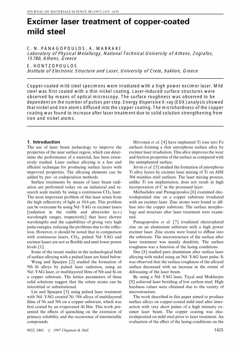
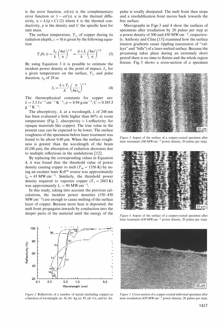
Micrographs in Figs 3 and 4 show the surfaces ofspecimens after irradiation by 20 pulses per step ata power density of 300 and 430 MWcm~2, respective-ly. Anthony and Cline [13] examined how the surfacetension gradients cause rippling (succession of ‘‘val-leys’’ and ‘‘hills’’) of a laser-melted surface. Because theprocessing takes place during an extremely shortperiod there is no time to flatten and the whole regionfreezes. Fig. 5 shows a cross-section of a specimen
Figure 3 Aspect of the surface of a copper-coated specimen afterlaser treatment (300 MWcm~2 power density, 20 pulses per step).
Figure 4 Aspect of the surface of a copper-coated specimen afterlaser treatment (430 MWcm~2 power density, 20 pulses per step).
Figure 5 Cross-section of a copper-coated mild steel specimen after
laser irradiation (430 MWcm~2 power density, 20 pulses per step).1427
laser irradiated at a laser power density of430 MWcm~2. The presence of ‘‘valleys’’ and ‘‘hills’’ isobvious. The ‘‘valleys’’ (dark areas) correspond to de-pressed surfaces under the centre of the beam and the‘‘hills’’ (light areas) to surfaces away from the centre ofthe beam. Examining Figs 3 and 4 it is observed thatthe widths of the ‘‘hills’’ (light areas) remain the sameand correspond to overlaps between successive steps(30%). The widths of the ‘‘valleys’’ (dark areas) arelarger in Fig. 3 than in Fig. 4. The changes in thewidths may be attributed to the different values oflaser power densities (the number of pulses for bothfigures is the same). The formation of ‘‘valleys’’ and‘‘hills’’ is enhanced by the creation of plasma in thecopper surface during its excimer laser radiation (pre-vious calculations show that the incident power dens-ities are sufficient to cause evaporation of the surfacelayer of copper).
Fig. 6 shows the surface roughness, R!, of the laser-
treated specimens as a function of the number ofpulses per step. Increasing the number of pulses leadsto increasing absorption of the incident laser energy(as the temperature rises absorption increases becauseof an increase in the photon population causing morephoton—electron energy exchanges [12]) resulting inan increase of the surface roughness.
In the X-ray diffraction data there was evidence forthe formation of copper oxide, CuO (Fig. 7). Theformation of the oxides may well have been due tostrong melt—environment interactions (the experi-ments were performed in ambient air and the incidentpower densities were much higher than the thresholdvalue of the power density necessary to melt copper).Also, the plasma creates a charged zone of copperatoms that easily react with oxygen atoms (oxygenmolecules decompose due to the high temperature)and form oxides.
During the laser treatment, heat is transferred awayfrom the surface to the internal layers of the coppercoating (copper has very high thermal conductivity).Transport of heat during the laser treatment stimu-lates the mechanism of diffusion in the solid state. Theresults of microanalysis of some laser-treated speci-mens are shown in Figs 8 and 9. It was observed thatiron atoms from the substrate diffuse into the thinnickel coating and then to the copper coating. Nickel
Figure 6 Surface roughness of laser-treated specimens as a function
of the number of pulses per step (300 MW cm~2 power density).1428
Figure 7 X-ray diffractometry data from the laser-treated surface(the thickness of the copper coating is 10 lm. Lasing conditions:300 MWcm~2 power density, 50 pulses per step).
Figure 8 Elemental concentration (at %) as a function of depth(EDX analysis). (e) Fe, (K) Ni, (n) Cu (thickness of copper coating,10 lm; lasing conditions: 430 MWcm~2 power density at 20 pulsesper step).
atoms were found to diffuse into the copper coating.Copper atoms were also found to migrate into the thinnickel coating. Copper and nickel diffusion into themild steel substrate was not detected.
Examining the results of EDX microanalysis, someobservations can be made:
1. As the diffusion distance increases, the number ofdiffused atoms becomes lower.
2. The concentration profiles of iron are a functionof the lasing conditions, in particular, of the powerdensity and the number of pulses per step (Figs 10 and11, respectively). Increasing each parameter separate-ly, shows that diffusion became weaker.
Figs 12 and 13 show the average microhardnessprofiles of copper with depth for different lasing condi-tions.
Comparing the microhardness values before and
after the laser treatment, it can be seen that there is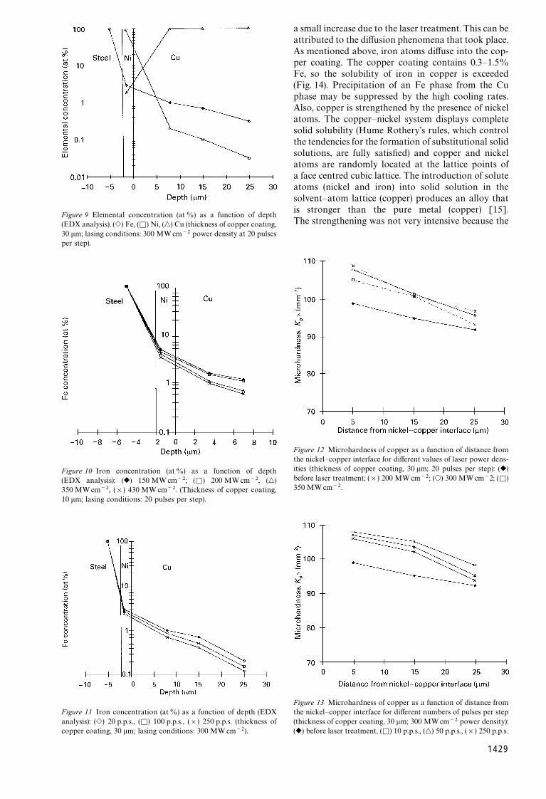
Figure 9 Elemental concentration (at %) as a function of depth(EDX analysis). (e) Fe, (K) Ni, (n) Cu (thickness of copper coating,30 lm; lasing conditions: 300 MWcm~2 power density at 20 pulsesper step).
Figure 10 Iron concentration (at%) as a function of depth(EDX analysis): (r) 150 MW cm~2; (K) 200 MWcm~2, (n)350 MWcm~2, (]) 430 MW cm~2. (Thickness of copper coating,10 lm; lasing conditions: 20 pulses per step).
Figure 11 Iron concentration (at%) as a function of depth (EDXanalysis): (e) 20 p.p.s., (K) 100 p.p.s., (]) 250 p.p.s. (thickness of
copper coating, 30 lm; lasing conditions: 300 MW cm~2).a small increase due to the laser treatment. This can beattributed to the diffusion phenomena that took place.As mentioned above, iron atoms diffuse into the cop-per coating. The copper coating contains 0.3—1.5%Fe, so the solubility of iron in copper is exceeded(Fig. 14). Precipitation of an Fe phase from the Cuphase may be suppressed by the high cooling rates.Also, copper is strengthened by the presence of nickelatoms. The copper—nickel system displays completesolid solubility (Hume Rothery’s rules, which controlthe tendencies for the formation of substitutional solidsolutions, are fully satisfied) and copper and nickelatoms are randomly located at the lattice points ofa face centred cubic lattice. The introduction of soluteatoms (nickel and iron) into solid solution in thesolvent—atom lattice (copper) produces an alloy thatis stronger than the pure metal (copper) [15].The strengthening was not very intensive because the
Figure 12 Microhardness of copper as a function of distance fromthe nickel—copper interface for different values of laser power dens-ities (thickness of copper coating, 30 lm; 20 pulses per step): (r)before laser treatment; (]) 200 MWcm~2; (s) 300 MWcm~2; (K)350 MWcm~2.
Figure 13 Microhardness of copper as a function of distance fromthe nickel—copper interface for different numbers of pulses per step(thickness of copper coating, 30 lm; 300 MWcm~2 power density):
(r) before laser treatment, (K) 10 p.p.s., (n) 50 p.p.s., (]) 250 p.p.s.1429
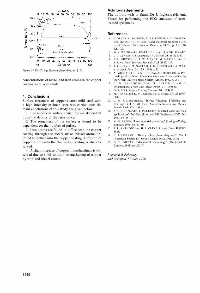
Figure 14 Fe—Cu equilibrium phase diagram [14].
concentrations of nickel and iron atoms in the coppercoating were very small.
4. ConclusionsSurface treatment of copper-coated mild steel witha high intensity excimer laser was carried out: themain conclusions of this study are given below.
1. Laser-induced surface structures are dependentupon the density of the laser power.
2. The roughness of the surface is found to bedependent on the number of pulses.
3. Iron atoms are found to diffuse into the coppercoating through the nickel strike. Nickel atoms arefound to diffuse into the copper coating. Diffusion ofcopper atoms into the thin nickel coating is also ob-served.
4. A slight increase of copper microhardness is ob-served due to solid solution strengthening of copper
by iron and nickel atoms.1430
AcknowledgementsThe authors wish to thank Dr J. Inglessis (HellenicForce) for performing the EDX analyses of laser-treated specimens.
References1. F . OLSEN, C. BAGGER, T. KRISTENSEN, H. JORGEN-
SEN and O. GREGERSEN, ‘‘Laser materials processing’’, 3rdedn (Technical University of Denmark, 1992) pp. 3.5, 3.10,3.11, 7.9.
2. W. K. WANG and F. SPAEPEN, J. Appl. Phys. 58 (1985) 4477.3. C.-J . LIN and F. SPAEPEN, Acta Metall. 34 (1986) 1367.4. J . -P . HIRVONEN, J . W. MAYER, M. NASTASI and D.
STONE, Nucl. Instrum. Methods B 23 (1987) 487.5. T. R. JERVIS, M. NASTASI , T. G. ZOCCO and J . A. MAR-
TIN, Appl. Phys. ¸ett. 53 (1988) p. 75.6. A. MICHAELIDIS and C. N. PANAGOPOULOS, in Pro-
ceedings of the Ninth Greek Conference on Lasers, edited bythe Greek Electro-optical Society, Athens, 1992, p. 250.
7. C. N. PANAGOPOULOS, G. CHRISTOU and G.
DAURELIO, ¹rans. Inst. Metal Finish. 71 (1993) 65.8. D. K. DAS, Surface Coating ¹echnol. 64 (1994) 11.9. M. TAYAL and K. MUKHERJEE, J. Mater. Sci. 29 (1994)
5699.10. L. M. WEISENBERG, ‘‘Surface Cleaning, Finishing and
Coating’’, Vol. 5, 9th Edn (American Society for Metals,Metals Park, OH, 1983).
11. J . T. LUXON and D. E. PARKER, ‘‘Industrial lasers and theirapplications’’, 2nd Edn (Prentice-Hall, Englewood Cliffs, NJ,1992) pp. 251—5.
12. W. M. STEEN, ‘‘Laser material processing’’ (Springer-Verlag,London, 1991) pp. 47—50.
13. T. R. ANTHONY and H. E. CLINE, J. Appl. Phys. 48 (1977)3888.
14. B. MASSALSKI, ‘‘Binary alloy phase diagrams’’, Vol. 1(American Society for Metals, Metals Park, OH, 1986).
15. G. E. DIETER, ‘‘Mechanical metallurgy’’ (McGraw-Hill,London, 1988) pp. 203—7.
Received 6 February
and accepted 17 July 1996.


![A distinct [18F]MPPF PET profile in amnestic mild cognitive impairment compared to mild Alzheimer's disease](https://static.fdokumen.com/doc/165x107/63361f3bb5f91cb18a0bb07c/a-distinct-18fmppf-pet-profile-in-amnestic-mild-cognitive-impairment-compared.jpg)