Evolution of biofilms during the colonization process of pyrite by Acidithiobacillus thiooxidans
13
APPLIED MICROBIAL AND CELL PHYSIOLOGY Evolution of biofilms during the colonization process of pyrite by Acidithiobacillus thiooxidans Dulce M. González & René H. Lara & Keila N. Alvarado & Donato Valdez-Pérez & Hugo R. Navarro-Contreras & Roel Cruz & Jessica Viridiana García-Meza Received: 23 February 2011 /Revised: 22 April 2011 /Accepted: 25 April 2011 /Published online: 20 July 2011 # Springer-Verlag 2011 Abstract We have applied epifluorescence principles, atomic force microscopy, and Raman studies to the analysis of the colonization process of pyrite (FeS 2 ) by sulfuroxid- izing bacteria Acidithiobacillus thiooxidans after 1, 15, 24, and 72 h. For the stages examined, we present results comprising the evolution of biofilms, speciation of S n 2− /S 0 species, adhesion forces of attached cells, production and secretion of extracellular polymeric substances (EPS), and its biochemical composition. After 1 h, highly dispersed attached cells in the surface of the mineral were observed. The results suggest initial non-covalent, weak interactions (e.g., van der Waal’ s, hydrophobic interactions), mediating an irreversible binding mechanism to electrooxidized massive pyrite electrode (eMPE), wherein the initial production of EPS by individual cells is determinant. The mineral surface reached its maximum cell cover between 15 to 24 h. Longer biooxidation times resulted in the progressive biofilm reduction on the mineral surface. Quantification of attached cell adhesion forces indicated a strong initial mechanism (8.4 nN), whereas subsequent stages of mineral colonization indicated stability of biofilms and of the adhesion force to an average of 4.2 nN. A variable EPS (polysaccharides, lipids, and proteins) secre- tion at all stages was found; thus, different architectural conformation of the biofilms was observed during 120 h. The main EPS produced were lipopolysaccharides which may increase the hydrophobicity of A. thiooxidans biofilms. The highest amount of lipopolysaccharides occurred between 15–72 h. In contrast with abiotic surfaces, the progressive depletion of S n 2− /S 0 was observed on biotic eMPE surfaces, indicating consumption of surface sulfur species. All observations indicated a dynamic biooxidation mechanism of pyrite by A. thiooxidans, where the biofilms stability and composition seems to occur independently from surface sulfur species depletion. Keywords Acidithiobacillus thiooxidans . Pyrite . Biofilms evolution . Electrooxidation . Interfacial analysis . Proteins quantification Introduction Interfacial mechanisms describing the development of leaching bacterial biofilms on mineral sulfides (SM) are of great significance for determining the bioleaching pathways of SM. The sulfur-oxidizing microorganisms (SOM: Archaea or Bacteria) play a major role in the cycling of sulfur in the biosphere. SOM processed surface sulfur species (e.g., elemental sulfur, S 0 ; polysulfides, S n 2− ) as a source of energy and reducing power; this metabolic capability has been applied in the mining industry for the bioleaching of metals D. M. González : R. H. Lara : R. Cruz : J. V. García-Meza (*) Geomicrobiology, Institute of Metallurgy, UASLP, Sierra Leona 550, Lomas 2°, 78210 San Luis Potosí, SLP, Mexico e-mail: [email protected] K. N. Alvarado Basics Sciences Laboratory, UASLP, Salvador Nava 110, 78210 SLP, Mexico D. Valdez-Pérez Institute of Physics, UASLP, Salvador Nava 110, 78210 SLP, Mexico H. R. Navarro-Contreras CIACyT, UASLP, Sierra Leona 550, Lomas 2°, 78210 SLP, Mexico Appl Microbiol Biotechnol (2012) 93:763–775 DOI 10.1007/s00253-011-3465-2
-
Upload
independent -
Category
Documents
-
view
1 -
download
0
Transcript of Evolution of biofilms during the colonization process of pyrite by Acidithiobacillus thiooxidans

APPLIED MICROBIAL AND CELL PHYSIOLOGY
Evolution of biofilms during the colonization processof pyrite by Acidithiobacillus thiooxidans
Dulce M. González & René H. Lara & Keila N. Alvarado &
Donato Valdez-Pérez & Hugo R. Navarro-Contreras &
Roel Cruz & Jessica Viridiana García-Meza
Received: 23 February 2011 /Revised: 22 April 2011 /Accepted: 25 April 2011 /Published online: 20 July 2011# Springer-Verlag 2011
Abstract We have applied epifluorescence principles,atomic force microscopy, and Raman studies to the analysisof the colonization process of pyrite (FeS2) by sulfuroxid-izing bacteria Acidithiobacillus thiooxidans after 1, 15, 24,and 72 h. For the stages examined, we present resultscomprising the evolution of biofilms, speciation of Sn
2−/S0
species, adhesion forces of attached cells, production andsecretion of extracellular polymeric substances (EPS), andits biochemical composition. After 1 h, highly dispersedattached cells in the surface of the mineral were observed.The results suggest initial non-covalent, weak interactions(e.g., van der Waal’s, hydrophobic interactions), mediatingan irreversible binding mechanism to electrooxidizedmassive pyrite electrode (eMPE), wherein the initialproduction of EPS by individual cells is determinant. Themineral surface reached its maximum cell cover between 15to 24 h. Longer biooxidation times resulted in the
progressive biofilm reduction on the mineral surface.Quantification of attached cell adhesion forces indicated astrong initial mechanism (8.4 nN), whereas subsequentstages of mineral colonization indicated stability of biofilmsand of the adhesion force to an average of 4.2 nN. Avariable EPS (polysaccharides, lipids, and proteins) secre-tion at all stages was found; thus, different architecturalconformation of the biofilms was observed during 120 h.The main EPS produced were lipopolysaccharides whichmay increase the hydrophobicity of A. thiooxidans biofilms.The highest amount of lipopolysaccharides occurred between15–72 h. In contrast with abiotic surfaces, the progressivedepletion of Sn
2−/S0 was observed on biotic eMPE surfaces,indicating consumption of surface sulfur species. Allobservations indicated a dynamic biooxidation mechanismof pyrite by A. thiooxidans, where the biofilms stability andcomposition seems to occur independently from surfacesulfur species depletion.
Keywords Acidithiobacillus thiooxidans . Pyrite . Biofilmsevolution . Electrooxidation . Interfacial analysis . Proteinsquantification
Introduction
Interfacial mechanisms describing the development ofleaching bacterial biofilms on mineral sulfides (SM) are ofgreat significance for determining the bioleaching pathwaysof SM. The sulfur-oxidizing microorganisms (SOM: Archaeaor Bacteria) play a major role in the cycling of sulfur in thebiosphere. SOM processed surface sulfur species (e.g.,elemental sulfur, S0; polysulfides, Sn
2−) as a source of energyand reducing power; this metabolic capability has beenapplied in the mining industry for the bioleaching of metals
D. M. González :R. H. Lara :R. Cruz : J. V. García-Meza (*)Geomicrobiology, Institute of Metallurgy, UASLP,Sierra Leona 550, Lomas 2°,78210 San Luis Potosí, SLP, Mexicoe-mail: [email protected]
K. N. AlvaradoBasics Sciences Laboratory, UASLP,Salvador Nava 110,78210 SLP, Mexico
D. Valdez-PérezInstitute of Physics, UASLP,Salvador Nava 110,78210 SLP, Mexico
H. R. Navarro-ContrerasCIACyT, UASLP,Sierra Leona 550, Lomas 2°,78210 SLP, Mexico
Appl Microbiol Biotechnol (2012) 93:763–775DOI 10.1007/s00253-011-3465-2

from SM, since SOM as Acidithiobacillus thiooxidansand Acidithiobacillus caldus may oxidize the intermediarysulfur species released after the iron has been oxidized byiron-oxidizing microorganisms (IOMs, e.g., Leptospirillumferrooxidans) (Rawlings et al. 1999; Dopson and Lindström1999). SOM also provide protons (bioacidification) tomaintain Fe(III) in its oxidized state (Schippers and Sand1999). The former explained why S0 was generally added topromote the growth of A. thiooxidans in some bioleachingprocess where mixed populations of leaching bacteria areadded (Liu et al. 2008). The degradation of an acid-insolubleSM as pyrite (FeS2) arises via the thiosulfate mechanism bymeans of electron extraction, by the indirect attack ofhydrated Fe(III) ions from the crystal lattice. The mainreduced sulfur obtained is the thiosulfate, which via a seriesof reactions subsequently yields sulfate and protons (viatetrathionate and other polythionates) or S0 (Schippers andSand 1999). S0 is produced in significant amounts (10–20%)during the chemical or electrochemical oxidation of pyriteat pH <2, moderate temperature and pressure, and inabsence of SOM (Hamilton and Wood 1981; Schippers andSand 1999; Rohwerder et al. 2003); thus, S0 generated mayact as a barrier against the diffusion of oxygen or Fe(III)ions, affecting global SM reactivity, and delaying thecomplete oxidation process of SM (Mustin et al. 1993;Dopson and Lindström 1999; Fowler and Crundwell 1999;Bevilaqua et al. 2002; Nava et al. 2008). Several researchesindicated that the attachment of cells as biofilms to SMsurfaces enhances bioleaching of SM (Pogliani and Donati1999; Sand et al. 2001; Rodríguez et al. 2003; Sharma et al.2003). A biofilm is a highly organized system of micro-organisms, embedded in a self-produced, gelatinous, andhighly hydrated matrix, composed of extracellular polymericsubstances (EPS; comprise 50–90% of the biofilm), ions,gases, colloidal and particulate compounds, and open waterchannels. Thus, the EPS of biofilms constitute an interfacialmatrix or media between the cell membrane and the mineralsurface whereon the microorganisms are attached and thechemical mineral transformations occur. The importance ofthe composition of such matrix during the microorganisms/substratum interaction is evidenced in the fact that, e.g.,attached cells of the SOM A. thiooxidans growing in sulfurproduce up to 85–100 times more EPS than planktonic cells(Harneit et al. 2006; Lara et al. 2010). According to Sand,Gehrke, and collaborators, the biofilm matrix of IOMsrepresents a reactive space wherein the electrochemicalmechanisms/surface reaction takes place, since EPS-complexed Fe(III) ions interacts with the pyrite surface byelectrostatic interactions, and just this EPS-complexed Fe(III) performed the oxidative attack of SM, through a“contact mechanism” (Kinzler et al. 2003; Rohwerder andSand 2007). However, the researchers also suggested thatSOM completely lack complexed Fe(III) or other positive
charged groups in their EPS, but relies on other differentmechanisms in addition to electrostatic forces (Rohwerder etal. 2003); possibly, through molecularly mediated specificEPS and outer membrane proteins, e.g., the adhesins (Chi etal. 2007; Chandraprabha et al. 2010), or by pili-mediatedbond to the mineral surface, as was observed for Acid-ithiobacillus ferrooxidans (Li et al. 2010).
A previous work applying on electrooxidized pyriteindicated that A. thiooxidans forms a monolayered biofilmafter 5 days of inoculation, wherein the contact resulted in astrong adhesion force (0.467 nN) between the cells and thealtered surface (Lara et al. 2010). The results suggested thatA. thiooxidans attaches directly to the electrooxidized pyritesurface during the dynamic interfacial mechanisms of S0
biooxidation. A. thiooxidans attachment does not requireEPS-complexed Fe(III) ions because the biooxidation of theelemental S0 to sulfate may occur via enzymatic catalysis,e.g., the sulfur-dioxygenase or sulfur oxygenase (catalyzingthe oxidation of S0 to sulfite, HSO3
−) and a sulfite acceptoroxidoreductase or dehydrogenase (HSO3
− to SO42−) (Kelly
1985; Schippers and Sand 1999; Rother et al. 2001;Rohwerder and Sand 2007). Direct contact of A. thioox-idans on the pyrite surface was previously suggested(Takakuwa et al. 1977; Konishi et al. 1995; Fowler andCrundwell 1999; Liu et al. 2003) and reported in pure andmixed cultures of A. thiooxidans (Florian et al. 2009;Echeverría and Demergasso 2009). For the studies on theinterfacial processes of biofilm colonization and biooxida-tive attack of SM, a set of combined methods has beenproposed; e.g., atomic force microscopy (AFM), epifluor-escence microscopy, and Raman spectroscopy (Mangold etal. 2008a, b; Edwards et al. 1999).
In our earlier work, we studied biofilms of the SOM A.thiooxidans on previously electrooxidized pyrite surfaces(electrooxidized massive pyrite electrode, eMPE), analyz-ing the role of EPS during cells attachment, the adhesionforces implied, and the status of surface sulfur speciesduring biooxidation processes; these analysis were doneusing confocal laser scanning microscopy (CLSM), AFM,and Raman spectroscopy, respectively (Lara et al. 2010).We used eMPE because (1) major biofilm was establishedon the eMPE after biotic assays rather than in thechemically altered MPE, and (2) allows a better develop-ment of the insoluble sulfur species, defined by the AFManalysis and easily identified by Raman spectroscopy.Now, the aims of this new work are twofold: first, todescribe the interfacial changes of pyrite during theevolution of biofilms in the mineral colonization processby A. thiooxidans; and second, to reexamine the role ofbacterial EPS during contact mechanisms between thisSOM and pyrite during the evolution, analyzing thebiochemical components of EPS during the differentstages of Sn
2−/S0 species biooxidation.
764 Appl Microbiol Biotechnol (2012) 93:763–775

Materials and methods
Mineral samples
Pyrite samples were obtained from Zacatecas, Mexico. Thesampled pyrite purity is of ∼99.5% wt, with minor amountsof chalcopyrite (CuFeS2, 0.3% wt) and sphalerite (ZnS,0.5% wt), according with mineralogical analysis performedafter chemical analysis of total acid-digested samples, X-ray diffraction patterns, and scanning electron microscopycoupled to energy dispersive X-ray spectroscopy analyses.Crystals of pyrite were selected for the construction ofmassive pyrite electrodes (MPE). These MPE were mineralsections of 1.2–1.5 cm2, mounted in epoxy resin with asilver epoxy electrical contact on the backside. The MPEpristine surface (un-oxidized) was polished until mirror-likepolishing surfaces are reached.
Potentiostatic oxidation of massive pyrite electrodes(eMPE)
Electrooxidation of MPE allows the overproduction of bothS0 and Sn
2− species directly from the pyrite under acidicconditions (Mycroft et al. 1990; Lara et al. 2010). Thus, theMPE surfaces were electrochemically oxidized accordingwith Lara et al. (2010), in acidified (pH 2) ATCC-125(American Type Culture Collection) growing medium. Theselected potential for these experiments was 1.11 V/SHE(during 3,600 s); using an autolab PGSTAT 30 coupled to aPC and a classic Pyrex® glass three electrode cell. Hence,the working electrode was the MPE, the counter electrodewas a graphite rod (Alfa Aesar, 99.9995% purity), and thereference electrode was a saturated sulfate electrode (SSE,0.615 V vs. The standard hydrogen electrode, SHE).Consequently, the formation of Sn
2−/S0 species on eMPEsurfaces was confirmed by Raman spectroscopy after eachelectrooxidation of MPE.
Bacterial strain and cultivation
The strain ATCC-8085 of A. thiooxidans was used in thisstudy. The microorganism was cultivated aerobically, untilthe exponential growth phase (∼108 cells/mL) at 28–30°Cin 50 mL of media ATCC-125 is reached, which per liter ofdistilled water contained: 10 g of S0, 3 g of KH2PO4, 0.4 gof (NH4)2SO4, 0.5 g of MgSO4·7H2O, 0.25 g ofCaCl2·2H2O, and 0.01 g of FeSO4·7H2O. The media wasdispensed into 250-mL Erlenmeyer flasks and weresterilized by autoclaving at 121°C for 15 min. The S0 wassterilized separately using 2–3 h of UV irradiation; thesterilization was done spreading carefully the S0 over acrystal surface, in an active laminar (horizontal) flowcabinet; and every hour, the dispersed S0 was shaken in
order to homogenize the exposure of S0 grains. Final pH ofmedium was adjusted to 2 with H2SO4. A planktonic(suspended) inoculum was used for the biotic experiments.
Biofilms formation
Because Sn2−/S0 species are required to sustain the
metabolic activity of the SOM A. thiooxidans, eMPEsurfaces were used as the source of S0/Sn
2− species, as itwas described in previous studies (Lara et al. 2010): eMPEsurfaces were sterilized by exposing to UV irradiation for2 h; afterward, the eMPE was placed in a flask with100 mL of the ATCC-125 at pH 2 without none S0 source,and with ∼108 cells/mL of A. thiooxidans. The culture wasincubated aerobically at 28–30°C and 150 rpm during 1,15, 24, and 72 h. These biotic assays were done intriplicate. An “abiotic” (uninoculated) control was alsocarried out in triplicate to compare between chemical andbiological oxidation of S0/Sn
2−. After each time, theresulted eMPEs were collected, dried with a direct currentof nitrogen, and preserved in a desiccator under inertconditions until their analysis.
Quantification of proteins and polysaccharides in EPSof biofilms
Quantification of proteins and polysaccharides composingthe EPS after 1, 15, 24, 72, and 120 h of microorganismcolonization was carried out using the well-known Bradford(1976) and Dubois (1959) methods, respectively. Each bioticeMPE was spoiled using a bistoury set to remove biofilms;eppendorf vials were used to collect cells under icedconditions (4°C). Vials were left inundated with 300 μL ofacidified ATCC-125 medium or 1,150 μL of saline solution(NaCl, 2%), according with protocols for proteins andpolysaccharides extraction methods, respectively. For EPSextraction, different eluents (NaCl, NaOH, Na2EDTA, andthe culture media) were assayed, and the stain Alcian Blue(specific for polysaccharides) to check the extractionefficiency of each eluent, according to Barranguet et al.(2004). The NaCl showed the highest extraction efficiencyof bacterial EPS from cells, at concentration of 2%. Forthe Bradford method, vials were centrifuged during 15 min(5,000 rpm), whereas 20 min (14,000 rpm) were allowedfor the Dubois method. Temperature was controlled duringthese procedures of extraction (4°C). Pellets were resus-pended using 300 μL of acidified ATCC-125 medium orphenol solution (5%) for washing procedures, accordingwith proteins and polysaccharides analysis, respectively.Bradford reaction was promoted by adding 3 mL ofBradford reagent into vials and mixed until reachhomogeneous solutions. Dubois reaction was promotedby adding 2.5 mL of H2SO4 in vials, followed by 15 min
Appl Microbiol Biotechnol (2012) 93:763–775 765

of ambient temperature. Subsequently, vials were placed inwater bath during 15 min (30°C). Proteins and carbohy-drates quantification was carried out by comparing UVmeasurements (UV-Visible Spectrophotometer 50 Bio)with a reference curve, constructed under similar con-ditions of analysis. The polysaccharide concentration inthe samples was determined by comparing the measuredabsorption values with those of a calibration range of freshα-glucose standards of known concentrations between 1and 100 mg/mL. Albumin was used as standards of proteinof known concentrations between 1 and 30 mg/mL. Allmaterials and solutions were previously sterilized byautoclaving and/or UV irradiation.
Surface analysis of eMPEs
eMPE’s surfaces were analyzed by AFM, CLSM, andRaman spectroscopy to evaluate the evolution of interfacialprocesses associated with four stages of mineral coloni-zation by A. thiooxidans. Before the essays, observationsand analysis of MPE were carried out for pristine (un-electrooxidized) and electrooxidized surfaces (previous toimmersion in ATCC-125). After 1, 15, 24, and 72 h ofbioassay, the same analyses were done for biotic (eMPEimmersed in culture media with A. thiooxidans) andabiotic control (eMPE in culture media without A.thiooxidans).
The AFM analysis of MPE and eMPEs was performedwith a Nanoscope Multimode IIIa digital instrument.Narrow and wide regions were visualized to obtaintopographic images by the tapping mode (scan rate between0.5 and 1 Hz). At least, images of 30 regions per MPEsurface were obtained. The silicon cantilever showed a freeresonance frequency between 275 and 325 kHz and aconstant between 31.18 and 44.53 N/m during theseexperiments. Force separation curves were acquired usingcontact mode sampling 1 μm2 areas in order to quantify theadhesion forces of attached cells in biofilms. At least 400curves were taken from each specific surface, and 10% ofthe obtained curves were randomly analyzed. The Si3N4
cantilever showed a free resonance frequency between 90and 115 kHz and a constant between 1.08 and 2.03 N/mduring the collection of these curves. Additionally, roughness(Ra, nm) and root mean square (Rq, nm) of eMPEs were alsoevaluated in each specific surface.
After AFM analysis, the same eMPEs exposed to A.thiooxidans (biotic surfaces) were analyzed by CLSM. Thebiofilms were previously fixed with two to three drops offormaldehyde (3%), stained during 30 min in the dark usinglectin Canavalia ensiformis (Con-A; tetramethylrhodamineconjugated; Molecular Probes, Eugene, OR, USA) and thelipophilic fluorescent stain Nile Red (NR; Sigma Aldrich)were used to stain polysaccharides (α-mannose and α-
glucose) and hydrophobic domains (as lipids) composingEPS, respectively. Several drops of 0.1 g/L of Con-A solutionwere added to the MPE’s surface, and after 75 min ofincubation at 30°C under dark conditions, the MPE’s surfacewas rinsed with HEPES (N-2-hydroxyethylpiperazine-N9-2-ethanesulfonic acid, at 10 mM and pH 7.4) buffer. Immedi-ately after staining with Con-A, several drops of NR at 10%in ethanol (w/v) were added to the MPE and after 10 min at30°C in the dark, the MPE’s surface was rinsed again withHEPES. This process was applied for eMPE’s surfacesobtained for the different stages of mineral colonization.Planktonic cells of A. thiooxidans were also stained, forcomparison purposes. The planktonic cells from the culturemedia were smeared on a slide by capillary action within asterile loop; immediately, the cells were fixed with formalde-hyde and stained as was described previously. After cellsstaining, the eMPE’s surfaces and the slide for planktonic cellswere examined using a CLSM (Leica DMI4000B) using a×63 immersion objective. CLSM images were acquired in 2D.Con-A was excited at 488 nm and the emitted signal wasdetected using a band pass filter for emission at 575 nm; NRwas excited 515–560 nm; emission, >590 nm. The relativesignal intensity of three groups in depth was resolved usingthe software program of the CLSM (Leica LAS AF). The datawere corrected for background signal.
The intensity of Con-A and NR fluorescence was used toestimate the relative contribution of extracellular polysac-charides (stained with Con-A) and lipids (stained with NR)to biofilm; the data were expressed in Arbitrary units (A.U.). Ithas been demonstrated that CLSM is useful for quantify EPSin biofilms (Zhang and Fang 2001) and that CLSM is moresensitive than the chemical extraction of EPS in young andless compact biofilms (Barranguet et al. 2004); however, anunderestimation of EPS by CLSM occurred for >35 μmthick and larger compact biofilms.
Finally, Raman spectroscopy analysis was carried out inpristine MPE, in electrooxidized MPE, in electrooxidizedMPE after the biotic and the abiotic assays, and in the non-electrooxidized MPE exposed to the culture media withoutA. thiooxidans. A triple monochromator Raman Jobin YvonT64000 spectrometer equipped with an optical microscope(Olympus BH2-UMA) was used. The detector was acharged coupled device cooled by liquid nitrogen. MPE’ssurfaces were excited by an Ar+ laser beam at 514 nm
Fig. 1 Raman spectra collected on abiotic control and biooxidizedeMPE surfaces: a, c, e, g are control abiotic surfaces after 1, 15, 24,and 72 h of immersion, respectively (without exposition to A.thiooxidans cells); b, d, f, h are biooxidized surfaces after 1, 15, 24,and 72 h of inoculation with A. thiooxidans. Some altered surfacesinvolve two types of mineral zones, as indicated by i and ii, accordingwith abundance of Raman spectra collected therein. Sixty seconds ofcollection time, λ=514 nm
b
766 Appl Microbiol Biotechnol (2012) 93:763–775

Appl Microbiol Biotechnol (2012) 93:763–775 767

(Stabilité 2017, Spectra Physics) with a power of 2 mW,and focused with a diameter of about 0.8 μm on the sample.Collection time was 60 s in each analysis. At least tenRaman spectra were collected from each specific surface.Raman performance and calibration was validated using aSi wafer by observing the well-known Si LO single sharppeak at 521 cm−1. Raman backscattering showed a signal/noise ratio greater than 100 for Si analysis, ensuring a goodRaman performance during mineral surface analysis (Laraet al. 2010). The vibrational range analyzed was 100–750 cm−1, as the S0/Sn
2− species show their main activemodes within this interval (e.g., Sasaki et al. 1998;Toniazzo et al. 1999).
Results
Raman analysis was carried out in order to evaluate thesulfur speciation on eMPE’s surfaces during abiotic andbiotic trials after 1, 15, 24, and 72 h of trail (Fig. 1). In allcases, Raman peaks at 343 (v2) and 381 (v1) cm−1 typicallyindicated pristine pyrite surfaces (e.g., Sasaki et al. 1998).Additionally, in the abiotic control, and after 1 h, thepresence of S0/Sn
2− species were confirmed because theassociated Raman peaks at 155 (v2), 222 (v3), and 455–475(v1; Fig. 1a) (Mycroft et al. 1990; Sasaki et al. 1998;Toniazzo et al. 1999) are now visible. This fact indicatedthe formation of two mixed sulfur-rich areas, where thepresence of S0 seems to be more abundant than Sn
2−
(diverse polysulfide species), accordingly with the sug-gested abundance from the frequency of appearance of theRaman spectra for S0 species collected in such areas(Fig. 1a, ii).
After 15 h, Raman spectra collected from the abioticcontrol indicated a better crystallization of S0 and minoramounts of Sn
2−, since Raman peaks at 155, 222, and 455–473 cm−1 (this last with some asymmetry) are sharp andwell defined (Fig. 1c); in contrast, Raman spectracollected after 24 h showed mostly an intense sharp peakat 455–475 cm−1 indicating the formation of Sn
2− speciesin most of oxidized mineral areas (Fig. 1e, i); however,minor amounts of S0/Sn
2− species were also identified(Fig. 1e, ii), according with the presence of additionalRaman peaks at 155 and 221 cm−1 (Fig. 1e) (Mycroft et al.1990).
Finally, analysis collected after 72 h of the abioticcontrol indicated exclusively the formation of Sn
2− species,(Fig. 1g). The Raman results discussed above suggested acyclic formation of variable amounts of S0/Sn
2− species onthe altered pyrite, restricted by kinetic processes only. SomeS0 species are progressively oxidized to sulfate ions,whereas Sn
2− produced S0 due to oxidizing conditions inacidic medium. The stability of low amounts of S0/Sn
2−
species (10–20%) during chemical oxidation of pyrite inacidic conditions has been indicated by Schippers andSand (1999). Other authors have pointed out the presenceof S0/Sn
2− species after chemical or electrochemicaloxidation of pyrite, under acidic conditions (Mycroft et al.1990; Sasaki et al. 1998). Consequently, the eMPEs hereprepared are adequate interfaces for inducing the mineralcolonization by sulfur-oxidizing A. thiooxidans cells.
The Raman spectra for the eMPE’s surfaces in the biotictrials after 1, 15, 24, and 72 h, are presented in Fig. 1b, d, f,and h, respectively. Surprisingly, the Raman spectracollected after 1 h indicated the clear formation of very-well-crystallized S0 species, according with sharp peaks at155, 222, and 470 cm−1, as well as minor peaks at 245 and437 cm−1 also for the S0 species (Fig. 1b). The precedentresults can be indicative of initial stage of S0/Sn
2−
biooxidation, and that a modification of the crystallinestructure of S0/Sn
2− seems to occur, in agreement with AFMobservations (Fig. 2b, areas surrounding attached cells).After 15 h different altered mineral zones were observed,some with Sn
2− species (Fig. 1d, ii) and others with a totaldepletion of sulfur species (Fig. 1d, i); these resultssuggested the progressive S0/Sn
2− consumption during thefirst 15 h, by A. thiooxidans. Finally, after 24 and 72 h ofbiotic assay, no peaks for reduced sulfur species wereobserved (Fig. 1f, h), indicating the total depletion orconsumption of S0/Sn
2− species.After Raman study, similar eMPE’s surfaces were
analyzed by AFM (Fig. 2). Figure 2a, d, g, and j showsimages collected from control (abiotic) surfaces after 1, 15,24, and 72 h, respectively. These surfaces had a similar Ra,as a result of the formation of the nanoscale size structuresof S0/Sn
2−; thus, changes in their structure aggregation andshape (e.g., Fig. 2a, d) could be associated with the variablecrystallization degree achieved during the S0/Sn
2− chemicalalteration. The former results are in agreement with thecyclic generation–consumption behavior of S0/Sn
2−, as seenby the Raman study discussed above, at different stages ofeMPE’s immersion in acidic ATCC-125 medium. Figure 2b,c, e, f, h, i, k, and l shows images collected from bioticsurfaces at 1, 15, 24, and 72 h of biooxidation. Here,eMPE’s surfaces suggested variable amounts of attachedcells. After 1 h, there were few clusters (Fig. 2b, c). Thisfact is in agreement with Ra and Rq values obtained for
Fig. 2 Three dimension images of AFM from abiotic control andbiooxidized eMPE surfaces: a, d, g, j are images from control surfacesafter 1, 15, 24, and 72 h of immersion, respectively (withoutexposition to A. thiooxidans cells); b, c, e, f, h, i, k, l are imagesfrom biooxidized surfaces after 1, 15, 24, and 72 h of inoculation withA. thiooxidans cells. Images were acquired in air using tapping modeand a scan rate of 0.5–1 Hz. Height of elements is shown on figure
b
768 Appl Microbiol Biotechnol (2012) 93:763–775

Appl Microbiol Biotechnol (2012) 93:763–775 769

eMPE’s surfaces after 1 h (biotic, areas surroundingattached cells; or abiotic) there were no significantvariations in these observed parameters (Table 1), thusindicating limited S0/Sn
2− consumption associated with thescant bacterial–mineral colonization at this stage. Never-theless, the small differences observed in shape between thenanoscale size structures in biotic (Fig. 2a) and abiotic(Fig. 2b) surfaces seems to be associated with bettercrystallization of S0 species in biotic surfaces, in agreementwith the Raman study (Fig. 1b).
After 15 h of biooxidation, cluster of attached cells maybe observed (Fig. 2e, f). In general overview, Ra and Rqvalues (Table 1), as well as Raman results confirmed theoccurrence of S0/Sn
2− biooxidation during the biologicalactivity of A. thiooxidans, and they are a clear evidence thatillustrates the progressive depletion of S0/Sn
2− on alteredpyrite surfaces, during evolution of mineral colonization.
In order to achieve a complete description of interfacialmechanisms associated with cells attachment, the adhesionforces was obtained for the identified stages of mineralcolonization. After 1 h, the adhesion force of biofilms was8.1 nN, whereas at longer times of biooxidation was 4.1,3.92, and 5.43 nN for 15, 24, and 72 h, respectively(Fig. 3).
Former studies were completed by CLSM analysis toanalyze the presence and development of biofilms on the
eMPE, as the AFM images suggested. Images collectedfrom planktonic (suspended) cells (Fig. 4a) and 120 h ofbiooxidation (Fig. 4f) were also included for comparisonpurposes. CLSM study confirmed the presence of clustersof attached cells (1 h, Fig. 4b) and the formation ofmonolayered biofilms (15 and 24 h, Fig. 4c and d);however, at 120 h (Fig. 4f), CLSM study confirmed theprogressive decrease of biofilms, and only dispersedattached cells were observed (Fig. 4e).
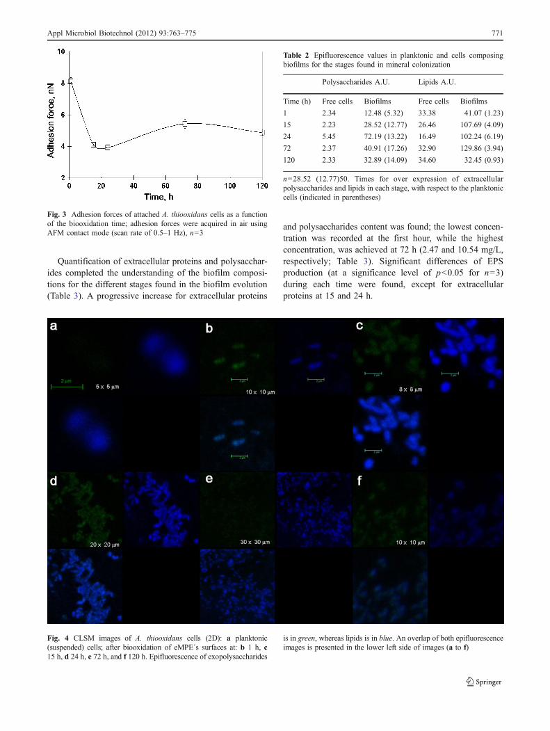
Epifluorescence analysis indicated variable secretion ofextracellular polysaccharides and lipids (as hydrophobicdomains) for the stages associated with biofilms evolution(Table 2). After 1 h of biooxidation, significant differencesof EPS production were found (at a significance level of p<0.05 for n=28) between cells forming biofilm and free(planktonic) cells; actually, 5.32 times more exopolysac-charides and 1.23 times more lipids, comparing with theplanktonic (suspended) cells. Longer times of biooxidationimplies changes in the quantity of these EPS, and themaximum secretion of exopolysaccharides and lipids wereachieved at 72 and 24 h, respectively. The higher secretionof EPS was not observed to be associated with the intensityof the strength of the cell attachment, i.e., in the initialadhesion stages, where the strongest adhesion force wasmeasured (Fig. 3), is far from the maximum EPSexpression.
Table 1 Evolution of root mean square (Rq) and roughness (Ra) values collected for stages found of biofilms evolution collected on eMPE areas
MPE surface Area (μm×μm) Number of areas RMS (Rq), nm Roughness (Ra), nm
Electrooxidized (1.11 V/SHE) 5×5 5 24.35±2.18 19.41±1.78
1×1 35 22.85±2.15 18.25±1.66
Abiotic assay (1 h) 5×5 27 70.01±5.98 56.34±4.45
1×1 34 51.79±10.17 42.30±8.89
Biotic assays (1 h, collected on top of attached cells) 5×5 5 89.33±11.88 64.95±7.89
1×1 14 59.40±18.22 46.99±15.71
Abiotic assay (15 h) 5×5 25 64.96±15.67 46.24±7.13
1×1 34 45.13±11.09 37.29±4.24
Biotic assays (15 h, surrounding attached cells) 5×5 14 57.07±8.56 35.27±9.91
1×1 25 40.32±13.27 32.18±10.54
Abiotic assay (24 h) 5×5 30 71.05±23.97 55.38±8.22
1×1 40 47.12±13.29 39.66±7.12
Biotic assays (24 h, surrounding attached cells) 5×5 17 24.80±6.93 18.16±5.68
1×1 33 16.10±4.94 12.57±3.82
Abiotic assay (72 h) 5×5 34 58.55±9.55 46.72±7.09
1×1 27 41.40±8.96 33.35±7.22
Biotic assays (72 h, surrounding attached cells) 5×5 9 29.61±5.38 23.15±3.99
1×1 16 25.65±3.64 20.74±3.25
Abiotic assay (120 h) 5×5 50 83.77±9.78 66.97±8.32
1×1 25 80.54±4.12 62.56±3.25
Biotic assays (120 h, surrounding attached cells) 5×5 25 45.13±3.58 36.08±3.01
1×1 45 37.41±4.83 30.27±4.72
770 Appl Microbiol Biotechnol (2012) 93:763–775
Quantification of extracellular proteins and polysacchar-ides completed the understanding of the biofilm composi-tions for the different stages found in the biofilm evolution(Table 3). A progressive increase for extracellular proteins
and polysaccharides content was found; the lowest concen-tration was recorded at the first hour, while the highestconcentration, was achieved at 72 h (2.47 and 10.54 mg/L,respectively; Table 3). Significant differences of EPSproduction (at a significance level of p<0.05 for n=3)during each time were found, except for extracellularproteins at 15 and 24 h.
Fig. 3 Adhesion forces of attached A. thiooxidans cells as a functionof the biooxidation time; adhesion forces were acquired in air usingAFM contact mode (scan rate of 0.5–1 Hz), n=3
Fig. 4 CLSM images of A. thiooxidans cells (2D): a planktonic(suspended) cells; after biooxidation of eMPE´s surfaces at: b 1 h, c15 h, d 24 h, e 72 h, and f 120 h. Epifluorescence of exopolysaccharides
is in green, whereas lipids is in blue. An overlap of both epifluorescenceimages is presented in the lower left side of images (a to f)
Table 2 Epifluorescence values in planktonic and cells composingbiofilms for the stages found in mineral colonization
Polysaccharides A.U. Lipids A.U.
Time (h) Free cells Biofilms Free cells Biofilms
1 2.34 12.48 (5.32) 33.38 41.07 (1.23)
15 2.23 28.52 (12.77) 26.46 107.69 (4.09)
24 5.45 72.19 (13.22) 16.49 102.24 (6.19)
72 2.37 40.91 (17.26) 32.90 129.86 (3.94)
120 2.33 32.89 (14.09) 34.60 32.45 (0.93)
n=28.52 (12.77)50. Times for over expression of extracellularpolysaccharides and lipids in each stage, with respect to the planktoniccells (indicated in parentheses)
Appl Microbiol Biotechnol (2012) 93:763–775 771

Discussion
A. thiooxidans forms biofilms strongly attached to thesurface of the MPE previously electrooxidized (eMPE),suggesting an intimate physical contact between thebacteria and the pyrite, which resulted in strong adhesionforces (range, 3.92–8.10 nN; Table 1). The colonizationinitiated after 1 h, consisting of clusters attached cells ontothe surface of the mineral (Fig. 2). This initial contactresulted in the strongest adhesion force recorded throughthe biofilm assay (8.1 nN). Subsequently longer coloniza-tion times resulted in stability of attached cells and thechanges in biofilms adhesion forces were negligible(Fig. 3).
At initial time, the minimal EPS production andsecretion was recorded but its results were significantlyhigher than the EPS of planktonic cells (p<0.05 for n=28).An increase in EPS content is directly associated with theincrease of epifluorescence in attached cells, reflectingconsiderable EPS thickness (Fig. 4); the data of epifluor-escence indicated that EPS mainly comprise hydrophobicdomains (as lipids; Table 2). The aforementioned resultssuggest initial non-covalent, weak interactions, mediatingan irreversible binding mechanism to eMPE, wherein theinitial production of EPS by individual cells is determinant.The former was supported by experimental data of differentauthors in systems with A. ferrooxidans: (a) the attachedcells to pyrite are more hydrophobic than suspended ones;the hydrophobicity brings down the interfacial tensionbetween cells and the surface of sulfur or pyrite, to enhancecontact and adhesion (Devasia et al. 1993; Devasia andNatarajan 2010); and (b) the attachment of the cells tohydrophobic substrata as pyrite and sulfur is dominated byvan der Waal’s attraction forces (Gehrke et al. 1998). Thus,hydrophobicity helps in adhesion (Donlan 2002).
After 15 h, the number of cells significantly increasedand a biofilm with a monolayer structure was observed;later, ca. the 24th hour, a biofilm with two layers of cells
was observed. A concomitant increase of the EPS produc-tion was recorded between the 15th and before the 120thhour (Tables 2 and 3). This highest content of EPS is relatedwith the increase of the cell number but also with theestablishment of a self-covered matrix of EPS in biofilms,as it was observed during CLSM studies and suggestedduring the AFM studies.
It has been demonstrated that the nature and quantity ofEPS produced and secreted by bioleaching cells is stronglyinfluenced by the culture conditions and the electrochemicalcharacteristics of the substratum (Donlan 2002), and that free-living (planktonic) cells produce less EPS than cell attachedto a mineral (Harneit and Sand 2007); also, that the mainEPS produced by bioleaching microorganisms grown onpyrite are neutral polysaccharides (glucose, rhammose,fucose, xylose, and mannose), lipids or saturated fatty acids,and lipopolysaccharides (Gehrke et al. 1998).
Interestingly, our results showed the dominance ofhydrophobic domains (as extracellular lipids) in the EPSof both, the free and biofilm-forming cells of A. thioox-idans, with a maximum within the 15–72 h (Tables 2 and3). Elevated level of lipids is closely associated with thelate stages of biofilm maturation in Mycobacterium tuber-culosis (Ojha et al. 2005) and lipids and lipopolysacchar-ides are essential for the biofilm adhesive stability underextreme environmental conditions. Previous report indicatedthat when A. ferrooxidans grew attached on sulfur or pyriteproduced more lipids (Vu et al. 2009) and that lipopolysac-charide may be a prerequisite for such attachment (Gehrke etal. 1998). Apparently, lipids enhance elemental sulfuroxidation acting as a wetting agent (Beebe and Umbreit1971). Yet, the high content of lipids increases thehydrophobicity of A. thiooxidans biofilms. Since S0 is alsohydrophobic, the sulfur–cell interaction is hydrophobic.Because lipids and hydrophobic domains strongly influencelipid-mediated adhesion mechanisms in biofilm, it isnecessary to investigate the effect of surfactants or thehydrophobicity of EPS in suspensions, by measuring liquid–liquid partition in aqueous and organic phase using.
Devasia et al. (1993) and Devasia and Natarajan (2010)distinguish proteinaceous compounds on the surface of A.ferrooxidans attached to pyrite, and conclude that aninduction of relatively hydrophobic proteins is requiredfor adhesion, changes the cell surface chemistry and hencethe utilization of sulfur, and is also a prerequisite forexopolysaccharides secretion.
Thus, extracellular polysaccharides and lipids play a keyrole in cell adherence, favoring hydrophobic interaction, butalso extracellular or surface proteins are involved inadhesion and are linked to electron transport; both are keyaspects in bacterial colonization and oxidation of insolublesubstrates (Bobadilla et al. 2011). The initial stage of cellattachment involves the highest adhesion force but also the
Table 3 Quantification of extracellular proteins and polysaccharidesfor the stages found in mineral colonization
Time (h) Proteins Polysaccharides
Absorbance mg/mL Absorbance mg/mL
0 0.002 0.031 0.003 0.17
1 0.034 0.596 0.016 1.29
15 0.043 1.37 0.067 4.94
24 0.043 1.416 0.075 5.52
72 0.055 2.473 0.145 10.54
120 0.029 0.14 0.036 2.77
n=3
772 Appl Microbiol Biotechnol (2012) 93:763–775

smallest extracellular protein concentration (Table 3); thisfact cannot be fully explored in the present work. However,it may be explained invoking the fact that adhesines areimportant molecules involved in any bacterial adhesionprocess, and in our case it may proceed as follows: anextracellular (outer membrane) 40-KDa protein was de-scribed by Jerez et al. (1992), such protein named Omp40participates in the adhesion of A. ferrooxidans to hydro-phobic sulfur aggregates (Arredondo et al. 1994). It ishypothesized that the secretion of an “Omp40-like” proteinby A. thiooxidans (Bobadilla et al. 2011) also occurs.Additionally, and more important, Ohmura et al. (1996)observed that the flagellum of A. ferrooxidans contains a40-KDa protein which could adhere to elemental sulfur,mediated by the disulfide bond between thiol groups in theprotein and elemental sulfur. More recently, Li et al. (2010)concluded that A. ferrooxidans colonizes and forms micro-colonies on pyrite by assessing pili-mediated twitchingmotility and a strong adherence; such pili present theprotein adhesin at its tip (Pointon et al. 2010). Pili arepresent in diverse groups of bacteria, but no studies on pilisand adhesines of A. thiooxidans have been performed yet.This process could be also implied at initial adhesion of A.thiooxidans cells to pyrite; however, such importantquestion should be clarified in subsequent work. However,another piece of information that may be related with theadhesion process is the fact that Bobadilla et al. (2011)reported the major secreted protein by A. thiooxidanscultured with elemental sulfur, the lipoprotein Licanantase,which seems to be related with the colonization andoxidation of sulfur.
We propose that A. thiooxidans cells follow four stagesduring the colonization of the eMPE (of 1.2–1.5 cm2): (1)an initial hydrophobic interaction with the sulfur and thecolonization of cell clusters throughout the surface; (2) thecells attached to the surface achieved a stable adhesionforce and the EPS production and secretion resulted in acolony of one layer of cells; (3) the mineral surface reachedits maximum cells covering sometime around 15–24 h; butthe well-structured Sn
2−/S0 species recorded after 1 h ofbiotic assay are totally depleted after 24 h (Fig. 1f, h); thisstarvation conditions and the high cell density, promote thetransition from surface-attached cells into a mature biofilm(Figs. 2e, f, h and 4d, e); (4) at the final stage (120 h), thenumber cells and the quantity of EPS of the biofilmsdecreased and the biofilm dissolves releasing cells; theindividual cells may colonize other surfaces (Monods andO’Toole 2009). During the colonization of the eMPE’ssurface, cells of A. thiooxidans progressively biooxidizedthe reduced sulfur species (Sn
2−/S0) of the eMPE, modify-ing the crystalline topography or texture of the surfacestructure of Sn
2−/S0as the AFM (Fig. 2) and Raman (Fig. 1)observations suggested and indicated, respectively.
Since in this experiment, the Fe(II)/Fe(III) ratio was thesame in both cultures with A. thiooxidans and in the abioticcontrol (0.01 g of iron; see Bacterial strain and cultivation),the aforementioned results indicated that the observeddifferences are clearly a consequence of the oxidation ofthe S0 generated on the electrooxidized MPE by thebiological activity of A. thiooxidans, and that S0 is stableduring pyrite weathering in acidic conditions (abioticassays). No passive layer was detected. The previous resultis of industrial interest, since in absence of a sulfur-oxidizing microorganisms as A. thiooxidans large amountsof jarosite and elemental sulfur may be produced duringother biochemical leaching processes, and the biofilmformed may be embedded by the jarosite deposition, as isobserved by Lei et al. (2009) during the biooxidationprocess of chalcopyrite by A. ferrooxidans. Also Li et al.(2010) reported an accumulation of secondary minerals thatsurrounded or partially encapsulated the A. ferrooxidanscells; such secondary minerals modified the oxidationprocess of pyrite.
Acknowledgments Financial support for this work comes from theMexican Council of Science and Technology (CONACyT; project no. 05-49321). The authors thank Dr. Amauri Pozos and Dr. Jaime Ruiz-Garcíafor access to the CLSM (Basics Sciences Laboratory) and the AFM(Colloids and Interfaces Laboratory) equipment at UASLP, respectively.The authors also thank Erasmo Mata-Martinez for mineral sectionpreparation, Francisco Galindo-Murillo for MPE preparation. René Laraalso thanks CONACyT for his postdoctoral fellowship.
References
Arredondo R, García A, Jerez CA (1994) Partial removal oflipopolysaccharide from Thiobacillus ferrooxidans affects itsadhesion to solids. Appl Environ Microbiol 60:2846–2851
Barranguet C, van Bausekom SAM, Veuger B, Neu TR, MandersEMM, Sinke JJ, Admiraal W (2004) Studying undisturbedphototrophic biofilms: still a technical challenge. Aquat MicrobiolEcol 34:1–9
Beebe JL, Umbreit WW (1971) Extracellular lipid of Thiobacillusthiooxidans. J Bacteriol 108(1):612–614
Bevilaqua D, Leite ALLC, García O, Tuovinen OH (2002) Oxidationof chalcopyrite by Acidithiobacillus ferrooxidans and Acid-ithiobacillus thiooxidans in shake flask. Process Biochem38:587–594
Bobadilla RA, Levican G, Parada P (2011) Acidithiobacillus thiooxidanssecretome containing a newly described lipoprotein Licanantaseenhances chalcopyrite bioleaching rate. Appl Microbiol Biotechnol89:771–780
Bradford MM (1976) A rapid and sensitive method for thequantitation of microgram quantities of protein utilizing theprinciple of protein-dye binding. Ann Biochem 72:248–254
Chandraprabha MN, Somasundaran P, Natarajan KA (2010) Modelingand analysis of nanoscale interaction forces between Acid-ithiobacillus ferrooxidans and AFM tip. Coll Surf B Biointerfaces75(1):310–318
Chi A, Valenzuela L, Beard S, Mackey AJ, Shabanowitz J, Hunt DF,Jerez CA (2007) Periplasmic proteins of the extremophile
Appl Microbiol Biotechnol (2012) 93:763–775 773

Acidithiobacillus ferrooxidans. Mol Cell Proteomics 6:2239–2251
Devasia P, Natarajan KA (2010) Adhesion of Acidithiobacillusferrooxidans to mineral surfaces. Int J Miner Proces 94:135–139
Devasia P, Natarajan KA, Sathyanarayana DN, Rao GR (1993)Surface chemistry of Thiobacillus ferrooxidans relevant toadhesion on mineral surfaces. Appl Environ Microbiol 59:4051–4055
Donlan MR (2002) Biofilms: microbial life on surfaces. Emerg InfectDis 8(9):881–890
Dopson M, Lindström EB (1999) Potential role of Thiobacillus caldusin arsenopyrite bioleaching. Appl Environ Microbiol 65(1):36–40
Dubois M (1959) Colorimetric method for determination of sugars andrelated substances. Anal Chem 28:350–356
Echeverría A, Demergasso C (2009) Assessment of microbial adhesionin mixed cultures to sulfide minerals using CARD-FISHtechniques. Adv Mat Reser Biohydrometallurgy 71–73:83–86
Edwards KJ, Goebel BM, Rodgers TM, Schrenk MO, Gihring TM,Cardona MM, Hu B, McGuire MM, Hamers RJ, Pace NR,Banfield JF (1999) Geomicrobiology of pyrite (FeS2) dissolu-tion: case study at Iron Mountain, California. Geomicrobiol J16:155–179
Florian B, Noël N, Sand W (2009) Biofilm of lithotrophic lechingbacteria visualizized by combined epifluorescence and atomic forcemicroscopy. Proceedings 5th ASM Conference in Biofilms 144
Fowler TA, Crundwell FK (1999) Leaching of zinc sulfide byThiobacillus ferrooxidans: bacterial oxidation of the sulfurproduct layer increases the rate of zinc sulfide dissolution athigh concentration of ferrous ions. Appl Environ Microbiol 65(12):5285–5292
Gehrke T, Telegdi J, Thierry D, Sand W (1998) Importance ofextracellular polymeric substances from Thiobacillus ferrooxi-dans for bioleaching. Appl Environ Microbiol 64:2743–2747
Hamilton IC, Wood R (1981) An investigation of surface oxidation ofpyrite and pyrrhotite by linear potential sweep voltammetry. JElectroanal Chem 118:327–343
Harneit K, Sand W (2007) Influence of growth substrate andattachment substratum on EPS and biofilms formation byAcidithiobacillus ferrooxidans A2. Adv Mat Res 20–21:385
Harneit K, Göksel A, Kock D, Klock JH, Gehrke T, Sand W (2006)Adhesion to metal sulfide surfaces by cells of Acidithiobacillusferrooxidans, Acidithiobacillus thiooxidans and Leptospirillumferrooxidans. Hydrometallurgy 83:245–254
Jerez CA, Seeger M, Amaro AM (1992) Phosphate starvation affectsthe synthesis of outer membrane proteins in Thiobacillusferrooxidans. FEMS Microbiol Lett 98:29–34
Kelly DP (1985) Physiology of the thiobacilli: elucidating the sulphuroxidation pathway. Microbiol Sci 2:105–109
Kinzler K, Gehrke T, Telegdi J, Sand W (2003) Bioleaching: a resultof interfacial processes caused by extracellular polymericsubstances (EPS). Hydrometallurgy 71:83–88
Konishi Y, Asai S, Yoshida N (1995) Growth kinetics of Thiobacillusthiooxidans on the surface of elemental sulfur. Appl EnvironMicrobiol 61(10):3617–3622
Lara RH, Valdez-Pérez D, Rodríguez AG, Navarro-Contreras HR,Cruz R, García-Meza JV (2010) Interfacial insights of pyritecolonized by Acidithiobacillus thiooxidans cells under acidicconditions. Hydrometallurgy 103:35–44
Lei J, Huaiyang Z, Xiaotong P, Zhonghao D (2009) The use ofmicroscopy techniques to analyze microbial biofilms of the bio-oxidized chalcopyrite surface. Mineral Eng 22:37–42
Li YQ, Wan DS, Huang SS, Leng FF, Yan L, Ni YTQ, Li HY (2010)Type IV pili of Acidithiobacillus ferrooxidans are necessary forsliding, twitching motility, and adherence. Curr Microbiol 60:17–24
Liu HL, Chen BY, Lan YW, Cheng YC (2003) SEM and AFM imagesof pyrite surfaces after bioleaching by the indigenous Thiobacillusthiooxidans. Appl Microbiol Biotechnol 62:414–420
Liu Y-G, Zhou M, Zeng G-M, Wang X, Li X, Fan T, Xu W-H (2008)Bioleaching of heavy metals from mine tailings by indigenoussulfur-oxidizing bacteria: effects of substrate concentration.Bioresour Technol 99:4124–4129
Mangold S, Harneit K, Rohwerder T, Claus G, Sand W (2008a) Novelcombination of atomic force microscopy and epifluorescencemicroscopy for visualization of leaching bacteria on pyrite. ApplEnviron Microbiol 74(2):410–415
Mangold S, Laxander M, Harneit K, Rohwerder T, Claus G, Sand W(2008b) Visualization of Acidithiobacillus ferrooxidans biofilmson pyrite by atomic force and epifluorescence microscopy undervarious experimental conditions. Hydrometallurgy 94:127–132
Monods RD, O’Toole GA (2009) The developmental model ofmicrobial biofilms: ten years of a paradigm up for review. TrendsMicrobiol 17(2):73–87
Mustin C, de Donato Ph, Berthelin J, Marion Ph (1993) Surfacesulphur as promoting agent of pyrite leaching by Thiobacillusferrooxidans. FEMS Microbiol Rev 11(1–3):71–74
Mycroft JR, Bancroft GM, McIntyre NS, Lorimer JW, Hill IR (1990)Detection of sulphur and polysulphides on electrochemicallyoxidized pyrite surfaces by X-ray photoelectron spectroscopy andRaman spectroscopy. J Electroanal Chem 292:139–152
Nava D, González I, Leinen D, Ramos-Barrado JR (2008) Surfacecharacterization by X-ray photoelectron spectroscopy and cyclicvoltammetry of products formed during the potentiostaticreduction of chalcopyrite. Electrochem Acta 53:4889–4899
Ohmura N, Tsugita K, Koizumi J-I, Saiki H (1996) Sulfur-bindingprotein of flagella of Thiobacillus ferrooxidans. J Bacteriol 178(19):5776–5780
Ojha A, Anand M, Bhatt A, Kremer L, Jacobs W, Hatfull GF (2005)GroEL1: a dedicated chaperone involved in mycolic acid biosyn-thesis during biofilms formation in mycobacteria. Cell 123:861–873
Pogliani C, Donati E (1999) The role of exopolymers in bioleachingof a non-ferrous metal sulphide. J Ind Microbiol Biotechnol 22(2):88–92
Pointon JA, Smith WD, Saalbach G, Crow A, Kehoe MA, BanfieldMJ (2010) Highly unusual thioester bond in a pilus adhesin isrequired for efficient host cell interaction. J Biol Chem285:33858–33866
Rawlings DE, Tributsch H, Hansford GS (1999) Reasons why‘Leptospirillum’-like species rather than Thiobacillus ferrooxi-dans are the dominant iron-oxidizing bacteria in many commer-cial processes for the biooxidation of pyrite and related ores.Microbiol 145:5–13
Rodríguez Y, Ballester A, Blázquez ML, González F, Muñoz JA(2003) New information on the pyrite bioleaching mechanisms atlow and high temperature. Hydrometallurgy 71:37–46
Rohwerder T, Sand W (2007) Mechanisms and biochemical funda-mentals of bacterial metal sulfide oxidation. In: Donati ER, SandW (eds) Microbial processing of metal sulfides. Springer, NewYork, pp 35–58
Rohwerder T, Gehrke T, Kinzler K, Sand W (2003) Bioleachingreview part A Progress in bioleaching: fundamentals andmechanisms of bacterial metal sulfide oxidation. Appl MicrobiolBiotechnol 63:239–248
Rother D, Henrich HJ, Quentmeier A, Bardischewsky F, Friedrich CG(2001) Novel genes of the sox gene cluster, mutagenesis of theflavoprotein SoxF, and evidence for a general sulfur-oxidizing systemin Paracoccus pantotrophus GB17. J Bacteriol 183:4499–4508
Sand W, Gehrke T, Jozsa P-G, Schippers A (2001) (Bio)chemistry ofbacterial leaching-direct vs. indirect bioleaching. Hydrometallurgy59:159–175
774 Appl Microbiol Biotechnol (2012) 93:763–775

Sasaki K, Tsunekawa M, Ohtsuka T, Konno H (1998) The role ofsulfur-oxidizing bacteria Thiobacillus thiooxidans in pyriteweathering. Coll Surf A 133:269–278
Schippers A, Sand W (1999) Bacterial leaching of metal sulfidesproceeds by two indirect mechanisms via thiosulfate or viapolysulfides and sulfur. Appl Environ Microbiol 65(1):319–321
Sharma PK, Das A, Hanumantha Rao K, Forssberg KSE (2003)Surface characterization of Acidithiobacillus ferrooxidans cellsgrown under different conditions. Hydrometalllurgy 71:285–292
Takakuwa S, Nishikawa T, Hosoda K, Tominaga N, Iwasaki H (1977)Promoting effect of molybdate on the growth of a sulfur
oxidizing bacterium, Thiobacillus thiooxidans. J Gen ApplMicrobiol 23:163–173
Toniazzo V, Mustin C, Portal JM, Humbert B, Benoit R, Erre R (1999)Elemental sulfur at the pyrite surfaces: speciation and quantifi-cation. Appl Surf Sci 143:229–237
Vu B, Chen M, Crawford RJ, Ivanova EP (2009) Bacterialextracellular polysaccharides involved in biofilms formation.Molecules 14(7):2535–2554
Zhang T, Fang HHP (2001) Quantification of extracellular polymericsubstances in biofilms by confocal laser scanning microscopy.Biotechnol Lett 23:405–409
Appl Microbiol Biotechnol (2012) 93:763–775 775