Distribution of Sulfate-Reducing Bacteria in a Stratified Fjord
Evaluation of the use of common sculpin (Myoxocephalus scorpius)organ histology as bioindicator for element exposure in the fjordof the mining area Maarmorilik, West Greenland
Christian Sonne a,n, Lis Bach a, Jens Søndergaard a, Frank F. Rigét a, Rune Dietz a,Anders Mosbech a, Pall S. Leifsson b, Kim Gustavson a
a Aarhus University, Faculty of Science and Technology, Department of Bioscience, Arctic Research Centre (ARC), Frederiksborgvej 399,P.O. Box 358, DK-4000 Roskilde, Denmarkb University of Copenhagen, Faculty of Health and Medical Sciences, Department of Veterinary Disease Biology, Bülowsvej 17,DK-1870 Frederiksberg, Denmark
a r t i c l e i n f o
Article history:Received 13 March 2014Received in revised form28 May 2014Accepted 30 May 2014
Keywords:ArsenicGillHealth effectsLeadLiverMercury
a b s t r a c t
The former Black Angel lead–zinc mine in Maarmorilik, West Greenland, is a historic example of howmining activity may result in a significant impact on the surrounding fjord system in terms of elevatedconcentrations of especially lead (Pb) and zinc (Zn) in seawater, sediments and surrounding biota. Inorder to shed light on the present contamination and possible effects in the fjord we initiated a range ofstudies including a pilot study on gill and liver morphology of common sculpins (Myoxocephalusscorpius) around Maarmorilik. Sculpins were caught and sampled at five different stations known torepresent a gradient of Pb concentrations. Fish livers from all specimens were analyzed for relevantelements in the area: Fe, Zn, As, Cu, Se, Cd, Pb, Ag, Hg, Co and Ni. Lead, As and Hg showed significantdifferences among the five stations. For 20% of the sculpins, Hg concentrations were in the range oflowest observed effect dose (LOED) of 0.1–0.5 μg/g ww for toxic threshold on reproduction andsubclinical endpoints. Likewise LOEDs for tissue lesions, LOEDs for biochemistry, growth, survival andreproduction were exceeded for Cd (0.42–1.8 μg/g ww) and for As (11.6 μg/g ww) in 28% and 85% of thesculpins, respectively. Similar to this, the no observed effect dose (NOED) for biochemistry was exceededfor Pb (0.32 μg/g ww) and for growth, mortality and reproduction for Zn (60–68 μg/g ww) in 33% and24% of the sculpins, respectively. For all sculpins, females were significantly larger than males and forfive of the elements (Fe, Co, Ni, Cu, Se) females had higher concentrations. The chronic lesions observedin liver (mononuclear cell infiltrates, necrosis, vacuolar hepatocytes, portal fibrosis, bile duct hyperplasia,active melanomacrophage centers) and gills (fusion and edema of secondary lamellae, laminartelangiectasis, mononuclear cell infiltrates, blebs) were similar to those in the literature studies forboth wild and laboratory exposed sculpins and other fish species carrying similar or higher Hgconcentrations. Ignoring sex and size, specimens with hepatic cell infiltrates had the highest concentra-tions of most elements, a relation that was also found for gill telangiectasis and Hg (all po0.05). Whencontrolling for sex and size, the prevalence of vacuolar hepatocytes and endoparasites was significantlyhighest at the three most contaminated stations and similar differences were found for liver necrosis. Wesuggest that beside exposure to mining-related elements, other environmental factors, such as parasites,might be co-factors in the development of the observed liver and gill lesions. Therefore, sculpin liver andgill pathology are likely to be suitable health indicators when biomonitoring gradients of mining andother element related activity effects; while a larger study is required to fully evaluate the relationships.
& 2014 Elsevier Inc. All rights reserved.
1. Introduction
Mining poses a well-known environmental concern due to thepotential release of elements and chemicals from extraction andprocessing of mineral ores. Evaluation of environmental impactsfrom mining both in the Arctic and worldwide has traditionallybeen based on concentrations of elements measured in sediments as
Contents lists available at ScienceDirect
journal homepage: www.elsevier.com/locate/envres
Environmental Research
http://dx.doi.org/10.1016/j.envres.2014.05.0310013-9351/& 2014 Elsevier Inc. All rights reserved.
n Corresponding author. Fax: þ45 87 15 50 15.E-mail address: [email protected] (C. Sonne).
Environmental Research 133 (2014) 304–311

well as in various biomonitoring species as macro algae, musselsand fish (Johansen et al., 1991; Rigét et al., 1997; Sanchez et al., 1998;Cairrao et al., 2007; O’Connor and Lauenstein, 2006; Torres et al.,2008), whereas less effort has been made to monitor toxicologicaleffects (Burd, 2002; Josefson et al., 2008).
The former Black Angel (BA) lead–zinc mine in Maarmorilik inWest Greenland (Fig. 1) was operated during the period 1973–1990 and is a historic example of how mining activity resulted in asignificant impact on the surrounding fjord system in terms ofelevated concentrations of especially lead (Pb) and zinc (Zn) inseawater, sediments and surrounding biota (e.g. Johansen et al.,1991, 2008; Schiedek et al., 2009; Søndergaard et al., 2011). Mainsources of contamination were subsea deposition of tailings andwaste rock, as well as dispersal of dust (Søndergaard et al., 2011).Despite a slow recovery, elevated concentrations of elements aretwo decades after mining closure still measured in marine biotasuch as blue mussels (Mytilus edulis), seaweed (Fucus vesiculosus)and sculpins (Myoxocephalus scorpius) within a distance of 12 kmfrom the mine, although concentrations are decreasing (Schiedeket al., 2009; Søndergaard et al., 2011). More detailed informationon the history of the BA mine, aspects of its decommissioning andthe present status can be found described elsewhere in theliterature (e.g. Johansen et al., 1991, 2008; Elberling et al., 2002;Søndergaard et al., 2011).
Research on element exposure to fish species and the potentialrisk of physiological and pathological effects have been conductedwidely in both laboratory experiments and on wild fish species inthe environment (Alam and Maughan, 1992; Clearwater et al.,2002; Greenfield et al., 2008; Iwasaki et al., 2010; Maceda-Veiga etal., 2013). Of the multiple elements occurring in the environment,special attention has been drawn to copper (Cu), cadmium (Cd),lead (Pb), zinc (Zn), arsenic (As) and mercury (Hg) since these havedocumented effects on, e.g., organ morphology, behavior, neuro-chemical and endocrine processes as well as reproduction (Mattaet al., 2001; Oost et al., 2003; Scheuhammer et al., 2007; Crumpand Trudeau, 2009; Sandheinrich and Wiener, 2011; Depew et al.,2012; Adams and Sonne, 2013). Near mining and industrial areas,fish species are commonly used as biomonitoring species for
assessing element dispersion and biological uptake near rivers,lakes, or other reservoirs directly influenced by the anthropogenicsources (Bengtsson and Larsson, 1986; Raldúa et al., 2007; Adamset al., 2010; Adams and Sonne, 2013). Such settings allow forstudies of biological effects caused by element exposure on variousbiochemical, cellular and organ processes, that can be documentedby histopathological analyses. Thus, as described above, althoughextensive literature exists on different fish species little is knownof the arctic common sculpin (Myoxocephalus scorpius). Thecommon sculpin is a highly common species in the fjords ofGreenland and is relatively sedentary at the seafloor and easy tocatch. This makes the common sculpin an ideal indicator speciesand though it has been included in monitoring programs forelement analyses, very little is known of fate and effects ofelements to this species. On the top of this, common sculpinsare sometimes caught and eaten by the population around thecoasts of Greenland.
In the present study we examined histopathology of liver andgills as possible effect endpoints and analyzed element concentra-tions in liver tissue of sculpins around the BA mining area in WestGreenland. The specific objectives of this study were (1) todetermine concentrations of 11 elements in liver tissue of sculpinscollected at gradient zones from the mining area, (2) to relatethese element concentrations to sex, size, and liver and gillhistology with regard to potential effects in sculpins in the zonesand (3) to evaluate if sculpins could be a useful species formonitoring toxic effects of mining activities in arctic areas.
2. Materials and methods
2.1. Study area and sample collection
The study was designed around the BA mine in Maarmorilik in West Greenland(W52, N71) (Fig. 1), an area that is described in detail elsewhere (Asmund,1992a,1992b; Elberling et al., 2002; Søndergaard et al., 2014). All sculpins (n¼33)were collected live by the use of fishing rods on the 28 August and 1 September2012 at five stations representing a gradient of pollution 1–40 km from the mine(Fig. 1 and Table 1 for GPS coordinates). Stations 1–3 are placed close to the mining
Fig. 1. A map of the Maarmorilik study area in West Greenland. The location of the tailings disposal site and the mining town (mining symbol) is shown. 1–5: Samplingstations. Source: Google maps.
C. Sonne et al. / Environmental Research 133 (2014) 304–311 305
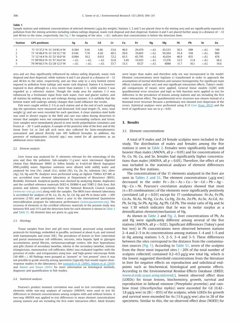
area and are thus significantly influenced by subsea tailing disposals, waste rockdisposal and dust dispersal; while stations 4 and 5 are placed in a distance of �12and 40 km to the mine, respectively, and are thus only to a very limited extentexposed to pollution from tailings and waste rock disposal. Station 4 is howeverexposed to dust although to a less extent than stations 1–3, while station 5 wasregarded as a reference station. Though the study area for stations 1–3 wasinfluenced by a freshwater input from melt water, river and glacier outlets; andall stations by melting sea ice and icebergs during summer; it is unlikely that thebottom water will undergo salinity changes that could influence the results.
Fish were caught within 2–3 h at each station and at the end of each samplingday the specimens were measured and dissected. Fish total length (TL, mm), totalweight (g) and sex were recorded for each specimen. A clean stainless-steel knifewas used to dissect organs in the field and care was taken during dissection toensure that samples were not contaminated by surrounding surfaces and tissue.Liver samples were immediately placed in new sterile polyethylene containers andfrozen at �20 1C until analysis. A sample of the posterior lobe of the liver and of thetissue from 1st or 2nd gill arch were also collected for histo-morphometricassessment and placed directly into 10% buffered formalin. In addition, thepresence of endoparasites (Ascarid spp.) was noted for all specimens as anadditional stress indicator.
2.2. Element analysis
Liver tissue was analyzed for 11 elements relevant for the mineralogy of thearea and thus the pollution. Sub-samples (1 g ww) were microwave digested(Anton Paar Multiwave 3000) in Teflon bombs in 4 ml/4 ml Merck SuprapureHNO3/milliQ water. Digestion solutions were diluted with milliQ water andanalyzed for iron (Fe), cobalt (Co), nickel (Ni), Cu, Zn, As, selenium (Se), silver(Ag), Cd, Hg and Pb. Analyses were performed using an Agilent 7500ce ICP-MS atour accredited trace element laboratory at Department of Bioscience (BIOS),Roskilde, Denmark. Analytical quality was checked by analyzing blanks, duplicatesand the certified reference materials DOLT-4, DORM-4 and TORT-2 (fish liver, fishprotein and lobster, respectively) from the National Research Council Canada(www.nrc-cnrc.gc.com) along with the samples. The BIOS trace element laboratoryis accredited for analyses of Ni, Cu, Zn, As, Se, Cd, Hg and Pb in biota with o20%precision (2 SD) and participates twice a year in the international Quasimemeintercalibration program for laboratory performance (www.quasimeme.org). Therecovery of elements in the certified reference materials in the present study wasbetween 95% and 111% and the detection limit for each element is shown in Table 2and Table S1. All element data are given in mg/g ww.
2.3. Histology
Tissue samples from liver and gill were trimmed, processed using standardprotocols for histology, embedded in paraffin, sectioned at about 4 mm, and stainedwith haematoxylin and eosin (HE). The prevalence of lesions in liver (interstitialand portal mononuclear cell infiltrates, necrosis, intra hepatic lipid or glycogenaccumulation, portal fibrosis, melanomacrophage centers, bile duct hyperplasia)and gills (fusion of secondary lamellae, edema in the secondary lamellae, laminartelangiectasis, mononuclear cell infiltrates, blebs) was evaluated together with thepresence of endo- and ectoparasites using low- and high-power microscope fields(50–400� ). All findings were grouped as “present” or “not present” since it wasnot possible to grade severity among specimens (typically that would require dose-response studies in the laboratory). See Leonardi et al. (2009), Adams et al. (2010)and Adams and Sonne (2013) for more information on histological methods,diagnoses and quantification in fish studies.
2.4. Statistical analyses
Pearson's product moment correlation was used to test correlations amongelements while one-way analyses of variance (ANOVA) were used to test fordifferences in element concentrations among sculpins with and without lesions. Atwo-way ANOVA was applied to test differences in mean element concentrationsamong stations and sex including the first order interaction effect. Adult females
were larger than males and therefore only sex was incorporated in the model.Element concentrations were log(base e) transformed in order to approach theassumptions of normal distribution and variance homogeneity. For significant maineffects (stations and/or sex) and non-significant interaction effects, Tukey's multi-ple comparisons of means were applied. General linear models (GLM) withquasibinomial error structure and logit as link function were applied to test fordifference in the prevalence of lesions among stations and sex including the firstorder interaction effect. The quasibinomial error structure was chosen instead of abinomial error structure because a preliminary test showed over dispersion of theerrors. Statistical analyses were performed using R (R Core Team, 2013) and thelevel of significance was set to po0.05.
3. Results
3.1. Element concentrations
A total of 9 males and 24 female sculpins were included in thestudy. The distribution of males and females among the fivestations is seen in Table 2. Females were significantly longer andheavier than males (ANOVA, all po0.05) and for concentrations ofFe, Co, Ni, Cu, and Se, females had significantly higher concentra-tions than males (ANOVA, all po0.05). Therefore, the effect of sexwas included in the statistical analyses of element differencesamong the five stations.
The concentrations of the 11 elements analyzed in the liver areseen in Tables 2 and S1. The element concentrations (mg/g ww)decreased in the order Fe4Zn4As4Cu4Se�Cd4Pb4Ag�Hg�Co4Ni. Pearson's correlation analyses showed that most(n¼83 combinations) of the elements were significantly positivelycorrelated (all po0.03) except for 17 combinations: Fe:As, Fe:Pb,Co:As, Ni:As, Ni:Hg, Cu:As, Cu:Hg, Zn:As, Zn:Pb, As:Se, As:Cd, As:Pb, Se:Hg, Se:Pb, Ag:Hg, Ag:Pb, Cd:Pb. The molar ratio of Hg and Sewas 1:10 which indicates that Se was in molar surplus as adetoxification element/mechanism of Hg.
As shown in Table 2 and Fig. 2, liver concentrations of Pb, Asand Hg were significantly different among several of the fivestations (ANOVA, all po0.02). Significant differences (Tukey's posthoc test) in Pb concentrations were observed between stations2–4 and 2–5 in As concentrations among stations 1–4 and 1–5 andin Hg among stations 1–5, 2–5, 3–4 and 3–5. These differencesbetween the sites correspond to the distance from the contamina-tion sources (Fig. 1). According to Table S1, seven of the sculpinsfrom the three most impacted sites (�20% of the total number ofsculpins collected) contained 0.2–0.5 μg/g ww total Hg, which isthe lowest suggested threshold concentrations from the literaturein liver for negative effects on reproduction and subclinical end-points such as biochemical, histological and genomic effects.According to the Environmental Residue-Effects Database (ERED;www.el.erdc.usace.army.mil/ered/), lowest observed effect dose(LOEDs) for tissue lesions, biochemistry, growth, survival andreproduction in fathead minnow (Pimephales promelas) and rain-bow trout (Oncorhynchus mykiss) were exceeded for Cd (0.42–1.8 μg/g ww) in 28 (�85%) of the sculpins, while LOEDs for growthand survival were exceeded for As (11.6 μg/g ww) also in 28 of thespecimens. Similar to this, the no observed effect dose (NOED) for
Table 1Sample stations and sediment concentrations of selected elements (mg/g dry weight). Stations 1, 2 and 3 are placed close to the mining area and are significantly exposed topollution from the mining activities including subsea tailings disposal, waste rock disposal and dust dispersal. Stations 4 and 5 are placed further away in a distance of �12and 40 km to the mine, respectively. See Fig. 1 for mapping of the sites. od.l.: indicates that concentration is below the detection limit.
Station GPS positions Ag As Cd Cr Cu Fe Hg Mg Ni Pb Se Zn
1 71113037.2″N–51124085.4″W 0.384 9.16 1.66 53.6 40.9 29,479 od.l. 43,533 36.5 588 od.l. 7492 71114048.8″N–51124032.7″W 0.144 7.70 4.26 40.2 29.0 19,663 od.l. 50,364 35.4 338 od.l.. 9663 71112014.4″N–51125010.0″W 0.084 10.2 0.611 72.2 43.5 45,795 od.l. 41,034 46.9 435 od.l. 4784 71109099.6″N–51157034.4″W od.l. od.l. od.l. 32.8 3.45 15,925 od.l. 17,276 12.5 11.8 od.l. 28.25 70199066.5″N–52128012.5″W od.l. od.l. od.l. 33.7 13.3 8127 od.l. 4088 11.7 16.1 od.l. 14.6
C. Sonne et al. / Environmental Research 133 (2014) 304–311306

biochemistry was exceeded for Pb (0.32 μg/g ww), and growth,mortality and reproduction for Zn (60–68 μg/g ww) in 33% and24% of the sculpins, respectively.
3.2. Liver histology
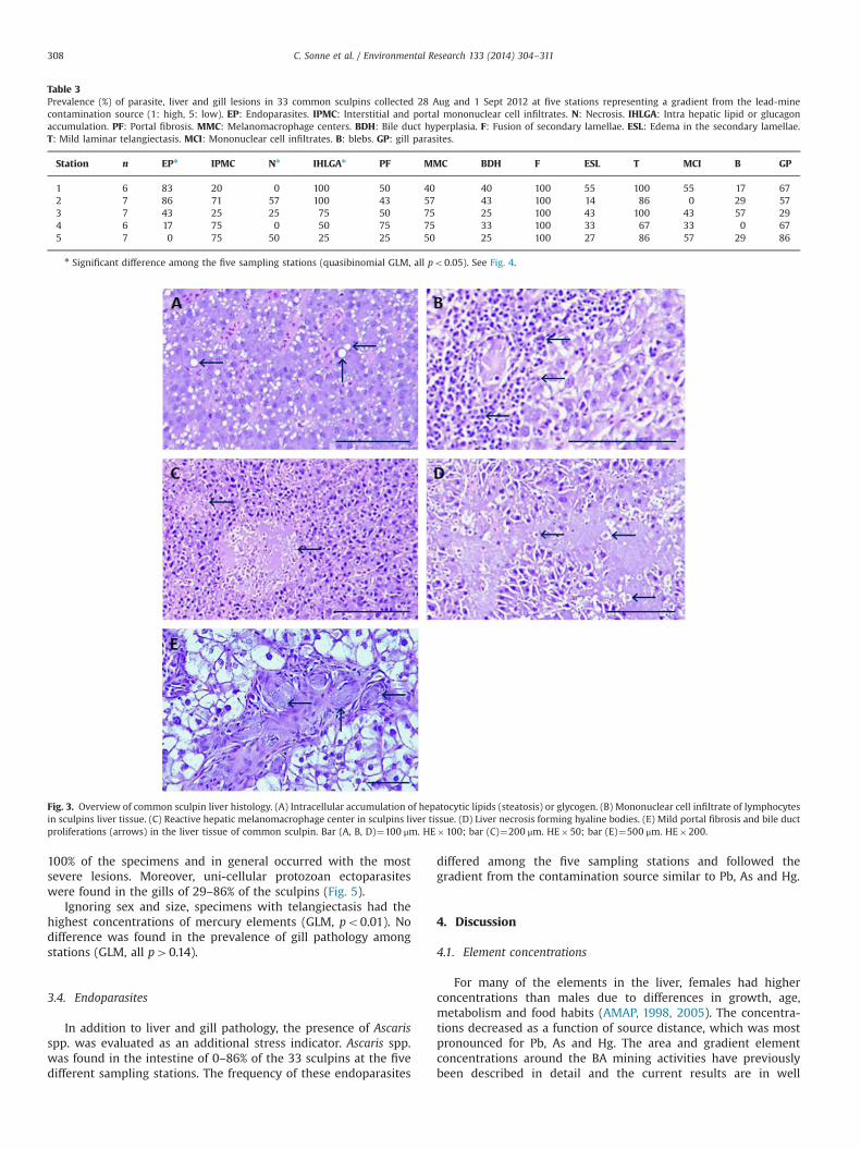
Liver tissue was fully evaluated in 33 sculpins (Table 3). Micro-structure of sculpin liver was consistent with that for other marineteleost species, and no exocrine pancreatic tissue was observednear the hepatic portal veins or in any other part of the liver(Fig. 3). Histological examination revealed six categories of liverchanges or lesions, two of parenchymal origin and four of inter-stitial or portal origin. Of the parenchymal changes, the moststriking lesion was the intracellular accumulation of hepatocyticlipids (steatosis) or glycogen (Fig. 3). These macrovesicular dro-plets were found in 25–100% of the specimens and were evenlydistributed in portal zones 1–3. Moreover, multifocal and diffuse(coagulative) necrosis was observed in 0–57% of the individualshaving formed hyaline bodies (Fig. 3). The four interstitial changesobserved were multifocal mononuclear cell infiltrates (Fig. 3), mildportal fibrosis and bile duct proliferations (Fig. 3) and multifocalmelanomacrophage centers (Fig. 3). These mononuclear infiltrateswere found in the interstitium similar to granulomas and in theportal area accompanied by mild necrosis and fibrosis. In additionto this, immune cells were found together with reactive multifocaland demarcated melanomacrophage centers. As observed in otherfish species, the melanomacrophage centers varied in size andwere characterized by HE-positive debris of macrophages andvacuolated cytoplasm.
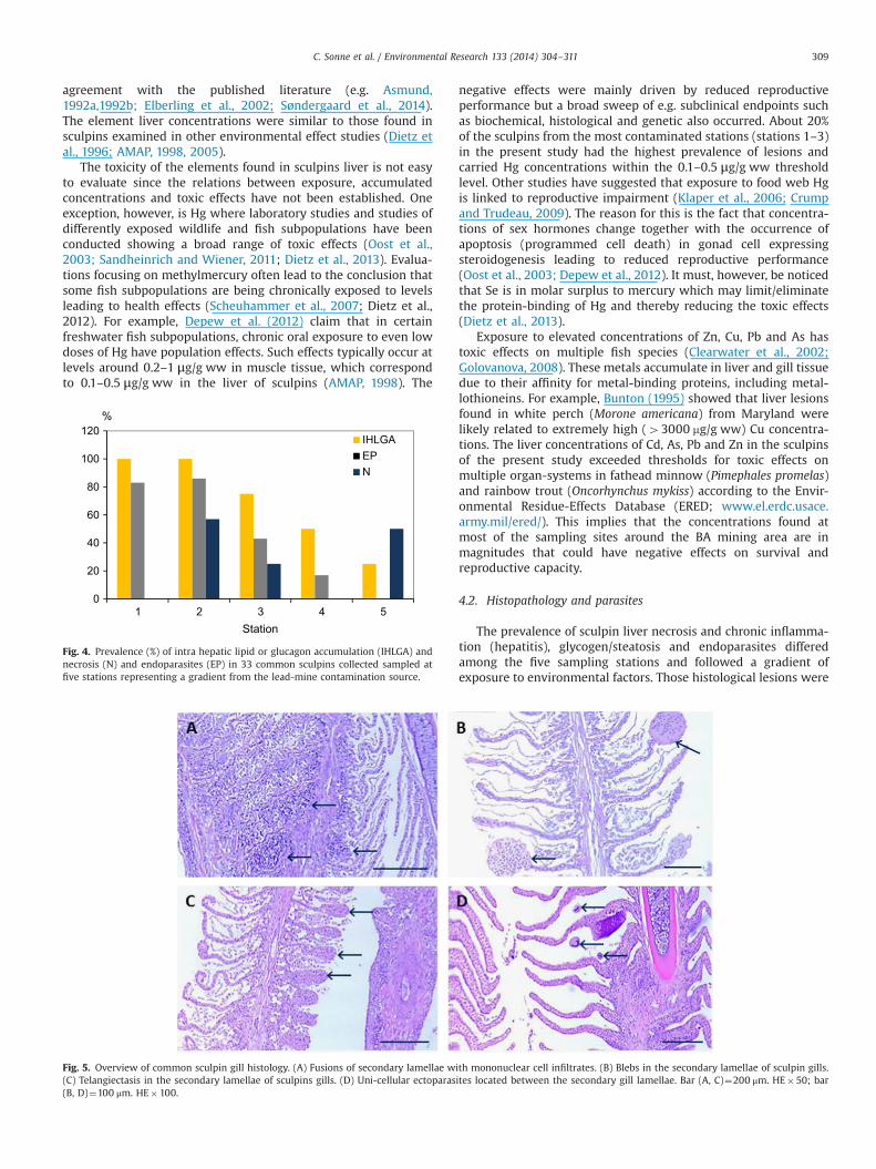
Ignoring sex and size, specimens with interstitial and portalmononuclear cell infiltrates had the highest concentrations ofmost elements (GLM, all po0.05). Controlling for sex and size,the prevalence of vacuolar hepatocytes and endoparasites wassignificantly highest at the three most element contaminatedstations and similar differences were found for liver necrosis(Fig. 4, GLM, all po0.05). This difference largely follow theconcentration gradient of elements; i.e. the higher the elementconcentrations in liver the higher the prevalence of lesions.
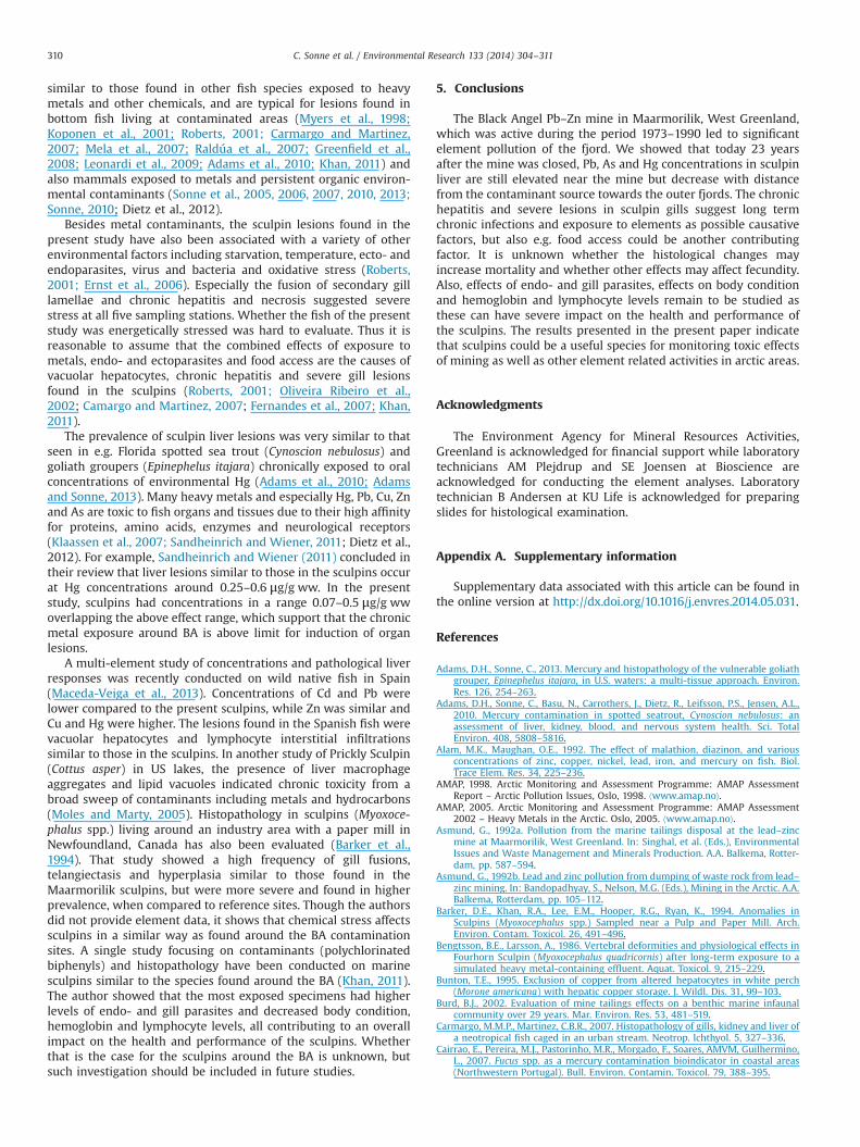
3.3. Gill histology
Gill tissue was fully evaluated in 33 sculpins (Table 3). Fivelesions were found in the secondary lamellae. The most pro-nounced lesions were fusion of the secondary lamellae most oftenfound together with mononuclear cell infiltrates and blebs (Fig. 5).In addition, severe intra cellular edema (vacuolization) was foundin 14–55% of the fish, while telangiectasis (Fig. 5) was found in 67–Ta
ble
2Biologicalinform
ation,m
orphom
etrics
andelem
entco
ncentrationsof
33co
mmon
sculpinssampled28
Augan
d1Se
pt20
12at
five
stationsrepresentingagrad
ientfrom
thelead
-mineco
ntaminationsource(Station
1:highload
s,station5:
low
load
,see
Fig.
1).A
lldatagive
nas
mea
n7
SD.d
.l.¼Detection
limitforelem
entan
alyses
determined
as3SD
onblan
ksamples.
F:female.
M:male.
Station
Sex
nLe
ngth
Weigh
tLive
rweigh
tAg
Asn
CdCo
CuFe
Hgn
Ni
Pbn
SeZn
cmg
gmg
/gww
d.l.
0.00
20.03
0.00
60.00
20.05
0.4
0.00
80.00
90.03
0.00
90.07
1F
534
75
4897
181
19.37
6.8
0.17
70.22
1107
108
0.87
70.65
0.07
70.04
2.54
72.20
727
480.19
70.18
0.01
70.00
0.447
0.60
1.06
70.33
407
12M
128
270
3.38
0.06
221.39
0.16
3.34
183
0.14
0.08
0.13
1.32
34
2F
436
76
5397
184
15.17
3.7
0.04
70.04
347
91.11
70.65
0.14
70.07
1.36
70.36
2057
167
0.16
70.08
0.02
70.01
0.47
70.30
1.04
70.24
487
7.9
M3
277
221
57
373.87
1.6
0.53
70.36
427
311.28
70.2
0.35
70.11
8.447
6.06
2807
390.15
70.06
0.15
70.03
1.71
70.17
2.15
70.24
577
6.98
3F
634
73
4527
106
14.27
9.2
0.08
70.07
677
561.89
71.84
0.06
70.03
1.45
70.68
1567
880.19
70.09
0.02
70.01
0.24
70.15
1.14
70.59
527
18M
125
188
3.8
0.17
240.71
0.48
5.55
207
0.29
0.13
0.84
1.72
45
4F
434
75
4087
180
14.77
4.3
0.08
70.11
247
150.71
70.59
0.06
70.05
1.31
70.49
1287
109
0.07
70.07
0.03
70.01
0.12
70.07
1.13
70.28
437
18M
227
–28
216–
236
3.3–
8.7
0.02
–0.19
8–31
1.45
–1.55
0.09
–0.11
1.34
–4.50
227–
278
0.07
–0.09
0.07
–0.11
0.06
–0.26
1.94
–1.27
30–46
5F
529
71
2757
618.07
4.5
0.16
70.15
157
51.08
70.48
0.08
70.05
3.22
72.15
1067
640.05
70.01
0.22
70.39
0.06
70.04
1.11
70.26
557
13M
223
–27
138–
223
1.8–
10.3
0.01
–0.12
7–21
0.49
–2.56
0.03
–0.24
0.68
–3.20
78–34
70.01
–0.10
0.03
–0.11
–0.80
–1.94
26–62
nSign
ificantdifference
amon
gthefive
samplin
gstations(A
NOVA,a
llpo
0.01
).Se
eFig.
2.
0.0
0.2
0.4
0.6
0.8
1.0
1.2
1.4
1 2 3 4 5
µg/g ww
Station
PbAs x 10-2
Hg
Fig. 2. Concentrations (mean7SD) of Pb, As and Hg in sculpin liver among the fivesampling stations representing a gradient away from the lead-zinc-mine contam-ination source (st 1: high, st 5: low).
C. Sonne et al. / Environmental Research 133 (2014) 304–311 307
100% of the specimens and in general occurred with the mostsevere lesions. Moreover, uni-cellular protozoan ectoparasiteswere found in the gills of 29–86% of the sculpins (Fig. 5).
Ignoring sex and size, specimens with telangiectasis had thehighest concentrations of mercury elements (GLM, po0.01). Nodifference was found in the prevalence of gill pathology amongstations (GLM, all p40.14).
3.4. Endoparasites
In addition to liver and gill pathology, the presence of Ascarisspp. was evaluated as an additional stress indicator. Ascaris spp.was found in the intestine of 0–86% of the 33 sculpins at the fivedifferent sampling stations. The frequency of these endoparasites
differed among the five sampling stations and followed thegradient from the contamination source similar to Pb, As and Hg.
4. Discussion
4.1. Element concentrations
For many of the elements in the liver, females had higherconcentrations than males due to differences in growth, age,metabolism and food habits (AMAP, 1998, 2005). The concentra-tions decreased as a function of source distance, which was mostpronounced for Pb, As and Hg. The area and gradient elementconcentrations around the BA mining activities have previouslybeen described in detail and the current results are in well
Table 3Prevalence (%) of parasite, liver and gill lesions in 33 common sculpins collected 28 Aug and 1 Sept 2012 at five stations representing a gradient from the lead-minecontamination source (1: high, 5: low). EP: Endoparasites. IPMC: Interstitial and portal mononuclear cell infiltrates. N: Necrosis. IHLGA: Intra hepatic lipid or glucagonaccumulation. PF: Portal fibrosis. MMC: Melanomacrophage centers. BDH: Bile duct hyperplasia. F: Fusion of secondary lamellae. ESL: Edema in the secondary lamellae.T: Mild laminar telangiectasis. MCI: Mononuclear cell infiltrates. B: blebs. GP: gill parasites.
Station n EPn IPMC Nn IHLGAn PF MMC BDH F ESL T MCI B GP
1 6 83 20 0 100 50 40 40 100 55 100 55 17 672 7 86 71 57 100 43 57 43 100 14 86 0 29 573 7 43 25 25 75 50 75 25 100 43 100 43 57 294 6 17 75 0 50 75 75 33 100 33 67 33 0 675 7 0 75 50 25 25 50 25 100 27 86 57 29 86
n Significant difference among the five sampling stations (quasibinomial GLM, all po0.05). See Fig. 4.
Fig. 3. Overview of common sculpin liver histology. (A) Intracellular accumulation of hepatocytic lipids (steatosis) or glycogen. (B) Mononuclear cell infiltrate of lymphocytesin sculpins liver tissue. (C) Reactive hepatic melanomacrophage center in sculpins liver tissue. (D) Liver necrosis forming hyaline bodies. (E) Mild portal fibrosis and bile ductproliferations (arrows) in the liver tissue of common sculpin. Bar (A, B, D)¼100 μm. HE�100; bar (C)¼200 μm. HE�50; bar (E)¼500 μm. HE�200.
C. Sonne et al. / Environmental Research 133 (2014) 304–311308
agreement with the published literature (e.g. Asmund,1992a,1992b; Elberling et al., 2002; Søndergaard et al., 2014).The element liver concentrations were similar to those found insculpins examined in other environmental effect studies (Dietz etal., 1996; AMAP, 1998, 2005).
The toxicity of the elements found in sculpins liver is not easyto evaluate since the relations between exposure, accumulatedconcentrations and toxic effects have not been established. Oneexception, however, is Hg where laboratory studies and studies ofdifferently exposed wildlife and fish subpopulations have beenconducted showing a broad range of toxic effects (Oost et al.,2003; Sandheinrich and Wiener, 2011; Dietz et al., 2013). Evalua-tions focusing on methylmercury often lead to the conclusion thatsome fish subpopulations are being chronically exposed to levelsleading to health effects (Scheuhammer et al., 2007; Dietz et al.,2012). For example, Depew et al. (2012) claim that in certainfreshwater fish subpopulations, chronic oral exposure to even lowdoses of Hg have population effects. Such effects typically occur atlevels around 0.2–1 μg/g ww in muscle tissue, which correspondto 0.1–0.5 μg/g ww in the liver of sculpins (AMAP, 1998). The
negative effects were mainly driven by reduced reproductiveperformance but a broad sweep of e.g. subclinical endpoints suchas biochemical, histological and genetic also occurred. About 20%of the sculpins from the most contaminated stations (stations 1–3)in the present study had the highest prevalence of lesions andcarried Hg concentrations within the 0.1–0.5 μg/g ww thresholdlevel. Other studies have suggested that exposure to food web Hgis linked to reproductive impairment (Klaper et al., 2006; Crumpand Trudeau, 2009). The reason for this is the fact that concentra-tions of sex hormones change together with the occurrence ofapoptosis (programmed cell death) in gonad cell expressingsteroidogenesis leading to reduced reproductive performance(Oost et al., 2003; Depew et al., 2012). It must, however, be noticedthat Se is in molar surplus to mercury which may limit/eliminatethe protein-binding of Hg and thereby reducing the toxic effects(Dietz et al., 2013).
Exposure to elevated concentrations of Zn, Cu, Pb and As hastoxic effects on multiple fish species (Clearwater et al., 2002;Golovanova, 2008). These metals accumulate in liver and gill tissuedue to their affinity for metal-binding proteins, including metal-lothioneins. For example, Bunton (1995) showed that liver lesionsfound in white perch (Morone americana) from Maryland werelikely related to extremely high (43000 mg/g ww) Cu concentra-tions. The liver concentrations of Cd, As, Pb and Zn in the sculpinsof the present study exceeded thresholds for toxic effects onmultiple organ-systems in fathead minnow (Pimephales promelas)and rainbow trout (Oncorhynchus mykiss) according to the Envir-onmental Residue-Effects Database (ERED; www.el.erdc.usace.army.mil/ered/). This implies that the concentrations found atmost of the sampling sites around the BA mining area are inmagnitudes that could have negative effects on survival andreproductive capacity.
4.2. Histopathology and parasites
The prevalence of sculpin liver necrosis and chronic inflamma-tion (hepatitis), glycogen/steatosis and endoparasites differedamong the five sampling stations and followed a gradient ofexposure to environmental factors. Those histological lesions were
0
20
40
60
80
100
120
1 2 3 4 5
%
Station
IHLGAEPN
Fig. 4. Prevalence (%) of intra hepatic lipid or glucagon accumulation (IHLGA) andnecrosis (N) and endoparasites (EP) in 33 common sculpins collected sampled atfive stations representing a gradient from the lead-mine contamination source.
Fig. 5. Overview of common sculpin gill histology. (A) Fusions of secondary lamellae with mononuclear cell infiltrates. (B) Blebs in the secondary lamellae of sculpin gills.(C) Telangiectasis in the secondary lamellae of sculpins gills. (D) Uni-cellular ectoparasites located between the secondary gill lamellae. Bar (A, C)¼200 μm. HE�50; bar(B, D)¼100 μm. HE�100.
C. Sonne et al. / Environmental Research 133 (2014) 304–311 309
similar to those found in other fish species exposed to heavymetals and other chemicals, and are typical for lesions found inbottom fish living at contaminated areas (Myers et al., 1998;Koponen et al., 2001; Roberts, 2001; Carmargo and Martinez,2007; Mela et al., 2007; Raldúa et al., 2007; Greenfield et al.,2008; Leonardi et al., 2009; Adams et al., 2010; Khan, 2011) andalso mammals exposed to metals and persistent organic environ-mental contaminants (Sonne et al., 2005, 2006, 2007, 2010, 2013;Sonne, 2010; Dietz et al., 2012).
Besides metal contaminants, the sculpin lesions found in thepresent study have also been associated with a variety of otherenvironmental factors including starvation, temperature, ecto- andendoparasites, virus and bacteria and oxidative stress (Roberts,2001; Ernst et al., 2006). Especially the fusion of secondary gilllamellae and chronic hepatitis and necrosis suggested severestress at all five sampling stations. Whether the fish of the presentstudy was energetically stressed was hard to evaluate. Thus it isreasonable to assume that the combined effects of exposure tometals, endo- and ectoparasites and food access are the causes ofvacuolar hepatocytes, chronic hepatitis and severe gill lesionsfound in the sculpins (Roberts, 2001; Oliveira Ribeiro et al.,2002; Camargo and Martinez, 2007; Fernandes et al., 2007; Khan,2011).
The prevalence of sculpin liver lesions was very similar to thatseen in e.g. Florida spotted sea trout (Cynoscion nebulosus) andgoliath groupers (Epinephelus itajara) chronically exposed to oralconcentrations of environmental Hg (Adams et al., 2010; Adamsand Sonne, 2013). Many heavy metals and especially Hg, Pb, Cu, Znand As are toxic to fish organs and tissues due to their high affinityfor proteins, amino acids, enzymes and neurological receptors(Klaassen et al., 2007; Sandheinrich and Wiener, 2011; Dietz et al.,2012). For example, Sandheinrich and Wiener (2011) concluded intheir review that liver lesions similar to those in the sculpins occurat Hg concentrations around 0.25–0.6 μg/g ww. In the presentstudy, sculpins had concentrations in a range 0.07–0.5 μg/g wwoverlapping the above effect range, which support that the chronicmetal exposure around BA is above limit for induction of organlesions.
A multi-element study of concentrations and pathological liverresponses was recently conducted on wild native fish in Spain(Maceda-Veiga et al., 2013). Concentrations of Cd and Pb werelower compared to the present sculpins, while Zn was similar andCu and Hg were higher. The lesions found in the Spanish fish werevacuolar hepatocytes and lymphocyte interstitial infiltrationssimilar to those in the sculpins. In another study of Prickly Sculpin(Cottus asper) in US lakes, the presence of liver macrophageaggregates and lipid vacuoles indicated chronic toxicity from abroad sweep of contaminants including metals and hydrocarbons(Moles and Marty, 2005). Histopathology in sculpins (Myoxoce-phalus spp.) living around an industry area with a paper mill inNewfoundland, Canada has also been evaluated (Barker et al.,1994). That study showed a high frequency of gill fusions,telangiectasis and hyperplasia similar to those found in theMaarmorilik sculpins, but were more severe and found in higherprevalence, when compared to reference sites. Though the authorsdid not provide element data, it shows that chemical stress affectssculpins in a similar way as found around the BA contaminationsites. A single study focusing on contaminants (polychlorinatedbiphenyls) and histopathology have been conducted on marinesculpins similar to the species found around the BA (Khan, 2011).The author showed that the most exposed specimens had higherlevels of endo- and gill parasites and decreased body condition,hemoglobin and lymphocyte levels, all contributing to an overallimpact on the health and performance of the sculpins. Whetherthat is the case for the sculpins around the BA is unknown, butsuch investigation should be included in future studies.
5. Conclusions
The Black Angel Pb–Zn mine in Maarmorilik, West Greenland,which was active during the period 1973–1990 led to significantelement pollution of the fjord. We showed that today 23 yearsafter the mine was closed, Pb, As and Hg concentrations in sculpinliver are still elevated near the mine but decrease with distancefrom the contaminant source towards the outer fjords. The chronichepatitis and severe lesions in sculpin gills suggest long termchronic infections and exposure to elements as possible causativefactors, but also e.g. food access could be another contributingfactor. It is unknown whether the histological changes mayincrease mortality and whether other effects may affect fecundity.Also, effects of endo- and gill parasites, effects on body conditionand hemoglobin and lymphocyte levels remain to be studied asthese can have severe impact on the health and performance ofthe sculpins. The results presented in the present paper indicatethat sculpins could be a useful species for monitoring toxic effectsof mining as well as other element related activities in arctic areas.
Acknowledgments
The Environment Agency for Mineral Resources Activities,Greenland is acknowledged for financial support while laboratorytechnicians AM Plejdrup and SE Joensen at Bioscience areacknowledged for conducting the element analyses. Laboratorytechnician B Andersen at KU Life is acknowledged for preparingslides for histological examination.
Appendix A. Supplementary information
Supplementary data associated with this article can be found inthe online version at http://dx.doi.org/10.1016/j.envres.2014.05.031.
References
Adams, D.H., Sonne, C., 2013. Mercury and histopathology of the vulnerable goliathgrouper, Epinephelus itajara, in U.S. waters: a multi-tissue approach. Environ.Res. 126, 254–263.
Adams, D.H., Sonne, C., Basu, N., Carrothers, J., Dietz, R., Leifsson, P.S., Jensen, A.L.,2010. Mercury contamination in spotted seatrout, Cynoscion nebulosus: anassessment of liver, kidney, blood, and nervous system health. Sci. TotalEnviron. 408, 5808–5816.
Alam, M.K., Maughan, O.E., 1992. The effect of malathion, diazinon, and variousconcentrations of zinc, copper, nickel, lead, iron, and mercury on fish. Biol.Trace Elem. Res. 34, 225–236.
AMAP, 1998. Arctic Monitoring and Assessment Programme: AMAP AssessmentReport – Arctic Pollution Issues, Oslo, 1998. ⟨www.amap.no⟩.
AMAP, 2005. Arctic Monitoring and Assessment Programme: AMAP Assessment2002 – Heavy Metals in the Arctic. Oslo, 2005. ⟨www.amap.no⟩.
Asmund, G., 1992a. Pollution from the marine tailings disposal at the lead–zincmine at Maarmorilik, West Greenland. In: Singhal, et al. (Eds.), EnvironmentalIssues and Waste Management and Minerals Production. A.A. Balkema, Rotter-dam, pp. 587–594.
Asmund, G., 1992b. Lead and zinc pollution from dumping of waste rock from lead–zinc mining. In: Bandopadhyay, S., Nelson, M.G. (Eds.), Mining in the Arctic. A.A.Balkema, Rotterdam, pp. 105–112.
Barker, D.E., Khan, R.A., Lee, E.M., Hooper, R.G., Ryan, K., 1994. Anomalies inSculpins (Myoxocephalus spp.) Sampled near a Pulp and Paper Mill. Arch.Environ. Contam. Toxicol. 26, 491–496.
Bengtsson, B.E., Larsson, A., 1986. Vertebral deformities and physiological effects inFourhorn Sculpin (Myoxocephalus quadricornis) after long-term exposure to asimulated heavy metal-containing effluent. Aquat. Toxicol. 9, 215–229.
Bunton, T.E., 1995. Exclusion of copper from altered hepatocytes in white perch(Morone americana) with hepatic copper storage. J. Wildl. Dis. 31, 99–103.
Burd, B.J., 2002. Evaluation of mine tailings effects on a benthic marine infaunalcommunity over 29 years. Mar. Environ. Res. 53, 481–519.
Carmargo, M.M.P., Martinez, C.B.R., 2007. Histopathology of gills, kidney and liver ofa neotropical fish caged in an urban stream. Neotrop. Ichthyol. 5, 327–336.
Cairrao, E., Pereira, M.J., Pastorinho, M.R., Morgado, F., Soares, AMVM, Guilhermino,L., 2007. Fucus spp. as a mercury contamination bioindicator in coastal areas(Northwestern Portugal). Bull. Environ. Contamin. Toxicol. 79, 388–395.
C. Sonne et al. / Environmental Research 133 (2014) 304–311310
Clearwater, S.J., Farag, A.M., Meyer, J.S., 2002. Bioavailability and toxicity of dietborne copper and zinc to fish. Comp. Biochem. Physiol. Part C 132, 269–313.
Crump, K.L., Trudeau, V.L., 2009. Mercury-induced reproductive impairment in fish.Environ. Toxicol. Chem. 28, 895–907.
Depew, D.C., Basu, N., Burgess, N.M., Campbell, L.M., Devlin, E.W., Drevnick, P.E.,Hammerschmidt, C.R., Murphy, C.A., Sandheinrich, M.B., Wiener, J.G., 2012.Toxicity of dietary methylmercury to fish: derivation of ecologically meaningfulthreshold concentrations. Environ. Toxicol. Chem. 31, 1536–1547.
Dietz, R., Rigét, F.F., Johansen, P., 1996. Lead, cadmium, mercury and selenium inGreenland marine animals. Sci. Total Environ. 186, 67–93.
Dietz, R., Basu, N., Braune, B., O‘Hara, T., Scheuhammer, T.M., Sonne, C., Andersen,M., Andreasen, C., Andriashek, D., Asmund, G., Aubail, A., Baagøe, H., Born, E.W.,Chan, H.M., Derocher, A.E., Grandjean, P., Knott, K., Kirkegaard, M., Lunn, N.,Messier, F., Obbard, M., Olsen, M.T., Peacock, E., Renzoni, A., Rigét, F.F., Skaare, J.U., Stern, G., Stirling, I., Taylor, M., Wiig, Ø., Aars, J., 2013. What are thetoxicological effects of mercury in Arctic biota? Sci. Total Environ. 443,775–790.
Elberling, B., Asmund, G., Kunzendorf, H., Krogstad, E.J., 2002. Geochemical trendsin metal-contaminated fjord sediments near a former lead–zinc mine in WestGreenland. Appl. Geochem. 12, 493–502.
Ernst, B., Hoeger, S.J., O‘Brien, E., Dietrich, D.R., 2006. Oral toxicity of themicrocystin-containing cyanobacterium, Planktothrix rubescens in Europeanwhitefish, Coregonus lavaretus. Aquat. Toxicol. 79, 31–40.
Fernandes, C., Fontaínhas-Fernandes, A., Monteiro, S.M., Salgado, M.A., 2007.Histopathological gill changes in wild leaping grey mullet, Liza saliens, fromthe Esmoriz-Paramos coastal lagoon, Portugal. Environ. Toxicol. 22, 443–448.
Golovanova, I.L., 2008. Effects of heavy metals on the physiological and biochemicalstatus of fishes and aquatic invertebrates. Inland Water Biol. 1, 93–101.
Greenfield, B.K., Teh, S.J., Ross, J.R.M., Hunt, J., Zhang, G., Davis, J.A., Ichikawa, G.,Crane, D., Hung, S.S.O., Deng, D., Teh, F.-C., Green, P.G., 2008. Contaminantconcentrations and histopathological effects in sacramento splittail (Pogo-nichthys macrolepidotus). Arch. Environ. Contam. Toxicol. 55, 270–281.
Iwasaki, Y., Hayashi, T., Kamo, M., 2010. Comparison of population-level effects ofheavy metals on fathead minnow (Pimephales promelas). Ecotoxicol. Environ.Chem. 73, 465–471.
Johansen, P., Hansen, M.M., Asmund, G., Nielsen, P.B., 1991. Marine organisms asindicators of heavy metal pollution-experience from 16 years of monitoring at alead zinc mine in Greenland. Chem. Ecol. 5, 35–55.
Johansen, P., Asmund, G., Riget, F., Johansen, K., 2008. Environmental Monitoring atthe Lead-zinc Mine in Maarmorilik, Northwest Greenland, 2007. NERI TechnicalReport No. 684. National Environmental Research Institute, Aarhus University.Available at: ⟨http://www.dmu.dk/Pub/FR684.pdf⟩. p. 54.
Josefson, A.B., Hansen, J.L.S., Asmund, G., Johansen, P., 2008. Threshold response ofbenthic macrofauna integrity to metal contamination in West Greenland. Mar.Pollut. Bull. 56, 1265–1274.
Khan, R.A., 2011. Chronic exposure and decontamination of a marine sculpin(Myoxocephalus scorpius) to polychlorinated biphenyls using selected bodyindices, blood values, histopathology, and parasites as bioindicators. Arch.Environ. Contam. Toxicol. 60, 479–485.
Klaassen, C.D., Amdur, M.O., Doull, J., 2007. Casarett and Doulls Toxicology: TheBasic Science of Poisons, 6th ed. McGraw-Hill, New York.
Klaper, R., Rees, C.B., Drevnick, P., Weber, D., Sandheinrich, M., Caravan, M.J., 2006.Gene expression changes related to endocrine function and decline in repro-duction in fathead minnow, Pimephales promelas, after dietary methylmercuryexposure. Environ. Health Perspect. 11, 1337–1343.
Koponen, K., Myers, M.S., Ritola, O., Huuskonen, S.E., Seppa-Lindstrom, P., 2001.Histopathology of feral fish from a PCB-contaminated freshwater lake. AMBIO30, 122–126.
Leonardi, M., Tarifeno, E., Vera, J., 2009. Diseases of the Chilean flounder,Paralichthys adspersus (Steindachner, 1867), as a biomarker of marine coastalpollution near the Itata River (Chile): Part II. Histopathological lesions. Arch.Environ. Contam. Toxicol. 56, 546–556.
Maceda-Veiga, A., Monroy, M., Navarro, E., Viscor, G., de Sostoa, A., 2013. Metalconcentrations and pathological responses of wild native fish exposed tosewage discharge in a Mediterranean river. Sci. Total Environ. 449, 9–19.
Matta, M.B., Linse, J., Cairncross, C., Francendese, L., Kocan, R.M., 2001. Reproductiveand transgenerational effects of methylmercury or Aroclor 1268 on Fundulusheteroclitus. Environ. Toxicol. Chem. 20, 327–335.
Mela, M., Randi, M.A.F., Ventura, D.F., Carvalho, C.E.V., Pelletier, E., Oliveira Ribeiro,C.A., 2007. Effects of dietary methylmercury on liver and kidney histology inthe neotropical fish Hoplias malabaricus. Ecotoxicol. Environ. Saf. 68, 426–435.
Moles, A., Marty, G.D., 2005. Physiological changes in prickly sculpin (Cottus asper)inhabiting a lake used by jet-propelled watercraft. Bull. Environ. Contam.Toxicol. 74, 1151–1158.
Myers, M.S., Johnson, L.L., Olson, O.P., Stehr, C.M., Horness, B.H., Collier, T.K., Mccain,B.B., 1998. Toxicopathic hepatic lesions as biomarkers of chemical contaminantexposure and effects in marine bottomfish species from the northeast andpacific coasts, USA. Mar. Pollut. Bull. 37, 92–113.
O‘Connor, T.P., Lauenstein, G.G., 2006. Trends in chemical concentrations in musselsand oysters collected along the US coast: update to 2003. Mar. Environ. Res. 62,261–285.
Oliveira Ribeiro, C.A., de Belger, L., Pelletier, E., Rouleau, C., 2002. Histopathologicalevidence of inorganic mercury and methyl mercury toxicity in the arctic charr,Salvelinus alpinus. Environ. Res. 90, 217–225.
Oost, R.V.D., Beyer, J., Vermeulen, N.P.E., 2003. Fish bioaccumulation and biomar-kers in environmental risk assessment: a review. Environ. Toxicol. Pharmacol.13, 57–149.
R Core Team, 2013. R: A Language and Environment for Statistical Computing. RFoundation for Statistical Computing, Vienna, Austria. URL: ⟨http://www.R-project.org/⟩.
Raldúa, D., Díez, S., Bayona, J.M., Damiá, B., 2007. Mercury levels and liver pathologyin feral fish living in the vicinity of a mercury cell chlor-alkali factory.Chemosphere 66, 1217–1225.
Rigét, F., Johansen, P., Asmund, G., 1997. Uptake and release of lead and zinc by bluemussels. Experience from transplantation experiments in Greenland. Mar.Pollut. Bull. 34, 805–815.
Roberts, R.J., 2001. Fish Pathology, 3rd ed. Elsevier, Edinburgh.Sanchez, J., Marino, N., Vaquero, M.C., Ansorena, J., Legorburu, I., 1998. Metal
pollution by old lead–zinc mines in Urumea river valley (Basque country,Spain). Soil, biota and sediment. Water Air Soil Pollut. 107, 303–319.
Sandheinrich, M.B., Wiener, J.G., 2011. Methylmercury in freshwater fish: recentadvances in assessing toxicity of environmentally relevant exposures. In: Beyer,N., Meador, J. (Eds.), Environmental Contaminants in Wildlife: InterpretingTissue Concentrations, 2nd ed. Taylor and Francis Publishers, Boca Raton,Florida.
Scheuhammer, A.M., Meyer, M.W., Sandheinrich, M.B., Murray, M.W., 2007. Effectsof environmental methylmercury on the health of wild birds, mammals, andfish. AMBIO 36, 12–19.
Schiedek, D., Asmund, G., Johansen, P., Rigét, F., Johansen, K., Strand, J., Mølvig, S.,2009. Environmental Monitoring at the Former Lead–Zinc Mine in Maarmorilik,Northwest Greenland, in 2008. NERI Technical Report No. 737. NationalEnvironmental Research Institute, Aarhus University, p. 70 ⟨http://www.dmu.dk/Pub/FR737.pdf⟩.
Søndergaard, J., Asmund, G., Johansen, P., Rigét, F.F., 2011. Long-term response of anarctic fiord system to lead–zinc mining and submarine disposal of mine waste(Maarmorilik, West Greenland). Mar. Environ. Res. 71, 331–341.
Søndergaard, J., Bach, L., Gustavson, K., 2014. Measuring bioavailable metals usingdiffusive gradients in thin films (DGT) and transplanted seaweed (Fucusvesiculosus), blue mussels (Mytilus edulis) and sea snails (Littorina saxatilis)suspended from monitoring buoys near a former lead–zinc mine in WestGreenland. Mar. Pollut. Bull. 78, 102–109.
Sonne, C., 2010. Health effects from long-range transported contaminants in Arctictop predators: an integrated review based on studies of polar bears andrelevant model species. Environ. Int. 36, 461–491.
Sonne, C., Dietz, R., Leifsson, P.S., Born, E.W., Kirkegaard, M., Rigét, F.F., Letcher, R.J.,Muir, D.C.G., Hyldstrup, L., 2005. Do Organohalogen Contaminants Contributeto Liver Histopathology in East Greenland Polar Bears (Ursus maritimus)?Environ. Health Perspect. 113, 1569–1574.
Sonne, C., Dietz, R., Leifsson, P.S., Born, E.W., Kirkegaard, M., Letcher, R.J., Muir, D.C.G., Rigét, F.F., Hyldstrup, L., 2006. Are organohalogen contaminants a co-factorin the development of renal lesions in East Greenland polar bears (Ursusmaritimus)? Environ. Toxicol. Chem. 25, 1551–1557.
Sonne, C., Dietz, R., Leifsson, P.S., Born, E.W., Kirkegaard, M., Rigét, F.F., 2007. Areliver and renal lesions in East Greenland polar bears, Ursus maritimus,associated with high mercury levels? Environ. Health 6, 11.
Sonne, C., Dam, M., Leifsson, P.L., Dietz, R., 2010. Liver and renal histopathology ofNorth Atlantic long-finned pilot whales (Globicephala melas) with high con-centrations of heavy metals and organochlorines. Toxicol. Environ. Chem. 92,969–985.
Sonne, C., Leifsson, P.S., Dietz, R., 2013. Liver and renal lesions in mercurycontaminated narwhals (Monodon monoceros) from North West Greenland.Toxicol. Environ. Chem. 95, 515–528.
Torres, M.A., Barros, M.P., Campos, S.C.G., Pinto, E., Rajamani, S., Sayre, R.T.,Colepicolo, P., 2008. Biochemical biomarkers in algae and marine pollution: areview. Ecotoxicol. Environ. Saf. 71, 1–15.
C. Sonne et al. / Environmental Research 133 (2014) 304–311 311
Copyright © 2022 FDOKUMEN