Evaluation of the impact of decellularization and sterilization ...
11
Acta of Bioengineering and Biomechanics Original paper Vol. 21, No. 3, 2019 DOI: 10.5277/ABB-01382-2019-02 Evaluation of the impact of decellularization and sterilization on tensile strength transgenic porcinedermal dressings KAMIL JOSZKO 1 , BOŻENA GZIK-ZROSKA 2 , EDYTA KAWLEWSKA 1 *, AGNIESZKA KLAMA-BARYŁA 3, 4 , SŁAWOMIR SUCHOŃ 1 , MICHAŁ BURKACKI 1 , WOJCIECH WOLAŃSKI 1 1 Department of Biomechatronics, Faculty of Biomedical Engineering, Silesian University of Technology, Zabrze, Poland. 2 Department of Biomaterials and Medical Devices Engineering, Faculty of Biomedical Engineering, Silesian University of Technology, Zabrze, Poland. 3 Dr Stanisław Sakiel Centre of Burns Treatment, Siemianowice Śląskie, Poland. 4 Silesian Higher Medical School in Katowice, Katowice, Poland. Purpose: The aim of this paper was to evaluate which method of acellularization and sterilization is optimal, in the meaning of which processes have the least impact on the deterioration of mechanical properties of porcine tissues used for xenogeneic applications. Meth- ods: The static tensile probe was conducted for 80 skin specimens obtained from transgenic swine, which are used as a wound dressing for skin recipient. Obtained data were subsequently analyzed with the use of statistical methods. Results: It was found that Young’s modulus for the samples after the sterilization process for the dispase substance and the mixed method (SDS + trypsin) were statistically significantly changed. In the case of dispase, Young’s modulus value before the sterilization process was 12.4 MPa and after the value increased to 28.0 MPa. For the mixed method (SDS + trypsin) before the sterilization process Young’s modulus value was 5.6 MPa and after it was increased to 6.3 MPa. The mixed method (SDS + trypsin) had the slightest effect on changing the mechanical properties of the samples before and after the sterilization process. Conclusions: It was confirmed that different methods of acellularization and the process of sterilization have an influence on the change of mechanical properties of the skin of transgenic swine. In the authors’ opinion, the mixed method (SDS + trypsin) should be recommended as the best one for the preparation of transgenic porcine dermal dressings because it ensures a smaller probability of dressing’s damage during a surgical procedure. Key words: skin tissue, burn wounds treatment, xenogeneic grafts, static tensile test, statistical analysis, reconstructive surgery 1. Introduction Skin is an organ which is essential for the proper functioning of the whole organism. On one hand, it provides the body with protection against the external environment, on the other hand, it enables contact with the surroundings. The skin is highly exposed to the impact of external factors, thus its main role is a protective function. Thanks to such features as re- sistance, elasticity and semi-permeability, it protects the human organism from mechanical injuries, infec- tions, the loss of physiological liquids and harmful radiation [5]. Moreover, it plays an important role in the organism’s metabolism of fat and vitamins as well as water and electrolyte balance. The skin also takes part in immunological processes, thermoregulation of the body and resorption. All these activities are dis- turbed as a result of burns, which cause not only changes in the scope of anatomy and physiology of the damaged organisms but also lead to endocrinological and immunological disorders. Among all kinds of wounds, burn wounds are characterized by the biggest disorders of all repair factors and, therefore, they are the most difficult to treat [19]. Nowadays, the most common way of treatment involves the application of biostatic (deprived of live cells) grafts obtained from human corpses or transgenic (genetically modified) swine. The process of preparation of biological dress- ings encompasses acellularization (removal of cells), ______________________________ * Corresponding author: Edyta Kawlewska, Department of Biomechatronics, Silesian University of Technology, Roosevelta 40, 41-800 Zabrze, Poland. Room 116, phone: +48322777437, e-mail: [email protected] Received: May 13th, 2019 Accepted for publication: July 22nd, 2019
-
Upload
khangminh22 -
Category
Documents
-
view
0 -
download
0
Transcript of Evaluation of the impact of decellularization and sterilization ...
Acta of Bioengineering and Biomechanics Original paperVol. 21, No. 3, 2019 DOI: 10.5277/ABB-01382-2019-02
Evaluation of the impact of decellularization and sterilizationon tensile strength transgenic porcinedermal dressings
KAMIL JOSZKO1, BOŻENA GZIK-ZROSKA2, EDYTA KAWLEWSKA1*, AGNIESZKA KLAMA-BARYŁA3, 4,SŁAWOMIR SUCHOŃ1, MICHAŁ BURKACKI1, WOJCIECH WOLAŃSKI1
1 Department of Biomechatronics, Faculty of Biomedical Engineering, Silesian University of Technology, Zabrze, Poland.2 Department of Biomaterials and Medical Devices Engineering, Faculty of Biomedical Engineering,
Silesian University of Technology, Zabrze, Poland.3 Dr Stanisław Sakiel Centre of Burns Treatment, Siemianowice Śląskie, Poland.
4 Silesian Higher Medical School in Katowice, Katowice, Poland.
Purpose: The aim of this paper was to evaluate which method of acellularization and sterilization is optimal, in the meaning of whichprocesses have the least impact on the deterioration of mechanical properties of porcine tissues used for xenogeneic applications. Meth-ods: The static tensile probe was conducted for 80 skin specimens obtained from transgenic swine, which are used as a wound dressingfor skin recipient. Obtained data were subsequently analyzed with the use of statistical methods. Results: It was found that Young’smodulus for the samples after the sterilization process for the dispase substance and the mixed method (SDS + trypsin) were statisticallysignificantly changed. In the case of dispase, Young’s modulus value before the sterilization process was 12.4 MPa and after the valueincreased to 28.0 MPa. For the mixed method (SDS + trypsin) before the sterilization process Young’s modulus value was 5.6 MPa andafter it was increased to 6.3 MPa. The mixed method (SDS + trypsin) had the slightest effect on changing the mechanical properties ofthe samples before and after the sterilization process. Conclusions: It was confirmed that different methods of acellularization and theprocess of sterilization have an influence on the change of mechanical properties of the skin of transgenic swine. In the authors’ opinion,the mixed method (SDS + trypsin) should be recommended as the best one for the preparation of transgenic porcine dermal dressingsbecause it ensures a smaller probability of dressing’s damage during a surgical procedure.
Key words: skin tissue, burn wounds treatment, xenogeneic grafts, static tensile test, statistical analysis, reconstructive surgery
1. Introduction
Skin is an organ which is essential for the properfunctioning of the whole organism. On one hand, itprovides the body with protection against the externalenvironment, on the other hand, it enables contactwith the surroundings. The skin is highly exposed tothe impact of external factors, thus its main role isa protective function. Thanks to such features as re-sistance, elasticity and semi-permeability, it protectsthe human organism from mechanical injuries, infec-tions, the loss of physiological liquids and harmfulradiation [5]. Moreover, it plays an important role inthe organism’s metabolism of fat and vitamins as well
as water and electrolyte balance. The skin also takespart in immunological processes, thermoregulation ofthe body and resorption. All these activities are dis-turbed as a result of burns, which cause not onlychanges in the scope of anatomy and physiology of thedamaged organisms but also lead to endocrinologicaland immunological disorders. Among all kinds ofwounds, burn wounds are characterized by the biggestdisorders of all repair factors and, therefore, they arethe most difficult to treat [19]. Nowadays, the mostcommon way of treatment involves the application ofbiostatic (deprived of live cells) grafts obtained fromhuman corpses or transgenic (genetically modified)swine. The process of preparation of biological dress-ings encompasses acellularization (removal of cells),
______________________________
* Corresponding author: Edyta Kawlewska, Department of Biomechatronics, Silesian University of Technology, Roosevelta 40,41-800 Zabrze, Poland. Room 116, phone: +48322777437, e-mail: [email protected]
Received: May 13th, 2019Accepted for publication: July 22nd, 2019
K. JOSZKO et al.88
sterilization and storage inadequate conditions. At pres-ent, xenotransplantation is becoming more and morepopular giving hope in the treatment of serious andextensive skin loss in a situation when there is a lackof human skin, both autologous and allogeneic [3].The usefulness of the application of xenogeneic graftshas been confirmed by numerous clinical investiga-tions [2], [4], [24], [25]. Taking the above into con-sideration, the researchers search for optimum meth-ods of storage and sterilization that would have thesmallest impact on the decreasing mechanical proper-ties of grafts. Before homing of the skin with the do-nor’s cells, the skin must be properly prepared. Thepreparation process consists of the removal of thedonor’s cells with simultaneous preservation of thestructure of the extracellular matrix and sterilization.There are many methods of the removal of cells (in otherwords, decellularization or acellularization) which mostoften include chemical, biological or mixed procedures[6], [8], [22].
The selection of the best method of the cell removalfrom tissues or organs depends on many factors, suchastissue cellularity, density, thickness or the content oflipids. The fact that should be borne in mind is thateach reagent will cause modifications of the composi-tion of the extracellular matrix and have an impact oncertain disturbances of the ultrastructure. Therefore, itis advisable to attempt the minimization of possibleundesirable effects [6].
Literature includes works concerning the determi-nation of mechanical properties of the skin [1], [12],[13], [20], [21]. There are also many works dealingwith the application of the determined mechanicalproperties of various biological structures to the pro-cess of modelling [7], [9], [10], [15], [18], [23]. How-ever, there is a lack of literature which would con-firm the investigations aiming at the determination ofmechanical properties of skins subjected to the pro-cedure of acellularization and sterilization. It seemsnecessary to conduct research which would definewhat exactly influences the changes of mechanicalproperties – whether it is a type of acellularizationmethod or the application of the process of sterilization.The developed skin grafts often have weak mechanicalproperties, which makes them vulnerable to damageduring the transplantation procedure. Also, after thetransplantation, they are exposed to the action of localshear and tensile forces [14]. Taking the above intoaccount, the aim of the study was to evaluate the meth-ods of decellularization of dressings from transgenicpigskin by determining and comparing the mechanicalproperties of the examined dressings. Such comparisonis aimed at determining which of the deacellalization
methods will ensure the best mechanical properties ofdressings for surgical procedures.
2. Materials and methods
2.1. Specimens preparation
Within the framework of this work 80 specimensobtained from transgenic swines were tested. The der-mis should be harvested from the animal just right aftereuthanasia. All samples for the tests were taken fromthe ridge of the pig maintaining the same arrangementof collagen fibres to exclude their influence on themeasured mechanical properties.
In the first stage, the harvesting site should beshaved with an electric shaver to remove the hair.Next, the site should be disinfected with a skin disin-fectant (e.g., skinsept, octanisept) and dermatomeshould be set at the optimal depth to remove the entireepidermis with as little dermis as possible. After har-vesting the epidermis, the target fragments of thedermis should be harvested at the same site derma-tome as well (set at the maximum depth). The dermisshall be used in further procedures and, before itsapplication in further stages of the procedure, it can bestored in water for injections as low as – 80 °C. Theywere subjected to procedures of the removal of cellsfrom the skin.This procedure is called decellulariza-tion. In order to remove cells from the tissue of animalorigin, four procedures may be applied:1. Chemical procedure (SDS, Triton X).2. Enzymatic procedure (Trypsin, Dispase).3. Physical procedure (Liquid nitrogen).4. Mixed procedure – a two-stage procedure to com-
bine the enzymatic method (first stage) and chemi-cal method (second stage).
Chemical procedures
In the chemical method of removing cells from pigskin, two different reagents were applied:1. The first method applied Triton-X reagent in the
concentration of 3%: in the first stage, 3% solutionwas prepared by dosing 30 mL of 10% Triton-Xsolution to be diluted in 70 mL of H2O.
2. The other method of chemical removal of cells frompig skin applies a 0.1% solution of SDS (sodiumdodecyl sulphate). In the first stage, a 1% SDS so-lution is prepared by dosing 1 g of the substance tobe diluted in 100 mL of water for injections. After
Evaluation of the impact of decellularization and sterilization on tensile strength transgenic porcinedermal dressings 89
preparing the 1% solution, it should be diluted10-fold to obtain a 0.1% SDS solution.
Enzymatic procedure
The enzymatic procedure applied the ready-to-useconcentrated reagent Triple 1x or 2.4 U/mL dispase.The prepared solutions must be filtered through 0.22µm filters. Such a filter should be fixed on a syringeafter the solution of relevant concentration has beendrawn into the syringe. Then the solution must befiltered into the target container. Pigskin should beharvested with a dermatome according to the above-described protocol, resulting in dividing the skin intofragments of predefined dimensions. Each skinfragment should be washed with water mixed withdetergent so that blood clots, residual tissues andother contaminants are removed. To degrease theskin, it should be washed with an alcohol solutionand then skin fragments should be washed with wa-ter to remove the detergent and alcohol. In the nextstage, the skin must be immersed in a container filledwith the solution and left for 24 h for shaking in4–8 °C. Afterwards, each skin fragment should besoaked in water to remove the solution. The washingshould continue until the decellularisation reagent isentirely removed.
Physical procedure
The physical method requires freezing in liquidnitrogen of temperature –196 °C. Pigskin should beharvested with a battery-supplied dermatome as de-scribed above, resulting in dividing the skin intofragments of predefined dimensions. Skin fragmentsshould be divided into equal pieces of 7 20 cm andthen placed in 50 mL falcon tubes and tightly lockedto obtain an anaerobic environment. Three cycles offreezing in liquid nitrogen are required with thespecimens placed in liquid nitrogen for 4 hours eachtime. Between the cycles, incubation should be held ata temperature from 20 to 37 °C for at least 5 minutesand preferably 10 to 30 minutes. Next, the falcontubes with the skin should be incubated as describedabove in ambient temperature for 48 hours.
Mixed procedure
In the first stage, cells are removed from tissues ac-cording to the paragraph enzymatic procedure, apply-ing one of the enzymes – either trypsin or dispase. Thenext stage involves the chemical procedure, accordingto the above-described protocol applying one of thechemical reagents: either Tritin-X or SDS. The effi-ciency of the decellularization process for each method
should be verified by histopathologic preparation withhaematoxylin-eosin staining.
Haematoxylin-eosin staining procedure
A fragment of the harvested pigskin should bebathed in a tray filled with alum haematoxylin for7–10 minutes and then processed by bathing prepa-rations in a tray with distilled water. Next, the prepara-tions must be rinsed under tap water for 15 minutes andthen bathed again in the tray with distilled water. Afterthis stage, the preparations are washed in a tray filledwith 1% eosin Y with one drop of acetic acid addedand finally washed in the tray with distilled wateragain. In order to verify the degree of removing thechemical solution from the preparation, the skinspecimen should be checked for the presence ofchemical substances in the preparation. After theprocess of decellularisation, half of the samples areto be subjected to the process of radiation sterilization(35 kGy) in compliance with the valid procedures ofthe Tissue Bank. Sterility of the tissues should beconfirmed microbiologically. The specimens must bestored at a temperature of –20 °C prior to mechanicaltests, samples were thawed in saline at room tem-perature 21 °C for 20 minutes. The same procedureshould be used before the implantation process in theoperating room.
2.2. Uniaxial tensile testing
The tests were conducted by means of a testingmachine MTS Insight 2. This is an electromechanicalmachine used for static tests and serves the purpose oflow-force measurements in the range up to 2 kN. Thetests were carried out on samples with a width of 20± 1.97 mm, length 120 mm, thickness 0.49 ± 0.05.The width of the samples was measured by a certifiedcalliper and the thickness of the samples was meas-ured with a thickness gauge. All samples tested werestored for a period of 2 months from the time of col-lection. Next, the prepared specimens were subjectedto a tensile test after the endings of the samples hadbeen fixed by means of special grips. The ends of thesample were wound on specially prepared rolls forthis type of testing, which were later placed in theholders of the testing machine. Those activities aimedto prevent the spontaneous slipping of the specimensfrom the clamps and to eliminate the phenomenon ofconcentration of stresses at the place of the sample'sfixation. Prior to testing, an initial distance betweenthe clamps was measured as well as the thickness andwidth of the specimens were determined. Next, the
K. JOSZKO et al.90
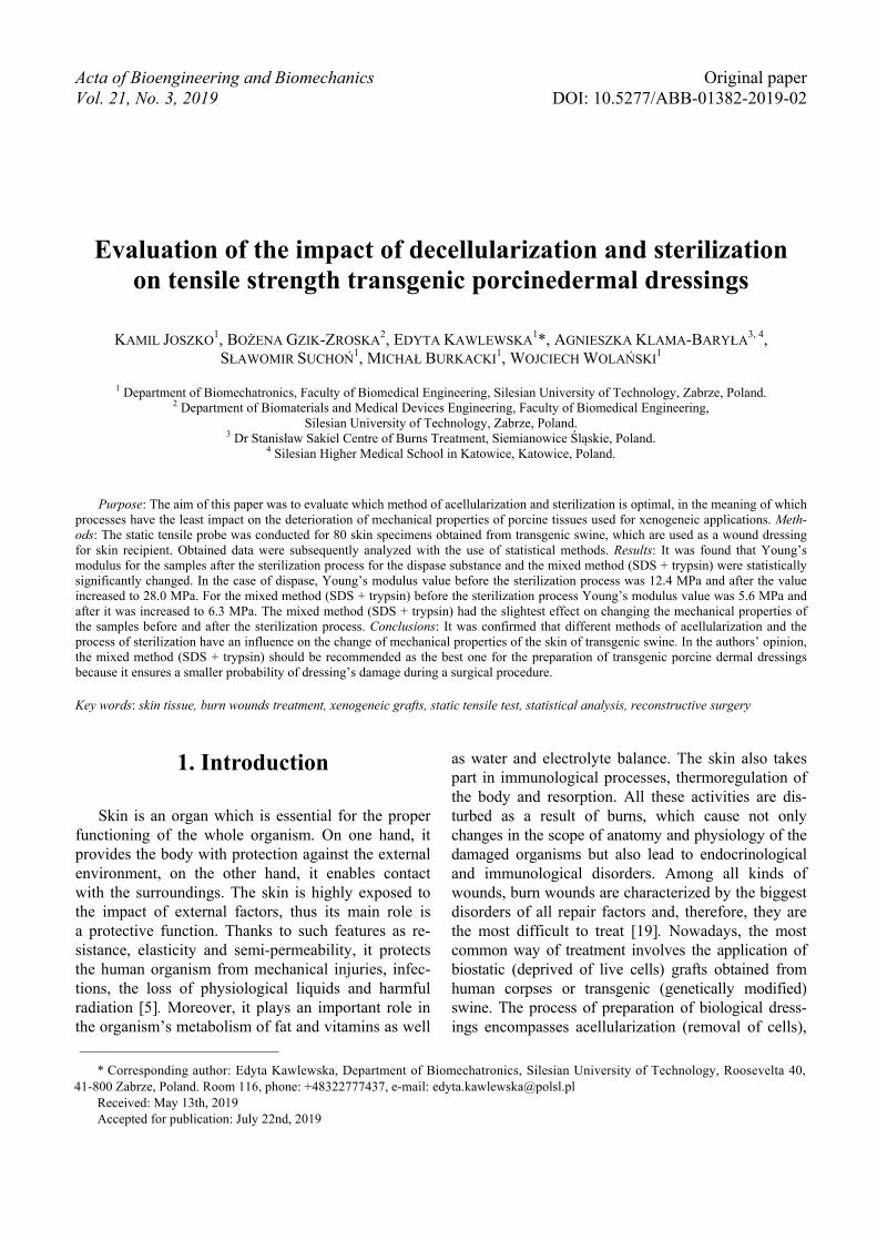
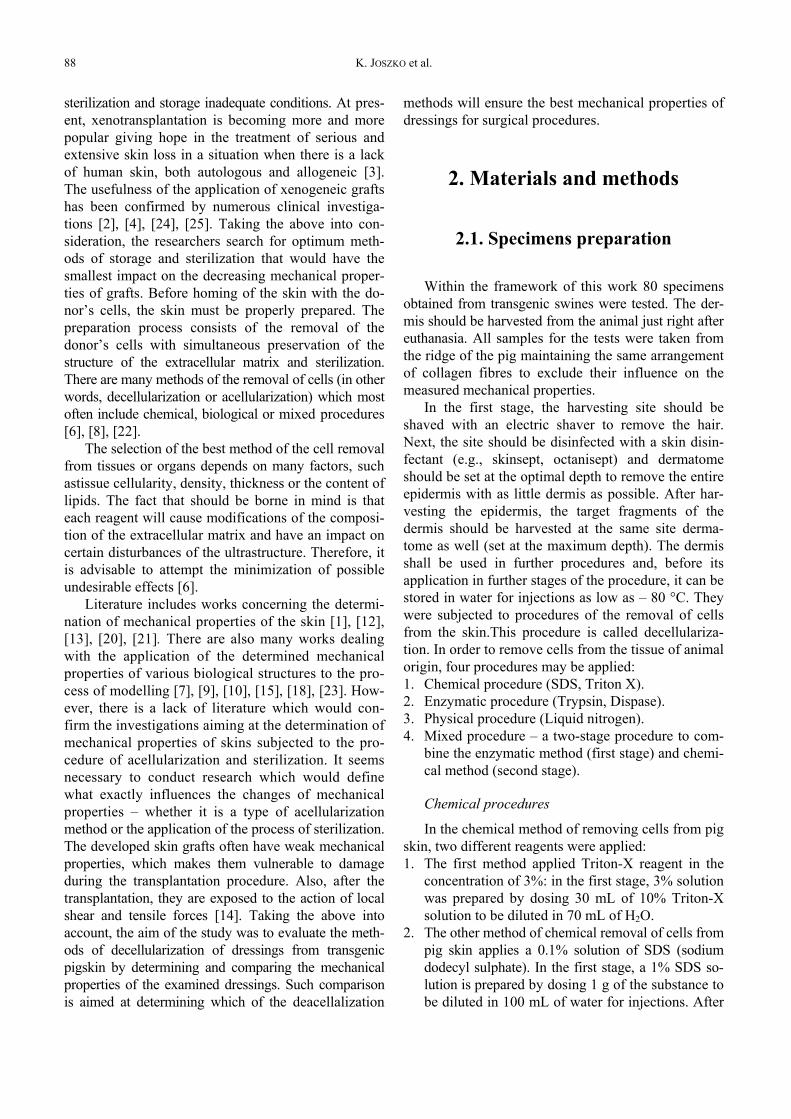
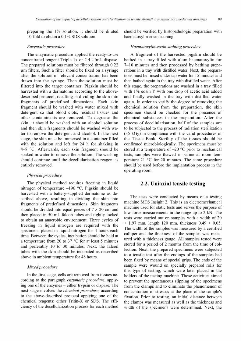
samples were stretched in the direction of the longitu-dinal axis (Fig. 1) in quasi-static conditions at a ve-locity of 5 mm/min [1], [12]. The room in which theinvestigations were carried out had a constant tem-perature of 21 °C. The results were recorded at a fre-quency of 10 Hz. As a result of the carried out tests,the value of Young’s modulus was determined on thebasis of the stress and deformation plot in the propor-tional range. The value of Young’s modulus corre-sponds to the tangent of the angle of inclination of thesegment proportional to the axis of deformation. Anexemplary graph is shown in Fig. 2. The presentedgraph depicts two areas of toe region and linear re-gion, like in [16], and is very similar to the periodon-tal biline characteristics. In the case of the analyzedmaterial toe region reaches about 1/3 of the total strainrange, which maps the viscoelastic properties of thebiological material. With regards to this range, mainlyelastin fibers are responsible for deformation, whilecollagen fibers stand for the region analyzed by the
Fig. 1. A method of fixation of a samplein the clamps of the strength testing machine
Fig. 2. An exemplary graph from static extension test
authors. It was assumed that the mechanical propertiesof the tested material were determined for a linearregion that corresponds to the model of a linearlyelastic material. The value of the maximum stress max
and strain max was read at point “P” which is markedin Fig. 2.
3. Results
3.1. Data obtainedin experimental tests
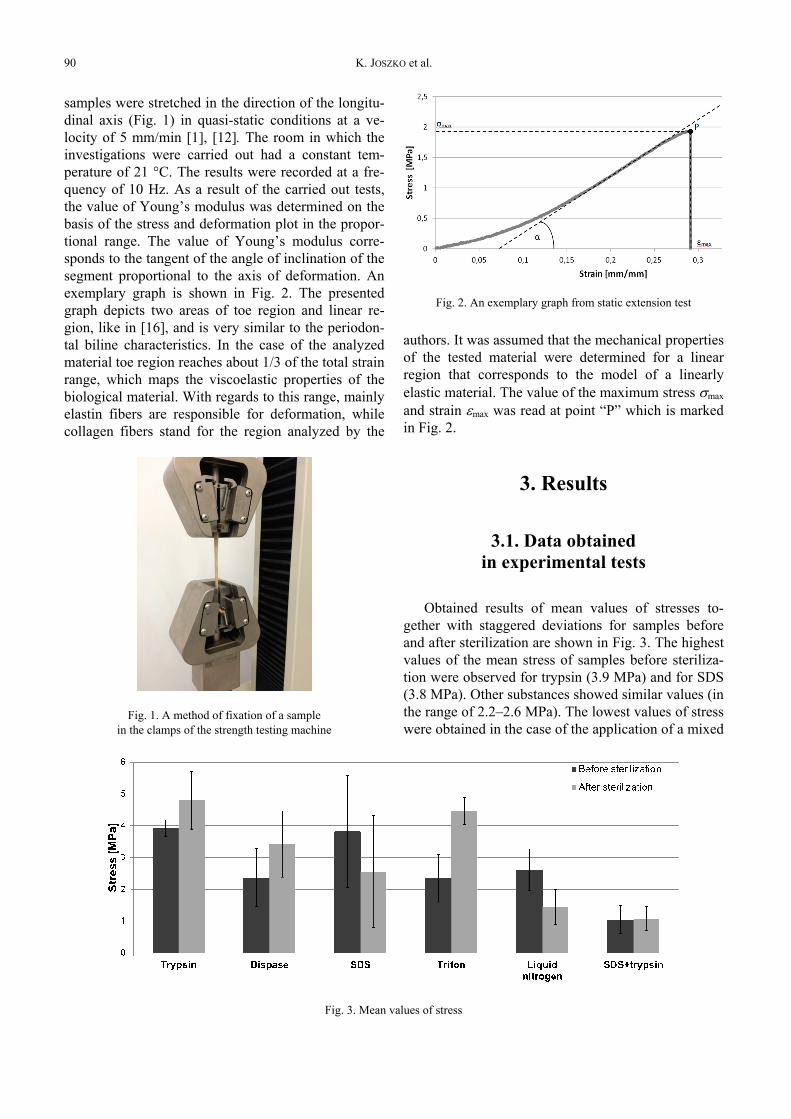
Obtained results of mean values of stresses to-gether with staggered deviations for samples beforeand after sterilization are shown in Fig. 3. The highestvalues of the mean stress of samples before steriliza-tion were observed for trypsin (3.9 MPa) and for SDS(3.8 MPa). Other substances showed similar values (inthe range of 2.2–2.6 MPa). The lowest values of stresswere obtained in the case of the application of a mixed
Fig. 3. Mean values of stress
Evaluation of the impact of decellularization and sterilization on tensile strength transgenic porcinedermal dressings 91
method (SDS and trypsin – 1.0 MPa). Specimenssubjected to the process of sterilization revealed thehighest mean stress in the case of the application oftrypsin (4.8 MPa) and Triton (4.5 MPa) and the lowestmean stress in the case of the mixed method (1.1 MPa)– similarly to the results obtained before sterilization.After sterilization, mean stresses rose for trypsin (by0.9 MPa), dispase (by 1.1 MPa), Triton (by 2.1 MPa)and SDS + trypsin (only by 0.03 MPa).
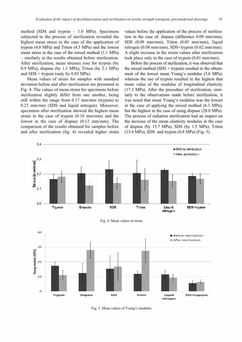
Mean values of strain for samples with standarddeviation before and after sterilization are presented inFig. 4. The values of mean strain for specimens beforesterilization slightly differ from one another, beingstill within the range from 0.17 mm/mm (trypsin) to0.23 mm/mm (SDS and liquid nitrogen). Moreover,specimens after sterilization showed the highest meanstrain in the case of trypsin (0.18 mm/mm) and thelowest in the case of dispase (0.12 mm/mm). Thecomparison of the results obtained for samples beforeand after sterilization (fig. 4) revealed higher strain
values before the application of the process of steriliza-tion in the case of: dispase (difference 0.09 mm/mm),SDS (0.09 mm/mm), Triton (0.05 mm/mm), liquidnitrogen (0.08 mm/mm), SDS+trypsin (0.02 mm/mm).A slight increase in the strain values after sterilizationtook place only in the case of trypsin (0.01 mm/mm).
Before the process of sterilization, it was observed thatthe mixed method (SDS + trypsin) resulted in the obtain-ment of the lowest mean Young’s modulus (5.6 MPa),whereas the use of trypsin resulted in the highest thatmean value of the modulus of longitudinal elasticity(17.3 MPa). After the procedure of sterilization, simi-larly to the observations made before sterilization, itwas noted that mean Young’s modulus was the lowestin the case of applying the mixed method (6.3 MPa),but the highest in the case of using dispase (28.0 MPa).The process of radiation sterilization had an impact onthe increase of the mean elasticity modulus in the caseof dispase (by 15.7 MPa), SDS (by 1.5 MPa), Triton(15.6 MPa), SDS and trypsin (0.8 MPa) (Fig. 5).
Fig. 4. Mean values of strain
Fig. 5. Mean values of Young’s modulus
K. JOSZKO et al.92
3.2. Statistical analysis
3.2.1. Data analysis:reagent, sterilization, strain
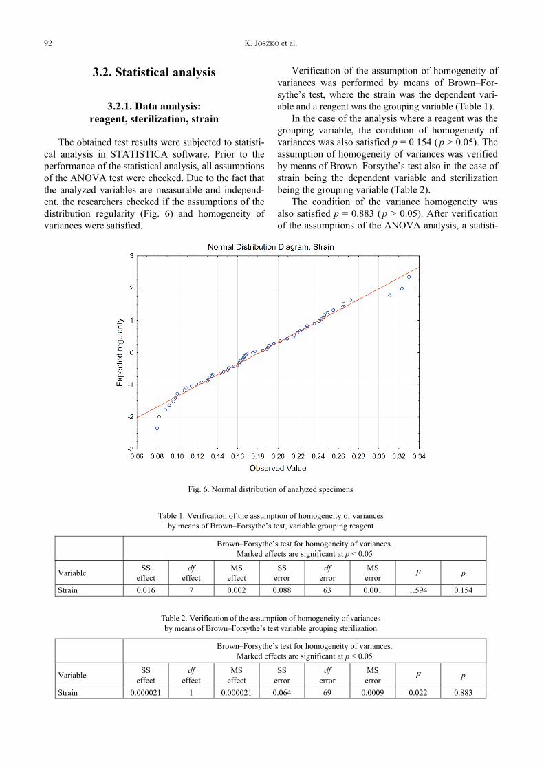
The obtained test results were subjected to statisti-cal analysis in STATISTICA software. Prior to theperformance of the statistical analysis, all assumptionsof the ANOVA test were checked. Due to the fact thatthe analyzed variables are measurable and independ-ent, the researchers checked if the assumptions of thedistribution regularity (Fig. 6) and homogeneity ofvariances were satisfied.
Verification of the assumption of homogeneity ofvariances was performed by means of Brown–For-sythe’s test, where the strain was the dependent vari-able and a reagent was the grouping variable (Table 1).
In the case of the analysis where a reagent was thegrouping variable, the condition of homogeneity ofvariances was also satisfied p = 0.154 ( p > 0.05). Theassumption of homogeneity of variances was verifiedby means of Brown–Forsythe’s test also in the case ofstrain being the dependent variable and sterilizationbeing the grouping variable (Table 2).
The condition of the variance homogeneity wasalso satisfied p = 0.883 ( p > 0.05). After verificationof the assumptions of the ANOVA analysis, a statisti-
Fig. 6. Normal distribution of analyzed specimens
Table 1. Verification of the assumption of homogeneity of variancesby means of Brown–Forsythe’s test, variable grouping reagent
Brown–Forsythe’s test for homogeneity of variances.Marked effects are significant at p < 0.05
Variable SSeffect
dfeffect
MSeffect
SSerror
dferror
MSerror F p
Strain 0.016 7 0.002 0.088 63 0.001 1.594 0.154
Table 2. Verification of the assumption of homogeneity of variancesby means of Brown–Forsythe’s test variable grouping sterilization
Brown–Forsythe’s test for homogeneity of variances.Marked effects are significant at p < 0.05
Variable SSeffect
dfeffect
MSeffect
SSerror
dferror
MSerror F p
Strain 0.000021 1 0.000021 0.064 69 0.0009 0.022 0.883
Evaluation of the impact of decellularization and sterilization on tensile strength transgenic porcinedermal dressings 93
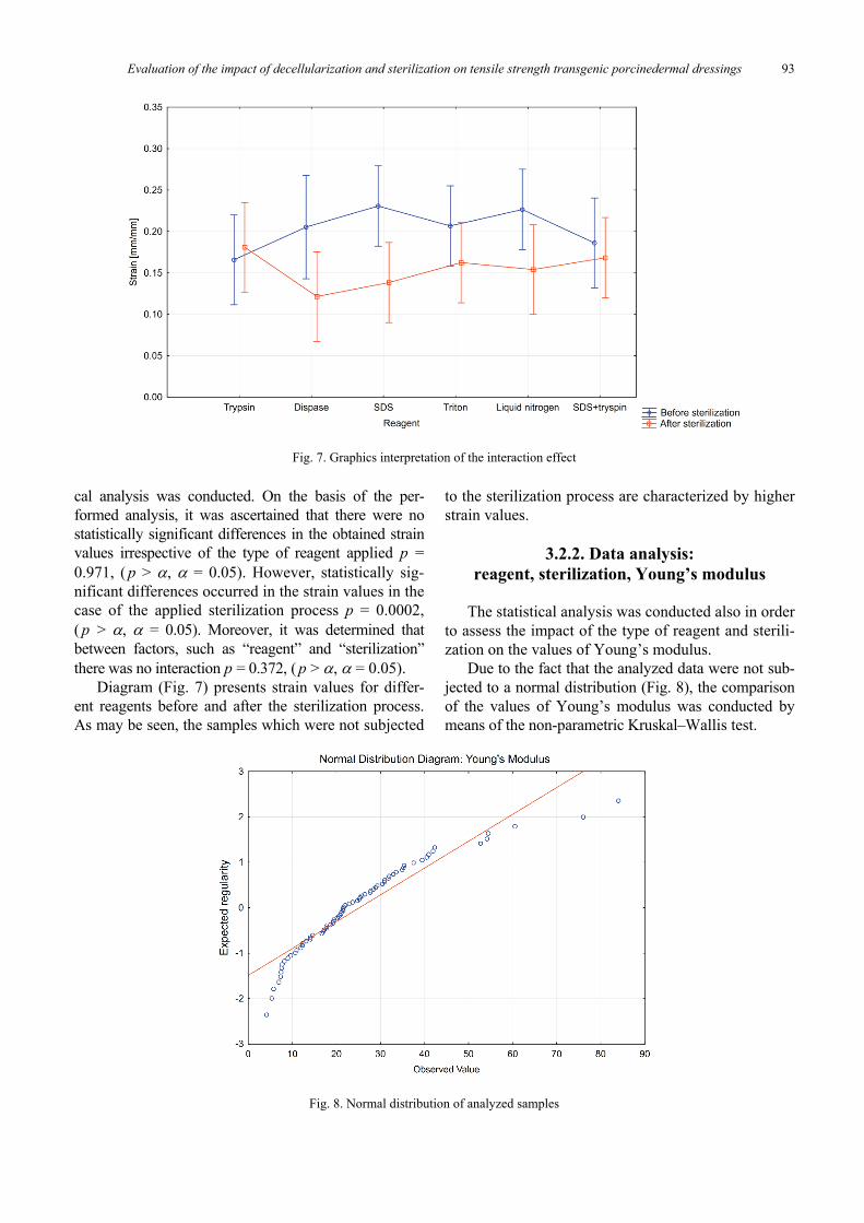
cal analysis was conducted. On the basis of the per-formed analysis, it was ascertained that there were nostatistically significant differences in the obtained strainvalues irrespective of the type of reagent applied p =0.971, ( p > , = 0.05). However, statistically sig-nificant differences occurred in the strain values in thecase of the applied sterilization process p = 0.0002,( p > , = 0.05). Moreover, it was determined thatbetween factors, such as “reagent” and “sterilization”there was no interaction p = 0.372, ( p > , = 0.05).
Diagram (Fig. 7) presents strain values for differ-ent reagents before and after the sterilization process.As may be seen, the samples which were not subjected
to the sterilization process are characterized by higherstrain values.
3.2.2. Data analysis:reagent, sterilization, Young’s modulus
The statistical analysis was conducted also in orderto assess the impact of the type of reagent and sterili-zation on the values of Young’s modulus.
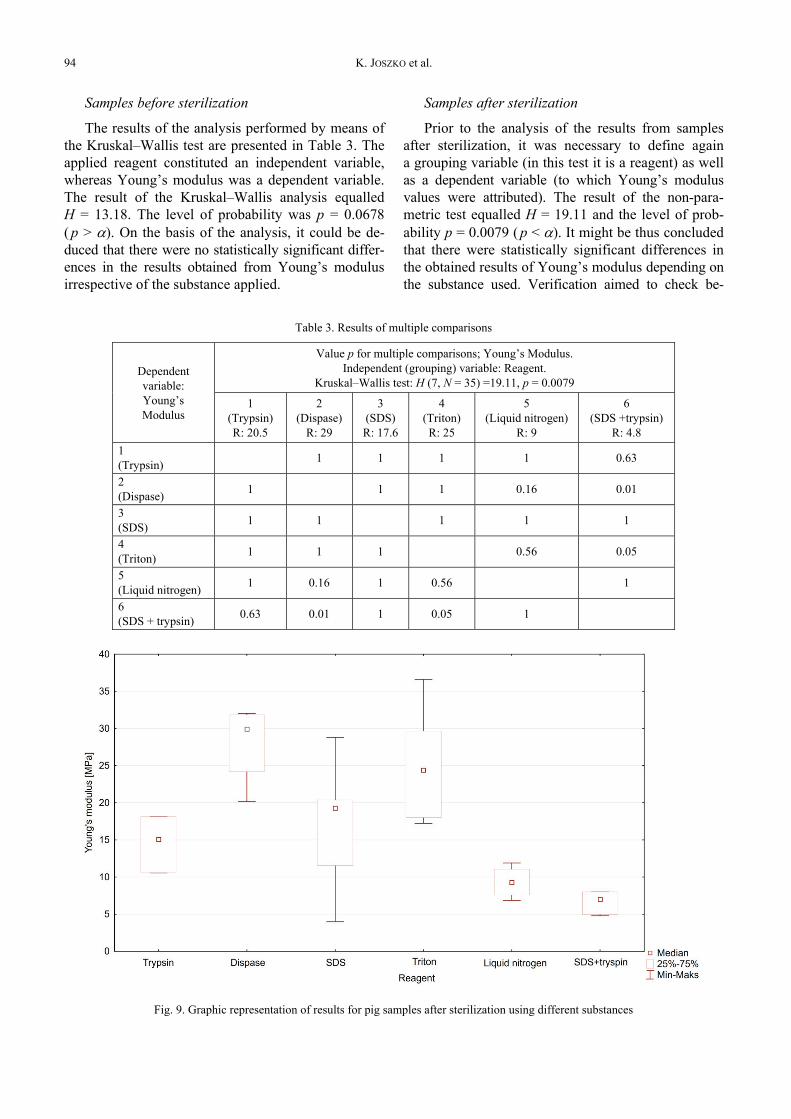
Due to the fact that the analyzed data were not sub-jected to a normal distribution (Fig. 8), the comparisonof the values of Young’s modulus was conducted bymeans of the non-parametric Kruskal–Wallis test.
Fig. 7. Graphics interpretation of the interaction effect
Fig. 8. Normal distribution of analyzed samples
K. JOSZKO et al.94
Samples before sterilization
The results of the analysis performed by means ofthe Kruskal–Wallis test are presented in Table 3. Theapplied reagent constituted an independent variable,whereas Young’s modulus was a dependent variable.The result of the Kruskal–Wallis analysis equalledH = 13.18. The level of probability was p = 0.0678( p > ). On the basis of the analysis, it could be de-duced that there were no statistically significant differ-ences in the results obtained from Young’s modulusirrespective of the substance applied.
Samples after sterilization
Prior to the analysis of the results from samplesafter sterilization, it was necessary to define againa grouping variable (in this test it is a reagent) as wellas a dependent variable (to which Young’s modulusvalues were attributed). The result of the non-para-metric test equalled H = 19.11 and the level of prob-ability p = 0.0079 ( p < ). It might be thus concludedthat there were statistically significant differences inthe obtained results of Young’s modulus depending onthe substance used. Verification aimed to check be-
Table 3. Results of multiple comparisons
Value p for multiple comparisons; Young’s Modulus.Independent (grouping) variable: Reagent.
Kruskal–Wallis test: H (7, N = 35) =19.11, p = 0.0079Dependentvariable:Young’sModulus
1(Trypsin)R: 20.5
2(Dispase)
R: 29
3(SDS)R: 17.6
4(Triton)R: 25
5(Liquid nitrogen)
R: 9
6(SDS +trypsin)
R: 4.81(Trypsin) 1 1 1 1 0.63
2(Dispase) 1 1 1 0.16 0.01
3(SDS) 1 1 1 1 1
4(Triton) 1 1 1 0.56 0.05
5(Liquid nitrogen) 1 0.16 1 0.56 1
6(SDS + trypsin) 0.63 0.01 1 0.05 1
Fig. 9. Graphic representation of results for pig samples after sterilization using different substances
Evaluation of the impact of decellularization and sterilization on tensile strength transgenic porcinedermal dressings 95
tween which reagents there were statistically significantdifferences. The verification was conducted by meansof multiple comparisons.
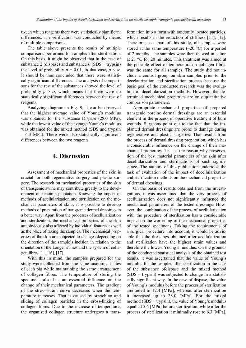
The table above presents the results of multiplecomparisons performed for samples after sterilization.On this basis, it might be observed that in the case ofsubstance 2 (dispase) and substance 6 (SDS + trypsin)the level of probability p = 0.01, in that case, p < .It should be thus concluded that there were statisti-cally significant differences. The analysis of compari-sons for the rest of the substances showed the level ofprobability p > , which means that there were nostatistically significant differences between the rest ofreagents.
Analyzing diagram in Fig. 9, it can be observedthat the highest average value of Young’s moduluswas obtained for the substance Dispase (28.0 MPa),while the lowest value of the average Young’s moduluswas obtained for the mixed method (SDS and trypsin– 6.3 MPa). There were also statistically significantdifferences between the two reagents.
4. Discussion
Assessment of mechanical properties of the skin iscrucial for both regenerative surgery and plastic sur-gery. The research on mechanical properties of the skinof transgenic swine may contribute greatly to the devel-opment of xenotransplantation. Knowing the impact ofmethods of acellularization and sterilization on the me-chanical parameters of skins, it is possible to developmethods of preparation of transgenic dermal dressings ina better way. Apart from the processes of acellularizationand sterilization, the mechanical properties of the skinare obviously also affected by individual features as wellas the place of taking the samples. The mechanical prop-erties of the skin are subjected to changes depending onthe direction of the sample’s incision in relation to theorientation of the Langer’s lines and the system of colla-gen fibres [1], [16], [17].
With this in mind, the samples prepared for thestudy were collected from the same anatomical sitesof each pig while maintaining the same arrangementof collagen fibres. The temperature of storing thespecimens also has an essential influence on thechange of their mechanical parameters. The gradientof the stress–strain curve decreases when the tem-perature increases. That is caused by stretching andsliding of collagen particles in the cross-linking ofcollagen fibres. Due to the increase of temperature,the organized collagen structure undergoes a trans-
formation into a form with randomly located particles,which results in the reduction of stiffness [11], [12].Therefore, as a part of this study, all samples werestored at the same temperature (–20 C) for a periodof 2 months. The samples were then thawed in salineat 21 C for 20 minutes. This treatment was aimed atthe possible effect of temperature on collagen fibreswas the same for all samples. The study did not in-clude a control group on skin samples prior to thedecelaurization and sterilization process because thebasic goal of the conducted research was the evalua-tion of decellularization methods. However, the de-termined mechanical properties are only quantitativecomparison parameters.
Appropriate mechanical properties of preparedtransgenic porcine dermal dressings are an essentialelement in the process of operative treatment of burnwounds. Surgeons point out to the fact that the im-planted dermal dressings are prone to damage duringregenerative and plastic surgeries. That results fromthe process of dermal dressing preparation, which hasa considerable influence on the change of their me-chanical properties. That is the reason why preserva-tion of the best material parameters of the skin afterdecellularization and sterilizations of such signifi-cance. The authors of this publication undertook thetask of evaluation of the impact of decellularizationand sterilization methods on the mechanical propertiesof dermal dressings.
On the basis of results obtained from the investi-gations, it was ascertained that the very process ofacellularization does not significantly influence themechanical parameters of the tested dressings. How-ever, the combination of the process of acellularizationwith the procedure of sterilization has a considerableimpact on the worsening of the mechanical propertiesof the tested specimens. Taking the requirements ofa surgical procedure into account, it would be advis-able that the dressings obtained after acellularizationand sterilization have the highest strain values andtherefore the lowest Young’s modulus. On the groundsof the conducted statistical analysis of the obtained testresults, it was ascertained that the value of Young’smodulus for the samples after sterilization in the caseof the substance ofdispase and the mixed method(SDS + trypsin) was subjected to change in a statisti-cally significant way. In the case of dispase, the valueof Young’s modulus before the process of sterilizationamounted to 12.4 [MPa], whereas after sterilizationit increased up to 28.0 [MPa]. For the mixedmethod (SDS + trypsin), the value of Young’s modulusequalled 5.6 [MPa] before sterilization, while after theprocess of sterilization it minimally rose to 6.3 [MPa].
K. JOSZKO et al.96
The mixed method (SDS+trypsin) had the smallestimpact on the change of specimen mechanical prop-erties before and after the process of sterilization.The difference between values obtained for Young’smodulus and strain amounted to approximately 12%and was the smallest in comparison with other sub-stances. Dermal dressings prepared by means of themixed method are characterized by a low value ofYoung’s modulus and a relatively high level of strainboth prior to and after the process of sterilization.Such mechanical properties are desirable in the caseof medical procedures of plastic and regenerativesurgeries due to the fact that such dressings will beless vulnerable and prone to damage during a surgicalprocedure.
5. Conclusions
Within the framework of these investigations, theresearchers carried out uniaxial tensile tests on der-mal specimens coming from transgenic swine whosegene conditioning the formation of an antigen Gal(1,3-galactosyltransferase) had been knocked out aswell as proteins of surface cells had been modified(gene 1,2-fucosyltransferase of a human being). Thetotal number of 80 specimens was tested by means of8 methods of acellularization. Half of the tested sam-ples were subjected to the process of radiation sterili-zation (35 kGy) in compliance with the valid proce-dures of the Tissue Bank. Sterility of tissues wasconfirmed microbiologically. On the basis of the con-ducted tests, it was confirmed that different methodsof acellularization and the process of sterilization havean influence on the change of mechanical propertiesof the skin of transgenic swine. In six out of eight ap-plied acellularization methods (Trypsin, SDS, Triton,Liquid nitrogen), the differences in obtained valueswere not statistically significant. However, in the caseof the mixed method (SDS + trypsin) and dispase,statistically significant differences were observed.From the point of view of the requirements of theprocedures of reconstructive and plastic surgeries,more favourable mechanical properties were obtainedfor the mixed method (SDS+trypsin) because it en-ables the obtainment of the lowest value of Young’smodulus, at the same time maintaining a relatively highlevel of strain. In the authors’ opinion, this methodshould be recommended as the best one for the prepa-ration of transgenic porcine dermal dressings becauseit ensures a smaller probability of dressing’s damageduring a surgical procedure.
Conflict of interest
The authors declare that the article content was composed inthe absence of any commercial or financial relationships thatcould be construed as a potential conflict of interest.
References
[1] BRUYERE K., DESTRADEA M., GILCHRISTA M.D., OTTENIO M.,NI ANNAIDH A., Characterising the anisotropic mechanicalproperties of excised human skin, J. Mech. Behav. Biomed.Mater, 2012, 5 (1), 139–148, DOI: https://doi.org/10.1016/j.jmbbm.2011.08.016
[2] BURKEY B., DAVIS W., GLAT P.M., Porcine xenograft treat-ment of superficial partial-thickness burns in paediatric pa-tients, J. Wound Care, 2016, 25 (2), S10–S15, DOI: 0.12968/jowc.2016.25.Sup2.S10.
[3] ESTEBAN-VIVES R., YOUNG M.T., ZHU T., BEIRIGER J.,PEKOR C., ZIEMBICKI J., CORCOS A., RUBIN P., GERLACH J.C.,Calculations for reproducible autologous skin cell-spray graft-ing, Burns, 2016, 42, 1756–1765.
[4] FENG X., SHEN R., TAN J., CHEN X., PAN Y., RUAN S., ZHANG F.,LIN Z., ZENG Y., WANG X., The study of inhibiting systematicinflammatory response syndrome by applying xenogenic (por-cine) acellular dermal matrix on second-degree burns, Burns,2007, 33, 477–479, DOI: 10.1016/j.burns.2006.08.011.
[5] GIEREK M., KAWECKI M., MIKUŚ K., KLAMA-BARYŁA A.,NOWAK M., Biological dressings as substitutes of the skin inthe treatment of burn wounds, Pol. J. Surg., 2013, 85 (6),354–359, DOI: https://doi.org/10.2478/pjs-2013-0054.
[6] GILBERT T.W., BADYLAK S.F., CRAPO P.M., An overviewof tissue and whole organ decellularization processes,Biomaterials, 2011, 32 (12), 3233–43, DOI: 10.1016/j.biomaterials.2011.01.057.
[7] GZIK-ZROSKA B., WOLANSKI W., GZIK M., Engineering--aided treatment of chest deformities to improve the processof breathing, Int. J. Numer. Meth. Bio., 2013, 29, 926–937,DOI: 10.1002/cnm.2563.
[8] HERSON M.R., PINO E., MATHOR M.B., BOURROUL S.C.,Sterilization of skin allografts by ionizing radiation, Cellular andMolecular Biology, 2002, 48(7), 803–807, PMID: 12619979.
[9] JOSZKO K., GZIK M., WOLAŃSKI W., GZIK-ZROSKA B.,KAWLEWSKA E., Biomechanical evaluation of human lumbarspine in spondylolisthesis, J. Appl. Biomed., 2018, 16 (1), 51–58,DOI 10.1016/j.jab.2017.10.004.
[10] KAJZER A., KAJZER W., GZIK-ZROSKA B., WOLAŃSKI W.,JANICKA I., DZIELICKI J., Experimental biomechanical as-sessment of plate stabilizers for treatment of pectus excava-tum, Acta Bioeng. Biomech., 2013, 15 (3), 113–121, DOI:10.5277/abb130314.
[11] KUMARASWAMYA N., KHATAMB H., REECEB G., FINGERET M.,MARKEYA M., RAVI-CHANDARC K., Mechanical response ofhuman female breast skin under uniaxial stretching, J. Mech.Behav. Biomed. Mater, 2017, 74, 164–175, DOI: 10.1016/j.jmbbm.2017.05.027.
[12] KUROPKA P., KOBIELARZ M., DUDEK A., KALETA--KURATEWICZ K., SZOSTEK S., ŻAK M., Determination of themechanical properties of the skin of pig foetuses with respectto its structure, Acta Bioeng. Biomech., 2011, 13 (2), 37–43,PMID: 21761809.
Evaluation of the impact of decellularization and sterilization on tensile strength transgenic porcinedermal dressings 97
[13] LIU X., Understanding the effect of skin mechanical proper-ties on the friction of human fingerpads, PhD thesis, 2013,University of Sheffield. URL: http://etheses.whiterose.ac.uk/id/eprint/3800.
[14] LYNCH K.A., BOYCE S.T., SANDER E.A., Development of themechanical properties of engineered skin substitutes aftergrafting to full-thickness wounds, J. Biomech. Eng., 2014,136 (5), 051008, DOI: 10.1115/1.4026290.
[15] MAŁACHOWSKI J., KWIATKOWSKI P., SUTKOWSKA E.,JAKUBAS-KWIATKOWSKA W., GOŁĘBIEWSKI S., GIL R., Theeffects of types of guidewires and pressure applied during stentimplantation in themain vessel on the incidence of damage tocoronary guidewires during angioplasty of coronary bifurca-tion lesions – Wide Beast study, J. Am. Coll. Cardiol., 2016,68 (18), B134–B135, DOI: 10.1016/j.jacc.2016.09.456.
[16] MILEWSKI G., HILLE A., Experimental strength analysis oforthodonticextrusion of human anterior teeth, Acta Bioeng.Biomech., 2012, 14 (1), 15–21.
[17] OTTENIOA M., TRANA D., NI ANNAIDH A., GILCHRISTD M.D.,BRUYERE K., Strain rate and anisotropy effects on the tensilefailure characteristics of human skin, J. Mech. Behav. Biomed.Mater, 2015, 41 (1), 241–250, DOI: https://doi.org/10.1016/j.jmbbm.2014.10.006.
[18] PEZOWICZ C., GŁOWACKI M., The mechanical propertiesof human ribs in young adult, Acta Bioeng. Biomech., 2012,14 (2), 53–60. DOI: 10.5277/abb120208.
[19] ROWAN M.P., CANCIO L.C., ELSTER E.A., BURMEISTER D.M.,ROSE L.F., CHAN R.K., CHRISTY R.J., CHUNG K.K., Burn
wound healing and treatment: review and advancements,J. Crit. Care, 2015, 19, 243, DOI: https://doi.org/10.1186/s13054-015-0961-2.
[20] SEFFEN K.A., LU T.J., XU F., Temperature-dependent me-chanical behaviors of skin tissue, Int. J. Comput. Sci., 2009,131 (7), 071001, DOI: 10.1115/1.3118766.
[21] SELBER J.C., BUTLER C.E., ADELMAN D.M., Bovine versusporcine acellular dermal matrix: A comparison of mechani-cal properties, Plast. Reconstr. Surg., 2014, 2(5), e155. DOI:10.1097/GOX.0000000000000072.
[22] SINGH D., SINGH A., SINGH R., Radiation sterilization oftissue allografts: a review, J. Radiol., 2016, 8 (4), 355–369,DOI: 10.4329/wjr.v8.i4.355.
[23] SZAREK A., KORYTKOWSKI M., RUTKOWSKI L., SCHERER R.,SZYPROWSKI J., Forecasting Wear of Head and Acetabulumin Hip Joint Implant, [in:] L. Rutkowski, M. Korytkowski,R. Scherer, R. Tadeusiewicz, L.A. Zadeh, J.M. Zurada(Eds.), Artificial Intelligence and Soft Computing, ICAISC2012. Lecture Notes in Computer Science, Vol. 7268,Springer, Berlin, Heidelberg, DOI: https://doi.org/10.1007/978-3-642-29350-4_41.
[24] YANG Y.W., OCHOA S.A., Use of porcine xenografts indermatology surgery: the Mayo Clinic experience, Der-matol. Surg., 2016, 42 (8), 985–91, DOI: 10.1097/DSS.0000000000000804.
[25] ZEYLAND J., LIPIŃSKI D., SŁOMSKI R., The current state ofxenotransplantation, J. Appl. Genet., 2015, 56 (2), 211–218,DOI: 10.1007/s13353-014-0261-6.