Evaluation of the Diagnostic Accuracy of Serum D-Dimer Levels in Pregnant Women with Adnexal Torsion
9
Diagnostics 2015, 5, 1-9; doi:10.3390/diagnostics5010001 diagnostics ISSN 2075-4418 www.mdpi.com/journal/diagnostics/ Article Evaluation of the Diagnostic Accuracy of Serum D-Dimer Levels in Pregnant Women with Adnexal Torsion Hasan Onur Topçu *, Can Tekin İskender, Ufuk Ceran, Oktay Kaymak, Hakan Timur, Dilek Uygur and Nuri Danışman Zekai Tahir Burak Women’s Health Education and Research Hospital, 1549 Cadde, Hardem Apartmanı, B Blok, Daire 12 Çiğdem-Çankaya, Ankara 06300, Turkey; E-Mails: [email protected] (C.T.I.); [email protected] (U.C.); [email protected] (O.K.); [email protected] (H.T.); [email protected] (D.U.); [email protected] (N.D.) * Author to whom correspondence should be addressed; E-Mail: [email protected]; Tel.: +90-532-635-95-38; Fax: +90-312-306-59-17. Academic Editor: Ludmilla A. Morozova-Roche Received: 15 September 2014 / Accepted: 20 November 2014 / Published: 5 January 2015 Abstract: We aimed to evaluate the diagnostic accuracy of serum D-dimer levels in pregnant women with adnexal torsion (AT). The pregnant women with ovarian cysts who suffered from pelvic pain were divided into two groups; the first group consisted of the cases with surgically proven as AT (n = 17) and the second group consisted of the cases whose pain were resolved in the course of follow-up period without required surgery (n = 34). The clinical characteristics and serum D-dimer levels were compared between the groups. Patients with AT had a higher rate of elevated serum white blood cell (WBC) count (57% vs. 16%, p = 0.04) and serum D-dimer levels (77% vs. 21%, p < 0.01) on admission in the study group than in the control group. Elevated D-dimer and cyst diameter larger than 5 cm yielded highest sensitivity (82% for each); whereas the presence of nausea and vomiting and elevated CRP had the highest specificity (85% and 88%, respectively). This is the first study that evaluates the serum D-dimer levels in humans in the diagnosis of AT, and our findings supported the use of D-dimer for the early diagnosis of AT in pregnant women. OPEN ACCESS
-
Upload
independent -
Category
Documents
-
view
2 -
download
0
Transcript of Evaluation of the Diagnostic Accuracy of Serum D-Dimer Levels in Pregnant Women with Adnexal Torsion
Diagnostics 2015, 5, 1-9; doi:10.3390/diagnostics5010001
diagnostics ISSN 2075-4418
www.mdpi.com/journal/diagnostics/
Article
Evaluation of the Diagnostic Accuracy of Serum D-Dimer Levels in Pregnant Women with Adnexal Torsion
Hasan Onur Topçu *, Can Tekin İskender, Ufuk Ceran, Oktay Kaymak, Hakan Timur,
Dilek Uygur and Nuri Danışman
Zekai Tahir Burak Women’s Health Education and Research Hospital, 1549 Cadde,
Hardem Apartmanı, B Blok, Daire 12 Çiğdem-Çankaya, Ankara 06300, Turkey;
E-Mails: [email protected] (C.T.I.); [email protected] (U.C.);
[email protected] (O.K.); [email protected] (H.T.); [email protected] (D.U.);
[email protected] (N.D.)
* Author to whom correspondence should be addressed; E-Mail: [email protected];
Tel.: +90-532-635-95-38; Fax: +90-312-306-59-17.
Academic Editor: Ludmilla A. Morozova-Roche
Received: 15 September 2014 / Accepted: 20 November 2014 / Published: 5 January 2015
Abstract: We aimed to evaluate the diagnostic accuracy of serum D-dimer levels in pregnant
women with adnexal torsion (AT). The pregnant women with ovarian cysts who suffered
from pelvic pain were divided into two groups; the first group consisted of the cases with
surgically proven as AT (n = 17) and the second group consisted of the cases whose pain
were resolved in the course of follow-up period without required surgery (n = 34). The clinical
characteristics and serum D-dimer levels were compared between the groups. Patients with
AT had a higher rate of elevated serum white blood cell (WBC) count
(57% vs. 16%, p = 0.04) and serum D-dimer levels (77% vs. 21%, p < 0.01) on admission in
the study group than in the control group. Elevated D-dimer and cyst diameter larger than 5
cm yielded highest sensitivity (82% for each); whereas the presence of nausea and vomiting
and elevated CRP had the highest specificity (85% and 88%, respectively).
This is the first study that evaluates the serum D-dimer levels in humans in the diagnosis of
AT, and our findings supported the use of D-dimer for the early diagnosis of AT in
pregnant women.
OPEN ACCESS
Diagnostics 2015, 5 2
Keywords: adnexal torsion; D-dimer; ovarian torsion; pregnancy
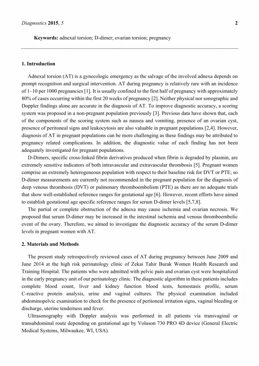
1. Introduction
Adnexal torsion (AT) is a gynecologic emergency as the salvage of the involved adnexa depends on
prompt recognition and surgical intervention. AT during pregnancy is relatively rare with an incidence
of 1–10 per 1000 pregnancies [1]. It is usually confined to the first half of pregnancy with approximately
80% of cases occurring within the first 20 weeks of pregnancy [2]. Neither physical nor sonographic and
Doppler findings alone are accurate in the diagnosis of AT. To improve diagnostic accuracy, a scoring
system was proposed in a non-pregnant population previously [3]. Previous data have shown that, each
of the components of the scoring system such as nausea and vomiting, presence of an ovarian cyst,
presence of peritoneal signs and leukocytosis are also valuable in pregnant populations [2,4]. However,
diagnosis of AT in pregnant populations can be more challenging as these findings may be attributed to
pregnancy related complications. In addition, the diagnostic value of each finding has not been
adequately investigated for pregnant populations.
D-Dimers, specific cross-linked fibrin derivatives produced when fibrin is degraded by plasmin, are
extremely sensitive indicators of both intravascular and extravascular thrombosis [5]. Pregnant women
comprise an extremely heterogeneous population with respect to their baseline risk for DVT or PTE; so
D-dimer measurements are currently not recommended in the pregnant population for the diagnosis of
deep venous thrombosis (DVT) or pulmonary thromboembolism (PTE) as there are no adequate trials
that show well-established reference ranges for gestational age [6]. However, recent efforts have aimed
to establish gestational age specific reference ranges for serum D-dimer levels [5,7,8].
The partial or complete obstruction of the adnexa may cause ischemia and ovarian necrosis. We
proposed that serum D-dimer may be increased in the intestinal ischemia and venous thromboembolic
event of the ovary. Therefore, we aimed to investigate the diagnostic accuracy of the serum D-dimer
levels in pregnant women with AT.
2. Materials and Methods
The present study retrospectively reviewed cases of AT during pregnancy between June 2009 and
June 2014 at the high risk perinatology clinic of Zekai Tahir Burak Women Health Research and
Training Hospital. The patients who were admitted with pelvic pain and ovarian cyst were hospitalized
in the early pregnancy unit of our perinatology clinic. The diagnostic algorithm in these patients includes
complete blood count, liver and kidney function blood tests, hemostasis profile, serum
C-reactive protein analysis, urine and vaginal cultures. The physical examination included
abdominopelvic examination to check for the presence of peritoneal irritation signs, vaginal bleeding or
discharge, uterine tenderness and fever.
Ultrasonography with Doppler analysis was performed in all patients via transvaginal or
transabdominal route depending on gestational age by Voluson 730 PRO 4D device (General Electric
Medical Systems, Milwaukee, WI, USA).
Diagnostics 2015, 5 3
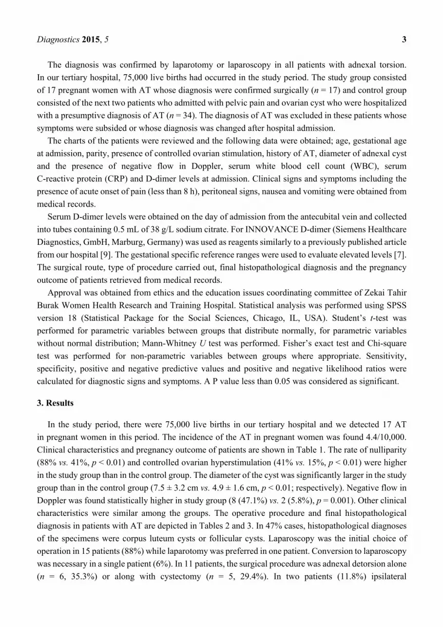
The diagnosis was confirmed by laparotomy or laparoscopy in all patients with adnexal torsion.
In our tertiary hospital, 75,000 live births had occurred in the study period. The study group consisted
of 17 pregnant women with AT whose diagnosis were confirmed surgically (n = 17) and control group
consisted of the next two patients who admitted with pelvic pain and ovarian cyst who were hospitalized
with a presumptive diagnosis of AT (n = 34). The diagnosis of AT was excluded in these patients whose
symptoms were subsided or whose diagnosis was changed after hospital admission.
The charts of the patients were reviewed and the following data were obtained; age, gestational age
at admission, parity, presence of controlled ovarian stimulation, history of AT, diameter of adnexal cyst
and the presence of negative flow in Doppler, serum white blood cell count (WBC), serum
C-reactive protein (CRP) and D-dimer levels at admission. Clinical signs and symptoms including the
presence of acute onset of pain (less than 8 h), peritoneal signs, nausea and vomiting were obtained from
medical records.
Serum D-dimer levels were obtained on the day of admission from the antecubital vein and collected
into tubes containing 0.5 mL of 38 g/L sodium citrate. For INNOVANCE D-dimer (Siemens Healthcare
Diagnostics, GmbH, Marburg, Germany) was used as reagents similarly to a previously published article
from our hospital [9]. The gestational specific reference ranges were used to evaluate elevated levels [7].
The surgical route, type of procedure carried out, final histopathological diagnosis and the pregnancy
outcome of patients retrieved from medical records.
Approval was obtained from ethics and the education issues coordinating committee of Zekai Tahir
Burak Women Health Research and Training Hospital. Statistical analysis was performed using SPSS
version 18 (Statistical Package for the Social Sciences, Chicago, IL, USA). Student’s t-test was
performed for parametric variables between groups that distribute normally, for parametric variables
without normal distribution; Mann-Whitney U test was performed. Fisher’s exact test and Chi-square
test was performed for non-parametric variables between groups where appropriate. Sensitivity,
specificity, positive and negative predictive values and positive and negative likelihood ratios were
calculated for diagnostic signs and symptoms. A P value less than 0.05 was considered as significant.
3. Results
In the study period, there were 75,000 live births in our tertiary hospital and we detected 17 AT
in pregnant women in this period. The incidence of the AT in pregnant women was found 4.4/10,000.
Clinical characteristics and pregnancy outcome of patients are shown in Table 1. The rate of nulliparity
(88% vs. 41%, p < 0.01) and controlled ovarian hyperstimulation (41% vs. 15%, p < 0.01) were higher
in the study group than in the control group. The diameter of the cyst was significantly larger in the study
group than in the control group (7.5 ± 3.2 cm vs. 4.9 ± 1.6 cm, p < 0.01; respectively). Negative flow in
Doppler was found statistically higher in study group (8 (47.1%) vs. 2 (5.8%), p = 0.001). Other clinical
characteristics were similar among the groups. The operative procedure and final histopathological
diagnosis in patients with AT are depicted in Tables 2 and 3. In 47% cases, histopathological diagnoses
of the specimens were corpus luteum cysts or follicular cysts. Laparoscopy was the initial choice of
operation in 15 patients (88%) while laparotomy was preferred in one patient. Conversion to laparoscopy
was necessary in a single patient (6%). In 11 patients, the surgical procedure was adnexal detorsion alone
(n = 6, 35.3%) or along with cystectomy (n = 5, 29.4%). In two patients (11.8%) ipsilateral

Diagnostics 2015, 5 4
salpingo-oophorectomy was performed and in one patient (5.9%) cystectomy and ovarian wedge
resection was performed.
Table 1. Comparison of clinical characteristics and pregnancy outcome between groups.
Variables Adnexal Torsion (n = 17) Control (n = 34) p
Age (years) 25.4 ± 4.6 26.0 ± 5.6 0.67
Nulliparity 15 (88%) 14 (41%) <0.01
Controlled ovarian hyperstimulation 7 (41%) 5 (15%) 0.04
Multiple pregnancy 3 (18%) 1 (3%) 0.10
Gestational period at admission
1st trimester 11 (65%) 18 (53%) - 2nd trimester 6 (35%) 16 (47%) 0.42 3rd trimester - - -
Cyst Diameter (cm) 7.5 ± 3.2 4.9 ± 1.6 <0.01
WBC (×103) (cells/mm3) 13.3 ± 6.9 10.3 ± 2.8 0.19 D-dimer (µg/mL)
Independent from trimester 1.83 ± 1.1 0.66 ± 0.37 <0.01
1st trimester 2.13 ± 1.1 0.60 ± 0.43 <0.01 2nd trimester 1.27 ± 0.8 0.73 ± 0.29 0.20
Pregnancy outcome *
Term delivery 10 (63%) 28 (85%) 0.14
Preterm delivery 3 (19%) 3 (9%) 0.38
Abortion 3 (19%) 2 (6%) 0.31
Values are given as mean ± standard deviation or number (percentage), p < 0.05 = statistically significant.
* excluding cases with elective termination of pregnancy.
Table 2. Details of the operative procedure in patients with adnexal torsion during pregnancy.
Operative Procedure Number of Patients Percentage (%)
Adnexal detorsion alone 6 35.3 Adnexal detorsion with cyst aspiration and IPL fixation 3 11.8 Adnexal detorsion with cystectomy 5 5.9 Adnexal detorsion with ipsilateral salpingectomy 2 5.9 Adnexal detorsion with cystectomy and ovarian wedge resection 1 5.9
IPL: Infundibulopelvic ligament.
Table 3. Final histopathological diagnosis in patients with adnexial torsion during pregnancy.
Pathological Diagnosis Number of Patients Percentage (%)
Corpus luteum cyst 6 35.3 Follicular cyst 2 11.8
Mature Cystic teratoma 1 5.9 Mucinous cystadenoma 1 5.9 Tubal ectopic pregnancy 1 5.9 No specimen obtained 6 35.3

Diagnostics 2015, 5 5
Patients with AT had a higher rate of elevated serum WBC count (47.1% vs. 17.6%, p = 0.04), serum
D-dimer levels (76.5% vs. 8.8%, p < 0.01) and nausea and vomiting (35.3% vs. 8.8%, p = 0.04) on
admission in the study group than in the control group. However, the rates of elevated CRP, acute onset
pain, and presence of peritoneal signs were similar (Table 4).
The diagnostic performance of physical findings is shown in Table 5. Elevated D-dimer and cyst
diameter larger than 5 cm yielded highest sensitivity (82% for each); whereas the presence of nausea
and vomiting and elevated CRP had the highest specificity (85% and 88%, respectively). The specificity,
positive and negative predictive values of elevated serum D-dimer were 79%, 66% and 90%, respectively.
Positive and negative likelihood ratios for D-dimer were 4.0 (95% confidence interval: 2.0–8.02) and
0.22 (0.08–0.63).
Table 4. Comparison of clinical and laboratory features among groups.
Variables Adnexal Torsion (n = 17) Control (n = 34) p
Acute onset pain (<8 h) 11 (64.7%) 16 (47.1%) 0.23
Cyst diameter > 5cm 14 (82.4%) 14 (41.2%) <0.001
Peritoneal signs 9 (52.9%) 13 (38.2%) 0.32
Nausea and vomiting 6 (35.3%) 3 (8.8%) 0.04
Negative flow in Doppler 8 (47.1%) 2 (5.8%) 0.001
Leukocytosis (WBC ≥ 12 × 103) (cells/mm3) 10 (71.4%)
8 (47.1%) 6 (17.6%) 0.04
Elevated CRP (≥8 mg/L) 5 (29.4%) 4 (11.7%) 0.14
* Elevated D-dimer 13 (76.5%) 3 (8.8%) 0.001
Values are given as number (percentage), p < 0.05 = statistically significant. WBC: white blood cell,
CRP: C-reactive protein, h: hours, * reference [7] was used to determine elevated levels.
Table 5. Comparison of clinical features in pregnant patients with adnexal torsion.
Variables
Sensitivity Specificity PPV NPV LR + LR −
% (95%
CI)
% (95%
CI)
% (95%
CI)
% (95%
CI) (95% CI) (95% CI)
Acute onset pain
(<8 h) 65 (38–86) 53 (35–70) 41 (22–61) 75 (53–90) 1.4 (0.83–2.3) 0.67 (0.33–1.37)
Cyst diameter > 5cm 82 (56–96) 59 (41–75) 50 (31–69) 87 (66–97) 2.0 (1,26–3.16) 0.30 (0.10–0.87)
Negative Doppler flow 35 (14–62) 76 (59–89) 43 (18–71) 70 (53–84) 1.5 (0.62–3.63) 0.85 (0.57–1.26)
Peritoneal signs 53 (28–77) 62 (44–78) 41 (21–63) 72 (53–87) 1.4 (0.75–2.6) 0.76 (0.43–1.35)
Nausea and vomiting 35 (14–62) 85 (69–95) 55 (24–83) 73 (56–85) 2.4 (0.85–6.75) 0.76 (0.52–1.11)
Leukocytosis (WBC ≥
12 × 103) (cells/mm3)
10 (71.4%)
47 (23–72) 82 (65–93) 57 (29–82) 76 (58–88) 2.7 (1.10–6.46) 0.64 (0.40–1.03)
Elevated CRP
(≥8 mg/L) 24 (7–50) 88 (73–97) 50 (16–84) 70 (54–83) 2.0 (0.57–7.03) 0.87 (0.65–1.16)
* Elevated D-dimer 82 (50–93) 79 (76–98) 66 (43–85) 90 (73–98) 4.0 (2.0–8.02) 0.22 (0.08–0.63)
CI: confidence interval, PPV: Positive predictive value, NPV: Negative predictive value, LR+: Positive
likelihood ratio, LR−: Negative likelihood ratio, h: hours, CRP: C-reactive protein, * reference [7] was used to
determine elevated levels.

Diagnostics 2015, 5 6
4. Discussion
Herein, we reported a study among pregnant women with ovarian cysts who were admitted to our
tertiary referral hospital suffering from pelvic pain. We compared the clinical and laboratory features in
pregnant patients with ovarian cysts whose diagnosis were surgically proven as adnexal torsion and
whose diagnosis were confirmed as permanent pelvic pain responded to the medical therapy in the course
of follow-up period. We determined that an increase of the serum D-dimer level was a novel promising
finding in the pregnant patients for the diagnosis of AT.
The management of the pelvic pain in women with ovarian cysts is still a complicated situation due
to the probability of the presence of AT because the clinicians should decide promptly to perform surgery
or not, in order to preserve the ovarian reserve. In addition, this situation is more complicated in pregnant
women with ovarian cysts due to its low incidence, and a likely false positive diagnosis and the likely
adverse effects of the surgery to the fetus. AT in pregnancy is a rare clinical entity with an incidence of
1–5/10,000 in spontaneous pregnancy [1]. During the study period, we determined that approximately
75,000 live births had occurred at our hospital; and we detected AT in 17 of 75,000 pregnancies, with
an incidence of 4.4/10,000 similar with Smorgick et al. [10]. The most common ovarian cysts in
pregnancy were reported in order as dermoid cysts, serous cystadenoma, mucinous cystadenoma and
low malign potential tumors (37.6%, 13%, 14.5%, 7.2%, respectively) [11]. In another study, the
majority of the ovarian cysts removed at the second trimester were dermoids (37%), cystadenomas
(24%), persistent corpus luteum cysts (20%), paraovarian cysts (5%) and endometriomas (5%) [12]. In
our study, the final pathological diagnoses of the cysts which resulted in twisted ovary were corpus
luteum cyst (54.5%), follicular cyst (18.2%), mature cystic teratoma (9.1%), mucinous cystadenoma
(9.1%) and tubal ectopic pregnancy (9.1%).
Controlled ovarian stimulation (COS) protocols may raise the AT rate by creating multiple cystic
lesions of the ovary. The incidence of the AT in the COS cycles rises from 6% to 16% [13,14]. Moreover,
AT was found to be with a high ratio (49%) in pregnancies conceived through the COS cycles [10].
Seven of 17 (41%) patients in AT group and 5 of 34 (15%) patients in control group were conceived through
COS protocols and this difference reached a significant value in our study, in compliance with previous
studies [10,15]. Commonly, the AT had occurred in the first trimester of the pregnancy [10,16–18]. In
this current report, AT occurred in 11 of 17 cases (65%) in the first trimester, and in 6 of 17 cases (35%)
in the second trimester. There were no AT cases in the third trimester similar with a past report [19].
Goh et al. [11] reported 8.3% AT rate in 126 pregnant patients in whom ovarian cyst diameter equal
or greater than 5 cm. In the previous studies, the diameter of the ovarian cysts greater than 5 cm was
reported as one of the most important parameters for the occurrence of AT [20,21]. In current study, we
had 14 of 17 (82.4%) pregnant patients in the study group and 14 of 34 (41.2%) pregnant patients in
control group whose ovarian cyst diameter was greater than 5 cm. The differences of the diameter of the
ovarian cysts between the groups were statistically significant. We also evaluated the pregnancy
outcomes including term delivery, preterm delivery and miscarriage rates. We did not detect any
increased risk of surgery to the pregnancy outcomes which is similar to a recent study [1].
In our study, nausea and vomiting and leukocytosis in AT group were found higher than in the control
group. Elevated CRP, peritoneal irritation signs and acute onset pain were the laboratory and clinical
parameters which were found to be similar in both groups. To achieve the differential diagnosis in
Diagnostics 2015, 5 7
pregnant women who had ovarian cysts and pelvic or abdominal pain; the peritoneal irritation signs,
acute onset pain or elevated CRP were found to be useless in those women. Nausea and vomiting and
leukocytosis were found to be beneficial clinical and laboratory parameters but they are nonspecific and
inadequate to achieve the diagnosis. Negative flow in Doppler was determined in 8 of 17 (47.1%) in
study group. Despite the utility of Doppler examination, the clinicians need more parameters to diagnose
AT. Additionally in pregnant women, because although low complication rates were reported in the
second trimester surgery, we believe that to make decision for surgery in pregnant women is harder than
in non-pregnant women.
The twisting of the ovary and its surrounding tissue may result in congestion due to the prior
obstruction of the venous return at first, and with the persistence of the obstruction, the partial or
complete obstruction may cause ischemia and ovarian necrosis. D-dimer is a product of fibrin turnover
and it rises quite quickly after a thromboembolic event and it has begun to be used widely after its
discovery as an indicator of intestinal ischemia and venous thromboembolic disorders in all body organs,
including the lungs, pelvis, upper extremities, thigh and calf [5]. To our knowledge, there is no report
about the serum maternal D-dimer levels in pregnant women with AT. To preserve ovarian reserve in
cases with AT, it is important to diagnose and to intervene early; thus a laboratory parameter that can
support early diagnosis, such as; D-dimer, which may be more important for early diagnosis. In an
experimental animal study, Kart et al. [22] demonstrated the increase of the D-dimer in AT. In our study,
elevated D-dimer levels were detected significantly higher in AT group than in the control group (13 of
17, 76.5%, 3 of 34, 8.8%, respectively). We think that further studies with larger populations are needed
to understand the diagnostic role of the serum D-dimer levels in pregnant women with AT.
Our study has several limitations. This is a retrospective study and the retrospective design of the
study may affect some results; however, our perinatology clinic has strict rules for diagnostic algorithms,
and these reduce the differences in the management of the cases. Our data depends on assay information;
this is a limitation of our study. Our trial has a small number of the study population and a follow up
D-dimer test may be performed to assess whether it will increase or decrease further; however, it is very
difficult to design a prospective study for a disease which has a low incidence. There is also a limitation
regarding the trials which investigate D-dimer levels due to the lack of the standardization on the assay.
The D-dimer value may change by using a different assay methodology. Standardization may be required
for the trials which evaluate the D-dimer levels. Some cases in the control group might have AT and it
may be resolved spontaneously, so this is another limitation of our study, however, there is no certain
way to diagnose spontaneously resolved ATs.
5. Conclusions
This is the first study that evaluates the serum D-dimer levels in humans in the diagnosis of AT, and
our findings supported that the D-dimer may be used for the early diagnosis of AT in pregnant women.
Acknowledgments
This project received no funding.

Diagnostics 2015, 5 8
Author Contributions
H.O.T. and C.T.I. designed the study and contributed to the development and implementation of the
study; C.T.I. conducted the analysis and interpretation of data together with H.T., H.O.T. and U.C.;
H.O.T. and C.T.I. drafted the manuscript; D.U. and N.D. have contributed to reviewing the manuscript.
All authors gave approval of the final version and can take public responsibility for the content of
the manuscript.
Conflicts of Interest
The authors declare no conflict of interest.
References
1. Hasson, J.; Tsafrir, Z.; Azem, F.; Bar-On, S.; Almog, B.; Mashiach, R.; Seidman, D.; Lessing, J.B.;
Grisaru, D. Comparison of adnexal torsion between pregnant and nonpregnant women. Am. J.
Obstet. Gynecol. 2010, 202, 536.e1–536.e6.
2. Koo, Y.J.; Kim, T.J.; Lee, J.E.; Kwon, Y.S.; Kim, H.J.; Lee, I.H.; Lim, K.T.; Lee, K.H.; Shim, J.U.;
Mok, J.E. Risk of torsion and malignancy by adnexal mass size in pregnant women. Acta Obstet.
Gynecol. Scand. 2011, 90, 358–361.
3. Huchon, C.; Staraci, S.; Fauconnier, A. Adnexal torsion: A predictive score for pre-operative
diagnosis. Hum. Reprod. 2010, 25, 2276–2280.
4. Yen, C.F.; Lin, S.L.; Murk, W.; Wang, C.J.; Lee, C.L.; Soong, Y.K.; Arici, A. Risk analysis of
torsion and malignancy for adnexal masses during pregnancy. Fertil. Steril. 2009, 91, 1895–1902.
5. Murphy, N.; Broadhurst, D.; Khashan, A.; Gilligan, O.; Kenny, L.; O’Donoghue, K.
Gestation-specific D-dimer reference ranges: A cross-sectional study. BJOG 2014,
doi:10.1111/1471-0528.12855.
6. Bourjeily, G. D-dimer use in venous thromboembolic disease in pregnancy. BJOG 2014,
doi:10.1111/1471-0528.12858.
7. Abbassi-Ghanavati, M.; Greer, L.G.; Cunningham, F.G. Pregnancy and laboratory studies: a
reference table for clinicians. Obstet. Gynecol. 2009, 114, 1326–1331.
8. Khalafallah, A.A.; Morse, M.; Al-Barzan, A.M.; Adams, M.; Dennis, A.; Bates, G.; Robertson, I.;
Seaton, D.; Brain, T. D-Dimer levels at different stages of pregnancy in Australian women: A single
centre study using two different immunoturbidimetric assays. Thromb. Res. 2012, 130,
E171–E177.
9. Oncel, M.Y.; Erdeve, O.; Calisici, E.; Oguz, S.S.; Canpolat, F.E.; Uras, N.; Dilmen, U. The effect
of whole-body cooling on hematological and coagulation parameters in asphyxic newborns.
Pediatr. Hematol. Oncol. 2013, 30, 246–252.
10. Smorgick, N.; Pansky, M.; Feingold, M.; Herman, A.; Halperin, R.; Maymon, R. The clinical
characteristics and sonographic findings of maternal ovarian torsion in pregnancy. Fertil. Steril.
2009, 92, 1983–1987.
11. Goh, W.A.; Rincon, M.; Bohrer, J.; Tolosa, J.E.; Sohaey, R.; Riano, R.; Davis, J.; Zalud, I. Persistent
ovarian masses and pregnancy outcomes. J. Matern. Fetal Neonatal Med. 2013, 26, 1090–1093.
Diagnostics 2015, 5 9
12. Giuntoli, R.L., 2nd; Vang, R.S.; Bristow, R.E. Evaluation and management of adnexal masses
during pregnancy. Clin. Obstet. Gynecol. 2006, 49, 492–505.
13. Zanetta, G.; Mariani, E.; Lissoni, A.; Ceruti, P.; Trio, D.; Strobelt, N.; Mariani, S. A prospective
study of the role of ultrasound in the management of adnexal masses in pregnancy. BJOG 2003,
110, 578–583.
14. Mashiach, S.; Bider, D.; Moran, O.; Goldenberg, M.; Ben-Rafael, Z. Adnexal torsion of
hyperstimulated ovaries in pregnancies after gonadotropin therapy. Fertil. Steril. 1990, 53, 76–80.
15. Hasiakos, D.; Papakonstantinou, K.; Kontoravdis, A.; Gogas, L.; Aravantinos, L.; Vitoratos, N.
Adnexal torsion during pregnancy: Report of four cases and review of the literature. J. Obstet.
Gynaecol. Res. 2008, 34, 683–687.
16. Rackow, B.W.; Patrizio, P. Successful pregnancy complicated by early and late adnexal torsion
after in vitro fertilization. Fertil. Steril. 2007, 87, 697.e9–697.e12.
17. Silja, A.; Gowri, V. Torsion of a normal ovary in the third trimester of pregnancy: A case report.
J. Med. Case Rep. 2008, 2, 378.
18. Prefumo, F.; Ciravolo, G. Adnexal torsion in late pregnancy. Arch. Gynecol. Obstet. 2009, 280,
473–474.
19. Hibbard, L.T. Adnexal torsion. Am. J. Obstet. Gynecol. 1985, 152, 456–461.
20. Bouguizane, S.; Bibi, H.; Farhat, Y.; Dhifallah, S.; Darraji, F.; Hidar, S.; Lassoued, L.; Chaieb, A.;
Khairi, H. [Adnexal torsion: A report of 135 cases]. J. Gynecol. Obstet. Biol. Reprod. 2003, 32,
535–540.
21. Pena, J.E.; Ufberg, D.; Cooney, N.; Denis, A.L. Usefulness of Doppler sonography in the diagnosis
of ovarian torsion. Fertil. Steril. 2000, 73, 1047–1050.
22. Kart, C.; Aran, T.; Guven, S.; Karahan, S.C.; Yulug, E. Acute increase in plasma D-dimer level in
ovarian torsion: An experimental study. Hum. Reprod. 2011, 26, 564–568.
© 2015 by the authors; licensee MDPI, Basel, Switzerland. This article is an open access article
distributed under the terms and conditions of the Creative Commons Attribution license
(http://creativecommons.org/licenses/by/4.0/).