The speaker and the perfective auxiliaries hafa and vera in Icelandic
Upload
independentCategory
view
1download
0
874 AJVR, Vol 74, No. 6, June 2013
Evaluation of osteochondral sample collection guided by computed tomography and magnetic
resonance imaging for early detection of osteoarthritis in centrodistal joints of young
Icelandic horses
Charles J. Ley, BVSc, MVetMed; Stina Ekman, DVM, PhD; Leif E. Dahlberg, MD, PhD; Sigríður Björnsdóttir, DVM, PhD; Kerstin Hansson, DVM, PhD
ObjectiveTo evaluate the use of CT and MRI for guidance of osteochondral sample col-lection for histologic detection of early osteoarthritic lesions in centrodistal (distal intertar-sal) joints of horses.Sample Right tarsal joints from the cadavers of 24 Icelandic horses aged 29 to 31 months.ProceduresCT and MRI were used to evaluate the extent of suspected osteoarthritic changes in centrodistal joints, which were graded with a semiquantitative system. The ana-tomic regions with the highest grade of change were identified, and osteochondral samples were obtained from these regions. Samples were also obtained from the same centrodistal joints at predetermined sites. Histologic examination of all samples was performed, with samples classified as negative or positive for osteoarthritis, and results were compared between sample collection methods. ResultsHistologic examination revealed osteoarthritic lesions in 29% (7/24) of centrodis-tal joints with the predetermined method and in 63% (15/24) with the image-guided meth-od. Significant associations were identified between histologic osteoarthritis detection and the summed image-guided sample collection site image grades, central osteophytes, ar-ticular cartilage thickness abnormalities, grade 2 articular mineralization front defects, and grade 2 marginal osteophytes. Conclusions and Clinical RelevanceCT and MRI aided the detection of focal changes suggestive of early-stage osteoarthritis in the centrodistal joints of equine cadavers and may be useful for detection of similar disease in live horses. The first morphological chang-es of centrodistal joint osteoarthritis were suspected to be in the articular cartilage and the articular mineralization front regions. (Am J Vet Res 2013;74:874–887)
Received November 8, 2012.Accepted January 14, 2013.From the Departments of Clinical Sciences (Ley, Hansson) and
Biomedical Sciences and Veterinary Public Health (Ekman), Swedish University of Agricultural Sciences, Uppsala, SE-750 07, Sweden; Joint and Soft Tissue Unit, Department of Clinical Sciences, Malmö Lund University, Skåne University Hospital, Malmö, SE-205 02, Sweden (Dahlberg); and Icelandic Food and Veterinary Authority, Austurvegur 64, 800 Selfoss, Iceland (Björnsdóttir).
Supported by grants from the Swedish Norwegian Foundation for Equine Research (H0847237) and the Royal Swedish Academy of Agriculture and Forestry (H10-0265-CFH).
The authors thank Anders Lundberg, Gunilla Arvidsson, Monica Segelsjö, Lars Ove Magnusson, Hans Kanbjer, Lars Hammarsten, Johan Karevik, Beate Hillman, and Agneta Boström for technical assistance and Claudia von Brömssen for statistical assistance.
All authors declare that they have no conflicts of interest with this study.
Address correspondence to Dr. Ley ([email protected]).
ABBREVIATIONS AUC Area under the curveROC Receiver operating characteristicROI Region of interestSTIR Short tau inversion recoveryWATSf 3-D nonspoiled fast field echo volume scan
Histologic examination with light microscopy is considered the criterion standard to detect
osteoarthritis in its earliest stages in humans and
other animals.1–11 In the early stages, articular cartilage changes develop in focal and specific regions of the affected joint,11–16 but the locations of these regions in particular joints and species are uncertain and are likely to vary among individuals. When osteochondral samples are collected from predetermined anatomic sites for histologic examination of joints early in the disease process, osteoarthritis may fail to be detected because the associated lesions may not be included in the collection site.17,18 For this reason, a method is required to guide osteochondral sample collection from patients with suspected osteoarthritis so that the focal regions of pathological change are assessed.12
12-11-0371r.indd 874 5/23/2013 10:22:35 AM

AJVR, Vol 74, No. 6, June 2013 875
In studies4,17–21 of orthopedic disease in horses, histologic evaluation of osteochondral samples obtained from predetermined sites has been used to further investigate and verify lesions identified through diagnostic imaging.4,17–21 Selection of these sites has been based on reports of anatomic locations with a high incidence of disease4 or high incidence of lesions detected with diagnostic imaging17,19,20 or on the use of evenly spaced multiple standard sites in regions considered important for the disease under investigation.18,21 In horses, image-guided sample collection and histologic examination have been used to verify specific cartilage and bone lesions detected with MRI scans of disarticulated carpal bones.22 Direct examination of the articular surfaces can be used to choose osteochondral regions appropriate for histologic examination,23 although early osteoarthritic changes may fail to be detected grossly22,24,25 and the arthrotomy required to make the surfaces visible can result in destruction of joint tissues that may contain important lesions.
Icelandic horses are excellent subjects for the investigation of osteoarthritis development because they have a high prevalence of naturally occurring centrodistal (distal intertarsal) joint osteoarthritis.26–28 Affected Icelandic horses usually develop clinical signs and radiologic changes at > 4 years of age, and disease development is suspected to be slow.26 Chondronecrosis has been identified by means of histologic examination in centrodistal joints of Icelandic horses as young as 6 months of age, and it has been proposed that osteoarthritis of the centrodistal joint starts to develop when the horses are young.4 The centrodistal joint is a low-motion joint supported by several short, strong extra- and intra-articular ligaments; it has a low-volume joint space and is surrounded by a firm, fibrous joint capsule.29,30 This structure means that arthrotomy of the centrodistal joint is possible only after extensive dissection of the periarticular soft tissues, resulting in the loss of the positional relationship of apposing articular surfaces and periarticular tissues. For this reason, when aspects of the centrodistal joint are investigated in equine cadavers, the region is commonly sawed into sagittal or oblique sagittal bone slabs without opening the joint to harvest an osteochondral sample of full joint width for histologic evaluation at predetermined sites.1,4,31 Although not clearly indicated, the predetermined sample sites in such studies1,4,31 appear to have been selected on the basis of reports32–34 of regions identified by radiography with a high incidence of osteoarthritic lesions. Gross examination and specific selection of bone slab sample sites from centrodistal tarsal joints is described,2 but it is unclear whether slabs were sawed before or after examination of joint surfaces or how the joint surfaces were accessed.
The objective of the study reported here was to compare osteoarthritis detection rates on histologic examination between predetermined and MRI- and CT-guided methods for sample collection from centrodistal joints of young Icelandic horses. The hypothesis was that, compared with the predetermined method, an image-guided method would result in a higher detection rate and that there would be specific imaging changes
associated with the presence of histologically detected osteoarthritis.
Materials and Methods
SpecimensThe right tarsal joints from cadavers of 24 Icelandic horses (age range, 29 to 31 months; 14 males and 10 females) obtained after slaughter at an abattoir were used for the study. Radiographic evidence of osteoarthritis in the centrodistal joint had been iden-tified in both parents for 15 horses, in neither parent for 5 horses, and in 1 joint of 1 parent for 4 horses. The radiographic classification of the parent horses was made by consensus by 3 of the authors (SB, KH, and CJL) on the basis of detection of 1 or more osteoarthri-tis radiographic changes (Appendix 1).
The skin was removed from the right hind limb of each cadaver, and the tarsal joints were separated by sawing through the midaspect of the tibia and disartic-ulating the limb at the level of the metatarsophalangeal joint. Tarsal joints were kept refrigerated and arrived at the diagnostic imaging facility 24 to 50 hours after horses were slaughtered.
CT and MRI protocols for centrodistal jointsMagnetic resonance imaging was performed by use of a 1.5-T magneta and a knee-foot coil,b with each disarticulated tarsal joint held in position by foam pads. The sequences used were WATSf and STIR. The WATSf (steady-state–free precession technique) involves the principle of selective excitation technique (water excitation pulse) and is similar to the water excitation 3-D fast low-angle shot scan.35 Steady-state imaging sequences can provide high-resolution 3-D images with excellent contrast and signal-to-noise ratio,36 and the addition of water excitation is an effective method for removing the fat signal in the image.37 The standard clinical settings of the WATSf sequence were modified to achieve images with an in-plane resolution of 0.3 mm, a through-plane resolution of 0.35 mm, and a satisfactory signal-to-noise ratio (Appendix 2). This allowed acquisition of high-resolution images with excellent definition of the articular cartilage and near-isotropic voxels. The total duration of each MRI examination was approximately 54 min/tarsal joint.
Computed tomography was performed within 2 hours after MRI by use of a 64-slice scannerc with a helical protocol. Each tarsal joint was positioned so its dorsal surface was facing the table top (slice thick-ness, 0.6 mm; slice increment, 0.4 mm; single collima-tion width, 0.6 mm; tube voltage, 120 kVp; tube cur-rent, 400 mA; exposure time, 500 milliseconds; sharp convolution kernel, B70f; focal spot, 1.2; pitch factor [spiral], 0.8; position in the gantry, head-foot-prone; matrix, 512 X 512 pixels, and reconstruction diameter, 124 mm). Orthogonal sagittal and dorsal images (slice thickness, 240 µm; matrix, 802 X 512 pixels) were made by means of 3-D multiplanar reconstruction with workstation software.d
Bone slab preparationTwelve to 16 hours after completion of the imaging studies, the centrodistal joints were manually sectioned with a bandsawe through parallel saw cuts to yield 5- to 10-mm-thick frontal plane bone
12-11-0371r.indd 875 5/23/2013 10:22:35 AM

876 AJVR, Vol 74, No. 6, June 2013
slabs. Positioning and angulations of the saw cuts were selected on the basis of a previously published technique.4 Bone slabs were immediately immersed in neutral-buffered 10% formalin or frozen and subsequently transferred into neutral-buffered 10% formalin.
CT protocol for bone slabsComputed tomogra-phy with a 64-slice scannerb was used to image cen-trodistal joint slabs 10 to 12 months after they had been prepared. A helical protocol was used with the following settings: slice thickness, 0.4 mm; slice incre-
ment, 0.2 mm; single collimation width, 0.3 mm; tube voltage, 120 kVp; tube current, 100 mA; exposure time, 1,000 milliseconds; sharp convolution ker-nel, U70u; focal spot, 0.7; pitch fac-tor (spiral), 0.5; position in the gantry, head-foot-prone; matrix, 512 X 512 pix-els; reconstruction diameter, 50 to 80 mm. Dorsal plane images (slice thick-ness, 400 µm; reconstruction diameter, 50 mm; matrix, 512 X 512 pixels) were reconstructed from raw data.
Predetermined sample collection protocolBefore sample collection was performed to validate the image-guided method, blocks that contained the full joint width were removed from 1 bone slab/joint with a bandsaw at the locations described by Björnsdóttir et al.4 The pathologist (SE) who removed the samples had no knowledge of the originating horse’s parentage, and no attempt was made to identify gross lesions. The centrodistal joint of the selected bone slab was removed by transversely dividing the central, third, and fourth tarsal bones. The joint was then divided into lateral and medial halves by sawing sagittally through the centrodistal fossa. This resulted in two 8- to 30-mm blocks representing lateral and medial aspects of the centrodistal joint.
Image-guided sample collection protocolA veterinary radiologist (CJL) used a semiquantitative grading system to evaluate the CT and MRI scans of each centrodistal joint, without knowl-edge of horse identity. During this pro-cess, CT and MRI scans were examined simultaneously, all image planes and se-quences were available, and all tools in the workstation softwared were available for use. The CT images were examined with a bone window (window level, 500 Hounsfield units; window width, 3,500 Hounsfield units).
Each centrodistal joint was divided into lateral, dorsal, medial, plantar, and plantaromedial regions by use of maxi-mum length and width measurements of the third tarsal bone and anatomic fea-tures (Figure 1). Images were examined systematically for changes considered to indicate lesions of the articular cartilage, subchondral bone, and joint margins. Lesions were graded on a 3-point scale
Figure 2Representative MRI images showing examples of features graded to as-sess the severity of osteoarthritis in centrodistal joints from cadavers of Icelandic horses. A and BTwo WATSf images showing grade 2 linear (A) and oval-shaped (B) hypointensities (arrows) in the articular cartilage region. Notice that fluid in a WATSf sequence is hyperintense; thus, this hypointense region cannot represent synovial fluid. C—A WATSf image showing thickening of the articular cartilage (grade 1). The line (length, 1.2 mm) shows the measurement of articular cartilage thickness. D—A STIR image showing focal hyperintensity (arrow) in the region of the joint space that was considered a collection of synovial fluid within an articular cartilage defect (grade 2). E—A WATSf image showing 2 central osteophytes (arrows) extending into the articular cartilage of the centrodistal joint from the articular mineralization front of the central tarsal bone. F—A WATSf image showing grade 2 hyperintensity (arrow) in the dorsolateral subchondral bone tissue of the central tarsal bone, close to the margin of the joint.
Figure 1Transverse CT images at the level of the proximal subchondral bone tissue of the third tarsal bone (T3) in a cadaveric specimen obtained from an Icelandic horse. A—Line measurements used to define medial (M), dorsal (D), lateral (L), plantar (Pl), and plantaromedial (PlM) regions of the centrodistal joint. The line made of the maxi-mal width of T3 was divided equally in 3, shown by equal lengths of white and gray lines marked x. Another line was made perpendicular to the lines x and was posi-tioned where the dorsal to plantar dimension of T3 was maximal, and the midpoint of this line was identified and shown by equal lengths of the white and gray lines marked y. To divide the medial, dorsal, lateral, and plantar regions, 2 black lines were drawn parallel to lines y and passing through the points of contact between the lines x. To divide the dorsal and plantar regions, 1 line was drawn parallel to lines x and passing through the point of contact between lines y. B—Lines x and y were then removed, leaving the black lines to define the medial, dorsal, lateral, and plantar regions. The plantaromedial region was defined as the articular region of the first and second tarsal bones. The lines dividing the regions were propagated to all transverse images so that regions could be identified in slices proximal and distal to the measured transverse slice. T1+2 = Fused first and second tarsal bone. T4 = Fourth tarsal bone.
12-11-0371r.indd 876 5/23/2013 10:22:36 AM

AJVR, Vol 74, No. 6, June 2013 877
as follows: grade 0, unremarkable appearance; grade 1, abnormal with a small or mild change; and grade 2, moderate to severe or large change (Appendix 3). For the features cartilage hypointensity, central osteophyte,
and intra-articular or chondral mineralization, only a grade of 0 (change not present) or 2 (change present) was assigned because it was not possible to either dif-ferentiate between grades 1 and 2 for these variables or
to separate grade 1 from grade 0. When multiple lesions of the same type were identified within a region, the lesion with the highest grade was recorded. Ten fea-tures were graded for each of the lateral, dorsal, and medial joint regions, and 9 features were graded for the plantar and plantaromedial joint regions. Joint le-sions were recorded in a table accord-ing to lesion grade and region, and ROIs were marked with a point. When the ap-propriate grade to assign was unclear on the basis of borderline measurements or the possibility existed of an artifact with the same appearance as a lesion, then the lower grade was given. Grade values were summed for individual horses and for joint regions.
MRI gradingThe WATSf sequence was used to detect regions of articular cartilage hypointensity. This feature was graded as either not present (grade 0) or present (grade 2). To differentiate this lesion from partial volume averaging artifacts originating from closely apposing articular mineralization fronts (interfaces between unmineralized and mineralized osteochondral tissue), a grade of 2 was given only when the border of the hypointensity could be clearly separated from both mineralization fronts or the hypointense area was considered too large to be caused by partial volume averaging (Figure 2).
Increases or decreases in articu-lar cartilage thickness were estimated through a combination of information from the WATSf and STIR sequences. Because of variations in the joint mar-gin shape and thus apparent joint space width, the marginal 1-mm regions of the joints were not included in the assess-ment of articular cartilage thickness. To be considered unremarkable, articular cartilage in a WATSf image was required to be moderately hyperintense with a mildly heterogeneous signal attribut-able to noise and partial volume averag-ing artifacts. In WATSf images, synovial fluid had an intensity that was mildly hyperintense relative to unremarkable articular cartilage. It was not possible to define the separate opposing articular cartilage layers in the WATSf images, so both cartilage layers were evaluated and measured as 1 structure. An unremark-able STIR signal in the region of the ar-ticular surfaces was considered to be one that was absent or appeared as a faint,
Figure 3Images of central osteophytes in a centrodistal joint from the cadaver of an Icelandic horse. A—A WATSf image showing a central osteophyte (white arrow) close to the dorsal margin of the centrodistal joint; bar = 1 mm. B—Photomicrograph of a tissue specimen obtained from the location of the central osteophyte marked in panel A. The bulging mineralization front, which is subchondral bone incompletely covered by mineralized articular cartilage (black arrow), corresponds to the central osteophyte marked in panel A. Multiple chondrocyte clusters (white arrowhead) are seen in the adjacent nonmineralized articular cartilage. A second central osteophyte is also seen (black arrowhead), and the adjacent nonmineralized articular cartilage is acellular with severe chondrocyte necrosis (asterisk). H&E stain; bar = 200 µm.
Figure 4Representative CT images showing features graded to assess the severity of osteoarthritis in centrodistal joints from cadavers of Icelandic horses. A—Grade 1 articular mineralization front defect (arrow). B—Grade 2 articular mineralization front defect (arrow). C—Grade 2 intra-articular and chondral mineralization (arrow). D—Grade 1 marginal osteophyte (arrow) on the dorsodistal margin of the central tarsal bone. E—Grade 2 marginal osteophytes (arrows) on the dorsodistal margin of the central tarsal bone and the dorsoproximal margin of the third tarsal bone. F—Grade 1 focal subchondral sclerosis (arrows). G—Grade 1 focal subchondral bone lysis (arrow) and intraosseus blood vessel (arrowhead). H—Grade 2 subchondral sclerosis, which involves a large proportion of the medial joint region (arrows). I—Two grade 2 subchon-dral cyst-like lesions (arrows). The smallest cyst is 1.5 mm in diameter, and the largest is 1.7 mm in diameter. An intraosseus blood vessel is also shown (arrowhead).
12-11-0371r.indd 877 5/23/2013 10:22:37 AM

878 AJVR, Vol 74, No. 6, June 2013
fine straight hyperintense line. Focal rounded to oval hyperintense areas identified by means of STIR in the region of the articular surfaces were considered to rep-resent accumulations of synovial fluid within articular cartilage defects.
When the hyperintense cartilage joint space was thicker than 1 mm in WATSf images and no evidence of synovial fluid collection was detected in STIR im-ages, a grade of 1 was assigned (Figure 2). Grade 2 was assigned when focal synovial fluid accumulation was detected in STIR images. When the hyperintense carti-lage and joint space could not be seen with the use of WATSf, ankylosis was assumed to be present and grade 2 was assigned. Given the considerable degree of natu-ral variation in the thickness of these structures in the plantar and plantaromedial regions, it was not possible to grade tissues in those regions.
Central osteophytes were defined as hypointense projections from the articular mineralization fronts toward the joint space19 as identified through WATSf sequences (Figures 2 and 3). This feature was classified as not present (grade 0) or present (grade 2). Care was taken to avoid confusing central osteophytes with partial volume averaging effects in regions where the articular surface was oblique to the pixel rows.
Grades were assigned for focal areas of hyperinten-sity in the subchondral bone tissue in STIR and WATSf MRI sequences. No attempt was made to differentiate cyst-like lesions from areas of edema-like signal, with grading based only on the maximal dimension of the hyperintense area. Hyperintense areas < 1 mm in di-mension were assigned a grade of 1, and hyperintense areas ≥ 1 mm were assigned grade of 2 (Figure 2).
CT gradingArticular aspect mineralization front defects were defined as concave regions in the articular aspect of the mineralization front (mineralized articu-lar cartilage or subchondral bone border). The depth of the defect was measured from a line drawn between the margins of the defect to the deepest point of the defect. Defects shallower or equal to 0.5 mm were classified as grade 1, and defects deeper than 0.5 mm were classified as grade 2 (Figure 4).
Mineralization detected in CT images in the area between the proximal and distal articular mineraliza-tion fronts was classified as intra-articular and chondral given the possibility that this change could be between the opposing articular surfaces (within the joint space) or within the nonmineralized articular cartilage. This feature was graded as not present (grade 0) or pres-ent (grade 2). To differentiate this lesion from partial volume averaging artifacts originating from closely ap-posing mineralization fronts, grade 2 was assigned only when the border of the hyperattenuating area could be defined from both mineralization fronts (Figure 4).
Marginal osteophytes were defined as hyperattenuating projections extending beyond the typical limits of the joint margins. Assignment of grade 1 and grade 2 osteophyte size was made with reference images obtained from centrodistal joints not included in this study.
Regions of subchondral sclerosis (hyperattenuation and loss of trabecular structure) and lysis (hypoattenuation) were judged subjectively in CT
images of the distal half of the central tarsal bone and the proximal halves of the first, second, and third tarsal bones, with reference to the usual patterns of equine tarsal subchondral bone thickness described in the literature.38 Grade 1 was assigned to focal regions of sclerosis or lysis, and grade 2 was assigned when the sclerosis or lysis was present in most of the anatomic region (Figure 4). Well-defined focal circular areas of hypoattenuation in the subchondral bone were considered subchondral cyst-like lesions, and these were graded separately according to size with the same size criterion as hyperintense areas in MRI images.
CT-guided determination of sample collection sitesAnatomic sites for osteochondral sample col-lection were selected from the anatomic region of the centrodistal joint with the highest grade total (referred to as the sample collection region). Because of the lack of grades for articular cartilage thickness in the plan-tar and plantaromedial joint regions, region totals were first compared by grades of all lesion types except the grade for articular cartilage thickness. When the plan-tar or the plantaromedial joint region had the highest total, then that region was selected. When the medial, dorsal, or lateral joint region had the highest total, then these areas were compared again by region totals that included all lesion types. When > 1 region shared the highest region total, then selection priority was given to the region with the highest grade lesions on the articu-lar aspect of the joint, instead of lesions on the articular margin.
The image-guided sample collection site was se-lected semiquantitatively from the sample collection region by taking into account the lesion grades, group-ings, and type. The aim was to identify 1 primary and 1 secondary image-guided sample collection site in sepa-rate regions of each centrodistal joint that were < 5 mm in diameter and contained the highest degree of patho-logical change. The secondary image-guided sample collection site was used only when it was found during evaluation of the bone slabs that the primary sample collection site had been destroyed by a bandsaw cut. Coregistration of the CT and MRI images with a manu-al rigid registration algorithmd on the basis of at least 4 anatomic landmarks was used during image evaluation so the locations of lesions identified by CT and MRI could be compared. Correct coregistration alignment was confirmed subjectively by scrolling through the coregistered images simultaneously.
When multiple lesions were located within a 5-mm-diameter area, the grades for each were added to give an image-guided sample collection site summed imaging grade. When multiple 5-mm-diameter areas had the same summed grade within a joint, selection priority was given to the site with the highest grade lesions on the articular aspect of the joint, over lesions on the articular margin. The 5-mm-diameter area that had the highest summed grade was identified as the primary image-guided sample collection site, and the second highest was used to identify the secondary site. The midpoints of these sites were marked with ROIs on the CT image. Representative CT and MRI images of the identified image-guided sample collection sites were saved in a presentation program.f
12-11-0371r.indd 878 5/23/2013 10:22:37 AM
AJVR, Vol 74, No. 6, June 2013 879
Identification of image-guided sample collection sites in CT images of bone slabsBone slab CT im-ages were coregistered by means a manual rigid reg-istration algorithm on the basis of at least 4 points to the tarsal CT images marked with the image-guided sample collection site position. Correct alignment of the bone slab and tarsal CT images was confirmed sub-jectively by scrolling through the coregistered series si-multaneously. With the tarsal CT images as a guide and the assistance of the localizer function in the imaging software,d locations of the image-guided sample collec-tion sites were marked as ROIs on the corresponding bone slab CT image.
Lesions that were visible in the tarsal CT images were always visible in the slab CT images (unless de-stroyed in the saw cut); therefore, lesion identification in the bone slab images was also used to locate the im-age-guided sample collection site. When MRI lesions were not visible in CT images, the image-guided sample collection site was selected with the localizer function of the imaging software after confirmation of the ac-curacy of the localizer by testing it on adjacent, clearly visible anatomic landmarks. When the primary image-guided sample collection site had been destroyed by a bandsaw cut, the secondary image-guided sample col-lection site was selected.
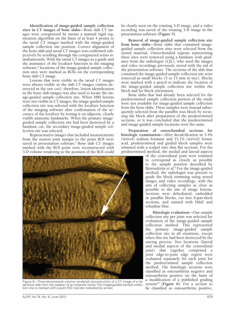
Representative images that included measurements from the nearest joint margin to the point ROI were saved in presentation software.f Bone slab CT images marked with the ROI point were reconstructed with 3-D volume rendering so the position of the ROI could
be clearly seen on the rotating 3-D image, and a video recording was saved of the rotating 3-D image in the presentation software (Figure 5).
Removal of image-guided sample collection site from bone slabsBone slabs that contained image-guided sample collection sites were selected from the stored material. Osteochondral regions representing these sites were removed using a bandsaw with guid-ance from the radiologist (CJL), who used the images and video recordings previously stored with the aid of the presentation software. The sections of the slab that contained the image-guided sample collection site were removed as small blocks (5 to 15 mm in size). Blocks were marked with a pencil to indicate the location of the image-guided sample collection site within the block and for block orientation.
Bone slabs that had already been selected for the predetermined sample collection portion of the study were not available for image-guided sample collection from the bone slabs. These samples were instead subse-quently selected from the paraffin wax block by recut-ting the block after preparation of the predetermined sections, or it was concluded that the predetermined and image-guided sample locations were the same.
Preparation of osteochondral sections for histologic examinationAfter decalcification in 3.4% (wt/vol) sodium formiate and 15.1% (wt/vol) formic acid, predetermined and guided block samples were trimmed with a scalpel into thin flat sections. For the predetermined method, the medial and lateral aspects
of the centrodistal joint were trimmed to correspond as closely as possible to the sample position described by Björnsdóttir et al.4 For the image-guided method, the radiologist was present to guide the block trimming using stored images and video recordings, with the aim of collecting samples as close as possible to the site of image lesions. Sections were dehydrated, embedded in paraffin blocks, cut into 6-µm-thick sections, and stained with H&E and toluidine blue.
Histologic evaluationOne sample collection site per joint was selected for evaluation of the image-guided sample collection method. This represented the primary image-guided sample collection site in all situations, except when this site had been destroyed by the sawing process. Two locations (lateral and medial aspects of the centrodistal joint) that together comprised a joint edge-to-joint edge region were evaluated separately for each joint for the predetermined sample collection method. The histologic sections were classified as osteoarthritis negative and osteoarthritis positive on the basis of a modification of a published grading system39 (Figure 6). For a section to be classified as osteoarthritis positive,
Figure 5Three-dimensional volume–rendered reconstruction of a CT image of a tar-sal bone slab from the cadaver of an Icelandic horse. The image-guided sample collec-tion site is marked with a point ROI (red dot indicated by arrow).
12-11-0371r.indd 879 5/23/2013 10:22:38 AM

880 AJVR, Vol 74, No. 6, June 2013
at least 3 of the following features had to be present (otherwise, the section was classified as osteoarthritis negative): chondrocyte necrosis, articular cartilage fibrillation, chondrocyte clusters with > 3 chondrocytes/lacuna, increase or decrease in articular cartilage thickness, osteochondral splitting, loss of toluidine blue staining, and tidemark (the interface between the mineralized and nonmineralized articular cartilage, which is considered to be equivalent to the articular mineralization front in the MRI and CT images) fragmentation, reduplication, or absence.
Sections were first evaluated independently by a veterinary pathologist (SE) and a veterinary radiologist (CJL). When there was disagreement between grade values, the final histologic grade was decided by con-sensus. All sections were coded so that the evaluators were unaware of the horses from which they had origi-nated. For each location, at least 1 H&E- and toluidine blue–stained section were available. When consider-able creasing, straining, or folding artifacts that inter-fered with evaluation were encountered, additional osteochondral sections were recut from the paraffin blocks until sections of suitable quality were obtained. A predetermined sample collection site was considered osteoarthritis positive when 1 or both of the lateral and
medial locations had been classified as osteoarthritis positive.
Statistical analysisOsteoarthritis detection results (ie, histologic osteoarthritis classification) for each joint as determined by means of the 2 sample collection methods were compared with results of the χ2 test for proportions.g To analyze the imaging grades, the histologic classification (osteoarthritis positive or negative) of specimens from the image-guided sample collection sites was used as the criterion (gold) standard and the sums of the imaging grades were grouped into 4 larger categories: category 1 for sums 1 to 3, category 2 for sums 4 to 6, category 3 for sums 7 to 9, and category 4 for sums 10 to 12. The association between the summed imaging grade categories for the image-guided sample collection sites and the histologic osteoarthritis classification of sites was assessed with logistic regression and production of ROC curves.40,h
The summed image grades were evaluated for their usefulness as a possible diagnostic tool for osteoarthritis detection by calculation of the AUC for the ROC curve. Diagnostic sensitivity and specificity, including 95% confidence intervals,41,42,i were calculated for image lesion types and grades detected within each image-
guided sample region, with results of histologic classification of each image-guided sample considered the criterion (gold) standard. The Fisher exact test43,j was used to identify image lesion types and grades that were associated with the histologic osteoarthritis classification. Values of P < 0.05 were considered significant for all statistical tests.
Results
Osteoarthritis detectionThe 2 osteochondral sample collection meth-ods (predetermined and image guided) for detection of osteoarthritis in the centrodistal joints of Icelandic horses yielded significantly (P = 0.02) different results. Osteoarthritic lesions were histo-logically detected in 7 of 24 (29%) joints when the samples were collected from the predetermined location without image guidance and in 15 (62%) joints when the sites from which samples were collected were identified with the aid of MRI and CT (Figure 7). All joints with osteoarthritic changes detected through predetermined sample collection also had osteoarthritic changes detected with the image-guided method. In 9 (38%) joints, histologic evidence of osteoar-thritis was not detected.
Abnormalities identified in joint imagesGrade 1 image abnormalities were detected in all 24 (100%) centrodis-tal joints, and grade 2 lesions were detect-ed in 19 (79%). Articular mineralization front defects were detected in all joints.
Figure 6Representative photomicrographs of tissue specimens from the centro-distal joints from cadavers of Icelandic horses to illustrate a protocol for identifying osteoarthritis. A—Healthy articular cartilage (osteoarthritis negative). H&E stain; bar = 500 µm. B—Osteoarthritis-positive specimen showing mild focal chondrocyte ne-crosis (asterisk) and focal chondrocyte clusters (arrows). H&E stain; bar = 500 µm. C—Osteoarthritis-positive specimen showing severe chondrocyte necrosis (aster-isks), fibrillation that extends deep into the articular cartilage, osteochondral splits (arrowheads), large chondrocyte clusters (arrows), and a decrease in articular cartilage thickness. H&E stain; bar = 500 µm. D—Osteoarthritis-positive specimen showing generalized loss of staining, severe chondrocyte necrosis (arrowheads), and large chondrocyte clusters (arrows). Toluidine blue stain; bar = 200 µm.
12-11-0371r.indd 880 5/23/2013 10:22:39 AM

AJVR, Vol 74, No. 6, June 2013 881
Marginal osteophytes (18/24 [75%]), articular cartilage hypointensity (15/24 [63%]), central osteophytes (14/24 [58%]), and articular cartilage thickness abnormalities (12/24 [50%]) were common. Intra-articular and chon-
dral mineralization (6/24 [25%]), subchondral sclerosis (6/24 [25%]), subchondral lysis (5/24 [21%]), subchon-dral cyst-like lesions (1/24 [4%]), and subchondral bone hyperintensity (1/24 [4%]) were less common.
Image grades for image-guided sam-ple collection sitesValues of summed image grades for the image-guided sam-ple collection sites ranged from 1 to 12 (Figure 7). Moderate to high numbers of samples had evidence of articular mineralization front defects, marginal osteophytes, articular cartilage hypoin-tensities, central osteophytes, articular cartilage thickness abnormalities, and intrachondral and articular mineral-ization (Table 1). On the other hand, low numbers of samples had signs of subchondral sclerosis, subchondral ly-sis, subchondral cyst, and subchondral hyperintensity.
Lesion location in image-guided sample collection sitesWhen im-age guidance was used, osteochondral samples were collected from the lateral (46%), medial (46%), and dorsal (8%) centrodistal joint regions. In 2 (8%) joints, the image-guided sample was
Figure 7Diagnostic imaging (MRI and CT) grades for and histologic classification of osteoarthritis in centrodistal joints from cadavers of Icelandic horses (n = 24). The data are ordered by the summed image grade calculated for the image-guided sample collection site identified for each horse (light gray), and bars are grouped per horse. Histologic classification was assigned as 0 for osteoarthritis-negative joints and 1 for osteoarthritis-positive joints and performed for sample collection sites chosen without imaging (black; predetermined sites) and sites chosen with MRI and CT guidance (dark gray; image-guided sites). Notice that the predetermined and image-guided sample collection sites for horse P were the same.
Sensitivity (%) for Specificity (%) for No. of osteoarthritis osteoarthritisImage feature specimens detection (95% CI) detection (95% CI) P value*
AMFD-1 16 53 (27–79) 11 (0–48) 0.18AMFD-2 7 47 (21–73) 100 (66–100) 0.022AMFD-1+2 23 100 (78–100) 11 (0–48) 0.38ACH-2 13 67 (38–88) 67 (30–93) 0.20ACT-1 3 20 (4–48) 100 (66–100) 0.27 ACT-2 9 53 (27–79) 89 (52–100) 0.08ACT-1+2 12 77 (45–92) 89 (52–100) 0.009IACM-2 5 33 (12–62) 100 (66–100) 0.12MO-1 5 20 (4–48) 78 (55–96) 1MO-2 7 47 (21–73) 100 (66–100) 0.022 MO-1+2 12 67 (38–88) 78 (40–97) 0.09CO-2 12 80 (52–97) 100 (70–100) < 0.001SCH-1 1 7 (0–32) 100 (66–100) 1SCH-2 0 — — —SCC-1 0 — — — SCC-2 1 7 (0–32) 100 (66–100) 1SCS-1 2 7 (0–32) 89 (52–100) 1SCS-2 1 0 89 (52–100) 0.38SCS-1+2 3 7 (0–32) 78 (40–97) 0.53SCL-1 1 7 (0–32) 100 (66–100) 1SCL-2 0 — — —
*P value results from Fisher exact tests of association between image lesion types and grades with the histologic osteoarthritis classification for the image-guided sample collection sites.
— = Not applicable. ACH = Articular cartilage hypointensity. ACT = Articular cartilage thickness abnormality. AMFD = Articular mineralization front defect. CI = Confidence interval. CO = Central osteophyte. IACM = Intra-articular or chondral mineralization. MO = Marginal osteophyte. SCC = Subchondral cyst-like lesion. SCH = Subchondral hyperintensity. SCL = Subchondral lysis. SCS = Subchondral sclerosis. 1 = Grade 1. 2 = Grade 2. 1 + 2 = Sum of grades 1 and 2.
For sensitivity and specificity calculations, histologic classification of the image-guided samples was used as the criterion standard.
Table 1Comparison of results for detection of various CT and MRI image features and grades and histologic classification (osteoarthritis positive or negative) of tissue specimens from image-guided sample collection sites in centrodistal joints from cadavers of Icelandic horses (n = 24).
12-11-0371r.indd 881 5/23/2013 10:22:39 AM

882 AJVR, Vol 74, No. 6, June 2013
within the bone slab selected for predetermined sample collection. In one of these joints, the location of the image-guided sample collection site was the same as the predetermined section location (Figure 7). In the other joint, the image-guided sample was located in a region of the paraffin block–embedded sample that was not included in the predetermined section; therefore, the paraffin block was recut with imaging guidance.
Histologic gradingSeventy-one osteochondral samples were evaluated, representing 24 image-guided samples, 24 predetermined samples collected from the medial aspect of the joint, and 24 predetermined sam-ples collected from the lateral aspect of the joint (one of which was the same as that obtained with image guid-ance). The 2 independent evaluators who classified histologic preparations as osteoarthritis positive or nega-tive had similar findings for 99% (70/71) of the osteo-chondral samples. For the sample with opposing find-ings, the evaluators subsequently reached agreement by consensus that a suspected abnormality was a staining artifact and the sample was classified as osteoarthritis negative.
Twenty-two joints were classified as osteoarthritis positive, and 26 joints were classified as osteoarthritis negative (Figure 7). All osteochondral samples classified as osteoarthritis positive included areas of chondronecro-sis, adjacent chondrone formation, and loss of toluidine blue staining (Figure 6). In some samples, fibrillation, tidemark fragmentation, osteochondral splitting, and fo-cal changes in cartilage thickness were seen. In 9 of the 26 (35%) joints classified as osteoarthritis negative, one of the individual evaluators (never both) detected 1 or 2 of the following features: articular cartilage thickness change, > 3 chondrocytes/lacuna, or loss of toluidine blue staining. Five of these samples were from predetermined collection sites, and 3 were from image-guided sample collection sites.
Agreement of image grades with histologic classificationAn association was detected between the guided-sample summed image grade categories and the histologic classification of osteoarthritis (positive vs negative; P = 0.018). The AUC calculated from the ROC curve was 0.89, indicating that the summed image grades were a good predictor of osteoarthritis detection by histologic evaluation.
An association was identified between histologic detection of osteoarthritis and the following lesions identified with imaging: central osteophyte (P < 0.001), combined grades 1 and 2 of articular cartilage thickness abnormalities (P = 0.009), grade 2 articular mineraliza-tion front defects (P = 0.022), and grade 2 marginal os-teophytes (P = 0.022).
The specificity of image lesions and grades for os-teoarthritis detection (with histologic classification of the image-guided samples as the criterion standard) was gen-erally high, with the exception of articular mineralization front defects grade 1 (very low specificity) and articular cartilage hypointensities (moderately low specificity; Table 1). Large variation was evident in the sensitivities of the various image lesions and grades for osteoarthritis detection, with image lesions and grades that were un-common (detected in < 7 joints) having low sensitivity.
DiscussionThe sensitivity of histologic examination for the
detection of early osteoarthritis is highly influenced by the sample collection method.12,44 In the present study involving centrodistal joints harvested from cadavers of Icelandic horses, use of an image-guided method to obtain osteochondral samples for histologic examination resulted in significantly more joints with an osteoarthritis-positive classification, compared with a predetermined sample collection method, indicating the superior sensitivity of the guided method. Thus, use of the predetermined sample collection method would be expected to underestimate the incidence of histologically diagnosed osteoarthritis in young Icelandic horses.
Our findings emphasized that sample collection methods for early osteoarthritis detection need to be based on information from the entire joint organ and have a resolution that is adequate to detect the mild and focal changes that are present early in the disease process. The image-guided method for sample collection is flexible and could be used for osteochondral sample collection from centrodistal joints of all breeds of horses. The method also has potential for application in other joints and other species. Not only can image-guided sample collection be used for solitary joints to determine the anatomic location of pathological changes, it could also be used for a series of adjacent joints, an entire limb, or even an entire animal.
The unique feature of the described image-guided method versus others is the CT examination of the sawn tarsal joint slabs. Images from those examina-tions provided a link between the images of the entire joint organ and the focal region of sample collection. The tarsal slab CT images were coregistered to the CT and MRI images of the entire tarsal joints with freely available computer software, allowing a simple meth-od to identify and mark focal changes on the slab CT images. The higher resolution of the slab CT images, compared with the resolution of the tarsal joint CT im-ages, meant that in situations when lesions were visible in CT images, lesion definition and clarity were better on the slab CT images. This high resolution provided added confidence when the lesion ROI was marked on the slab CT images.
The use of 3-D volume rendering of the slab CT images was considered enormously helpful during sample collection from the bone slabs. The volume-rendered images showed the surface features of the slabs clearly from all angles, so that sawing of the slab into a smaller sample block could be guided by multiple surface features. Distances between surface features and joint margins were easily measured, and these measurements were helpful for guiding collection of samples that contained lesions. The use of CT and MRI in combination to evaluate joints for osteoarthritis is uncommon,45 but with CT providing superior images of mineralized tissue and MRI providing superior images of soft tissues, there are considerable advantages in the combined use of these 2 modalities.
Surprisingly, few image-guided sample sites (2/24) were identified in the bone slabs selected for
12-11-0371r.indd 882 5/23/2013 10:22:39 AM

AJVR, Vol 74, No. 6, June 2013 883
predetermined sample collection. The method that was used to identify the predetermined locations for centrodistal joint sample collection4 appears to have been based on reports32,34 of regions of high incidence for osteoarthritic lesions as established by radiography. Because radiographic assessment of the distal portion of the tarsus has only limited agreement with high-detail radiologic assessment of sawn joint slabs,1 it is possible that tarsal radiography does not allow accurate identification of the site of osteoarthritis within the centrodistal joint. Alternatively, early-stage osteoarthritic lesions detected by high-detail radiography, CT, and MRI may not always progress to the more advanced stages of the disease that are detectable in radiographs. The distribution of focal osteoarthritic changes in centrodistal joints also likely varies among the horses used in studies because of age, breed, and conformation. For these reasons, sample collection methods that involve image guidance should provide more secure and reliable results when the location of focal lesions in a population is uncertain.
Although the predetermined sample collection method we used included the full joint width in histologic sections, the sensitivity of the image-guided sample collection method (where small focal regions were removed) was considerably higher. When predetermined blocks and sections are used, it is recommended that the samples obtained for histologic examination should extend from one edge of the joint to the other.11 The design of our study did not allow for joint edge-to-joint edge samples for both sample collection methods. A joint edge-to-joint edge sample collection approach could be used for the image-guided method when sawing of the whole joint into slabs is done after image assessment and lesion localization. Use of such a technique may result in further improvements in the sensitivity of the image-guided method for detection of osteoarthritic lesions.
To evaluate the sensitivity and specificity of a diagnostic method, a criterion or gold standard is required, and currently, there is not agreement for a single whole-organ osteoarthritis criterion standard.46 This situation is made more complicated by the lack of a consistent definition for early-stage osteoarthritis.46,47 We considered the microscopic morphological changes in the joints that were classified as osteoarthritis positive to have represented early-stage osteoarthritis. The histologic changes were comparable with grades 1 to 3 of the Osteoarthritis Research Society International system11 and a recently proposed definition of early-stage osteoarthritis as detected by means of histologic examination in humans.48 The changes were also reflective of lesion scores 1 to 4 of a proposed microscopic grading system for osteoarthritis in histologic preparations of articular cartilage specimens from equine metacarpo- and metatarsotarsophalangeal and carpal joints.39
The histologic classification system that we used required at least 3 features of osteoarthritis to be pres-ent in a histologic specimen before it could be classi-fied as osteoarthritis positive. This system minimized the risk of misclassification of joints as osteoarthritis positive when they actually had features caused by reversible osteochondral injury, processing artifacts,
or typical morphological variation. Several of the osteoarthritis features evaluated, including chondro-cyte necrosis, articular cartilage fibrillation, osteochon-dral splitting, and tidemark fragmentation, could be considered strong evidence for the presence of early-stage osteoarthritis, even when seen in isolation. Such stronger features were never seen in the study without the presence of chondrocyte clusters, loss of toluidine blue staining, or articular cartilage thickness changes within the same sample.
One or 2 abnormal microscopic features including articular cartilage thickness change, > 3 chondrocytes/lacuna, or loss of toluidine blue staining were detected by either of the 2 examiners in 35% of osteochondral samples classified as osteoarthritis negative. Complete disagreement occurred between examiners for the pres-ence of these features in these joints, indicating a high degree of uncertainty. Although some of these changes may have represented early-stage osteoarthritis, we consider it more likely that the changes were a result of variations in chondrocyte grouping related to the young age of the horses, anatomic variation in articular cartilage thickness, and staining artifacts.
To investigate the sensitivity and specificity of imaging features for the detection of osteoarthritis, we considered the histologic classification of the image-guided sample collection sites as the criterion standard and compared those findings with the image features and grades. When the summed image-guided sample collection site grade values were used, there was good correlation between the image grades and histologic classification of osteoarthritis as well as a high AUC for the ROC curve. Thus, focal groups of suspected osteoarthritis features in the images were an excellent indicator of histologic osteoarthritis detection. The proportion of osteochondral samples with central osteophytes, articular cartilage thickness abnormalities (grades 1 and 2 combined), grade 2 articular mineralization front defects, and grade 2 marginal osteophytes detected by CT and MRI was significantly higher in the image-guided samples with a histologic classification of osteoarthritis, thus validating these imaging features as changes associated with osteoarthritis. All of these image features were highly specific for histologic osteoarthritis classification, and the sensitivity varied from high for central osteophytes and articular cartilage thickness abnormalities to moderate for grade 2 articular mineralization front defects and grade 2 marginal osteophytes.
All joints in the study had at least 1 abnormal imaging feature; consequently, all image-guided samples contained at least 1 abnormal imaging feature, but in 38% of the joints, osteoarthritis was not histologically detected. The abnormal imaging feature identified may not have been caused by osteoarthritis, or the sample collection method may have failed to extract the tissue containing this feature. Several steps in the sample collection process have the potential to yield mild variations in sites of sample locations that were assessed microscopically. These include identification of the sample site on the actual bone slab from the 3-D reconstructed CT images (which was manual and somewhat subjective), mechanical limitations of saw cut angles and positions, and positioning of
12-11-0371r.indd 883 5/23/2013 10:22:39 AM

884 AJVR, Vol 74, No. 6, June 2013
the decalcified sample within the paraffin block. The problem of mild variations in sample location would be expected to have the largest effect in the earliest osteoarthritic changes given that these are most likely the most focal changes.11–15
Pathological changes referred to as subchondral bone pits, subchondral bone defects, subchondral bone plate irregularities, and osteochondral junction defects, which appear to correspond with the lesions that we have called articular mineralization front defects, are described in reports concerning high-detailed radiography, MRI, and histologic studies of equine centrodistal joints1,4,49 and MRI and CT assessment of the equine carpal25 and metacarpophalangeal joints.45 We chose to call these lesions articular mineralization front defects because the margin seen between nonmineralized and mineralized tissue on the articular aspect in a CT image should represent the articular front of the mineralized cartilage or, if the mineralized cartilage is not present, the articular front of the subchondral bone plate. Strong associations between subchondral defects and bone plate irregularities and the histologic abnormalities of the articular cartilage have been detected in studies1,4 of the equine centrodistal joints, and such findings are in agreement with our findings for grade 2 articular mineralization front defects.
In contrast to grade 2 articular mineralization front defects, grade 1 defects were detected in 67% (16/24) of image-guided sample collection sites, but osteoarthritis was detected in only 8 of these samples. Milder grade 1 articular mineralization front defects may have occu-pied a smaller area than the grade 2 defects, resulting in a higher chance to be missed during the sample collec-tion process, but it appears more likely that a propor-tion of horses have mild concave contours in regions of the articular mineralization front but healthy overlying nonmineralized articular cartilage. This is supported by the subjective observation from histologic evaluations that several samples classified as osteoarthritis nega-tive had a mildly concave shape of the tidemark and chondro-osseus junction but unremarkable nonminer-alized articular cartilage morphological characteristics. Whether the mild concavities in the mineralization front represented natural anatomic variation or wheth-er this shape represented remodeling in response to load in these regions requires additional investigation.
Abnormal imaging features of the subchondral bone regions (lysis, sclerosis, cyst-like lesions, and MRI hyperintensity) were uncommonly detected in the osteoarthritis-positive image-guided sample collection sites. Other equine joint studies22,45,50 in which CT and MRI protocols similar to ours were used have revealed subchondral bone changes in relation to osteoarthritis,22,45,50 so rather than poor sensitivity of the imaging techniques in our study explaining the discrepancy, we consider that subchondral bone abnormalities are rare in early-stage osteoarthritis of the centrodistal joint in young Icelandic horses. This low prevalence of subchondral bone abnormalities is in agreement with investigations of the distal aspect of the tarsus in young horses with high-resolution radiography and histologic examination1,4 and gives strong support to the theory that the first morphological changes of
centrodistal joint osteoarthritis in horses are in the articular cartilage and the articular mineralization front regions, rather than in the subchondral bone.
Previous reports1,4,31,49 of centrodistal joint osteoarthritis have not mentioned intra-articular and chondral mineralizations as lesions detected with imaging, but such lesions have reportedly been detected in metacarpo- and metatarsophalangeal joints.51 Intra-articular and chondral mineralization have also not been identified in histologic studies1,2,4,31 of the centrodistal joints, but it is possible that the decalcification processing and microtome sectioning of paraffin blocks may result in this lesion type not being readily detectable through histologic examination.51 No statistical association was found between intra-articular and chondral mineralizations and histologic osteoarthritis detection, but it is noteworthy that every time this image lesion was identified (5 image-guided specimens), osteoarthritis was always detected histologically. We therefore suspect that intra-articular and chondral mineralizations are a relatively uncommon change in the early stages of osteoarthritis and that these mineralizations are an osteoarthritis-specific change. It is possible that intra-articular or chondral mineralization developed in the joints that had the more advanced pathological changes in the study horses.
Articular cartilage hypointensity as a diagnos-tic feature had poor specificity for predicting a histo-logic classification of osteoarthritis. Because this type of lesion is small and focal, the image-guided sample collection method may have failed to extract the tis-sue containing the lesion. More likely, however, is that some of the articular cartilage hypointensities were the result of image artifacts. The MRI sequence WATSf was used for the evaluation of this feature, and when this imaging sequence is used at a high resolution, a mod-erate amount of noise is present in the images. This noise appears as dark areas in the bright regions of car-tilage signal and thus has a similar appearance to a car-tilage hypointensity. Partial volume averaging can also result in dark pixels in the region of the junction be-tween the hyperintense articular cartilage and hypoin-tense bone, particularly in areas where the articular car-tilage layers are curved and thin.
To test the reliability of the histologic osteoarthri-tis classification system, 1 experienced and 1 inexperi-enced person evaluated the sections on histologic ex-amination independently. The excellent agreement of the evaluators shows that the primary endpoint of the study, osteoarthritis detection, was robust and allowed effective separation of nonarthritic from arthritic joints.
To use diagnostic images for detection of osteoar-thritis in joints, a clear separation needs to exist be-tween the diseased and nondiseased groups, and the repeatability and reliability of the evaluation method need to be demonstrated. The purpose of the present study was not to develop an image-guided grading system that would result in the classification of joints as osteoarthritis positive or negative but rather to in-vestigate image-guided sample collection as a method for obtaining samples to screen for early-stage osteoar-thritis in joints. For this reason, the repeatability and reliability of the image grading were not investigated.
12-11-0371r.indd 884 5/23/2013 10:22:40 AM

AJVR, Vol 74, No. 6, June 2013 885
Nevertheless, the study provided a starting point for the development of an image-grading system for the detection of early-stage osteoarthritis in centrodistal joints. The association of the image changes, including central osteophytes, articular cartilage thickness abnor-malities, grade 2 articular mineralization front defects, and grade 2 marginal osteophytes, with osteoarthritis-positive histologic results provides validation that these imaging lesions are related to early-stage osteoarthritis and therefore should be used in such a grading system. An image-based grading system for osteoarthritis in the distal joints of the tarsus would be of great value because this would provide a noninvasive method to evaluate osteoarthritic lesions and allow these lesions to be monitored over time.
Imaging of the distal region of the tarsus can be performed in live horses with MRI, when the horses are standing and low-field magnets are used or when they are recumbent and low- or high-field magnets are used. Computed tomography can also be used in recumbent horses. Anesthesia, considerable lifting and manipulation of horses, and use of specially designed tables are required to examine recumbent horses, whereas standing examinations are performed by sedating the horses, walking them to the magnet, and positioning the affected limb in the magnet. Horse motion is an important cause of artifacts in MRI images, particularly when the proximal regions of limbs are examined with horses standing, and the higher the spatial resolution of an image is, the more susceptible that image is to degradation due to motion.52 Motion is minimized by anesthetizing the subjects, although the longer the period of anesthesia, the greater the risk of anesthetic-related problems.53 Motion-correction techniques can be used in standing MRI to reduce motion artifacts.52 The small size and focal nature of many of the lesions in the early stages of osteoarthritis mean that submillimeter spatial resolution is required to detect any morphological changes. The CT protocols we used could be applied to live anesthetized horses to detect morphological changes, and this would allow validated osteoarthritic lesions detected by imaging to be monitored over time.
For MRI to achieve the required resolution, the scan times for most sequences must be long; consequently, effective methods to minimize motion artifacts will be required. The MRI protocols in the present study were only possible in cadaveric specimens because of the long acquisition time (WATSf sequence, 50 minutes). Higher magnetic field strengths result in higher tissue signal; therefore, high-resolution images can be acquired with shorter scan times.54,55 However, the availability of machines for use in live horses with field strengths > 1.5 T is extremely limited and scan times will still be long. An attribute of MRI is the large range of sequences possible and the ability of specific sequences to show specific tissue changes with high-contrast resolution. Investigation of such sequences in low- and high-field MRI, together with validation through the use of image-guided histologic evaluation, may identify methods that can detect early osteoarthritic changes in images that have a lower spatial resolution than currently required to detect morphological changes.
An image-guided sample collection method is rec-ommended when histologic examination will be used as the criterion standard for the detection of early-stage osteoarthritis in centrodistal joints of horses. Computed tomography of slab samples removed from joints can be used effectively to guide removal of small osteochondral blocks for histologic processing. The combination of a significant association between the imaging lesions of the articular mineralization front, articular cartilage thick-ness, and marginal osteophytes, compared with the lack of changes detected in subchondral bone, leads us to specu-late that the morphological changes of centrodistal joint osteoarthritis begin in the articular cartilage and the artic-ular mineralization front regions. The association between the histologic osteoarthritis classification and the imaging lesions of central osteophytes, abnormal articular cartilage thickness, grade 2 marginal osteophytes, and grade 2 ar-ticular mineralization front defects provides evidence to support the use of these imaging lesions for the detection of osteoarthritis of the centrodistal joint in horses.
a. Intera, Philips Medical Systems, Best, The Netherlands.b. Q-Knee/Foot Coil, Philips Medical Systems, Best, The
Netherlands.c. Definition, Siemens Medical Systems, Erlangen, Germany.d. OsiriX, version 3.8, Pixmeo, Geneva, Switzerland.e. KT-400, Klaukkala, Finland.f. PowerPoint 2008, Microsoft, Redmond, Wash.g. Minitab 15 statistical software program, Minitab Inc, State
College, Pa.h. Proc Logistic, SAS, version 9.2, SAS Institute Inc, Cary, NC.i. Interactive Statistical Calculation Pages. Available at: statpages.
org/ctab2x2.html. Accessed Oct 16, 2012.j. Proc Freq, SAS, version 9.2, SAS Institute Inc, Cary, NC.
References
1. Laverty S, Stover SM, Belanger D, et al. Radiographic, high de-tail radiographic, microangiographic and histological findings of the distal portion of the tarsus in weanling, young and adult horses. Equine Vet J 1991;23:413–421.
2. Watrous BJ, Hultgren BD, Wagner PC. Osteochondrosis and ju-venile spavin in equids. Am J Vet Res 1991;52:607–612.
3. Batiste DL, Kirkley A, Laverty S, et al. High-resolution MRI and micro-CT in an ex vivo rabbit anterior cruciate ligament transection model of osteoarthritis. Osteoarthritis Cartilage 2004;12:614–626.
4. Björnsdóttir S, Ekman S, Eksell P, et al. High detail radiogra-phy and histology of the centrodistal joint of young Icelandic horses. Equine Vet J 2004;36:5–11.
5. Calvo E, Palacios I, Delgado E, et al. Histopathological correla-tion of cartilage swelling detected by magnetic resonance imag-ing in early experimental osteoarthritis. Osteoarthritis Cartilage 2004;12:878–886.
6. Bittersohl B, Mamisch TC, Welsch GH, et al. Experimental model to evaluate in vivo and in vitro cartilage MR imaging by means of histological analyses. Eur J Radiol 2009;70:561–569.
7. McGibbon CA, Trahan CA. Measurement accuracy of focal car-tilage defects from MRI and correlation of MRI graded lesions with histology: a preliminary study. Osteoarthritis Cartilage 2003;11:483–493.
8. Loeuille D, Gonord P, Guingamp C, et al. In vitro magnetic res-onance microimaging of experimental osteoarthritis in the rat knee joint. J Rheumatol 1997;24:133–139.
9. Gahunia HK, Babyn P, Lemaire C, et al. Osteoarthritis staging: comparison between magnetic resonance imaging, gross pathol-ogy and histopathology in the rhesus macaque. Osteoarthritis Cartilage 1995;3:169–180.
10. Vande Berg BC, Lecouvet FE, Malghem J. Frequency and to-pography of lesions of the femoro-tibial cartilage at spiral CT
12-11-0371r.indd 885 5/23/2013 10:22:40 AM

886 AJVR, Vol 74, No. 6, June 2013
arthrography of the knee: a study in patients with normal knee radiographs and without history of trauma. Skeletal Radiol 2002;31:643–649.
11. Pritzker KPH, Gay S, Jimenez SA, et al. Osteoarthritis cartilage histopathology: grading and staging. Osteoarthritis Cartilage 2006;14:13–29.
12. Veje K, Hyllested-Winge JL, Ostergaard K. Topographic and zonal distribution of tenascin in human articular cartilage from femoral heads: normal versus mild and severe osteoarthritis. Os-teoarthritis Cartilage 2003;11:217–227.
13. Dieppe P, Kirwan J. The localization of osteoarthritis. Br J Rheu-matol 1994;33:201–203.
14. Bae WC, Payanal MM, Chen AC, et al. Topographic patterns of cartilage lesions in knee osteoarthritis. Cartilage 2010;1:10–19.
15. Brama PA, Tekoppele JM, Bank RA, et al. Topographical map-ping of biochemical properties of articular cartilage in the equine fetlock joint. Equine Vet J 2000;32:19–26.
16. Palmer JL, Bertone AL, Malemud CJ, et al. Site-specific proteo-glycan characteristics of third carpal articular cartilage in ex-ercised and nonexercised horses. Am J Vet Res 1995;56:1570–1576.
17. Murray RC, Blunden TS, Schramme MC, et al. How does mag-netic resonance imaging represent histologic findings in the equine digit? Vet Radiol Ultrasound 2006;47:17–31.
18. Smith MA, Dyson SJ, Murray RC. Reliability of high- and low-field magnetic resonance imaging systems for detection of carti-lage and bone lesions in the equine cadaver fetlock. Equine Vet J 2012;44:684–691.
19. Olive J, D’Anjou M-A, Girard C, et al. Imaging and histologi-cal features of central subchondral osteophytes in racehorses with metacarpophalangeal joint osteoarthritis. Equine Vet J 2009;41:859–864.
20. Olive J, D’Anjou M, Girard C, et al. Fat-suppressed spoiled gra-dient-recalled imaging of equine metacarpophalangeal articular cartilage. Vet Radiol Ultrasound 2010;51:107–115.
21. Blunden A, Dyson S, Murray R, et al. Histopathology in horses with chronic palmar foot pain and age-matched controls. Part 1: navicular bone and related structures. Equine Vet J 2006;38:15–22.
22. Anatasiou A, Skioldebrand E, Ekman S, et al. Ex vivo magnetic resonance imaging of the distal row of equine carpal bones: as-sessment of bone sclerosis and carpal damage. Vet Radiol Ultra-sound 2003;44:501–512.
23. Schmitz N, Laverty S, Kraus VB, et al. Basic methods in histopa-thology of joint tissues. Osteoarthritis Cartilage 2010;18(suppl 3):S113–S116.
24. Brommer H, Rijkenhuizen AB, Brama PA, et al. Accuracy of diagnostic arthroscopy for the assessment of cartilage dam-age in the equine metacarpophalangeal joint. Equine Vet J 2004;36:331–335.
25. Lacourt M, Gao C, Li A, et al. Relationship between carti-lage and subchondral bone lesions in repetitive impact trau-ma-induced equine osteoarthritis. Osteoarthritis Cartilage 2012;20:572–583.
26. Eksell P, Axelsson M, Brostrom H, et al. Prevalence and risk factors of bone spavin in Icelandic horses in Sweden: a radio-graphic field study. Acta Vet Scand 1998;39:339–348.
27. Björnsdottir S, Axelsson M, Eksell P, et al. Radiographic and clinical survey of degenerative joint disease in the distal tarsal joints in Icelandic horses. Equine Vet J 2000;32:268–272.
28. Björnsdottir S, Aronson T, Lord P. Culling rate of Icelandic hors-es due to bone spavin. Acta Vet Scand 2003;44:161–169.
29. Sisson S. Equine syndesmology. Sisson and Grossman’s the anat-omy of the domestic animals. 5th ed. Philadelphia: Saunders, 1975;349–375.
30. Auer JA. Tarsus. In: Auer JA, Stick JA, eds. Equine surgery. 4th ed. St Louis: Elsevier Saunders, 2012;1388–1409.
31. Barneveld A, van Weeren PR. Early changes in the distal inter-tarsal joint of Dutch Warmblood foals and the influence of exer-cise on bone density in the third tarsal bone. Equine Vet J Suppl 1999;31:67–73.
32. Verschooten F, Schramme M. Radiological examination of the tarsus. Equine Vet Educ 1994;6:323–332.
33. Eksell P, Uhlhorn H, Carlsten J. Evaluation of different pro-
jections for radiographic detection of tarsal degenerative joint disease in Icelandic Horses. Vet Radiol Ultrasound 1999;40:228–232.
34. Hartung K, Münzer, B., Keller, H. Radiologic evaluation of spav-in in young trotters. Vet Radiol Ultrasound 1983;24:153–155.
35. Kim H, Lee SH, Kang CH, et al. Evaluation of the chondroma-lacia patella using a microscopy coil: comparison of the two- dimensional fast spin echo techniques and the three-dimension-al fast field echo techniques. Korean J Radiol 2011;12:78–88.
36. Hargreaves BA, Gold GE, Beaulieu CF, et al. Comparison of new sequences for high-resolution cartilage imaging. Magn Reson Med 2003;49:700–709.
37. Kornaat PR, Doornbos J, van der Molen AJ, et al. Magnetic reso-nance imaging of knee cartilage using a water selective balanced steady-state free precession sequence. J Magn Reson Imaging 2004;20:850–856.
38. Branch MV, Murray RC, Dyson SJ, et al. Is there characteris-tic distal tarsal subchondral bone plate thickness pattern in horses with no history of hind limb lameness? Equine Vet J 2005;37:450–455.
39. McIlwraith CW, Frisbie DD, Kawcak CE, et al. The OARSI his-topathology initiative—recommendations for histological as-sessments of osteoarthritis in the horse. Osteoarthritis Cartilage 2010;18(suppl 3):S93–S105.
40. SAS/STAT92 user’s cuide. 2nd ed. Cary, NC: SAS Institute Inc, 2009;3257–3472.
41. Fleiss JL. Sampling method I: naturalistic or cross-sectional studies. In: Statistical methods for rates and proportions. 2nd ed. New York: John Wiley & Sons, 1981;56–82.
42. Clopper CJ, Pearson ES. The use of confidence or fiducial limits illustrated in the case of the binomial. Biometrika 1934;26:404–413.
43. SAS/STAT92 user’s guide. 2nd ed. Cary, NC: SAS Institute Inc, 2009;1676–1826.
44. Mankin HJ, Dorfman H, Lippiello L, et al. Biochemical and met-abolic abnormalities in articular cartilage from osteo-arthritic human hips. II. Correlation of morphology with biochemical and metabolic data. J Bone Joint Surg Am 1971;53:523–537.
45. Olive J, D’Anjou M, Alexander K, et al. Comparison of magnetic resonance imaging, computed tomography, and radiography for assessment of noncartilaginous changes in equine metacarpo-phalangeal osteoarthritis. Vet Radiol Ultrasound 2010;51:267–279.
46. Hunter DJ, Arden N, Conaghan PG, et al. Definition of osteoar-thritis on MRI: results of a Delphi exercise. Osteoarthritis Carti-lage 2011;19:963–969.
47. Luyten F, Denti M, Filardo G, et al. Definition and classification of early osteoarthritis of the knee. Knee Surg Sports Traumatol Arthrosc 2012;1–6.
48. Madry H, Luyten FP, Facchini A. Biological aspects of early os-teoarthritis. Knee Surg Sports Traumatol Arthrosc 2012;20:407–422.
49. Branch MV, Murray RC, Dyson SJ, et al. Magnetic resonance imaging of the equine tarsus. Clin Tech Equine Pract 2007;6:96–102.
50. Raes EV, Bergman EH, van der Veen H, et al. Comparison of cross-sectional anatomy and computed tomography of the tar-sus in horses. Am J Vet Res 2011;72:1209–1221.
51. Boyde A, Riggs CM, Bushby AJ, et al. Cartilage damage involv-ing extrusion of mineralisable matrix from the articular calcified cartilage and subchondral bone. Eur Cell Mater 2011;21:470–478.
52. McKnight AL, Manduca A, Felmlee JP, et al. Motion-correction techniques for standing equine MRI. Vet Radiol Ultrasound 2004;45:513–519.
53. Young SS, Taylor PM. Factors influencing the outcome of equine anaesthesia: a review of 1,314 cases. Equine Vet J 1993;25:147–151.
54. Masi JN, Sell CA, Phan C, et al. Cartilage MR imaging at 3.0 versus that at 1.5 T: preliminary results in a porcine model. Ra-diology 2005;236:140–150.
55. Bolen G, Audigié F, Spriet M, et al. Qualitative comparison of 0.27T, 1.5T, and 3T magnetic resonance images of the normal equine foot. J Equine Vet Sci 2010;30:9–20.
12-11-0371r.indd 886 5/23/2013 10:22:40 AM

AJVR, Vol 74, No. 6, June 2013 887
Parameter WATSf STIR
Echo time (ms) 10.1 60Inversion time (ms) — 140Repetition time (ms) 21.7 3,422Flip angle (°) 50 90No. of signal acquisitions 4 3Frequency (Hz) 63.9 63.9 Field of view (mm) 80 100Slice thickness and gap* (mm) 0.7 and –0.35 3 and 3.3Phase encoding steps 272 125Frequency encoding steps 272 160Reconstruction matrix (pixels) 512 X 512 288 X 288Scan time (min) 50 4
*For slice gap, –0.35 refers to contiguous slices with a 0.35-mm overlap.
= Not applicable.
Appendix 2Settings used during MRI (WATSf and STIR) of centrodistal joints from equine cadavers.
ModalityLesion (sequences used) Grade 0 Grade 1 Grade 2
Articular cartilage hypointensity MRI (WATSf) Unremarkable intensity — Focal hypointense signal within the of cartilage articular cartilage
Articular cartilage thickness MRI (WATSf WATSf: moderately WATSf, hyperintense WATSf: hyperintense layer not and joint space and STIR) hyperintense and < 1 mm and ≥ 1 mm thick visible; or STIR: focal collection thick; STIR: no signal of hyperintense signal between or fine hyperintense line articular surfaces
Central osteophyte MRI (WATSf) ND — Hypointense projections from the articular mineralization front
Subchondral bone hyperintensity MRI (WATSf ND Maximal dimension Maximal dimension and STIR) of hyperintense area, of hyperintense area < 1 mm ≥ 1 mm Articular mineralization CT ND ≤ 0.5 mm depth > 0.5 mm depth front defect Intra-articular and CT ND — Mineral density material between chondral mineralization the articular mineralization fronts Marginal osteophyte CT ND Small Large
Subchondral cyst-like lesion CT ND Maximal dimension of Maximal dimension of focal circular area of focal circular area of hypoattenuation, hypoattenuation, ≥ 1mm < 1 mm Subchondral sclerosis CT ND Focal sclerosis Generalized sclerosis
Subchondral lysis CT ND Focal lysis Generalized lysis
— = Not applicable. ND = Not detected.
Appendix 3Grading system used to characterize the type and severity of suspected osteoarthritic features in centrodistal joints from equine cadavers as identified via CT and MRI.
Classification Description
Negative No evidence of subchondral bone (all features required) sclerosis or lysis Smooth articular surfaces No evidence of joint space narrowing Smooth angular to rounded joint margins
Positive Subchondral bone sclerosis or lysis (at least 1 feature Rough irregular articular surfaces required) Narrow or uneven joint space Marginal osteophytes Periarticular bone bridge or ankylosis
Appendix 1Classification system used to define the presence or absence of radiographic centrodistal joint osteoarthritis in parent horses.
12-11-0371r.indd 887 5/23/2013 10:22:40 AM
Copyright © 2022 FDOKUMEN