Evaluation of different detection methods of biofilm formation in the clinical isolates
7
ORIGINAL ARTICLE 305 Evaluation of different detection methods of biofilm formation in the clinical isolates Authors Afreenish Hassan 1 Javaid Usman 2 Fatima Kaleem 1 Maria Omair 3 Ali Khalid 1 Muhammad Iqbal 4 1 MPhil Microbiology, Department of Microbiology, National University of Sciences and Technology (NUST), Islamabad, Army Medical College, Rawalpindi, Pakistan 2 Professor of Pathology, Department of Microbiology, NUST, Islamabad, Army Medical College, Rawalpindi, Pakistan 3 FCPS Microbiology, Department of Microbiology, NUST, Islamabad, Army Medical College, Rawalpindi, Pakistan 4 ST, Senior Technician, Department of Microbiology, NUST, Islamabad, Army Medical College, Rawalpindi, Pakistan Submitted on: 01/03/2011 Approved on: 05/03/2011 Correspondence to: Afreenish Hassan Department of Microbiology National University of Sciences and Technology, Islamabad, Army Medical College, Rawalpindi, Pakistan [email protected] We declare no conflict of interest. ©2011 Elsevier Editora Ltda. All rights reserved. ABSTRACT Background: Microorganisms growing in a biofilm are associated with chronic and recurrent hu- man infections and are highly resistant to antimicrobial agents. ere are various methods to detect biofilm production like Tissue Culture Plate (TCP), Tube method (TM), Congo Red Agar meth- od (CRA), bioluminescent assay, piezoelectric sensors, and fluorescent microscopic examination. Objective: is study was conducted to compare three methods for the detection of biofilms. Method: e study was carried out at the Department of Microbiology, Army Medical College, National University of Sciences and Technology, Pakistan, from January 2010 to June 2010. A total of 110 clinical isolates were subjected to biofilm detection methods. Isolates were identified by standard microbiological procedures. Biofilm detection was tested by TCP, TM and CRA. Antibiotic suscep- tibility test of biofilm producing bacteria was performed by using the Kirby-Bauer disc diffusion technique according to CLSI guidelines. Results: e TCP method was considered to be superior to TM and CRA. From the total of 110 clinical isolates, TCP method detected 22.7% as high, 41% mod- erate and 36.3% as weak or non-biofilm producers. We have observed higher antibiotic resistance in biofilm producing bacteria than non-biofilm producers. Conclusion: We can conclude from our study that the TCP method is a more quantitative and reliable method for the detection of biofilm forming microorganisms as compared to TM and CRA methods, and it can be recommended as a general screening method for detection of biofilm producing bacteria in laboratories. Keywords: biofilms; bacteria; anti-bacterial agents. INTRODUCTION Biofilms are defined as microbially derived ses- sile communities characterized by the cells that are irreversibly attached to a substratum or to each other. ey are embedded in a matrix of extracellular polymeric substances (EPS) they have produced, and exhibit an altered phe- notype with respect to growth rate and gene transcription. 1 Within a biofilm, bacteria com- municate with each other by production of chemotactic particles or pheromones, a phe- nomenon called quorum sensing. 2 Availability of key nutrients, chemotaxis towards surface, motility of bacteria, surface adhesins and pres- ence of surfactants are some factors which in- fluence biofilm formation. 2 Microorganisms growing in a biofilm are intrinsically more re- sistant to antimicrobial agents than planktonic cells. High antimicrobial concentrations are required to inactivate organisms growing in a biofilm, as antibiotic resistance can increase 1,000 fold. 3 According to a publication by the National Institutes of Health, more than 80% of all infections involve biofilms. 4 Biofilms are as- sociated with many medical conditions includ- ing indwelling medical devices, dental plaque, upper respiratory tract infections, peritonitis, and urogenital infections. 5 Both Gram-positive and Gram-negative bacteria have the capability to form biofilms. Bacteria commonly involved include Enterococcus faecalis, Staphylococcus aureus, Staphylococcus epidermidis, Streptococ- cus viridans, Escherichia coli, Klebsiella pneu- moniae, Proteus mirabilis and Pseudomonas aeruginosa. 6 ere are various methods to detect biofilm production. ese include the Tissue Culture Plate (TCP), 7 Tube method (TM), 8 Congo Red Agar method (CRA), 9 bio- luminescent assay, 10 piezoelectric sensors, 11 and fluorescent microscopic examination. 12
-
Upload
independent -
Category
Documents
-
view
3 -
download
0
Transcript of Evaluation of different detection methods of biofilm formation in the clinical isolates
OR
IGIN
AL
A
RTI
CLE
305
Evaluation of different detection methods of biofilm formation in the clinical isolates
AuthorsAfreenish Hassan1
Javaid Usman2
Fatima Kaleem1
Maria Omair3
Ali Khalid1
Muhammad Iqbal4
1MPhil Microbiology, Department of Microbiology, National University of Sciences and Technology (NUST), Islamabad, Army Medical College, Rawalpindi, Pakistan 2Professor of Pathology, Department of Microbiology, NUST, Islamabad, Army Medical College, Rawalpindi, Pakistan 3FCPS Microbiology, Department of Microbiology, NUST, Islamabad, Army Medical College, Rawalpindi, Pakistan 4ST, Senior Technician, Department of Microbiology, NUST, Islamabad, Army Medical College, Rawalpindi, Pakistan
Submitted on: 01/03/2011Approved on: 05/03/2011
Correspondence to:Afreenish HassanDepartment of MicrobiologyNational University ofSciences and Technology,Islamabad, Army Medical College, Rawalpindi, [email protected]
We declare no conflict of interest.
©2011 Elsevier Editora Ltda. All rights reserved.
ABSTRACT
Background: Microorganisms growing in a biofilm are associated with chronic and recurrent hu-man infections and are highly resistant to antimicrobial agents. There are various methods to detect biofilm production like Tissue Culture Plate (TCP), Tube method (TM), Congo Red Agar meth-od (CRA), bioluminescent assay, piezoelectric sensors, and fluorescent microscopic examination. Objective: This study was conducted to compare three methods for the detection of biofilms. Method: The study was carried out at the Department of Microbiology, Army Medical College, National University of Sciences and Technology, Pakistan, from January 2010 to June 2010. A total of 110 clinical isolates were subjected to biofilm detection methods. Isolates were identified by standard microbiological procedures. Biofilm detection was tested by TCP, TM and CRA. Antibiotic suscep-tibility test of biofilm producing bacteria was performed by using the Kirby-Bauer disc diffusion technique according to CLSI guidelines. Results: The TCP method was considered to be superior to TM and CRA. From the total of 110 clinical isolates, TCP method detected 22.7% as high, 41% mod-erate and 36.3% as weak or non-biofilm producers. We have observed higher antibiotic resistance in biofilm producing bacteria than non-biofilm producers. Conclusion: We can conclude from our study that the TCP method is a more quantitative and reliable method for the detection of biofilm forming microorganisms as compared to TM and CRA methods, and it can be recommended as a general screening method for detection of biofilm producing bacteria in laboratories. Keywords: biofilms; bacteria; anti-bacterial agents.
INTRODUCTION
Biofilms are defined as microbially derived ses-sile communities characterized by the cells that are irreversibly attached to a substratum or to each other. They are embedded in a matrix of extracellular polymeric substances (EPS) they have produced, and exhibit an altered phe-notype with respect to growth rate and gene transcription.1 Within a biofilm, bacteria com-municate with each other by production of chemotactic particles or pheromones, a phe-nomenon called quorum sensing.2 Availability of key nutrients, chemotaxis towards surface, motility of bacteria, surface adhesins and pres-ence of surfactants are some factors which in-fluence biofilm formation.2 Microorganisms growing in a biofilm are intrinsically more re-sistant to antimicrobial agents than planktonic cells. High antimicrobial concentrations are required to inactivate organisms growing in
a biofilm, as antibiotic resistance can increase 1,000 fold.3 According to a publication by the National Institutes of Health, more than 80% of all infections involve biofilms.4 Biofilms are as-sociated with many medical conditions includ-ing indwelling medical devices, dental plaque, upper respiratory tract infections, peritonitis, and urogenital infections.5 Both Gram-positive and Gram-negative bacteria have the capability to form biofilms. Bacteria commonly involved include Enterococcus faecalis, Staphylococcus aureus, Staphylococcus epidermidis, Streptococ-cus viridans, Escherichia coli, Klebsiella pneu-moniae, Proteus mirabilis and Pseudomonas aeruginosa.6 There are various methods to detect biofilm production. These include the Tissue Culture Plate (TCP),7 Tube method (TM),8 Congo Red Agar method (CRA),9 bio-luminescent assay,10 piezoelectric sensors,11 and fluorescent microscopic examination.12
BJID-4-JUN ARTE FINAL.indd 305 28/07/11 13:30
306
We screened 110 organisms by three different methods, which could be used in a routine clinical laboratory, for determining their ability to form biofilm.
OBJECTIVES
The study was conducted to detect biofilm forming microor-ganisms isolated from clinical specimens by three different methods (TCP, TM, CRA) and to compare these methods for biofilm detection.
MATERIALS AND METHODS
Place and duration of the studyThe study was conducted at the Department of Microbiol-ogy, Army Medical College, National University of Sciences and Technology, Pakistan, from January 2010 to June 2010.
Selection of the isolatesA total of 110 clinical isolates were subjected to biofilm de-tection methods. Organisms were selected on the following criteria: those isolated from the pus, intravenous and urinary catheter tips, urine, sputum and nasobronchial lavage speci-mens, and those showing increased resistance to commonly available antibiotics by Kirby-Bauer disc diffusion method. Urinary catheter tips, intravenous catheter tips, nasobron-chial lavage specimens and few of the pus specimens were related to medical devices (Table 1). All of the specimens were received from patients with nosocomial infections ad-mitted to the hospital.
Isolates were identified by standard microbiological pro-cedures (Gram staining, colonial morphology, catalase test, cytochrome oxidase reaction, motility, biochemical tests). Reference strain of positive biofilm producer Staphylococcus epidermidis ATCC 35984, Staphylococcus aureus ATCC 35556, Pseudomonas aeruginosa ATCC 27853, Escherichia coli ATCC 35218 and Staphylococcus epidermidis ATCC 12228 (non-slime producer) were used as control. Biofilm detection was done by the following methods:
Tissue culture plate methodThis quantitative test described by Christensen et al.7 is con-sidered the gold-standard method for biofilm detection.13 Organisms isolated from fresh agar plates were inoculated in 10 mL of trypticase soy broth with 1% glucose. Broths were incubated at 37oC for 24 h. The cultures were then diluted 1:100 with fresh medium. Individual wells of sterile 96 well-flat bottom polystyrene tissue culture treated plates (Sigma-Aldrich, Costar, USA) were filled with 200 µL of the diluted cultures. The control organisms were also incubated, diluted and added to tissue culture plate. Negative control wells con-tained inoculated sterile broth. The plates were incubated at 37oC for 24 h. After incubation, contents of each well were removed by gentle tapping. The wells were washed with
0.2 mL of phosphate buffer saline (pH 7.2) four times. This removed free floating bacteria. Biofilm formed by bacteria adherent to the wells were fixed by 2% sodium acetate and stained by crystal violet (0.1%). Excess stain was removed by using deionized water and plates were kept for drying. Opti-cal density (OD) of stained adherent biofilm was obtained by using micro ELISA autoreader (model 680, Biorad, UK) at wavelength 570 nm. The experiment was performed in triplicate and repeated three times. The interpretation of biofilm production was done according to the criteria of Stepanovic et al.14 (Table 2).
Tube methodDescribed by Christensen et al.,8 this is a qualitative method for biofilm detection. A loopful of test organ-isms was inoculated in 10 mL of trypticase soy broth with 1% glucose in test tubes. The tubes were incubated at 37oC for 24 h. After incubation, tubes were decanted and washed with phosphate buffer saline (pH 7.3) and dried. Tubes were then stained with crystal violet (0.1%). Ex-cess stain was washed with deionized water. Tubes were dried in inverted position. The scoring for tube method was done according to the results of the control strains. Biofilm formation was considered positive when a visible film lined the wall and the bottom of the tube. The amount of biofilm formed was scored as 1-weak/none, 2-moder-ate and 3-high/strong. The experiment was performed in triplicate and repeated three times.
Congo Red Agar methodFreeman et al.9 have described a simple qualitative method to detect biofilm production by using Congo Red Agar (CRA) medium. CRA medium was prepared with brain heart infusion broth (Oxoid, UK) 37 g/L, sucrose 50 g/L, agar No. 1 (Oxoid, UK) 10 g/L and Congo Red indicator (Oxoid, UK) 8 g/L. First Congo Red stain was prepared as a concentrated aqueous solution and autoclaved (121oC for 15 minutes) separately from the other medium constitu-ents. Then it was added to the autoclaved brain heart infu-sion agar with sucrose at 55oC.5 CRA plates were inoculated with test organisms and incubated at 37oC for 24 h aerobi-cally. Black colonies with a dry crystalline consistency indicated biofilm production.5 The experiment was per-formed in triplicate and repeated three times.
Antibiotic susceptibility test of biofilm produc-ing bacteria was done on Mueller Hinton agar (Oxoid, UK) using the following antibiotic discs: ampicillin, cotrimoxazole, ciprofloxacin, aztreonam, meropenem, cefoperazone-sulbactam, chloramphenicol, vancomy-cin, erythromycin, amoxicillin-clavulanic acid, oxacil-lin, linezolid, penicillin. All antibiotic discs were ob-tained from Oxoid, UK. Escherichia coli ATCC 25922, Pseudomonas aeruginosa ATCC 27853 and Staphylococcus
Evaluation of different detection methods of biofilm formation in the clinical isolates
BJID-4-JUN ARTE FINAL.indd 306 28/07/11 13:30

307
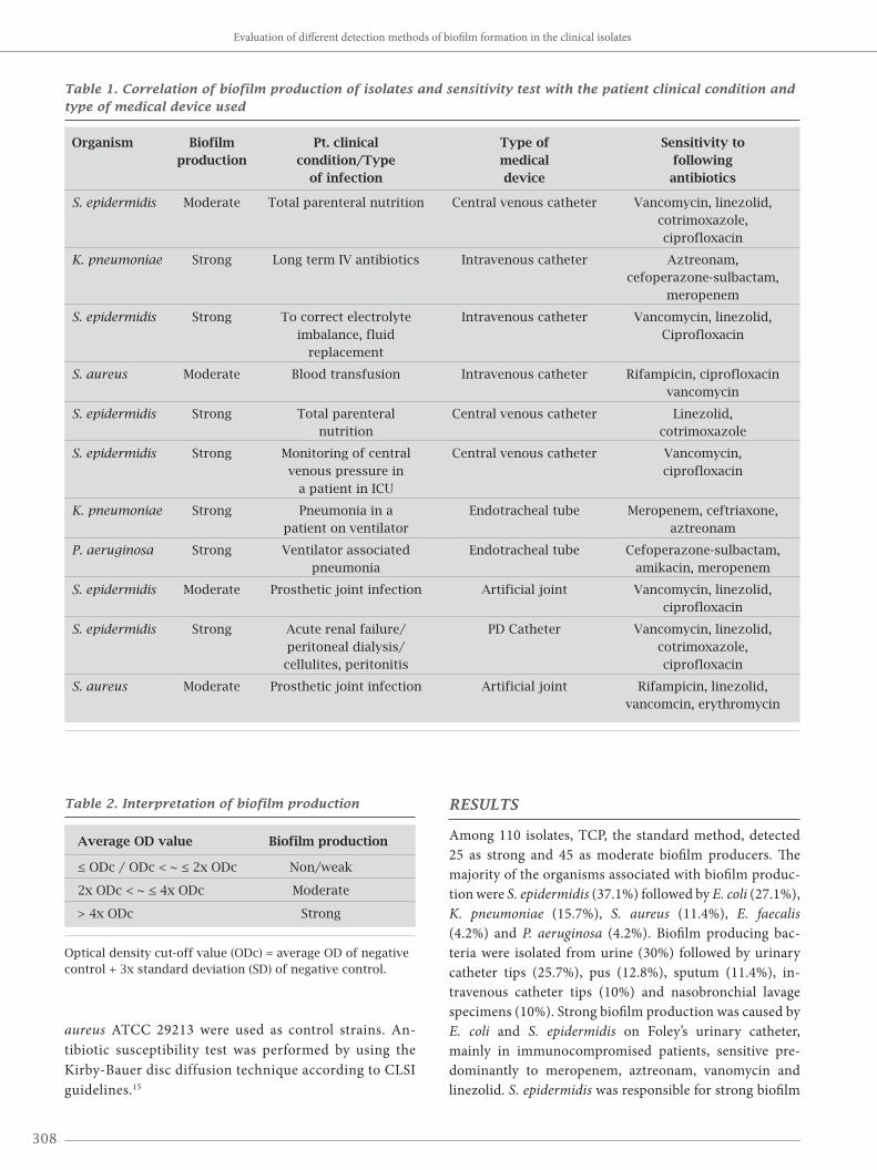
Table 1. Correlation of biofilm production of isolates and sensitivity test with the patient clinical condition and type of medical device used
Organism Biofilm Pt. clinical Type of Sensitivity to
production condition/Type medical following
of infection device antibiotics
E. coli Strong Urine retention due Foley’s urinary Amikacin, meropenem,
to stroke catheter cefoperazone-sulbactam
S. epidermidis Strong Care of a metastatic Urinary catheter Vancomycin,
terminal disease linezolid, ciprofloxacin
E. coli Strong For relief of bladder Foley’s urinary catheter Aztreonam, meropenem
outlet obstruction due
to urethral stricture
E. faecalis Moderate Neurogenic bladder Foley’s urinary catheter Vancomycin, linezolid,
cotrimoxazole
E. coli Strong Benign prostatic Foley’s urinary catheter Ciprofloxacin,
hypertrophy aztreonam, meropenem
E. coli Strong Post-operation to Foley’s urinary catheter Aztreonam, meropenem,
monitor output ciprofloxacin
S. epidermidis Strong Urologic surgery Foley’s urinary catheter Vancomycin,
linezolid, ciprofloxacin
K. pneumoniae Strong Urine retention due Foley’s urinary catheter Cotrimoxazole,
to calculus disease ciprofloxacin,
ceftriaxone, meropenem
E. coli Strong Urine retention due to Foley’s urinary catheter Amikacin,
cerebrovascular disease ceftriaxone, meropenem
K. pneumoniae Strong Hip fracture surgery Foley’s urinary catheter Ciprofloxacin,
ceftriaxone, meropenem
E. coli Strong Post-operation Foley’s urinary catheter Meropenem, amikacin,
to monitor output aztreonam, ceftriaxone
S. epidermidis Strong Care for traumatic Foley’s urinary catheter Vancomycin, linezolid,
spinal cord injury cotrimoxazole,
ciprofloxacin
S. epidermidis Moderate Neurogenic bladder Foley’s urinary catheter Linezolid, ciprofloxacin,
cotrimoxazole
E. coli Strong Total abdominal Foley’s urinary catheter Ceftriaxone, meropenem
hysterectomy surgery
S. epidermidis Moderate Bladder outlet Foley’s urinary catheter Vancomycin, linezolid,
obstruction due cotrimoxazole,
to urethral stricture ciprofloxacin
E. coli Strong Urologic surgery Foley’s urinary catheter Meropenem, ceftriaxone
E. coli Strong Benign prostatic Foley’s urinary catheter Ciprofloxacin, aztreonam,
hypertrophy meropenem
K. pneumoniae Strong Palliative care in a Foley’s urinary catheter Aztreonam, meropenem
incontinent impaired
patient
S. epidermidis Strong Monitoring of central Central venous catheter Linezolid, ciprofloxacin
venous pressure
in an acutely ill patient
(Cont.)
Hassan, Usman, Kaleem et al.
Braz J Infect Dis 2011; 15(4):305-311
BJID-4-JUN ARTE FINAL.indd 307 28/07/11 13:30
308
Table 1. Correlation of biofilm production of isolates and sensitivity test with the patient clinical condition and type of medical device used
Organism Biofilm Pt. clinical Type of Sensitivity to production condition/Type medical following of infection device antibiotics
S. epidermidis Moderate Total parenteral nutrition Central venous catheter Vancomycin, linezolid, cotrimoxazole, ciprofloxacin
K. pneumoniae Strong Long term IV antibiotics Intravenous catheter Aztreonam, cefoperazone-sulbactam, meropenem
S. epidermidis Strong To correct electrolyte Intravenous catheter Vancomycin, linezolid, imbalance, fluid Ciprofloxacin replacement
S. aureus Moderate Blood transfusion Intravenous catheter Rifampicin, ciprofloxacin vancomycin
S. epidermidis Strong Total parenteral Central venous catheter Linezolid, nutrition cotrimoxazole
S. epidermidis Strong Monitoring of central Central venous catheter Vancomycin, venous pressure in ciprofloxacin a patient in ICU
K. pneumoniae Strong Pneumonia in a Endotracheal tube Meropenem, ceftriaxone, patient on ventilator aztreonam
P. aeruginosa Strong Ventilator associated Endotracheal tube Cefoperazone-sulbactam, pneumonia amikacin, meropenem
S. epidermidis Moderate Prosthetic joint infection Artificial joint Vancomycin, linezolid, ciprofloxacin
S. epidermidis Strong Acute renal failure/ PD Catheter Vancomycin, linezolid, peritoneal dialysis/ cotrimoxazole, cellulites, peritonitis ciprofloxacin
S. aureus Moderate Prosthetic joint infection Artificial joint Rifampicin, linezolid, vancomcin, erythromycin
aureus ATCC 29213 were used as control strains. An-tibiotic susceptibility test was performed by using the Kirby-Bauer disc diffusion technique according to CLSI guidelines.15
RESULTS
Among 110 isolates, TCP, the standard method, detected 25 as strong and 45 as moderate biofilm producers. The majority of the organisms associated with biofilm produc-tion were S. epidermidis (37.1%) followed by E. coli (27.1%), K. pneumoniae (15.7%), S. aureus (11.4%), E. faecalis (4.2%) and P. aeruginosa (4.2%). Biofilm producing bac-teria were isolated from urine (30%) followed by urinary catheter tips (25.7%), pus (12.8%), sputum (11.4%), in-travenous catheter tips (10%) and nasobronchial lavage specimens (10%). Strong biofilm production was caused by E. coli and S. epidermidis on Foley’s urinary catheter, mainly in immunocompromised patients, sensitive pre-dominantly to meropenem, aztreonam, vanomycin and linezolid. S. epidermidis was responsible for strong biofilm
Evaluation of different detection methods of biofilm formation in the clinical isolates
Table 2. Interpretation of biofilm production
Average OD value Biofilm production
≤ ODc / ODc < ~ ≤ 2x ODc Non/weak
2x ODc < ~ ≤ 4x ODc Moderate
> 4x ODc Strong
Optical density cut-off value (ODc) = average OD of negative control + 3x standard deviation (SD) of negative control.
BJID-4-JUN ARTE FINAL.indd 308 28/07/11 13:30
309
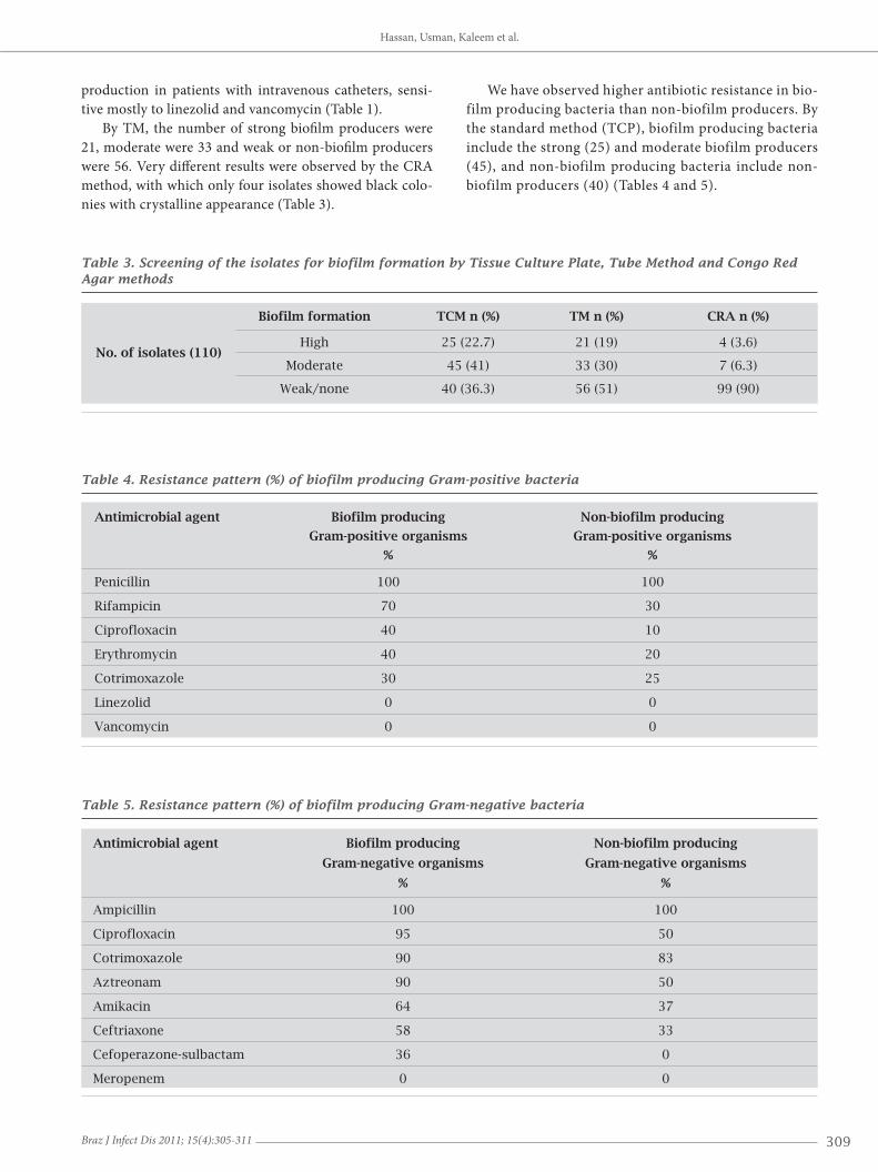
Table 3. Screening of the isolates for biofilm formation by Tissue Culture Plate, Tube Method and Congo Red Agar methods
Biofilm formation TCM n (%) TM n (%) CRA n (%)
No. of isolates (110) High 25 (22.7) 21 (19) 4 (3.6)
Moderate 45 (41) 33 (30) 7 (6.3)
Weak/none 40 (36.3) 56 (51) 99 (90)
Table 4. Resistance pattern (%) of biofilm producing Gram-positive bacteria
Antimicrobial agent Biofilm producing Non-biofilm producing
Gram-positive organisms Gram-positive organisms
% %
Penicillin 100 100
Rifampicin 70 30
Ciprofloxacin 40 10
Erythromycin 40 20
Cotrimoxazole 30 25
Linezolid 0 0
Vancomycin 0 0
Table 5. Resistance pattern (%) of biofilm producing Gram-negative bacteria
Antimicrobial agent Biofilm producing Non-biofilm producing
Gram-negative organisms Gram-negative organisms
% %
Ampicillin 100 100
Ciprofloxacin 95 50
Cotrimoxazole 90 83
Aztreonam 90 50
Amikacin 64 37
Ceftriaxone 58 33
Cefoperazone-sulbactam 36 0
Meropenem 0 0
production in patients with intravenous catheters, sensi-tive mostly to linezolid and vancomycin (Table 1).
By TM, the number of strong biofilm producers were 21, moderate were 33 and weak or non-biofilm producers were 56. Very different results were observed by the CRA method, with which only four isolates showed black colo-nies with crystalline appearance (Table 3).
We have observed higher antibiotic resistance in bio-film producing bacteria than non-biofilm producers. By the standard method (TCP), biofilm producing bacteria include the strong (25) and moderate biofilm producers (45), and non-biofilm producing bacteria include non-biofilm producers (40) (Tables 4 and 5).
Hassan, Usman, Kaleem et al.
Braz J Infect Dis 2011; 15(4):305-311
BJID-4-JUN ARTE FINAL.indd 309 28/07/11 13:30
310
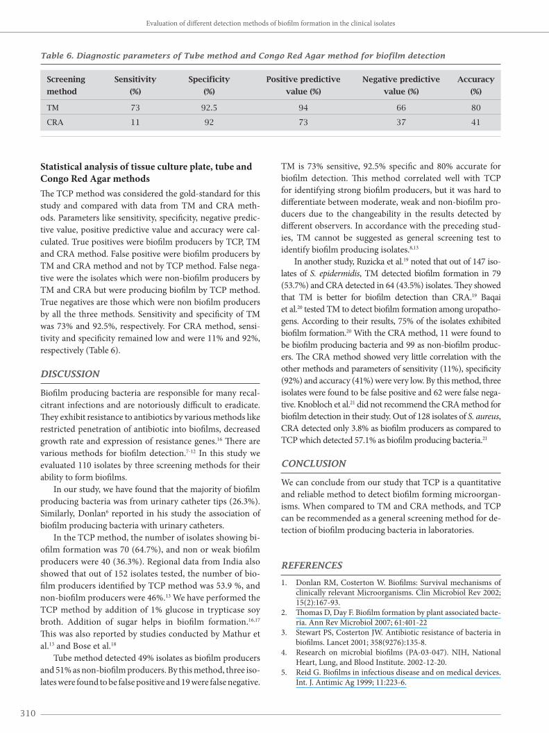
Statistical analysis of tissue culture plate, tube and Congo Red Agar methodsThe TCP method was considered the gold-standard for this study and compared with data from TM and CRA meth-ods. Parameters like sensitivity, specificity, negative predic-tive value, positive predictive value and accuracy were cal-culated. True positives were biofilm producers by TCP, TM and CRA method. False positive were biofilm producers by TM and CRA method and not by TCP method. False nega-tive were the isolates which were non-biofilm producers by TM and CRA but were producing biofilm by TCP method. True negatives are those which were non biofilm producers by all the three methods. Sensitivity and specificity of TM was 73% and 92.5%, respectively. For CRA method, sensi-tivity and specificity remained low and were 11% and 92%, respectively (Table 6).
DISCUSSION
Biofilm producing bacteria are responsible for many recal-citrant infections and are notoriously difficult to eradicate. They exhibit resistance to antibiotics by various methods like restricted penetration of antibiotic into biofilms, decreased growth rate and expression of resistance genes.16 There are various methods for biofilm detection.7-12 In this study we evaluated 110 isolates by three screening methods for their ability to form biofilms.
In our study, we have found that the majority of biofilm producing bacteria was from urinary catheter tips (26.3%). Similarly, Donlan6 reported in his study the association of biofilm producing bacteria with urinary catheters.
In the TCP method, the number of isolates showing bi-ofilm formation was 70 (64.7%), and non or weak biofilm producers were 40 (36.3%). Regional data from India also showed that out of 152 isolates tested, the number of bio-film producers identified by TCP method was 53.9 %, and non-biofilm producers were 46%.13 We have performed the TCP method by addition of 1% glucose in trypticase soy broth. Addition of sugar helps in biofilm formation.16,17 This was also reported by studies conducted by Mathur et al.13 and Bose et al.18
Tube method detected 49% isolates as biofilm producers and 51% as non-biofilm producers. By this method, three iso-lates were found to be false positive and 19 were false negative.
TM is 73% sensitive, 92.5% specific and 80% accurate for biofilm detection. This method correlated well with TCP for identifying strong biofilm producers, but it was hard to differentiate between moderate, weak and non-biofilm pro-ducers due to the changeability in the results detected by different observers. In accordance with the preceding stud-ies, TM cannot be suggested as general screening test to identify biofilm producing isolates.8,13
In another study, Ruzicka et al.19 noted that out of 147 iso-lates of S. epidermidis, TM detected biofilm formation in 79 (53.7%) and CRA detected in 64 (43.5%) isolates. They showed that TM is better for biofilm detection than CRA.19 Baqai et al.20 tested TM to detect biofilm formation among uropatho-gens. According to their results, 75% of the isolates exhibited biofilm formation.20 With the CRA method, 11 were found to be biofilm producing bacteria and 99 as non-biofilm produc-ers. The CRA method showed very little correlation with the other methods and parameters of sensitivity (11%), specificity (92%) and accuracy (41%) were very low. By this method, three isolates were found to be false positive and 62 were false nega-tive. Knobloch et al.21 did not recommend the CRA method for biofilm detection in their study. Out of 128 isolates of S. aureus, CRA detected only 3.8% as biofilm producers as compared to TCP which detected 57.1% as biofilm producing bacteria.21
CONCLUSION
We can conclude from our study that TCP is a quantitative and reliable method to detect biofilm forming microorgan-isms. When compared to TM and CRA methods, and TCP can be recommended as a general screening method for de-tection of biofilm producing bacteria in laboratories.
REFERENCES
1. Donlan RM, Costerton W. Biofilms: Survival mechanisms of clinically relevant Microorganisms. Clin Microbiol Rev 2002; 15(2):167-93.
2. Thomas D, Day F. Biofilm formation by plant associated bacte-ria. Ann Rev Microbiol 2007; 61:401-22
3. Stewart PS, Costerton JW. Antibiotic resistance of bacteria in biofilms. Lancet 2001; 358(9276):135-8.
4. Research on microbial biofilms (PA-03-047). NIH, National Heart, Lung, and Blood Institute. 2002-12-20.
5. Reid G. Biofilms in infectious disease and on medical devices. Int. J. Antimic Ag 1999; 11:223-6.
Table 6. Diagnostic parameters of Tube method and Congo Red Agar method for biofilm detection
Screening Sensitivity Specificity Positive predictive Negative predictive Accuracy
method (%) (%) value (%) value (%) (%)
TM 73 92.5 94 66 80
CRA 11 92 73 37 41
Evaluation of different detection methods of biofilm formation in the clinical isolates
BJID-4-JUN ARTE FINAL.indd 310 28/07/11 13:30
311
6. Donlan RM. Biofilms and device-associated infections. Emerg Infect Dis 2001; 7(2): 277-81.
7. Christensen GD, Simpson WA, Younger JA et al. Adherence of coagulase negative Staphylococci to plastic tissue cultures: a quantitative model for the adherence of Staphylococci to medical devices. J Clin Microbiol 1995;22:996-1006.
8. Christensen GD, Simpson WA, Bisno AL, Beachey EH. Adher-ence of slime producing strains of Staphylococcus epidermidis to smooth surfaces. Infect Immun1982; 37:318-26.
9. Freeman J, Falkiner FR, Keane CT. New method for detecting slime production by coagulase negative staphylococci. J Clin Pathol 1989; 42:872-4.
10. Donlan RM, Murga R, Bell M et al. Protocol for detection of biofilms on needleless connectors attached to central venous catheters. J Clin Microbiol 2001; 39:750-3.
11. Aparna MS, Yadav S. Biofilms: microbes and disease. Braz J Infect Dis 2008; 12(6):526-30.
12. Zufferey J, Rime B, Francioli P, Bille J. Simple method for rapid diagnosis of catheter associated infection by direct Acridine or-ange staining of catheter tips. J Clin Microbiol 1988; 26:175-7.
13. Mathur T, Singhal S, Khan S, Upadhyay DJ, Fatma T, Rattan A. Detection of biofilm formation among the clinical isolates of staphylococci: an evaluation of three different screening meth-ods. Indian J Med Microbiol 2006; 24(1) :25-9.
14. Stepanovic S, Vukovi D, Hola V et al. Quantification of biofilm in microtiter plates: overview of testing conditions and practi-cal recommendations for assessment of biofilm production by Staphylococci. APMIS. 2007 115:891-9.
15. Bauer AW, Kirby M M, Sherris JC, Jurek M. Antibiotic sus-ceptibility testing by a standardized single method. Am J Clin Pathol 1966; 45:493-6.
16. Kim L. Riddle of biofilm resistance. Antimic Ag Chemother 2001; 45(4):999-1007.
17. Eftikhar F, Speert DP. Biofilm formation by persistent and non-persistent isolates of Staphylococcus epidermidis from a neonatal intensive care unit. J Hosp Infect 2009; 71(2):112-6.
18. Bose S, Khodke M, Basak S, Mallick SK. Detection of biofilm producing staphylococci: need of the hour. J Clin Diagn Res 2009; 3:1915-20.
19. Ruzicka F, Hola V, Votava M et al. Biofilm detection and clini-cal significance of Staphylococcus epidermidis isolates. Folia Microbiol (Praha) 2004; 49(5):596-600.
20. Baqai R, Aziz M, Rasool G. Urinary tract infection in diabetic patients and biofilm formation of uropathogens. Infect Dis J Pakistan. 2008 17(1):7-9.
21. Knobloch JK, Horsetkotte MA, Rohde H, Mack D. Evaluation of different detection methods of biolfilm formation in Staphy-lococcus aureus. Med Microbial Immunol 2002; 191(2):101-6.
Hassan, Usman, Kaleem et al.
Braz J Infect Dis 2011; 15(4):305-311
BJID-4-JUN ARTE FINAL.indd 311 28/07/11 13:30