Lake Trout (Salvelinus namaycush) Populations in Lake Superior and Their Restoration in 1959–1993
Upload
independentCategory
view
1download
0
Comparative Biochemistry and Physiology, Part B 175 (2014) 1–8
Contents lists available at ScienceDirect
Comparative Biochemistry and Physiology, Part B
j ourna l homepage: www.e lsev ie r .com/ locate /cbpb
Evaluation of chitinolytic activities andmembrane integrity in gut tissuesof Arctic charr (Salvelinus alpinus) fed fish meal and zygomycete biomass
Rani Abro a, Kristina Sundell b, Erik Sandblom b, Henrik Sundh b, Eva Brännäs c, Anders Kiessling a,Jan Erik Lindberg a, Torbjörn Lundh a,⁎a Department of Animal Nutrition and Management, Swedish University of Agricultural Sciences, P.O. Box 7024, S-75007 Uppsala, Swedenb Department of Biological and Environmental Sciences, University of Gothenburg, P.O. Box 463, S-40530 Gothenburg, Swedenc Department of Wildlife, Fish and Environmental Studies, Swedish University of Agricultural Sciences, S-90 83 Umeå, Sweden
Abbreviations: GlcN, glucosamine; NAG, N-acetylp-nitrophenyl N-acetyl-β-D-glucose aminide; DMAB, p-FM, fish meal; FZ, fish meal and zygomycetes; GI, gaparacellular permeability; TER, transepithelial resistance.⁎ Corresponding author. Tel.: +46 18 672137; fax: +4
E-mail address: [email protected] (T. Lundh).
http://dx.doi.org/10.1016/j.cbpb.2014.06.0031096-4959/© 2014 Elsevier Inc. All rights reserved.
a b s t r a c t
a r t i c l e i n f oArticle history:Received 2 December 2013Received in revised form 19 May 2014Accepted 6 June 2014Available online 17 June 2014
Keywords:ChitinZygomycetesIntestinal barrier functionsAmino acid absorptionPlasma cortisol
Chitinolytic activity, nutrient uptake and intestinal barrier functions were investigated in gut tissues of Arcticcharr (Salvelinus alpinus) fed iso-nitrogenous diets based on fish meal, with or without inclusion of zygomycetes(Rhizopus oryzae). We found that gut tissue of Arctic charr had significant chitinase activity, of both endo- andexo-chitinase iso-forms. Moreover, the distribution pattern along the GI tract of Arctic charr differed betweenendo-chitinase and exo-chitinase. The endo-chitinase activity in stomach tissue and in the distal intestine wasseveral hundred-fold higher than the exo-chitinase activity in stomach tissue. The greatest exo-chitinase activitywas found in the distal intestine. The zygomycete-based diet resulted in higher chitinolytic activity in gut tissuecompared to the fish meal-based diet. Disturbed intestinal integrity and increased uptake rate of the amino acidlysine were observed in the distal, but not proximal, intestine of fish fed the zygomycete-based feed.
© 2014 Elsevier Inc. All rights reserved.
1. Introduction
Farming of Arctic charr (Salvelinus alpinus) is increasing in theNordic countries, including Sweden. This is primarily due to increasedconsumer demand, the rapid development of culture procedures andthe ability of this fish species to grow at low temperatures (BrännäsandWiklund, 1992; Dick and Yang, 1992). The Arctic charr is a carnivo-rous salmonid fish with similar protein requirement as Atlantic salmon(Salmo salar) (Klemetsen et al., 2000). Diets for Arctic charr have there-fore traditionally been similar in composition to salmon feed. Thus,while fish meal has been an important protein component of suchdiets, alternative ingredients to fish meal are now required to establishmore sustainable fish farming systems and to reduce feed costs(Naylor et al., 2000; Kristofersson and Anderson, 2006).
Zygomycetes (Rhizopus oryzae) is a microfungal species that is richin protein (40–50% depending on the culture conditions) and has anamino acid profile similar to that of fish meal (Edebo, 2008; Bankeforset al., 2011; Van Leeuwen et al., 2012). Thus, zygomycete biomass
-D-glucosamine; pNP-GlcNAc,dimethyl aminobenzaldehyde;strointestinal; Papp, apparent
6 18 672995.
may have great potential to replace fish meal protein in fish feed(Edebo, 2008; Ferreira et al., 2012). Zygomycetes have been widelyused for food production (i.e. tempe), as well as for the extraction oforganic substances such as lactic acid, fumaric acid and extracellularenzymes (amylases, cellulases, proteases, lipases) (Sues et al., 2005;Dijksterhuis and Samson, 2006). In addition to proteins, zygomycetescontain large but varying quantities of chitosan (glucosamine polymers)and chitin (N-acetylglucosamine polymers). The variability in chitosanand chitin content (45–85%) is dependent on fungal strain and cultivationconditions (Hjorth, 2005; Edebo, 2008).
Chitin, a heteropolysaccharide composed of the hexosamineβ-(1–4)-linked N-acetylglucosamine molecules, is the second most abundantcompound after cellulose in living organisms world-wide. It is essentialas a supportive material in the exo-skeleton of insects and aquaticinvertebrates (Lindsay et al., 1984). In several fish species, dietary sup-plementation with chitin influences growth and nutrient digestibility,but the results are not unambiguous and differ among species (Ringoet al., 2012). For example, cyprinids do not seem to be affected by lowinclusions of chitin (about 2%), but higher concentrations reduce growthand feed digestibility in tilapia (Shiau, andYu, 1999). Even so, salmonidslike Atlantic salmon, chum salmon (Oncorhynchus keta) and rainbowtrout (Oncorhynchus mykiss) seem to have a low ability to utilise chitin,with reduced growth performancewhen fish meal is replaced with krillmeal containing a high chitin content (Ringo et al., 2012). Althoughadult rainbow trout are unable to digest chitin (Buddington, 1980), it

Table 1Composition and proximate analysis of the fishmeal diet (FM) and zygomycete diet (FZ)(% kg−1 dry mass).
Diet
FM FZ
IngredientsFish meal 58.0 23.0Fish oil 13.0 13.0Zygomycete 00.0 48.0Rapeseed oil 7.0 7.0Sesame oil 1.0 1.0Wheat 8.0 0.0Alginate 5.0 0.0Gelatine 6.5 6.5Mineral/vitamin mix 1.0 1.0Titanium dioxide 0.5 0.5
Proximate analysisCrude protein 49.0 46.7Crude fat 25.5 27.3Ash 12.7 9.5
2 R. Abro et al. / Comparative Biochemistry and Physiology, Part B 175 (2014) 1–8
has been reported that the inclusion of 6% chitin enhances growth inrainbow trout juveniles (Lellis and Barrows, 2000). To the best of ourknowledge, the ability of Arctic charr to hydrolyse chitin and utilisethe degradation products as a nutrient supply has not been reported todate.
The chitinolytic enzymes, chitinase (EC.3.2.1.14) and chitobiase(EC.3.2.1.30) are responsible for the biological degradation of chitin.These enzymes comprise two main groups, i.e. the endo-chitinases(chitinase), which are involved in the random catalytic breakdown ofchitin to produce chitin-oligosaccharides, and the exo-chitinases(chitobiase), which cause the cleavage of chitin-oligosaccharides intomonomers of N-acetylglucosamine (NAG) and glucosamine (GlcN)(Cohen-Kupiec and Chet, 1998; Kang et al., 1999; Filho et al., 2002).Lysozyme also exerts chitinolytic activity in the gut, but is of lesserimportance (Fange et al., 1979).
The enzymatic actions of chitinase and chitobiase are mainly associ-ated with the alimentary tract of the fish (Clark et al., 1988; Jeuniaux,1993). Chitinolytic activity has been reported in the gastric mucosa,intestinal mucosa, pyloric caeca, pancreas, liver and lymphomyeloidtissues of the followingfish species: Anoplopoma fimbria, Coryphaenoidesarmatus, Tarletonebeania crenularis, Sebastes chlorostictus, Microstomuspacificus, Rachycentron canadum and Lympanyctus ritterii (Gutowskaet al., 2004; Krogdahl et al., 2005; Fines and Holt, 2010).
The intestine of fish is vital not only for food digestion and nutrientuptake (Sundell and Rønnestad, 2011), but also for regulation of ionand water balance (Sundell and Sundh, 2012). As it is continuouslyexposed to feed and the surrounding water, the intestinal epitheliumis faced with the challenges of selectively absorbing nutrients, vitaminsand minerals, while at the same time efficiently preventing passage ofharmful molecules and organisms (Segner et al., 2012). This defencemechanism is referred to as the gastrointestinal barrier (Baumgart andDignass, 2002), which consists of three major parts: the extrinsic, theintrinsic and the immunological barrier. Of these, the extrinsic barrierconstitutes the secretion ofmucus and bicarbonate aswell as the endog-enous microflora, while the intrinsic barrier constitutes the epithelialcell monolayer and the tight junction complexes. Together, these twoparts create the primary physical barrier between the lumen and thecirculation.
The gastrointestinal tract of salmonids, as of other fish, is a majorroute of infection for several pathogens (Jutfelt et al., 2006, 2008;Sundh et al., 2009, 2011). Therefore, the maintenance of high integrityof the primary barriers is required for farming of healthy fish withminimised disease susceptibility. The intestinal primary barrier is ofparticular interest for possible effects of new feed ingredients, as it isthe first tissue to be exposed. Harmful feed ingredients can result inadverse effects on nutrient uptake, intestinal barrier integrity and/orlocal inflammation, which may lead to increased risk of infection anddisease susceptibility (Jutfelt et al., 2007; Knudsen et al., 2008).
For example, replacing dietary fish meal with soybean meal influ-ences the immune response, feed intake, digestibility, feed conversion,growth and enzyme activity in Atlantic salmon (Krogdahl et al., 2003).It has also been reported that dietary soya saponins increase gut perme-ability and are involved in soybean-induced enteritis in Atlantic salmon(Knudsen et al., 2008). However, new feed ingredients as alternatives tosoybean could also have positive effects on intestinal barrier function.Feed ingredients rich in chitosan and chitin, built up by hexosamines,are particularly interesting from a pre-biotic viewpoint. The extrinsicmucus barrier covering the epithelial layers consists ofmucins, a hetero-geneous group of large glycoproteins containing O-linked carbohy-drates (Linden et al., 2008). A diet rich in hexosamines, like NAG, maythus serve as building blocks formucin synthesis and is hence a possiblecandidate for a strengthening of the extrinsic barrier and thereby im-proving intestinal integrity. In fact, chitin oligosaccharides have beenshown to enhance the barrier function of the epithelial cell membraneand induce the secretion of mucin in intestinal epithelial mucosal tissuein humans and pigs (Deters et al., 2008).
Themain objective of the present studywas to investigate the abilityof Arctic charr to utilise chitin/chitosan-rich zygomycete diet, bymeasuring chitinolytic activity and thereby the possibility to utilisethe liberated GlcN and NAG as an energy substrate. A further objectivewas to study the effect of inclusion of zygomycetes on intestinal primarybarrier functions since the chitosan molecules may interact with theintestinal barrier system.
2. Materials and methods
2.1. Fish rearing
A total of 120 Arctic charr (S. alpinus) fingerlings with a mean massof 105± 0.5 gwere collected from the production tanks and distributedevenly among six 1m3
fibreglass tanks. Two diets (see Section 2.2)wereformulated and fed to groups of 20 fish each in triplicate tanks (thewater temperature was maintained at 6 ± 0.5 °C. The fish acceptedthe experimental diets, and no mortality was observed during theexperimental period. Fish in each tank were weighed separately in thebeginning and at the end of the experiment, although individual fishwere not marked. The experiment was approved by the EthicalCommittee for Animal Experiments in the Umeå region.
2.2. Diets
Two iso-nitrogenous diets based on fish meal (FM) and zygomycetebiomass (FZ) diets were formulated (Table 1). Zygomycetes werecultured on spent sulphite liquor, a by-product from sulphite pulpmills, and the microbial biomass was harvested, washed and driedbefore formulation, as reported by Bankefors et al. (2011). The twoexperimental diets were hand-made using a meat grinder, and the feedwas chopped to a pellet size of 2 mm. The fish were fed continuouslyby using band feeders with a total daily allowance of 2% of the initialbody weight for the four-week duration of the experiment. No equip-ment for measuring uneaten feed was available, but visual inspectionsuggested that the feed was accepted by the fish.
2.3. Chemical analysis
Samples of feed ingredients and diets were analysed with standardmethods AOAC (1997). Dry matter was determined by drying in anoven at 105 °C for 24 h. Nitrogen (N) was determined by the Kjeldahlmethod, and crude protein (CP) was calculated as N × 6.25. Crude fat(EE) contentwas analysed using the Soxhletmethod after acid hydrolysisof the sample. Crude fibre (CF) content was determined by the standardmethod AOAC (1997). Ash content was determined by incineration in a
3R. Abro et al. / Comparative Biochemistry and Physiology, Part B 175 (2014) 1–8
muffle furnace at 550 °C for 12 h. Gross energy content (MJ kg−1) wasdetermined with a bomb calorimeter (Calorimeter Parr 6300, Parr In-strument Company, Moline, IL, USA).
2.4. Determination of N-acetylglucosamine and glucosamine in experimentaldiets
NAG and GlcN are building blocks of the chitin and chitosanmolecules and can therefore be used as a measure of dietary chitinand chitosan content, respectively. The method used for GlcN andNAG analysis was reported previously by Zamani et al. (2008) and istherefore described only briefly. Chitin and chitosan are not soluble inalkali; FM and FZ samples were therefore incubated in 0.5 M NaOHovernight at 90 °C. The alkali insoluble material (AIM) was separatedby centrifugation and washed with distilled water. The AIMwas hydro-lysed by a two-step method with concentrated sulphuric acid at roomtemperature for 90 min and with diluted sulphuric acid at 120 °C for60 min, followed by degradation with nitrous acid. For measuringNAG (representing chitin) the concentration of acetic acid in coldsulphuric acid hydrolyzates wasmeasured by HPLC. GlcN (representingchitosan) were measured colorimetrically after the addition of 3-methyl-2-benzothiozolone-hydrazone-hydrochloride and FeCl3, whichcreates a blue colour complex with anhydromannose (formed afternitric acid treatment).
2.5. Sample preparation
Fishwere sampled during four consecutive days. On days 1–3, a totalof 12 fish from each dietary treatment (four fish per tank, or eight fishper sampling occasion) were sampled for their live intestines, foranalyses of intestinal nutrient transport and barrier functions. On day4, the 16 remaining fish per tank were sampled for collection of blood,for cortisol analyses, and gastrointestinal tissues, and for enzymeanalysis.
2.5.1. Sampling of live intestineThe live intestine samples for in vitro intestinal physiology assess-
ment were obtained using an Ussing chamber set-up (Sundell et al.,2003; Sundell and Sundh, 2012). The four fish from each tank wererandomly sampled inonequickdipnetting and immediately anesthetisedwith Tricane methane sulphonate (MS-222) solution (50 mg L−1). Theanesthetised fish were then killed with a sharp blow to the head, thebody cavity was opened and the mesenteries and adipose tissue wereremoved using blunt dissection. The intestine from the last pyloriccaecum to the anus was carefully removed and opened longitudinally.The intestine was washed in ice-cold Ringer solution for FW-adaptedAtlantic salmon (140 mM NaCl, 2.5 mM KCl, 1.5 mM CaCl2, 0.8 MgSO4,10 mM glucose, 20 mM glutamine, 5 mM HEPES and pH set to 7.8with 1.5 M TRIS-base), and divided into a proximal and a distal part atthe level of the ileorectal valve. Both intestinal segments were pinnedto a temperature-controlled stripping board, mucosa side down, andthe serosa and parts of the muscular layers were peeled off usingblunt dissection. The peeling procedure maximised oxygen availabilityto the intestinal epithelium and minimised translocation distance forsubstances during the experiment. The peeled intestinal segmentswere briefly stored on ice in Ringer solution, before being mounted inthe Ussing chambers (see Section 2.9 below).
2.5.2. Sampling for blood and gastrointestinal enzyme analysisFishwere rapidly sampled from the tanks by one quick dip netting of
an excess number of fish. From these, nine fish were randomly selectedand immediately anesthetised with Tricane methane sulphonate(MS-222) solution (50mgL−1). The anesthetisedfishwere then quicklykilled with a sharp blow to the head, and blood samples were with-drawn from the caudal vessels using a heparinised syringe and needle.The blood was centrifuged at 7000 g, and the plasma was transferred
to new tubes, snap-frozen in liquid N2 and stored at −80 °C until fur-ther analysis. Thereafter the fish were cut open and the gastrointestinaltract was separated into stomach, pyloric caeca, and proximal and distalintestines. The tissue sampleswere immediately frozen in liquidnitrogen,transferred to a−80 °C freezer and kept until sample preparation. Thesamples were thawed, weighed and kept on ice. The tissues weresuspended in extraction buffer (1:20 w/v) containing malic acid(1 M), sodium hydroxide, sodium chloride, calcium chloride (40 mM)and sodium azide (0.1%). Samples were homogenised using an ultraturrax tube disperser (IKA, Germany), dispersed 3 × 30 s, followed bycentrifugation at 15,800 g for 10 min. The supernatant was used forenzyme activity measurement. The different dilutions of the sampleswere prepared with phosphate extraction buffer, and chitinolyticactivity was analysed.
2.6. Assays of chitinolytic activity
The chitinolytic (endo- and exo-chitinase) activity of gastrointestinalsamples was determined using two different substrates. Chitin fromcrab shell (Sigma # C9752) was used as substrate to assess endo-chitinase activity, and 4-nitrophenyl N-acetyl-β-D-glucose aminide(pNP-GlcNAc) (Sigma# N9376) was used as substrate to assess exo-chitinase activity.
Endo-chitinase activity was determined using a modification of theprocedure described by Jeuniaux (1966) and Gutowska et al. (2004),for use on a microtitre plate reader. Duplicate homogenate solutionswere used for measuring the NAG production and for blank assays.The test assay solution contained 250 μL supernatant from the centri-fuged homogenate, 250 μL chitin suspension and 250 μL distilledwater. In order to determine the total chitinolytic activity, one assayaliquot contained 250 μL β-glucosidase (6 units per mL) instead ofdistilled water as described above. The glass tubes containing assaysolution were placed on a rotary shaker for incubation at 20 °C for 2 h.Thereafter, the tubes with the assay solution were boiled for 10 min tostop the reaction, cooled to room temperature, and centrifuged for30 min at 13,600 g. A sample of the supernatant (0.5 mL) was trans-ferred to a new tube containing 0.1 mL 0.8 M borate buffer (K2B4O7)to maintain pH 9.8 in the solution. The solution was boiled for 3 minand immediately cooled in tap water. A 3 mL aliquot of p-dimethyl-amino-benzaldehyde (DMAB) solution was added, and after mixingthe solution was incubated at 37 °C for 20 min.
The formation of NAG was measured at 550 nm at room tempera-ture on amicrotitre plate reader (Multiskan FC; Thermo Fisher ScientificInc., IL, USA). The absorbance values obtained from chitinolytic activitywere calculated by using a standard curve of NAG (Sigma# G7274)with a concentration of 5–200 μM. The NAG concentration of standardand blank aliquots was measured as described by Reissig et al. (1955),with the modification of addition of chitin from crab shell suspensionand water as blank assay. For calculations, the software Skan It 3.1(Thermo Fisher Scientific Inc., IL, USA) was used. Chitinolytic activitywas expressed in μg NAG g−1 wet tissue h−1.
2.7. Plasma cortisol analysis
Plasma cortisol levelsweremeasured in un-extracted plasma using aradio immunoassay (RIA), a procedure originally described by Young(1986) and modified and validated by Sundh et al. (2011). The cortisolantibody (Code: S020; Lot: 1014-180182)was purchased fromGuildhayLtd. (Guildford, Surrey, UK) and validated for cross-reactivity withcortisone and corticosterone, as well as for inter- and intra-assay varia-tion for cortisol, as described by Sundh et al. (2011). For the determina-tion of radioactivity in the samples, a β-counter (Wallac 1409 LiquidScintillation Counter, Turku, Finland) was used. The intra- and inter-assay coefficients of variation for cortisol were 3.9% and 5.4%,respectively. The detection limit was 0.8 ng mL−1.

Table 2Analysis of alkali-insoluble material (AIM), glucosamine (GlcN) and N-acetyl glucosamine(NAG) in the fish meal diet (FM) and zygomycete diet (FZ), expressed as mg g−1 DM.
Diet
FM FZ
Alkali-insoluble material AIM/sample 155 104Glucosamine (chitosan) GlcN/AIM 12 63
GlcN/sample 1.9 6.6N-acetyl glucosamine (chitin) NAG/AIM 49 163
NAG/sample 8.0 17.0
4 R. Abro et al. / Comparative Biochemistry and Physiology, Part B 175 (2014) 1–8
2.8. Total protein concentration
Total protein concentration in gut tissues was measured using aprotein assay kit (Micro BCA™, Pierce, Rockford, IL, USA) with bovineserum albumin as standard, according to the manufacturer'sinstructions.
2.9. Ussing chamber experiment
Electrical characteristics of the intestinal epithelium, in combinationwith transfer rates of the hydrophilic marker molecule 14C-mannitoland the radioactively labelled amino acid 3H-Lysine across the intestinalepithelial segments, weremeasured using a set of custom-made Ussingchambers (Sundell et al., 2003). In brief, the peeled intestinal segmentswere mounted in Ussing chambers and electrical characteristics of thetissues; i.e. transepithelial potential (TEP), transepithelial resistance(TER) and short circuit current (SCC), were monitored every 5 min. Anew Ussing chamber measurement system was used in the presentstudy, the UCC-401 (UCC-Labs Ltd., Göteborg Sweden). This system isspecifically developed for fish intestines with low TEP as described bySundell and Sundh (2012). The method applies alternating adaptiveDC voltage (U) to the epithelium, generating corresponding currents(I). The U/I pairs obtained are then fitted to a straight line using theleast squares method. The slope of the line represents the TER and thevoltage where it intercepts U = 0 shows the SCC. Undisturbed TEPwas measured continuously throughout the experiment. The intestinalarea of exposure was 0.75 cm2, and oxygenation and stirring wasensured by an air-lift (compressed air) on both sides of the intestinalsegments. The temperature in the Ussing chambers was kept at thefish acclimation temperature by the use of cooling mantles.
After mounting the intestinal segments they were left for 60 min toallow stabilisation of the electrical parameters. Thereafter, the experi-ment was started by renewing the Ringer solution on the serosal side,and replacing the Ringer solution on the mucosal side with Ringercontaining the hydrophilic marker molecule 14C-mannitol (specificactivity, 0.042 MBq mL−1) and 3H-lysine in combination with coldlysine at a concentration of 500 μMin thefinal Ringer solution, renderinga specific activity of 2 × 10⁸ MBq mol−1. A 50 μL portion of the serosalRinger was sampled at time points 0, 20, 30, 60, 80 and 90 min. Radio-activity was assessed in a liquid scintillation counter (Wallac 1409Liquid Scintillation Counter, Turku, Finland) after adding5mLOptiphaseHigh Safe III (Wallac, Turku, Finland).
The rate of accumulation of 14C-mannitol on the serosal side wasused to assess the apparent paracellular permeability (Papp; Eq. (1)) ofthe intestinal segments, as 14C-mannitol is an inert and hydrophilicmolecule mainly translocating the epithelium through the paracellularpathway:
Papp ¼ dQ=dt � 1=ACo ð1Þ
where dQ/dt is the appearance rate of 14C-mannitol in the serosal com-partment (mol s−1) of the Ussing chamber, A is the area of intestinalsurface exposed in the chamber (0.75 cm2) and Co is the initial concen-tration on the mucosal side (mol mL−1).
The accumulation rate of 3H-lysine in relation to the specific activitywas used to calculate the total amount of lysine transported from themucosal to the serosal side of the epithelium, i.e. the lysine absorption.
2.10. Statistical analysis
Initial and final fish weights were analysed using a three-factorialanalysis of variance (ANOVA) in a general linear model (GLM) (Minitab12, State College PA, USA), with diet and time as fixed factors and thefactor tank nested within diet. Chitinolytic activity and Ussing chamberdata were analysed using three-factorial analyses of variance (ANOVA)in a general linear model (GLM) (SAS, Cary NC, USA). Normality and
homogeneity of variance were tested for the residuals and predictedvalues in the GLM model used. Data not passing normality and/orhomogeneity tests were transformed (sqrt or log10 transformationwas sufficient in all cases). The model used for chitinolytic activity andUssing chamber data included diet and GI regions as fixed factors andthe factor tank nested within diet. Significant interactions betweenfactors were investigated using Bonferroni corrected multiple pair-wise comparisons as post hoc tests. Plasma cortisol levelswere subjectedto a standard t-test (IBM SPSS 20, NY, US). Data are presented asmeans ± SEM unless otherwise indicated, and p b 0.05 was consideredstatistically significant.
3. Results
3.1. Diet chitin analysis
The chitin content in diets FM and FZ was 1.9 and 6.6 mg g−1,respectively and the chitosan content was 8.0 and 17.0 mg g−1
respectively. The results are expressed as the content of GlcN and NAG(Table 2).
3.2. General
The fish increased in weight by 15 ± 3% (mean ± SD) during the4-week experimental period. No difference was found in fish growthbetween diets (interaction between diet and time, p = 0.592). Therewas no difference between the fish tanks (interaction between tankand time, p = 0.766), although one tank containing the FM diet startedwith fish on average 13% larger than in the other five tanks (mean110.2 g versus 97.1 ± 11.6 g; mean ± SD).
3.3. Plasma cortisol levels
The plasma cortisol levels were generally low; 2.8± 0. 6 ngmL−1 inthe group fed FM diet and 6.1 ± 2.2 ng mL−1 in the group fed FZ diet.There was no difference (p = 0.136) in plasma cortisol levels betweendiets.
3.4. Endo-chitinase activity
Endo-chitinase activity in gut samples associated with feedingdiets FM and FZ is shown in Fig. 1A. There was a difference betweenGI regions, with significantly higher endo-chitinase activity in thestomach compared with the other gut tissues (p b 0.001). Most impor-tantly, there was also an interaction between diet and GI regions(p b 0.001). The highest endo-chitinase activity in fish fed FM diet wasobserved in the stomach tissue (1288 μg NAG g−1 wet tissue h−1),which was almost 2.5 times higher than for fish fed FZ diet(478 μg NAG g−1 wet tissue h−1). The lowest activity was found inthe proximal tissue (7 μg NAG g−1 wet tissue h−1) in fish fed FM diet,which was approximately 20 times lower than in fish fed FZ diet(155 μg NAG g−1 wet tissue h−1). In the fish fed FZ diet the endo-chitinase activity showed a different pattern with the highest activity

Stomach
Pyloric
caec
a
Proxim
alDist
al1
10
100
1000
10000
Endo-chitinase activity
GI region
µg N
AG
g-1
h-1
aAB
b B
b
AB
b A
Diet p>0.05GI region p<0.001Diet * GI region p<0.001
*
A
Stomach
Pyloric
caec
a
Proxim
alDist
al0.1
1
10
100
1000
Exo-chitinase activity
GI region
µg N
AG
g-1
h-1
Diet p>0.05GI region p<0.001Diet * GI region p>0.05
B
Fig. 1. Chinolytic activity in gut tissues of Arctic charr fed a fish meal-based (white bars) and a zygomycete-based (grey bars) diet was assessed as μg NAG formed per g wet tissue h−1,endo-chitinase activity (A) measured using chitin from shrimp shell as substrate and exo-chitinase activity (B), measured using synthetic substrate 4-nitrophenyl N-acetyl-β-D-glucoseaminide. Overall effects are indicated as text in the diagram. When the interaction between diet and GI regions was significant, the dataset was subjected to post-hoc analysis and lettersabove bars indicate statistical relationship. Lower case letters indicate differences between tissues within fish meal-based diet. Upper case letters indicate differences between tissueswithin zygomycete-based diet. Asterisk (*) indicates a difference between diets within GI regions. Results are presented as mean ± SEM (n = 9). Note the logarithmic scale.
5R. Abro et al. / Comparative Biochemistry and Physiology, Part B 175 (2014) 1–8
in the distal part, followed by stomach, proximal and pyloric caeca indescending order.
Overall, the specific endo-chitinase activity showed a similar patternto the chitinase activity in wet tissue samples. The addition ofβ-glucosidase had no effect on the endo-chitinase activity.
3.5. Exo-chitinase activity
Exo-chitinase activity in gut samples associated with feedingdiets FM and FZ is shown in Fig. 1B. The activity is expressed as μgN-acetyl glucose amine monomers formed from the substrate 4-nitrophenyl N-acetyl-β-D-glucose aminide per gram wet tissue andhour (NAG g−1 wet tissue h−1), in order to be comparable with earlierpublished reports.
There was no difference (p N 0.05) in exo-chitinase activity betweendiets. However, there was a difference between gastrointestinal (GI)regions, with higher exo-chitinase activity in the distal intestinecompared with the stomach, pylorus and proximal intestine (Fig. 1B;p b 0.001). There was a tendency for an interaction between diet andGI regions (P = 0.061), suggesting that the two diets tested mighthave produced different effects in different parts of the GI tract. Thehighest exo-chitinase activity was observed in the distal intestine withan activity of 436 μg NAG g−1 wet tissue h−1 in fish fed diet FM, and525 μg NAG g−1 wet tissue h−1 in fish fed diet FZ, whereas the lowest
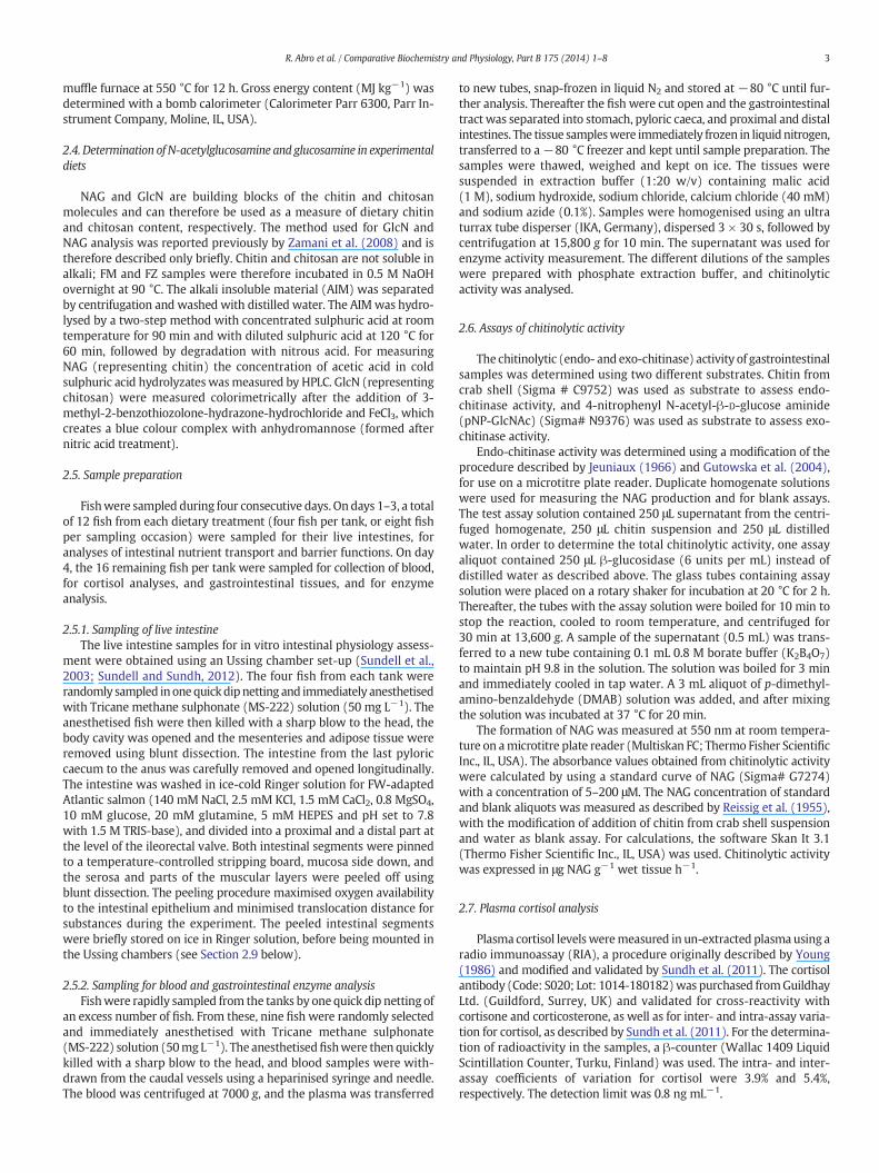
Proximal Distal0.0
2.0
4.0
6.0
8.0Papp mannitol
10-6
× cm
s-1
Diet p>0.05GI region p<0.001Diet * GI region p=0.089
A
GI regionProximal
0
50
100
150
200
Ω *
cm2
Diet p>0.05GI region p<0.001Diet * GI region p<0.05
aA
B
GI
Fig. 2. Intestinal barrier function assessed as the apparent permeability for mannitol (Papp; A),intestines of Arctic charr fed a fish meal-based (white bars) and a zygomycete-based (grey bardiet and GI regions was significant, the dataset was subjected to post-hoc analysis and letters atissues within fish meal-based diet. Upper case letters indicate differences between tissues wregions. Results are presented as mean ± SEM (n= 11–12).
activity was found in the stomach (0.9 μg NAG g−1 wet tissue h−1) forboth treatment groups.
Overall, the specific chitinase activity showed a similar pattern to thechitinase activity in wet tissue samples.
3.6. Intestinal barrier functions and nutrient uptake
The GLM analysis revealed no overall effects of diet (p N 0.05) on theparacellular permeability, as assessed by Papp. However, there was adifference between GI regions, with higher Papp values in the distalthan in the proximal intestine (p b 0.001; Fig. 2A). Therewas a tendencyfor an interaction between diet and GI regions (p = 0.089), suggestingthat the two diets might have exerted different effects on differentintestinal regions. The Papp values in the distal intestine were higher infish fed diet FZ than in fish fed diet FM, whereas no effect was seen inthe proximal intestine (Fig. 2A).
The patterns shown for paracellular permeability, as assessed byPapp for mannitol, were confirmed by the TER (Fig. 2B), the secondparacellular permeability marker. The statistical evaluation revealedno differences for factor diet (p N 0.05), but a significant effect wasfound for factor GI region (p b 0.001). A significant interaction betweenthe two factors was observed (p b 0.05). The proximal regions showedlower TER than the distal. In the distal intestine the FZ diet tended to de-crease TER (p = 0.06), whereas no statistical differences could beobserved in the proximal intestine.
Distal
TER
b
B
regionProximal Distal
0.0
0.1
0.2
0.3
0.4
0.5L-lysine transport
nmol
min
-1 c
m-2
Diet p>0.05GI region p<0.05Diet * GI region p<0.05
a
A
b
A*
GI region
C
transepithelial resistance (TER; B) and the L-lysine uptake (C) in the proximal and distals) diet. Overall effects are indicated as text in the diagram. When the interaction betweenbove bars indicate statistical relationship. Lower case letters indicate differences betweenithin zygomycete-based diet. Asterisk (*) indicates a difference between diets within GI

6 R. Abro et al. / Comparative Biochemistry and Physiology, Part B 175 (2014) 1–8
The statistical evaluation revealed no overall effects of diet (p N 0.05)on lysine uptake (Fig. 2C), but the uptake was effected by GI-region(p b 0.05) and a significant interaction between the two factorswas observed (p b 0.05). Multiple post-hoc comparisons revealedsignificantly higher lysine absorption in the proximal intestine com-pared with the distal intestine in fish fed the FM diet. The FZ dietincreased lysine absorption significantly in the distal intestine com-pared with the FM diet, whereas in the proximal intestine therewas an opposite tendency.
4. Discussion
For the first time, the present study shows that the gut tissue ofArctic charr has significant chitinase activity, of both the endo- andexo-chitinase iso-forms. This is an important trait and a prerequisitefor efficient utilisation of chitin-containing food. A lack of this abilitywould have a negative impact on digestibility, nutrient utilisation andgrowth performance of fish fed chitin-rich feed. Chitinase activity hasbeen reported to be present in gut tissue of other fish species, such asrainbow trout, juvenile cobia (R. canadum), parmaturus (Paramaturusxaniurus) and pachycara (Pachycara sp.) (Lindsay et al., 1984; Gutowskaet al., 2004; Fines and Holt, 2010). In contrast, it was recently reportedthat juvenile turbot (Psetta maxima) do not possess chitinase activity(Kroeckel et al., 2012).
In agreementwith Gutowska et al. (2004), we found that the activityof chitinase varied between GI regions. Furthermore, in the Arctic charrthe distribution pattern along the GI tract differed for endo-chitinaseand exo-chitinase. The exo-chitinase activity in the stomach showedthe lowest absolute value. In comparison, the endo-chitinase activityof both stomach and distal intestine was 100-fold higher. When com-paring exo-chitinase activity between intestinal regions, thedistal intes-tine had 100-fold higher activity than did the stomach. This distributionpattern of higher exo-chitinase activitymore distally in the gastro intes-tinal tract is plausible, as random cleavage of the chitin molecule byendo-chitinase is a pre-requisite for the release of glucosamine byexo-chitinase. The accompanying high endo-chitinase activity in thedistal intestine of Arctic charr indicates that it has the ability to efficientlyutilise any chitin escaping hydrolysis in the anterior parts of the GI tract.
The growth performance of Arctic charr was similar between fishfed a zygomycete-containing diet (FZ), high in chitin, and fish fed afish meal diet (FM). This is in agreement with one previous study onrainbow trout (Lindsay et al., 1984). Other studies have reported thatthe inclusion of chitin in the diet of red sea bream, Japanese eel, yellow-tail and black sea bream had positive effects on growth performance(Kono et al., 1987; Esteban et al., 2001; Om et al., 2003). In contrast,dietary chitin and chitosan have also been reported to have negativeeffects on growth performance in tilapia, rainbow trout, Atlantic salmon,chum salmon and giant tiger prawn (Kono et al., 1987; Shiau and Yu,1998; Bullock et al., 2000). The varying responses in growth perfor-mance among studies may reflect species differences in chitinolyticactivity, but can also be related to differences in molecular weight anddegree of acetylation of the chitosan molecules included in the feed. Inaddition, reduced growth from dietary chitin and chitosan may resultfrom impaired overall nutrient digestibility (Lindsay et al., 1984; Shiauand Yu, 1999) and/or disturbed nutrient absorption. The latter was sug-gested by thepresent study,where theuptake of L-lysine in theproximalintestine, the main nutrient-absorbing intestinal region, was impaired.
As expected, FZ had a higher content of GlcN and NAG than did FM(Ferreira et al., 2012). Thus, the FZ diet contained close to 10 timesmore chitin and chitosan than the FM diet. Overall, however, this wasnot reflected in differences in chitinase activity across GI regionsbetween the two diets. Even so, the interactions between diet and GIregions indicated that the diets may have different impacts on chitinaseactivity. For exo-chitinase activity, the absolute values were higher infish fed diet FZ than in fish fed diet FM in all tissues except for the prox-imal intestine.
The enzymatic degradation of chitin into small oligosaccharides,with negligible activity towards pNP-GlcNAc substrate, demonstratesthe endo-chitinase nature of the enzyme. In the presence of onlyendo-chitinases, the addition of β-glucosidase could enhance the truechitinolytic activity (Kang et al., 1999). In the present study, the additionofβ-glucosidase did not result in any increase in endo-chitinase activity.Similar results have been reported for several marine fish species(Gutowska et al., 2004). The paracellular permeability of the distalintestine was increased by the FZ diet, whereas the proximal intestinalregion was unaffected. This indicates that FZ diet resulted in disturbedintegrity and a leakier distal intestine in Arctic charr. This decrease inintestinal barrier function can be an effect of the exposure of theintestinal mucosa to the zygomycete raw material containing chitinmolecules (Lennartsson, 2012) which may be acting as mechanicallyirritating factors (Mydland et al., 2009). It has also been shown thatchitosan increases the paracellular permeability of the gastrointestinalepithelia by affecting the protein expression of the tight junction (TJ)complex (Thanou et al., 2001; Chopra et al., 2006; Rosenthal et al.,2012). The re-location of the TJ protein ZO-1 from the membrane tothe cytoskeleton is suggested to cause increased pore size of the TJcomplex and thus increased paracellular permeability of the epithelialcell layer (Smith et al., 2004). In the present study the content of chitosanwas 3.5 fold higher in the FZ diet than in FM diet. The activity of endo-and exo-chitinase was higher in the distal- than in the proximal-regionwhich also coincides with higher Papp for 14C-mannitol and a decreasedTER in the distal intestine. The results from the present study are inaccordance with previous results reported for mammalian cell cultures,treated with chitosan molecules of different grades of deacetylationand molecular weight (Schipper et al., 1996).
The increased disturbance in barrier function of the distal intestinecould also be a secondary stress response, emanating from primarystress experienced by the fish in response to a sub-optimal or non-palatable feed. In several other studies on salmonid fish, increasedintestinal paracellular permeability, i.e. disturbed barrier function, hasbeen shown to be a consistent secondary response to different typesof long-term stressors (Sundh et al., 2009, 2010). A disturbed intestinalintegrity is apparent also after the primary stress response, i.e. increasein the plasma cortisol levels, has returned to basal levels (Sundh et al.,2010). In the current study, no differences in plasma cortisol levelswere seen after a fourweek exposure to FZ, both dietary groups showedmean plasma cortisol levels of 2–6 ng/mL which is within the range ofunstressed fish (Woodward and Strange, 1987; Pickering and Pottinger,1989; Barry et al., 1993). Irrespective of cause, decreased intestinalbarrier function may constitute a threat to the welfare of the fish andcan increase the risk of infection in the long-term (Segner et al., 2012).
As discussed above, the intestinal paracellular permeability, i.e. theTJ permeability, was assessed in two different ways, as Papp for thehydrophilic marker molecule 14C-mannitol and by measuring thetransepithelial resistance, TER. Overall, TER was higher in the distalcompared to the proximal intestine, irrespective of diet. This pattern isconsistent with other salmonid species (Sundell et al., 2003; Jutfeltet al., 2006; Sundh et al., 2010; Sundell and Sundh, 2012). In the presentstudy, however, also the Papp was higher in the distal compared to theproximal intestine, which may seem contradictory. The selectivity inTJ permeability to charged and uncharged molecules is complex anddiscrepancies between the measures of the paracellular pathwaywhen assessing the electrical parameter TER or the Papp of a hydrophilicmolecule have been observed in both fish and mammalian studies(Günzel and Yu, 2013; Sundell et al., 2003; Sundh et al., 2011; Sundhet al., 2010; Van Itallie et al., 2008 ). The different claudin isoformsdisplay different numbers and types of charged amino acid residueslining the TJ pore formed between the adjacent cells, which constitutethe passageway formolecules through the paracellular pathway. Differ-ential expression of claudin isoforms is therefore suggested to be part ofthe explanation for differences in ion and size selectivity of TJs (Günzeland Yu, 2013). In fish, 56 different claudin isoforms have been described

7R. Abro et al. / Comparative Biochemistry and Physiology, Part B 175 (2014) 1–8
in the pufferfish (Fugu rubripes) (Loh et al., 2004) and 26 isoforms inAtlantic salmon (Tipsmark et al., 2008). However, to elucidate theprotein composition of TJs in the intestinal epithelium of fish and theirrole in the regulation of TJ permeability and selectivity further studiesare needed.
The zygomycete-based feed resulted in increased uptake rate of theamino acid L-lysine in the distal intestine, whereas no dietary effectswere seen in the proximal intestine. Previous studies on salmonid fishshow that amino acid absorption is several-fold higher in the proximalcompared with the distal intestine (Collie, 1985; Jutfelt et al., 2007)and the general picture is that the pyloric and proximal regions arethe main sites for a regulated, saturable and active amino acid absorp-tion in fish (Loretz, 1995). In the Arctic charr, there was a tendencytowards a downregulation of this active uptake of lysine in responseto the Zygomycete-based diet, but the effects were not statisticallysignificant. On the other hand, in the distal intestine there was a signif-icant increase in L-lysineuptake rate in response to the Zygomycete diet.Considering that the FZdiet induced increased paracellular permeabilityin the distal intestine, this increased L-lysine uptake is probably a resultof an overall increase in diffusional passage through a more leakyepithelium. Interestingly, the Arctic charr seem to deviate from the gen-eral picture, in that the difference in L-lysine absorption rate betweenthe proximal and distal intestines was low compared to that reportedfor other salmonid species.
5. Conclusions
Arctic charr showed a capacity to produce endogenous exo- and endo-chitinolytic enzymes in their gut tissues. The zygomycete ingredientenhanced the chitinase activity, with the exo-chitinase activity beingthe highest in the distal intestine. The paracellular permeability of theintestinal epithelium in the distal intestine was increased by the FZdiet. An increased exo-chitinase activity more distal in the gastrointes-tinal tract will probably result in an increased amount of chitosanbeing present. This in turnmay be themechanism behind the increasedparacellular permeability observed, as chitosan is known to affect andopen up tight junction complexes. The present results indicate thatthe gastrointestinal function of Arctic charr may be sensitive to chitin-rich feed based on the zygomycete R. oryzae.
Acknowledgements
The studywas supported by Swedish ResearchCouncil Formas (229-2009-526) (Swedish, Research Council for Environment, AgriculturalSciences and Spatial Planning). The authors are grateful to Dr. LarsEdebo, Department of Clinical Microbiology, University of Gothenburg,Sweden, for producing the zygomycete biomass, to Patrik Lennartssonfor glucosamine and N-acetylglucosamin analysis, and to David Morri-son for some of the statistical analyses. Rani Abro was funded by the Is-lamic Development Bank.
References
AOAC, 1997. Animal feeds. In: Cunniff, P.A. (Ed.), Official Methods of Analysis. Associationof Official Analytical Chemists International. AOAC, Arlington, VA, USA (Chapter 4, 1,1102 pp.).
Bankefors, J., Kaszowska, M., Schlechtriem, C., Pickova, J., Brannas, E., Edebo, L., Kiessling,A., Sandstrom, C., 2011. A comparison of the metabolic profile on intact tissue andextracts of muscle and liver of juvenile Atlantic salmon (Salmo salar L.)— applicationto a short feeding study. Food Chem. 129, 1397–1405.
Barry, T.P., Lapp, A.F., Kayes, T.B., Malison, J.A., 1993. Validation of a microtitre plate ELISAfor measuring cortisol in fish and comparison of stress responses of rainbow trout(Oncorhynchus mykiss) and lake trout (Salvelinus namaycush). Aquaculture 117,351–363.
Baumgart, D.C., Dignass, A.U., 2002. Intestinal barrier function. Curr Opin Clin Nutr MetabCare 5, 685–694.
Brännäs, E., Wiklund, B.S., 1992. Low temperature growth potential of Arctic charr andrainbow trout. Nord. J. Fresh W. Res. 77–81.
Buddington, R.K., 1980. Hydrolysis-resistant organic-matter as a reference for measure-ment of fish digestive efficiency. Trans. Am. Fish. Soc. 109, 653–656.
Bullock, G., Blazerb, V., Tsukuda, S., Summerfelt, S., 2000. Toxicity of acidified chitosan forcultured rainbow trout (Oncorhynchus mykiss). Aquaculture 185, 273–280.
Chopra, S., Mahdi, S., Kaur, J., Iqbal, Z., Talegaonkar, S., Ahmad, F.J., 2006. Advances andpotential applications of chitosan derivatives as mucoadhesive biomaterials inmodern drug delivery. J. Pharm. Pharmacol. 58, 1021–1032.
Clark, J., Quayle, K.A., Macdonald, N.L., Stark, J.R., 1988. Metabolism in marine flatfish. 5.Chitinolytic activities in dover sole, Solea solea (L.). Comp. Biochem. Physiol. B 90,379–384.
Cohen-Kupiec, R., Chet, I., 1998. The molecular biology of chitin digestion. Curr. Opin.Biotechnol. 9, 270–277.
Collie, Nathan L., 1985. Intestinal nutrient transport in coho salmon (Oncorhynchuskisutch) and the effects of development, starvation, and seawater adaptation. J.Comp. Physiol. B. 156 (2), 163–174.
Deters, A., Petereit, F., Schmidgall, J., Hensel, A., 2008. N-Acetyl-D-glucosamine oligosac-charides induce mucin secretion from colonic tissue and induce differentiation ofhuman keratinocytes. J. Pharm. Pharmacol. 60, 197–204.
Dick, T.A., Yang, X., 1992. A-linolenic acid metabolism of fish. Nutrition 218–220.Dijksterhuis, J., Samson, R.A., 2006. Activation of ascospores by novel food preservation
techniques. Adv. Food Mycol. 571, 247–260.Edebo, L., 2008. Zygomecete for fish feed. World patent, WO 2008/002231.Esteban, M.A., Cuesta, A., Ortuno, J., Meseguer, J., 2001. Immunomodulatory effects of
dietary intake of chitin on gilthead seabream (Sparus aurata L.) innate immunesystem. Fish Shellfish Immunol. 11, 303–315.
Fange, R., Lundblad, G., Lind, J., Slettengren, K., 1979. Chitinolytic enzymes in the digestive-system of marine fishes. Mar. Biol. 53, 317–321.
Ferreira, J.A., Lennartsson, P.R., Niklasson, C., Lundin, M., Edebo, L., Taherzadeh, M.J., 2012.Spent sulphite liquor for cultivation of an edible Rhizopus sp. Bioresources 7, 173–188.
Filho, B.P.D., Lemos, F.J.A., Secundino, N.F.C., Pascoa, V., Pereira, S.T., Pimenta, P.F.P., 2002.Presence of chitinase and beta-N-acetylglucosaminidase in the Aedes aegypti: achitinolytic system involving peritrophic matrix formation and degradation. InsectBiochem. Mol. Biol. 32, 1723–1729.
Fines, B.C., Holt, G.J., 2010. Chitinase and apparent digestibility of chitin in the digestivetract of juvenile cobia, Rachycentron canadum. Aquaculture 303, 34–39.
Günzel, D., Yu, A.S.L., 2013. Claudins and the modulation of tight junction permeability.Physiol. Rev. 93, 525–569.
Gutowska, M.A., Drazen, J.C., Robison, B.H., 2004. Digestive chitinolytic activity in marinefishes of Monterey Bay, California. Comp. Biochem. Physiol. A 139, 351–358.
Hjorth, H., 2005. The Cell Wall of Rhizopus oryzae: Structure, Properties and Applications.(PhD thesis) Gothenburg University, Department of Clinical Bacteriology, Gothenburg,Sweden.
Jeuniaux, C., 1966. Chitinases. Methods Enzymol. 8, 644–650.Jeuniaux, C., 1993. In: Muzzareli, R.A.A. (Ed.), Chitinolytic Systems in the Digestive Tract of
Vertebrates: A Review. European Chitin Society, Lyon and Ancoana, pp. 233–244.Jutfelt, F., Olsen, R.E., Glette, J., Ringo, E., Sundell, K., 2006. Translocation of viable
Aeromonas salmonicida across the intestine of rainbow trout, Oncorhynchus mykiss(Walbaum). J. Fish Dis. 29, 255–262.
Jutfelt, F., Olsen, R.E., Bjornsson, B.T., Sundell, K., 2007. Parr-smolt transformation anddietary vegetable lipids affect intestinal nutrient uptake, barrier function and plasmacortisol levels in Atlantic salmon. Aquaculture 273, 298–311.
Jutfelt, F., Sundh, H., Glette, J., Mellander, L., Bjornsson, B.T., Sundell, K., 2008. The involve-ment of Aeromonas salmonicida virulence factors in bacterial translocation across therainbow trout, Oncorhynchus mykiss (Walbaum), intestine. J. Fish Dis. 31, 141–151.
Kang, S.C., Park, S., Lee, D.G., 1999. Purification and characterization of a novel chitinase fromthe entomopathogenic fungus,Metarhizium anisopliae. J. Invertebr. Pathol. 73, 276–281.
Klemetsen, A., Amundsen, P.-A., et al., 2000. Atlantic salmon Salmo salar L., brown troutSalmo trutta L. and Arctic charr Salvelinus alpinus (L.): a review of aspects of theirlife histories. Ecol. Freshw. Fish 12 (1), 1–59.
Knudsen, D., Jutfelt, F., Sundh, H., Sundell, K., Koppe, W., Frokiaer, H., 2008. Dietary soyasaponins increase gut permeability and play a key role in the onset of soyabean-induced enteritis in Atlantic salmon (Salmo salar L.). Br. J. Nutr. 100, 120–129.
Kono, M., Matsui, T., Shimizu, C., 1987. Effects of chitin, chitosan and cellulose as dietsupplements on the growth of cultured fish Nippon Suisan Gakkaishi, pp. 125–129.
Kristofersson, D., Anderson, J.L., 2006. Is there a relationship between fisheries andfarming? Interdependence offisheries, animal production andaquaculture.Mar. Policy30, 721–725.
Kroeckel, S., Harjes, A.G.E., Roth, I., Katz, H., Wuertz, S., Susenbeth, A., Schulz, C., 2012.When a turbot catches a fly: evaluation of a pre-pupae meal of the Black SoldierFly (Hermetia illucens) as fish meal substitute — growth performance and chitindegradation in juvenile turbot (Psetta maxima). Aquaculture 364, 345–352.
Krogdahl, A., Bakke-McKellep, A.M., Baeverfjord, G., 2003. Effects of graded levels of stan-dard soybean meal on intestinal structure, mucosal enzyme activities, and pancreaticresponse in Atlantic salmon (Salmo salar L.). Aquacult. Nutr. 9, 361–371.
Krogdahl, A., Hemre, G.I., Mommsen, T.P., 2005. Carbohydrates in fish nutrition: digestionand absorption in postlarval stages. Aquacult. Nutr. 11, 103–122.
Lellis, W.A., Barrows, F.T., 2000. Effect of dietary ingredient substitution on dorsal finerosion of steelhead. North Am. j. Aquacult. 62, 135–138.
Lennartsson, P.R., 2012. Zygomycetes and Cellulose Residuals: Hydrolysis, Cultivation andApplications. Thesis for the Degree Doctoral of Philosophy. Chalmers University ofTechnology, Gothenburg, Sweden.
Linden, S.K., Sutton, P., Karlsson, N.G., Korolik, V., McGuckin, M.A., 2008. Mucins in themucosal barrier to infection. Mucosal Immunol. 1 (3), 183–197.
Lindsay, G.J.H., Walton, M.J., Adron, J.W., Fletcher, T.C., Cho, C.Y., Cowey, C.B., 1984. Thegrowth of rainbow trout (Salmo gairdneri) given diets containing chitin and itsrelationship to chitinolytic enzymes and chitin digestibility. Aquaculture 37, 315–334.

8 R. Abro et al. / Comparative Biochemistry and Physiology, Part B 175 (2014) 1–8
Loh, Y.H., Christoffels, A., Brenner, S., Hunziker, W., Venkatesh, B., 2004. Extensiveexpansion of the claudin gene family in the teleost fish, Fugu rubripes. Genome Res.14, 1248–1257.
Loretz, C.A., 1995. Electrophysiology of ion transport in teleost intestinal cells. FishPhysiol. XX, 25–56.
Mydland, L.T., Skugor, S., Martinsen, T.S., Landsverk, S., Kiessling, A., Edebo, L., Krasnov, A.,2009. Alterations in gene expression in gut tissue in Rainbow trout (Oncorhynchusmykiss) fedmyceliumbiomass from fungi (Rhizopus oryzae). International Symposiumon Genomics in Aquaculture, July 5–7, 2009, Bodø, Norway.
Naylor, R.L., Goldburg, R.J., Primavera, J.H., Kautsky, N., Beveridge, M.C., Clay, J., Folke, C.,Lubchenco, J., Mooney, H., Troell, M., 2000. Effect of aquaculture on world fishsupplies. Nature 405, 1017–1024.
Om, A.D., Ji, H., Umino, T., Nakagawa, H., Okada, K., Asano, M., Nakagawa, A., 2003. Effectof dietary chitin on lipid metabolism and vitality of black sea bream juvenile(Acanthopagrus schlegeli). ITE Lett. 691–698.
Pickering, A.D., Pottinger, T.G., 1989. Stress responses and disease resistance in salmonidfish— effects of chronic elevation of plasma cortisol. Fish Physiol. Biochem. 7, 253–258.
Reissig, J.L., Strominger, J.L., Leloir, L.F., 1955. A modified colorimetric method for theestimation of N-acetylamino sugars. J. Biol. Chem. 217, 959–966.
Ringo, E., Zhou, Z., Olsen, R.E., Song, S.K., 2012. Use of chitin and krill in aquaculture— theeffect on gut microbiota and the immune system: a review. Aquac. Nutr. 18, 117–131.
Rosenthal, R., Gunzel, D., Finger, C., Krug, S.M., Richter, J.F., Schulzke, J.D., Fromm, M.,Amasheh, S., 2012. The effect of chitosan on transcellular and paracellular mecha-nisms in the intestinal epithelial barrier. Biomaterials 33, 2791–2800.
Schipper, N.G.M., Vårum, K.M., Artursson, P., 1996. Chitosans as absorption enhancers forpoorly absorbable drugs. 1: influence of molecular weight and degree of acetylationon drug transport across human intestinal epithelial (Caco-2) cells. Pharm. Res. 13,1686–1692.
Segner, H., Sundh, H., Buchmann, K., Douxfils, J., Sundell, K., Mathieu, C., Ruane, N., Jutfelt,F., Toften, H., Vaughan, L., 2012. Health of farmed fish: its relation to fish welfare andits utility as welfare indicator. Fish Physiol Biochem 38, 85–105.
Shiau, S.Y., Yu, Y.P., 1998. Chitin but not chitosan supplementation enhances growth ofgrass shrimp, Penaeus monodon. J. Nutr. 128, 908–912.
Shiau, S.Y., Yu, Y.P., 1999. Dietary supplementation of chitin and chitosan depressesgrowth in tilapia, Oreochromis niloticus × O. aureus. Aquaculture 179, 439–446.
Smith, J., Wood, E., Dornish, M., 2004. Effect of chitosan on epithelial cell tight junctions.Pharm. Res. 21, 43–49.
Sues, A., Millati, R., Edebo, L., Taherzadeh, M.J., 2005. Ethanol production from hexoses,pentoses, and dilute-acid hydrolyzate by Mucor indicus. FEMS Yeast Res. 5, 669–676.
Sundell, K.S., Rønnestad, I., 2011. Intestinal absorption. In: Editor-in-Chief: Anthony, P.F.(Ed.), Encyclopedia of Fish Physiology. San Diego, Academic Press, pp. 1311–1321.
Sundell, K.S., Sundh, H., 2012. Intestinal fluid absorption in anadromous salmonids:importance of tight junctions and aquaporins. Front. Physiol. 3, 388.
Sundell, K., Jutfelt, F., Agustsson, T., Olsen, R.E., Sandblom, E., Hansen, T., Bjornsson, B.T.,2003. Intestinal transport mechanisms and plasma cortisol levels during normaland out-of-season parr–smolt transformation of Atlantic salmon, Salmo salar.Aquaculture 222, 265–285.
Sundh, H., Olsen, R.E., Fridell, F., Gadan, K., Evensen, O., Glette, J., Taranger, G.L., Myklebust,R., Sundell, K., 2009. The effect of hyperoxygenation and reduced flow in fresh waterand subsequent infectious pancreatic necrosis virus challenge in sea water, on theintestinal barrier integrity in Atlantic salmon, Salmo salar L. J. Fish Dis. 32, 687–698.
Sundh,H., Kvamme, B.O., Fridell, F., Olsen, R.E., Ellis, T., Taranger, G.L., et al., 2010. Intestinalbarrier function of Atlantic salmon (Salmo salar L.) post smolts is reduced by commonsea cage environments and suggested as a possible physiological welfare indicator.BMC Physiol. 10, 22.
Sundh, H., Calabrese, S., Jutfelt, F., Niklasson, L., Olsen, R.E., Sundell, K., 2011. Translocationof infectious pancreatic necrosis virus across the intestinal epithelium of Atlanticsalmon (Salmo salar L.). Aquaculture 82–95.
Thanou, M., Verhoef, J.C., Junginger, H.E., 2001. Chitosan and its derivatives as intestinalabsorption enhancers. Adv. Drug Deliv. Rev. 50, S91–S101.
Tipsmark, C.K., Kiilerich, P., Nilsen, T.O., Ebbesson, L.O.E., Stefansson, S.O., Madsen, S.S.,2008. Branchial expression patterns of claudin isoforms in Atlantic salmon duringseawater acclimation and smoltification. Am. J. Physiol. Regul. Integr. Comp. Physiol.294, R1563–R1574.
Van Itallie, C.M., Holmes, J., Bridges, A., Gookin, J.L., Coccaro, M.R., Proctor, W., Colegio, O.R.,Anderson, J.M., 2008. The density of small tight junction pores varies among celltypes and is increased by expression of claudin-2. J Cell Sci 121, 298–305.
Van Leeuwen, J., Rasmussean,M.L., Sakaran, S., Koza, C.R., Erickson, D.T.,Mitra, D., Jin, B., 2012.Fungal treatment of crop processing wastewaters with value-added co-products. In:Gopalakrishnan, K., Brown, R.C. (Eds.), Sustainable Bioenergy and Bioproducts.Springer London, London, pp. 13–44.
Woodward, C.C., Strange, R.J., 1987. Physiological stress responses in wild and hatchery-reared rainbow-trout. Trans. Am. Fish. Soc. 116, 574–579.
Young, G., 1986. Cortisol secretion invitro by the interrenal of coho salmon (Oncorhynchuskisutch) during smoltification — relationship with plasma thyroxine and plasmacortisol. Gen. Comp. Endocrinol. 63, 191–200.
Zamani, A., Jeihanipour, A., Edebo, L., Niklasson, C., Taherzadeh, M.J., 2008. Determinationof glucosamine and N-acetyl glucosamine in fungal cell walls. J. Agric. Food Chem. 56,8314–8318.
Copyright © 2022 FDOKUMEN