Haematological changes in the Mudfish, Clarias gariepinus (Burchell) exposed to Malachite green
Upload
independentCategory
view
0download
0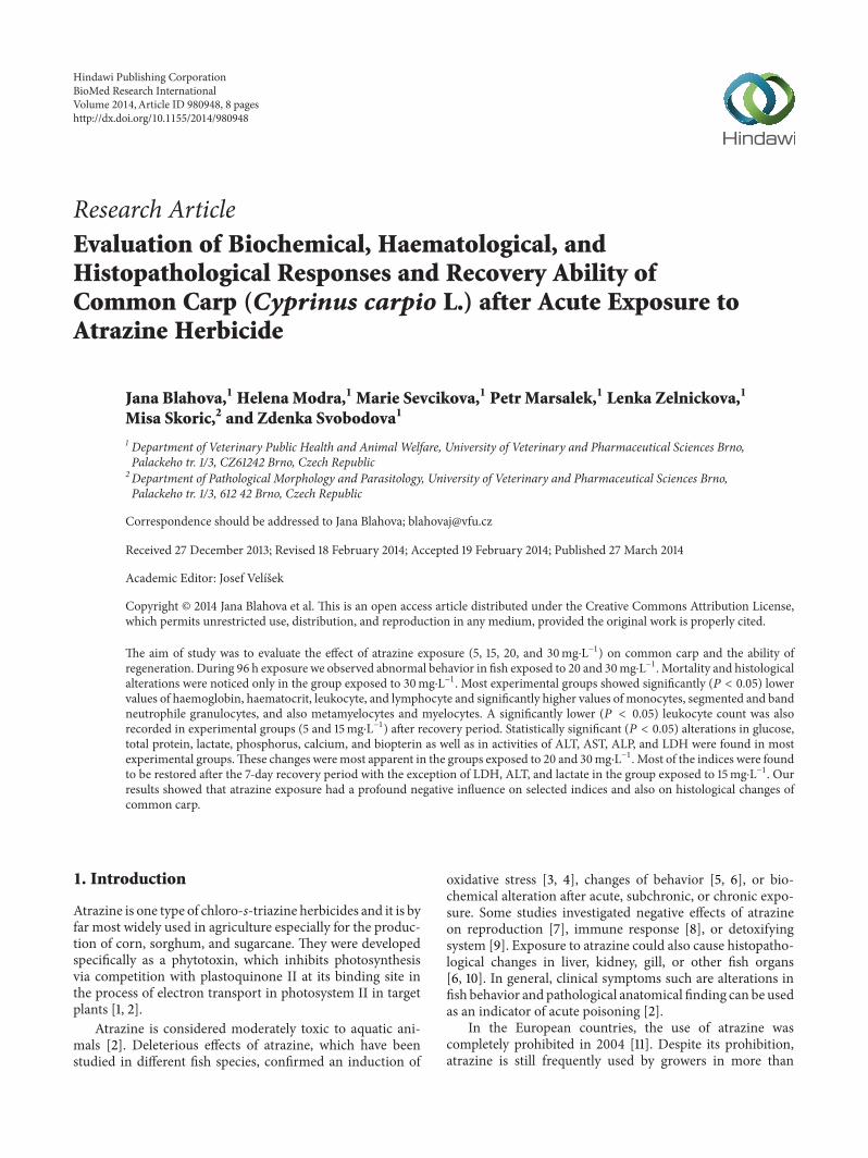
Research ArticleEvaluation of Biochemical, Haematological, andHistopathological Responses and Recovery Ability ofCommon Carp (Cyprinus carpio L.) after Acute Exposure toAtrazine Herbicide
Jana Blahova,1 Helena Modra,1 Marie Sevcikova,1 Petr Marsalek,1 Lenka Zelnickova,1
Misa Skoric,2 and Zdenka Svobodova1
1 Department of Veterinary Public Health and Animal Welfare, University of Veterinary and Pharmaceutical Sciences Brno,Palackeho tr. 1/3, CZ61242 Brno, Czech Republic
2 Department of Pathological Morphology and Parasitology, University of Veterinary and Pharmaceutical Sciences Brno,Palackeho tr. 1/3, 612 42 Brno, Czech Republic
Correspondence should be addressed to Jana Blahova; [email protected]
Received 27 December 2013; Revised 18 February 2014; Accepted 19 February 2014; Published 27 March 2014
Academic Editor: Josef Velısek
Copyright © 2014 Jana Blahova et al. This is an open access article distributed under the Creative Commons Attribution License,which permits unrestricted use, distribution, and reproduction in any medium, provided the original work is properly cited.
The aim of study was to evaluate the effect of atrazine exposure (5, 15, 20, and 30mg⋅L−1) on common carp and the ability ofregeneration. During 96 h exposure we observed abnormal behavior in fish exposed to 20 and 30mg⋅L−1. Mortality and histologicalalterations were noticed only in the group exposed to 30mg⋅L−1. Most experimental groups showed significantly (𝑃 < 0.05) lowervalues of haemoglobin, haematocrit, leukocyte, and lymphocyte and significantly higher values of monocytes, segmented and bandneutrophile granulocytes, and also metamyelocytes and myelocytes. A significantly lower (𝑃 < 0.05) leukocyte count was alsorecorded in experimental groups (5 and 15mg⋅L−1) after recovery period. Statistically significant (𝑃 < 0.05) alterations in glucose,total protein, lactate, phosphorus, calcium, and biopterin as well as in activities of ALT, AST, ALP, and LDH were found in mostexperimental groups.These changes weremost apparent in the groups exposed to 20 and 30mg⋅L−1. Most of the indices were foundto be restored after the 7-day recovery period with the exception of LDH, ALT, and lactate in the group exposed to 15mg⋅L−1. Ourresults showed that atrazine exposure had a profound negative influence on selected indices and also on histological changes ofcommon carp.
1. Introduction
Atrazine is one type of chloro-s-triazine herbicides and it is byfar most widely used in agriculture especially for the produc-tion of corn, sorghum, and sugarcane. They were developedspecifically as a phytotoxin, which inhibits photosynthesisvia competition with plastoquinone II at its binding site inthe process of electron transport in photosystem II in targetplants [1, 2].
Atrazine is considered moderately toxic to aquatic ani-mals [2]. Deleterious effects of atrazine, which have beenstudied in different fish species, confirmed an induction of
oxidative stress [3, 4], changes of behavior [5, 6], or bio-chemical alteration after acute, subchronic, or chronic expo-sure. Some studies investigated negative effects of atrazineon reproduction [7], immune response [8], or detoxifyingsystem [9]. Exposure to atrazine could also cause histopatho-logical changes in liver, kidney, gill, or other fish organs[6, 10]. In general, clinical symptoms such are alterations infish behavior and pathological anatomical finding can be usedas an indicator of acute poisoning [2].
In the European countries, the use of atrazine wascompletely prohibited in 2004 [11]. Despite its prohibition,atrazine is still frequently used by growers in more than
Hindawi Publishing CorporationBioMed Research InternationalVolume 2014, Article ID 980948, 8 pageshttp://dx.doi.org/10.1155/2014/980948

2 BioMed Research International
70 countries, with the top being the United States, Brazil,Argentina, Mexico, and China [1, 2]. Atrazine is listed as apriority substance in the European Water Framework. Theannual average and the maximum allowable concentrationshave been proposed for inland and other surface waters (0.6and 2 𝜇g⋅L−1, resp.) [12].
Although the s-triazine ring makes atrazine moleculeresistant, this herbicide undergoes physical, chemical, andbiological degradation in the soil, as well as in the watercolumn and in the sediment associated with water bodies[1]. The half-life of atrazine in soil, water, and sedimentsranges from few weeks to more than 2 years dependingon physicochemical properties of the environment. Atrazinemay degrade into many metabolites, each of varying per-sistence and toxicity. The most common metabolites arehydroxyatrazine, deethylatrazine, deisopropylatrazine, dide-alkylatrazine, and deethylhydroxyatrazine [13]. Due to thepersistence and mobility, atrazine and its degradation prod-ucts are still detected in surface water and groundwater ofcountries, in which the application of atrazine was banned.According to the CzechHydrometeorological Institute, watersamples from Czech rivers in years 2005, 2006, 2007, and2008 showed rates of occurrence of 88, 86, 46, and 60% insurface water, respectively, and the maximum concentrationsin these years ranged from 0.3 to 1.0𝜇g L−1. In the countries,where atrazine is still frequently used, the concentrations instreams, rivers, and lakes are severely higher. Obtained levelsof atrazine in water are episodic with major peaks in springand early summer after the field application in April andMay[1, 3].
The aim of the present study was to investigate the toxiceffect of atrazine on common carp (Cyprinus carpio L.)following 96 h exposure. Furthermore, the ability of organ-ism regeneration was studied using 7-day recovery period(depuration in fresh water). The toxic effects were evaluatedon the basis of results of haematological, biochemical, andhistopathological examination of test organisms.
2. Materials and Methods
2.1. Design of Experiment. The total of 80 common carps (C.carpio) (mean body weight 88.4 ± 2.6 g) was obtained from alocal hatchery (Pohorelice, PCL, Czech Republic) and usedin the acute toxicity test with atrazine of 99.0% chemicalpurity (A2S, France). At first, the fish were randomly dis-tributed into 8 aquarium tanks (𝑛 = 10) with dechlorinatedwater (volume of 100 L). After one week acclimatization tolaboratory conditions, the fish were exposed to a range ofatrazine concentrations (5, 15, 20, and 30mg⋅L−1) for 96 h.In control and experimental groups with 5 and 15mg⋅L−1of atrazine we used 20 fish for each concentration; in theexperimental groups with 20 and 30mg⋅L−1 of atrazine weused only 10 fish for each concentration.The solution volumeswere replaced once a day. The atrazine stock solutions wereprepared in dimethyl sulfoxidewith the final concentration of0.005% (HPLCgrade). Atrazine concentrationwas controlledby chromatographic analysis and did not decrease below 80%of the original concentration during the experiment. During
the test, conditions of fish and water were checked at 24 hintervals and the number of dead fish was recorded for eachconcentration. The values of water quality were as follows:temperature 22 ± 1∘C, oxygen saturation above 60%, and pH7.6–8.1. After 96 h exposure, individual blood samples weretaken from several fish in control (𝑛 = 12), 5mg⋅L−1 (𝑛 = 12)and 15mg⋅L−1 (𝑛 = 12) of atrazine groups and from all fishin 20mg⋅L−1 (𝑛 = 10) a 30mg⋅L−1 (𝑛 = 4) of atrazineexperimental groups. Blood samples were obtained by acardiac puncture and stabilized with 50 IU of heparin sodiumsalt per one mL of blood. The fish were killed by severing thespinal cord and selected organs (liver, gill, kidney, and skin)were taken for histopathological examination.The remainingfish in control (𝑛 = 8), 5mg⋅L−1 (𝑛 = 8) and 15mg⋅L−1(𝑛 = 8) of atrazine groups was subjected to dechlorinatedwater for another 7 days to assess the recovery ability. Atthe end of the recovery period, individual blood samplesand selected organs for histopathological examination weretaken. Experimental procedures were in a compliance withthe national legislation—Act No. 246/1992 Coll., on theProtection of Animals against Cruelty, as amended [14] andDecree No. 419/2012 Coll., on the Protection, Breeding, anduse of Experimental Animals, as amended [15].
2.2. Haematological and Biochemical Indices of Blood Samples.The haematological indices were determined in heparinizedblood and included erythrocyte count (RBC), haemoglobinconcentration (Hb), haematocrit value (PCV), leukocytecount (WBC), and differential leukocyte count. This proce-dure was carried out using the unified method for haema-tological examination of fish [16]. Blood plasma, which wasobtained from heparinized blood samples by centrifuga-tion (4∘C, 800×g, 10min), was used for the determina-tion of selected biochemical indices. Biochemical indicesin blood plasma included glucose, total protein, albumin,alanine aminotransferase (ALT), aspartate aminotransferase(AST), alkaline phosphatase (ALP), lactate, lactate dehy-drogenase (LDH), phosphorus, and calcium which weredetermined using biochemical analyzer Konelab 20i andcommercial kits (Biovendor PCL, Czech Republic). Bloodplasma was also used for the determination of biopterinand neopterin concentrations. The analysis of pterins wasbased on high performance liquid chromatography withfluorometric detection. For neopterin and biopterin analysis,300 𝜇L of trichloroacetic acid (5%) was added to 300 𝜇L ofstandard or plasma. The samples were centrifuged at 800×gfor 10min at 20∘C. The supernatant was filtered througha 0.45 𝜇m nylon filter and used for analysis. Elution wasperformed on a 150×4.6mm, 5 𝜇mZorbax Eclipse XBD-C18column (Agilent Technologies, Santa Clara, CA). Isocraticelution was performed at a flow rate of 1mL⋅min−1 withwater/acetonitrile (96 : 4) at 35∘C. Fluorescence detection at353 nm and 438 nm for excitation and emission, respectively,was used to detect pterins selectively. The chromatographicanalysis was accomplished by means of an Alliance 2695chromatographic system (Waters, Milford, MA) with an FD2475 fluorescent detector (Waters, Milford, MA). Neopterin,

BioMed Research International 3
biopterin, and trichloroacetic acid were purchased fromSigma-Aldrich (St. Louis, MO). All solvents were of HPLCgrade purity (Chromservis Ltd., CZ).The detection limits forneopterin and biopterin were 0.23 and 0.41 ng⋅mL−1, respec-tively.The limits of quantification for neopterin and biopterinwere 1.85 and 2.45 ng⋅mL−1, respectively. The coefficient ofvariation was 3.8%.
2.3. Histopathological Examination. The selected fish tissues(liver, gill, cranial and caudal kidney, and skin) were preparedfor histopathological examination, fixed in buffered 10%neutral formalin, dehydrated, embedded in paraffin wax,sectioned on a microtome at a thickness of 4 𝜇m, and stainedwith haematoxylin and eosin.
2.4. Determination of Atrazine in Water. Measurement ofatrazine concentration in water samples was conducted usinggas chromatography with ion trap tandem mass spectrom-etry. Sample preparation was based on simple liquid-liquidextraction into cyclohexane (1 : 1). The separation, identifica-tion, and quantification of atrazine were carried out usinga Varian 450-GC gas chromatograph with 220-MS ion trapmass spectrometer and VF-5ms (30m× 0.25mm) column(Varian, Inc., USA). A 1𝜇L of aliquot of sample extractwas injected in splitless mode. The injector temperature was250∘C.The initial oven temperature was set at 50∘C for 1min,increased in a rate of 30∘C per min to 130∘C for 1min,increased in a rate of 16∘C per min to 230∘C, held for 1min,increased in a rate of 60∘C per min to 280∘C, and heldfor 1min. The total run time was 13.75min. The certifiedstandard of atrazine was purchased from Dr. EhrenstorferGmbH (Germany). All solvents were of GC/MS grade purity(Chromservis Ltd., CZ).
2.5. Statistical Analysis. Statistical assessment was carried outusing Unistat 5.6 for Excel software (Czech Republic). Datawere tested for normality (Shapiro-Wilk test) and a one-way analysis of variance (ANOVA) was applied to test thedifferences in measured indices among experimental groupsfor both tests, that is, for 96 h toxicity test and recoverytest together. Individual differences among the means weretested successively using multiple comparison Tukey HSDtest. The differential leukocyte count did not satisfy normaldistribution; therefore a nonparametric Kruskal-Wallis testwas applied. Significance was accepted at 𝑃 < 0.05. All dataare reported as mean ± standard error of mean (SEM).
3. Results
3.1. Mortality and Fish Behavior. Changes in behavior wereobserved already after one hour of exposure to atrazine inthe groups of fish in the highest concentrations (20 and30mg⋅L−1). The behavioral changes were also observed inthe group exposed to 15mg⋅L−1 after 24 hours, but thesechanges were not so intensive. Abnormal behavior includedreduced reflexes, erratic swimming, loss of equilibrium, andaccelerated respiration. In the highest concentration, the fishwere lying on their side andweremoving only in this position.
Figure 1: Experimental carp after 96 h exposure to 30mg⋅L−1 ofatrazine: transudate in the body cavity.
No changes in behavior were observed in the control groupand experimental group exposed to 5mg⋅L−1 of atrazine aswell as in all groups during the recovery period. Mortalitywas observed only in the highest concentration of atrazine(30mg⋅L−1), in which the total mortality for 96 hours was60%. In this group, fish began to die after 48 hours of atrazineexposure. Transudate in the body cavity and an increasedinjection of visceral vessels were found during autopsy inthe fish exposed to the highest concentration of atrazine(30mg⋅L−1) (Figure 1).
3.2. Haematological Profile of Blood. The results of haema-tological examination of blood samples from acute toxicitytest and recovery period are given in Table 1. It is evidentthat the acute exposure to atrazine resulted in significantchanges in almost all haematological indices, especially inthe groups exposed to the highest concentrations of atrazine(20 and 30mg⋅L−1). A nonsignificant decrease in erythrocytecount in the group exposed to 30mg⋅L−1 subsequently led to asignificant decrease (𝑃 < 0.05) in haemoglobin concentrationand haematocrit value in this group compared to the control.A significant decrease (𝑃 < 0.05) in white blood cells wasobserved in all experimental groups exposed to atrazine for96 h. Significantly lower (𝑃 < 0.05) leukocyte count was alsorecorded in experimental groups exposed to 5 and 15mg⋅L−1of atrazine after seven days of recovery period in pure watercompared to the control group. Acute exposure to atrazineresulted in an intensive, significant (𝑃 < 0.05) alteration ofdifferential leukocyte counts compared to the control group;no changes were noticed during the recovery period. Aseveralfold significant decrease (𝑃 < 0.05) in lymphocyteswas found in experimental groups (15, 20, and 30mg⋅L−1)during 96 h toxicity test. On the other hand, we obtaineda severalfold significant increase (𝑃 < 0.05) in almostall experimental groups during 96 h atrazine exposure inthe case of monocytes, segmented and band neutrophilegranulocytes, metamyelocytes, and myelocytes.
3.3. Biochemical Profile of Blood Plasma. Results of bloodplasma biochemical indices are shown in Table 2. After 96 hexposure, there were found significant (𝑃 < 0.05) alterationsin the concentrations of glucose, total protein, albumin,

4 BioMed Research International
Table 1: Results of haematological examination during atrazine exposure and recovery period.
Indices 96 h toxicity test 7-day recovery test in pure waterControl 5mg⋅L−1 15mg⋅L−1 20mg⋅L−1 30mg⋅L−1 Control 5mg⋅L−1 15mg⋅L−1
RBC (T⋅L−1) 2.07 ± 0.12ab 1.55 ± 0.16b 2.21 ± 0.19ab 2.49 ± 0.10a 1.48 ± 0.46b 1.64 ± 0.06b 1.73 ± 0.17b 1.75 ± 0.12ab
Hb (g⋅L−1) 74.80 ± 1.35a 73.48 ± 1.88a 73.68 ± 3.63a 75.46 ± 2.95a 56.05 ± 7.19b 74.80 ± 1.35a 73.48 ± 1.88a 73.68 ± 3.63a
PCV (l⋅L−1) 0.24 ± 0.01b 0.24 ± 0.01b 0.22 ± 0.01b 0.23 ± 0.01b 0.18 ± 0.01a 0.23 ± 0.01b 0.25 ± 0.01b 0.23 ± 0.01b
WBC (G⋅L−1) 25.88 ± 1.73a 13.92 ± 1.36b 11.88 ± 1.05b 13.36 ± 1.34b 12.88 ± 4.30b 21.17 ± 1.06a 16.75 ± 2.25b 18.10 ± 1.80b
Lymphocytes (G⋅L−1) 7.18 ± 0.09a 5.99 ± 0.24ab 4.38 ± 0.31b 3.32 ± 0.33b 2.61 ± 0.83b 6.73 ± 0.17a 6.66 ± 0.13a 7.12 ± 0.09a
Monocytes (G⋅L−1) 0.08 ± 0.02a 0.08 ± 0.02a 0.14 ± 0.04a 0.12 ± 0.04a 0.30 ± 0.09b 0.09 ± 0.03a 0.10 ± 0.03a 0.10 ± 0.03a
NG-Segment (G⋅L−1) 0.12 ± 0.05a 0.43 ± 0.12b 0.52 ± 0.10b 0.48 ± 0.15b 0.51 ± 0.18b 0.16 ± 0.05a 0.21 ± 0.04a 0.18 ± 0.05a
NG-Bands (G⋅L−1) 0.08 ± 0.02a 0.26 ± 0.05a 0.72 ± 0.11b 0.92 ± 0.12b 1.46 ± 0.43b 0.08 ± 0.05a 0.12 ± 0.04a 0.07 ± 0.02a
Metamyelocytes (G⋅L−1) 0.11 ± 0.04a 0.64 ± 0.10b 1.58 ± 0.20c 1.94 ± 0.24c 1.83 ± 0.23c 0.26 ± 0.08a 0.32 ± 0.09a 0.19 ± 0.03a
Myelocytes (G⋅L−1) 0.09 ± 0.02a 0.18 ± 0.06a 0.37 ± 0.07b 0.79 ± 0.15b 0.85 ± 0.33b 0.07 ± 0.02a 0.09 ± 0.03a 0.05 ± 0.02a
Data are expressed as mean ± standard error of mean. Significant differences (𝑃 < 0.05) are indicated by different alphabetic superscripts (a legend: RBC:erythrocyte count; Hb: haemoglobin concentration; PCV: haematocrit value; WBC: leukocyte count; NG: neutrophile granulocytes).
Table 2: Results of biochemial examination during atrazine exposure and recovery period.
Indices 96 h toxicity test 7-day recovery test in pure waterControl 5mg⋅L−1 15mg⋅L−1 20mg⋅L−1 30mg⋅L−1 Control 5mg⋅L−1 15mg⋅L−1
Glucose (mmol⋅L−1) 3.42 ± 0.20a 9.15 ± 0.62b 23.03 ± 1.30c 22.47 ± 2.32c 28.02 ± 3.79c 3.55 ± 0.24a 3.55 ± 0.26a 3.39 ± 0.23a
Total protein (g⋅L−1) 19.59 ± 1.11a 20.97 ± 0.91a 26.52 ± 0.91b 23.12 ± 1.34ab 17.13 ± 4.7a 22.40 ± 0.62ab 19.36 ± 0.40a 21.72 ± 0.47ab
Albumin (g⋅L−1) 6.02 ± 0.39ab 6.99 ± 0.53bc 8.05 ± 0.29b 3.87 ± 0.70a 4.59 ± 2.12ac 5.93 ± 0.40ab 5.65 ± 0.25ac 6.20 ± 0.32bc
ALT (𝜇katal⋅L−1) 0.17 ± 0.01a 0.27 ± 0.03acd 0.46 ± 0.09bc 0.50 ± 0.07bd 0.60 ± 0.15b 0.25 ± 0.01ac 0.26 ± 0.02ac 0.49 ± 0.06bd
AST (𝜇katal⋅L−1) 2.33 ± 0.13a 2.45 ± 0.38a 3.33 ± 0.31ac 5.15 ± 0.36b 4.73 ± 0.67bc 3.31 ± 0.33ac 3.27 ± 0.14ac 2.81 ± 0.27a
ALP (𝜇katal⋅L−1) 0.21 ± 0.03a 0.32 ± 0.05ab 0.42 ± 0.02b 0.37 ± 0.03bc 0.24 ± 0.07ac 0.30 ± 0.02ab 0.30 ± 0.03ab 0.35 ± 0.03bc
LDH (𝜇katal⋅L−1) 3.98 ± 0.58ab 3.02 ± 0.61ab 4.57 ± 1.13ab 10.15 ± 0.89c 5.84 ± 1.35b 5.26 ± 0.65b 2.22 ± 0.13a 2.02 ± 0.21a
Lactate (mmol⋅L−1) 1.64 ± 0.30b 0.73 ± 0.10ad 0.90 ± 0.10ab 0.94 ± 0.10ab 0.40 ± 0.10a 1.45 ± 0.20bd 1.62 ± 0.13b 2.63 ± 0.20c
Phosphorus (mmol⋅L−1) 2.07 ± 0.08c 1.28 ± 0.11ad 1.12 ± 0.06a 1.55 ± 0.08bd 1.07 ± 0.10a 1.76 ± 0.09bc 1.62 ± 0.10bd 1.88 ± 0.07bc
Calcium (mmol⋅L−1) 2.04 ± 0.02bc 2.06 ± 0.03c 1.86 ± 0.03b 1.52 ± 0.03a 1.51 ± 0.03a 1.90 ± 0.08bc 1.91 ± 0.02bc 2.03 ± 0.03bc
Data are expressed as mean ± standard error of mean. Significant differences (𝑃 < 0.05) are indicated by different alphabetic superscripts (a legend: ALT:alanine aminotransferase; AST: aspartate aminotransferase; ALP: alkaline phosphatase; LDH: lactate dehydrogenase).
lactate, phosphorus, and calcium as well as in the activitiesof ALT, AST, ALP. and LDH compared to the control group.Most of the biochemical parameterswere found to be restoredafter a 7-day recovery period. Only in the case of LDH andALT activities and the concentration of lactate, the indices inexperimental group exposed to 15mg⋅L−1 of atrazine for 96 hwere significantly different (𝑃 < 0.05) even after the recoveryperiod in comparison to the control group.
The results of pterins concentration in blood plasmaare shown in Table 3. Because of an insufficient amount ofblood samples taken fromfish in experimental group exposedto 30mg⋅L−1, the analysis of pterins was not performed inthis concentration. Acute atrazine exposure resulted in anincrease in biopterin content only in the experimental groupexposed to 20mg⋅L−1. No changes were found in neopterinconcentration.
3.4. HistopathologicalExamination. Histopathological exam-ination revealed pathological lesions in fish only in the exper-imental group with the highest concentration of atrazine(30mg⋅L−1). Morphological changes were observed in theliver and represented by moderate to marked dystrophic
Table 3: Results (mean± standard error ofmean) of pterins in bloodplasma during atrazine exposure and recovery period.
Neopterin (mmol⋅L−1) Biopterin (mmol⋅L−1)96 h toxicity test
Control 25.61 ± 1.36a 47.53 ± 3.56ac
5mg⋅L−1 27.99 ± 2.81a 49.29 ± 4.40a
15mg⋅L−1 19.09 ± 1.48a 49.32 ± 5.71ac
20mg⋅L−1 27.22 ± 3.66a 74.90 ± 6.24b
7-day recovery test in pure waterControl 24.18 ± 1.89a 36.60 ± 3.71ac
5mg⋅L−1 21.69 ± 1.12a 31.56 ± 2.62c
15mg⋅L−1 22.91 ± 1.63a 34.97 ± 4.34ac
Significant differences (𝑃 < 0.05) are indicated by different alphabeticsuperscripts.
lesions of hepatocytes. There were morphological signs ofthe initial cell injury represented by hydropic to vacuolardegeneration of hepatocytes, dilatation of capillaries, andhyperaemia. Affected liver tissues were histopathologically

BioMed Research International 5
Control 30mg L−1
Figure 2: Results of histopathological examination of liver samples (arrows: diffuse hydropic to vacuolar degeneration of hepatocytes).
Control 30mg L−1
Figure 3: Results of histopathological examination of gill samples (a: severe lamellar teleangiectasis; dilated lamellar capillaries filled witherythrocytes; b: rupture of dilated lamellar capillary and pooling of the blood with formation of thrombi).
compared with tissue sections from negative control group(Figure 2).
Marked lesions were also observed in the gill samples andrepresented by a severe multifocal lamellar teleangiectasis asa result of the rupture of the retaining pillar cells and dilationof the lamellar capillary and pooling of the blood with for-mation of thrombi. Affected tissues were histopathologicallycompared with tissue sections from negative control group(Figure 3). Tissues and organs of the fish in experimentalgroups exposed to atrazine at the concentrations of 5, 15, and20mg⋅L−1 exhibited no pathomorphological changes.
4. Discussion
Although the atrazine use has been banned in the CzechRepublic and also in the European Union few years ago,the residues of this persistent compound are still found inthe surface waters. The presented study showed that acuteexposure to relatively high concentrations of atrazine couldnegatively affect the health status of common carp, which isconnected with the changes in behavior, alteration in bio-chemical and haematological indices, and also pathological-anatomical findings in selected tissues of common carp.
In the highest concentrations (20 and 30mg⋅L−1), thealterations in behavior were observed after one hour of
atrazine exposure. Behavior changes were also registered inthe experimental group exposed to 15mg⋅L−1 of atrazine,but this abnormal behavior was observed after 24 hoursof exposure. During the recovery period, no behavioralalterations were observed in the experimental groups. Behav-ior changes during exposure period were also reported byChapadense et al. [17], who evaluated the toxic effects ofatrazine on fish species tambaqui (Colossoma macropomum).They documented abnormal behavior such is lethargy, lossof equilibrium, increase in the frequency of opercular move-ments, and increase in the thickness of the inferior lips after48 h of exposure to atrazine at the range of 20–25mg⋅L−1.Moreover, they observed presence of hemorrhages in theeyes, lips, or even the whole body. Similar trends connectedwith uncoordinated behavior were also documented in fresh-water fish Channa punctatus after 96 h exposure to sublethalconcentrations of atrazine at the range of 4.2–10.6mg⋅L−1[18]. At the initial exposure, fish were alerted and stoppedswimming; after some time they tried to avoid the toxiceffects of atrazine with fast swimming and jumping and atthe end of the experiment; fish lost their balance and becameexhausted and lethargic. They also continuously secretedmassive amounts of mucus from whole body continuouslyand the body pigmentation was decreased. Steinberg et al.[19] documented that exposure of zebrafish to atrazine caused

6 BioMed Research International
changes in behavior, such as a significant preference of darkhabitats. Surprisingly, this effect was already detected in theenvironmental concentration (5 𝜇g⋅L−1), in which 79% of thefish records were taken in the dark part.
Atrazine is rapidly metabolized in the liver and kidneyand then excreted without any significant accumulation inthe tissues of fish organism. Some studies also documentedalterations of the gill tissues because of their direct contactwith water, which allows entering the substance throughthem into the fish body [2, 13]. Therefore the exposure toatrazine in fish is most often associated with the degenerativechanges in the kidney and gills and also with the alteration inthe liver tissues [20–23]. Besides, atrazine has been proposedto exert adverse effects on the reproductive fitness of fish.Thiscan lead to gonad abnormalities in both male and female fish[2, 10]. Fischer-Scherl et al. [20] documented alterations ofdifferent components of renal corpuscles and renal tubulesin rainbow trout (Oncorhynchus mykiss) after exposure toatrazine at the range of 5–40𝜇g⋅L−1 for 28 days. They alsofound necrosis of endothelial cells and renal hemopoietictissue in experimental group exposed to the concentrationsat the range of 80–2800𝜇g⋅L−1. Similar results were obtainedafter exposure of rare minnow (Gobiocypris rarus) to atrazinefor 28 days; histological observation revealed lesions inkidney tissues including an extensive expansion in the lumen,degenerative and necrotic changes of tubular epithelia, andshrinkage of the glomeruli at the concentration of 10𝜇g⋅L−1.The same concentration also resulted in obvious lesionsin the gill including hyperplasia, necrosis in epitheliumregion, aneurysm, and lamellar fusion [23]. Similarly, thegills changes were observed after acute 6 h exposure toatrazine in freshwater fish Gnathonemus petersii and theywere represented by the breaks in the epitheliumat 0.5mg⋅L−1and developed into deep pits at 5.0mg⋅L−1 [24]. Steatosis inliver tissue was observable in grey mullet (Liza ramanda)after subchronic exposure to 170 𝜇g⋅L−1 of atrazine. But infish exposed to atrazine and then returned to clean water,a reversal of the induced alterations and enhancement ofthe hepatic metabolism linked to detoxication mechanismsindicated hepatic recovery [22]. Mela et al. [4] studied theeffects of acute atrazine exposure at the range of 2–100𝜇g⋅L−1in neotropical catfish (Rhamdia quelen) and histopathologyexamination of liver tissues revealed leukocyte infiltration,hepatocyte vacuolization like steatosis and necrosis areas,leading to raised lesion index levels in all tested concen-trations. Surprisingly, the highest concentration of atrazinedid not cause the alteration in kidney tissue in the currentexperiment, but we observed degenerative changes in livertissue represented by marked hydropic to vacuolar degenera-tion of hepatocytes, dilatation of capillaries, and hyperaemia.Moreover, we obtained a severalfold significant increase inthe activities of all liver enzymes such as ALT, AST, and ALPdetected in plasma samples of all experimental groups and,also in the case of ALT, the significant increase in its activityrecorded after the recovery period in the experimental groupexposed to 15mg⋅L−1 of atrazine. The other significant alter-ations represented bymultifocal lamellar teleangiectasis werefound in the gill tissues of fish from the experimental group
exposed to 30mg⋅L−1 of atrazine. The cause of the damagemost likely results from the fact that atrazine denatures theepithelium of the gill filaments and this damage disrupts theion regulatory function of the gills [13].
In our study, we also observed the formation of transudatein the body cavity; this finding was recorded only in theexperimental group exposed to the 30mg⋅L−1 of atrazine.We can assume that the presence of transudate in the bodycavity can be connected with a decrease in oncotic pressurecaused by hypoproteinaemia and hypoalbuminaemia, whichwere found in this experimental group. Similar results weredocumented in the toxicological studies with other triazinepesticides [25], in which the formation of transudate was alsoassociated with the damage of epithelial cells of renal tubules[26].
In response to a stressor such as pesticide exposure,the fish undergo a series of biochemical and physiologicalchanges in an attempt to compensate the challenge imposedon them and thus cope with the stress. Blood is one of theimportant pathophysiological reflectors of the whole bodyorganism. Therefore, blood parameters such as haemato-logical and biochemical indices can be used as importantmarkers of diagnosing the structural and functional statusof fish exposed to pesticides, which induce stress reaction[27, 28]. In our study, acute exposure to atrazine resultedin significant concentration-dependent changes in almost allhaematological and biochemical indices and in the case ofsome indices these alterations were irreversible after 7-daydepuration in atrazine-free water.
Leukocytes are involved in the regulation of immuno-logical function and their alterations can be connected withimmunotoxic potential of substances [2, 29]. Changes inleukocyte profile indicate the response of fish organismto stress reaction [30]. It appears that there are distinctdifferences in the sensitivity of fish in terms of their immuneresponse to atrazine exposure [2]. In our study, the mainhaematological responses of common carp to the effects ofacute exposure of atrazinewere a significant decrease inwhiteblood cells and lymphocytes and a significant increase inmonocytes, segmented and band neutrophile granulocytes,and metamyelocytes and myelocytes counts. These changeswere observed especially in the experimental groups exposedto the highest concentrations of atrazine and, only in thecase of count of white blood cells, this alteration continuedalso during the recovery period. Declines in both leukocytesand lymphocytes counts indicate a reduction of nonspecificimmunity and a stress condition of common carp afteratrazine exposure. A decrease in leukocytes and lymphocyteswas also reported by Velisek et al. [25, 26] after acuteexposure to metribuzin in common carp and rainbow trout(Oncorhynchus mykiss).
In the erythrocyte profile, we observed alterations espe-cially in the experimental group exposed to the highest con-centration of atrazine. A significant decrease in haematocritvalue and haemoglobin concentration can be interpreted asa compensatory response that reduces the oxygen carryingcapacity to maintain gas exchange in the damaged gilllamellae. Our results are in line with those found by other

BioMed Research International 7
authors, who assessed the effects of selected pesticides onhaematological profile of fish blood [25, 26, 30–32].
Our results of blood plasma biochemical investiga-tion indicated strong alterations in almost all indices as astress response to atrazine exposure; the main biochemicalresponses were observed in glucose level and in the activitiesof liver enzymes. An increase in glucose levels detected inall experimental groups after 96 hours of atrazine exposurereflects an intense metabolic stress response of fish organism.The highest enhancement, which wasmore than dozen times,was observed in fish exposed to the highest concentration(30mg⋅L−1). In spite of this, the glucose concentration wasfound to be restored to physiological levels after a 7-dayrecovery period. Similar trend was also observed in severalstudies, in which acute exposures of fish organisms todifferent pesticides were studied [25, 33].
In our experiment, we recorded significant elevations inthe activities of all enzymes as conventional indicators of liverinjury in fish exposed to different atrazine concentrations.The most pronounced alteration was found in ALT activityand the significantly increased activity of the enzymewas alsoobserved after the recovery period. Alanine aminotransferaseis predominantly present in hepatocytes and therefore itsincreased activity after the recovery period could reflect thedamage of liver tissue [33]. Similar results were found byNeskovic et al. [21] who assessed effects of subacute (14days) exposure to atrazine at the range of 1.5–6mg⋅L−1 incommon carp. In all analyses of blood serum, the activityof ALP demonstrated a significant increase ranging from75 to 200% compared to the control group. On the otherhand, the ALP activities in tissues (heart, liver, and kidney)showed significant decrease compared to the control group,which was associated with histopathological alteration foundin these tissues.
Furthermore, we found a decline in the concentrations ofplasmaminerals (calcium and phosphorus), which is in closeagreement with the findings of the study by Prasad andReddy[34]. The authors observed significant changes of blood min-erals in Tilapia mossambicus indicating disturbances in thehydromineral balance of the fish as a consequence of atrazineexposure. During the experiment, we recorded elevationsof total protein and albumin especially in the experimentalgroup exposed to 15mg⋅L−1 of atrazine followed by thedecline in these indices in experimental groups exposed tothe highest concentrations. The protein concentration couldbe used as an indicator of general state of fish health and alsoas an indicator of stress [30].
Neopterin and biopterin belong to a group of uncon-jugated pterins and their levels in body fluids are oftenused as a biochemical marker of cell mediated immunity[35]. Unfortunately, there is only one study, which dealswith the possibility to use the monitoring of pterins aspotential markers of immune stress in fish organisms afterpesticide exposure [36]. The authors studied the effects ofsubchronic exposure to prochloraz in common carp andthey reported an increase in neopterin concentration, whilebiopterin concentration was not influenced. In the presentstudy, we found an alteration only in the case of biopterin
level in the experimental group exposed to 20mg⋅L−1 after96 hours. Our findings suggest an influencing of the fishimmune system by acute atrazine exposure, which is furtherdocumented by changes in the count of white blood cells inthe exposed groups.
5. Conclusion
In summary, this study gives further evidence that atrazineis toxic to fish organism and causes changes in biochemicaland haematological indices and alteration of behavior andpathological anatomical finding of common carp. On theother hand, the results of the recovery test showed a highregeneration potential of organisms and confirmed that theresponses of common carp were in most indices reversiblewhen exposure was terminated. Irreversible changes wereobserved only in the activity of liver enzyme alanine amino-transferase and the count of white blood cells (especiallylymphocytes), which confirms a hepatotoxic and immuno-suppressive potential of atrazine.
Conflict of Interests
The authors declare that there is no conflict of interestsregarding the publication of thispaper.
Acknowledgments
This research was supported by GACR P502/12/P163. Theauthors would like to thank Veronika Sımova, DVM, forpaper improvement and English correction.
References
[1] H. M. LeBaron, J. E. McFarland, and O. C. Burnside, Eds.,The Triazines Herbicides 50 Years Revolutionizing Agriculture,Elsevier, Oxford, UK, 2008.
[2] K. R. Solomon, J. A. Carr, L. H. Du Preez et al., “Effects ofatrazine on fish, amphibians, and aquatic reptiles: a criticalreview,” Critical Reviews in Toxicology, vol. 38, no. 9, pp. 721–772, 2008.
[3] J. Blahova, L. Plhalova, M. Hostovsky et al., “Oxidative stressresponses in zebrafish Danio rerio after subchronic exposure toatrazine,” Food andChemical Toxicology, vol. 61, pp. 82–85, 2013.
[4] M. Mela, I. C. Guiloski, H. B. Doria et al., “Effects of herbicideatrazine in neotropical catfish (Rhamdia quelen),” Ecotoxicologyand Environmental Safety, vol. 93, pp. 13–21, 2013.
[5] P. Saglio and S. Trijasse, “Behavioral responses to atrazine anddiuron in goldfish,” Archives of Environmental Contaminationand Toxicology, vol. 35, no. 3, pp. 484–491, 1998.
[6] L. Plhalova, J. Blahova, I. Mikulikova et al., “Effects of sub-chronic exposure to atrazine on zebrafish (Danio rerio),” PolishJournal of Veterinary Science, vol. 15, pp. 417–423, 2012.
[7] D. E. Tillitt, D. M. Papoulias, J. J. Whyte, and C. A. Richter,“Atrazine reduces reproduction in fatheadminnow (Pimephalespromelas),” Aquatic Toxicology, vol. 99, no. 2, pp. 149–159, 2010.
[8] L. C. Kreutz, L. J. G. Barcellos, E. D. dos Santos, M. Pivato, andR. Zanatta, “Innate immune response of silver catfish (Rhamdiaquelen) exposed to atrazine,” Fish and Shellfish Immunology, vol.33, no. 4, pp. 1055–1059, 2012.

8 BioMed Research International
[9] Y. Fu, M. Li, C. Liu et al., “Effect of atrazine and chlorphyrofosexposure on cytochrome P450 contents and enzyme activitiesin common carp gills,” Ecotoxicology and Environmental Safety,vol. 94, pp. 28–36, 2013.
[10] M.G. Paulino,N. E. S. Souza, andM.N. Fernandes, “Subchronicexposure to atrazine induces biochemical and histopathologicalchanges in the gills of a neotropical freshwater fish, Prochiloduslineatus,” Ecotoxicology and Environmental Safety, vol. 80, pp.6–13, 2012.
[11] European Commission, “COMMISSION DECISION of 10March 2004 concerning the non-inclusion of atrazine in AnnexI to Council Directive 91/414/EEC and the withdrawal ofauthorisations for plant protection products containing thisactive substance (No. 2004/248/EC),” OJL, 78, 16.03,pp. 53–55,2004.
[12] European Commission, “Directive 2008/105/EC of the Euro-pean Parliament and of the Council of 16 December 2008on environmental quality standards in the field of waterhggh83/513/EEC, 84/156/EEC, 84/491/EEC, 86/280/EEC andamending Directive 2000/60/EC of the European Parliamentand of the Council,” OJL, 348, 24.12, pp. 84–97, 2008.
[13] M. Graymore, F. Stagnitti, and G. Allinson, “Impacts of atrazinein aquatic ecosystems,” Environment International, vol. 26, no.7-8, pp. 483–495, 2001.
[14] Act No. 246/1992 Coll., on the Protection of Animals againstCruelty, as amended (Czech).
[15] Decree No. 419/2012 Coll., on the Protection, Breeding and useof Experimental Animals, as amended (Czech).
[16] Z. Svobodova, D. Pravda, and H. Modra, Methods of Haema-tological Examination of Fish, University of Veterinary andPharmaceutical Sciences Brno, Brno, Czech Republic, 2012(Czech).
[17] P. F. G. Chapadense, F. D. J. Castro, J. A. Almeida, and S. E.Moron, “Toxicity of atrazine herbicide in Colossoma macrop-omum,” Revista Brasileira de Saude e Producao Animal, vol. 10,pp. 398–405, 2009.
[18] C. D. Nwani, W. S. Lakra, N. S. Nagpure, R. Kumar, B.Kushwaha, and S. K. Kumar, “Toxicity of the herbicide atrazine:effects on lipid peroxidation and activities of antioxidantenzymes in the freshwater fish Channa punctatus (Bloch),”International Journal of Environmental Research and PublicHealth, vol. 7, no. 8, pp. 3298–3312, 2010.
[19] C. E. W. Steinberg, R. Lorenz, and O. H. Spieser, “Effects ofatrazine on swimming behavior of zebrafish,Brachydanio rerio,”Water Research, vol. 29, no. 3, pp. 981–985, 1995.
[20] T. Fischer-Scherl, A. Veeser, R. W. Hoffmann, C. Kuhnhauser,R. Negele, and T. Ewringmann, “Morphological effects of acuteand chronic atrazine exposure in rainbow trout (Oncorhynchusmykiss),”Archives of Environmental Contamination and Toxicol-ogy, vol. 20, no. 4, pp. 454–461, 1991.
[21] N. K. Neskovic, I. Elezovic, V. Karan, V. Pokeksic, and M.Budimir, “Acute and subacute toxicity of atrazine to carp(Cyprinus carpio L.),” Ecotoxicology and Environmental Safety,vol. 25, no. 2, pp. 173–182, 1993.
[22] S. Biagianti-Risbourg and J. Bastide, “Hepatic perturbationsinduced by a herbicide (atrazine) in juvenile grey mulletLiza ramada (Mugilidae, Teleostei): an ultrastructural study,”Aquatic Toxicology, vol. 31, no. 3, pp. 217–229, 1995.
[23] L. Yang, J. Zha, W. Li, Z. Li, and Z. Wang, “Atrazine affectskidney and adrenal hormones (AHs) related genes expressionsof rareminnow (Gobiocypris rarus),”Aquatic Toxicology, vol. 97,no. 3, pp. 204–211, 2010.
[24] B. M. Alazemi, J. W. Lewis, and E. B. Andrews, “Gill damage inthe freshwater fish Gnathonemus petersii (family: Mormyridae)exposed to selected pollutants: an ultrastructural study,” Envi-ronmental Technology, vol. 17, no. 3, pp. 225–238, 1996.
[25] J. Velisek, Z. Svobodova, V. Piackova, and E. Sudova, “Effectsof acute exposure to metribuzin on some hematological, bio-chemical and histopathological parameters of common carp(Cyprinus carpio L.),” Bulletin of Environmental Contaminationand Toxicology, vol. 82, no. 4, pp. 492–495, 2009.
[26] J. Velisek, Z. Svobodova, V. Piackova et al., “Effects ofmetribuzin on rainbow trout (Oncorhynchus mykiss),” Veteri-narni Medicina, vol. 53, no. 6, pp. 324–332, 2008.
[27] S. Adhikari, B. Sarkar, A. Chatterjee, C. T. Mahapatra, and S.Ayyappan, “Effects of cypermethrin and carbofuran on certainhematological parameters and prediction of their recovery in afreshwater teleost, Labeo rohita (Hamilton),” Ecotoxicology andEnvironmental Safety, vol. 58, no. 2, pp. 220–226, 2004.
[28] D. H. Evans and J. B. Claiborne, Eds., The Physiology of Fishes,CRC Press, Boca Raton, Fla, USA, 2005.
[29] G. O. Evans, Ed., Animal Hematotoxicology—A Practical Guidefor Toxicologists and Biomedical Researchers, CRC Press, BocaRaton, Fla, USA, 2009.
[30] M. Ramesh, R. Srinivasan, and M. Saravana, “Effect of atrazine(Herbicide) on blood parameters of common carp Cyprinuscarpio (Actinopterygii: Cypriniformes),” African Journal ofEnvironmental Science and Technology, vol. 3, no. 12, pp. 453–458, 2009.
[31] T. A. V. Prasad, T. Srinivas, G. M. Rafi, and D. C. Reddy, “Effectin vivo of atrazine on haematology and O
2
consumption in fish,Tilapia mossambica,” Biochemistry International, vol. 23, no. 1,pp. 157–161, 1991.
[32] J. H. Jee, F. Masroor, and J. C. Kang, “Responses ofcypermethrin-induced stress in haematological parameters ofKorean rockfish, Sebastes schlegeli (Hilgendorf),” AquacultureResearch, vol. 36, no. 9, pp. 898–905, 2005.
[33] I. Mikulikova, H. Modra, J. Blahova et al., “Recovery ability ofcommon carp (Cyprinus carpio) after a short-term exposure toterbuthylazine,” Polish Journal of Veterinary Science, vol. 16, no.1, pp. 17–23, 2013.
[34] T. A.V. Prasad andD.C. Reddy, “Atrazine toxicity on hydromin-eral balance of fish, Tilapia mossambicus,” Ecotoxicology andEnvironmental Safety, vol. 28, no. 3, pp. 313–316, 1994.
[35] G. Hoffmann, B. Wirleitner, and D. Fuchs, “Potential role ofimmune system activation-associated production of neopterinderivatives in humans,” Inflammation Research, vol. 52, no. 8,pp. 313–321, 2003.
[36] P.Marsalek, I.Mikulıkova, H.Modra, and Z. Svobodova, “Effectof prochloraz fungicide on neopterin and biopterin levels inblood plasma of common carp,”Acta Veterinaria Brno. In press.

Submit your manuscripts athttp://www.hindawi.com
PainResearch and TreatmentHindawi Publishing Corporationhttp://www.hindawi.com Volume 2014
The Scientific World JournalHindawi Publishing Corporation http://www.hindawi.com Volume 2014
Hindawi Publishing Corporationhttp://www.hindawi.com
Volume 2014
ToxinsJournal of
VaccinesJournal of
Hindawi Publishing Corporation http://www.hindawi.com Volume 2014
Hindawi Publishing Corporationhttp://www.hindawi.com Volume 2014
AntibioticsInternational Journal of
ToxicologyJournal of
Hindawi Publishing Corporationhttp://www.hindawi.com Volume 2014
StrokeResearch and TreatmentHindawi Publishing Corporationhttp://www.hindawi.com Volume 2014
Drug DeliveryJournal of
Hindawi Publishing Corporationhttp://www.hindawi.com Volume 2014
Hindawi Publishing Corporationhttp://www.hindawi.com Volume 2014
Advances in Pharmacological Sciences
Tropical MedicineJournal of
Hindawi Publishing Corporationhttp://www.hindawi.com Volume 2014
Medicinal ChemistryInternational Journal of
Hindawi Publishing Corporationhttp://www.hindawi.com Volume 2014
AddictionJournal of
Hindawi Publishing Corporationhttp://www.hindawi.com Volume 2014
Hindawi Publishing Corporationhttp://www.hindawi.com Volume 2014
BioMed Research International
Emergency Medicine InternationalHindawi Publishing Corporationhttp://www.hindawi.com Volume 2014
Hindawi Publishing Corporationhttp://www.hindawi.com Volume 2014
Autoimmune Diseases
Hindawi Publishing Corporationhttp://www.hindawi.com Volume 2014
Anesthesiology Research and Practice
ScientificaHindawi Publishing Corporationhttp://www.hindawi.com Volume 2014
Journal of
Hindawi Publishing Corporationhttp://www.hindawi.com Volume 2014
Pharmaceutics
Hindawi Publishing Corporationhttp://www.hindawi.com Volume 2014
MEDIATORSINFLAMMATION
of
Copyright © 2022 FDOKUMEN