European Annals of Allergy and Clinical Immunology
41
EDITORS IN CHIEF L. Cecchi (Firenze – Italy) P. Carreiro-Martins (Lisbon – Portugal) HONORARY EDITOR A. Sabbah (Angers – France) ASSOCIATE EDITORS R. Rodrigues Alves (Lisbon – Portugal) A. Tedeschi (Milano – Italy) EDITORIAL BOARD M. Morais-Almeida (Lisbon – Portugal) R. Asero (Milano – Italy) M.B. Bilò (Ancona – Italy) F. Bonifazi (Ancona – Italy) L.M. Borrego (Lisbon – Portugal) K. Brockow (München – Germany) Á.A. Cruz (Salvador – Brasil) L. Delgado (Oporto – Portugal) P. Demoly (Montpellier – France) G. D’Amato (Napoli – Italy) M. Drouet (Angers – France) M. Fernandez-Rivas (Madrid – Spain) A. Fiocchi (Milano – Italy) J. Fonseca (Oporto – Portugal) D. Macchia (Firenze – Italy) F. Mastrandrea (Taranto – Italy) M. Maurer (Berlin – Germany) G. Moscato (Pavia – Italy) A. Musarra (Reggio Calabria – Italia) C. Nunes (Portimao – Portugal) M. Olivieri (Verona – Italy) P. Parronchi (Firenze – Italy) G. Passalacqua (Genova – Italy) G. Pauli (Strasbourg – France) E. Pedro (Lisbon – Portugal) A. Perino (Torino – Italy) L.K. Poulsen (Copenaghen – Denmark) O. Quercia (Faenza – Italy) A. Romano (Roma – Italy) E. Scala (Roma – Italy) D. Solé (Sao Paulo – Brazil) A. Todo Bom (Coimbra – Portugal) S. Voltolini (Genova – Italy) SCIENTIFIC COMMITTEE L. Antonicelli (Italy) A. Bener (Turkey) H. Bazin (Belgium) J. Bellanti (USA) C. Geller-Bernstein (Israel) M. Cugno (Italy) B. David (France) S. Durham (UK) G.P. Girolomoni (Italy) R. Jarish (Austria) S.G.O. Johansson (Sweden) F. Levi-Shaffer (Israel) P. Lowenstein (Denmark) J.L. Malo (Canada) A.G. Palma-Carlos (Portugal) G. Scadding (UK) G. Scadding (UK) E. Stevens (Belgium) R. van Ree (Amsterdam) FOUNDER AND CORRESPONDING MEMBER G.M. Halpern (USA) European Annals of Allergy and Clinical Immunology THE OFFICIAL JOURNAL OF AAIITO ASSOCIAZIONE ALLERGOLOGI IMMUNOLOGI ITALIANI TERRITORIALI E OSPEDALIERI THE OFFICIAL JOURNAL OF SPAIC SOCIEDADE PORTUGUESA DE ALERGOLOGIA E IMUNOLOGIA CLINICA www.eurannallergyimm.com Editors in Chief and Managing Directors Lorenzo Cecchi P. Carreiro-Martins Chief Business & Content Officer Ludovico Baldessin Publishing Editor Chiara Scelsi [email protected] Ph. 039 (0)2-88184.257 Production Manager Walter Castiglione [email protected] Ph. 0039 (0)2-88184.222 Sales Stefano Busconi [email protected] Ph. 0039 (0)2-88184.404 Subscription [email protected] Ph. 0039 (0)2-88184.317 Italy subscription: 60 euro World subscription: 85 euro Printing Rotomail Italia S.p.A., Strada Rivoltana (SP 14), 12/AB 20060 Vignate (MI), Italy EDRA SpA Via G. Spadolini, 7 20141 Milano - Italy Tel. 0039 (0)2-88184.1 Fax 0039 (0)2-88184.301 www.edizioniedra.it “European Annals of Allergy and Clinical Immunology” registered at Tribunale di Milano - n. 336 on 22.10.2014 © 2019 Associazione Allergologi Immunologi Italiani Territoriali e Ospedalieri - AAIITO. Published by EDRA SpA. All rights reserved. To read our Privacy Policy please visit www.edraspa.it/privacy The contents of this Journal are indexed in PubMed, Scopus, Embase and Web of Science ® AAIITO Associazione Allergologi Immunologi Italiani Territoriali e Ospedalieri SPAIC Sociedade Portuguesa de Alergologia e Imunologia Clínica DIRECTORY BOARD President Antonino Musarra Designate President Riccardo Asero DIRECTORY BOARD President Elisa Pedro Past President Luís Delgado Vice Presidents Emilia Faria João Fonseca Pedro Martins Vice Presidents Francesco Murzilli Treasurer Oliviero Quercia Past President Maria Beatrice Bilò Treasurer Rodrigo Rodrigues Alves Secretary-General Manuel Branco Ferreira Secretary-Adjunct Ana Morête Members Michele Giovannini Maria Carmela Montera Lionello Muratore Battista Roberto Polillo Danilo Raffaele Villalta Susanna Voltolini Maria Teresa Zedda Members Rita câmara Ângela Gaspar Daniel Machado
-
Upload
khangminh22 -
Category
Documents
-
view
2 -
download
0
Transcript of European Annals of Allergy and Clinical Immunology
EDITORS IN CHIEFL. Cecchi (Firenze – Italy)
P. Carreiro-Martins (Lisbon – Portugal)
HONORARY EDITORA. Sabbah (Angers – France)
ASSOCIATE EDITORSR. Rodrigues Alves (Lisbon – Portugal)
A. Tedeschi (Milano – Italy)
EDITORIAL BOARDM. Morais-Almeida (Lisbon – Portugal)
R. Asero (Milano – Italy)M.B. Bilò (Ancona – Italy)
F. Bonifazi (Ancona – Italy)L.M. Borrego (Lisbon – Portugal)
K. Brockow (München – Germany)Á.A. Cruz (Salvador – Brasil)
L. Delgado (Oporto – Portugal)P. Demoly (Montpellier – France)
G. D’Amato (Napoli – Italy)M. Drouet (Angers – France)
M. Fernandez-Rivas (Madrid – Spain)A. Fiocchi (Milano – Italy)
J. Fonseca (Oporto – Portugal)D. Macchia (Firenze – Italy)
F. Mastrandrea (Taranto – Italy)M. Maurer (Berlin – Germany)
G. Moscato (Pavia – Italy)A. Musarra (Reggio Calabria – Italia)
C. Nunes (Portimao – Portugal)M. Olivieri (Verona – Italy)
P. Parronchi (Firenze – Italy)G. Passalacqua (Genova – Italy)
G. Pauli (Strasbourg – France)E. Pedro (Lisbon – Portugal)
A. Perino (Torino – Italy)L.K. Poulsen (Copenaghen – Denmark)
O. Quercia (Faenza – Italy)A. Romano (Roma – Italy)
E. Scala (Roma – Italy)D. Solé (Sao Paulo – Brazil)
A. Todo Bom (Coimbra – Portugal)S. Voltolini (Genova – Italy)
SCIENTIFIC COMMITTEEL. Antonicelli (Italy)
A. Bener (Turkey)H. Bazin (Belgium)
J. Bellanti (USA)C. Geller-Bernstein (Israel)
M. Cugno (Italy)B. David (France)
S. Durham (UK)G.P. Girolomoni (Italy)
R. Jarish (Austria)S.G.O. Johansson (Sweden)
F. Levi-Shaffer (Israel)P. Lowenstein (Denmark)
J.L. Malo (Canada)A.G. Palma-Carlos (Portugal)
G. Scadding (UK)G. Scadding (UK)
E. Stevens (Belgium)R. van Ree (Amsterdam)
FOUNDER AND CORRESPONDING MEMBERG.M. Halpern (USA)
European Annalsof Allergy and
Clinical Immunology
THE OFFICIAL JOURNAL OF AAIITOASSOCIAZIONE ALLERGOLOGI IMMUNOLOGI ITALIANI TERRITORIALI E OSPEDALIERI
THE OFFICIAL JOURNAL OF SPAICSOCIEDADE PORTUGUESA DE ALERGOLOGIA E IMUNOLOGIA CLINICA
www.eurannallergyimm.comEditors in Chief and Managing DirectorsLorenzo CecchiP. Carreiro-Martins
Chief Business & Content OfficerLudovico Baldessin
Publishing EditorChiara [email protected]. 039 (0)2-88184.257
Production ManagerWalter [email protected]. 0039 (0)2-88184.222
SalesStefano [email protected]. 0039 (0)2-88184.404
[email protected] Ph. 0039 (0)2-88184.317 Italy subscription: 60 euroWorld subscription: 85 euro
PrintingRotomail Italia S.p.A., Strada Rivoltana (SP 14), 12/AB 20060 Vignate (MI), Italy
EDRA SpAVia G. Spadolini, 720141 Milano - ItalyTel. 0039 (0)2-88184.1Fax 0039 (0)2-88184.301www.edizioniedra.it
“European Annals of Allergy and Clinical Immunology” registered at Tribunale di Milano - n. 336 on 22.10.2014
© 2019 Associazione Allergologi Immunologi Italiani Territoriali e Ospedalieri - AAIITO. Published by EDRA SpA. All rights reserved.
To read our Privacy Policy please visit www.edraspa.it/privacy
The contents of this Journal are indexed in PubMed, Scopus, Embase and Web of Science®
AAIITOAssociazione Allergologi Immunologi Italiani Territoriali e Ospedalieri
SPAIC Sociedade Portuguesa de Alergologia e Imunologia Clínica
Directory BoarD
PresidentAntonino Musarra
Designate PresidentRiccardo Asero
Directory BoarD
PresidentElisa Pedro
Past PresidentLuís Delgado
Vice PresidentsEmilia FariaJoão FonsecaPedro Martins
Vice PresidentsFrancesco Murzilli
TreasurerOliviero Quercia
Past PresidentMaria Beatrice Bilò
TreasurerRodrigo Rodrigues Alves
Secretary-General Manuel Branco Ferreira
Secretary-Adjunct Ana Morête
MembersMichele GiovanniniMaria Carmela MonteraLionello MuratoreBattista Roberto PolilloDanilo Raffaele VillaltaSusanna VoltoliniMaria Teresa Zedda
MembersRita câmaraÂngela GasparDaniel Machado

Author Guidelines
European Annals of Allergy and Clinical Immunology will accept for publication suitable manuscripts dealing with the ae-tiology, diagnosis, and treatment of allergic and immunologic diseases. These might include the study of methods of con-trolling immunologic and allergic reactions, human and ani-mal models of hypersensitivity and other aspects of basic and applied clinical allergy in its broadest sense.We encourage case reports that focus on topic(s) of extreme contemporary interest. Paper reporting the results of drug trials will be considered.European Annals of Allergy and Clinical Immunology also publishes solicited and usolicited review articles on subjects of topical interest to clinical and experimental allergy.
Manuscript
We request that all manuscripts should be submitted online through our web-based peer review system. Please go to: http://eaaci.edmgr.com.Submitted contributions are accepted for publication on the ba-sis of scientific interest and relevance, at the final discretion of the Editors in Chief, who will have blinded written evaluations from at least two anonymous reviewers. Once a manuscript has been accepted for publication, Authors will receive an electronic page proof for review and approval, following which the manuscript is published in the print jour-nal and on the journal website.Following acceptance, Authors are also requested to return both completed and signed Journal Publishing Agreement and Con-flict of interest disclosure forms by e-mail to: [email protected]
Full Authors Guidelines, online Submission System link, Jour-nal Publishing Agreement and Conflict of interest forms are available on Journal website: www.eurannallergyimm.comTyped manuscripts at 30 lines per page: maximum lenght 10 pages, around 300 lines.Manuscripts should be typewritten (double spacing) on one side of the paper; on a separate sheet, should bear the title of the paper, name, postal and e-mail address of the Author, together with the name of institution where the work was done.Generally, papers should be divided into the following parts and in the order indicated:1. Summary and key words: english, limited to 15 lines.2. Introduction: containing the reasons for doing the work.3. Materials and methods.4. Results: these should be given concisely; the use of tables and
figures to illustrate the same results will only rarely be allowed.5. Discussion: the presentation of results should be separated
from a discussion of their significance.6. References.
Units and Abbreviations
European Annals of Allergy and Clinical Immunology rec-ognizes the adoption of the International Systems of Units (SI-Units). Abbreviations to be put in a glossary at the foot of page 1 on the text.
References
References should be in the order:• the order number corresponding with that of appearance in
the text;• the author’s name(s), followed by initial or first name;• the title of the work, in the original language;• for journals: usual title abbreviations according to interna-
tional nomenclature and in the order: year, volume number, issue number (in parenthesis), first and last page numbers of the work.
For example:Bodtger U, Linnegerg A. Remission of allergic rhinitis: An 8-year observational study. J Allergy Clin Immunol 2004; 114(6): 1384-1388.• for books: name of the author/editor, title, publisher/institu-
tion, town where published, year of publication, first and last page numbers of the work.
For example:Paupe J, Scheinman P (Eds.). Allergologie Pédiatrique. Flam-marion, Paris, 1988: 324-342.
Illustrations
• Figures always on separate numbered sheets and legends on the back in pencil
• Figures always saved on separate numbered files• Figures, diagrams: JPG, 300 dpi minimum• Radiographs: JPG, 300 dpi minimum
All tables, figures, radiographs, etc. must be referenced in the text.Legends should be put on a separate sheet, saved on a separate file and have the same numbers as the figures.
The “pdf” of the article will be sent to the author by e-mail.
EDRA SpAVia Spadolini, 720141 Milano - ItalyTel. 0039 (0)2-88184.1Fax 0039 (0)2-88184.301www.eurannallergyimm.com

ReviewMigrants and allergy: a new view of the atopic march . . . . . . . . . . . . . . . . . . . . . . . 100B. Biagioni, g. Vitiello, S. Bormioli, D. tarrini, c lomBarDi, o. roSSi, P. Parronchi
Original ArticlesThe growing importance of real-life studies in allergen immunotherapy . . . . . . . . . . . . . . 115c. incorVaia, S. BarBeri, e. PaStorello, g. ciPranDi
Venom Immunotherapy: a 20-year experience with an ultra-rush protocol (210-min) . . . . . . . 122J. coSme, a. SPínola-SantoS, m.c. Pereira-SantoS, m. Pereira-BarBoSa
Letters to the editorA successful topical treatment for cutaneous inflammatory diseases: an additional or alternative therapy to topical steroids . . . . . . . . . . . . . . . . . . . . . . . 129a. tammaro, F. magri, c. chello, D. giorDano, F.r. PariSella, g. De marco, S. PerSechino
Methylchloroisothiazolinone / methylisothiazolinone: epidemiological retrospective study . . . . . 131a.i. lorente-laVirgen, c. almeiDa, J. BernaBeu, V. Valero, r. lorente
When to stop biologicals. Severe asthma exacerbation after mepolizumab discontinuation . . . . . 135D. BagnaSco, t. aloè, F. ScliFò, m.g. Ferrantino, F. marugo, F. arcaDiPane, a. manFreDi, g.W. canonica, g. PaSSalacqua
tAble of Contents
g.schincaglia
Testo inserito
.

R E V I E W Eur Ann AllErgy Clin immunol Vol 51, n 3, 100-114, 2019
of what was previously unknown is essential to develop memory, although of a non virtuous functional phenotype. However, in addition to the individual predisposition based on genes eventu-ally shared with the personal ethnic membership, several external factors may influence this initial process of immune recognition. Actually, atopy also ‘marches’ along with climate changes and consequent spread of new allergens in previously ‘untouched’ countries. At the same time, climate-affecting emissions might enhance allergenicity of environmental proteins. Finally, bio-diversity is impoverished that means not only the reduction of animal or vegetable variety, but also modification (and decrease) of our privileged relationship with the ‘old friends’ hosted inside our bowel, at the surface of our skin or respiratory tract. In this review these new concepts about the ‘march’ of atopy will be considered.
The ‘march’ of allergy: genetics and migration
Atopy might be interpreted as the result of the influence of environmental factors on a genetically predisposed individ-
Introduction
Atopy march is usually interpreted as the progression of the clinical manifestations of atopic diseases through the different ages (1). Even if this concept is world-wide accepted and truly demonstrated by several papers (2), since a couple of decades the significance of the ‘march’ of atopy could be interpreted in an alternative way, that means the progression and widespread of atopic disorders through the world because of the expansion of formerly considered wellness-related diseases in “in march-popu-lations”. Migration flows in association with the progressive ame-lioration of life conditions in several countries with a previous subsistence economy, seem to be the new gateway of ‘westerni-zation’. Thus, movement of populations in a South-North and East-West gradient together with the shift from rural to indus-trialized economy along with the GDP gradient represent an in-teresting model to approach how the environment can modulate the genetic background. It is generally assumed that exposure to new allergens is the necessary pre-requisite to develop sensitiza-tion and eventually allergic disease, because immune recognition
B. Biagioni1, g. Vitiello1, S. Bormioli1, D. tarrini1, c. lomBarDi2, o. roSSi3, P. Parronchi1
Migrants and allergy: a new view of the atopic march1Dipartimento di Medicina Sperimentale e Clinica, Università degli Studi di Firenze, Firenze, Italy2Unità Dipartimentale di Allergologia-Immunologia Clinica e Pneumologia, Fondazione Poliambulanza, Brescia, Italy 3SOD Immunoallergologia, AOU-Careggi, Firenze, Italy
Keywords
migrants; parasitic infestation; allergy; genetics; epidemiology; biodiversity
Corresponding authorPaola Parronchi, Dipartimento di Medicina Sperimentale e Clinica, Università degli Studi di Firenze, Largo Brambilla, 3 50134 Firenze, Italy Phone: +39 055 7947520E-mail: [email protected]
Doi10.23822/EurAnnACI.1764-1489.96
SummaryAtopy is the result of the influence of environmental factors on genetically predisposed individuals. Migration flows represent an interesting model to study the possible reciprocal roles of genes and environment. In this review the following issues influencing the development of allergic sensitization and/or atopic disorders in migrants will be investigated: 1) ethnicity, genetic poly-morphisms and risk of atopy; 2) double faceted effects of parasitic infestations; 3) biodiversity loss and industrial progress. Moreover, an extensive revision of the literature about the relationship between the migratory status and allergy development is provided.
101Migrants and allergy: a new view of the atopic march
ual. Twin studies have offered the best evidence of the heri-tability of this predisposition, with almost 80% concordance in monozygotic twins versus 20% in dizygotic ones (3). On the other hand, the higher prevalence of allergy in developed, rather than less affluent, countries may account for the influ-ence of environmental factors (4). Which one of these two factors exhibits the prominent impact is still unknown (5). The study of migration flows may be a way to provide a more in-depth knowledge on this issue. In particular, it may help to replicate the concept of “atopy march”, that is the age-related progression of atopic disorders. Differences in the rate of atopic disorders have been demon-strated among different ethnic groups but the impact of genet-ics might be underestimated because genome-wide association studies (GWAS) have been mainly focused on individuals of European ancestry (5). Atopic dermatitis (AD) has been exten-sively studied from a genetic point of view by GWAS approach and best represents a clear example of that underestimation. Suppression of filaggrin (FLG) in keratinocytes has been asso-ciated with skin barrier deficiencies and early-onset AD in Eu-ropeans (6). The higher prevalence of FLG mutations observed in Northern Europeans could indeed favor the penetration of UV-B rays with the consequence of more vitamin D3 synthe-sis or increased immunity towards infectious diseases, such as tuberculosis and plague (5). Similar FLG mutations have not been found in other ethnicities, but black children living in the United Kingdom show a 6-fold higher risk of AD when compared to the local population (7) and Chinese immigrated children have a 2-fold higher annual incidence of AD than lo-cal Australian population (8). Surprisingly, no FLG mutations were found in subjects of African descent who share the same rate of FLG level deficiency with Europeans (9). On the con-trary, mutations in the FLG2 gene, closely related to FLG, have been demonstrated in African Americans with AD but not in Europeans, but native Africans were not included in this study (10). These FLG2 mutations are eventually completely differ-ent from those found in subjects of Chinese ancestry which, in turn, are even different from those found in Japanese people. Nevertheless, differences in the prevalence of atopy exist in dif-ferent ethnicities (5). Studies from the United States highlight that African ancestry seems to be a risk factor for atopy (11). Disease-associated single-nucleotide polymorphisms (SNPs) may account for these disparities. Actually, SPNs variants in the IL4, IL4 receptor and IL13 coding genes, known to be strictly related to the type 2 immunity, were found to be more frequent in African subjects than in Europeans (48% vs 12%, respectively) (12), possibly representing an evolutionary foot-print. It has been hypothesized that the IL4 589 variant (C>T) (rs2243250) may be associated with a lower risk of malaria in-fection (13) as well as the Gln551Arg (rs1801275) (14) and Ile50Val (rs1805010) (15) variants of the IL4 receptor coding
gene and C-1112T (rs1800925) (16) variant of the IL13 gene may confer increased resistance to parasitic infestation as skew-ing immunity towards the type 2 response is more protective. Several other genes have been related to atopic disorders in their whole (IL5, TSLP, FOXP3, IL10, IFNG, CCL11, CCL26, FCER2, CD4, IGHG4, RNASE2, RNASE3, KCNE4), but stud-ies on their mutations in different ethnicities are still lacking (5).
The ‘march’ of allergy: parasites and migration
As stated above, these mutation differences might be explained by a balanced selection process of atopic-related genes as the result of multiple environmental factors, such as the pressure of pathogens (17). In this context, parasitic infestations have ac-quired a major interest during the last few years. Parasitic infes-tations can positively or negatively influence the development of atopy by respectively stimulating or suppressing the immune response. Factors claimed for this dual activity are the type of helminth, the concentration levels, the time of exposure and the genetic susceptibility of the host (17). Few clear examples of this “Janus” activity of parasite infestation on allergy are present in the literature. A first example has been represented by the Falascia people, an Ethiopian ethnic group of Jewish faith. In 1984 and 1991 two immigration waves brought almost 30,000 Ethiopians to Israel (18). At their arrival, most of them presented parasitic infestation and very high IgE levels, with no symptoms of allergy or asthma (19). The entire population of immigrants received anti-parasite treatment and re-evaluation 3-years later found a 11% prevalence of allergy, mainly allergic rhinitis and asthma. In addition, in an Ethiopian cohort that lived in Israel for more than 8 years, the prevalence of asthma was higher than in the local population itself (20). Similarly, a regular anti-helminthic treatment of Venezuelan children was associated with the increase of atopic sensitization to house dust mite from 17% to 68% after a follow-up period of two years (21). Another study conducted in Ethiopia on 12,876 individuals, found that the presence of parasitic infestation independently reduced the risk of wheeze-onset in already atopic subjects (22).On the other side, these data were not confirmed in other stud-ies carried out in the Tropics, where urbanization, Western life-style and a great range of infections including heavy parasitic infestation are present at the same time (17). As an example, in Ecuadorian children a 12-month anti-helminthic treatment did not promote any allergic sensitization during the follow-up pe-riod (23). Moreover, and intriguingly, a study in China demon-strated that infestation with A. lumbricoides was a risk factor for the development of asthma and aeroallergen sensitization (24). Nonetheless, it is important to underline that the molecular basis of the high prevalence of sensitization to common aller-gens when assessed with specific IgE levels among population living in helminth-endemic areas can be due to cross-reactive

102 B. Biagioni, G. Vitiello, S. Bormioli, D. Tarrini, C. Lombardi, O. Rossi, P. Parronchi
carbohydrate determinants (CCDs), complex N-glycans on plant and invertebrate glycoproteins, also present on parasites surfaces. This has been demonstrated analyzing molecular IgE profile on ImmunoCAP-ISAC in the serum of Indonesian chil-dren with virtually absent clinical allergy, but extremely high prevalence (65% to 85%) of sensitization to common airborne and food allergens (25). Although many epidemiological studies demonstrated that ascariasis is a risk factor for atopy and asth-ma as cross-reactive with house dust mite tropomyosin (26), basic research clearly demonstrated that helminths have immu-nomodulatory effects, playing a protective role against the de-velopment of allergy, through the induction of interleukin-10 and TGF-beta, the expansion of peripheral regulatory T cells and the production of high level of IgE and IgG4 by plasmacells (17,27,28) (Figure 1). From an evolutionary prospective, parasitic products are able to downregulate T-cell receptor–MHC interactions favoring a Th2 response in the same way as long-lasting exposure to low-dose allergens with low affinity for the T-cell receptor activates the type 2 response in allergy (29). Actually, baseline levels of total IgE are usually high in immigrants from less developed countries (18). On this favored type-2 background, any change in lifestyle and habits and/or the exposure to new allergens in-stead of continuous exposure to parasite products, may make immigrants even more susceptible to atopic disorders than the local population itself. Indeed, in Western countries, pollut-ants, dietary changes and different socioeconomic factors in the absence of infectious stimuli may be able to redirect immunity towards the inappropriate type 2 response to allergens (Figure 1). This hypothesis is strengthened by the demonstration of
a direct proportion between rate of sensitization and time of residence in the new country (30-32). Inclusion of different ethnicities in cohort studies and clinical trials as well as a clear definition of what the term atopy means (symptoms versus simple sensitization), would be key to defin-ing the real impact of genetics, gene-environment connections and parasitic infestations on atopy development.
The “migration” of allergy: role of environmental factors
While it remains irrefutable that genetic predisposition has its weight, it is now widely accepted how genetics together with en-vironmental exposure are key to shaping the immune system, especially during early life. Environmental exposure is in fact necessary to promote development and progression of allergic diseases (33). In 1958 Sherman stated that “Sensitization is never found to those allergens whose distribution precludes the exposure of the patient” (34). At that time, scientists were just beginning to deal with the consequences of imported ragweed pollen (Ambrosia) from America to Europe (35), and the sen-sitization that shortly followed exposure to an Ambrosia-naive population. The same could be witnessed the other way round, as described early on by Hughes in a series of 60 patients who immigrated to Canada, and developed sensitization to ragweed after at least one season of contact to the weed pollen (35).
The concept of biodiversity
Biodiversity is defined as the variability among living organisms from all sources, including inter alia, terrestrial, marine and oth-
Figure 1 - The “Janus” activity of parasitic infestation on the development of allergic diseases.
Factory and worm icons made by Freepik, available from www.flaticon.com
INDUCTION OF ATOPIC DISORDERS
PROTECTION FROM ATOPIC DISORDERS
PARASITIC INFESTATION
PROTECTIVE FACTORSImmune- response• Balanced Th2• High IgE and IgG4• High Treg cells• High IL-10 and TGF-beta
Environment• Farmstyle life during childhood• Exposure to endotoxins
PREDISPOSING FACTORSGenetics• IL4, IL4R and IL13 polymorphisms• FLG2 mutations
Environment• Pollution• Alergens• Lifestyle and diet• Western life habits

103Migrants and allergy: a new view of the atopic march
er aquatic ecosystems and the ecological complexes of which they are part. This includes diversity within species, between species and of ecosystems, a definition provided at the Convention on Biological Diversity in 1992. As a fact, the concept that biodi-versity loss could lead to disease was introduced only recently in 2011, when the connection between two global megatrends, biodiversity loss and inflammatory diseases, was eviscerated (36).Although the neonate immune system has been vastly regarded as immature, it has been recently shown that strong antigen-ic stimuli can indeed induce efficient protective Th1 responses similarly to adults (37). The expression of cell activation mark-ers, such as inducible T cell co-stimulator ligand (ICOS-L) and regulator markers, such as programmed death ligand 1 (PD-L1), has been found on dendritic cells of the neonatal lung in rats, implying the capacity of taking up antigens and processing them with a fine regulation of the immune response (38). On this basis, environmental exposure may exert an enormous im-pact on the immune system from an extreme early age onwards. This concept was evident from early on, when in 1989 the ‘hy-giene hypothesis’ theory was introduced, stating that increased early-life exposure to infections and larger family size lead to a decreased risk of allergic disease development (39). Over a decade later, the ‘old friends’ hypothesis was proposed, where an explanation for the increase in allergic diseases was linked to the loss of symbiotic relationships with beneficial parasites and bacteria (1,40). Just a year later, in 2005, the ‘microflora-micro-biota’ concept was introduced, blaming a reduced microbiome diversity for altered epithelial and immune cells (41). Indeed, numerous cohort studies dating back from 2001 to 2016, car-ried out in Europe and Australia, have shown that alterations in the gut microbiota during infancy and early childhood are associated with allergic disease (42-44). We now know that our intestinal tract is loaded with up to 1014 microbes (45) and data from metagenomic sequencing showed that every individual gives hospitality to at least 160 species of bacteria with a total number of bacterial species, identified in a sample of 124 Euro-peans, between 1000 to 1150 (46). Diminished early life expo-sure to the environmental microbiota could be responsible for priming the naïve immune system towards a Th2-predominant state, thus increasing the risk of developing allergic disorders. Indeed, microbiota has been claimed to modulate immune-re-sponses through a so-called metabolic control (e.g. the action of short-chain fatty acids), being able to promote T regulatory cells and release hormones thus reducing the expression of pro-in-flammatory cytokine (47,48). Noteworthy, not only the number but also the variety of com-mensals seems to be relevant to prevent or favor allergies. Along with the ‘biodiversity hypothesis’, it is now accepted that “con-tact with natural environments enriches the human microbi-ome, promotes immune balance and protects from allergy and inflammatory disorders”, as written by Haahtela T. in a recent
review on this topic (49). In this view two layers of biodiversity are identified: the outer (soil, natural waters, plants and animals), and the inner layer (gut, skin, airways). Biodiversity was further defined into three categories: macro-, micro- and genetic-di-versity, the macro-diversity being the only easily observed, but the all three necessary for a global balance. Studies investigating the role of macro-diversity are important for understanding the delicate but intricate relationship between the inner-outer layer balance. In 1998 the Karelia Allergy Study began to substantiate the increase of the allergy incidence in the Finnish population (50). To this end a number of interesting studies were set up to evaluate the Finnish and Russian Karelia populations, living in adjacent areas of northern Europe, geoclimatically similar but so-cio-economically distinct. A once united population faced severe separation: the Russian population living in a small-scale agricul-tural lifestyle, while the Finnish population started to urbanize (51). Comparing the occurrence of asthma and allergy across the border, sensitization was low in both populations in individuals born at the time of the War. However, a linear increase was ob-served for allergy only in the Finnish younger generations (52). Another study carried out on children and adolescents from Finland and Estonia came to the same conclusion: the greener the environment, the lower the risk of allergy (53). These docu-mented allergy gaps all presented in a relatively short period of time and between genetically close, if not identical, populations, supporting the view that not all can be explained by the genome, but rather in the change of lifestyle and environment (49).Urbanization infallibly results in the loss of biodiversity and in-creased air pollution alongside improved socioeconomic condi-tions and better sanitation infrastructure, which in turn posi-tively correlate with an increased incidence of atopy. In a large birth cohort in New Zealand, exposure to green areas and diverse vegetation has recently demonstrated to prevent from asthma de-velopment (54). However, these results have been not so clearly replicated and showed variations by region. In a study involving 2,472 children participating in the ongoing INfancia y Medio Ambiente (INMA) cohort located in Spain, higher residential surrounding greenness and higher proximity to green spaces were overall negatively associated with wheezing in the Euro-Si-berian region. At the same time, in the Mediterranean region, higher residential proximity to green spaces was associated with a reduced risk for bronchitis (55). Opposite results on allergy risk were reported in a study based on individual data from Swed-ish (BAMSE), Australian (MACS), Dutch (PIAMA), Canadian (CAPPS and SAGE), and German (GINIplus and LISAplus) birth cohorts, involving a total of 13,016 individuals, which ex-amined cohort-specific and combined associations of residential greenness with allergic rhinitis and aeroallergen sensitization. Here, residential greenness appeared to be associated with child-hood allergic rhinitis and aeroallergen sensitization with the ef-fect varying by location (56).

104 B. Biagioni, G. Vitiello, S. Bormioli, D. Tarrini, C. Lombardi, O. Rossi, P. Parronchi
Inner layer modifications are also being associated with various adverse health effects. It was recently confirmed that alterations in the composition of the gut and skin microbiota as effect of reduced biodiversity are associated with various inflammatory conditions such as asthma, allergic and inflammatory bowel dis-eases (IBD), type 1 diabetes, and obesity (57), due to different immunopathologic mechanisms but all showing how tolerance mechanisms can rapidly become impaired in microbe-poor en-vironments. These concepts has been acquired into the 2013 World Allergy Organization (WAO) Statement, where the loss of macro-diversity is associated with shrinking of micro-diversi-ty, which is in turn associated with alterations of the indigenous microbiota (58). Their proposal highlighted an exponentially inverse relationship between biodiversity and the asthma/aller-gic rhinitis incidence: as biodiversity plummeted to record-lows, the incidence of allergic diseases sky-rocketed.Finally, several epidemiological studies with large evidence sug-gested that exposure to high microbial loads in early life, such as a farming lifestyle, the presence of older siblings, and pet own-ership, may be protective against atopy by shifting the immune milieu back towards a healthy Th1/Th2 balance (59).
Rural-Industrial Gap
As stated above, the phenotype of allergic diseases is heavily in-fluenced by environmental exposure (33). When discussing the concept of biodiversity and its impact on the regulation of the immune system throughout an individual life, there is no better model to study than that of the consequences of immigration and/or emigration, a phenomenon which has lead hundreds of thousands across the world to become exposed to a complete new indoor and outdoor environment. Immigration and emi-gration to industrialized areas and the adoption of a more West-ern lifestyle has quickly led to behavioral, environmental, and dietary changes as the process of migration exposes immigrants to changes in socioeconomic, cultural, psychological, and, nota-bly, environmental factors (60). The prevalence of diseases can greatly vary in settings with different socioeconomic conditions but also between regions, countries, and centers within a city or country (61). Studies have shown disparities in health and disease indices between migrant and native populations living in the same geographical location, especially regarding allergy (62).Comparisons between rural versus non rural microbial exposure in children in studies from all over the world, such as PAR-SIFAL and GABRIELA, or from studies conducted in Russia, China, Finland and more, have all come to one halting con-clusion: early bacterial exposure carries a protective role in ex-posed individuals (52,63,64). One of the most striking study comparing microbial exposure in Amish and Hutterite children, respectively living on traditional or on large modern communal farms, showed a very low in the former and a significantly high-
er asthma incidence in the latter ones (5% vs 23%, respectively) (65). In this case, the importance of the origin and nature of bacterial exposure was highlighted, in fact it is supposed that the environmental protecting factor in Amish people consists of the higher endotoxin levels in household airborne dust. A Korean study involving 13,11 subjects divided into 3 groups according to the degree of urbanization (urban, semirural and rural) of the area where they resided, showed a positive correlation between sensitization to allergens (in particular house dust mite) and de-gree of urbanization (urban 17.2%, semirural 9.8% and rural 6.0%) (66). The environmental and behavioral changes deriving, as an ex-ample, from urbanization, time spent indoors or antibiotic use result in increased exposure to air and traffic pollution, fungi, in-fectious agents, tobacco smoke, and other early-life and lifelong risk factors for the development and exacerbation of asthma and allergic diseases (33). Not surprisingly, migrants from rural areas moving to urban areas in developed countries actually show low-er risks of allergic diseases compared to native urban residents.This rural-urban gradient was well demonstrated also in a Dan-ish study in which 1,236 male participants were divided into four groups depending on area of upbringing (city, town, ru-ral area and farm) and assessed for allergy sensitization by skin prick tests (SPT) and specific serum IgEs towards inhalant al-lergens, clearly showing how exposure to a less urbanized child-hood was associated with a lower risk of allergic sensitization and disease as an adult (67). Along with this first observation, a large body of evidence from Switzerland, Austria, Finland and Germany suggested that living on a farm and having con-tact with livestock is associated with protection against atopy, hay fever and asthma. The prevalence of atopy and hay fever has been reported to be reduced by between 31% and 69% in farmers’ children (68–70). However, whether the presence of livestock or just agricultural farming is protective, still remains unclear. In a pioneering study from Italy, exposure to oro-fe-cal and food-borne microbes was inasmuch able to prevent the development of atopy (71). Moreover, a study carried out on Australian children found allergy protection depending on the type of farming (72). In another study carried out in China, rural children exposed to farming and higher endotoxin levels had decreased asthma risk compared with urban children (64). Although childhood farm-living have a lifelong protective ef-fect on the prevalence of allergic rhinitis, it was also shown that an increasing prevalence of this disease goes hand in hand with the increasing degree of urbanization regardless of previous farm exposure (70). A cross-sectional study in the Chinese city of Suzhou observed that migrants from the countryside had lower rates of asthma and allergic symptoms compared to the local population (73). Interestingly enough, migrant children had higher rates of asthma compared to their parents, high-lighting once again the critical role of early-life environmental

105Migrants and allergy: a new view of the atopic march
factors in the pathogenesis of allergic disorders. The concept of rural-industrial gap is somehow even clear in less affluent coun-tries, where urbanization is relatively lower when compared to Western countries. Early studies conducted in South-East Asia in the 1980s, highlighted the higher prevalence of asthma in the cities (74,75). A study carried out in Mongolia in 2005 identified that the geographical distribution of allergic diseases was directly proportional to the level of urbanization of the area (76). Regarding Africa, a pioneering study in 1979 showed for the first time a greater prevalence of asthma in children of Xhosia ethnicity grew up in Cape Town when compared to peers residing in villages (3.17% versus 0.14%) (77). Subse-quent studies in Kenya and Ethiopia broadly confirmed the urban-rural distribution of allergies (78,79). Similar data have been found in Latin America, with a prevalence of atopic der-matitis, allergic rhinitis and asthma up to 4-times higher than in the rural cohort (80). An interesting industrial-rural distri-bution for allergic-related diseases has been shown in several studies (81,82) carried also in Canada and USA.Evaluating when and what happens when rurally-born and grown individuals move to a more industrial area is an intrigu-ing point. A study conducted in Denmark demonstrated a decrease in the risk of allergic sensitization as well as rhinitis, and allergic asthma in adulthood with decreasing degree of an urbanized childhood (67). An Italian study better documented this concept in migrants from Albania. In spite of the low preva-lence of allergic diseases in the country of origin, the prevalence of sensitization to local allergens and nasal symptoms increased in a time-dependent manner once migrants moved to the new urban setting (Italy)(83): more time passed, the higher the in-cidence of sensitization. Moreover, age at the time of migration does influence the risk of atopy and the rate of allergy acqui-sition (60). Subsequently, it was showed how children below the age of 4 years at the time of migration to Italy had a higher prevalence of rhinitis, atopic dermatitis, food allergy and allergic sensitization compared to children who migrated after the age of four (84). Viceversa, in this cohort an older age at the time of migration was associated with a shorter time to the onset of allergic symptoms from migration.Migrants also offer a view on the environmental dynamics to the development of allergic diseases. It is clear that first-gen-eration immigrants have a lower allergy prevalence compared to the native population, second-generation immigrants have a higher allergy prevalence compared to first-generation immi-grants and second-generation immigrants with 2 foreign-born parents have a lower allergy prevalence than those with only 1 foreign-born parent (85-87). A clear correlation between immigration and insurgence of allergic symptoms was well shown in a study where first-generation immigrants acquired the sensitization profiles and allergic disease prevalence of the host country (88). These data were confirmed in another
cross-sectional Italian study, involving 21 allergy units in Italy, where the onset of allergic respiratory symptoms were shown to occur after immigration in 83% of adult immigrants, who had otherwise been asymptomatic while living in their native country (89). Going back to the rural versus urban gap, it is possible that ur-banization associated with high levels of vehicle emissions may be responsible of increased pollen-induced respiratory allergy in urban compared to rural populations (90). Literature vastly describes the role of outdoor (but also indoor) air pollutants in causing adverse health effects. It was claimed that moving from a rural to an urban area leads to exposure to a mixture of natu-ral (wildfires, volcanoes, biological decay, dust storms) and hu-man-made pollutants (motor vehicles, biomass burning, power plants, industrial facilities, waste incinerators, pesticides). In ad-dition to this, sulfur dioxide, nitrogen oxides, carbon monoxide, and particulate matter are typical outdoor air pollutants from fuel combustion or motor vehicle emissions (91). Indoor air pollution is becoming increasingly troublesome due to the habit of some societies to spend the vast majority of time indoors, where tobacco smoke, solid fuels, stoves, construction materi-als, ambient particulate matter and biological materials can be found (92). Some studies investigated the pollutants capacity to directly promote the development of allergic disease. Traffic-re-lated air pollution (TRAP) and tobacco smoke in allergic disease and asthma are indeed able to cause asthma exacerbation in chil-dren (93). Few studies have shown how near-roadway exposure is associated with increased asthma prevalence, chronic lower respiratory symptoms, phlegm production, bronchitis, wheeze, and medication use (94), decreased lung function, lifetime diag-noses and symptoms of allergic rhinitis or allergic sensitization among school-aged children (95). In another study, an estimat-ed 14% of incident childhood asthma and 15% of childhood asthma exacerbations were caused by exposure to pollutants from roads with high vehicle traffic (96). Finally, other studies have shown how a large portion (40-83%) of the increased risk of aeroallergen sensitization by age 4 and increased risk of food allergy by age 8 could be linked to TRAP exposure (97). Despite the number of studies, however, meta-analyses of American and European cohorts observed substantial heterogeneity across studies that limited the ability to draw conclusions about the re-lationship between TRAP exposure and allergic outcomes (98).It has been claimed that air pollutants, such as CO2, O3 and NO2 levels, interact with airborne allergens enhancing the risk of allergic sensitization and exacerbation of symptoms in sensi-tized individuals. Climate change, especially the global warming phenomenon, is one of the most important factors acting on allergic disease risk because it affects air quality, plant distribu-tion and production, pollen count and fungal growth, being responsible for modifications of both allergenicity and season onset of aeroallergens spreading (4). Dietary factors may be

106 B. Biagioni, G. Vitiello, S. Bormioli, D. Tarrini, C. Lombardi, O. Rossi, P. Parronchi
important in modulating immune-responses. Migration is usu-ally associated with a change in dietary habits, and therefore the impact of these changes on allergy development has been investigated. Few studies have confirmed that the consumption of fast-food and take-away foods combined with the low intake of fruit and vegetables in the diet correlate with the increase in asthma and other atopic disorders prevalence (99). On the oth-er hand, Mediterranean diet, especially if started since the first years of life, would exert to be a protective factor for atopic dis-eases (100). Some authors have observed that the alteration of the intestinal microbiota may represent one of the mechanisms by which the consumption of food in industrialized societies results in harmful effects (101).
Migration status and allergic disease
Despite infection and parasitic pressure, asthma and allergic dis-eases in general are also increasing in low and middle-income countries, where the complexity and severity of atopic diseases especially affect the youth population who carry on the greatest part of this epidemiological tendency (102). Migration status could heavily contribute to this trend, due to the abrupt expo-sure of the migrant populations to a new set of pollutants and allergens and the dramatic changes in diet, housing conditions and accessibility to medical services after the arrival in a new country (103). This phenomenon has an enormous economic impact as nearly one-seventh of the world population is now liv-ing in a different location from the birthplace. With one billion
people having moved in 2018, migration is a global reality and one of the greatest political issue in the contemporary world. The “disease load” among migrants is heterogeneous and dy-namic because of a variety of interacting factors, such as genetic background as we have already touched on, pre-migration state of health, socio-economic and environmental conditions, local disease patterns, cultural norms and behaviours, access to medi-cations before and during the migration process (81).In the last two decades several papers on migrants have been published (Figure 2) with great heterogeneity regarding types of immigrant communities, comparison between local population and immigrants, which is often sporadic, possible lack of the country of origin. All these items may represent a big challenge for data synthesis (87). As a fact, the key variables affecting the external validity of migration studies and their results are rep-resented by heterogeneity of immigrant population in terms of ethnicity and country of origin. Overall, the findings of this great amount of published studies suggest that the burden of allergic diseases and asthma in immi-grants from less affluent countries is lower than in the high-in-come host country. The ISAAC study involving 48 countries and 111 centres found that 6- to 7-year-old and 13- to 14-year-old individuals recently migrated to Western countries (high-in-come and high-allergy prevalence), had a lower prevalence of asthma and allergic disorders compared to local population (82). These results were not confirmed by the European Com-munity Respiratory Health Survey (ECRHS), involving adults aged 20–45 years living in countries with high-asthma preva-
Figure 2 - A representation of the Countries where the studies about immigrants have been performed.

107Migrants and allergy: a new view of the atopic march
lence (Europe, USA, Australia, and New Zealand), founding no differences in asthma prevalence between resident migrants and non-migrants (104). However, these studies both present at least two limitations: sensitization instead of true allergy was taken into account in the ISAAC study, whereas in ECRHS asthma symptoms were considered, without any phenotyping into allergic and non-allergic one. Most of the migration studies found that the prevalence of aller-gic disorders in immigrants tends to coincide with the general prevalence of the local population through a time-dependent process, confirming the strong role of the environment on the development of allergic sensitization and disease initiation. Analysing the ISAAC study, the protective effect of migration against allergy progressively declined with the increase of resi-dence duration in the host country (82).The main factors influencing atopic risk in migrant populations are resumed in box 1 and Figure 3.
Box 1 Main factors influencing atopic risk in migrant populations
Figure 3 - Main factors influencing atopic risk in immigrant people: A. The protective effect of migration is confined to migrants moving from a low-income country to a country with a high prevalence of allergic disease; B. The risk of developing asthma and allergic disease increases with the duration of residence in the host country; C. 1st generation immigrants have a lower allergy prevalence compared to 2nd generation. Regarding the 2nd generation, people with two foreign-born parents have a lower allergy prevalence compared to those with only one foreign-born parent.
B. Time of residence in host countryA. Affluence status of country of origin
D. Immigrant generation
ImmigrantsAutochthonus
C. Age at the time of arrival in host country
● The protective effect of migration is confined for migrants moving from a low-income country to a country with a high prevalence of allergic diseases
● The risk of developing asthma and allergic disease increases with the duration of residence in the host country (4-7 years)
● The risk of developing atopic disease is increased in people with younger age at the time of migration
● First-generation immigrants have a lower allergy prevalence compared to the local population and to second-generations. Regarding the second generation, people with two foreign-born parents have a lower allergy prevalence compared to those with only one foreign-born parent

108 B. Biagioni, G. Vitiello, S. Bormioli, D. Tarrini, C. Lombardi, O. Rossi, P. Parronchi
Asthma and Allergic Respiratory Diseases in migration studies
We summarize herein the principal findings of the most sig-nificant studies about asthma and allergic respiratory diseases in immigrants, grouping by the continent of the host country where the study was conducted and, when possible, dividing by ethnicity/country-of-origin of the immigrated study group.
North AmericaIn 2007-2008 the National Survey of Children’s Health stud-ied 91,642 children with a cross-sectional questionnaire about atopic diseases, founding that children born outside the Unit-ed States had significantly lower odds of any atopic disorders than their peers born in the USA (logistic regression OR, 0.48; 95% CI, 0.38-0.61), including ever-asthma (OR 0.53; CI 0.39-0.72), current-asthma (OR 0.34; CI 0.23-0.51) and hay fever (OR 0.39; CI 0.27-0.55) (105).The National Health Interview Survey conducted early on be-tween 1997 and 2011 demonstrated that foreign-born Ameri-can adults from all regions of birth had lower odds of ever-asth-ma (adj OR 0.52, 95% CI 0.49–0.55) or current-asthma (adj OR 0.50, 95% CI 0.46–0.54) than US-born adults and that this risk increases after prolonged US residency. In fact, adult immigrants with prolonged residency in the USA (beyond 10 years) had greater odds of developing ever-asthma (OR 1.28, 95% CI 1.18–1.38) and current-asthma (OR 1.70, 95% CI 1.31–2.19) compared to those who had lived in the USA for less than 4 years (106).Data analysis from the Canadian Community Health Survey demonstrated a lower prevalence of self-reported and physi-cian-diagnosed non-food allergies among immigrants compared with non-immigrants, with diminishing difference along with the longer duration of residence (107).A recent population-based retrospective cohort study in Ontar-io found that asthma incidence was lower among immigrants compared with long-term residents (IRR 0.30; 95% CI 0.30-0.30). On the other hand, Ontario-born children of immigrants from different world regions had significantly higher asthma incidence when compared to children of long-term residents (IRR 1.44; 95% CI 1.43-1.45).
Mexican Immigrants in USA
In the Chicago Asthma School Study, living in the USA instead of Mexico in the first year of life was associated with higher prevalence of physician-diagnosed asthma (OR 1.79, 95% CI 1.09–2.94). Along with this, long-term Mexican immigrants living in the USA for more than 10 years, had an increased risk of asthma compared with short-term immigrants, independent-ly of country of residence in the first year of life (OR 1.93; 95% CI 1.00–3.73) (108).
Chinese Immigrants in USA
Adult Chinese immigrants living in the USA participating in the cross-sectional Community Assessment of Freeway Expo-sure and Health (CAFEH) study were less likely to have asthma (OR 0.20, 95% CI 0.09–0.48) compared to US-born whites thus showing that first generation immigrant status may be pro-tective with a long-lived effect, as long as two decades (109).
Chinese Immigrants in Canada
Elaboration of data from the International Study of Asthma and Allergies in Childhood (ISAAC) phase III in 2008, com-paring the prevalence of asthma and asthma-related symptoms (current wheezing, ever-wheezing, ever-asthma, wheezing at-tacks) in Chinese adolescents born in Canada, Chinese ado-lescents immigrated to Canada and Chinese adolescents living in China, demonstrated that asthma symptoms were lowest among mainland China residents, greater for Canada-immi-grated and highest among Canada-born individuals. In detail, the prevalence of asthma in Chinese adolescent immigrants was 7.7% when residing in Canada for less than 7 years, 11.2% when living in Canada since more than 7 years and 15.9% when born in Canada (trend p=0.006) (110).
AustraliaEarly studies in the 1980s and 1990s observed that children born outside Australia had a lower incidence of asthma than natives. Moreover, foreign children tend to manifest a severe phenotype following a time-dependent pattern (111,112).
Asian Immigrants in Australia
It has been showed that in patients under 20 years of age the prevalence of asthma is distributed according to a gradient, from the more prevalent to the less prevalent, non-Asian Australians – Asian Australians – Asian migrants. However, this distribution was different when considering the prevalence of allergic rhini-tis, which resulted more prevalent in Asian migrants and that was directly correlated with the levels of serum IgE (113,114).
AsiaThe prevalence of allergic rhinitis in a cohort of new migrants from mainland China to Singapore was 9% compared to great-er than 40% in Singapore-born subjects. The prevalence of al-lergic rhinitis increased up to 22% in the immigrated group after 8 years residence in Singapore. Moreover, less than 30% of China-born new immigrants were sensitized to house dust mites in comparison with 80% of Singapore-born subjects. However, after 3–8 years of residence, house dust mites in Chi-na-born migrants climbed to 50%, further increasing to 60% after more than 8 years. This study masterfully showed the

109Migrants and allergy: a new view of the atopic march
time-depending influence of the environment on the allergic sensitization process (115).The prevalence of asthma at age 17 on Israeli adolescents of the Israel defence forces was higher in native born Israelis compared with Ethiopians (4.7% vs 2.6% respectively, p<0.0005) or im-migrants from the former Soviet Union (FSU). The younger age of immigrants from Ethiopia and FSU when arriving to Israel, the higher was the prevalence of asthma at the age of 17 (20).
EuropeA different approach was used in a 2006 study, analysing the number of prescribed inhaled corticosteroids (ICS) as indica-tor of asthma in Swedish residents of different origin. A 3- to 4-fold higher rates of asthma medication was found in Interna-tional adoptees and Sweden-born children from foreign-born parents when compared with foreign-born children. The odds ratios of asthma medication use declined persistently with age at immigration (116). More recently, in a selected disadvantaged immigrant population with highly precarious housing and po-tentially harmful environmental exposures (indoor moisture or mould, smoking), the atopic burden was indeed very high, also underlining the importance of unmet medical needs of certain immigrated communities (117).One of the first Italian study conducted in Milan observed that a very high percentage of immigrants from outside Europe (84.5%) reported allergic/asthmatic symptoms developed after an average period of 4 years and 7 months from their arrival in Italy, while being asymptomatic in their country of origin. Aeroallergen sensitization patterns were similar to the local pop-ulation (118). In 2011 it was found that new immigrants to Northern Italy compared to the resident population displayed a time-dependent increase in the number of sensitizing aeroaller-gens, which correlated with the duration of residence (30). The Viadana study, on the contrary, enrolled children aged 3–14 years living in Northern Italy who were compared to children born in Italy from Italian parents, demonstrating that immi-grant children had a lower incidence rate of wheezing (7.9 vs. 36.6 per 1,000 persons/year) (119). In a wider cross-sectional study involving 21 Italian allergy units prevalently sited in the North and a very few in the Centre, taking into account immi-grants referred to allergy services for respiratory symptoms, the onset of allergic respiratory disease occurred in 83% of previ-ously asymptomatic adult immigrants. A higher rate of mono-sensitization was observed without any other relevant difference into the sensitization pattern, even though asthma and rhinitis were more severe in immigrants than in Italians (89). In a fur-ther and more recent cross-sectional multicentre study on rhini-tis/asthma involving children born in Italy from Italian parents in comparison with children born either in Italy or abroad but from immigrants, the latter group showed a lower prevalence of rhinitis compared to Italians (68.3% vs. 87.6%, p=0.003) with-
out any difference in terms of severity. However, significant dif-ferences in the pattern of sensitization was observed, inasmuch as immigrant children were more frequently sensitized to HDM than grass pollen (73.3% vs. 51%; p=0.002) (120).
Albanian Immigrants in Italy
In a pioneering Italian study on the Albanian immigrant com-munity living in Southern Italy, a lower prevalence of reported asthma and sensitization compared to the local Italian popula-tion was found (83). Over the time, the Albanian population developed allergic symptoms due to an increased prevalence of sensitization to local aeroallergens and acquired a pattern of sen-sitization typical of the host country (olive tree pollen).
Other Atopic Diseases in migration studies
The evidences about the prevalence, development and burden of atopic disorders other than respiratory diseases in migrant populations are definitely lower in numbers and more fragment-ed. We reported some interesting findings below.
AnaphylaxisA Danish register-based study using nationwide data revealed that hospitalizations for anaphylaxis were less frequent in non-Western immigrants compared to Danish-born people (121).In a recent study held in Australia about allergy and anaphylax-is, Asia-born children migrated to Australia exhibited a lower risk of food allergy (OR 0.33, 95%CI 0.20-0.55), eczema (OR 0.37, 95%CI 0.24-0.57) and asthma (OR 0.29, 95% CI 0.21-0.40) than non-Asian children. However, they were more likely at risk of anaphylaxis induced by both food and other triggers (122). The triggers of anaphylaxis differ between Asia- com-pared to non-Asian children, as lower for milk, peanuts and tree nuts but higher for soy, wheat and non-food related.
Food allergy The population-based Health Nuts study in Melbourne (Aus-tralia) showed that peanut allergy was more frequent in infants with at least one parent born in East Asia than infants with both two Australia-born parents (OR 3.4, 95% CI 2.2–5.1) and comparable to children with at least one UK/Europe-born par-ent (OR 0.8, 95% CI 0.4–1.5) (123).In the Canadian 2005-2006 National Health and Examination Survey investigating parental nativity, US-born children and adolescents had higher odds of sensitization to food, compared to outside US-born children, (OR 2.05, 95% CI 1.49-2.83, p<0.001). In this case, levels of specific IgE to either milk, egg or peanuts of 0.35 kU/L or greater were considered. In for-eign-born children, those arrived at 2 years of age or less had

110 B. Biagioni, G. Vitiello, S. Bormioli, D. Tarrini, C. Lombardi, O. Rossi, P. Parronchi
higher odds of food sensitization than the older ones (OR 2.68, 95% CI 1.19-6.08, p=0.02), even if the US-born children with immigrant parents continued to be at the highest risk (OR 1.53, 95% CI 1.05-2.24, p=0.02) (86).
Eczema Data from the German Interview and Examination Survey for Children and Adolescents (KIGGS) suggest that migration sta-tus has a significant inverse association with eczema (OR 0.63, 95% CI 0.49–0.80) (124).This finding has been replicated in the multicentre ISAAC study, where children migrated at 10 years of age or older had lower odds of eczema (OR 0.69, 95% CI 0.56–0.86) compared to children migrated at the age of 2 years or younger. Neverthe-less, eczema in children migrated at the age of 2 or older was more likely to be severe than earlier migration (82).In USA the already mentioned National Survey of Children’s Health found that outside US-born compared to US-born chil-dren had lower odds of eczema and food allergies in addition to lower respiratory allergic disorders (105).In Italy, two already mentioned papers also accounted for al-lergic manifestations other than respiratory diseases. The first one is the cross-sectional multicentre study on adult immigrants with the finding of a lower prevalence of atopic eczema, food and drug reactions (89). The second one, the Viadana study, carried on children living in Northern Italy, confirmed that im-migrant children had a lower incidence of eczema compared to Italy-born children from Italian parents (5.5 vs. 28.4 per 1,000 persons/year) (119).
Conclusion and future perspectives
Understanding the development of asthma and the allergic sen-sitization in the context of migration is a unique opportunity to reveal the complexity of gene-environment interactions, to identify risk factors and therefore to possibly find prevention strategies. In general, however, high-quality studies are still lack-ing, especially studies describing the longitudinal trajectory of illness in allergy (87). This should be advanced in future stud-ies dealing more with basic immunological research, as unique findings might emerge following exposures and health status of immigrants before, during and after migration process and disclosing variation in their immune responses more in depth. Nevertheless, migration needs to be treated as a determinant of health and addressed as a global health priority, because the way the world will face human mobility in the near future will determine public health for the next decades (81).A participatory approach in which migrants and local commu-nities are included in the research process must be encouraged in order to best take care of the ever-rising number of sensitized patients, to predict who will become allergic and, among these, who will develop a more severe phenotype of atopy.
Conflict of Interest
The authors declare that they have no conflict of interest
References
1. Lambrecht BN, Hammad H. The immunology of the aller-gy epidemic and the hygiene hypothesis. Nat Immunol. 2017 Sep;18(10):1076–1083.
2. Platts-Mills TAE. The allergy epidemics: 1870-2010. J Allergy Clin Immunol. 2015 Jul;136(1):3–13.
3. Ober C, Yao T-C. The genetics of asthma and allergic disease: a 21st century perspective. Immunol Rev. 2011 Jul;242(1):10–30.
4. Cecchi L, D’Amato G, Annesi-Maesano I. External exposome and allergic respiratory and skin diseases. J Allergy Clin Immunol. 2018 Mar;141(3):846–857.
5. Gupta J, Johansson E, Bernstein JA, Chakraborty R, Khurana Her-shey GK, Rothenberg ME, et al. Resolving the etiology of atopic disorders by using genetic analysis of racial ancestry. J Allergy Clin Immunol. 2016 Sep;138(3):676–699.
6. Irvine AD, McLean WHI, Leung DYM. Filaggrin Mutations Associated with Skin and Allergic Diseases. N Engl J Med. 2011 Oct;365(14):1315–1327.
7. Ben-Gashir MA, Hay RJ. Reliance on erythema scores may mask severe atopic dermatitis in black children compared with their white counterparts. Br J Dermatol. 2002 Nov;147(5):920–925.
8. Mar A, Tam M, Jolley D, Marks R. The cumulative incidence of atopic dermatitis in the first 12 months among Chinese, Vietnam-ese, and Caucasian infants born in Melbourne, Australia. J Am Acad Dermatol. 1999 Apr;40(4):597–602.
9. Thawer-Esmail F, Jakasa I, Todd G, Wen Y, Brown SJ, Kroboth K, et al. South African amaXhosa patients with atopic dermatitis have decreased levels of filaggrin breakdown products but no loss-of-function mutations in filaggrin. J Allergy Clin Immunol. 2014 Jan;133(1):280–282.e2.
10. Margolis DJ, Gupta J, Apter AJ, Ganguly T, Hoffstad O, Papa-dopoulos M, et al. Filaggrin-2 variation is associated with more persistent atopic dermatitis in African American subjects. J Allergy Clin Immunol. 2014 Mar;133(3):784–789.
11. Akinbami LJ, Moorman JE, Bailey C, Zahran HS, King M, John-son CA, et al. Trends in asthma prevalence, health care use, and mortality in the United States, 2001-2010. NCHS Data Brief. 2012 May;(94):1–8.
12. Obeng BB, Hartgers F, Boakye D, Yazdanbakhsh M. Out of Af-rica: what can be learned from the studies of allergic disorders in Africa and Africans? Curr Opin Allergy Clin Immunol. 2008 Oct;8(5):391–397.
13. Luoni G, Verra F, Arcà B, Sirima BS, Troye-Blomberg M, Coluzzi M, et al. Antimalarial antibody levels and IL4 polymorphism in the Fulani of West Africa. Genes Immun. 2001 Nov;2(7):411–414.
14. Hershey GK, Friedrich MF, Esswein LA, Thomas ML, Chatila TA. The association of atopy with a gain-of-function mutation in the alpha subunit of the interleukin-4 receptor. N Engl J Med. 1997 Dec;337(24):1720–1725.
15. Mitsuyasu H, Izuhara K, Mao XQ, Gao PS, Arinobu Y, Enomoto T, et al. Ile50Val variant of IL4R alpha upregulates IgE synthesis and associates with atopic asthma. Nat Genet. 1998 Jun;19(2):119–120.
16. Zhou G, Zhai Y, Dong X, Zhang X, He F, Zhou K, et al. Haplo-type Structure and Evidence for Positive Selection at the Human IL13 Locus. Mol Biol Evol. 2004 Oct;21(1):29–35.

111Migrants and allergy: a new view of the atopic march
17. Caraballo L, Zakzuk J, Lee BW, Acevedo N, Soh JY, Sánchez-Borg-es M, et al. Particularities of allergy in the Tropics. World Allergy Organ J. 2016 Dec;9(1):20.
18. Rottem M, Geller-Bernstein C, Shoenfeld Y. Atopy and Asthma in Migrants: The Function of Parasites. Int Arch Allergy Immunol. 2015;167(1):41–46.
19. Iancovici Kidon M, Stein M, Geller-Bernstein C, Weisman Z, Steinberg S, Greenberg Z, et al. Serum immunoglobulin E levels in Israeli-Ethiopian children: environment and genetics. Isr Med Assoc J. 2005 Dec;7(12):799–802.
20. Pereg D, Tirosh A, Lishner M, Goldberg A, Shochat T, Con-fino-Cohen R. Prevalence of asthma in a large group of israeli ado-lescents: Influence of country of birth and age at migration. Allergy Eur J Allergy Clin Immunol. 2008;63(8):1040–1045.
21. Lynch N, Hagel I, Perez M, Diprisco M, Lopez R, Alvarez N. Effect of anthelmintic treatment on the allergic reactivity of children in a tropical slum. J Allergy Clin Immunol. 1993 Sep;92(3):404–411.
22. Scrivener S, Yemaneberhan H, Zebenigus M, Tilahun D, Girma S, Ali S, et al. Independent effects of intestinal parasite infection and domestic allergen exposure on risk of wheeze in Ethiopia: a nested case-control study. Lancet (London, England). 2001 Nov;358(9292):1493–1499.
23. Cooper PJ, Chico ME, Vaca MG, Moncayo A-L, Bland JM, Mafla E, et al. Effect of albendazole treatments on the preva-lence of atopy in children living in communities endemic for geohelminth parasites: a cluster-randomised trial. Lancet. 2006 May;367(9522):1598–1603.
24. Palmer LJ, Celedón JC, Weiss ST, Wang B, Fang Z, Xu X. Ascaris lumbricoides infection is associated with increased risk of child-hood asthma and atopy in rural China. Am J Respir Crit Care Med. 2002 Jun;165(11):1489–1493.
25. Wiria AE, Wahyuni S, Hamid F, van Ree R, Supali T, Wammes LJ, et al. Molecular diagnostics and lack of clinical allergy in helminth-endemic areas in Indonesia. J Allergy Clin Immunol. 2017;140(4):1196–1199.e6.
26. Acevedo N, Sánchez J, Erler A, Mercado D, Briza P, Kennedy M, et al. IgE cross-reactivity between Ascaris and domestic mite aller-gens: the role of tropomyosin and the nematode polyprotein ABA-1. Allergy. 2009 Nov;64(11):1635–1643.
27. Schnoeller C, Rausch S, Pillai S, Avagyan A, Wittig BM, Lodden-kemper C, et al. A helminth immunomodulator reduces allergic and inflammatory responses by induction of IL-10-producing macrophages. J Immunol. 2008 Mar;180(6):4265–4272.
28. Larson D, Cooper PJ, Hübner MP, Reyes J, Vaca M, Chico M, et al. Helminth infection is associated with decreased basophil responsiveness in human beings. J Allergy Clin Immunol. 2012 Jul;130(1):270–272.
29. Milner JD. TCR Signaling Abnormalities in Human Th2-Associ-ated Atopic Disease. Front Immunol. 2018 Apr;9.
30. Burastero SE, Masciulli A, Villa AM. Early onset of allergic rhinitis and asthma in recent extra-European immigrants to Milan, Ita-ly: The perspective of a non-governmental organisation. Allergol Immunopathol (Madr) [Internet]. 2011;39(4):232–9. Available from: http://dx.doi.org/10.1016/j.aller.2010.07.004
31. Lombardi C, Penagos M, Senna G, Canonica GW, Passalacqua G. The clinical characteristics of respiratory allergy in immigrants in Northern Italy. Int Arch Allergy Immunol. 2008;147(3):231–234.
32. Biagioni B, Vitiello G, Bormioli S, Niccolini V, Rossi O, Parronchi P. Race and allergy: A new epidemic? Allergy. 2018;73(S105):691.
33. Murrison LB, Brandt EB, Myers JB, Hershey GKK. Environmen-tal exposures and mechanisms in allergy and asthma development. J Clin Invest [Internet]. 2019; Available from: http://www.ncbi.nlm.nih.gov/pubmed/30741719
34. Rapports présentés au IIIème Congrès international d’allergologie, Paris, 1958. ( Proceedings presented at the IIIrd International Con-gress of Allergology.). J Am Med Assoc. 1959 May;170(1):135.
35. Hughes RF. Incidence of hay fever in recent immigrants to Canada. Can Med Assoc J. 1958 Apr;80(8):651–653.
36. Von Hertzen L, Hanski I, Haahtela T. Natural immunity. EMBO Rep. 2011 Oct;12(11):1089–1093.
37. Roux X, Remot A, Petit-Camurdan A, Nahori M-A, Kiefer-Biasiz-zo H, Marchal G, et al. Neonatal lung immune responses show a shift of cytokines and transcription factors toward Th2 and a deficit in conventional and plasmacytoid dendritic cells. Eur J Im-munol. 2011 Oct;41(10):2852–2861.
38. Gollwitzer ES, Marsland BJ. Impact of Early-Life Exposures on Immune Maturation and Susceptibility to Disease. Trends Immu-nol. 2015 Nov;36(11):684–696.
39. Strachan DP. Hay fever, hygiene, and household size. BMJ. 1989 Nov;299(6710):1259–60.
40. Rook GAW. Hygiene Hypothesis and Autoimmune Diseases. Clin Rev Allergy Immunol. 2012 Feb;42(1):5–15.
41. Liu AH. Revisiting the hygiene hypothesis for allergy and asthma. J Allergy Clin Immunol. 2015 Oct;136(4):860–865.
42. Björkstén B, Sepp E, Julge K, Voor T, Mikelsaar M. Allergy devel-opment and the intestinal microflora during the first year of life. J Allergy Clin Immunol. 2001 Oct;108(4):516–520.
43. Prescott SL, Pawankar R, Allen KJ, Campbell DE, Sinn JK, Fioc-chi A, et al. A global survey of changing patterns of food allergy burden in children. World Allergy Organ J. 2013 Dec;6(1):21.
44. Ismail IH, Boyle RJ, Licciardi P V., Oppedisano F, Lahtinen S, Robins-Browne RM, et al. Early gut colonization by Bifidobac-terium breve and B. catenulatum differentially modulates eczema risk in children at high risk of developing allergic disease. Pediatr Allergy Immunol. 2016 Dec;27(8):838–846.
45. Shreiner A, Huffnagle GB, Noverr MC. The “Microflora Hypoth-esis” of Allergic Disease. In: GI Microbiota and Regulation of the Immune System. New York, NY: Springer New York; p. 113–134.
46. Ley RE, Peterson DA, Gordon JI. Ecological and Evolutionary Forces Shaping Microbial Diversity in the Human Intestine. Cell. 2006 Feb;124(4):837–848.
47. Poutahidis T, Kearney SM, Levkovich T, Qi P, Varian BJ, Lakritz JR, et al. Microbial symbionts accelerate wound healing via the neuropeptide hormone oxytocin. PLoS One. 2013;8(10):e78898.
48. Yano JM, Yu K, Donaldson GP, Shastri GG, Ann P, Ma L, et al. In-digenous bacteria from the gut microbiota regulate host serotonin biosynthesis. Cell. 2015 Apr;161(2):264–276.
49. Haahtela T. A Biodiversity Hypothesis. Allergy. 2019 Mar; 50. Haahtela T, Laatikainen T, Alenius H, Auvinen P, Fyhrquist N,
Hanski I, et al. Hunt for the origin of allergy - comparing the Finnish and Russian Karelia. Clin Exp Allergy. 2015 May;45(5):891–901.
51. Sameer A, Shera I, Siddiqi M, Nayak N, Rasool R, Nissar S, et al. Role of skin prick test in allergic disorders: A prospective study in Kashmiri population in light of review. Indian J Dermatol. 2013;58(1):12.
52. Ruokolainen L, Paalanen L, Karkman A, Laatikainen T, von Hert-zen L, Vlasoff T, et al. Significant disparities in allergy prevalence and microbiota between the young people in Finnish and Russian Karelia. Clin Exp Allergy. 2017 May;47(5):665–674.

112 B. Biagioni, G. Vitiello, S. Bormioli, D. Tarrini, C. Lombardi, O. Rossi, P. Parronchi
53. Ruokolainen L, von Hertzen L, Fyhrquist N, Laatikainen T, Leh-tomäki J, Auvinen P, et al. Green areas around homes reduce atopic sensitization in children. Allergy. 2015 Feb;70(2):195–202.
54. Donovan GH, Gatziolis D, Longley I, Douwes J. Vegetation di-versity protects against childhood asthma: results from a large New Zealand birth cohort. Nat Plants. 2018 Jun;4(6):358–364.
55. Tischer C, Gascon M, Fernández-Somoano A, Tardón A, Lertx-undi Materola A, Ibarluzea J, et al. Urban green and grey space in relation to respiratory health in children. Eur Respir J. 2017 Jun;49(6):1502112.
56. Fuertes E, Markevych I, Bowatte G, Gruzieva O, Gehring U, Becker A, et al. Residential greenness is differentially associated with childhood allergic rhinitis and aeroallergen sensitization in seven birth cohorts. Allergy. 2016 Oct;71(10):1461–1471.
57. Gholizadeh P, Mahallei M, Pormohammad A, Varshochi M, Gan-barov K, Zeinalzadeh E, et al. Microbial balance in the intestinal microbiota and its association with diabetes, obesity and allergic disease. Microb Pathog. 2019 Feb;127:48–55.
58. Demain J, von Hertzen L, Portnoy J, Pawankar R, Benjaponpitak S, Haahtela T, et al. The biodiversity hypothesis and allergic dis-ease: world allergy organization position statement. World Allergy Organ J. 2013;6(1):1–18.
59. Karvonen AM, Hyvärinen A, Rintala H, Korppi M, Täubel M, Doekes G, et al. Quantity and diversity of environmental micro-bial exposure and development of asthma: a birth cohort study. Allergy. 2014 Aug;69(8):1092–1101.
60. Tham EH, Loo EXL, Zhu Y, Shek LP-C. Effects of Migration on Allergic Diseases. Int Arch Allergy Immunol. 2019;178(2):128–140.
61. Mallol J, Crane J, von Mutius E, Odhiambo J, Keil U, Stewart A. The International Study of Asthma and Allergies in Childhood (ISAAC) Phase Three: A global synthesis. Allergol Immunopathol (Madr). 2013 Mar;41(2):73–85.
62. Kennedy S, Kidd MP, McDonald JT, Biddle N. The Healthy Im-migrant Effect: Patterns and Evidence from Four Countries. J Int Migr Integr. 2015 May;16(2):317–332.
63. Birzele LT, Depner M, Ege MJ, Engel M, Kublik S, Bernau C, et al. Environmental and mucosal microbiota and their role in child-hood asthma. Allergy. 2017 Jan;72(1):109–119.
64. Feng M, Yang Z, Pan L, Lai X, Xian M, Huang X, et al. Associ-ations of Early Life Exposures and Environmental Factors With Asthma Among Children in Rural and Urban Areas of Guang-dong, China. Chest. 2016 Apr;149(4):1030–1041.
65. Stein MM, Hrusch CL, Gozdz J, Igartua C, Pivniouk V, Murray SE, et al. Innate Immunity and Asthma Risk in Amish and Hut-terite Farm Children. N Engl J Med. 2016 Aug;375(5):411–421.
66. Song W-J, Sohn K-H, Kang M-G, Park H-K, Kim M-Y, Kim S-H, et al. Urban–rural differences in the prevalence of allergen sensi-tization and self-reported rhinitis in the elderly population. Ann Allergy, Asthma Immunol. 2015 Jun;114(6):455–461.
67. Elholm G, Linneberg A, Husemoen LLN, Omland Ø, Grønager PM, Sigsgaard T, et al. The Danish urban-rural gradient of al-lergic sensitization and disease in adults. Clin Exp Allergy. 2016 Jan;46(1):103–111.
68. Downs SH, Marks GB, Mitakakis TZ, Lëuppi JD, Car NG, Peat JK. Having lived on a farm and protection against allergic diseases in Australia. Clin Exp Allergy. 2001 Apr;31(4):570–5.
69. Riedler J, Braun-Fahrländer C, Eder W, Schreuer M, Waser M, Maisch S, et al. Exposure to farming in early life and develop-
ment of asthma and allergy: a cross-sectional survey. Lancet. 2001 Oct;358(9288):1129–1133.
70. Eriksson J, Ekerljung L, Lötvall J, Pullerits T, Wennergren G, Rön-mark E, et al. Growing up on a farm leads to lifelong protection against allergic rhinitis. Allergy. 2010 Nov;65(11):1397–1403.
71. Matricardi PM, Rosmini F, Riondino S, Fortini M, Ferrigno L, Rapicetta M, et al. Exposure to foodborne and orofecal microbes versus airborne viruses in relation to atopy and allergic asthma: epidemiological study. BMJ. 2000 Feb;320(7232):412–417.
72. Holt PG, Sly PD, Björkstén B. Atopic versus infectious diseases in childhood: a question of balance? Pediatr Allergy Immunol. 1997 May;8(2):53–58.
73. Li L, Spengler JD, Cao S-J, Adamkiewicz G. Prevalence of asthma and allergic symptoms in Suzhou, China: Trends by domestic mi-grant status. J Expo Sci Environ Epidemiol. 2017 Dec;
74. Zhu JL. [Primary investigation on asthma in 142,035 inhabi-tants both in Shanghai urban and Jiashan rural areas (author’s transl)]. Zhonghua Jie He He Hu Xi Xi Ji Bing Za Zhi. 1981 Dec;4(6):329–332.
75. Goh KT, Lun KC, Chong YM, Ong TC, Tan JL, Chay SO. Prev-alence of respiratory illnesses of school children in the industri-al, urban and rural areas of Singapore. Trop Geogr Med. 1986 Dec;38(4):344–350.
76. Viinanen A, Munhbayarlah S, Zevgee T, Narantsetseg L, Naidans-uren T, Koskenvuo M, et al. Prevalence of asthma, allergic rhino-conjunctivitis and allergic sensitization in Mongolia. Allergy. 2005 Nov;60(11):1370–1377.
77. Van Niekerk CH, Weinberg EG, Shore SC, Heese H V, Van Schalkwyk J. Prevalence of asthma: a comparative study of urban and rural Xhosa children. Clin Allergy. 1979 Jul;9(4):319–324.
78. Yemaneberhan H, Bekele Z, Venn A, Lewis S, Parry E, Brit-ton J. Prevalence of wheeze and asthma and relation to atopy in urban and rural Ethiopia. Lancet (London, England). 1997 Jul;350(9071):85–90.
79. Ng’ang’a LW, Odhiambo JA, Mungai MW, Gicheha CM, Nderitu P, Maingi B, et al. Prevalence of exercise induced bronchospasm in Kenyan school children: an urban-rural comparison. Thorax. 1998 Nov;53(11):919–926.
80. Rodriguez A, Vaca M, Oviedo G, Erazo S, Chico ME, Teles C, et al. Urbanisation is associated with prevalence of childhood asth-ma in diverse, small rural communities in Ecuador. Thorax. 2011 Dec;66(12):1043–1050.
81. Abubakar I, Aldridge RW, Devakumar D, Orcutt M, Burns R, Barreto ML, et al. The UCL–Lancet Commission on Mi-gration and Health: the health of a world on the move. Lancet. 2018;392(10164):2606–2654.
82. Garcia-marcos L, Robertson CF, Anderson HR, Ellwood P, Wil-liams HC, Wong GWK. Does migration affect asthma, rhino-conjunctivitis and eczema prevalence? Global findings from the international study of asthma and allergies in childhood. Int J Ep-idemiol. 2014;43(6):1846–1854.
83. Ventura MT, Munno G, Giannoccaro F, Accettura F, Chironna M, Lama R, et al. Allergy, asthma and markers of infections among Albanian migrants to Southern Italy. Allergy Eur J Allergy Clin Immunol. 2004;59(6):632–636.
84. Lombardi C, Fiocchi A, Raffetti E, Donato F, Canonica GW, Pas-salacqua G. Cross-sectional comparison of the characteristics of re-spiratory allergy in immigrants and Italian children. Pediatr Allergy Immunol. 2014 Aug;25(5):473–480.

113Migrants and allergy: a new view of the atopic march
85. Eldeirawi K, McConnell R, Furner S, Freels S, Stayner L, Hernan-dez E, et al. Associations of doctor-diagnosed asthma with immi-gration status, age at immigration, and length of residence in the United States in a sample of Mexican American School Children in Chicago. J Asthma. 2009 Oct;46(8):796–802.
86. Keet CA, Wood RA, Matsui EC. Personal and parental nativity as risk factors for food sensitization. J Allergy Clin Immunol [In-ternet]. 2012;129(1):169–175.e5. Available from: http://dx.doi.org/10.1016/j.jaci.2011.10.002
87. Cabieses B, Uphoff E, Pinart M, Antó JM, Wright J. A systematic review on the development of asthma and allergic diseases in rela-tion to international immigration: The leading role of the environ-ment confirmed. PLoS One. 2014;9(8).
88. Jasso G, Massey DS, Rosenzweig MR, Smith JP. Immigrant Health: Selectivity and Acculturation. In: Anderson NB, Bulatao RA CB, editor. Critical Perspectives on Racial and Ethnic Differences in Health in Late Life. Washington DC; 2004.
89. Lombardi C, Canonica GW, Passalacqua G. The possible in-fluence of the environment on respiratory allergy: A survey on immigrants to Italy. Ann Allergy, Asthma Immunol [Internet]. 2011;106(5):407–11. Available from: http://dx.doi.org/10.1016/j.anai.2011.01.023
90. D’Amato G, Vitale C, Lanza M, Molino A, D’Amato M. Climate change, air pollution, and allergic respiratory diseases. Curr Opin Allergy Clin Immunol. 2016 Oct;16(5):434–440.
91. Cohen AJ, Brauer M, Burnett R, Anderson HR, Frostad J, Es-tep K, et al. Estimates and 25-year trends of the global burden of disease attributable to ambient air pollution: an analysis of data from the Global Burden of Diseases Study 2015. Lancet. 2017 May;389(10082):1907–1918.
92. Gligorovski S, Abbatt JPD. An indoor chemical cocktail. Science (80- ). 2018 Feb;359(6376):632–633.
93. Carlsten C, Rider CF. Traffic-related air pollution and allergic dis-ease: an update in the context of global urbanization. Curr Opin Allergy Clin Immunol. 2017 Apr;17(2):85–9.
94. Chen Z, Salam MT, Eckel SP, Breton C V, Gilliland FD. Chronic effects of air pollution on respiratory health in Southern California children: findings from the Southern California Children’s Health Study. J Thorac Dis. 2015 Jan;7(1):46–58.
95. Jung D-Y, Leem J-H, Kim H-C, Kim J-H, Hwang S-S, Lee J-Y, et al. Effect of Traffic-Related Air Pollution on Allergic Disease: Re-sults of the Children’s Health and Environmental Research. Allergy Asthma Immunol Res. 2015;7(4):359.
96. Perez L, Declercq C, Iñiguez C, Aguilera I, Badaloni C, Bal-lester F, et al. Chronic burden of near-roadway traffic pollution in 10 European cities (APHEKOM network). Eur Respir J. 2013 Sep;42(3):594–605.
97. Codispoti CD, LeMasters GK, Levin L, Reponen T, Ryan PH, Biagini Myers JM, et al. Traffic pollution is associated with early childhood aeroallergen sensitization. Ann Allergy, Asthma Immu-nol. 2015 Feb;114(2):126–133.e3.
98. Bowatte G, Lodge C, Lowe AJ, Erbas B, Perret J, Abramson MJ, et al. The influence of childhood traffic-related air pollution exposure on asthma, allergy and sensitization: a systematic re-view and a meta-analysis of birth cohort studies. Allergy. 2015 Mar;70(3):245–256.
99. Ellwood P, Asher MI, García-Marcos L, Williams H, Keil U, Rob-ertson C, et al. Do fast foods cause asthma, rhinoconjunctivitis and eczema? Global findings from the International Study of Asthma
and Allergies in Childhood (ISAAC) phase three. Thorax. 2013 Apr;68(4):351–360.
100. De Batlle J, Garcia-Aymerich J, Barraza-Villarreal A, Antó JM, Romieu I. Mediterranean diet is associated with reduced asthma and rhinitis in Mexican children. Allergy. 2008 Oct;63(10):1310–1316.
101. Poutahidis T, Kleinewietfeld M, Smillie C, Levkovich T, Perrotta A, Bhela S, et al. Microbial reprogramming inhibits Western di-et-associated obesity. PLoS One. 2013;8(7):e68596.
102. Pawankar R, Canonica GW, Holgate ST, Lockey RF. Aller-gic diseases and asthma. Curr Opin Allergy Clin Immunol. 2011;12(1):39–41.
103. Amato G, Rottem M, Dahl R, Blaiss MS, Ridolo E, Cecchi L, et al. Climate change, migration, and allergic respiratory diseases: An update for the allergist. World Allergy Organ J. 2011;4(7):121–125.
104. Soriano JB, Tobias A, Chin S, Burney P, Anto JM, Sunyer J. Symptoms of asthma, bronchial responsiveness and atopy in mi-grants in Europe. Am J Respir Crit Care Med. 2000;161:A498.
105. Silverberg JI, Simpson EL, Durkin HG, Joks R. Prevalence of al-lergic disease in foreign-born American children. JAMA Pediatr. 2013;167(6):554–560.
106. Silverberg JI, Durkin HG, Joks R. Association between birth-place, prevalence, and age of asthma onset in adults: A United States population-based study. Ann Allergy, Asthma Immunol [Internet]. 2014;113(4):410–417.e1. Available from: http://dx.doi.org/10.1016/j.anai.2014.07.006
107. Yao J, Sbihi H. Prevalence of non-food allergies among non-im-migrants, long-time immigrants and recent immigrants in Cana-da. Can J Public Heal. 2016;107(4–5):e461–466.
108. Eldeirawi KM, Persky VW. Associations of physician-diagnosed asthma with country of residence in the first year of life and other immigration-related factors: Chicago Asthma School Study. Ann Allergy, Asthma Immunol [Internet]. 2007;99(3):236–43. Avail-able from: http://dx.doi.org/10.1016/S1081-1206(10)60659-X
109. Corlin L, Woodin M, Thanikachalam M, Lowe L, Brugge D. Evi-dence for the healthy immigrant effect in older Chinese immigrants: A cross-sectional study. BMC Public Health. 2014;14(1):1–8.
110. H.-Y. W, G.W.K. W, Y.-Z. C, A.C. F, J.M. G, Y. M, et al. Prev-alence of asthma among Chinese adolescents living in Canada and in China. Cmaj [Internet]. 2008;179(11):1133–42. Avail-able from: http://www.cmaj.ca/cgi/reprint/179/11/1133%5Cn-http://ovidsp.ovid.com/ovidweb.cgi?T=JS&PAGE=reference&D=emed8&NEWS=N&AN=2008555126
111. Peat JK, Woolcock AJ, Leeder SR, Blackburn CR. Asthma and bronchitis in Sydney schoolchildren. II. The effect of so-cial factors and smoking on prevalence. Am J Epidemiol. 1980 Jun;111(6):728–735.
112. Powell CVE, Nolan TM, Carlin JB, Bennett CM, Johnson PDR. Respiratory symptoms and duration of residence in immigrant teenagers living in Melbourne, Australia. Arch Dis Child. 1999 Aug;81(2):159–162.
113. Leung RC, Carlin JB, Burdon JG, Czarny D. Asthma, allergy and atopy in Asian immigrants in Melbourne. Med J Aust. 1994 Oct;161(7):418–425.
114. Leung R. Asthma and migration. Respirology. 1996 Jun;1(2):123–126.
115. Andiappan AK, Puan KJ, Lee B, Nardin A, Poidinger M, Con-nolly J, et al. Allergic airway diseases in a tropical urban envi-

114 B. Biagioni, G. Vitiello, S. Bormioli, D. Tarrini, C. Lombardi, O. Rossi, P. Parronchi
ronment are driven by dominant mono-specific sensitization against house dust mites. Allergy Eur J Allergy Clin Immunol. 2014;69(4):501–509.
116. Bråbäck L, Vogt H, Hjern A. Migration and asthma medication in international adoptees and immigrant families in Sweden. Clin Exp Allergy. 2011;41(8):1108–1115.
117. Richter JC, Jakobsson K, Taj T, Oudin A. High burden of atopy in immigrant families in substandard apartments in Sweden-on the contribution of bad housing to poor health in vulnerable populations. World Allergy Organ J. 2018;11(1):1–9.
118. Tedeschi A, Barcella M, Dal Bo GA, Miadonna A. Onset of al-lergy and asthma symptoms in extra-European immigrants to Milan, Italy: Possible role of environmental factors. Clin Exp Al-lergy. 2003;33(4):449–454.
119. Rava M, Marcon A, Cazzoletti L, Gisondi P, de Marco R, Pironi V, et al. Incidence of respiratory and allergic symp-toms in Italian and immigrant children. Respir Med [Inter-net]. 2010;105(2):204–10. Available from: http://dx.doi.org/10.1016/j.rmed.2010.09.009
120. Lombardi C, Fiocchi A, Raffetti E, Donato F, Canonica GW, Passalacqua G, et al. Cross-sectional comparison of the charac-teristics of respiratory allergy in immigrants and Italian children. Pediatr Allergy Immunol. 2014;25(5):473–480.
121. Norredam M, Sheikh A, Dynnes Svendsen K, Holm Petersen J, Garvey LH, Kristiansen M. Differences in hospital attendance for anaphylaxis between immigrants and non-immigrants: a co-hort study. Clin Exp Allergy. 2016;46(7):973–80.
122. Wang Y, Allen KJ, Suaini NHA, Peters RL, Ponsonby AL, Koplin JJ. Asian children living in Australia have a different profile of allergy and anaphylaxis than Australian-born children: A State-wide survey. Clin Exp Allergy. 2018;48(10):1317–1324.
123. Koplin JJ, Peters RL, Ponsonby AL, Gurrin LC, Hill D, Tang MLK, et al. Increased risk of peanut allergy in infants of Asian-born parents compared to those of Australian-born parents. Al-lergy Eur J Allergy Clin Immunol. 2014;69(12):1639–1647.
124. Apfelbacher CJ, Diepgen TL, Schmitt J. Determinants of ecze-ma: Population-based cross-sectional study in Germany. Allergy Eur J Allergy Clin Immunol. 2011;66(2):206–213.

O R I G I N A L A R T I C L E Eur Ann AllErgy Clin immunol Vol 51, n 3, 115-121, 2019
c. incorVaia1, S. BarBeri2, e. PaStorello3, g. ciPranDi4
The growing importance of real-life studies in allergen immunotherapy1Cardiac/Pulmonary Rehabilitation, ASST Pini-CTO, Milan, Italy 2Department of Paediatrics, Fatebenefratelli Hospital, Milan, Italy3Unit of Allergology and Immunology, Niguarda Ca’ Granda Hospital, Milan, Italy4Allergy Clinic, Casa di Cura Villa Montallegro, Genoa, Italy
Keywords
allergen immunotherapy; respiratory allergy; real-life; real-world; clinical practice
Corresponding authorGiorgio CiprandiVia Boselli 516146 Genoa, ItalyPhone: +39 0523 997390E-mail: [email protected]
Doi10.23822/EurAnnACI.1764-1489.84
SummaryReal-life studies offer the opportunity of obtaining outcomes suitable in clinical practice, as controlled trials do not mirror the real patients’ population observed in clinical practice. This concept is particularly appropriate for allergen immunother-apy (AIT). Therefore, the current review will present and discuss the most recent and relevant studies published on this topic. Globally, 15 real-life studies on AIT efficacy are available until now, the total of patients amounts to 9090, with an average number of 699 patients per study. This high number significantly decreases the possibility that the observations from real-life study are casual, and confers to such studies a key role in the next years to assess issues other than efficacy and safety, especially those scantly investigated thus far.
sessed by meta-analyses, but the validity and applicability of the observations resulting from RCTs data, especially in the context of real-life settings, is debatable (5). To confirm the applicability to common practice of AIT products demonstrated as effective in RCTs, real-life (also defined real-world) studies are needed. This model was increasingly used in recent years and, especial-ly when based on large populations of patients, provides very useful data to optimize the prescription and the performance of AIT in clinical practice. Here, we will discuss the significance of the outcomes that were achieved in such studies.
Real-life studies on AIT
The first paper mentioning the term real-life in its title was pub-lished in 2004. This study assessed the treatment outcome in 192 patients with allergic rhinitis (AR) with or without asth-ma treated only with drugs, and in 319 patients treated with sublingual immunotherapy (SLIT) (6). The results showed that SLIT approximately halved the symptom-medication scores compared to the score registered in drug-treated patients. Since then, several real-life studies were performed, including 11 stud-ies on SCIT (7-17), 15 on SLIT (6,18-29) and 5 on both (30-
Background
The concept of real-life studies was introduced in the 1970s as an optional approach to laboratory studies (1), but in the following years the actual reference to be used was the random-ized controlled trial (RCT), which was pioneered in the 1940s (2) and became the gold standard to establish the efficacy of a medical treatment, such as the “evidence”. The basis of an RCT is the random allocation of patients participating to the trial to receive either the treatment under investigation or placebo (a treatment already demonstrated as effective may be also used). The double-blind fashion results in clear advantages in terms of minimization of causality and bias commonly affecting open studies. In 1998, a level II evidence was attributed to a single RCT and a level I (the highest) evidence was attributed to a sys-tematic review of RCTs (3). However, the advantage of the rigid control and patients’ selectivity in RCTs is counterbalanced by the unlikely applicability to patients managed in routine clin-ical practice, because the “highly selected population of RCTs only partially represents the real-life population” (4). This issue plainly concerns also allergen immunotherapy (AIT) for respira-tory allergy, which has clear evidence of efficacy and safety as as-

116 C. Incorvaia, S. Barberi, E. Pastorello, G. Ciprandi
34). Tables I to III show the main characteristics of these stud-ies. We analysed the issues highlighted in real-life studies, which are represented by efficacy, safety and tolerability, quality of life, patient adherence and compliance to treatment, economic as-pects, and physicians’ prescription attitude.
Efficacy and safety of SCIT and SLIT
The major measures to assess efficacy of immunotherapy in RCTs are symptom and medication scores. Actually, in most real-life studies the major aim was efficacy assessment. In 11 studies (3 on SCIT, 7 on SLIT and 1 on both) only efficacy was evaluated, while in 3 studies (1 SCIT and 2 SLIT) also safety was evaluated. In other 2 studies, safety was the only object of assessment. Thus, a global number of 14 real-life studies on ef-ficacy (4 on SCIT, 9 on SLIT and 1 on both) are available. In particular, two studies (1 on SLIT and 1 on SCIT and SLIT) included very large number of patients. Zielen et al. performed a retrospective multiple regression anal-ysis of data from a German prescription database consisting
Table I - Real-life studies on subcutaneous immunotherapy.
Author, year (ref ) Study population Issue addressed Results
Zeldin et al. 2008 (7) 133 pts of all ages efficacy and safety significant decrease of symptoms and medication scores, mild to moderate reactions in 8%
Petersen et al. 2010 (8) willingness to pay patients with allergy select themselves appropriately according to need
Petersen et al. 2013 (9) 248 pts of all ages quality of life (QoL) improvement of QoL and decrease of sick days
Pfaar et al. 2015 (10) 2927 children and adolescents
safety local reactions in 16.3%, systemic reactions in 1.6%
El-Qutob et al. 2016 (11) 409 pts of all ages efficacy by physician-completed visual analogue scale (VAS)
58.1% clinical improvement
Droessart et al. 2016 (12) 800 pts of all ages SCIT efficacy compared to drug treatment after a 3- year course
persistent symptoms in 18% of SCIT vs. 51% of drug treated, drug use in 30% of SCIT vs. 61% of drug treated
Li et al. 2016 (13) 272 pts of all ages efficacy by symptom severity scores and VAS after 12 months
significant improvement in symptom scores and VAS
Gelincik et al. 2017 (14) 204 adult pts adherence to SCIT 87.3% of pts were considered adherent
Reiber et al., 2017 (15) 307 adult pts tolerability adverse events in 23.3% of pts (mild-moderate in 14.8%, severe in 8.5%)
Allen-Ramey et al. 2017 (16)
6710 pts of all ages healthcare costs continued SCIT use associated to lower costs (decreased emergency room visits, inpatient stays, decreased oral corticosteroid use) compared with early discontinuation
Yang et al. 2018 (17) 311 pts of all ages adherence to a 3-year SCIT course
global adherence rate at year 3 64.6%; 19% of pts dropped out in year 1, 10% in year 2, and 6.4% in year 3; higher adherence in children
of 2851 patients treated with grass pollen SLIT tablets, and 71,275 control patients (25) in a time horizon of 7 years. As in-dicators, changes over time in symptomatic drug consumption after SLIT stopping, use of medications for asthma, and time of asthma onset in patients with AR were used. The results showed a significant difference in favour of SLIT for all comparators. In particular, the use of symptomatic drugs for AR compared to the pre-treatment period was 18.8% lower (p < 0.01) in SLIT treated patients than in controls, the asthma medication use decreased by an additional 16.7% (p = 0.004) after treatment withdrawal in SLIT treated compared with controls, and the onset of asthma was less frequent (odds ratio 0.696, p = 0.002) in SLIT treated patients than in controls. The authors overall conclusions highlighted that the treatment with grass pollen SLIT tablets results in better disease control and less frequent onset of asthma in patients with AR, as well as in slower disease progression in patients with asthma. The other large real-life study included, using the same German prescription database and a time horizon of 2-6 years, a retrospective cohort of 9001 patients treated with SLIT or SCIT for birch pollen-associated

117The growing importance of real-life studies in allergen immunotherapy
Table II - Real-life studies on sublingual immunotherapy.
Author, year (ref ) Study population Issue addressed Results
Marogna et al. 2004 (6) 511 pts of all ages SLIT efficacy compared with drug treatment
significant improvement of clinical scores in the SLIT group
Marogna et al. 2007 (18) 65 adult pts duration of SLIT efficacy 7-8 years after its stopping
significant difference in symptom-medication scores compared with untreated pts
Sieber et al. 2010 (19) 1052 adult pts efficacy and safety of high dose SLIT
consistent improvement in symptom and medication score, better results with ultra-rush schedule. Adverse events in 24% of patients during titration with no difference between schedules
Trebuchon et al. 2012 (20) 1289 pts of all ages SLIT effectiveness on rhinitis and asthma, compliance
symptoms of rhinitis and/or asthma improved in 66% and 63% of pts, respectively, concomitant reduction in medication intake. Compliance 84%
Wessel et al. 2012 (21) 628 pts of all ages SLIT safety during 3-year treatment with 1-grass tablet
reactions requiring SLIT discontinuation or symptomatic medication: 15 (14 at initiation and 1 at reintroduction); mild- moderate reactions in 46.2%, 14.4% and 1.8% of pts, during the 1st, 2nd and 3rd year of SLIT, respectively
Pastorello et al. 2013 (22) 47 pts of all ages SLIT efficacy in pts unresponsive to drugs
significant decrease of mean medication score (from 4.2 ± 1.3 before to 2.4 ± 2.0 after SLIT); significant increase in patient satisfaction after SLIT
Shah-Hosseini et al. 2015 (23)
1408 pts of all ages SLIT effectiveness and safety
significant reduction of symptoms (49.9%) compared with the preceding pollen; mild-moderate reactions in 15.3% of pts
Zielen et al. 2018 (24) 2851 SLIT treated and 71275 untreated pts
SLIT long-term efficacy, asthma onset in pts with rhinitis
medication for rhinitis 18.8% lower and medication for asthma 16.6% lower in SLIT treated at cessation; asthma onset significantly less frequent (p = 0. 002) in SLIT treated
Janson et al. 2018 (25) 207 adult pts three-year completion of SLIT
55% of pts completed the SLIT course, 24% were still on treatment, 22% discontinued prematurely; asthma improvement twice as common among pts who completed
Schafer et al. 2017 (26) 253 adult pts SLIT efficacy and patient’s satisfaction
significant improvement of symptoms from baseline, reduced need for medications, good satisfaction
Novakova et al. 2017 (27) 191 adult pts QoL after a 3-year course of SLIT
significant improvement of QoL compared with baseline
Nadir Bahceciler et al. 2017 (28)
90 children steroid sparing effects in children with asthma
inhaled corticosteroids avoided in 70% of children; significantly higher avoidance in longer SLIT duration
Kiotseridis et al. 2018 (29) 399 pts of all ages adherence and QoL in a 3-year course of SLIT
55% of pts completed the SLIT course; improvement of QoL at study end
AR and asthma and 45,005 matched patients treated only with symptomatic drugs as controls. Six different birch or tree (hazel, alder) pollen extracts were prescribed, including SLIT drops, natural pollen SCIT and 4 SCIT allergoid preparations (36). The multiple-regression analysis showed that at completion of the 6 years follow-up 65.4% of AIT treated patients used no more symptomatic drugs for AR compared with 47.4% of con-trols (p < 0.001), and 49.1% of AIT treated patients used no
more drugs for asthma compared with 35.1% of controls (p < 0.001). Also, the risk of new-onset asthma was significantly lesser in AIT treated vs. controls (odds ratio 0.83, p = 0.001). The very large number of patients analysed in these two studies ensures the reliability of the efficacy data supporting a major role of AIT in the treatment of patients with respiratory allergy. Concerning the safety, though the number of studies was low (2 studies with safety as the only object of assessment, 4 studies

118 C. Incorvaia, S. Barberi, E. Pastorello, G. Ciprandi
analysing both efficacy and safety) the overall population in-vestigated included 6148 patients. Such figure guarantees the reliability of the observations that indicate a very good profile of safety in real-life conditions. In fact, most adverse reactions, which ranged from 16.3% to 49.9% in the different studies, concerned local reactions in the oral mucosa, while systemic re-actions were rare (7,10,19,21-23). Of interest, no fatal anaphy-lactic reactions to SCIT were reported. Such reactions have been a critical issue in the past, but the identification of the major risk factors, the highest being associated to the presence of uncon-trolled asthma at the moment of the allergen extract injection, made the occurrence of anaphylactic reaction very rare (37). Based on the data from the available studies, it is likely that precautions to prevent anaphylaxis are adopted also in real life.
Other issues investigated
A single study evaluated as a measure of efficacy the steroid sparing effect of SLIT. This issue was previously explored con-cerning anti-asthmatic drugs, such as montelukast (38) also in controlled trials of allergen immunotherapy (39). The study by Nadir Bahceciler et al. evaluated 90 monosensitised or polysen-sitised children with asthma treated with single or 2-simultane-ous and multiple-pollen-mix allergen SLIT, which resulted in 70% avoidance of inhaled corticosteroids. No significant dif-ference was detected between mono- and poly-sensitized chil-dren. The rates of avoidance in mono-allergen, pollen-mixture
and 2-simultaneous-allergen SLIT were 93.6, 83.3 and 73.7%, respectively. A significantly higher avoidance (p = 0.0001) was observed in children with longer-duration SLIT (28). Another aspect evaluated in a single study was the ability to pre-vent the development of asthma in subjects treated for AR. The data were obtained from German National Health Insurance based on a cohort of 118,754 patients with rhinitis but with-out asthma, who were stratified to received AIT (SCIT or SLIT) or only drugs. In the 2431 AIT treated patients, a new asthma diagnosis was done in 1.4% of subjects, with a risk of asthma was significantly lower in AIT treated (risk ratio 0.60; 95% CI, 0.42-0.84) compared with patients treated only with drugs (31). The other topics were addressed in multiple studies. The most investigated was adherence and compliance, which was the subject of 5 studies (15,18,26,30). In a short-term study on SCIT, 87.2% of patients were considered adherent (15), while in a 3-year study the adherence at the last year was 64.66% (18). In the two 3-years studies on adherence to SLIT, the same outcome was reported, 55% of patients completing the entire treatment (26,30). The only study comparing SCIT to SLIT reported a compliance rate of 58.7% in SCIT treat-ed and 11.6% in SLIT treated patients (34). Except the first SCIT study, the rate of adherence in real-life is apparently low-er than reported in controlled trials, but this is not surprising, based on the much more stringent criteria used to monitor the patients recruited in trials (40). The effects on quality of life (QoL) were analysed in 3 real-life studies. The first was a
Table III - Real-life studies on subcutaneous and sublingual immunotherapy.
Author, year (ref ) Study population Issue addressed Results
Schmidt et al. 2015 (30) 118,754 pts of all ages with allergic rhinitis but without asthma
risk of developing asthma newly diagnosed asthma in 1.4% of pts treated with AIT for 3 or more years
McDonell et al. 2015 (31) 18,805 pts of all ages AIT prescription for grass pollen allergy
SCIT is the preferred AIT in Germany but there was a marked increase in prescription of SLIT tablets
Schwanke et al. 2017 (32) 105 adult pts QoL at initiation of AIT and after 6 d 12 months
improvement of QoL with both SCIT and SLIT but statistical significance only with SCIT
Wahn et al. 2017 (33) 1029 pts of all ages AIT prescription in polysensitized pts
98% of physicians prescribed SCIT or SLIT, 58% with single-allergen and 42% with multiple-allergens; 74% of them were aware of latest AIT guidelines
Musa et al. 2017 (34) 236 adult pts compliance at 3-year course of AIT
compliance of 58.7% with SCIT, 11.6% with SLIT
Wahn et al. 2018 (35) 9001 SLIT treated and 45,005 matched untreated pts
SLIT long-term efficacy, asthma onset in pts with rhinitis
significantly reduced AR and significantly decreased risk of new-onset asthma

119The growing importance of real-life studies in allergen immunotherapy
prospective assessment of 248 patients with AR on the changes in QoL measured by the disease specific Rhino-conjunctivitis Quality of Life Questionnaire (RQLQ) after a SCIT course. The mean RQLQ-score significantly reduced from 3.02 at baseline to 2.00 at follow-up (9). Novakova et al. prospec-tively evaluated by RQLQ 191 adult patients with moderate to severe mite-induced or grass pollen-induced undergoing SLIT for 3 years. The mean RQLQ score decreased signifi-cantly from baseline to end of treatment for both mite (from 2.95 to 0.76) and grass pollen (from 2.83 to 1.22) SLIT (28). The study by Schwanke et al. used the same questionnaire to compare the variations of QoL in 29 SCIT treated and 11 SLIT treated patients with respiratory allergy from initiation to 6- and 12-months immunotherapy. In both groups of pa-tients there was an improvement in QOL, but the change in the RQLQ score from both baseline to 6 months and baseline to 1 year was significant only in the SCIT group (p = 0.002). After 1 year of treatment, both SCIT and SLIT achieved the minimally important difference from baseline in the overall RQLQ score (33).Economic aspects have increasing importance in any medical treatment. The first real-life study was limited to assessing the willingness to pay for SCIT in patients with respiratory al-lergy, concluding that subjects with allergy select themselves appropriately according to need and not to other characteris-tics, such as income or education (8). The more recent study by Allen-Ramey et al. evaluated medical and pharmacy claims from a US Database from January 2009 through February 2014 for adults and paediatric patients with more than 7 or less than 7 injection visits for SCIT within 60 days from start-ing (17). Each cohort included 6710 patients. Patients receiv-ing more than 7 injections (continuers) used significantly less oral corticosteroids than patients receiving less than 7 injec-tions (27.7% vs. 29.6%, p = 0.018). Other significant differ-ences in favour of continuers included less respiratory-related emergency room visit, less outpatient visits in front of higher mean total rhinitis-related costs compared with discontinuers ($ 1918 vs. $ 646, p < 0.001). However, when adjusted with a generalized linear model, these costs were significantly lower among continuers (p < 0 .001). Lastly, two studies addressed in large populations the physi-cians prescribing attitudes. The first study, including 18,805 patients, reported that SCIT is the preferred AIT for grass pollen allergy in Germany, though a marked increase in pre-scription of SLIT occurred when sublingual tablets were made available (31). The other study estimated the AIT prescription in 1029 polysensitized patients. SCIT or SLIT were prescribed by 98% of physicians, using single allergens in 58% and mul-tiple allergens in 42% of cases. The awareness of the updated AIT guidelines was ascertained in 74% of physicians (34).
Conclusions
AIT has received full evidence of efficacy and safety by a num-ber of meta-analyses of placebo-controlled trials. Limiting the examination to the more recent meta-analyses, the evidence concerned both SCIT and SLIT. The analysis by Calderon et al., comprising 51 trials on SCIT in patients with AR, resulted in a highly significant reduction of symptoms and medication scores in active treatment (p < 0.00001 in both parameters), while severe systemic reactions requiring adrenaline occurred in 0.13% of patients (41). Radulovic et al. included 49 tri-als on SLIT in patients with AR: the same level of statistical significance (p < 0.00001) was detected for symptoms and medication need, along with the absence of severe systemic reactions requiring adrenaline administration (42). Dhami et al. performed a systematic review and meta-analysis including 89 trials (54 SCIT, 34 SLIT and 1 both treatments) on the efficacy of AIT in allergic asthma. Short-term symptom scores and medication scores were reduced, as shown by a standard-ized mean difference (SMD) of -1.11 (95% CI -1.66, -0.56) and -1.21 (95% CI -1.87, -0.54), respectively, though with po-tential publication bias. AIT resulted in a “of adverse events, systemic reactions being more frequent with SCIT but with no fatalities (43). This suggests that AIT in its two routes of administration is clearly indicated as an effective treatment in patients with AR or asthma. However, as hinted above, the ef-ficacy and safety assessed by meta-analyses of rigidly controlled RCTs is unlikely applicable to common clinical practice, the average patient easily lacking the characteristics to be included in a trial. Thus, real-life studies are essential to favour the ap-propriate choices in daily practice in patients with respiratory allergy. A central aspect is represented by the number of pa-tients: in the meta-analysis of 89 trials on the efficacy of SCIT and SLIT in asthma, a total of 7413 patients were enrolled, resulting in an average number of 83 patients per trial. Instead, in the 13 real-life studies on AIT efficacy available until now, the total of patients amounts to 9090, with an average num-ber of 699 patients per study. This significantly decreases the possibility that the observations from real-life study are casual, and confers to such studies a key role in the next years to as-sess issues other than efficacy and safety, especially those scantly investigated thus far. Still, the real-life model has its pitfalls. For example, the lack of inclusion and exclusion criteria may result in marked differences in the proportion of patients in the groups to be compared (in the study by Zielen et al. the rate of patients in pediatric age was 48.6% in the SLIT group and 7.5% in the “non-AIT” group) (25).
Conflict of interest
The authors declare that they have no conflict of interest

120 C. Incorvaia, S. Barberi, E. Pastorello, G. Ciprandi
References
1. Levi L. Stress and distress in response to psychosocial stimuli. Lab-oratory and real life studies on sympatho-adrenomedullary and related reactions. Acta Med Scand 1972; Supp. 528(4):1-166.
2. Streptomycin in Tuberculosis Trials Committee. Streptomycin treatment of pulmonary tuberculosis. A Medical Research Coun-cil investigation. Br Med J 1948; 4582,769-782.
3. National Health and Medical Research Council. A guide to the development, implementation and evaluation of clinical practice guidelines. Canberra, Commonwealth of Australia 1998:11-16.
4. Saturni S, Bellini F, Blaido F, et al. Randomized Controlled Tri-als and real life studies. Approaches and methodologies: a clinical point of view. Pulm Pharmacol Ther 2014; 27(2):129-138.
5. Larenas-Linnemann D, Luna-Pech JA. What you should not miss from the systematic reviews and meta-analyses on allergen-specific immunotherapy in 2017. Curr Opin Allergy Clin Immunol 2018; 18(3):168-176.
6. Marogna M, Spadolini I, Massolo A, Canonica GW, Passalaqua G. Randomized controlled open study of sublingual immunother-apy for respiratory allergy in real-life: clinical efficacy and more. Allergy 2004; 59(11):1205-1210.
7. Zeldin Y, Weiler Z, Magen E, Tiosano L, Kidon MI. Safety and ef-ficacy of allergen immunotherapy in the treatment of allergic rhini-tis and asthma in real life. Isr Med Assoc J 2008; 10(12):869-872.
8. Petersen KD, Gyrd-Hansen D, Linneberg A, et al. Willingness to pay for allergy-vaccination among Danish patients with respira-tory allergy. Int J Technol Assess Health Care 2010; 26(1):20-29.
9. Petersen KD, Kronborg C, Larsen JN, Dahl R, Gyrd-Hansen D. Patient related outcomes in a real life prospective follow up study: Allergen immunotherapy increase quality of life and reduce sick days. World Allergy Organ J 2013; 6(1):15.
10. Pfaar O, Sager A, Robinson DS. Safety and effect on reported symptoms of depigmented polymerized allergen immunotherapy: a retrospective study of 2927 paediatric patients. Pediatr Allergy Immunol 2015; 26(3):280-286.
11. Zidarn M, Košnik M, Šilar M, Bajrović N, Korošec P. Sustained effect of grass pollen subcutaneous immunotherapy on suppres-sion of allergen-specific basophil response; a real-life, nonrandom-ized controlled study. Allergy 2015; 70(5);547-555.
12. El-Qutob D, Moreno F, Subtil-Rodríguez A. Specific immuno-therapy for rhinitis and asthma with a subcutaneous hypoallergen-ic high-dose house dust mite extract: results of a 9-month therapy. Immunotherapy 2016; 8(8):867-876.
13. Droessaert V, Timmermans M, Dekimpe E, et al. Real-life study showing better control of allergic rhinitis by immunotherapy than regular pharmacotherapy. Rhinology 2016; 54(3):214-220.
14. Li X, Wang X, Lin X, et al. Semi-depot house-dust mite allergen extract for Chinese with allergic rhinitis and asthma. Am J Rhinol Allergy 2016; 30(3):201-208.
15. Gelincik A, Demir S, Olgaç M, et al. High adherence to subcu-taneous immunotherapy in a real-life study from a large tertiary medical center. Allergy Asthma Proc 2017; 38(6):78-84.
16. Reiber R, Wolf H, Schnitker J, Wüstenberg E. Tolerability of an immunologically enhanced subcutaneous immunotherapy rrepa-ration in patients treated with concomitant allergy immunother-apy: a non-interventional observational study. Drugs Real World Outcomes 2017; 4(1):65-74.
17. Allen-Ramey F, Mao J, Blauer-Peterson C, Rock M, Nathan R, Halpern R. Healthcare costs for allergic rhinitis patients on aller-
gy immunotherapy: a retrospective observational study. Curr Med Res Opin 2017; 33(11):2039-2047.
18. Yang Y, Wang Y, Yang L, et al. Risk factors and strategies in non-adherence with subcutaneous immunotherapy: a real-life study. Int Forum Allergy Rhinol 2018. (Epub ahead of print).
19. Marogna M, Bruno M, Massolo A, Falagiani P. Long-lasting ef-fects of sublingual immunotherapy for house dust mites in aller-gic rhinitis with bronchial hyperreactivity: A long-term (13-year) retrospective study in real life. Int Arch Allergy Immunol 2007; 142(1):70-78.
20. Sieber J, Köberlein J, Mösges R. Sublingual immunotherapy in dai-ly medical practice: effectiveness of different treatment schedules - IPD meta-analysis. Curr Med Res Opin 2010; 26(4):925-932.
21. Trebuchon F, David M, Demoly P. Medical management and sublingual immunotherapy practices in patients with house dust mite-induced respiratory allergy: a retrospective, observational study. Int J Immunopathol Pharmacol 2012; 25(1):193-206.
22. Wessel F, Chartier A, Meunier JP, Magnan A. Safety and tolera-bility of an SQ-standardized Gasss Allergy immunotherapy tablet (GRAZAX®) in a real-life setting for three consecutive seasons - the GRAAL trial. Clin Drug Investig 2012; 32(7):451-463.
23. Pastorello EA, Losappio L, Milani S, et al. 5-grass pollen tablets achieve disease control in patients with seasonal allergic rhinitis unresponsive to drugs: a real-life study. J Asthma Allergy 2013; 6(5):127-133.
24. Shah-Hosseini K, Mioc K, Hadler M, Karagiannis E, Mösges R. Op-timum treatment strategies for polyallergic patients - analysis of a large observational trial. Curr Med Res Opin 2015; 31(12):2249-2259.
25. Zielen S, Devillier P, Heinrich J, Richter H, Wahn U. Sublingual immunotherapy provides long-term relief in allergic rhinitis and reduces the risk of asthma: a retrospective, real-world database analysis. Allergy 2018; 73(1):165-177.
26. Janson C, Sundbom F, Arvidsson P, Kämpe M. Sublingual grass allergen specific immunotherapy: a retrospective study of clinical outcome and discontinuation. Clin Mol Allergy 2018; 16,14.
27. Schäfer U, Kienle-Gogolok A, Hadler M, Karagiannis E, Schnitzer S. Treatment Satisfaction During Sublingual Immunotherapy with a Five-Grass Pollen Tablet for Allergic Rhinoconjunctivitis: A Pro-spective, Non-Interventional Study. Drugs Real World Outcomes 2017; 4(2):109-117.
28. Novakova SM, Staevska MT, Novakova PI, et al. Quality of life im-provement after a three-year course of sublingual immunotherapy in patients with house dust mite and grass pollen induced allergic rhinitis: results from real-life. Health Qual Life Outcomes 2017; 15(1):189.
29. Nadir Bahceciler N, Galip N, Babayigit A. Steroid sparing effect of sublingual immunotherapy: real life study in mono/polisensitized children with asthma. Immunotherapy 2017; 9(15):1263-1269.
30. Kiotseridis H, Arvidsson P, Backer V, Braendholt V, Tunsäter A. Adherence and quality of life in adults and children during 3-years of SLIT treatment with Grazax-a real life study. NPJ Prim Care Respir Med 2018; 28(1):4.
31. Schmitt J, Schwarz K, Stadler E, Wüstenberg EG. Allergy immu-notherapy for allergic rhinitis effectively prevents asthma: results from a large retrospective cohort study. J Allergy Clin Immunol 2015; 136(6):1511-1516.
32. McDonell AL, Wahn U, Demuth D, et al. Allergy immunother-apy prescribing trends for grass pollen-induced allergic rhinitis in Germany: a retrospective cohort analysis. Allergy Asthma Clin Im-munol 2015; 11(1):19.
121The growing importance of real-life studies in allergen immunotherapy
33. Schwanke T, Carragee E, Bremberg M, Reisacher WR. Quali-ty-of-life outcomes in patients who underwent subcutaneous im-munotherapy and sublingual immunotherapy in a real-world clini-cal setting. Am J Rhinol Allergy 2017; 31(5):310-316.
34. Wahn U, Calderon MA, Demoly P. Real-life clinical practice and management of polysensitized patients with respiratory allergies: a large, global survey of clinicians prescribing allergen immunother-apy. Expert Rev Clin Immunol 2017; 13(3):283-289.
35. Musa F, Al-Ahmad M, Arifhodzic N, Al-Herz W. Compliance with allergen immunotherapy and factors affecting compliance among patients with respiratory allergies. Hum Vaccin Immunother 2017; 13(3):514-517.
36. Wahn U, Bachert C, Heinrich J, Richter H, Zielen S. Real-world benefit of allergen immunotherapy for birch pollen-associated al-lergic rhinitis and asthma. Allergy 2018; (Epub ahead of print).
37. James C, Bernstein DI. Allergen immunotherapy: an updated review of safety. Curr Opin Allergy Clin Immunol 2017; 17(1):55-59.
38. Ahmed S, Atia NN. Simultaneous determination of montelukast as sparing therapy with some inhaled corticosteroids in plasma of asthmatic patients. J Pharm Biomed Anal 2013; 74(8):250-256.
39. Compalati E, Braido F, Canonica GW. An update on aller-gen immunotherapy and asthma. Curr Opin Pulm Med 2014; 20(1):109-117.
40. Incorvaia C, Mauro M, Leo G, Ridolo E. Adherence to Sublin-gual Immunotherapy. Curr Allergy Asthma Rep 2016; 16(2):12.
41. Calderon MA, Alves B, Jacobson M, Hurwitz B, Sheikh A, Durham S. Allergen injection immunotherapy for seasonal aller-gic rhinitis. Cochrane Database Syst Rev 2017; (1):CD001936.
42. Radulovic S, Wilson D, Calderon M, Durham S. Systematic reviews of sublingual immunotherapy (SLIT) 2011; 66(6):740-752.
43. Dhami S, Kakourou A, Asamoah F, et al. Allergen immunother-apy for allergic asthma: A systematic review and meta-analysis. Allergy 2017; 72(12):1825-1848.

O R I G I N A L A R T I C L E Eur Ann AllErgy Clin immunol Vol 51, n 3, 122-128, 2019
J. coSme1, a. SPínola-SantoS1, m.c. Pereira-SantoS2,3, m. Pereira-BarBoSa1,3
Venom Immunotherapy: a 20-year experience with an ultra-rush protocol (210-min)1Serviço de Imunoalergologia, Hospital de Santa Maria, Centro Hospitalar Universitário de Lisboa Norte (CHULN), Lisboa, Portugal2Laboratório de Imunologia Clínica, Instituto de Medicina Molecular, Faculdade de Medicina, Universidade de Lisboa, Lisboa, Portugal3Clínica Universitária de Imunoalergologia, Faculdade de Medicina, Universidade de Lisboa, Lisboa, Portugal
Keyword
adverse reactions; honey-bee; paper wasp; ultra-rush; wasp
Corresponding authorJoana CosmeServiço de Imunoalergologia Hospital de Santa Maria Av. Prof. Egas Moniz 1649-035 Lisboa, PortugalFax: +351 210 405 808E-mail: [email protected]
Doi10.23822/EurAnnACI.1764-1489.85
SummaryBackground. Ultra-rush (UR) are induction protocols used in venom immunotherapy (VIT). Objectives. To evaluate the adverse reactions during a 210-minutes UR and deter-mine possible risk factors. Methods. Retrospective study of 129 patients submitted to UR with VIT in the last 20 years. Results. In 114 (88.4%) patients, the 101.1 μg maintenance dose was reached in 210 minutes. Systemic reactions (SR) occurred in 22% of patients (71% mild). There were no severe SR, late reactions or fatalities. Adrenaline was administered in 10% of all UR. The SR were more frequent with honey bee VIT and had greater severity in the patients with a previous severe systemic sting reaction. No significant difference in the risk of SR was found with other demographic, clinical or laboratory factors. There were 5% of large local reactions (LLR), these being more frequent in females. Conclusion. Most SR during UR were mild with no need for adrenaline treatment. The honey bee venom and the severity of the anaphylaxis during the field sting were the only SR’s risk factors for systemic adverse reactions during the UR.
VIT is usually administered by subcutaneous injections with aqueous extracts and comprises an induction phase and a main-tenance phase necessary to ensure a sustained effect over time (1). Since its development, several induction protocols have been proposed for VIT. These protocols differ from one another in the time required to reach the maintenance dose and in the interval between the injections (8,9,10). The risk of a new systemic sting reaction implies the need for the patient to reach the protection dose as quickly as possible. Thus, slow, conventional protocols with an induction period of 4 to 6 months and intervals between doses of 3 to 7 days have been progressively replaced by faster protocols (11,12). The latest include cluster or rush-modified protocols in which induction lasts generally 6 weeks (administra-tion of 2 injections separated for 30 minutes every 3 to 7 days), rush protocols in which the induction lasts less than a week and the ultra-rush (UR) protocols in which the induction may last from 120 minutes to 2 days with doses being administered at intervals ranging from 20 minutes to 2 hours (11,12,13).
Introduction
It is estimated that between 56.6% to 94.5% of the general population has been stung, at least once in life, by a Hymenop-tera (1,2). In Portugal, the most common Hymenoptera are the honey bee (Apis mellifera), the wasp (Vespula species) and the paper wasp (Polistes dominula).Hymenoptera stings can be associated with local or system-ic reactions (2,3). Local reactions that have a diameter greater than 10 cm and are maintained for a period of more than 24 hours, are called large local reactions (LLR) (1,2). The systemic reactions include anaphylactic, toxic and uncommon reactions (1,2). The most common reactions after Hymenoptera stings are LLR and systemic anaphylactic reactions (1,4,5).Venom immunotherapy (VIT) is well recognized as the most efficient treatment to prevent further Hymenoptera systemic allergic reactions (1), being associated with a long-term protec-tion in 85-90% of cases (1,6,7).

123Venom Immunotherapy: a 20-year experience with an ultra-rush protocol (210-min)
UR protocols allow a complete and rapid desensitization with a smaller number of injections and hospital visits. In these proto-cols the protective dose of 100 μg venom is reached in a quicker way, decreasing the potential risk of an anaphylactic sting reac-tion (1,8,14). Over the years, several studies have shown that this induction protocol is safe and effective (15,16,17,18). Nev-ertheless, there are still some concerns related to the occurrence of severe systemic reactions during the UR protocols.The objective of the present study is to evaluate the frequency of local and systemic reactions associated with a 210-minute UR protocol with VIT and to identify possible risk factors for these reactions.
Materials and methods
Population
Retrospective study of 129 patients submitted to VIT using a 210-minutes UR protocol, from June 1998 to June 2018, in an Immunoallergology Department. Demographic, clinical and laboratory data were collected from the patients’ file. VIT was prescribed to patients with a previous history of immediate sys-temic reaction after a Hymenoptera sting and sensitization to at least one of these venoms demonstrated by skin tests and/or specific IgE measurement, according to the criteria established by EAACI (1). The severity of the sting reactions was classified according to Mueller (19).A written informed consent was obtained from all patients and/or their legal representatives before their diagnostic and therapeu-tic evaluation. Data were anonymized, and their confidentiality guaranteed, and this study protocol was approved by the Ethical Board of Centro Hospitalar Universitário de Lisboa Norte.
Skin tests
The skin tests with Apis mellifera, Vespula spp. and Polistes spp. venoms were performed with Stallergenes® or Bial-Aristegui / Roxall® extracts, at least three weeks after the last sting reac-tion (1). The skin prick tests were performed using a 100 μg/mL concentration and with 0.9% NaCl as negative control and 10 mg/ml histamine as a positive control. The intradermal tests were performed with increasing concentrations from 0.001 to 1 μg/ml and with a negative control (20).
Lab results
Specific IgE for Apis mellifera, Vespula spp. and Polistes spp. were determined in the sera of the patients by ImmunoCAP, Thermo Fisher Scientific (Uppsala, Sweden). All results > 0.35 kUL were considered positive. Basal serum tryptase was also determined and a tryptase value of < 11.4 ng/mL was considered normal.
Venom immunotherapy ultra-rush protocol
The induction protocol used was the 210-minute UR proposed by Birnbaum (21). In this, a cumulative dose of 101.1 μg, divid-ed by 6 injections, is given as follows: an initial dose of 0.1 μg, followed by 1, 10 and 20 μg at 30-minute intervals. Then 30 and 40 μg were given every 60 minutes. The maintenance dose of 100 μg was repeated 15 days after the UR and administered at 4-6-week intervals over a period of 3 to 5 years, as established in the EAACI guidelines (1).All injections were given by trained medical personnel in an Immunoallergology Day Hospital, equipped with material for the treatment of anaphylactic reactions. All patients had a ve-nous access with saline during the procedure. Heart rate, blood pressure and peripheral oxygen saturation were continuously monitored. Patients received pretreatment with oral H1 antihis-tamine (cetirizine 10 mg, ebastine 10 mg or other equivalent 2nd generation H1 antihistamine) in the 2 days prior to UR and in the morning of the UR.Therapy with ACE inhibitors or with cardio-selective beta blockers in patients with stable cardiovascular disease is contin-ued during UR and VIT.
Classification of adverse reactions
Systemic reactions in UR were stratified according to the Mueller classification (19) and treated with intravenous corticosteroids, antihistamines and, if necessary, with intramuscular adrenaline. The UR was not finished in patients with systemic grade III or IV adverse reactions and in patients with grade I or II systemic reactions that had an unsuccessful response to its treatment. Re-garding local adverse reactions, only LLR (mean diameter > 10 cm) were considered. All local reactions were treated with ice, topical corticosteroids and oral antihistamine.
Statistical analysis
Statistical analysis was performed using version 24 of SPSS® software for Windows (SPSS Inc., Chicago, Ill). The median value and the first and third quartiles [Q1, Q3] are presented for the results. The Chi-square test or the Mann-Whitney U test were used to calculate differences between variables and p values < 0.05 were considered statistically significant.
Results
Characteristics of the population
In the last 20 years, 129 patients (73% men, median age 42 years, minimum age 10 years, maximum age 74 years) were sub-mitted to VIT with a 210-minutes UR protocol in our Immu-

124 J. Cosme, A. Spínola-Santos, M.C. Pereira-Santos, M. Pereira-Barbosa
noallergology Department. Nine of these were under 18 years of age (90% boys, minimum age 10, median age 16 years).All patients had a previous history of anaphylaxis after insect sting (29% grade III and 34% grade IV). Demographic and clinical data and the results of in vivo and in vitro diagnostic tests are summarized in table I. None of the patients from our population had mastocytosis or any mast cell disorder. A total of 96 patients (74.4%) received VIT with honey bee, 19 (14.7%) with wasp and 14 (10.9%) with paper wasp. UR was performed
Table I – Demographic and clinical characterization of the studied population
Honey bee Wasp Paper wasp Total
Patients - n (%) 96 (74.4) 19 (14.7) 14 (10.9) 129 (100)
Age - [Q1;Q3] 41 [30.3; 55.5] 52 [35; 57] 43.5 [31.8; 57.3] 42 [31; 56.5]
Age group (≤45/>45) – n (%) 56 (58.3)/ 40 (41.7) 7 (35.8)/ 12 (63.2) 7 (50)/ 7 (50) 70 (54.3)/ 59 (45.4)
Gender
Male - n (%) 72 (75) 10 (52.6) 12 (85.7) 94 (72.9)
Female - n (%) 24 (25) 9(47.4) 2 (14.3) 35 (27.1)
Atopy - n (%) 37 (38.5) 6 (31.6) 5 (35.7) 48 (37.2)
Asthma - n (%) 11 (11.5) 1 (5.3) 1 (7.1) 13 (10.1)
Cardiovascular disease- n (%) 14 (14.6) 4 (21.1) 3 (21.4) 21 (16.3)
Beekeeper - n (%) 66 (68.8) - - 66 (68.8)
Beekeeper direct family member - n (%) 16 (16.7) - - 16 (16.7)
Severity of anaphylactic reaction after Hymenoptera sting – n (%)
Grade I 9 (9.4) 3 (15.8) 1 (7.1) 13 (10.1)
Grade II 32 (33.3) 1 (5.3) 2 (14.3) 35 (27.1)
Grade III 27 (28.1) 5 (26.3) 5 (35.7) 37 (28,7)
Grade IV 28 (29.2) 10 (52.6) 6 (42.9) 44 (34.1)
Use of adrenaline after Hymenoptera sting
Yes - n (%) 31 (39.7) 6 (46.2) 1 (10) 38 (37.6)
Basal tryptase (ng/mL) - [Q1;Q3] 3,6 [2.3; 5.7] 5.4 [2; 9.5] 3.3 [1.8; 5.6] 3.6 [2; 5.8]
Apis mellifera sIgE (kU/L) - [Q1;Q3] 11.5 [3.9; 31.2] - - -
Vespula spp. sIgE (kU/L) - [Q1;Q3] - 8.1 [1.2; 21.4] - -
Polistes spp. sIgE (kU/L) - [Q1;Q3] - - 8.7 [1.7; 21.7] -
Positivity of Hymenoptera venom skin tests - n (%)
Skin prick tests 12 (9.3) 0 1 (0,8) 13 (10)
ID 0.001 μg/mL 12 (9.3) 2 (1.6) 0 14 (10.9)
ID 0.01 μg/mL 35 (27.1) 9 (7) 8 (6.2) 52 (40.3)
ID 0.1 μg/mL 25 (19.4) 5 (3.9) 3 (2.3) 33 (25.5)
ID 1 μg/mL 10 (7.9) 2 (1.6) 1 (0.8) 13 (10)
Abbreviations: ID – Intradermal tests, – Median; sIgE – specific IgE
with aqueous extracts purified from Hymenoptera venom (97% produced by Stallergenes® or Bial-Aristegui / Roxall®).
Adverse reactions during the ultra-rush protocol
From a total of 129 patients who underwent UR, 114 (88.4%) achieved a dose of 101.1 μg and 94 (72.9%) did not present any systemic reaction or large local reaction during the protocol (table II). There were no fatalities or late reactions.
g.schincaglia
Testo inserito
.
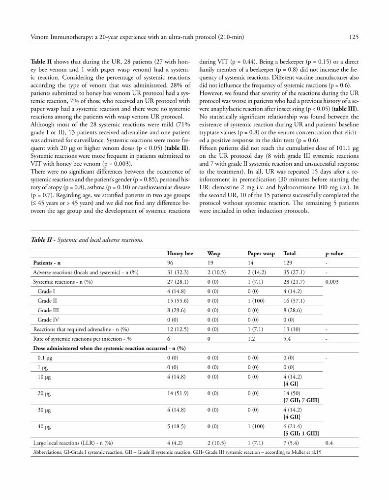
125Venom Immunotherapy: a 20-year experience with an ultra-rush protocol (210-min)
Table II shows that during the UR, 28 patients (27 with hon-ey bee venom and 1 with paper wasp venom) had a system-ic reaction. Considering the percentage of systemic reactions according the type of venom that was administered, 28% of patients submitted to honey bee venom UR protocol had a sys-temic reaction, 7% of those who received an UR protocol with paper wasp had a systemic reaction and there were no systemic reactions among the patients with wasp venom UR protocol. Although most of the 28 systemic reactions were mild (71% grade I or II), 13 patients received adrenaline and one patient was admitted for surveillance. Systemic reactions were more fre-quent with 20 μg or higher venom doses (p < 0.05) (table II). Systemic reactions were more frequent in patients submitted to VIT with honey bee venom (p = 0.003). There were no significant differences between the occurrence of systemic reactions and the patient’s gender (p = 0.85), personal his-tory of atopy (p = 0.8), asthma (p = 0.10) or cardiovascular disease (p = 0.7). Regarding age, we stratified patients in two age groups (≤ 45 years or > 45 years) and we did not find any difference be-tween the age group and the development of systemic reactions
during VIT (p = 0.44). Being a beekeeper (p = 0.15) or a direct family member of a beekeeper (p = 0.8) did not increase the fre-quency of systemic reactions. Different vaccine manufacturer also did not influence the frequency of systemic reactions (p = 0.6). However, we found that severity of the reactions during the UR protocol was worse in patients who had a previous history of a se-vere anaphylactic reaction after insect sting (p < 0.05) (table III).No statistically significant relationship was found between the existence of systemic reaction during UR and patients’ baseline tryptase values (p = 0.8) or the venom concentration that elicit-ed a positive response in the skin tests (p = 0.6).Fifteen patients did not reach the cumulative dose of 101.1 μg on the UR protocol day (8 with grade III systemic reactions and 7 with grade II systemic reaction and unsuccessful response to the treatment). In all, UR was repeated 15 days after a re-inforcement in premedication (30 minutes before starting the UR: clemastine 2 mg i.v. and hydrocortisone 100 mg i.v.). In the second UR, 10 of the 15 patients successfully completed the protocol without systemic reaction. The remaining 5 patients were included in other induction protocols.
Table II - Systemic and local adverse reactions.
Honey bee Wasp Paper wasp Total p-value
Patients - n 96 19 14 129 -
Adverse reactions (locals and systemic) - n (%) 31 (32.3) 2 (10.5) 2 (14.2) 35 (27.1) -
Systemic reactions - n (%) 27 (28.1) 0 (0) 1 (7.1) 28 (21.7) 0.003
Grade I 4 (14.8) 0 (0) 0 (0) 4 (14.2)
Grade II 15 (55.6) 0 (0) 1 (100) 16 (57.1)
Grade III 8 (29.6) 0 (0) 0 (0) 8 (28.6)
Grade IV 0 (0) 0 (0) 0 (0) 0 (0)
Reactions that required adrenaline - n (%) 12 (12.5) 0 (0) 1 (7.1) 13 (10) -
Rate of systemic reactions per injection - % 6 0 1.2 5.4 -
Dose administered when the systemic reaction occurred - n (%)
0.1 μg 0 (0) 0 (0) 0 (0) 0 (0) -
1 μg 0 (0) 0 (0) 0 (0) 0 (0)
10 μg 4 (14.8) 0 (0) 0 (0) 4 (14.2)[4 GI]
20 μg 14 (51.9) 0 (0) 0 (0) 14 (50)[7 GII; 7 GIII]
30 μg 4 (14.8) 0 (0) 0 (0) 4 (14.2)[4 GII]
40 μg 5 (18.5) 0 (0) 1 (100) 6 (21.4)[5 GII; 1 GIII]
Large local reactions (LLR) - n (%) 4 (4.2) 2 (10.5) 1 (7.1) 7 (5.4) 0.4
Abbreviations: GI-Grade I systemic reaction, GII – Grade II systemic reaction, GIII- Grade III systemic reaction – according to Muller et al.19

126 J. Cosme, A. Spínola-Santos, M.C. Pereira-Santos, M. Pereira-Barbosa
There were 7 LLR (table II): 4 with honey bee venom, 2 with wasp and one with paper wasp. From these, one reaction was identified after the administration of the 30-μg and the remain-ing after the 40-μg injection. All patients with LLR finished the 210-minutes UR in the first attempt. These reactions were more frequent in females (p = 0.02). We did not find any other factor that had a significant association with the occurrence of LLR.
Discussion
This study is a retrospective survey of the last 20 years of all patients undergoing VIT using a 210-minutes UR protocol. From a total of 129 patients included, 114 (88%) achieved the dose of 101.1 μg in the planned period of time, while 15 (12%) patients did not complete the UR in the first attempt. Of these, two thirds (n = 10) reached the cumulative dose of 101.1 μg in a further UR, 15 days later, with a premedication reinforcement and using the same UR protocol.In contrast to other studies published to date (16,17,21,22) our study has much more patients submitted honey bee VIT, and also 13 patients that underwent an UR protocol with paper wasp venom. These are particular aspects related to the environ-mental exposure to insects’ stings in our country.The UR protocols allow a rapid desensitization and are as-sociated with fewer injections and hospital visits. Addition-ally, they are associated with a reduced risk of anaphylaxis in case the patient is re-stung before reaching the protective dose
(1,10,14). Despite being safe, these fast protocols are not to-tally risk free, as they can be associated with systemic and/or local adverse reactions.Regarding the systemic reactions, we documented a frequency of systemic reactions of 22%, most of them being mild. We also found that only 10% of all our patients were treated with adrenaline during the protocol, and no fatalities were reported.
The frequency of systemic reactions during VIT UR protocols (≤ 210 minutes) reported in previously published studies ranges from 0% to 30% (14,21,22,23).Our results are similar to the ones reported by Birnbaum et al (21), who documented a frequency of systemic reactions of 30% in a subset of patients treated with honey bee ven-om and of 6.1% in patients treated with wasp venom, using a 210-minutes UR protocol. Roll et al (22), on the other hand, reported a lower frequency of systemic reactions. These au-thors analyzed 67 patients (total of 80 UR procedures) that received VIT with honey bee or wasp venoms and found a total of 12.5% of systemic adverse reactions (bee 5% and wasp 7.5%). Although, in this study (22), the overall percentage of adverse reactions is inferior to ours, the number of reactions in patients treated with wasp VIT is higher than the number of reactions that occurred in patients treated with honey bee VIT. This data is not in agreement to what is published in most studies where bee venom alone is a risk factor for systemic reactions (1,24,25,26). An explanation for this difference in systemic reactions between honey bee and wasp VIT could be the degree of purity of the Hymenoptera venom vaccines. It has been demonstrated through laboratory studies that honey bee vaccines have a lower concentration of non-allergenic pro-teins, whereas in the wasp vaccines the allergenic proteins are diluted with non-allergenic proteins from the venom bag (11).Several studies have been developed in order to identify possible clinical or laboratory risk factors for the occurrence of systemic adverse reactions (24,26). In our study, in addition to the venom administered, we verified that there is a relationship between the severity of anaphylaxis after honey bee sting and the severity of the systemic reaction during UR with honey bee venom (p = 0.04). Birnbaum et al. (24) also demonstrated that patients who had grade III or IV anaphylaxis after Hymenoptera sting more frequently devel-
Table III - Relation between the severity of the reaction after Hymenoptera sting and the severity of anaphylactic reaction during VIT-UR.
Severity of the reaction@ after insect sting ⁄ Severity of the reaction @during VIT-UR*
Severity of the reaction after insect sting
Grade I(n=13)
Grade II(n=35)
Grade III(n=37)
Grade IV(n=44)
Seve
rity
of
the
reac
tion
du
ring
the
VIT
-UR
Without reaction (n=101) 13⁄11
35⁄25
37⁄30
44⁄35
Grade I 13⁄1
35⁄3
37⁄0
44⁄0
Grade II 13⁄1
35⁄7
37⁄2
44⁄6
Grade III 13⁄0
35⁄0
37⁄5
44⁄3
Grade IV 13⁄0
35⁄0
37⁄0
44⁄0
*p-value <0.05

127Venom Immunotherapy: a 20-year experience with an ultra-rush protocol (210-min)
oped a grade III or IV systemic reaction during the UR. This data may point out to the need of a reinforcement in premedica-tion before UR in the subset of patients with a previous history of more severe systemic reaction after insect sting.We did not find any relationship between the occurrence of systemic reactions and the patients’ age (p = 0.44), gender (p = 0.85), personal history of atopy (p = 0.8), asthma (p = 0.1) or of cardiovascular disease (p = 0.7). Besides this, no statistically significant relationship was found between the ex-istence of systemic reactions during the UR protocol and the results of the skin tests (p = 0.6) or the level of the baseline tryptase (p = 0.8). These data are in line with previous reports (5,24,25,27,28).Regarding LLR, we also did not find any significant association between their occurrence and the patients’ age, previous history of atopy, asthma or cardiovascular disease or, also, with the type of venom administered to the patient. However, as previous-ly reported (20,29), we found that LLR were more frequent in females (p = 0.02). However, there were only 7 LLR reactions which limits interpretation of these results.Another aspect that deserves mention is the fact that 10 of the 15 patients who did not complete the UR in a first attempt, achieved the 101.1 μg dose in a second UR session, which increased our success rate to 96%. It was not possible to identify any risk factors that led to the failure of the first UR in these patients.
Conclusion
In conclusion, over the past 20 years, in our Immunoallergology Department, 129 patients underwent a 210-minutes VIT UR protocol, with an overall completion rate of 96%, considering the 114 patients that completed VIT UR at a first attempt and the 10 patients that completed it at a second attempt with pre-medication reinforcement. This protocol was carried out in the Day Hospital and was performed by trained medical staff with quick access to the necessary equipment for the treatment of an anaphylactic reaction. UR protocols allow a quicker achieve-ment of the protective dose; however, it is not a risk-free proce-dure. In our study, we documented a frequency of 22% of sys-temic reactions, most of them being mild and without need for adrenaline. The only predictive factors for a systemic reaction that we found in our study were the use of honey bee venom and the severity of systemic insect sting reaction.
Acknowledgements
The authors would like to thank to Elisa Pedro, Insect Allergy Clinic coordinator in CHULN, for letting us have access to all patients’ data and also for the scientific revision of the paper. We also like to thank Manuel Branco Ferreira for the scientific revision of the manuscript.
Conflict of Interest
The authors declare that they have no conflict of interest
References
1. Sturm GJ, Varga EM, Roberts G, Mosbech H, Bilò MB, Akdis CA et al. EAACI guidelines on allergen immunotherapy: Hymenop-tera venom allergy. Allergy 2018; 73(4):744-764.
2. Biló BM, Rueff F, Mosbech H, Bonifazi F, Oude-Elberink JN. Diagnosis of Hymenoptera venom allergy. Allergy 2005; 60(11):1339-1349.
3. Jakob T, Rafei-Shamsabadi D, Spillner E, Müller S. Diagnostics in Hymenoptera venom allergy: current concepts and developments with special focus on molecular allergy diagnostics. Allergo J Int 2017; 26(3):93-105.
4. Jennings A, Duggan E, Perry IJ, Hourihane JO. Epidemiology of allergic reactions to hymenoptera stings in Irish school children. Pediatr Allergy Immunol 2010; 21(8):1166-1170.
5. Incorvaia C, Senna G, Mauro M, Bonadonna P, Marconi I, Asero R, et al. Prevalence of allergic reactions to Hymenoptera stings in northern Italy. Eur Ann Allergy Clin Immunol 2004; 36(10):372-374.
6. Muller U, Heibling A, Berchtold E. Immunotherapy with honey-bee venom and yellow jacket venom is different regarding efficacy and safety. J Allergy Clin Immunol 1992; 89(2):529-535.
7. Pereira Santos MC, Pedro E, Spínola Santos A, Branco Ferreira M, Palma Carlos ML, Palma Carlos AG. Immunoblot studies in allergic patients to hymenoptera venom before and during immu-notherapy. Eur Ann Allergy Clin Immunol 2005; 37(7):273-278.
8. Rueff F, Przybilla B. Ultrarush immunotherapy in patients with Hymenoptera venom allergy. J Allergy Clin Immunol 2001; 107(5):928-929.
9. Brehler R, Wolf H, Kutting B, Schnitker J, Luger T. Safety of a two-day ultrarush insect venom immunotherapy protocol in com-parison with protocols of longer duration and involving a larger number of injections. J Allergy Clin Immunol 2000; 105(6 Pt 1):1231-1235.
10. Wenzel J, Meissner-Kraemer M, Bauer R, Bieber T, Gerdsen R. Safety of rush insect venom immunotherapy. The results of a ret-rospective study in 178 patients. Allergy 2003; 58(11):1176-1179.
11. Bonifazi F, Jutel M, Biló BM, Birnbaum J, Muller U. Prevention and treatment of hymenoptera venom allergy: guidelines for clini-cal practice. Allergy 2005; 60(12):1459-1470.
12. Ludman SW, Boyle RJ. Stinging insect allergy: current perspectives on venom immunotherapy. J Asthma Allergy 2015; 8:75-86.
13. Verburg M, Oldhoff JM, Klemans RJ, Lahey-de Boer A, de Bru-in-Weller MS, Röckmann H et al. Rush immunotherapy for wasp venom allergy seems safe and effective in patients with mastocyto-sis. Eur Ann Allergy Clin Immunol 2015; 47(6):192-196.
14. Patella V, Florio G, Giuliano A, Oricchio C, Spadaro G, Marone G et al. Hymenoptera Venom Immunotherapy: Tolerance and Effica-cy of an Ultrarush Protocol versus a Rush and a Slow Conventional Protocol. J Allergy (Cairo) 2012; 2012:1921-1992.
15. Gutiérrez Fernández D, Moreno-Ancillo A, Fernández Meléndez S, Domínguez-Noche C, Gálvez Ruiz P, Alfaya Arias T et al. Insect Venom Immunotherapy: Analysis of the Safety and Tolerance of 3 Build-up Protocols Frequently Used in Spain. J Investig Allergol Clin Immunol 2016(6); 26:366-373.

128 J. Cosme, A. Spínola-Santos, M.C. Pereira-Santos, M. Pereira-Barbosa
16. Schiavino D, Nucera E, Pollastrini E, De Pasquale T, Buonomo A, Bartolozzi F et al. Specific ultrarush desensitization in Hymenop-tera venom-allergic patients. Ann Allergy Asthma Immunol 2004; 92(4):409-413.
17. Bożek A, Kołodziejczyk K. Safety of specific immunotherapy us-ing an ultra-rush induction regimen in bee and wasp allergy. Hum Vaccin Immunother 2018; 14(2):288-291.
18. Gruzelle V, Ramassamy M, Bulai Lidiveanu C, Didier A, Mailhol C, Guilleminault L. Safety of ultra-rush protocols for hymenoptera venom immunotherapy in systemic mastocytosis. Allergy 2018; 73(11):2260-2263.
19. Mueller HL. Diagnosis and treatment of insect sensitivity. J Asth-ma Res 1966; 3(4):331-333.
20. Golden DB, Demain J, Freeman T, Graft D, Tankersley M, Tra-cy J, et al. Stinging insect hypersensitivity: A practice parameter update 2016. Ann Allergy Asthma Immunol 2017; 118(1):28-54.
21. Birnbaum J, Ramadour M, Magnan A, Vervloet D. Hymenoptera ultra-rush venom immunotherapy (210 min): a safety study and risk factors. Clin Exp Allergy 2003; 33(1):58-64.
22. Roll A, Hofbauer G, Ballmer-Weber BK, Schmid-Grendelmeier P. Safety of specific immunotherapy using a four-hour ultra-rush induction scheme in bee and wasp allergy. J Investig Allergol Clin Immunol 2006; 16(2):79-85.
23. Nittner-Marszalska M, Cichocka-Jarosz E, Małaczyńska T, Kraluk B, Rosiek-Biegus M, Kosinska M et al. Safety of Ultrarush Venom
Immunotherapy: Comparison Between Children and Adults. J In-vestig Allergol Clin Immunol 2016; 26(1):40-47.
24. Birnbaum J, Charpin D, Vervloet D. Rapid Hymenoptera venom immunotherapy: comparative safety of three protocols. Clin Exp Allergy 1993; 23(3):226-230.
25. Bousquet J, Ménardo JL, Velasquez G, Michel FB. Systemic reac-tions during maintenance immunotherapy with honey bee venom. Ann Allergy 1988; 61(1):63-68.
26. Lockey RF, Turkeltaub PC, Olive ES, Hubbard JM, Baird-Warren IA, Bukantz SC. The Hymenoptera venom study. III: Safety of venom immunotherapy. J Allergy Clin Immunol 1990; 86(5):775-780.
27. Verburg M, Oldhoff JM, Klemans RJ, Lahey-de Boer A, de Bru-in-Weller MS, Röckmann H et al. Rush immunotherapy for wasp venom allergy seems safe and effective in patients with mastocyto-sis. Eur Ann Allergy Clin Immunol 2015(6); 47:192-196.
28. Mosbech H, Mueller U. Side-effects of insect venom immuno-therapy: results from an EAACI multicenter study. European Academy of Allergology and Clinical Immunology. Allergy 2000; 55(11):1005-1010.
29. Rueff F, Przybilla B, Bilo MB, Müller U, Scheipl F, Aberer W, et al. Predictors of side effects during the build-up phase of venom immunotherapy for Hymenoptera venom allergy: the impor-tance of baseline serum tryptase. J Allergy Clin Immunol 2010; 126(1):105-111.
L E T T E R TO T H E E D I T O R Eur Ann AllErgy Clin immunol Vol 51, n 3, 129-130, 2019
a. tammaro1, F. magri1, c. chello1, D. giorDano1, F.r. PariSella2, g. De marco1, S. PerSechino1
A successful topical treatment for cutaneous inflammatory diseases: an additional or alternative therapy to topical steroids
1Dermatology Unit, NESMOS Department, S. Andrea Hospital, University of Rome “Sapienza”, Rome, Italy2University of Queensland, Brisbane, Australia
Keywords
Cream; Psoriasis; Eczema; Omega
Corresponding authorAntonella TammaroSant’Andrea HospitalVia di Grottarossa 1035, 00189 Roma, Italy Phone: +39 06 3377 5907E-mail: [email protected]
Doi10.23822/EurAnnACI.1764-1489.79
Exclusion criterion was a proven allergy to one of the three components of the cream: hemp seed oil, macadamia oil, rosa mosqueta oil. The patients applied 1 ml of the cream, twice a day, on the inflammated lesions. Patients came to control visit after 1, 3, 7 and 12 weeks. The evaluation of the treatment efficacy has been estimated with a three-levels score: complete healing, par-tial healing (using %) with description, absence of visible effect.The emollient and hydrating cream of the study was composed by water, cetearyl alcohol, cannabis sativa seed oil, rosa moscha-ta seed oil, macadamia ternifolia seed oil, panthenol, propylene glycol, hydrolyzed glycosaminoglycans, proline, magnesium aspartate, citric acid, disodium EDTA, cetrimonium chloride, 2-bromo-2-nitropropane-1,3 diol, phenoxyethanol.After 12 weeks, we observed a complete healing with restitutio ad integrum in 30 patients (figure 1 and 2). Among this group, one patient was affected by radiodermitits and another one by ex-
To the Editor
Topical steroids are commonly used in several cutaneous pathol-ogies, such as psoriasis, atopic dermatitis, irritative and allergic contact dermatitis, particularly in acute phase and often for long periods. However, topical corticosteroids can be associated with adverse effects, such as acneiform lesions, erythema, teleangiec-tasias, hypertrichosis, perioral dermatitis, photosensitivity and burning sensation (1). The goal of our study was to find an alternative non-steroidal an-ti-inflammatory treatment for the management of cutaneous dis-eases. We selected 50 patients to test our non-steroidal inflammato-ry topical product. The patients were affected by mild or moderate psoriasis, atopic dermatitis and irritative contact dermatitis. The patients were not performing neither systemic nor topical treatment at the moment of our observation. The age of our patients (both male and female) was between 20 and 65.

130 A. Tammaro, F. Magri, C. Chello, D. Giordano, F.R. Parisella, G. De Marco, S. Persechino
tramammary vulvar Paget. Both showed complete healing using our cream. In the other 20 patients, we revealed a partial healing. According to the guidelines, steroid creams should be used for limited periods to avoid the onset of side effects (1). The cream of our study was composed by three different oils: cannabis sativa seed oil, rosa moschata seed oil and macadamia ternifolia seed oil. These oils are composed by omega-6 and -3 fatty acids, respectively linoleic acid and α-linolenic acid. Fur-thermore, it contained tocopherol, carotenoids and vitamin A, which have natural antioxidant action.Cutaneous diseases, such as psoriasis and atopic dermatitis, are characterized by an alteration of the skin barrier, which is normally constituted by ceramides, free fatty acids and cholesterol. For this reason, the use of our product, containing essential fatty acids, has been helpful for the reconstitution of the cutaneous barrier.Moreover, the components of our cream had also an anti-in-flammatory effect. Indeed, essential omega-3 fatty acid has an-ti-inflammatory and immune-modulating actions and can be helpful in various conditions: atherosclerosis, blood pressure al-terations, platelet function alterations, rheumatoid arthritis and cutaneous diseases (2).These fatty acids are capable of partly inhibiting many aspects of inflammation, including leucocyte chemotaxis, adhesion mole-cule expression, and leucocyte-endothelial adhesive interactions and production of pro-inflammatory cytokines (3).
In conclusion, our cream showed anti-inflammatory actions and could reintegrate the cutaneous barrier. For this reason, it could be a valid substitute to topical steroids in case of mild or moderate cutaneous lesions. It could be also useful as a co-adju-vant approach in case of patients who are performing a topical or systemic steroid treatment. Lastly, this cream could represent an additional therapy in case of cutaneous lesions, such as radi-odermitits or Paget’s (4).
Conflict of interest
The authors declare that they have no conflict of interest
References
1. 1. Rohini S, Sameer A and Mashqoor W. Misuse of topical corti-costeroids on facial skin. A study of 200 patients. J Dermatol Case Rep 2017; 11(1):5-8.
2. 2. Mori TA, Beilin LJ. Omega-3 fatty acids and inflammation. Curr Atheroscler Rep 2004; 6(6):461-467.
3. 3. Calder PC. Omega-3 fatty acids and inflammatory processes: from molecules to man. Biochem Soc Trans 2017 Sep 12. pii: BST20160474. doi: 10.1042/BST20160474.
4. 4. Tammaro A, Giulianelli V, Romano I, DE Vito E, Parisella FR, Giubettini M, Persechino S. Successful use of radiotherapy in the treatment of extramammary Paget’s disease in vulvar area. G Ital Dermatol Venereol 2017; 152(2):181-118.
Figure 2 - Cutaneous aspect after 12-week topical therapy.Figure 1- Bilateral, erythematous and squamous lesions of the palms.

L E T T E R TO T H E E D I T O R Eur Ann AllErgy Clin immunol Vol 51, n 3, 131-134, 2019
a.i. lorente-laVirgen1, c. almeiDa2, J. BernaBeu1, V. Valero3, r. lorente3
Methylchloroisothiazolinone / methylisothiazolinone: epidemiological retrospective study1Dermatology Department, Virgen del Rocio University Hospital, Seville, Spain2Statistical and Methodology Department, FISEVI, Seville University, Seville, Spain3Industrial Engineering School, Extremadura University, Badajoz, Spain
Keywords
allergic contact dermatitis; atopic dermatitis; preservants; methylchloroisothiazolinone / methylisothiazolinone (MCI/ MI)
Corresponding authorAna I. Lorente-LavirgenDermatology Department Virgen del Rocio University HospitalAvda. Manuel Siurot, S/n41013 Sevilla, SpainPhone: +34 661 570 058Fax: +34 661 570 058E-mail: [email protected]
Doi10.23822/EurAnnACI.1764-1489.92
certified allergen supplied by True Test® (TRUE test; Mekos Laboratories ApS, Denmark). Patches were applied to the up-per back using Scanpore® and left on for 48 h; then read on day D2 (48 h), and D4 (96h). We considered positive responses those in which erythema, infiltrated papules or vesicles were de-tected. All patients’ personal data were firstly anonymized and manually entered into a computerized database (Microsoft Ex-cel 2011 for Mac, version 14.0.0). Data were analysed with the Statistical Package for Social Sciences (SPSS) program 25.0; sev-eral statistical analyses were performed when needed (chi-square test), and p < 0.05 was considered statistically significant. This study has been prepared respecting the fundamental principles established in the Declaration of Helsinki (1946). The research project has undergone evaluation by the Committee of Ethics and research clinic of our University Hospital and all patients have been informed about the project.
To the Editor
Allergic contact dermatitis (ACD) is a common problem con-fronted in clinical practice and several authors have started to point to MCI/MI as an emerging allergen primary present in many daily cosmetic (1,2) since late 80s. The present study was carried out to described the epidemiological characteristic of patients with ACD to MCI/MI during a 10 years period. We want to elucidate factors associated and identify the clinical and epidemiological features of patients with ACD to MCI/MI. During a period of ten years, every adult patient with signs or symptoms of eczema attending our Contact Dermatitis De-partment, located in the University Hospital Virgen del Rocío (Seville, Spain), were recruited. They underwent a clinical his-tory, dermatological exploration and patch test with the stan-dard series of the Spanish Contact Dermatitis and Skin Allergy Research Group (GEIDAC) (3). All patients were tested with

132 A.I. Lorente-Lavirgen, C. Almeida, J. Bernabeu, V. Valero, R. Lorente
A total of 142 patients had a positive patch test reaction to MCI/MI. The allergic contact dermatitis to MCI/MI diagnosis was based on medical history and a relevant patch test reaction, which was found in 118 patients, representing 8% of the total.The age range of patients was from 14 to 89 years (mean age 51 years); 58.5% of the patients were males (n = 69) and 41.5% (n = 49) were females. By age group, sensitization to MCI/MI was more frequent in adults over 40 years old (79.6%). The most frequent location was hands (62.9%, n = 73), followed by diffuse involvement of different body parts (16.4%, n = 21); 11.3% of patients had facial lesions (n = 13), 4.3% (n = 5) had axillar eczema, 4.3% (n = 5) had foot eczema, and only 0.9% (n = 1) had genitalia-perianal eczema. No statistically significant differences were found.Furthermore, 12.7% had a pre-patch test diagnosis of atopic dermatitis (n = 15), 8.5% psoriasis (n = 10), and 0.9% sebor-rheic dermatitis (n = 1). We found that patients with atopic dermatitis had three times increased relative risk to and tested positive for MCI/MI with statistically significant differences (p = 0.016, OR 3, confidence interval of 95% [1.2/6.4]).Of the patients, 70.7% (n = 83) were in careers with manual ac-tivities involving wet work, compared with 29.3% (n = 35) who did not use their hands in their professional activities. The most frequently registered profession was cleaners/janitors (40.2%, n = 37), followed by office workers (19.6%, n = 18). Only 5 pa-tients were identified as having occupational dermatitis (4%), of which 2 were related to the use of paints and 3 were associated with cosmetic or household products used in their work. No statistically significant differences were found between locations or professions and a positive sensitization to MCI/MI.The most common sources of exposure related to MCI/MI found in our group were bath products (46.6%, n = 55), baby wipes (26.27%, n = 31), household cleaning products (14.40%, n = 17), and beauty products (12.70%, n = 15).Finally, we studied whether patients with positive patch test re-sults for MCI/MI had concomitant sensitization to other sub-stances, particularly other preservatives. These data are included in table I. We found that patients with positive patch test results for Quaternium 15 had an increased relative risk to or had sen-sitization to MCI/MI with statistically significant differences (p < 0.0001, OR 8, IC 95% [3.7/17.8]). Furthermore, patients with positive patch test results for formaldehyde also had an increased relative risk sensitization to MCI/MI with statistically significant differences (p < 0.0001, OR 6, IC 95% [2.5/13.2]).Finally, in the MOAHLFA index (male, occupational derma-titis, atopic dermatitis, hand, leg, > 40 years old), 58.5% were men, 4.24% had occupational dermatitis, 57.7% had atopic dermatitis, 62.9% had hand eczema, 0% had leg eczema, 9.5% had facial eczema, and 79.6 % were over 40 years old. A recent meta-analysis published in 2018 revealed that at least 20% of the general population have contact-allergic dermatitis
due to common environmental allergens (4). Hypersensitivity reactions to metals are one of the most common (5), but a con-cerning increased sensitization to MCI/MI had been detected since the late ’80s. Lundov et al. (6,7) revealed that MI ACD is estimated to have a prevalence of 1.5% in 2011, which has been increasing in the next years, mainly affecting middle-aged women with hand and facial dermatitis most often associated with cosmetics. In 2014, Aerts et al. showed an increase in sensi-tization rate to MCI/MI of 4.5% (7). Our group’s sensitization rate to MCI/MI is 8%, significantly higher than in other papers (6,7,8). Most studies on ACD to MCI/MI revealed a predom-inance in women (7,9) whereas in our study group, we found little predominance of males affected (58.5%, n = 69). We think this can be a point of confusion because men were asked more frequently in our study sample. Atopic dermatitis was the most common dermatological disease registered in our series in con-trast with dyshidrotic eczema in the de Unamuno series, which studied a population similar to ours (10). Furthermore, we de-scribed that patients with AD had three times higher risk to sensitization to MCI/MI probably associated with skin barrier alterations in this group of patients.The most frequent concomitant allergen in the de Unamuno (10) group and other series was nickel sulfate whereas it was formaldehyde and Quaternium 15 in our study group, although we also detected a high number of patients with concomitant positive patch test to disclosure cobalt and nickel sulfate (table I). In fact, we want to emphasize that it is particularly interesting that formaldehyde and the formaldehyde releaser Quaternium 15 are not only allergens more often concomitant with MCI/MI sensitization, but we also found statistically significant dif-ferences between these allergens and MCI/MI. These substanc-es are preservatives used to prevent contamination in cosmetics and household products. Warshaw et al. (5) reported in 2013 that formaldehyde, Quaternium 15, and MCI/MI were the pre-servatives that most frequently tested positive to the patch test in the North American Contact Dermatitis Group (NACDG) (5). A positive sensitization to MCI/MI was much more preva-lent in janitors and cleaners (n = 37, 40.2%), followed by office workers (n = 18, 19.6%) and builders (n = 8, 8.7%). Thus, we conclude that humidity affects the process of allergic contact dermatitis, but MCI/MI is so prevalent in daily cosmet-ic products that further studies are needed to elucidate whether this is associated with professions or daily contact with cosmetic and cleaning products. In addition, our result supports that the prevalence of MCI/MI is increasing in all developed countries. Many other studies are necessary to explain risk factors for ACD to preservatives.
Conflict of interest
The authors declare that they have no conflict of interest.

133Methylchloroisothiazolinone / methylisothiazolinone: epidemiological retrospective study
Table I - Patients with positive patch test to MCI/MI and concomitant sensitisation to other substance in patch-test.
Allergens MCI/MI negative (N)
MCI/MI negative (%)
MCI/MI positive (N)
MCI/MI positive (%)
potassium dichromate negative 1252 91.7% 110 93.2%
positive 114 8.3% 8 6.8%
Caine mix negative 1349 98.8% 113 95.8%
positive 17 1.2 % 5 4.2%
fragrance mix negative 1302 95.3% 110 93.2%
positive 64 4.7% 8 6.7%
balsam of Peru negative 1333 97.6% 113 95.8%
positive 33 2.4% 5 4.2%
cobalt dichloride negative 1260 92.2% 109 92.4%
positive 108 7.8% 9 7.6%
paraben mix negative 1357 99.3% 117 99.2%
positive 9 0.7% 1 0.8%
carba mix negative 1338 98.0% 112 94.9%
positive 28 2.% 6 5.1%
Quaternium-15 negative 1349 98.8% 107 90.7%
positive 17 1.2% 11 9.3%
p-phenylenediamine negative 1317 96.4% 113 95.8%
positive 49 3.6% 5 4.2%
formaldehyde negative 1347 98.6% 109 92.4%
positive 19 1.4% 9 7.6%
thiuram mix negative 1345 98.5% 116 98.3%
positive 21 1.5% 2 1.7%
diazolidinyl urea, Germall II negative 1358 99.4% 117 99.2%
positive 8 0.6% 1 0.8%
imidazolidinyl urea, Germall 115 negative 1361 99.6% 117 99.2%
positive 5 0.4% 1 0.8%
Funding/Support
No commercial funding or material support was received for this investigation.
References
1. Garcia-Gavin J, Vansina S, Kerre S, et al. Methylisothiazolinone, an emerging allergen in cosmetics? Contact Dermat 2010; 63:96-101.
2. Gardner KH, Davis MD, Richardson DM, et al. The hazards of moist toilet paper: allergy to the preservative methylchloroiso-thiazolinone / methylisothiazolinone. Arch Dermatol 2010; 146:886-890.
3. Hervella-Garces M, García-Gavín J, Silvestre-Salvador JF, et al. The Spanish standard patch test series: 2016 update by the Span-ish Contact Dermatitis and Skin Allergy Research Group (GEI-DAC). Actas Dermosifiliogr 2016; 107:559-566.
4. Alinaghi F, Bennike NH, Egeberg A, Thyssen JP, Johansen JD. Prevalence of contact allergy in the general population: A sys-tematic review and meta-analysis. Contact Dermatitis 2018; 29 doi:10.1111/cod.13119.
5. Warshaw EM, Belsito DV, Taylor JS, et al. Dermatitis. North American Contact Dermatitis Group patch test results: 2009 to 2010 Dermatitis 2013; 24:50-59.
6. Lundov MD, Krongarrd T, Menn’e TL. Johansen JD. Methy-isothiazolinone contact allergy: a review. Br J Dermatol 2011; 165:1178-1182.

134 A.I. Lorente-Lavirgen, C. Almeida, J. Bernabeu, V. Valero, R. Lorente
7. Aerts O, Baexk M, Constandt L et al. The dramatic increase in the rate of methylisothiazolinone contact allergy in Belgium: a multi-centre study. Contact Dermatitis 2014; 71:41-48.
8. Wilkinson JD, Shaw S, Anderson KE, et al. Monitoring levels of preservative sensitivity in Europe. A 10-year overview (1991-2000). Contact Dermatitis. 2002; 46:207-210.
9. Murad A, Marren P. Prevalence of methylchloroisothiazolinone and methylisothiazolinone contact allergy in facial dermatitis: a single centre Irish study. J Eur Acad Dermatol 2016; 30:60-62.
10. B. de Unamuno, V. Zaragoza Ninet, C. Sierra y J. de la Cuadra. Estudio descriptivo de la sensibilización a metilcloroisotiazolin-ona/metilisotiazolinona en una unidad de alergia cutánea. Actas Dermosifiliogr 2014; 105:854-859.

L E T T E R TO T H E E D I T O R Eur Ann AllErgy Clin immunol Vol 51, n 3, 135-137, 2019
D. BagnaSco1, t. aloè1, F. ScliFò2, m.g. Ferrantino3, F. marugo4, F. arcaDiPane3, a. manFreDi1, g.W. canonica5, g. PaSSalacqua1
When to stop biologicals. Severe asthma exacerbation after mepolizumab discontinuation
1Allergy and Respiratory Diseases, IRCCS Policlinico San Martino, University of Genoa, Genoa, Italy2Emergency Department, Ospedale Policlinico San Martino, Genoa, Italy3Respiratory Diseases, Azienda Ospedaliera Nazionale SS. Antonio e Biagio e C. Arrigo, Alessandria, Italy4Respiratory Diseases, Ospedale La Colletta, Arenzano, Italy5Personalized Medicine Clinic Asthma and Allergy, Humanitas Clinical and Research Center, Department of Biomedical Sciences, Humanitas University, Rozzano, Milan, Italy
Keyword
mepolizumab; real-life; discontinuation; severe asthma; monoclonal antibodies
Corresponding AuthorDiego BagnascoAllergy and Respiratory diseasesIRCCS Policlinico San MartinoLargo Rosanna Benzi 1016132, Genoa ItalyPhone: +39 010 5555 805E-mail: [email protected]
Doi10.23822/EurAnnACI.1764-1489.88
other factors through the action of several cytokines, including interleukin (IL) 5 (4). Severe asthma also involves unrecog-nized or underestimated social costs, for instance due to the effects of systemic corticosteroids or work absenteeism (5). The novel biological drugs for severe asthma are able to reduce the direct and indirect costs, while maintaining a satisfactory control of disease. Mepolizumab (MEP), a humanized mAb against IL-5 was recently tested and commercialized for severe asthmatic patients (6,7). We describe herein the clinical case of a patient with severe hypereosinophilic asthma, successfully treated with MEP, first during the MENSA (6) study, then with its open-label exten-sion and commercial availability, who had a sudden episode of asthma exacerbation after MEP withdrawal after more than 3
To the Editor
Severe asthma affects 5% to 10% of all asthmatic patients (1). To be defined as severe an asthmatic, patient must be treated with the best standard therapies at maximal doses, without achieving a full control, and therefore requiring also oral cor-ticosteroids (OCS) (2). In Western Countries, severe asthma is responsible for at least 50% of the socio-economic burden, including both direct and indirect costs (3). The development of monoclonal antibodies (mAbs) allowed to expand the ther-apeutic approach in patients affected by this disease, especially in reducing the need for OCS and the exacerbation rate. Cer-tainly, the most studied immunological aspect is that linked to the “type 2” inflammation, which involves eosinophils and

136 D. Bagnasco, T. Aloè, F. Sclifò, M.G. Ferrantino, F. Marugo, F. Arcadipane, A. Manfredi, G.W. Canonica, G. Passalacqua
years of therapy. Written informed consent was obtained from the patient for publication of this case report.A never-smoker 64-year-old Caucasian male was followed-up by our clinic since about 10 years due to severe uncontrolled asthma. Asthma appeared around the age of 40, without any previous evidence of respiratory symptoms. Of note, between 40 and 60 years of age, the patient underwent 5 surgical in-terventions for nasal polyposis. Besides, an episode of isolated hypereosinophilia without symptoms was recorded. During clinical and historical examination, we found gastro-oesopha-geal reflux and a positive skin prick test to Parietaria. There was no evidence of occupational exposure to pulmonary pathogens. In the 2 years before enrolling him in MENSA study respira-tory symptoms became increasingly severe, with a significant limitation to daily activity, despite the maximal therapy (flut-icasone / salmeterol 50/500 mcg twice daily, theophylline 300 mg/die, proton-pump inhibitors and nasal steroids). To exclude that symptoms are related to a poor adherence to the therapy, we carefully checked it. In the two years before enrolling him in MENSA, a mean of 4 asthma exacerbations per year were ascertained, with multiple hospital admissions. Finally, at the end of 2012 the patient was enrolled in the MEA 115588 trial (MENSA) (6), and treated with MEP. The baseline characteristic before MEP administra-tion, was an increased number of blood eosinophils (770 cell/μl), a normal lung function, a mild hypertension and a normal saturation. At the beginning of 2013 the patient was random-ized and included in the study. After the first month of therapy a clinical improvement could be observed, with an improvement in FEV
1 (from 2.45 L to 2.87 L). Afterwards the patient con-
tinued, with subjective and clinical benefit, with no more exac-
erbations or systemic steroids need. At the end of the study, he received regularly MEP for compassionate use then, after mar-keting, he continued for other 8 months without symptoms or drug related adverse events.After 4 years and 11 months of therapy, at the end of November 2017, with a full control of asthma symptoms, no exacerbations, no OCS therapy and normal lung function, we attempted MEP discontinuation. After about 2 months of control we could assist to a progressive deterioration: worsening of respiratory symp-toms with dyspnoea even at rest, wheezing, cough, and noctur-nal awakenings due to shortness of breath and a normal haemo-globin saturation. His blood eosinophils level increased to 820 cell/μl and lung function test showed a progressive impairment. A home-based treatment with OCS (50 mg/day Prednisone), aerosolized steroids and bronchodilators was started, without a clinical improvement. The patient was hospitalized and treated with intravenous steroid and aminophylline. During the hos-pitalization, a bacterial superinfection occurred, for which an antibiotic therapy was set. A progressive objective and subjective improvement of symptoms was seen, with a reduction of blood eosinophils, and 500 mL FEV
1 improvement. After this, the
MEP therapy was re-started during hospitalization and, in his clinical stability, patient was discharged. Currently, the patient is on regular MEP treatment, and remains well controlled, with-out exacerbations or OCS treatment needed (Table I).All clinical trials with MEP, after appropriately selecting patients (serum eosinophils > 300/μl), confirmed the positive results in reducing exacerbations (6,7), in sparing OCS (8), in FEV1 improvement and in quality of life (6-8). In the current trials, a reassuring safety profile was also demonstrated with a small number of adverse events, similar to the placebo groups (9).
Table I - Main data at baseline, after MEP therapy (MEA 115588, compassionate use and marketing), during and after exacerbation.
2013 baseline Nov 2017
DIS
CO
NT
INU
ATIO
N
Hospitalization (Feb 2018)
RE
AD
MIN
IST
RAT
ION
8 months later (Oct 2018)
weight 65 kg 69 kg 69 kg 69 kg
height 173 cm 172 cm 172 cm 172 cm
serum eosinophils 770 cell/μl 150 cell/μl 800 cell/μl 110 cell/μl
FEV1 (%) 79% 96% 74% 92%
FEV1 (L) 2.45 L 2.73 L 2.09 L 2.58 L
FVC (%) 85% 121% 104% 115%
FVC (L) 3.38 4.25 L 3.80 L 4.21 L
FEV1/FVC 72 61.5 55 61.3
ACT score 13 24 15 24
exacerbations / 12 monts 4 0 n.a. n.a.
hospitalization 1 0 n.a. n.a.(n.a. = not available)

137When to stop biologicals. Severe asthma exacerbation after mepolizumab discontinuation
The available clinical trials with MEP clearly evidenced the ef-ficacy of the biological drug in reducing exacerbations of severe asthma. One of the most important point to be clarified is how long to continue the administration of MEP (10). In vitro ob-servations suggested a hypothetical risk of rebound of airways eosinophilic inflammation after discontinuation (11). Also, an increased stimulus of TH
2 cells to synthetize IL-5 was shown,
associated to an upregulated expression of IL-5R by eosinophils and a persistence of preformed IL-5 in complex with the drug (12). In vivo, an observational study performed on 56 subject (27 on active arm) who suspend MEP after the trial, reported a slightly greater exacerbation rate at 12 months (non statistically significant) (3.1 vs. 3.9; rate ratio, 1.25; 95% CI, 0.71-1.91; p = 0.54). An increase of overall frequency of severe exacerbations, only in treated patients and not in placebo, from 0.56/patient at 0 to 3 months to 1.2/patient (p = 0.007) at 3 to 6 months (rate ratio, 2.11; 95% CI, 1.76-2.54; p < 0.001). As for exacerbations, also for blood eosinophils count, the increasing of value resulted higher in 0 to 3 months (p < 0.001) and 3 to 6 months (p = 0.004) in treated patients (11). As previously observed in the case herein described, the increase in blood eosinophils paralleled the clinical exacerbation. To better understand also how to continue with the administration of the therapy, the experience in real life will be very useful. Indeed, consistent data are available about the patients enrolled in clinical trials with biologicals, but few data in the real-life setting, especially about the duration of the therapy. In addition, the characteristics of real-life patients resulted to be different from those described in clinical trials (13,14). The duration of the therapy is one of the main challenges in the management of these drugs. In fact, in the asthmatic patient, in which therapy is usually modulated according to the control of symptoms, the behaviour to be applied with biological drugs is not yet defined. Although it is common ground that a suspen-sion of biologicals should be taken into account, at the moment the timing is not clear. With clinical practice it was possible to highlight that, after drug discontinuation, several patients re-sponded by keeping control and others returning to have symp-toms. A careful study of the biological parameters of patients treated with these drugs will be necessary in order to look for biomarkers that allow us to predict control of drug withdrawal.This clinical case is an interesting example of how cautiously MEP should be discontinued in clinical practice, being aware of the risk of exacerbations immediately after withdrawal.
Conflict of Interest
The authors declare that they have no conflict of interest
Acknowledgements
CIPRO (centro interprofessionale pneumologico ricerca ed or-ganizzazione), IRCCS Policlinico San Martino, Genova, Italy.
Funding
No funding has been received for the writing or editing of this manuscript.
References
1. Menzella F, Galeone C, Lusuardi M, Simonazzi A, Castagnetti C, Ruggiero P, Facciolongo N. Near-fatal asthma responsive to mepolizumab after failure of omalizumab and bronchial thermo-plasty. Ther Clin Risk Manag 2017; 13:1489-1493.
2. Chung KF, Wenzel SE, Brozek JL, Bush A, Castro M, Sterk PJ, et al. International ERS/ATS guidelines on definition, evaluation and treatment of severe asthma. Eur Respir J 2014; 43(2):343-373.
3. Accordini S, Corsico AG, Braggion M, Gerbase MW, Gislason D, Gulsvik A, et al. The cost of persistent asthma in Europe: an international population-based study in adults. Int Arch Allergy Immunol 2013; 160(1):93-101.
4. Caminati M, Pham DL, Bagnasco D, Canonica GW. Type 2 im-munity in asthma. World Allergy Organ J 2018; 11(1):13.
5. Barry LE, Sweeney J, O’Neill C, Price D, Heaney LG.The cost of systemic corticosteroid-induced morbidity in severe asthma: a health economic analysis. Respir Res 2017; 18(1):129.
6. Ortega HG, Liu MC, Pavord ID, Brusselle GG, Mark FitzGerald J, Chetta A, et al. Mepolizumab treatment in patients with severe eosinophilic asthma. N Engl J Med 2014; 371:1198-1207.
7. Pavord ID, Korn S, Howarth P, Bleecker ER, Buhl R, Keene ON, et al. Mepolizumab for severe eosinophilic asthma (DREAM): a multicentre, double-blind, placebo-controlled trial. Lancet 2012; 380:651-659.
8. Bel EH, Wenzel SE, Thompson PJ, Prazma CM, Keene ON, Yancey SW, et al. Oral glucocorticoid-sparing effect of mepoli-zumab in eosinophilic asthma. N Engl J Med 2014; 371:1189-1197.
9. Passalacqua G, Matucci A, Vultaggio A, Bagnasco D, Mincarini M, Maggi E, Canonica GW. The safety of monoclonal antibodies in asthma. Expert Opin Drug Saf 2016; 15(8):1087-1095.
10. Emma R, Morjaria JB, Fuochi V, Polosa R, Caruso M. Mepoli-zumab in the management of severe eosinophilic asthma in adults: current evidence and practical experience. Ther Adv Respir Dis 2018; 12:1753466618808490.
11. Haldar P, Brightling CE, Singapuri A, Hargadon B, Gupta S, Monteiro W, et al. Outcomes after cessation of mepolizumab therapy in severe eosinophilic asthma: a 12-month follow-up anal-ysis. J Allergy Clin Immunol 2014; 133(3):921-923.
12. Stein ML, Villanueva JM, Buckmeier BK, Yamada Y, Filipov-ich AH, Assa’ad AH, Rothenberg ME. Anti-IL-5 (mepolizum-ab) therapy reduces eosinophil activation ex vivo and increases IL-5 and IL-5 receptor levels. J Allergy Clin Immunol 2008; 121:1473-1483.
13. Bagnasco D, Milanese M, Rolla G, Lombardi C, Bucca C, Heffler E, et al. Anti-IL-5 therapy in real life. The North-Western Italian experience 3 and comparison with the regulatory trials. World Al-lergy Organization Journal 2018; 11:34.
14. Caminati M, Senna G, Guerriero M, Dama AR, Chieco-Bianchi F, Stefanizzi G, et al. Omalizumab for severe allergic asthma in clinical trials and real-life studies: what we know and what we should address. Pulm Pharmacol Ther 2015; 31:28-35.