Estrogen Protects against Oxidative Multiorgan Damage in Rats with Chronic Renal Failure
16
PLEASE SCROLL DOWN FOR ARTICLE This article was downloaded by: [TÜBTAK EKUAL] On: 20 October 2009 Access details: Access Details: [subscription number 772814176] Publisher Informa Healthcare Informa Ltd Registered in England and Wales Registered Number: 1072954 Registered office: Mortimer House, 37-41 Mortimer Street, London W1T 3JH, UK Renal Failure Publication details, including instructions for authors and subscription information: http://www.informaworld.com/smpp/title~content=t713597293 Estrogen Protects against Oxidative Multiorgan Damage in Rats with Chronic Renal Failure Özgür Kasmay a ; Göksel ener b ; Bar Çakr a ; Meral Yüksel c ; ule Çetinel d ; Gazi Contuk d ; Berrak Ç. Yeen a a Department of Physiology, Marmara University School of Medicine, Istanbul, Türkiye b Department of Pharmacology, Marmara University School of Pharmacy, Istanbul, Türkiye c Department of Medical Laboratory, Vocational School of Health Related Professions, Istanbul, Türkiye d Department of Histology & Embryology, Marmara University School of Medicine, Istanbul, Türkiye Online Publication Date: 01 September 2009 To cite this Article Kasmay, Özgür, ener, Göksel, Çakr, Bar, Yüksel, Meral, Çetinel, ule, Contuk, Gazi and Yeen, Berrak Ç.(2009)'Estrogen Protects against Oxidative Multiorgan Damage in Rats with Chronic Renal Failure',Renal Failure,31:8,711 — 725 To link to this Article: DOI: 10.3109/08860220903134563 URL: http://dx.doi.org/10.3109/08860220903134563 Full terms and conditions of use: http://www.informaworld.com/terms-and-conditions-of-access.pdf This article may be used for research, teaching and private study purposes. Any substantial or systematic reproduction, re-distribution, re-selling, loan or sub-licensing, systematic supply or distribution in any form to anyone is expressly forbidden. The publisher does not give any warranty express or implied or make any representation that the contents will be complete or accurate or up to date. The accuracy of any instructions, formulae and drug doses should be independently verified with primary sources. The publisher shall not be liable for any loss, actions, claims, proceedings, demand or costs or damages whatsoever or howsoever caused arising directly or indirectly in connection with or arising out of the use of this material.
Transcript of Estrogen Protects against Oxidative Multiorgan Damage in Rats with Chronic Renal Failure

PLEASE SCROLL DOWN FOR ARTICLE
This article was downloaded by: [TÜBTAK EKUAL]On: 20 October 2009Access details: Access Details: [subscription number 772814176]Publisher Informa HealthcareInforma Ltd Registered in England and Wales Registered Number: 1072954 Registered office: Mortimer House,37-41 Mortimer Street, London W1T 3JH, UK
Renal FailurePublication details, including instructions for authors and subscription information:http://www.informaworld.com/smpp/title~content=t713597293
Estrogen Protects against Oxidative Multiorgan Damage in Rats with ChronicRenal FailureÖzgür Kasmay a; Göksel ener b; Bar Çakr a; Meral Yüksel c; ule Çetinel d; Gazi Contuk d; Berrak Ç. Yeen a
a Department of Physiology, Marmara University School of Medicine, Istanbul, Türkiye b Department ofPharmacology, Marmara University School of Pharmacy, Istanbul, Türkiye c Department of MedicalLaboratory, Vocational School of Health Related Professions, Istanbul, Türkiye d Department of Histology &Embryology, Marmara University School of Medicine, Istanbul, Türkiye
Online Publication Date: 01 September 2009
To cite this Article Kasmay, Özgür, ener, Göksel, Çakr, Bar, Yüksel, Meral, Çetinel, ule, Contuk, Gazi and Yeen, BerrakÇ.(2009)'Estrogen Protects against Oxidative Multiorgan Damage in Rats with Chronic Renal Failure',Renal Failure,31:8,711 — 725
To link to this Article: DOI: 10.3109/08860220903134563
URL: http://dx.doi.org/10.3109/08860220903134563
Full terms and conditions of use: http://www.informaworld.com/terms-and-conditions-of-access.pdf
This article may be used for research, teaching and private study purposes. Any substantial orsystematic reproduction, re-distribution, re-selling, loan or sub-licensing, systematic supply ordistribution in any form to anyone is expressly forbidden.
The publisher does not give any warranty express or implied or make any representation that the contentswill be complete or accurate or up to date. The accuracy of any instructions, formulae and drug dosesshould be independently verified with primary sources. The publisher shall not be liable for any loss,actions, claims, proceedings, demand or costs or damages whatsoever or howsoever caused arising directlyor indirectly in connection with or arising out of the use of this material.

Renal Failure, 31:711–725, 2009 Copyright © Informa UK Ltd.ISSN: 0886-022X print / 1525-6049 onlineDOI: 10.3109/08860220903134563
711
LRNFLABORATORY STUDY
Estrogen Protects against Oxidative Multiorgan Damage in Rats with Chronic Renal Failure
Estrogen Alleviates CRF-Induced Oxidative DamageÖzgür KasimayMarmara University School of Medicine, Department of Physiology, Istanbul, Türkiye
Göksel SenerMarmara University School of Pharmacy, Department of Pharmacology, Istanbul, Türkiye
Baris ÇakirMarmara University School of Medicine, Department of Physiology, Istanbul, Türkiye
Meral YükselVocational School of Health Related Professions, Department of Medical Laboratory, Istanbul, Türkiye
Sule Çetinel and Gazi ContukMarmara University School of Medicine, Department of Histology & Embryology, Istanbul, Türkiye
Berrak Ç. YegenMarmara University School of Medicine, Department of Physiology, Istanbul, Türkiye
The impact of sex dimorphism on chronic renal failure(CRF)-induced oxidative multiorgan damage and the effects ofestradiol (E2) loss and E2 supplementation on the progress of CRFwere studied. Sprague-Dawley rats underwent 5/6 nephrectomy(CRF), and a group of female rats had bilateral ovariectomy(OVX), while the sham-operated rats had no nephrectomy orOVX. Rats received either estradiol propionate (50 μg/kg/day) orvehicle for six weeks. Serum BUN levels were elevated in bothmale and female CRF groups treated with vehicle, while creati-nine level was not significantly changed in the female CRFgroup. CRF-induced elevation in serum TNF-a of male rats wasabolished when the animals were treated with E2, while OVXexaggerated TNF-a response. In OVX and male rats with CRF,E2 treatment reversed the malondialdehyde elevations in all thestudied tissues (kidney, heart, lung, ileum, brain, liver, and gastroc-nemius muscle), while depletion of glutathione in these tissues
was prevented by E2 treatment. Similarly, increased levels ofmyeloperoxidase activity, lucigenin chemiluminescence, andcollagen in most of the tissues were reversed by E2 treatment.The findings show that the extent of tissue injuries was relativelyless in females, while ovariectomy exacerbated all the indices ofoxidative injury. Moreover, the administration of E2, with itspotent anti-oxidant and anti-inflammatory effects, markedlyimproved CRF-induced systemic inflammatory outcomes in bothmale and female rats by depressing tissue neutrophil infiltrationand modulating the release of inflammatory cytokines.
Keywords estradiol, chronic renal failure, TNF-alpha,myeloperoxidase, lipid peroxidation
INTRODUCTION
Chronic renal failure (CRF) is well known to be asso-ciated with oxidative stress, which promotes the productionof reactive oxygen species (ROS) and cytokine release. InCRF, the imbalance between increased production of ROSand the limited or decreased antioxidant capacity results in apersistent formation of toxic lipid peroxidation products,[1]
Received 30 March 2009; revised 21 May 2009; accepted 21June 2009.
Address correspondence to Berrak Ç. Yegen, M.D., MarmaraUniversity, School of Medicine, Haydarpasa, Istanbul 34668,Türkiye; Tel.: +90 216 414 47 36; Fax: +90 216 414 47 31;E-mail: [email protected]
Downloaded By: [TÜBTAK EKUAL] At: 07:43 20 October 2009

712 Ö. Kasimay et al.
with a subsequent risk of damage to organic moleculessuch as proteins, nucleic acids, and lipids, both in thevicinity of their origin and far from the site of their forma-tion, contributing to the development of serious complica-tions in CRF.[2,3] Data obtained from many experimentalmodels of renal disease in animals indicate that renal dam-age is tolerated differently by the two sexes, while severalepidemiological studies indicate a sexual dimorphism inthe progression of human renal disease.[4] Although theincidence of chronic renal disease is lower in premeno-pausal women compared with age-matched men,[5,6] therisk for development and progression of renal disease inwomen is increased with menopause,[7,8] suggesting theimportant contribution of ovarian sex hormones in the reg-ulation of renal function. There is substantial evidence thatthe sexual dimorphism in kidney aging, with females pro-tected, is associated with beneficial vascular actions[9] andantioxidant properties of estrogens.[10]
Estrogen, which is a fat-soluble hormone that cancontribute to membrane fluidity by direct interactionswith phospholipids, exerts potentially vasoprotective effectsthrough modulation of pro-oxidant and anti-oxidantenzyme expression and activity.[11,12] It has also beensuggested that estrogen can suppress free radical-inducedperoxidation chain reactions because of the similarity instructure to vitamin E, namely, the presence of the hydroxylgroup in the phenolic A ring.[13,14] Substantive datashow that estrogen supplementation decreases oxidativedamage and plays a major role in the protection of theheart,[15] brain,[16,17] liver,[18] and skeletal muscle[19] indifferent models of oxidant injury. We have previouslyreported that 17b-estradiol (E2) protects the liver andintestines against sepsis-induced damage,[20] improvesthe healing of gastric and colonic injury,[21] and amelio-rates burn-induced liver and lung injury in rats.[22]
However, the effect of E2 treatment on the oxidativecomplications of CRF has not (to our knowledge) beenpreviously addressed.
Based on the aforementioned observations, the aim ofthe present study was to determine the impact of sexdimorphism on CRF-induced oxidative multiorgan dam-age, and elucidate the effects of E2 loss in female rats andE2 supplementation in both sexes using biochemical andhistopathological analysis.
MATERIALS AND METHODS
Animals
Both sexes of Sprague Dawley rats (200–250 g) werehoused in a room at a mean temperature of 22 ± 2°C with a12-hour light-dark cycle with free access to standard pellet
chow and water. The study was approved by the MarmaraUniversity Animal Experiments Ethical Committee.
Surgery and Experimental Design
All surgical procedures were performed under ketamineanesthesia (100 mg/kg ketamine and 0.75 mg/kg chlorprom-azine, intraperitoneally) using strict hemostasis and aseptictechniques. In order to induce CRF, male (n = 16) andfemale (n = 24) rats underwent 5/6 nephrectomy by surgicalresection of the upper and lower thirds of the left kidney, fol-lowed by right nephrectomy, as described by Vaziri et al.[23]
During the same operation, a group of female rats (n = 16)had bilateral ovariectomy (OVX). In the sham-operated rats(n = 16), peritoneal cavity was exposed via a ventral abdom-inal incision during which both kidneys in all animals andthe ovaries in females were gently manipulated.
In the CRF groups, rats were injected subcutaneouslywith either estradiol propionate (50 μg/kg/day in olive oil)or olive oil (1 mL/kg) for six weeks, while the sham-operatedrats received injections of oil for the same period. Ratswere decapitated at the end of six weeks, and blood wasobtained for the determination of tumor necrosis factor(TNF)-a, blood urea nitrogen (BUN), and creatinine.Samples from kidney, heart, lung, brain, ileum, liver, andskeletal muscle (m. gastrocnemius) were taken for themeasurement of tissue malondialdehyde and glutathionelevels, myeloperoxidase activity, and reactive oxygen spe-cies. Additional tissue samples were placed in 10% form-aldehyde for histological evaluation and for the determinationof tissue collagen content as an index of tissue fibroticactivity.
Measurement of BUN, TNF-a, and Creatinine Levels
Blood urea nitrogen[24] and serum creatinine[25] concen-trations were determined spectrophotometrically using anautomated analyzer. Serum levels of TNF-a were quantifiedaccording to the manufacturer’s instructions and guidelinesusing enzyme-linked immunosorbent assay (ELISA) kit spe-cific for the previously mentioned rat cytokines (BiosourceInternational, Nivelles, Belgium). These particular assay kitswere selected because of their high degree of sensitivity,specificity, inter- and intra-assay precision, and small amountof serum sample required conducting the assay.
Malondialdehyde and Glutathione Assays
Tissue samples were homogenized with ice-cold150 mM KCl for the determination of malondialdehyde
Downloaded By: [TÜBTAK EKUAL] At: 07:43 20 October 2009
Estrogen Alleviates CRF-Induced Oxidative Damage 713
(MDA) and glutathione (GSH) levels. The MDA levelswere assayed for products of lipid peroxidation by moni-toring thiobarbituric acid reactive substance formation, asdescribed previously.[26] Lipid peroxidation was expressedin terms of MDA equivalents using an extinction coeffi-cient of 1.56 × 10⁵ M−1 cm−1, and results were expressedas nmol MDA/g tissue. GSH measurements were per-formed using a modification of the Ellman procedure.[27]
Briefly, after centrifugation at 2,000 g for 10 min, 0.5 mlof supernatant was added to 2 ml of 0.3 mol/LNa2HPO4.2H2O solution. A 0.2 ml solution of dithiobisni-trobenzoate (0.4 mg/mL 1% sodium citrate) was added,and the absorbance at 412 nm was measured immediatelyafter mixing. GSH levels were calculated using an extinc-tion coefficient of 1.36 × 10⁴ M−1 cm−1. Results wereexpressed in μmol GSH/g tissue.
Measurement of Tissue Myeloperoxidase Activity
The activity of tissue myeloperoxidase (MPO), anenzyme found predominantly in the azurophilic granulesof polymorphonuclear leukocytes (PMN), correlates sig-nificantly with the number of PMN determined histochem-ically.[28] Therefore, MPO activity is frequently utilized toestimate tissue PMN accumulation in inflamed tissues.MPO activity was measured in tissues in a procedure simi-lar to that documented by Hillegas et al.[29] Briefly, tissuesamples were homogenized in 50 mM potassium phos-phate buffer (PB, pH 6.0), and centrifuged at 41,400 g (10min); pellets were suspended in 50 mM PB containing0.5% hexadecyltrimethylammonium bromide (HETAB).After three freeze and thaw cycles, with sonication betweencycles, the samples were centrifuged at 41,400 g for 10 min.Aliquots (0.3 ml) were added to 2.3 ml of reaction mixturecontaining 50 mM PB, o-dianisidine, and 20 mM H2O2solution. One unit of enzyme activity was defined as theamount of MPO present that caused a change in absor-bance measured at 460 nm for 3 min. MPO activity wasexpressed as U/g tissue.
Chemiluminescence (CL) Assay
To assess the role of reactive oxygen species intissue damage, lucigenin chemiluminescence was mea-sured as an indicator of O2
−• formation. Lucigenin (bis-N-methylacridiniumnitrate) was obtained from Sigma(St Louis, Missouri, USA). Measurements were made atroom temperature using Junior LB 9509 luminometer(EG&G Berthold, Germany). Specimens were put intovials containing PBS-HEPES buffer (0.5 M PBS contain-ing 20 mM HEPES, pH 7.2). Counts were obtained at 1 min
intervals and the results were given as the area under curve(AUC) for a counting period of 5 min. Counts was cor-rected for wet tissue weight (rlu/mg tissue).[30]
Collagen Measurement
Tissue collagen was measured as a free radical-induced fibrosis marker. Tissue samples that were cut witha razor blade and fixed in 10% formalin were embedded inparaffin, and 15 μm-thick sections were obtained. The col-lagen content was determined according to the methodpublished by Lopez de Leon and Rojkind,[31] which isbased on the selective binding of the dyes Sirius Red andFast Green FCF to collagen and non-collagenous compo-nents, respectively. Both dyes were eluted readily andsimultaneously using 0.1 N NaOH-methanol (1:1, v/v).Finally, the absorbances at 540 and 605 nm were used todetermine the amount of collagen and protein, respectively.
Histological Analysis
For light microscopic investigations, tissue specimensfrom kidney, heart, lung, brain, ileum, liver, and gastroc-nemius muscle were fixed in 10% formaldehyde, dehy-drated in alcohol series, cleared in toluene, and embeddedin paraffin. Paraffin sections (5 μm) were stained withhematoxylin and eosin (H&E) and examined under a phot-omicroscope (Olympus BH 2, Tokyo, Japan). All tissuesections were examined microscopically for the charac-terization of histopathological changes by an experi-enced histologist who was unaware of the treatmentconditions.
Statistical Analysis
Statistical analysis was carried out using GraphPadPrism 4.0 (GraphPad Software, San Diego, California,USA). Each group consisted of eight animals. All datawere expressed as means ± SEM. Groups of data werecompared with an analysis of variance (ANOVA) followedby Turkey’s multiple comparison tests. Values of p < 0.05were regarded as significant.
RESULTS
Serum BUN and creatinine levels were assessed toevaluate the extent of chronic renal failure. BUN levelswere elevated in both male (p < 0.001) and female (p < 0.05)CRF groups treated with vehicle, while ovariectomy caused
Downloaded By: [TÜBTAK EKUAL] At: 07:43 20 October 2009

714 Ö. Kasimay et al.
an additional increase in BUN of female rats with CRF (p <0.001; see Table 1). Estradiol treatment did not alter BUNlevels in male or female CRF groups. Serum creatininelevel was significantly increased in the vehicle-treated maleCRF group (p < 0.001), but it was not significantly changedin the corresponding female group. Estradiol treatment orOVX did not change creatinine levels in rats with CRF.
In the vehicle-treated male rats with CRF, serumTNF-a level was significantly increased with respect tosham-operated control group (p < 0.01), but this CRF-induced elevation in TNF-a of male rats was abolishedwhen the animals were treated with estradiol (p < 0.01). Infemale CRF rats, similar to male group, TNF-a level wasalso elevated (p < 0.05) when the animals were treatedwith vehicle. On the other hand, this increase in TNF-awas exaggerated in the female rats with OVX (p < 0.001)and reversed by E2 treatment back to the level of non-OVX group (p < 0.001; see Table 1).
In ovariectomized female rats with CRF and inmale CRF rats, MDA levels indicating enhanced lipidperoxidation were increased in all tissues except thegastrocnemius muscle (p < 0.05–0.001; see Figure 1).However, MDA levels in none of the tissues werechanged in female rats with intact ovaries. Estradioltreatment, on the other hand, reversed the MDA eleva-tions in all the studied tissues of both male and femaleCRF groups significantly (p < 0.05–0.001). Gastrocnemiusmuscle MDA levels that were not changed by CRF ineither the female or male rats were further depressedwith estradiol treatment (p < 0.05).
Chronic renal failure resulted in the depletion ofantioxidant GSH in the kidney, brain, ileum, and liverof male and ovariectomized female rats (p < 0.05–0.001),
while estradiol treatment in these groups increased tissueGSH contents back to control levels (p < 0.05–0.001; seeFigure 2). Similarly, decreased GSH (p < 0.01) in thegastrocnemius muscle of male CRF rats was replenishedback to control levels by E2 treatment (p < 0.01). How-ever, skeletal muscle and cardiac GSH contents were notchanged by CRF, OVX, or E2 in the female rats. In thelung and heart tissues of male CRF rats, GSH content wasnot changed with respect to control group, but was furtherelevated by estradiol treatment (p < 0.01). Likewise,reduced pulmonary GSH in the female CRF rats withOVX was elevated by E2 treatment (p < 0.01).
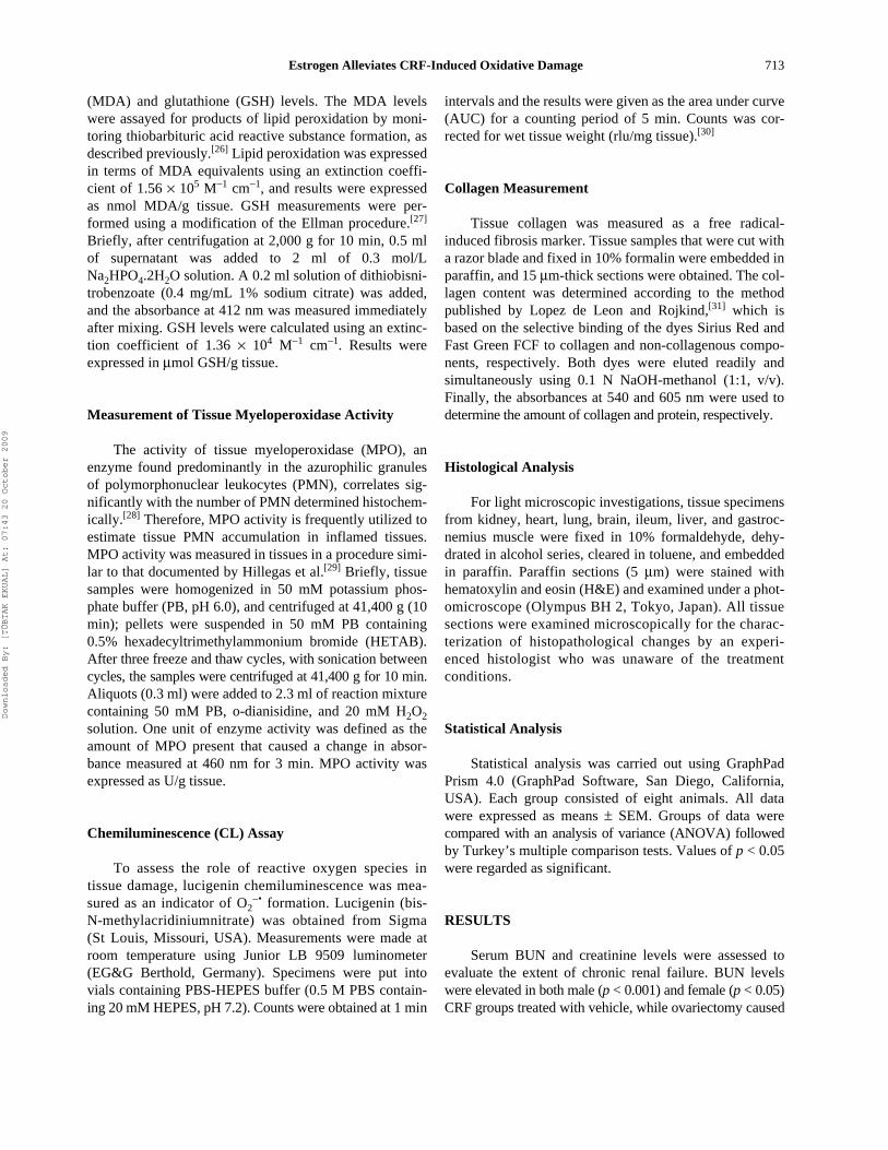
Myeloperoxidase activity representing the poly-morphonuclear leukocyte accumulation in the target tissuewas significantly elevated in all the studied tissues of maleCRF rats (p < 0.05–0.001), while E2 treatment reducedMPO activity in all of these tissues except the gastrocne-mius muscle (p < 0.05–0.001; see Figure 3). Similarly,increased MPO activity observed in all tissues of femaleCRF rats with OVX and in the muscle of CRF rats withintact ovaries was depressed by E2 (p < 0.01–0.001).
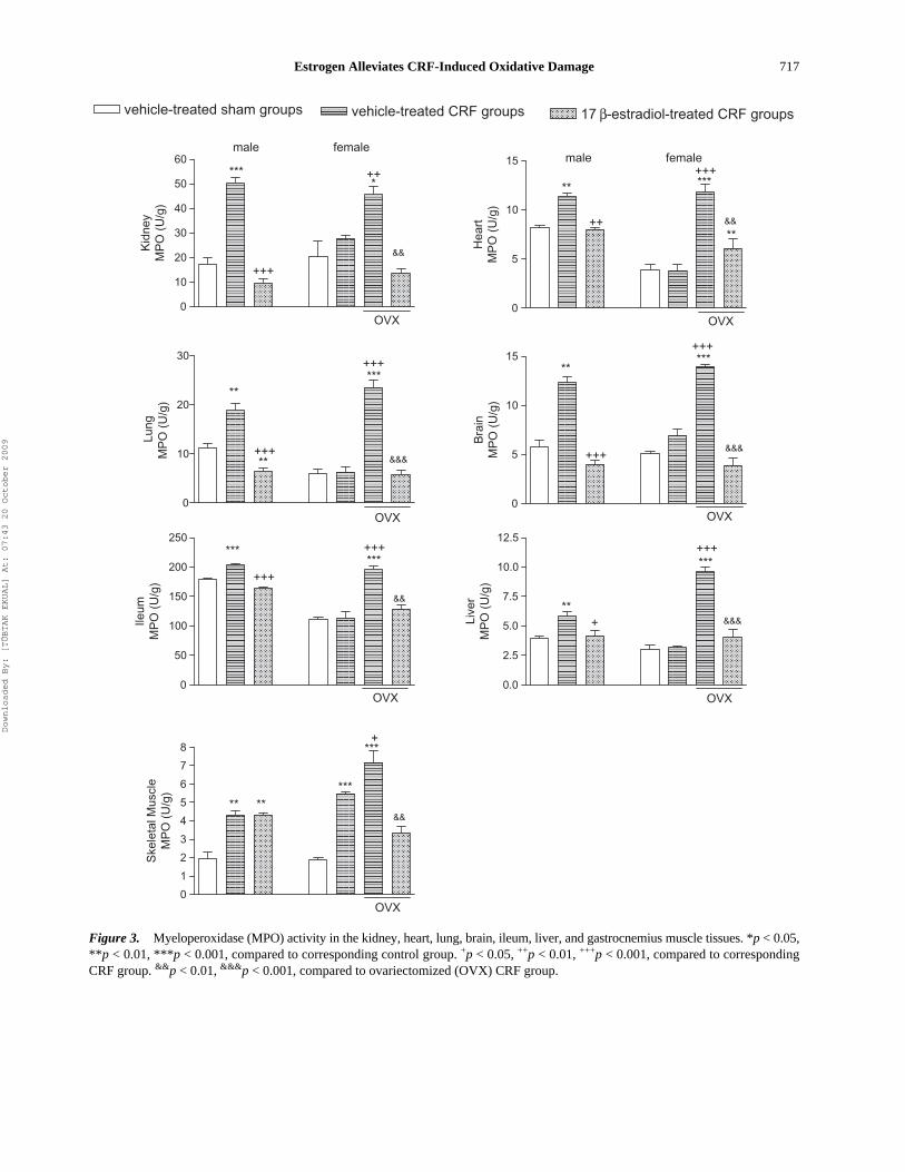
Lucigenin chemiluminescence (CL) was measured toelucidate the involvement of radical formation in CRF-induced tissue damage. Increased levels of lucigenin CL inthe brain, muscle, liver, and ileum of female and maleCRF groups (p < 0.05–0.001) were reversed by E2 treat-ment (p < 0.01–0.001; see Figure 4). Additionally, pulmo-nary CL level in male rats and renal CL in female rats,which were elevated by CRF, were also reversed by E2treatment (p < 0.05–0.01). CRF has not altered CL levelsin the kidney and heart tissues of male rats, while nosignificant change was noted in the cardiac and pulmo-nary tissues of female rats with CRF.
Table 1 Serum blood urea nitrogen (BUN), creatinine, and TNF-a levels in the experimental groups
Male Female
Sham-operated
CRF CRF
Sham-operated
Vehicle-treated
OVX
Vehicle-treated
Estradiol-treated
Vehicle-treated
Estradiol-treated
BUN (mg/dL) 31 ± 1.2 64.5 ± 2.8‡ 60 ± 11* 39 ± 3.2 61.5 ± 2.7* 67.3 ± 2.9‡& 67.3 ± 1.5†
Creatinine (mg/dL) 0.4 ± 0.1 0.6 ± 0.01‡ 0.6 ± 0.08‡ 0.5 ± 0.1 0.6 ± 0.01 0.7 ± 0.06 0.6 ± 0.01TNF-a (pg/mL) 3.2 ± 0.6 8.2 ± 1.1† 4.7 ± 0.4|| 4.6 ± 1.4 7.6 ± 0.8* 13.2 ± 0.8‡# 7.4 ± 0.4*§
For each group, n = 8.*p < 0.05, †p < 0.01, ‡p < 0.001, compared to corresponding sham-operated control group.§p < 0.05, ||p < 0.01, compared to corresponding vehicle-treated CRF group.#p < 0.01, compared to CRF group with OVX.Abbreviations: CRF = chronic renal failure, OVX = ovariectomy.
Downloaded By: [TÜBTAK EKUAL] At: 07:43 20 October 2009

Estrogen Alleviates CRF-Induced Oxidative Damage 715
Figure 1. Malondialdehyde (MDA) levels in the kidney, heart, lung, brain, ileum, liver, and gastrocnemius muscle tissues. *p < 0.05,**p < 0.01, ***p < 0.001, compared to corresponding control group. +p < 0.05, ++p < 0.01, +++p < 0.001, compared to correspondingCRF group. &p < 0.05, &&p < 0.01, compared to ovariectomized (OVX) CRF group.
vehicle-treated sham groups vehicle-treated CRF groups 17 β-estradiol-treated CRF groups
0.0
2.5
5.0
7.5
10.0
12.5
OVX
**
&
++**
++
Lung
MD
A (
nmol
/g)
0
25
50
75
100
125
OVX
+**
*
&&&*
Bra
inM
DA
(nm
ol/g
)
0
10
20
30
OVX
*
**+++
**
&&
++
Ileum
MD
A (
nmol
/g)
0
10
20
30
40
50
60
70
80
90
OVX
++
****++
&&
Live
rM
DA
(nm
ol/g
)
0
1
2
3
4
5
6
7
OVX
+
*
&
+
Ske
leta
l Mus
cle
MD
A (
nmol
/g)
0
5
10
15
20
25
30
35
40
45
OVX
**+
***+++
*+&
Kid
ney
MD
A (
nmol
/g)
male female
0
5
10
15
OVX
***
+++
***+++
&&&
Hea
rtM
DA
(nm
ol/g
)
male female
Downloaded By: [TÜBTAK EKUAL] At: 07:43 20 October 2009

716 Ö. Kasimay et al.
Figure 2. Gluthatione (GSH) levels in the kidney, heart, lung, brain, ileum, liver, and gastrocnemius muscle tissues. *p < 0.05,**p < 0.01, ***p < 0.001, compared to corresponding control group. +p < 0.05, ++p < 0.01, +++p < 0.001, compared to correspondingCRF group. &p < 0.05, &&p < 0.01, &&&p < 0.001, compared to ovariectomized (OVX) CRF group.
vehicle-treated sham groups vehicle-treated CRF groups 17 β-estradiol-treated CRF groups
0
1
2
OVX
**+
***+++
*
&&
Lung
GS
H (
μmol
/g)
0
1
2
OVX
***&&&
++**
Bra
inG
SH
(μm
ol/g
)
0
1
2
3
4
5
6
7
OVX
* * +*
+&&
Live
rG
SH
(μm
ol/g
)
0
1
2
OVX
**
++
Ske
leta
l Mus
cle
GS
H (
μml/g
)
0
1
2
3
4
OVX
***
**
+++
**
***+
+&
*
Ileum
GS
H (
μmol
/g)
0.0
0.5
1.0
1.5
2.0
2.5
3.0
3.5
male
OVX
*
*++
+++***
&&&
Kid
ney
GS
H (
μmol
/g)
female
0.0
0.5
1.0
1.5
2.0
2.5
OVX
**++
Hea
rtG
SH
(μm
ol/g
)
male female
Downloaded By: [TÜBTAK EKUAL] At: 07:43 20 October 2009
Estrogen Alleviates CRF-Induced Oxidative Damage 717
Figure 3. Myeloperoxidase (MPO) activity in the kidney, heart, lung, brain, ileum, liver, and gastrocnemius muscle tissues. *p < 0.05,**p < 0.01, ***p < 0.001, compared to corresponding control group. +p < 0.05, ++p < 0.01, +++p < 0.001, compared to correspondingCRF group. &&p < 0.01, &&&p < 0.001, compared to ovariectomized (OVX) CRF group.
vehicle-treated sham groups vehicle-treated CRF groups 17 β-estradiol-treated CRF groups
0
10
20
30
OVX
**
**
***
+++
+++
&&&
Lung
MP
O (
U/g
)
0
5
10
15
OVX
*****
+++
+++
&&&
Bra
inM
PO
(U
/g)
0
50
100
150
200
250
OVX
***
+++***+++
&&
Ileum
MP
O (
U/g
)
0.0
2.5
5.0
7.5
10.0
12.5
OVX
**+
***+++
&&&Live
rM
PO
(U
/g)
0
1
2
3
4
5
6
7
8
OVX
**&&
+
***
***
**
Ske
leta
l Mus
cle
MP
O (
U/g
)
0
10
20
30
40
50
60male
OVX
***
+++
*++
&&Kid
ney
MP
O (
U/g
)female
0
5
10
15
OVX
**
++
***
**
+++
&&
Hea
rtM
PO
(U
/g)
male female
Downloaded By: [TÜBTAK EKUAL] At: 07:43 20 October 2009
718 Ö. Kasimay et al.
Figure 4. Lucigenin chemiluminescence levels as indicators of O2 formation in the kidney, heart, lung, brain, ileum, liver, andgastrocnemius muscle tissues. *p < 0.05, **p < 0.01, ***p < 0.001, compared to corresponding control group. +p < 0.05, ++p < 0.01,+++p < 0.001, compared to corresponding CRF group. &p < 0.05, &&p < 0.01, &&&p < 0.001, compared to ovariectomized (OVX) CRFgroup.
vehicle-treated sham groups vehicle-treated CRF groups 17 β-estradiol-treated CRF groups
0
5
10
15
20
25
OVX
*
Lung
Luci
geni
n C
L (r
lu/m
g)
0
10
20
OVX
*
*
+
+++&
Bra
inLu
cige
nin
CL
(rlu
/mg)
0
10
20
30
40
OVX
*
**++
*
&&Ileum
Luci
geni
n C
L (r
lu/m
g)
0
5
10
15
OVX
***
**
+++**++&&&
Live
rLu
cige
nin
CL
(rlu
/mg)
0
10
20
OVX
++
*
++
**
+
Ske
leta
l Mus
cle
Luci
geni
n C
L (r
lu/m
g)
0
5
10
15
20
25
OVX
male
***+++
***
++&&
**
Kid
ney
Luci
geni
n C
L (r
lu/m
g)female
0
5
10
15
20
25
OVX
+&
Hea
rtLu
cige
nin
CL
(rlu
/mg)
male female
Downloaded By: [TÜBTAK EKUAL] At: 07:43 20 October 2009
Estrogen Alleviates CRF-Induced Oxidative Damage 719
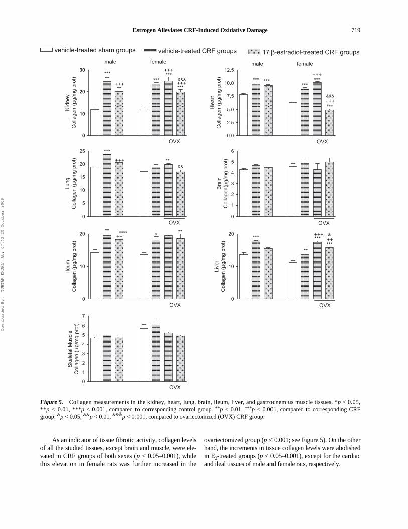
As an indicator of tissue fibrotic activity, collagen levelsof all the studied tissues, except brain and muscle, were ele-vated in CRF groups of both sexes (p < 0.05–0.001), whilethis elevation in female rats was further increased in the
ovariectomized group (p < 0.001; see Figure 5). On the otherhand, the increments in tissue collagen levels were abolishedin E2-treated groups (p < 0.05–0.001), except for the cardiacand ileal tissues of male and female rats, respectively.
Figure 5. Collagen measurements in the kidney, heart, lung, brain, ileum, liver, and gastrocnemius muscle tissues. *p < 0.05,**p < 0.01, ***p < 0.001, compared to corresponding control group. ++p < 0.01, +++p < 0.001, compared to corresponding CRFgroup. &p < 0.05, &&p < 0.01, &&&p < 0.001, compared to ovariectomized (OVX) CRF group.
vehicle-treated sham groups vehicle-treated CRF groups 17 β-estradiol-treated CRF groups
0
10
20
30
male female
OVX
+++
******
***+++
+++***
&&&
Kid
ney
Col
lage
n ( μ
g/m
g pr
ot)
0.0
2.5
5.0
7.5
10.0
12.5male female
OVX
*** ******
***
***
+++
+++&&&
Hea
rtC
olla
gen
( μg/
mg
prot
)0
5
10
15
20
25
OVX
+++&&
***
**
Lung
Col
lage
n ( μ
g/m
g pr
ot)
0
1
2
3
4
5
6
OVXB
rain
Col
lage
n(μg
/mg
prot
)
0
10
20
OVX
**++**** **
*
Ileum
Col
lage
n ( μ
g/m
g pr
ot)
0
10
20
OVX
*** ++***
*****
+++ &
Live
rC
olla
gen
( μg/
mg
prot
)
0
1
2
3
4
5
6
7
OVX
Ske
leta
l Mus
cle
Col
lage
n ( μ
g/m
g pr
ot)
Downloaded By: [TÜBTAK EKUAL] At: 07:43 20 October 2009
720 Ö. Kasimay et al.
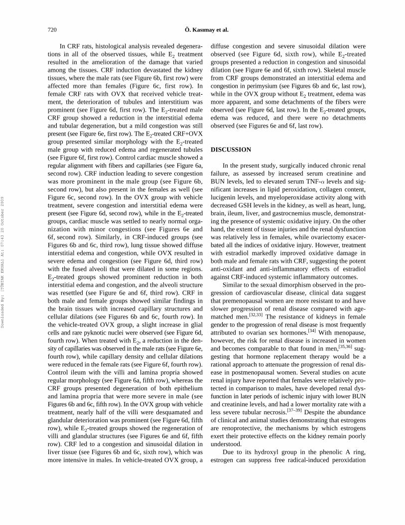
In CRF rats, histological analysis revealed degenera-tions in all of the observed tissues, while E2 treatmentresulted in the amelioration of the damage that variedamong the tissues. CRF induction devastated the kidneytissues, where the male rats (see Figure 6b, first row) wereaffected more than females (Figure 6c, first row). Infemale CRF rats with OVX that received vehicle treat-ment, the deterioration of tubules and interstitium wasprominent (see Figure 6d, first row). The E2-treated maleCRF group showed a reduction in the interstitial edemaand tubular degeneration, but a mild congestion was stillpresent (see Figure 6e, first row). The E2-treated CRF+OVXgroup presented similar morphology with the E2-treatedmale group with reduced edema and regenerated tubules(see Figure 6f, first row). Control cardiac muscle showed aregular alignment with fibers and capillaries (see Figure 6a,second row). CRF induction leading to severe congestionwas more prominent in the male group (see Figure 6b,second row), but also present in the females as well (seeFigure 6c, second row). In the OVX group with vehicletreatment, severe congestion and interstitial edema werepresent (see Figure 6d, second row), while in the E2-treatedgroups, cardiac muscle was settled to nearly normal orga-nization with minor congestions (see Figures 6e and6f, second row). Similarly, in CRF-induced groups (seeFigures 6b and 6c, third row), lung tissue showed diffuseinterstitial edema and congestion, while OVX resulted insevere edema and congestion (see Figure 6d, third row)with the fused alveoli that were dilated in some regions.E2-treated groups showed prominent reduction in bothinterstitial edema and congestion, and the alveoli structurewas resettled (see Figure 6e and 6f, third row). CRF inboth male and female groups showed similar findings inthe brain tissues with increased capillary structures andcellular dilations (see Figures 6b and 6c, fourth row). Inthe vehicle-treated OVX group, a slight increase in glialcells and rare pyknotic nuclei were observed (see Figure 6d,fourth row). When treated with E2, a reduction in the den-sity of capillaries was observed in the male rats (see Figure 6e,fourth row), while capillary density and cellular dilationswere reduced in the female rats (see Figure 6f, fourth row).Control ileum with the villi and lamina propria showedregular morphology (see Figure 6a, fifth row), whereas theCRF groups presented degeneration of both epitheliumand lamina propria that were more severe in male (seeFigures 6b and 6c, fifth row). In the OVX group with vehicletreatment, nearly half of the villi were desquamated andglandular deterioration was prominent (see Figure 6d, fifthrow), while E2-treated groups showed the regeneration ofvilli and glandular structures (see Figures 6e and 6f, fifthrow). CRF led to a congestion and sinusoidal dilation inliver tissue (see Figures 6b and 6c, sixth row), which wasmore intensive in males. In vehicle-treated OVX group, a
diffuse congestion and severe sinusoidal dilation wereobserved (see Figure 6d, sixth row), while E2-treatedgroups presented a reduction in congestion and sinusoidaldilation (see Figure 6e and 6f, sixth row). Skeletal musclefrom CRF groups demonstrated an interstitial edema andcongestion in perimysium (see Figures 6b and 6c, last row),while in the OVX group without E2 treatment, edema wasmore apparent, and some detachments of the fibers wereobserved (see Figure 6d, last row). In the E2-treated groups,edema was reduced, and there were no detachmentsobserved (see Figures 6e and 6f, last row).
DISCUSSION
In the present study, surgically induced chronic renalfailure, as assessed by increased serum creatinine andBUN levels, led to elevated serum TNF-a levels and sig-nificant increases in lipid peroxidation, collagen content,lucigenin levels, and myeloperoxidase activity along withdecreased GSH levels in the kidney, as well as heart, lung,brain, ileum, liver, and gastrocnemius muscle, demonstrat-ing the presence of systemic oxidative injury. On the otherhand, the extent of tissue injuries and the renal dysfunctionwas relatively less in females, while ovariectomy exacer-bated all the indices of oxidative injury. However, treatmentwith estradiol markedly improved oxidative damage inboth male and female rats with CRF, suggesting the potentanti-oxidant and anti-inflammatory effects of estradiolagainst CRF-induced systemic inflammatory outcomes.
Similar to the sexual dimorphism observed in the pro-gression of cardiovascular disease, clinical data suggestthat premenopausal women are more resistant to and haveslower progression of renal disease compared with age-matched men.[32,33] The resistance of kidneys in femalegender to the progression of renal disease is most frequentlyattributed to ovarian sex hormones.[34] With menopause,however, the risk for renal disease is increased in womenand becomes comparable to that found in men,[35,36] sug-gesting that hormone replacement therapy would be arational approach to attenuate the progression of renal dis-ease in postmenopausal women. Several studies on acuterenal injury have reported that females were relatively pro-tected in comparison to males, have developed renal dys-function in later periods of ischemic injury with lower BUNand creatinine levels, and had a lower mortality rate with aless severe tubular necrosis.[37–39] Despite the abundanceof clinical and animal studies demonstrating that estrogensare renoprotective, the mechanisms by which estrogensexert their protective effects on the kidney remain poorlyunderstood.
Due to its hydroxyl group in the phenolic A ring,estrogen can suppress free radical-induced peroxidation
Downloaded By: [TÜBTAK EKUAL] At: 07:43 20 October 2009
Estrogen Alleviates CRF-Induced Oxidative Damage 721
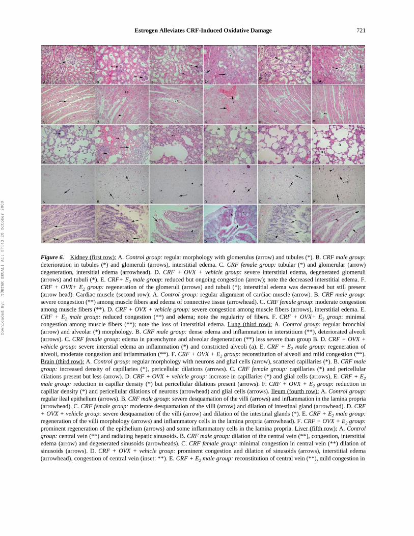
Figure 6. Kidney (first row): A. Control group: regular morphology with glomerulus (arrow) and tubules (*). B. CRF male group:deterioration in tubules (*) and glomeruli (arrows), interstitial edema. C. CRF female group: tubular (*) and glomerular (arrow)degeneration, intersitial edema (arrowhead). D. CRF + OVX + vehicle group: severe interstitial edema, degenerated glomeruli(arrows) and tubuli (*). E. CRF+ E2 male group: reduced but ongoing congestion (arrow); note the decreased interstitial edema. F.CRF + OVX+ E2 group: regeneration of the glomeruli (arrows) and tubuli (*); interstitial edema was decreased but still present(arrow head). Cardiac muscle (second row): A. Control group: regular alignment of cardiac muscle (arrow). B. CRF male group:severe congestion (**) among muscle fibers and edema of connective tissue (arrowhead). C. CRF female group: moderate congestionamong muscle fibers (**). D. CRF + OVX + vehicle group: severe congestion among muscle fibers (arrows), interstitial edema. E.CRF + E2 male group: reduced congestion (**) and edema; note the regularity of fibers. F. CRF + OVX+ E2 group: minimalcongestion among muscle fibers (**); note the loss of interstitial edema. Lung (third row): A. Control group: regular bronchial(arrow) and alveolar (*) morphology. B. CRF male group: dense edema and inflammation in interstitium (**), deteriorated alveoli(arrows). C. CRF female group: edema in parenchyme and alveolar degeneration (**) less severe than group B. D. CRF + OVX +vehicle group: severe interstial edema an inflammation (*) and constricted alveoli (a). E. CRF + E2 male group: regeneration ofalveoli, moderate congestion and inflammation (**). F. CRF + OVX + E2 group: reconstitution of alveoli and mild congestion (**).Brain (third row): A. Control group: regular morphology with neurons and glial cells (arrow), scattered capillaries (*). B. CRF malegroup: increased density of capillaries (*), pericellular dilations (arrows). C. CRF female group: capillaries (*) and pericellulardilations present but less (arrow). D. CRF + OVX + vehicle group: increase in capillaries (*) and glial cells (arrows), E. CRF + E2
male group: reduction in capillar density (*) but pericellular dilations present (arrows). F. CRF + OVX + E2 group: reduction incapillar density (*) and pericellular dilations of neurons (arrowhead) and glial cells (arrows). Ileum (fourth row): A. Control group:regular ileal epithelium (arrows). B. CRF male group: severe desquamation of the villi (arrows) and inflammation in the lamina propria(arrowhead). C. CRF female group: moderate desquamation of the villi (arrow) and dilation of intestinal gland (arrowhead). D. CRF+ OVX + vehicle group: severe desquamation of the villi (arrow) and dilation of the intestinal glands (*). E. CRF + E2 male group:regeneration of the villi morphology (arrows) and inflammatory cells in the lamina propria (arrowhead). F. CRF + OVX + E2 group:prominent regeneration of the epithelium (arrows) and some inflammatory cells in the lamina propria. Liver (fifth row): A. Controlgroup: central vein (**) and radiating hepatic sinusoids. B. CRF male group: dilation of the central vein (**), congestion, interstitialedema (arrow) and degenerated sinusoids (arrowheads). C. CRF female group: minimal congestion in central vein (**) dilation ofsinusoids (arrows). D. CRF + OVX + vehicle group: prominent congestion and dilation of sinusoids (arrows), interstitial edema(arrowhead), congestion of central vein (inset: **). E. CRF + E2 male group: reconstitution of central vein (**), mild congestion in
Downloaded By: [TÜBTAK EKUAL] At: 07:43 20 October 2009

722 Ö. Kasimay et al.
chain reactions[13,14] and protect against atherosclerosis bybinding free radicals.[40] Being a fat-soluble hormone,estrogen can contribute to membrane fluidity by directinteractions with phospholipids and attenuate the peroxi-dation of vascular smooth muscle cells and peroxidation-induced cell growth and migration.[41] On the other hand,age-related decline in estrogen secretion is accompaniedby excessive oxidative stress and peroxidative cell dam-age,[42] which are known features of CRF. The results ofthe present study show that lipid peroxidation productsmeasured as MDA levels in kidney, heart, lung, intestine,brain, and liver were significantly increased in the malerats with CRF, while the unchanged tissue MDA levels infemale rats were increased when ovaries were removed.Thus, the current results suggest that female gender is pro-tective, but ovariectomy attenuates the protective effect offemale gender on the development of CRF-induced oxida-tive injury. In addition, the administration of E2 causedsignificant inhibition of MDA production in both malesand females, indicating a reduction in lipid peroxidationand cellular injury. In vitro, high concentrations of estra-diol were shown to inhibit renal cell-mediated low-densitylipoprotein oxidation.[43] Accordingly, hormone replacementtherapy was shown to have a favorable effect on oxidativestress in postmenopausal women with CRF, as evidencedby plasma MDA level and total antioxidant capacity.[44]
Similarly, female rats in experimental polycystic kidneydisease exhibited normal renin status, lesser elevation ofrenal endothelin (ET-1), and proportionately lesser changesin vascular endothelial growth factor (VEGF) and endot-helial nitric oxide synthase (eNOS), while ovariectomywas shown to attenuate the protective effect of female genderwith a trend toward upregulation of ET-1 and a significant
down-regulation of VEGF and eNOS.[45] Furthermore, E2supplementation was shown to be protective in the agingkidney by attenuating the downregulation of eNOS proteinabundance and renal inflammation.[46]
In uremia, toxic metabolites in blood activate PMNleukocytes, which then exhibit a burst of oxygen con-sumption,[47] generating oxygen-derived reactive interme-diates, superoxide anion, hydrogen peroxide, nitric oxide,and hypochlorous acid (HOCl).[48] In the present study, inaccordance with elevated tissue MDA levels, lucigeninchemiluminescence levels showed marked elevations inmost of the studied target tissues of CRF rats, indicating theinvolvement of superoxide anion in associated tissuedamages,[30] while increased MPO activity clearly dem-onstrates that CRF-induced multiple organ damage isneutrophil-dependent. Activated neutrophils release MPO,causing production of large amounts of HOCl, which oxi-dizes and damages macromolecules, including proteins,lipids, carbohydrates, and nucleic acids.[48] Our results arein agreement with similar studies showing that increasedMPO activity plays a role in CRF-induced oxidative injury,and treating the animals with anti-inflammatory or antiox-idant agents prevents tissue damage by inhibiting neutrophilinfiltration.[49,50] Moreover, the anti-inflammatory effectof E2 observed in all the CRF-injured tissues appears toinclude an inhibition on the accumulation of neutrophilsand on the generation of superoxide radicals. Similarly, E2was shown to inhibit neutrophil infiltration and ICAM-1expression in endotoxin-induced glomerular inflammatoryresponse.[51]
Inflammatory diseases of the renal glomerulus andtubulointerstitium are characterized by destructive andrestorative processes, mediated by cytokines, including TNF-a,
Figure 6. (Continued) sinusoids (arrows). F. CRF + OVX + E2 group: regenerated central vein (**) and sinusoid (arrows)morphology, mild inflammation at the periphery of hepatic canal. Skeletal muscle (last row): A. Control group: regular alignmentof muscle fibers and connective tissue (*) among them. B. CRF male group: congestion among muscle fibers (*) and dilation ofendomysium (arrowheads). C. CRF female group: minimal congestion (*) in the connective tissue, but moderate deterioration ofgeneral morphology. D. CRF + OVX + vehicle group: dilation in connective tissue (*);note detachments in the muscle fibers.E. CRF+ E2 male group: cross-section of muscle bundle; note the regular morphology (arrow) and minimal congestion (*). F. CRF + OVX+ E2 group: ongoing congestion (*) among muscle fibers; note the regular alignment of muscle fibers (arrow). HE: ×200, inset: ×400.
Downloaded By: [TÜBTAK EKUAL] At: 07:43 20 October 2009

Estrogen Alleviates CRF-Induced Oxidative Damage 723
that instruct target cells to alter their proliferation, differ-entiation, and migration.[52] In chronically uremic patients,it was demonstrated that TNF-a gene expression is enhancedin circulating blood cells, suggesting the contribution ofsystemic inflammatory response in the complications ofCRF.[53] Accordingly, in another study, diminishing TNF-aexpression significantly reduced ischemia-induced renalfibrosis and inflammation and improved renal functions.[54]
On the other hand, the administration of E2 depressed ele-vations in serum TNF-a levels following burn-injury[22] ortrauma-hemorrhage.[55] In addition, E2 inhibited the pro-duction of TNF-a by the unstimulated and hydrogen per-oxide-stimulated macrophages from males and females.[56]
Parallel to these findings, the present data demonstrate thatelevated serum TNF-a levels of CRF rats are depressed byexogenous E2 treatment, suggesting that E2-induced allevi-ation of CRF-induced oxidative damage involves the mod-ulation of the synthesis of TNF-a.
To protect against the adverse effects of ROS, mam-malian cells are equipped with various antioxidant scaven-gers. Apart from the major enzymatic antioxidants, such assuperoxide dismutase, GSH peroxidase, and catalase, estro-gens are also classified as nonenzymatic antioxidants thatdelay or inhibit age-associated pathological and biologicalchanges.[57] Several experimental studies have shown thatantioxidant capacity of tissues is decreased in animals withCRF,[58,59] while marked reductions in plasma glutathioneconcentration and glutathione peroxidase activity, as wellas marked increases in the ratios of oxidized to reducedGSH were observed in CRF patients.[60,61] Current find-ings show that the reduction in GSH content of the studiedtissues of male and ovariectomized rats may be due to itsconsumption during CRF-induced oxidative stress. On theother hand, administration of E2, through its antioxidantproperties and by inhibiting lipid peroxidation, maintainedthe glutathione levels and thereby protected the tissuesagainst oxidative stress.
Estrogens influence the matrix deposition and scar-ring by stimulating the activity of collagen degradingenzymes[62] and regulating the genes involved in extracel-lular matrix metabolism,[63] thereby contributing to theprogression of renal disease. In vitro studies confirm thatestrogens inhibit the synthesis of type I and type IV col-lagen in renal tissue culture[64] via the suppression oftransforming growth factor b (TGF-b) signal.[65] Accord-ingly, in the TGF-b transgenic mouse, which developsextensive glomerulosclerosis, estradiol supplementationwas shown to inhibit renal scarring.[66] In parallel to thesestudies, current findings demonstrate that elevated collagencontents in the renal tissue as well as in the cardiac, pul-monary, ileal, and hepatic tissues of the male and femalerats with CRF were reduced when the rats were treated withE2. Taken together, it appears that estradiol supplementation
may delay the progression of chronic renal disease by limit-ing the oxidant-induced collagen accumulation.
A vast number of studies supporting the notion thatsystemic inflammation is activated during the progress ofchronic renal failure provide a strong rationale for the useof anti-inflammatory treatments in uremia. The findings ofthe current study illustrate that exogenously administeredestrogen acts as a potent antioxidant and reduces CRF-induced oxidative tissue damage by depressing tissue neu-trophil infiltration and modulating the release of inflammatorycytokines. Extensive experimental and clinical studies willbe necessary to further elucidate intracellular signals inducedby estrogens on the target cells and thus to establish possi-bilities of therapeutic intervention in the progress to end-stagerenal disease.
ACKNOWLEDGMENTS
The authors declare that there is no conflict of interest.
REFERENCES
1. Cottone S, Lorito MC, Riccobene R, Nardi E, Mulè G,Buscemi S, Geraci C, Guarneri M, Arsena R, Cerasola G.Oxidative stress, inflammation and cardiovascular disease inchronic renal failure. J Nephrol. 2008;21:175–179.
2. Siems W, Quast S, Carluccio F, Wiswedel I, Hirsch D,Augustin W, Hampi H, Riehle M, Sommerburg O. Oxidativestress in chronic renal failure as a cardiovascular risk factor.Clin Nephrol. 2002;58 (Suppl. 1):S12–S19.
3. Sener G, Paskaloglu K, Satiroglu H, Alican I, Kaçmaz A,Sakarcan A. L-carnitine ameliorates oxidative damage due tochronic renal failure in rats. J Cardiovasc Pharmacol. 2004;43:698–705.
4. Silbiger S, Neugarten J. Gender and human chronic renaldisease. Gend Med. 2008;5 (Suppl. A):S3–S10.
5. Seliger SL, Davis C, Stehman-Breen C. Gender and the pro-gression of renal disease. Curr Opin Nephrol Hypertens.2001;10:219–225.
6. Neugarten J. Gender and the progression of renal disease.J Am Soc Nephrol. 2002;13:2807–2809.
7. Schulman IH, Aranda P, Raij L, Veronesi M, Aranda FJ,Martin R.. Surgical menopause increases salt sensitivity ofblood pressure. Hypertension. 2006;47:1168–1174.
8. Reckelhoff JF, Zhang H, Srivastava K, Granger JP. Genderdifferences in hypertension in spontaneously hypertensiverats: Role of androgens and androgen receptor. Hypertension.1999;34:920–923.
9. Baylis C. Sexual dimorphism of the aging kidney: Role ofnitric oxide deficiency. Physiology (Bethesda). 2008;23:142–150.
10. Irwin RW, Yao J, Hamilton RT, Cadenas E, Brinton RD,Nilsen J. Progesterone and estrogen regulate oxidative
Downloaded By: [TÜBTAK EKUAL] At: 07:43 20 October 2009

724 Ö. Kasimay et al.
metabolism in brain mitochondria. Endocrinology. 2008;149:3167–3175.
11. Gragasin FS, Xu Y, Arenas IA, Kainth N, Davidge ST.Estrogen reduces angiotensin II-induced nitric oxide syn-thase and NAD(P)H oxidase expression in endothelial cells.Arterioscler Thromb Vasc Biol. 2003;23:38–44.
12. Strehlow K, Rotter S, Wassmann S, Adam O, Grohe C,Laufs K, Bohm M, Nickenig G. Modulation of antioxidantenzyme expression and function by estrogen. Circ Res. 2003;93:170–177.
13. Sugioka K, Shimosegawa Y, Nakano M. Estrogens as naturalantioxidants of membrane phospholipid peroxidation. FEBSLett. 1987;210:37–39.
14. Green PS, Gordon K, Simpkins JW. Phenolic A ring require-ment for the neuroprotective effects of steroids. J SteroidBiochem Mol Biol. 1997;63:229–235.
15. Zhai P, Eurell TE, Cotthaus R, Jeffery EH, Bahr JM,Gross DR. Effect of estrogen on global myocardial ischemia-reperfusion injury in female rats. Am J Physiol Heart CircPhysiol. 2000;279:H2766–H2775.
16. Behl C. Oestrogen as a neuroprotective hormone. Nat RevNeurosci. 2002;3:433–442.
17. Feng Z, Zhang JT. Long-term melatonin or 17beta-estradiolsupplementation alleviates oxidative stress in ovariecto-mized adult rats. Free Radic Biol Med. 2005;39:195–204.
18. Lee MY, Jung SC, Lee JH, Han HJ. Estradiol-17beta pro-tects against hypoxia-induced hepatocyte injury throughER-mediated upregulation of Bcl-2 as well as ER-independentantioxidant effects. Cell Res. 2008;18:491–499.
19. Persky AM, Green PS, Stubley L, Howell CO, Zaulyanov L,Brazeau GA, Simpkins JW. Protective effect of estrogensagainst oxidative damage to heart and skeletal muscle in vivoand in vitro. Proc Soc Exp Biol Med. 2000;223:59–66.
20. Sener G, Arbak S, Kurtaran P, Gedik N, Yegen BC. Estrogenprotects the liver and intestines against sepsis-induced injuryin rats. J Surg Res. 2005;128(1):70–78.
21. Günal O, Oktar BK, Ozçinar E, Sungur M, Arbak S, Yegen B.Estradiol treatment ameliorates acetic acid-induced gastricand colonic injuries in rats. Inflammation. 2003;27:351–359.
22. Ozveri ES, Bozkurt A, Haklar G, Cetinel S, Arbak S, Yegen C,Yegen BC. Estrogens ameliorate remote organ inflammationinduced by burn injury in rats. Inflamm Res. 2001;50:585–591.
23. Vaziri ND, Ni Z, Oveisi F, Liang K, Pandian R. Enhancednitric oxide inactivation and protein nitration by reactiveoxygen species in renal insufficiency. Hypertension 2002;39:135–141.
24. Talke H, Schubert GE. Enzymatic urea determination in theblood and serum in the Warburg optical test. Wiener Kli-nische Wochenschrift. 1965;43:174–175.
25. Slot C. Plasma creatinine determination: A new and specificJaffe reaction method. Scand J Clin Lab Invest. 1965;17:381–387.
26. Beuge JA, Aust SD. Microsomal lipid peroxidation. Methodsin Enzymology. 1978;52:302–311.
27. Beutler E. Glutathione. In Beutler E (ed.). Red blood cellmetabolism: A manual of biochemical methods. New York:Grune & Stratton;1975:112–114.
28. Bradley PP, Priebat DA, Christersen RD, Rothstein G.Measurement of cutaneous inflammation. Estimation of neu-trophil content with an enzyme marker. J Invest Dermatol.1982;78:206–209.
29. Hillegass LM, Griswold DE, Brickson B, Albrightson-Winslow C. Assessment of myeloperoxidase activity in wholerat kidney. J Pharm Methods. 1990;24:285–295.
30. Haklar G, Ulukaya-Durakbasa C, Yüksel M, Dagli T,Yalcin AS. Oxygen radicals and nitric oxide in rat mesen-teric ischemia-reperfusion: Modulation by L-arginine andN-nitro-L-arginine methyl ester. Clin Exp Pharmacol Physiol.1998;25:908–912.
31. Lopez De Leon A, Rojkind M. A simple micromethod forcollagen and total protein determination in formalin-fixedparraffin-embedded sections. J Histochem Cytochem. 1985;33:737–743.
32. Tozawa M, Iseki K, Iseki C, Kinjo K, Ikemiya Y,Takishita S. Blood pressure predicts risk of developingend-stage renal disease in men and women. Hypertension.2003;41:1341–1345.
33. Neugarten J. Gender and the progression of renal disease.J Am Soc Nephrol. 2002;13:2807–2809.
34. Ball P, Emons G, Kayser H, Teichmann J. Metabolic clear-ance rates of catechol estrogens in rats. Endocrinology. 1983;113:1781–1783.
35. Schulman IH, Aranda P, Raij L, Veronesi M, Aranda FJ,Martin R. Surgical menopause increases salt sensitivity ofblood pressure. Hypertension. 2006;47:1168–1174.
36. Reckelhoff JF, Zhang H, Srivastava K, Granger JP. Genderdifferences in hypertension in spontaneously hypertensiverats: Role of androgens and androgen receptor. Hypertension.1999;34:920–923.
37. Park KM, Kim JI, Ahn Y, Bonventre AJ, Bonventre JV.Testosterone is responsible for enhanced susceptibility ofmales to ischemic renal injury. J Biol Chem. 2004;279:52282–52292.
38. Müller V, Losonczy G, Heemann U, Vannay A, Fekete A,Reusz G, Tulassay T, Szabó AJ. Sexual dimorphism in renalischemia–reperfusion injury in rats: Possible role of endothelin.Kidney Int. 2002;62:1364–1371.
39. Fekete A, Vannay A, Ver A, et al. Sex differences in thealterations of Na(+), K(+)-ATPase following ischaemia–reperfusion injury in the rat kidney. J Physiol. 2004;555:471–480.
40. Barp J, Araujo ASR, Fernandes TR, et al. Myocardial antiox-idant and oxidative stres changes due to sex hormones. BrazJ Med Biol Res. 2002;35:1075–1081.
41. Dubey RK, Tyurina YY, Tyurin VA, et al. Estrogen andtamoxifen metabolites protect smooth muscle cell membranephospholipids against peroxidation and inhibit cell growth.Circ Res. 1999;84:229–239.
42. Hamden K, Carreau S, Ellouz F, Masmoudi H, El FA. Pro-tective effect of 17beta-estradiol on oxidative stress and liverdysfunction in aged male rats. J Physiol Biochem. 2007;63:195–201.
43. Chiang K, Parthasarathy S, Santanam N. Estrogen, neutro-phils and oxidation. Life Sci. 2004;75:2425–2438.
Downloaded By: [TÜBTAK EKUAL] At: 07:43 20 October 2009

Estrogen Alleviates CRF-Induced Oxidative Damage 725
44. Chang SP, Yang WS, Lee SK, Min WK, Park JS, Kim SB.Effects of hormonal replacement therapy on oxidative stressand total antioxidant capacity in postmenopausal hemodialysispatients. Ren Fail. 2002;24:49–57.
45. Stringer KD, Komers R, Osman SA, Oyama TT, Lindsley JN,Anderson S. Gender hormones and the progression of exper-imental polycystic kidney disease. Kidney Int. 2005;68(4):1729–1739.
46. Maric C, Xu Q, Sandberg K, Hinojosa-Laborde C. Age-related renal disease in female dahl salt-sensitive rats isattenuated with 17b-estradiol supplementation by modulat-ing nitric oxide synthase expression. Gend Med. 2008;5:147–159.
47. Westhuyzen J, Adams CE, Fleming SJ. Evidence for oxida-tive stress during in vitro dialysis. Nephron. 1995;70:49–54.
48. Kettle AJ, Winterbourn CC. Myeloperoxidase: A key reg-ulator of neutrophil oxidant production. Redox Rep. 1997;3:3–15.
49. Sener G, Sakarcan A, Sehirli O, Eksioglu-Demiralp E, Sener E,Ercan F, Gedik N, Yegen BC. Chronic renal failure-inducedmultiple-organ injury in rats is alleviated by the selectiveCysLT1 receptor antagonist montelukast. Prostaglandinsand Other Lipid Mediators. 2007;83:257–267.
50. Lim CS, Vaziri ND. Iron and oxidative stress in renal insuffi-ciency. Am J Nephrol. 2004;24:569–575.
51. Faas MM, Bakker WW, Valkhof N, Schuiling GA. Effect ofestradiol and progesterone on the low-dose endotoxin-inducedglomerular inflammatory response of the female rat. Am JReprod Immunol. 1999;41:224–231.
52. Baud L, Ardaillou R. Tumor necrosis factor in renal injury.Miner Electrolyte Metab. 1995;21:336–341.
53. Guarnieri G, Antonione R, Biolo G. Mechanisms of malnu-trition in uremia. J Ren Nutr. 2003;13:153–157.
54. Cau J, Favreau F, Zhang K. FR167653 improves renalrecovery and decreases inflammation and fibrosis afterrenal ischemia reperfusion injury. J Vasc Surg. 2009;49:728–740.
55. Hsieh CH, Nickel EA, Chen J, Schwacha MG, ChoudhryMA, Bland KI, Chaudry IH. Mechanism of the salutaryeffects of estrogen on kupffer cell phagocytic capacity fol-lowing trauma-hemorrhage: Pivotal role of Akt activation. JImmunol. 2009;182:4406–4414.
56. Huang H, He J, Yuan Y, Aoyagi E, Takenaka H, Itagaki T,Sannomiya K, Tamaki K, Harada N, Shono M, Shimizu I,Takayama T. Opposing effects of estradiol and progesteroneon the oxidative stress-induced production of chemokine andproinflammatory cytokines in murine peritoneal macroph-ages. J Med Invest. 2008;55:133–141.
57. Borrás C, Sastre J, García-Sala D, Lloret A, Pallardó FV,Viña J. Mitochondria from females exhibit higher antioxi-dant gene expression and lower oxidative damage thanmales. Free Radic Biol Med. 2003;34:546–552.
58. Sener G, Paskaloglu K, Toklu H, Kapucu C, Ayanoglu-Dulger G, Kacmaz A, Sakarcan A. Melatonin ameliorateschronic renal failure-induced oxidative organ damage in rats.J Pineal Res. 2004;36:232–241.
59. Calunga JL, Zamora ZB, Borrego A, Río S, Barber E,Menéndez S, Hernández F, Montero T, Taboada D. Ozonetherapy on rats submitted to subtotal nephrectomy: Role ofantioxidant system. Mediators Inflamm. 2005;2005:221–227.
60. Ceballos-Picot I, Witko-Sarsat V, Merad-Boudia M, Nguyen AT,Thévenin M, Jaudon MC, Zingraff J, Verger C, Jungers P,Descamps-Latscha B. Glutathione antioxidant system as amarker of oxidative stress in chronic renal failure. FreeRadic Biol Med. 1996;21:845–853.
61. Himmelfarb J, McMenamin E, McMonagle E. Plasma ami-nothiol oxidation in chronic hemodialysis patients. KidneyInt. 2002;61:705–716.
62. Guccione M, Silbiger S, Lei J, Neugarten J. Estradiol upregu-lates mesangial cell MMP-2 activity via the transcription factorAP-2. Am J Physiol Renal Physiol. 2002;282:F164–F169.
63. Potier M, Elliot SJ, Tack I, et al. Expression and regulationof estrogen receptors in mesangial cells: Influence on matrixmetalloproteinase-9. J Am Soc Nephrol. 2001;12:241–251.
64. Neugarten J, Acharya A, Lei J, Silbiger S. Selective estrogenreceptor modulators suppress mesangial cell collagen syn-thesis. Am J Physiol Renal Physiol. 2000;279:F309–F318.
65. Zdunek M, Silbiger S, Lei J, Neugarten J. Protein kinaseCK2 mediates TGF-b1-stimulated type IV collagen genetranscription and its reversal by estradiol. Kidney Int. 2001;60:2097–2108.
66. Blush J, Lei J, Ju W, Silbiger S, Pullman J, Neugarten J.Estradiol reverses renal injury in Alb/TGF-b1 transgenic mice.Kidney Int. 2004;66:2148–2154.
Downloaded By: [TÜBTAK EKUAL] At: 07:43 20 October 2009