Essential role of the chaperonin folding compartment in vivo
11
Essential role of the chaperonin folding compartment in vivo Yun-Chi Tang 1,2 , Hung-Chun Chang 1,3 , Kausik Chakraborty, F Ulrich Hartl* and Manajit Hayer-Hartl* Department of Cellular Biochemistry, Max Planck Institute of Biochemistry, Martinsried, Germany The GroEL/GroES chaperonin system of Escherichia coli forms a nano-cage allowing single protein molecules to fold in isolation. However, as the chaperonin can also mediate folding independently of substrate encapsulation, it remained unclear whether the folding cage is essential in vivo. To address this question, we replaced wild-type GroEL with mutants of GroEL having either a reduced cage volume or altered charge properties of the cage wall. A stepwise reduction in cage size resulted in a gradual loss of cell viability, although the mutants bound non-native protein efficiently. Strikingly, a mild reduction in cage size increased the yield and the apparent rate of green fluor- escent protein folding, consistent with the view that an effect of steric confinement can accelerate folding. As shown in vitro, the observed acceleration of folding was dependent on protein encapsulation by GroES but inde- pendent of GroES cycling regulated by the GroEL ATPase. Altering the net-negative charge of the GroEL cage wall also strongly affected chaperonin function. Based on these findings, the GroEL/GroES compartment is essential for protein folding in vivo. The EMBO Journal (2008) 27, 1458–1468. doi:10.1038/ emboj.2008.77; Published online 17 April 2008 Subject Categories: proteins Keywords: chaperonin; confinement; GroEL; protein folding Introduction A subset of cytosolic proteins, including at least 13 essential proteins, are strictly dependent on the chaperonin GroEL and its co-factor GroES for folding (Kerner et al, 2005). This likely explains why GroEL and GroES are essential for growth of E. coli (Fayet et al, 1989). Based on mechanistic studies in vitro, GroEL and GroES form a nano-compartment for single protein molecules to fold unimpaired by aggregation (Mayhew et al, 1996; Weissman et al, 1996). Moreover, recent experimental and theoretical studies provided evidence that transient enclosure of unfolded protein in the chaperonin cage may alter the folding energy landscape, resulting in accelerated folding for some proteins (Brinker et al, 2001; Baumketner et al, 2003; Takagi et al, 2003; Tang et al, 2006; Lucent et al, 2007). However, as GroEL/GroES also assists protein folding by a binding-and-release mechanism that is independent of encapsulation, it has remained unclear whether the chaperonin cage is essential in vivo (Chaudhuri et al, 2001; Paul et al, 2007). The GroEL/GroES system has been the subject of extensive structural and functional analysis (reviewed in Hartl and Hayer-Hartl, 2002; Horwich et al, 2007). GroEL is an B800 kDa cylindrical complex consisting of two stacked heptameric rings of B57 kDa subunits. Each subunit of GroEL is composed of an equatorial ATPase domain, an apical domain and an intermediate hinge-like domain. The apical domains expose hydrophobic amino-acid residues towards the central cavity for the binding of non-native protein. GroES, a single heptameric ring of B10 kDa sub- units, caps the substrate-bound ring of GroEL. This step is dependent on ATP binding to the interacting GroEL ring (cis-ring) and results in the displacement of bound substrate into an enclosed cage generally large enough for proteins up to B60kDa. Importantly, upon binding of ATP and GroES, GroEL undergoes a dramatic conformational change resulting in an enlarged hydrophilic cavity with a net-negative charge of 42. Substrate protein is allowed to fold inside this cage for about 10s, the time needed for the hydrolysis of the seven ATP bound in the GroEL cis-ring. Once hydrolysis is com- plete, ATP binding to the trans-ring causes the dissociation of GroES and release of the enclosed substrate. Incompletely folded protein is rapidly recaptured by an open GroEL ring for another folding attempt inside the cage. However, proteins that are too large to be encapsulated, such as yeast mitochon- drial aconitase (82 kDa) and E. coli maltodextrin glucosidase (69 kDa), can nevertheless utilize GroEL for folding by cycling on the GroEL ring in trans to GroES (Chaudhuri et al, 2001; Paul et al, 2007). Indeed, several proteins larger than 60 kDa were found to interact with GroEL/GroES com- plexes (Kerner et al, 2005), suggesting that trans-folding may be used more widely (Horwich et al, 2007). Since trans- folding depends on the regulatory function by GroES, this mechanism could also explain the essential role of GroEL and GroES in cell physiology. To determine whether folding by encapsulation is an essential element of chaperonin function in vivo, we tested a series of GroEL mutants with a reduced cavity size or altered cavity wall charge for their ability to functionally replace wild-type (WT)-GroEL. A stepwise reduction in cage size resulted in a gradual loss of cell viability. Mutant GroEL that was no longer able to encapsulate substrate failed to support cell growth, although protein binding and the ability to support trans-folding was preserved. Thus protein folding Received: 3 January 2008; accepted: 27 March 2008; published online: 17 April 2008 *Corresponding author. FU Hartl or M Hayer-Hartl, Cellular Biochemistry, MPI of Biochemistry, Am Klopferspitz 18, Martinsried 82152, Germany. Tel.: þ 49 89 8578 2233; Fax: 49 89 8578 2211 or Tel.: þ 49 89 8578 2204; Fax: 49 89 8578 2211; E-mails: [email protected] or [email protected] 1 These authors contributed equally to this work 2 Present address: Center for Cancer Research, Massachusetts Institute of Technology, Cambridge, MA 02139, USA 3 Present address: Department of Biology, Massachusetts Institute of Technology, 77 Massachusetts Avenue, Cambridge, MA 02139, USA The EMBO Journal (2008) 27, 1458–1468 | & 2008 European Molecular Biology Organization | All Rights Reserved 0261-4189/08 www.embojournal.org The EMBO Journal VOL 27 | NO 10 | 2008 & 2008 European Molecular Biology Organization EMBO THE EMBO JOURNAL THE EMBO JOURNAL 1458
-
Upload
biochem-mpg -
Category
Documents
-
view
0 -
download
0
Transcript of Essential role of the chaperonin folding compartment in vivo

Essential role of the chaperonin foldingcompartment in vivo
Yun-Chi Tang1,2, Hung-Chun Chang1,3,Kausik Chakraborty, F Ulrich Hartl*and Manajit Hayer-Hartl*
Department of Cellular Biochemistry, Max Planck Instituteof Biochemistry, Martinsried, Germany
The GroEL/GroES chaperonin system of Escherichia coli
forms a nano-cage allowing single protein molecules to
fold in isolation. However, as the chaperonin can also
mediate folding independently of substrate encapsulation,
it remained unclear whether the folding cage is essential
in vivo. To address this question, we replaced wild-type
GroEL with mutants of GroEL having either a reduced cage
volume or altered charge properties of the cage wall. A
stepwise reduction in cage size resulted in a gradual loss
of cell viability, although the mutants bound non-native
protein efficiently. Strikingly, a mild reduction in cage size
increased the yield and the apparent rate of green fluor-
escent protein folding, consistent with the view that an
effect of steric confinement can accelerate folding. As
shown in vitro, the observed acceleration of folding was
dependent on protein encapsulation by GroES but inde-
pendent of GroES cycling regulated by the GroEL ATPase.
Altering the net-negative charge of the GroEL cage wall
also strongly affected chaperonin function. Based on these
findings, the GroEL/GroES compartment is essential for
protein folding in vivo.
The EMBO Journal (2008) 27, 1458–1468. doi:10.1038/
emboj.2008.77; Published online 17 April 2008
Subject Categories: proteins
Keywords: chaperonin; confinement; GroEL; protein folding
Introduction
A subset of cytosolic proteins, including at least 13 essential
proteins, are strictly dependent on the chaperonin GroEL and
its co-factor GroES for folding (Kerner et al, 2005). This likely
explains why GroEL and GroES are essential for growth of
E. coli (Fayet et al, 1989). Based on mechanistic studies in
vitro, GroEL and GroES form a nano-compartment for single
protein molecules to fold unimpaired by aggregation
(Mayhew et al, 1996; Weissman et al, 1996). Moreover, recent
experimental and theoretical studies provided evidence that
transient enclosure of unfolded protein in the chaperonin
cage may alter the folding energy landscape, resulting in
accelerated folding for some proteins (Brinker et al, 2001;
Baumketner et al, 2003; Takagi et al, 2003; Tang et al, 2006;
Lucent et al, 2007). However, as GroEL/GroES also assists
protein folding by a binding-and-release mechanism that is
independent of encapsulation, it has remained unclear
whether the chaperonin cage is essential in vivo (Chaudhuri
et al, 2001; Paul et al, 2007).
The GroEL/GroES system has been the subject of extensive
structural and functional analysis (reviewed in Hartl and
Hayer-Hartl, 2002; Horwich et al, 2007). GroEL is an
B800 kDa cylindrical complex consisting of two stacked
heptameric rings of B57 kDa subunits. Each subunit of
GroEL is composed of an equatorial ATPase domain, an
apical domain and an intermediate hinge-like domain. The
apical domains expose hydrophobic amino-acid residues
towards the central cavity for the binding of non-native
protein. GroES, a single heptameric ring of B10 kDa sub-
units, caps the substrate-bound ring of GroEL. This step
is dependent on ATP binding to the interacting GroEL ring
(cis-ring) and results in the displacement of bound substrate
into an enclosed cage generally large enough for proteins up
to B60 kDa. Importantly, upon binding of ATP and GroES,
GroEL undergoes a dramatic conformational change resulting
in an enlarged hydrophilic cavity with a net-negative charge
of 42. Substrate protein is allowed to fold inside this cage for
about 10 s, the time needed for the hydrolysis of the seven
ATP bound in the GroEL cis-ring. Once hydrolysis is com-
plete, ATP binding to the trans-ring causes the dissociation of
GroES and release of the enclosed substrate. Incompletely
folded protein is rapidly recaptured by an open GroEL ring for
another folding attempt inside the cage. However, proteins
that are too large to be encapsulated, such as yeast mitochon-
drial aconitase (82 kDa) and E. coli maltodextrin glucosidase
(69 kDa), can nevertheless utilize GroEL for folding by
cycling on the GroEL ring in trans to GroES (Chaudhuri
et al, 2001; Paul et al, 2007). Indeed, several proteins larger
than 60 kDa were found to interact with GroEL/GroES com-
plexes (Kerner et al, 2005), suggesting that trans-folding may
be used more widely (Horwich et al, 2007). Since trans-
folding depends on the regulatory function by GroES, this
mechanism could also explain the essential role of GroEL and
GroES in cell physiology.
To determine whether folding by encapsulation is an
essential element of chaperonin function in vivo, we tested
a series of GroEL mutants with a reduced cavity size or
altered cavity wall charge for their ability to functionally
replace wild-type (WT)-GroEL. A stepwise reduction in cage
size resulted in a gradual loss of cell viability. Mutant GroEL
that was no longer able to encapsulate substrate failed to
support cell growth, although protein binding and the ability
to support trans-folding was preserved. Thus protein foldingReceived: 3 January 2008; accepted: 27 March 2008; publishedonline: 17 April 2008
*Corresponding author. FU Hartl or M Hayer-Hartl, CellularBiochemistry, MPI of Biochemistry, Am Klopferspitz 18, Martinsried82152, Germany. Tel.: þ 49 89 8578 2233; Fax: 49 89 8578 2211 orTel.: þ 49 89 8578 2204; Fax: 49 89 8578 2211;E-mails: [email protected] or [email protected] authors contributed equally to this work2Present address: Center for Cancer Research, Massachusetts Institute ofTechnology, Cambridge, MA 02139, USA3Present address: Department of Biology, Massachusetts Institute ofTechnology, 77 Massachusetts Avenue, Cambridge, MA 02139, USA
The EMBO Journal (2008) 27, 1458–1468 | & 2008 European Molecular Biology Organization | All Rights Reserved 0261-4189/08
www.embojournal.org
The EMBO Journal VOL 27 | NO 10 | 2008 &2008 European Molecular Biology Organization
EMBO
THE
EMBOJOURNAL
THE
EMBOJOURNAL
1458

by the encapsulation mechanism is an essential function of
the chaperonin system.
Results
Reducing GroEL cavity size and net charge results in
loss of E. coli viability
To test whether substrate enclosure in the chaperonin cage is
essential for folding in vivo, we made use of a series of GroEL
mutants with gradually reduced cavity size and encapsula-
tion capacity (Tang et al, 2006). In these mutants, the flexible
C-terminal [GGM]4 repeat sequences (B1.3 kDa per GroEL
subunit) that protrude from the equatorial GroEL domains
into the central cavity are either deleted (EL-DC) or repeated
up to four times (EL-2[GGM]4 to EL-4[GGM]4). This results in
either the deletion of B9 kDa or the addition of upto
B27 kDa mass per heptameric GroEL ring. The GroEL mu-
tants were co-expressed with WT-GroES under IPTG control
in an MC4100 E. coli strain, in which the chromosomal groE
operon is under the tight control of the arabinose (PBAD)
promoter (Kerner et al, 2005). IPTG induction leads to two to
threefold higher chaperonin levels than expression under the
endogenous groE promoter. Growth of this strain in the
absence of arabinose is only observed when functional
GroEL/GroES is expressed (Figure 1). Expression of WT-
GroEL alone in the absence of arabinose failed to support
cell growth at 371C, confirming that both GroEL and GroES
are indispensable (Fayet et al, 1989). Mutants ELDC and
EL-2[GGM]4, either lacking the disordered [GGM]4 tails or
having a tail duplication, complemented as efficiently as
WT-GroEL/GroES (Figure 1). However, further reduction in
cavity size resulted in a gradual loss of viability, with an
estimated 10- and 1000-fold drop in cell numbers for mutants
EL-3[GGM]4 and EL-4[GGM]4, respectively (Figure 1). This
effect was also observed at growth temperatures of 30 and
421C (data not shown).
Next, we tested the ability of GroEL mutants with a
reduced negative net charge of the cis-cavity wall to function-
ally replace WT-GroEL. These mutants are impaired in the
folding of specific model substrates in vitro but are active in
folding other proteins; they are functional in substrate bind-
ing, GroES-mediated encapsulation and release (Tang et al,
2006). In EL-NNQ, the cis-cavity net charge of �42 of WT-
GroEL is reduced to �21, due to the mutation of three
negatively charged amino acids to neutral (D359N, D361N
and E363Q) in each subunit of the heptameric GroEL rings.
These changes did not impair the function of GroEL in vivo
(Figure 1). However, EL-3N3Q, containing three additional
negative-to-neutral mutations (E252Q, D253N, E255Q), failed
to complement. Failure to support cell growth was also
observed for EL-KKK2, in which three negatively charged
amino acids are mutated to positive (D359K, D361K, E363K)
(Figure 1). Both, EL-3N3Q and EL-KKK2 have a cis-cavity net
charge of 0.
The failure of the GroEL mutants with reduced cavity size
or net-charge to support E. coli growth may have resulted
from their inability to either encapsulate essential cytosolic
proteins or to provide a physical cavity environment con-
ducive to their folding, respectively. Alternatively, some of
the mutations may have affected the ability of GroEL to
capture non-native substrate or may have changed the
allosteric properties of the GroEL ATPase (Yifrach and
Horovitz, 2000).
GroEL cavity mutants are functional in substrate
binding
A series of experiments was conducted to explore the possi-
ble impact of the cavity mutations on the ability of GroEL to
capture non-native proteins. We first tested the binding
capacity of the GroEL cavity size-mutants in the absence of
ATP in an in vitro aggregation prevention assay with mito-
chondrial rhodanese as a model substrate (Martin et al,
1991). This protein aggregates rapidly upon dilution from
denaturant, as followed by an increase in absorbance at
320 nm (Figure 2A). Although GroEL suppressed aggregation
completely, a 5–25% residual aggregation was noted for the
Vector
GroEL
GroEL/GroES
EL∆C/GroES
EL-2[GGM]4/GroES
EL-3[GGM]4/GroES
EL-4[GGM]4/GroES
Arabinose
5x 103104 101102
IPTG
103104 101102
EL-NNQ/GroES
EL-3N3Q/GroES
EL-KKK2/GroES
cis-cavityvolume (%) Net charge
100
100
104
96
91
87
100
100
100
–42
–42
–42
–42
–42
–42
–21
0
0 mut
ants
Cha
rge
Cav
ity s
ize
mut
ants
– –
Figure 1 In vivo functionality of GroEL cavity-mutants. Constructs encoding the proteins indicated were transformed into E. coli MC4100 SC3KanR cells. Cells were grown in the presence of arabinose for expression of WT-GroEL/GroES. Serial dilutions corresponding to cell numbersindicated were plated on arabinose-containing plates for continued expression of WT-GroEL/GroES or IPTG-containing plates for expression ofGroEL-mutants/GroES at 371C as described in Materials and methods.
Essentiality of GroEL folding cage in vivoY-C Tang et al
&2008 European Molecular Biology Organization The EMBO Journal VOL 27 | NO 10 | 2008 1459

cavity size-mutants scaling with the length of the C-terminal
extensions (Figure 2A, left panel). Doubling the concentra-
tion of chaperonin relative to rhodanese resulted in nearly
complete suppression of aggregation (Figure 2A, right panel).
Fully efficient aggregation prevention was also observed for
the cavity charge mutants (Supplementary Figure S1).
As prevention of aggregation provides only a semi-quanti-
tative measure of GroEL substrate affinity, substrate binding
was analysed directly by testing the ability of the GroEL
mutants to form binary complexes with proteins varying in
size, including rhodanese (33 kDa), and the authentic GroEL
substrates METK (S-adenosyl methionine synthetase; 42 kDa)
and SYT (threonyl-tRNA synthetase; 74 kDa) of E. coli
(Kerner et al, 2005). Denatured proteins were diluted into
buffer solution containing an equimolar concentration of
either WT-GroEL, EL-2[GGM]4 or EL-4[GGM]4, followed by
size-exclusion chromatography. Efficient complex formation
with mutant GroEL was observed with all substrates tested,
whereas aggregated proteins formed in the absence of GroEL
were not recovered from the sizing column (data not shown).
The ability of EL-2[GGM]4 to capture the unfolded proteins
was reduced by 1–5% and that of EL-4[GGM]4 by 9–25%
relative to WT-GroEL (Figure 2B). The slightly earlier elution
of substrate complexes with EL-4[GGM]4, which was most
pronounced with SYT (74 kDa), may suggest that the bound
substrate protrudes from the smaller cavity more than in the
case of WT-GroEL or EL-2[GGM]4.
As an additional measure of substrate binding, we tested
the capacity of the GroEL mutants to inhibit the spontaneous
refolding of maltose-binding protein (MBP) upon dilution
from denaturant. The GroEL and single-ring (SR)-EL cavity-
size-mutants up to 3[GGM]4 showed nearly WT efficiency in
GroELRho = 1:1
0. 0
0. 2
0. 4
0. 6
0. 8
1. 0
Rho
dane
se a
ggre
gatio
n(r
elat
ive
to s
pont
aneo
us)
Time (min)0 2 4 6 8 10
Time (min)0 2 4 6 8 10
0.0
0.2
0.4
0.6
0.8
1.0
Spontaneous EL∆C
GroELEL-2[GGM]4
EL-3[GGM]4EL-4[GGM]4
GroELRho = 2:1
L 95 8 1063 4 3 4 3 47 11 12
Rhodanese (33 kDa)
L 95 8 106 7 1112
METK (42 kDa)
N-Rho
L 95 8 106 7 1112
SYT (74 kDa)
N-METK N-SYTGroEL
GroEL
EL-2[GGM]4
EL-4[GGM]4
85%
93%
93%
70%
88%
93%
78%
89%
90%
GroEL GroEL
1 2 3 4 5 6 7 8 9 10 11 12 13 14 15 16 17 18 19 20 21 22 23 24 25 26 27 28 29 300
20
40
60
80
100
AconitaseGroEL
T S P T S P T S P T S P T S P T S P T S P T S P T S P T S P
WT ∆C WT 2[GGM]4 3[GGM]4 4[GGM]4 3N3Q NNQ KKK2GroELGroES − − + + + + + + + +
−
Aco
nita
se p
rote
in(%
of c
ontr
ol)
A
B
C
Figure 2 Substrate binding and trans-folding of GroEL cavity-mutants. (A) Prevention of rhodanese (Rho) aggregation in vitro was measuredat an equimolar ratio of GroEL/rhodanese or at a twofold molar excess of GroEL (see Materials and methods). Aggregation after 10 min ofrhodanese dilution from denaturant in the absence of chaperonin was set to 1. (B) Binary complexes of GroEL and GroEL-mutants withrhodanese, METK or SYT, produced by dilution of the denatured substrate proteins into GroEL-containing buffer, were analysed by size-exclusion chromatography. GroEL-bound substrates were quantified by immunoblotting with the loading control (L) set to 100%. Fractionationof GroEL and free native substrate proteins is indicated. (C) Capacity of GroEL cavity-mutants to support the soluble expression of the trans-folding substrate, yeast aconitase. Aconitase was overexpressed in E. coli BL21 cells with or without co-overexpression of WT-GroEL and GroEL-mutants with or without GroES at 371C as indicated. Total (T), supernatant (S) and pellet (P) fractions of cells were analysed on SDS–PAGE andaconitase was quantified by densitometry. Standard deviations of at least three independent experiments are shown.
Essentiality of GroEL folding cage in vivoY-C Tang et al
The EMBO Journal VOL 27 | NO 10 | 2008 &2008 European Molecular Biology Organization1460

slowing MPB refolding, whereas the 4[GGM]4 variants in-
hibited with slightly reduced efficiency (Supplementary
Figure S2). In contrast, all the cavity size-mutants completely
inhibited the refolding of a slow-folding double mutant of
MBP, whose folding properties resemble those of authentic
GroEL substrates (Supplementary Figure S3; Tang et al,
2006). In summary, based on a variety of assays, the sub-
strate-binding efficiency of the 2[GGM]4 variants is equiva-
lent to WT-GroEL and that of the 3[GGM]4 and 4[GGM]4
variants is reduced by B10 and B25%, respectively. These
results differ from recent findings by Farr et al (2007), who
reported a 70–90% reduction in the binding efficiency for a
GroEL mutant with 4[GGM]4 tails.
To assess the functionality of the GroEL cavity-mutants in
substrate binding and GroES cycling in vivo, we analysed
their ability to support protein folding without encapsulation
by the so-called trans-mechanism. In this mechanism, sub-
strates such as yeast aconitase (82 kDa) fold by cycling on the
GroEL ring in trans to GroES (Chaudhuri et al, 2001). Upon
expression of yeast aconitase in E. coli, only B17% of the
protein was recovered in the soluble fraction (Figure 2C,
lanes 1–3). Whereas co-expression of WT-GroEL alone did
not improve the folding yield, co-expression of both GroEL
and GroES increased the yield of soluble aconitase to B39%
(Figure 2C, lanes 4–6 and 10–12), consistent with the GroES
dependence of trans-folding (Chaudhuri et al, 2001). The
GroEL cavity size- and charge-mutants all enhanced the
yield of soluble aconitase to the same extent (Figure 2C,
lanes 7–30), confirming that they are functional in substrate
binding and ATP-dependent GroES cycling in vivo.
The results from the in vitro binding analyses and the
functional in vivo assays demonstrate that the GroEL cavity-
mutants preserve the ability to bind and release non-native
proteins. Thus, the failure of the size-mutant, EL-4[GGM]4,
and the charge-mutants, EL-3N3Q and EL-KKK2, to replace
WT-GroEL in vivo is probably due to their impaired ability to
mediate folding of essential proteins by the encapsulation
mechanism (Figure 1).
GroEL size-mutants have a reduced capacity to
encapsulate substrates in vivo
Most E. coli proteins that interact with GroEL for folding are
smaller than 60 kDa in size and thus could be enclosed in the
chaperonin cage (Kerner et al, 2005). To test whether redu-
cing the cavity volume limits the size range of substrates that
can be encapsulated in vivo, we applied an established pull-
down assay to isolate mutant-GroEL/GroES complexes with
enclosed substrate from cell extracts. A His6-tagged version of
GroES from Methanosarcina mazei (Mm) was transiently co-
overexpressed with the GroEL size-mutants in E. coli MC4100
cells, expressing endogenous GroEL/GroES. MmGroES is
fully functional in E. coli but results in a more stable inter-
action with GroEL, facilitating the isolation of complexes
upon rapid conversion of ATP to ADP by glucose/hexokinase
during cell lysis (Kerner et al, 2005). Substrate proteins up to
B60 kDa are enclosed in the isolated GroEL/MmGroES com-
plexes (Figure 3A), protected against externally added pro-
tease (Kerner et al, 2005).
No chaperonin complexes were isolated in the absence of
His6-MmGroES expression (Figure 3B, lane 1). The GroEL
mutants bound MmGroES with an efficiency similar to WT-
GroEL, as revealed by immunoblotting of pull-down reactions
(Figure 3B, top panel). As shown by immunoblotting against
specific substrates, WT-GroEL and ELDC permitted the effi-
cient encapsulation of proteins ranging from 31 to 50 kDa
(Figure 3B, lanes 2 and 3). EL-2[GGM]4 showed a substan-
tially reduced encapsulation for METK (B42 kDa) and XYLA
(B50 kDa) (Figure 3B, lane 4). EL-3[GGM]4 was still able to
enclose the B31 kDa protein, GATY, but displayed a strongly
reduced encapsulation efficiency for the larger substrates
(Figure 3B, lane 5). EL-4[GGM]4 showed a partial reduction
in encapsulation already with the smallest substrate
(B31 kDa) and essentially complete loss of encapsulation
with the larger proteins (Figure 3B, lane 6). As the cavity
size-mutants efficiently bind unfolded protein and form
stable substrate/GroEL complexes (Figure 2 and
Supplementary Figures S2 and S3), we conclude that the
stepwise reduction in cavity size is responsible for the
gradual loss in substrate encapsulation by GroES, thus ex-
plaining the reduced function of EL-3[GGM]4 and the loss of
function of EL-4[GGM]4 in vivo (Figure 1).
Enhanced folding of GFP in a smaller GroEL cavity
Reducing the size of the GroEL/GroES cage may accelerate
the folding of relatively small proteins (B30 kDa), due to
GroEL
GATY (30.8 kDa)
HEM2 (35.5 kDa)
ADD (36.4 kDa)
METK (41.8 kDa)
XYLA (49.7 kDa)
Contro
l
GroEL
EL-2[G
GM]4
EL-∆C
EL-3[G
GM]4
EL-4[G
GM]4
ADP ADPGroEL
GroES
HH HH HHHHHH
HH
3 41 2 5 6
Figure 3 Encapsulation efficiency of endogenous substrates byGroEL cavity size-mutants. (A) Schematic representation of com-plexes of GroEL with His6-tagged MmGroES arrested in the ADPstate with substrate enclosed within the cis-cavity. (B) E. coliMC4100 cells overexpressing GroEL cavity size-mutants and His6-tagged MmGroES were lysed, and chaperonin complexes containingendogenous substrates were isolated as described in Materials andmethods. The substrate proteins indicated were detected by immu-noblotting. GroEL was detected with the anti-serum against XYLA,which has strong GroEL cross-reactivity. To control for specificity,the same isolation procedure was performed with cells overexpres-sing WT-GroEL and non-His6-tagged MmGroES.
Essentiality of GroEL folding cage in vivoY-C Tang et al
&2008 European Molecular Biology Organization The EMBO Journal VOL 27 | NO 10 | 2008 1461
steric confinement of folding intermediates (Hayer-Hartl and
Minton, 2006; Tang et al, 2006). If the chaperonin cavity has a
critical role in folding, this effect may also lead to an increase
in folding yield in vivo. To test this possibility, we chose WT
green fluorescent protein (GFP; 27 kDa) as an aggregation-
prone model substrate (Wang et al, 2002). Overexpression of
WT-GroEL and GroES at 301C enhanced the solubility of GFP
from 10 to 40% (Figure 4A, lanes 2 and 8) and increased GFP
fluorescence sevenfold (Figure 4B), consistent with previous
findings (Wang et al, 2002). Strikingly, co-overexpression
with EL-2[GGM]4/GroES, which has a moderately reduced
cavity size, improved the solubility of GFP to 60% (Figure 4A,
lane 11) and increased GFP fluorescence more than tenfold
(Figure 4B). On the other hand, EL-3[GGM]4 was less efficient
than WT-GroEL, and EL-4[GGM]4 was inactive in supporting
GFP folding (Figure 4A and B). Deleting the C-terminal tails
in EL-DC also markedly reduced folding yield (Figure 4A, lane
5 and Figure 4B).
Refolding experiments in vitro, monitoring the gain in GFP
fluorescence, were performed to investigate this effect in
more detail. As GFP refolding is biphasic (Fukuda et al,
2000; Iwai et al, 2001), apparent folding rates are presented
as the weighted average of fast and slow phases (B27�10�3
and B5�10�3 s�1, respectively), based on their relative
amplitudes. Spontaneous refolding of denatured GFP resulted
in the recovery of B35% native protein (Figure 4C), as
refolding was limited by aggregation. WT-GroEL/GroES and
ATP increased the yield by B50% and accelerated folding
approximately twofold (Figure 4C and D). The improved
yield was predominately due to an increase in the amplitude
of the fast-folding phase. A substantial further increase in
yield and a moderate rate enhancement was observed with
EL-2[GGM]4, whereas folding efficiency was markedly re-
duced with EL-3[GGM]4 and EL-4[GGM]4 (Figure 4C and D).
As shown below, accelerated folding was more pronounced
with the SR-EL variant of 2[GGM]4 (Figure 6B). In contrast to
the observations in vivo (Figure 4A and B), refolding with
ELDC in vitro was similarly efficient as WT-GroEL (Figure 4C
and D). This discrepancy may be due to the coupling of
folding and fluorophore formation in vivo, whereas the
protein used for the in vitro experiments has a preformed
fluorophore. These results demonstrate that the mutations
changing the size of the GroEL cavity affect both the yield and
rate of GFP folding. A moderate reduction in cavity size
appears to be optimal for the folding of GFP, whereas further
reduction is inhibitory, presumably restricting rearrangement
necessary for folding.
Restriction of substrate motility in GroEL mutants with
reduced cavity size
Steady-state fluorescence anisotropy measurements with GFP
(27 kDa) and a fusion protein of mouse dihydrofolate reduc-
tase and GFP (DHFR–GFP, B50 kDa) were performed to
evaluate the effect of steric confinement by the chaperonin
cage. GroES-mediated encapsulation of the proteins was
performed in non-cycling single-ring (SR) versions of WT-
GroEL and cavity size-mutants. SR-EL is functionally
similar to WT-GroEL, but undergoes only one round of ATP
GFP
GroEL
GroELEL-
2[GGM]4
T S P
Vector EL-∆C
T S P T SP T S P T S P T S P
EL-3[GGM]4
EL-4[GGM]4
Rel
ativ
e G
FP
fluor
esce
nce
02468
1012
EL-
∆C
Gro
EL
EL-
2[G
GM
] 4
EL-
3[G
GM
]4
EL-
4[G
GM
]4
Vec
tor
1 2 3 4 5 6 7 8 9 10 11 12 13 14 15 16 17 18
EL-
∆C
Gro
EL
EL-
2[G
GM
] 4
EL-
3[G
GM
]4
EL-
4[G
GM
]4
Spo
nt.
App
aren
t rat
e of
GF
P f
oldi
ng (
×10–3
s–1
)
0
10
20
25
30
35
15
5
Time (min)
GF
P fl
uore
scen
ce (
rela
tive
to n
ativ
e)
0.0
0.2
0.4
0.6
0.8
Spontaneous EL∆C
GroE LEL-2[GGM]4
EL-3[GGM]4EL-4[GGM]4
0 2 63 4 51
Figure 4 Enhanced folding of GFP upon mild reduction of GroEL cavity size. Solubility (A) and fluorescence (B) of WT-GFP upon co-overexpression with GroEL cavity size-mutants and GroES in E. coli MC4100 cells at 301C. Cells were grown and analysed as in Figure 2C (seeMaterials and methods). Total (T), supernatant (S) and pellet (P) fractions from equal amounts of cells were analysed by SDS–PAGE andCoomassie staining. GFP fluorescence was measured in cell lysates containing equal amounts of total protein with the activity in the vectoronly control set to 1. Standard deviations of at least three independent experiments are shown. (C) and (D) Kinetics and yield of GFP refoldingwith GroEL cavity size-mutants and GroES in vitro. Refolding yields are plotted with the native GFP control set to 1. Refolding traces were fittedto a double exponential equation and apparent rates are plotted as the weighted average of the slow and fast rates based on their relativeamplitudes. Standard deviations of at least three independent experiments are shown.
Essentiality of GroEL folding cage in vivoY-C Tang et al
The EMBO Journal VOL 27 | NO 10 | 2008 &2008 European Molecular Biology Organization1462

− GroES + GroES
minPK 0 5 10 20 0 5 10 20
SR∆C
SR-EL
SR-2[GGM]4
SR-3[GGM]4
SR-4[GGM]4
DHFR-GFP (50 kDa)
SR
-EL∆
C
SR
-EL
SR
-2[G
GM
]4
SR
-3[G
GM
]4
SR
-4[G
GM
]4
PK
pro
tect
ion
of
DH
FR
–G
FP
(%
)
0
20
40
60
80
100
Ani
sotr
opy
GFP (27 kDa)
Nat
ive
Spo
nt.
SR
-EL∆
C
SR
-EL
SR
-2[G
GM
]4
SR
-3[G
GM
]4
SR
-4[G
GM
]4
0.27
0.28
0.29
0.30
0.31DHFR-GFP (50 kDa)
0.27
0.28
0.29
0.30
0.31
Nat
ive
Spo
nt.
SR
-EL∆
C
SR
-EL
SR
-2[G
GM
]4
SR
-3[G
GM
]4
SR
-4[G
GM
]4
Ani
sotr
opy
Figure 5 Motility and protease protection of substrate protein upon encapsulation in SR-EL cavity size-mutants. Steady-state fluorescenceanisotropy of GFP (A) and DHFR–GFP fusion protein (B) upon addition of GroES and AMP-PNP to binary SR-EL-substrate complexes.Anisotropy values of native and spontaneously refolded proteins are shown as controls (see Materials and methods). Standard deviations of atleast three independent experiments are shown. (C) and (D) Protease protection of DHFR–GFP upon addition of GroES and AMP-PNP to binarySR-EL-substrate complexes. Proteinase K (PK) protected DHFR–GFP was detected by immunoblotting with anti-GFP antibody and quantified bydensitometry. Standard deviations of three independent experiments are shown.
Spontaneous SR-EL∆C
SR-EL SR-2[GGM]4
SR-3[GGM]4SR-4[GGM]4
Rho
dane
se a
ctiv
ity (
rela
tive
to S
R-E
L)
Time (min)0 5 10 15 20 3025
0.0
0.2
0.4
0.6
0.8
1.0
0
0.04
0.12
0.16
0.20
0.08
Rho
dane
se fo
ldin
g
rat
e (m
in–1
)
SR
-EL∆
C
SR
-EL
SR
-2[G
GM
]4
SR
-3[G
GM
]4
SR
-4[G
GM
]4
Spo
nt.
SR
-EL∆
C
SR
-EL
SR
-2[G
GM
]4
SR
-3[G
GM
]4
SR
-4[G
GM
]4
Spo
nt.
App
aren
t rat
e of
GF
P
fold
ing
(×10
–3 s
–1)
05
1015202530
454035
GF
P fl
uore
scen
ce (
rela
tive
to n
ativ
e)
Time (min)0 6 82 4 10
0.0
0.2
0.4
0.6
0.8
Figure 6 Accelerated folding of GFP and rhodanese in cavity size-mutants is independent of ongoing ATP-hydrolysis. Kinetics and yield of WT-GFP refolding (A, B) and of rhodanese refolding (C, D) with SR-EL cavity size-mutants and GroES/ATP in vitro. Refolding yields are plottedwith the native GFP and rhodanese control set to 1, respectively. Refolding traces for GFP were fitted as in Figure 4D and refolding traces forrhodanese were fitted to a single exponential equation. Note that there is essentially no spontaneous renaturation of rhodanese under theexperimental conditions. Standard deviations of at least three independent experiments are shown.
Essentiality of GroEL folding cage in vivoY-C Tang et al
&2008 European Molecular Biology Organization The EMBO Journal VOL 27 | NO 10 | 2008 1463

hydrolysis, forming a stable complex with GroES and encap-
sulated substrate (Weissman et al, 1996). Substantially higher
anisotropy values were obtained for GFP upon enclosure in
the stable cis-cavity than for the free native or spontaneously
refolded protein (Figure 5A), reflecting the reduced mobility
of the molecule inside the chaperonin cage. Enclosure in SR-
2[GGM]4, SR-3[GGM]4 and SR-4[GGM]4 resulted in a gradual
increase in GFP anisotropy, indicating that the stepwise
extension of the C-terminal GroEL sequences results in an
increased steric confinement of the enclosed protein
(Figure 5A). Encapsulation in SR-2[GGM]4 also caused an
increase in anisotropy for the larger protein DHFR–GFP, but
the anisotropy values observed with SR-3[GGM]4 and SR-
4[GGM]4 were as low as those of free DHFR–GFP (Figure 5B).
This suggests that GroES-mediated encapsulation of DHFR–
GFP in SR-3[GGM]4 and SR-4[GGM]4 is no longer possible
and that the protein is instead displaced from the chaperonin
complex.
To address this possibility, we tested the accessibility of
DHFR–GFP to proteinase K (PK), based on previous observa-
tions that substrate protein encapsulated in SR-EL by GroES is
protease protected (Hayer-Hartl et al, 1996; Weissman et al,
1996). Non-native DHFR–GFP bound to chaperonin in the
absence of GroES was readily degraded, whereas encapsula-
tion in SR-DC, SR-EL and SR-2[GGM]4 resulted in complete
protease protection (Figure 5C and D). In contrast, when
bound to SR-3[GGM]4 or SR-4[GGM]4, addition of GroES
resulted in only 25–30% protease protection (Figure 5C and
D), indicating that stable encapsulation no longer occurred.
The residual protease resistance may be due to the folding or
misfolding of some DHFR–GFP upon its displacement from
chaperonin.
Together, these results provide direct evidence that redu-
cing cavity size effectively restricts the motility of the en-
closed 27 kDa GFP. Whereas a mild reduction in cavity
volume in the EL-2[GGM]4 variant is associated with en-
hanced GFP folding (Figure 4), the more marked volume
reduction in EL-3[GGM]4 results in the exclusion of substrate
proteins of B35 kDa and greater (Figures 3 and 5).
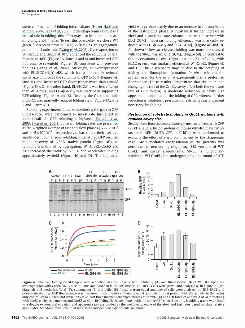
Relationship of folding rate and GroEL ATPase activity
The chaperonin cycle is allosterically regulated by ATP bind-
ing and hydrolysis (Yifrach and Horovitz, 2000). GroES binds
and unbinds from GroEL approximately every 10–15 s, the
time needed for completion of ATP-hydrolysis in the GroEL
cis-ring. GroES dissociation results in the release of enclosed
substrate, with incompletely folded protein rapidly rebinding
to an open GroEL ring. Many substrates interact with GroEL
for several reaction cycles and it has been suggested that
rebinding serves to repeatedly unfold kinetically trapped
intermediates, thereby allowing faster folding speeds (‘itera-
tive annealing’) (Thirumalai and Lorimer, 2001). To test
whether the acceleration of folding of B30 kDa proteins
observed upon reducing GroEL cavity size is dependent on
GroES cycling, we performed in vitro refolding experiments
with SR-EL size-mutants, in which folding occurs upon a
single round of encapsulation (Weissman et al, 1996).
Importantly, the rate acceleration of GFP folding observed
with the cycling GroEL system (Figure 4C and D) was
reproduced with the non-cycling SR-EL size-mutants (Figure
6A and B). Again, optimal folding rate and yield was obtained
with the 2[GGM]4 size-mutant. Similarly, a folding rate
acceleration of rhodanese (33 kDa) was observed with SR-
EL-2[GGM]4/GroES (Figure 6C and D). As under the experi-
mental conditions, the SR-2[GGM]4/GroES complex encapsu-
lates substrate stably (Figure 5C and D), the ability of this
cavity size-mutant to enhance the folding kinetics of GFP and
rhodanese is independent of iterative GroEL ATPase cycles.
The use of SR-EL in these experiments also rules out possible
effects of substrate or residual denaturant on the steady-state
GroEL ATPase as the cause of accelerated folding.
It has also been suggested that an increase of the GroEL
ATPase activity could contribute to the effects on folding rate
observed with the GroEL size-mutants by altering the dwell
time of substrate in the cis-cavity (Farr et al, 2007). ATPase
measurements showed that extension of the C-terminal
GroEL sequences results in a two- to threefold increase of
the maximal GroEL ATPase activity, dependent on assay
conditions (Figure 7A and Supplementary Figure S4).
However, the ATPase activity did not increase linearly with
the length of the C-terminal extensions, as reported by Farr
et al (2007), but rather reached a plateau with the 2[GGM]4
mutant. Importantly, the ability of GroES to inhibit the
ATPase activity by B50% was preserved. The dependence
of ATPase activity on ATP concentration was similar for WT-
GroEL and GroEL tail extension-mutants, with half-maximal
rates being observed at 6–12 mM ATP (Supplementary Figure
S4). As the tail extension-mutants have similar ATPase activ-
ities, the decrease in folding rate and yield observed with EL-
3[GGM]4 and EL-4[GGM]4 relative to EL-2[GGM]4 (Figure 4)
is unrelated to the ATPase activation but is rather a conse-
quence of the reduction of cis-cavity size. Although, in the
case of EL-2[GGM]4, the enhanced ATPase may have in-
creased substrate turnover and thus the yield of GFP refolding
in vivo (Figure 4A and B) (Tehver and Thirumalai, 2008), it is
likely that the reduced cavity size contributed to this effect.
This is supported by our observation with SR-2[GGM]4 in
which folding rate is independent of ongoing ATP hydrolysis
(Figure 6).
To test whether the activation of the GroEL ATPase affects
the folding rate upon a single round of encapsulation, we
generated GroEL cavity size-mutants containing the addi-
tional mutation D398A in the ATPase domain.
GroEL(D398A) binds ATP and GroES but hydrolyzes ATP
extremely slowly (Rye et al, 1997). As a result, substrate
encapsulation and folding occurs upon single-round encap-
sulation as in SR-EL, but with very slow ATP-hydrolysis.
As expected, GroEL(D398A), EL-2[GGM]4(D398A) and
EL-3[GGM]4(D398A) had negligible ATP-hydrolysis rates of
B2% of WT-GroEL (Figure 7B). The rate and yield of GFP
folding with GroEL(D398A) was reduced by B25% com-
pared with WT-GroEL. Importantly, EL-2[GGM]4(D398A) re-
sulted in an B50% increase in folding rate and yield relative
to GroEL(D398A). This effect was reversed with EL-
3[GGM]4(D398A) (Figure 7C), reproducing the results ob-
served with the GroEL and SR-EL tail extension-mutants
(Figures 4D and 6B). Similar results were obtained with
rhodanese. Again folding was accelerated with EL-
2[GGM]4(D398A) and slowed with EL-3[GGM]4(D398A)
(Figure 7D).
These experiments failed to demonstrate a dependence of
folding rate on the GroEL ATPase over a range of ATPase
activities from close to 0 to B3-fold higher than WT, both in
the cycling and non-cycling systems. This is consistent with a
Essentiality of GroEL folding cage in vivoY-C Tang et al
The EMBO Journal VOL 27 | NO 10 | 2008 &2008 European Molecular Biology Organization1464
theoretical simulation of rhodanese folding during GroEL/
GroES cycling. In this simulation, GroEL is in excess over
protein substrate, such that essentially all non-native protein
is GroEL associated. Folding is assumed to occur only upon
protein encapsulation in the GroEL/GroES cage, at a rate
equivalent to spontaneous folding (Brinker et al, 2001). A
two- to fivefold increase in steady-state ATPase is predicted to
have little effect on folding rate, whereas a further rate
increase begins to slow folding due to the reduction of the
dwell time of rhodanese in the cis-cavity and a corresponding
increase of the dwell time in the GroEL-bound state
(Figure 7E and Supplementary Figure S5). Reducing the
ATPase activity has no effect on folding rate, consistent
with the finding that a single round of encapsulation by SR-
EL results in fully efficient folding.
Discussion
Essential role of GroEL/GroES as a folding
compartment
The GroEL/GroES chaperonin system functions as a protein
folding cage based on experiments in vitro (Mayhew et al,
1996; Weissman et al, 1996), but whether folding by encap-
sulation underlies the essentiality of GroEL and GroES for
E. coli growth (Fayet et al, 1989) had remained unclear. To
determine whether the chaperonin cavity is dispensable for E.
coli growth, we tested the in vivo functionality of GroEL
mutants displaying modified cavity properties. GroEL ver-
sions with a reduced cavity size, precluding efficient encap-
sulation of substrates greater than B30 kDa, were no longer
able to support E. coli growth. Likewise, removal of the net
Gro
EL
EL-
2[G
GM
]4
EL-
3[G
GM
]4
EL∆
C
EL-
4[G
GM
]4
− GroES+ GroES
AT
Pas
e ac
tivity
(pi/G
roE
L m
in–1
)
0
10
20
30
40
50
Gro
EL
(D39
8A)
Gro
EL
EL-
2[G
GM
]4(D
398A
)E
L-3[
GG
M] 4
(D39
8A)
RateYield
Rhodanese
0.000.050.100.150.200.250.30
Rho
dane
se y
ield
(rel
ativ
e to
con
trol
)
0.00.20.40.60.81.01.2
Rho
dane
se fo
ldin
g r
ate
(min
–1)
AT
Pas
e ac
tivity
(pi/G
roE
L m
in–1
)
Gro
EL
(D39
8A)
Gro
EL
EL-
2[G
GM
]4(D
398A
)E
L-3[
GG
M]4
(D39
8A)
0
1015202530
5
Time (min)0 10 20 30 40
Nat
ive
prot
ein
(a.u
.)
0.0
0.2
0.4
0.6
0.8
1.0
WT×0.1WT×2.0WT×5.0WT×10WT×20
WT
0
10
20
30
40
GF
P fl
uore
scen
ce(r
elat
ive
to c
ontr
ol)
0.00.20.40.60.81.01.2
Gro
EL
(D39
8A)
Gro
EL
EL-
2[G
GM
]4(D
398A
)E
L-3[
GG
M] 4
(D39
8A)
RateYield
GFP
App
aren
t rat
e G
FP
fol
ding
(×1
0–3
s–1 )
− GroES+ GroES
Figure 7 Accelerated folding by GroEL cavity size-mutants is independent of the rate of ATP hydrolysis. Steady-state ATPase activities of GroELcavity size-mutants (A) and ATPase-deficient GroEL(D398A) cavity size-mutants (B) at 251C. ATPase rates are indicated in ATP hydrolyzed perGroEL tetradecamer per min (see Materials and methods). Refolding yields and rates of WT-GFP (C) and rhodanese (D) with GroEL(D398A)cavity size-mutants and GroES/ATP. Refolding traces for GFP were fitted as in Figure 4D and refolding traces for rhodanese were fitted to asingle exponential equation. The refolding yield obtained with WT-GroEL/GroES was set to 1. Standard deviations of at least three independentexperiments are shown. (E) Simulation of rhodanese folding kinetics dependent on GroEL/GroES cycling rate at excess chaperonin oversubstrate. The binding rate of unfolded protein to GroEL was set to 2�107 M�1 s�1 (Rye et al, 1999) and binding of GroES to GroEL-substratecomplexes was set to 1�106 M�1 s�1 (KC and SS, unpublished data, 2007). The rate of rhodanese refolding with WT-GroEL/GroES or SR-EL/GroES was set to 2.5�10�3 s�1 (Figure 7D). The normal ATPase-induced cycling rate was fixed at 0.07 s�1 with an approximate half-time of10 s. The simulation was performed using chemical kinetics simulator (CKS) (http://www.almaden.ibm.com/st/computational_science/ck).The ATPase-induced cycling rate was varied between 0.1- and 20-fold of the normal rate and refolding rates are plotted. The concentrationsused for the simulation were GroEL 1mM, GroES 2mM and rhodanese 0.5mM (see Supplementary Figure S5 for a kinetic model for thesimulation).
Essentiality of GroEL folding cage in vivoY-C Tang et al
&2008 European Molecular Biology Organization The EMBO Journal VOL 27 | NO 10 | 2008 1465

negative charge of the cis-cavity wall resulted in loss of GroEL
function. Importantly, these mutants preserved the ability to
bind and release non-native substrate protein and GroES. The
10–25% reduction in substrate-binding capacity of GroEL-
3[GGM]4 and 4[GGM]4 cannot account for the failure of these
variants to support normal growth when considering that
E. coli tolerates an B90% reduction in GroEL levels without
significant growth impairment (McLennan et al, 1993; Kerner
et al, 2005). Thus, our findings demonstrate the essential role
of the chaperonin compartment in protein folding in vivo.
The GroEL/GroES folding cage probably provides its essential
function by preventing protein aggregation during folding
and by reducing kinetic folding barriers encountered by a
subset of substrates.
Significance of GroEL cavity size and charge
The volume capacity of the GroEL/GroES cage is generally
sufficient for proteins up to B60 kDa. A special case is the
encapsulation of an B86 kDa assembly intermediate of the
heterodimeric mitochondrial branched-chain a-ketoacid de-
hydrogenase, consisting of B50.7 and B35.5 kDa subunits
(Chen et al, 2006). On the other hand, single protein mole-
cules of B69 and B74 kDa cannot be encapsulated (Kerner
et al, 2005; Paul et al, 2007). An additional mass of B18 kDa
per GroEL ring generated by triplicating the C-terminal
[GGM]4 repeat sequences in mutant EL-3[GGM]4 resulted in
a 10-fold drop in cell number in the absence of WT-GroEL. EL-
4[GGM]4, having an added mass of B27 kDa per GroEL ring,
was essentially unable to support E. coli growth (B1000-fold
drop in cell number upon loss of WT-GroEL). Analysis of
isolated chaperonin complexes showed that EL-3[GGM]4
partially or completely excludes proteins greater than
B35 kDa and EL-4[GGM]4 proteins greater than B30 kDa.
Consistent with these findings, the majority of GroEL-depen-
dent E. coli proteins are between B20 and 50 kDa, with six of
the 13 substrates predicted to have essential function exceed-
ing B31 kDa in size (Kerner et al, 2005). Thus, the failure of
EL-4[GGM]4 to support E. coli growth can be explained by the
exclusion of these essential proteins from the GroEL/GroES
cage, resulting in their misfolding. The capacity of EL-
2[GGM]4 to replace WT-GroEL is consistent with the ability
of this mutant to encapsulate proteins up to at least B40 kDa
(Tang et al, 2006 and this study); this would include all
essential E. coli proteins predicted to be GroEL dependent,
with the exception of D-amino acid dehydrogenase
(B48 kDa) and topoisomerase IV (PARC, B84 kDa) (Kerner
et al, 2005). Notably, PARC is too large to be encapsulated
even by WT-GroEL and may follow the trans-folding mechan-
ism described for yeast aconitase (Chaudhuri et al, 2001).
As demonstrated with EL-2[GGM]4, moderately restricting
cavity space can accelerate the folding of proteins of
B30 kDa, such as GFP, resulting in substantially higher
folding yields in vivo. Various mechanistic components may
contribute to enhancing folding speed, including an effect of
steric confinement, which would entropically destabilize the
unfolded state and favor the formation of compact folded
protein (Baumketner et al, 2003; Takagi et al, 2003).
Anisotropy measurements provided direct evidence that mul-
tiplication of the C-terminal GroEL sequences restricts the
motility of substrate protein upon encapsulation. For proteins
of B30 kDa (GFP and rhodanese), encapsulation in
EL-2[GGM]4 results in productive confinement, whereas
restricting motility further is inhibitory, presumably by hin-
dering necessary rearrangement steps.
Independent evidence for the essential role of the chaper-
onin folding compartment was provided by our analysis of
GroEL mutants with altered charge properties of the cavity
wall. The inner surface of the GroEL ring exposes numerous
positively and negatively charged amino-acid residues with a
marked net negative charge of 42 (minus 6 per GroEL
subunit). Many of the negatively charged residues are highly
conserved among GroEL homologues (Brocchieri and Karlin,
2000). Whereas GroEL mutant EL-NNQ, having a reduced net
charge of minus 21, fully supported E. coli growth, the
complete removal of the negative net charge in EL-3N3Q or
EL-KKK2 resulted in the loss of in vivo functionality. In
contrast, these GroEL charge-mutants were unaffected in
their ability to support the soluble expression of the trans-
folding substrate aconitase. These results suggest that the
negative charge property of the cavity is critical in the folding
of some essential proteins by the encapsulation mechanism.
The majority of GroEL-dependent E. coli proteins, including
eight essential proteins, are negatively charged (pI values of
5.1–6.2) (Kerner et al, 2005) and thus could experience a
repulsive force from the cavity wall that may facilitate fold-
ing. Such an effect may benefit many GroEL substrates, but it
has also been shown that changing a single cavity-exposed
aromatic residue on GroES to arginine (Y71R) can signifi-
cantly improve the folding of a specific protein, GFP (Wang
et al, 2002). This finding, together with our present results,
suggests that a mutual adaptation of the physical properties
of the chaperonin cavity and the natural GroEL substrate
complement has occurred during evolution.
Folding rate acceleration and ATPase turnover
There are currently two models for acceleration of folding by
the GroEL/GroES system. Although mechanistically distinct,
these two models are not mutually exclusive. The central
element of the ‘iterative annealing’ hypothesis suggests that
the GroES-mediated movement of the apical GroEL domains
exerts a stretching force on bound substrate protein, thereby
actively unfolding kinetically trapped, misfolded intermedi-
ates (Thirumalai and Lorimer, 2001). This effect, occurring in
every chaperonin ATPase cycle, would speed up folding by
reducing the half-life of slow folding species and allowing
their repartitioning with kinetically more effective folding
routes. In contrast, ‘cage-mediated annealing’ (Tang et al,
2006) posits that the physical environment of the chaperonin
cavity is critical in enhancing folding speed. To distinguish
between these two models, we explored the dependence of
folding rate on GroEL ATPase activity for GFP and rhodanese.
Specifically, we tested the premise of the iterative annealing
model that a faster ATPase rate, as observed in the GroEL
size-mutants, would speed up folding by increasing the
number of potential unfolding events. Contrary to this as-
sumption, we found that the folding rates achieved with the
cycling GroEL/GroES and the non-cycling SR-EL/GroES sys-
tems are identical within experimental error. Importantly, the
rate enhancement of folding achieved by reducing the size of
the cis-cavity is also observed with SR-EL/GroES and is thus
independent of the ATPase acceleration.
In the presence of GroES, SR-EL carries out a single round
of ATP-hydrolysis and then becomes arrested in the ADP state
(Weissman et al, 1996). To test whether the rate of this
Essentiality of GroEL folding cage in vivoY-C Tang et al
The EMBO Journal VOL 27 | NO 10 | 2008 &2008 European Molecular Biology Organization1466
ATPase reaction may be critical for folding speed, we per-
formed experiments with the D398A mutant of GroEL, which
binds ATP normally but hydrolyzes it extremely slowly.
Importantly, the accelerating effect of the tail extension-
mutants on folding was preserved in GroEL(D398A), estab-
lishing that the effect of geometric confinement on folding
speed is independent of ATP-hydrolysis by GroEL and only
requires ATP-dependent GroES binding. Based on these re-
sults, it would appear that the main role of the GroEL ATPase
is to drive the conformational transition of GroEL from the
substrate binding-active to the folding-active state, allowing
for folded protein to be discharged in a timely fashion.
As in a normal ATPase cycle the substrate protein spends
only B1 s or less in the folding-inactive GroEL-bound state
and the vast majority of the cycle time (10–15 s) encapsulated
within the folding-active cis-cavity, moderately increasing the
GroEL ATPase would have little effect on folding rate. A
simulation of the effect of ATPase rate on the folding speed
of rhodanese supported this conclusion (Figure 7E and
Supplementary Figure S5). Whereas a two- to fivefold in-
crease in cycling rate was predicted to slow folding by only a
small extent, a 10- to 20-fold increase would reduce the dwell
time of the substrate in the cis-cavity more substantially and
approximately double the half-time of folding for a protein
like rhodanese. In contrast, decelerating the ATPase rate was
shown to have virtually no effect on folding rate as long as
GroEL is in excess over substrate, consistent with the ob-
servation that SR-EL/GroES allows folding at essentially the
same rate as WT-GroEL/GroES. However, under conditions of
substrate excess in vivo, both decelerating or accelerating the
GroEL ATPase rate may reduce the folding yield for aggrega-
tion-sensitive substrates (Jewett and Shea, 2008; Tehver and
Thirumalai, 2008). Prolonging the dwell time of substrate
inside the cage would reduce the capacity of chaperonin to
fold multiple substrate molecules, whereas reducing the
dwell time may prevent completion of folding inside the
cage and thus lead to the release of aggregation-prone inter-
mediates.
Materials and methods
Strains and plasmidsGroEL mutants were constructed in a pCH vector backbone andpurified as specified earlier (Chang et al, 2005; Tang et al, 2006).The GroEL ATPase-deficient mutants, EL-D398A, EL-2[GGM]4-D398A and EL-3[GGM]4-D398A, were generated by site-directedmutagenesis. Chaperonin constructs for in vivo co-expression wereprepared by inserting mutated groEL DNA fragments from pCH–GroEL into the pOFXtac-SL2 vector (Castanie et al, 1997). TheDHFR–GFP fusion construct was cloned by introducing a mouseDHFR PCR fragment into the pCH–L16–GFP vector encoding aC-terminal His6-tag (Chang et al, 2005). WT GFP and yeastaconitase (a kind gift from S Rospert) were generated by subcloningthe respective open reading frames into the arabinose promoter-controlled vector pBAD18 (Guzman et al, 1995).
ProteinsChaperone proteins METK and SYT were purified as describedpreviously (Hayer-Hartl et al, 1996; Kerner et al, 2005). WT GFP andDHFR–GFP were purified using a Ni-NTA column (Qiagen). Bovinemitochondrial rhodanese was from Sigma. Polyclonal antibodiesagainst GroEL substrates were produced in rabbits.
In vivo complementation assayA GroEL depletion strain, MC4100 SC3 KanR, in which thechromosomal groE promoter was replaced with the araC gene and
the pBAD promoter, was used (Kerner et al, 2005). E. coli MC4100SC3 cells harbouring pOFXtac-SL2 plasmids for expression ofGroEL/GroES or GroEL-mutants/GroES were grown in LB/0.05%arabinose medium at 301C to OD600¼B1 (B7 h), when cellnumbers were determined to be similar among all strains tested(B5�108 cells ml�1). Serial dilutions of cell suspension werespotted onto LB-spectinomycin selection plates containing 0.05%arabinose (for expression of WT-GroEL/GroES) or 0.1 mM IPTG (forexpression of GroEL-mutants/GroES). Plates were incubated forB16 h at 30, 37 or 421C.
Rhodanese aggregation prevention assayRhodanese (50 mM) was denatured for 1 h at 251C in denaturationbuffer (6 M GuHCl, 20 mM Tris–HCl, pH 7.5, 20 mM KCl, 5 mMMgCl2, 5 mM DTT) and diluted 100-fold into buffer A (20 mM Tris–HCl, pH 7.5, 200 mM KCl, 5 mM Mg(OAc)2), or buffer A containing0.5 or 1 mM GroEL variants. Aggregation was monitored by lightscattering at 320 nm.
Size-exclusion chromatographyRhodanese, METK or SYT (50mM each) was denatured for 1 h at251C in denaturation buffer and diluted 100-fold into buffer A withor without 0.5mM GroEL or GroEL-mutants and subjected to gelfiltration on a Superdex-200 size-exclusion column (GE Healthcare).Fractions (50ml) were analysed by SDS–PAGE and immunoblottingfor the protein substrates. Fractionation of GroEL was visualized byPonceau staining. Recoveries of protein substrates were determinedby densitometry relative to the loading control.
Analysis of protein solubility in vivopOFXtac constructs expressing GroEL/GroES or GroEL-mutants/GroES were co-transformed into E. coli BL21 cells with thearabinose-controlled expression plasmids for yeast aconitase andWT GFP. Cells were grown in LB medium at 301C (for GFPexpression) and 371C (for aconitase expression) to anOD600¼B0.5, and chaperonins were induced with 0.1 mM IPTGfor 1 h before the induction of substrate proteins with 0.2%arabinose for 2 h. Spheroplasts were prepared and analysed asdescribed (Chang et al, 2005). GFP fluorescence was measured intotal spheroplast lysates using 96-well plates (20 mg total protein perwell) and analysed with a Synergy HT UV/VIS fluorescence platereader (Bio-Tek) (Chang et al, 2005).
In vivo capture of GroEL substratesGroEL/GroES-substrate complexes were isolated from live E. colispheroplasts co-expressing C-terminally His6-tagged M. mazeiGroES (MmGroES) and GroEL cavity size-mutants as describedpreviously (Kerner et al, 2005). Control pull-downs were performedwith lysate expressing WT-GroEL and non-His6-tagged MmGroES toevaluate non-specific binding to the beads. Endogenoussubstrates contained in chaperonin complexes were detected byimmunoblotting.
Fluorescence anisotropyGFP and DHFR–GFP (25mM each) was denatured in 6 M GuHCl,20 mM Tris, pH 7.5, 20 mM KCl and 100-fold diluted into low-saltbuffer B (20 mM Tris, pH 7.5, 20 mM KCl, 5 mM Mg(OAc)2) in theabsence or presence of SR-EL variants (1 mM). Encapsulation wasachieved by addition of GroES (2mM), and 5 mM ATP or AMP-PNP,followed by incubation for 30 min at 251C. Steady-state anisotropywas monitored at the emission wavelength of 508 nm (slit width5 nm) with an excitation wavelength at 398 nm (slit width 2 nm)using a Fluorolog 3 spectrofluorometer (Spex).
Proteinase K protection of GroEL–GroES–substratecomplexesDHFR–GFP (25 mM) was denatured as above and diluted 100-foldinto buffer B in the presence of a fourfold molar excess of SR-EL at251C. Reactions were incubated with or without GroES in thepresence of 5 mM AMP-PNP for 2 min and treated with proteinase K(2 mg ml�1) for 0–20 min at 251C (Hayer-Hartl et al, 1996; Tang et al,2006). Protease protection of substrate protein was determined byimmunoblotting.
Essentiality of GroEL folding cage in vivoY-C Tang et al
&2008 European Molecular Biology Organization The EMBO Journal VOL 27 | NO 10 | 2008 1467

In vitro refolding assaysGreen fluorescent protein (25 mM) was denatured in 20 mM Tris, pH7.5, 20 mM KCl, 6 M GuHCl and refolded upon 100-fold dilution intobuffer A or buffer B in the absence or presence of GroEL (0.5mM) orSR-EL (1mM) as described in the figure legends. Chaperonin-assisted refolding was initiated by the addition of GroES (a twofoldmolar excess over GroEL or SR-EL) and 5 mM ATP at 251C. GFPfluorescence was monitored on a Fluorolog 3 Spectrofluorometer(Spex) as described above.
Rhodanese (50mM) was denatured in denaturation buffer andrefolded upon 100-fold dilution into buffer A or buffer Bsupplemented with chaperones as indicated in the figure legendsand folding was initiated as above. Refolding was stopped atdifferent times by the addition of 50 mM CDTA, followed bycolorimetric rhodanese assay (Hayer-Hartl et al, 1996).
ATPase assayGroEL (0.2mM oligomer) was incubated in 20 mM Tris–HCl, pH 7.5,200 mM KCl, 5 mM MgCl2 for 5 min at 251C; when indicated, GroES
was present at a twofold molar excess over GroEL. The reaction wasinitiated by the addition of 2 mM ATP. ATPase activities werefollowed for 0–20 min, with time points taken every 2.5 min.Reactions were stopped by the addition of 15 mM CDTA. Quanti-fication of liberated inorganic phosphate was measured by themalachite green assay (Lanzetta et al, 1979).
Supplementary dataSupplementary data are available at The EMBO Journal Online(http://www.embojournal.org).
Acknowledgements
We thank D Wischnewski and N Wischnewski for technical assis-tance with protein purification. Financial support from the DeutscheForschungsgemeinschaft (SFB 594), the Ernst-Jung Foundation andthe Korber Foundation is acknowledged.
References
Baumketner A, Jewett A, Shea JE (2003) Effects of confinement inchaperonin assisted protein folding: rate enhancement by de-creasing the roughness of the folding energy landscape. J Mol Biol332: 701–713
Brinker A, Pfeifer G, Kerner MJ, Naylor DJ, Hartl FU, Hayer-Hartl M(2001) Dual function of protein confinement in chaperonin-assisted protein folding. Cell 107: 223–233
Brocchieri L, Karlin S (2000) Conservation among HSP60 sequencesin relation to structure, function, and evolution. Prot Sci 9: 476–486
Castanie HP, Berges H, Oreglia J, Prere MF, Fayet O (1997) A set ofpBR322-compatible plasmids allowing the testing of chaperone-assisted folding of proteins overexpressed in Escherichia coli.Anal Biochem 254: 150–152
Chang HC, Kaiser CM, Hartl FU, Barral JM (2005) De novo folding ofGFP fusion proteins: high efficiency in eukaryotes but not inbacteria. J Mol Biol 353: 397–409
Chaudhuri TK, Farr GW, Fenton WA, Rospert S, Horwich AL (2001)GroEL/GroES-mediated folding of a protein too large to beencapsulated. Cell 107: 235–246
Chen DH, Song JL, Chuang DT, Chiu W, Ludtke SJ (2006) Anexpanded conformation of single-ring GroEL–GroES complexencapsulates an 86 kDa substrate. Structure 14: 1711–1722
Farr GW, Fenton WA, Horwich AL (2007) Perturbed ATPase activityand not ‘close confinement’ of substrate in the cis cavity affectsrates of folding by tail-multiplied GroEL. Proc Natl Acad Sci USA104: 5342–5347
Fayet O, Ziegelhoffer T, Georgopoulos C (1989) The GroES andGroEL heat shock gene products of Escherichia coli are essentialfor bacterial growth at all temperatures. J Bacteriol 171:1379–1385
Fukuda H, Arai M, Kuwajima K (2000) Folding of green fluorescentprotein and the cycle3 mutant. Biochemistry 39: 12025–12032
Guzman LM, Belin D, Carson MJ, Beckwith J (1995) Tight regula-tion, modulation, and high-level expression by vectors containingthe arabinose p-BAD promoter. J Bacteriol 177: 4121–4130
Hartl FU, Hayer-Hartl M (2002) Molecular chaperones in thecytosol: from nascent chain to folded protein. Science 295:1852–1858
Hayer-Hartl M, Minton AP (2006) A simple semiempirical model forthe effect of molecular confinement upon the rate of proteinfolding. Biochem 45: 13356–13360
Hayer-Hartl MK, Weber F, Hartl FU (1996) Mechanism of chaper-onin action: GroES binding and release can drive GroEL-mediatedprotein folding in the absence of ATP hydrolysis. EMBO J 15:6111–6121
Horwich AL, Fenton WA, Chapman E, Farr GW (2007) Two familiesof chaperonin: physiology and mechanism. Ann Rev Cell DevelopBiol 23: 115–145
Iwai H, Lingel A, Pluckthun A (2001) Cyclic green fluorescentprotein produced in vivo using an artificially split PI-PfuI inteinfrom Pyrococcus furiosus. J Biol Chem 276: 16548–16554
Jewett AI, Shea J-E (2008) Do chaperonins boost protein yieldsby accelerating folding or preventing aggrgation? Biophys J 94:2987–2993 doi:10.1529/biophysj.107.113209
Kerner MJ, Naylor DJ, Ishihama Y, Maier T, Chang HC, Stines AP,Georgopoulos C, Frishman D, Hayer-Hartl M, Mann M, Hartl FU(2005) Proteome-wide analysis of chaperonin-dependent proteinfolding in Escherichia coli. Cell 122: 209–220
Lanzetta PA, Alvarez LJ, Reinach PS, Candia OA (1979) An im-proved assay for nanomole amounts of inorganic phosphate.Anal Biochem 100: 95–97
Lucent D, Vishal V, Pande VS (2007) Protein folding underconfinement: a role for solvent. Proc Natl Acad Sci USA 104:10430–10434
Martin J, Langer T, Boteva R, Schramel A, Horwich AL, Hartl FU(1991) Chaperonin-mediated protein folding at the surface ofGroEL through a ‘molten globule’-like intermediate. Nature 352:36–42
Mayhew M, Da Silva ACR, Martin J, Erdjument-bromage H, Tempst P,Hartl FU (1996) Protein folding in the central cavity of the GroEL–GroES chaperonin complex. Nature 379: 420–426
McLennan NF, Girshovich AS, Lissin NM, Charters Y, Masters M(1993) The strongly conserved carboxyl-terminus glycine-methio-nine motif of the Escherichia coli GroEL chaperonin is dispensa-ble. Mol Microbiol 7: 49–58
Paul S, Singh C, Mishra S, Chaudhuri TK (2007) The 69 kDaEscherichia coli maltodextrin glucosidase does not getencapsulated underneath GroES and folds through transmechanism during GroEL/GroES-assisted folding. FASEB J 21:2874–2885
Rye HS, Burston SG, Fenton WA, Beechem JM, Xu Z, Sigler PB,Horwich AL (1997) Distinct actions of cis and trans ATP withinthe double ring of the chaperonin GroEL. Nature 388: 792–798
Rye HS, Roseman AM, Chen S, Furtak K, Fenton WA, Saibil HR,Horwich AL (1999) GroEL-GroES cycling: ATP and nonnativepolypeptide direct alternation of folding-active rings. Cell 97:325–338
Takagi F, Koga N, Takada S (2003) How protein thermodynamicsand folding mechanisms are altered by the chaperonin cage:molecular simulations. Proc Natl Acad Sci USA 100: 11367–11372
Tang YC, Chang HC, Roeben A, Wischnewski D, Wischnewski N,Kerner MJ, Hartl FU, Hayer-Hartl M (2006) Structural features ofthe GroEL–GroES nano-cage required for rapid folding of encap-sulated protein. Cell 125: 903–914
Tehver R, Thirumalai D (2008) Kinetic model for the couplingbetween allosteric transitions in GroEL and substrate proteinfolding and aggregation. J Mol Biol 377: 1279–1295
Thirumalai D, Lorimer GH (2001) Chaperonin-mediated proteinfolding. Ann Rev Biophys Biomol Struct 30: 245–269
Wang JD, Herman C, Tipton KA, Gross CA, Weissman JS (2002)Directed evolution of substrate-optimized GroEL/ES chaperonins.Cell 111: 1027–1039
Weissman JS, Rye HS, Fenton WA, Beechem JM, Horwich AL (1996)Characterization of the active intermediate of a GroEL–GroES-mediated protein folding reaction. Cell 84: 481–490
Yifrach O, Horovitz A (2000) Coupling between protein folding andallostery in the GroE chaperonin system. Proc Natl Acad Sci USA97: 1521–1524
Essentiality of GroEL folding cage in vivoY-C Tang et al
The EMBO Journal VOL 27 | NO 10 | 2008 &2008 European Molecular Biology Organization1468