Erratum to: Increased Hippocampal Expression of the Divalent Metal Transporter 1 (DMT1) mRNA...
10
Increased Hippocampal Expression of the Divalent Metal Transporter 1 (DMT1) mRNA Variants 1B and +IRE and DMT1 Protein After NMDA-Receptor Stimulation or Spatial Memory Training Paola Haeger A ´ lvaro A ´ lvarez Nancy Leal Tatiana Adasme Marco Tulio Nu ´n ˜ez Cecilia Hidalgo Received: 8 May 2009 / Revised: 15 July 2009 / Accepted: 21 July 2009 Ó Springer Science+Business Media, LLC 2009 Abstract Iron is essential for crucial neuronal functions but is also highly toxic in excess. Neurons acquire iron through transferrin receptor-mediated endocytosis and via the divalent metal transporter 1 (DMT1). The N-terminus (1A, 1B) and C-terminus (?IRE, -IRE) splice variants of DMT1 originate four protein isoforms, all of which supply iron to cells. Diverse physiological or pathological condi- tions induce differential DMT1 variant expression, which are cell-type dependent. Hence, it becomes relevant to ascertain if activation of neuronal plasticity processes that require functional N-methyl D-aspartate (NMDA) receptors, including in vitro stimulation of NMDA recep- tor-mediated signaling and spatial memory training, selectively modify DMT1 variant expression. Here, we report for the first time that brief (5 min) exposure of pri- mary hippocampal cultures to NMDA (50 lM) increased 24 h later the expression of DMT1-1B and DMT1?IRE, but not of DMT1-IRE mRNA. In contrast, endogenous DMT1 mRNA levels remained unaffected following 6 h incubation with brain-derived nerve factor. NMDA (25–50 lM) also enhanced DMT1 protein expression 24–48 h later; this enhancement was abolished by the transcription inhibitor actinomycin D and by the NMDA receptor antagonist MK-801, implicating NMDA receptors in de novo DMT1 expression. Additionally, spatial memory training enhanced DMT1-1B and DMT1?IRE expression and increased DMT1 protein content in rat hippocampus, where the exon1A variant was not found. These results suggest that NMDA receptor-dependent plasticity processes stimulate expression of the iron trans- porter DMT1-1B?IRE isoform, which presumably plays a significant role in hippocampal spatial memory formation. Keywords Iron transport Synaptic plasticity mRNA splicing variants DMT1 protein Morris water maze Spatial learning BDNF Hippocampal pyramidal neurons Abbreviations ACSF Artificial cerebrospinal fluid Act D Actinomycin D BDNF Brain-derived neurotrophic factor DMT1 Divalent metal transporter 1 GFAP Glial fibrillary acidic protein IRE Iron response element IRP Iron regulatory protein MAP2 Microtubule-associated protein 2 MWM Morris Water Maze NF-Y Nuclear factor Y NFjB Nuclear factor kappa B NMDA N-Methyl D-aspartate Electronic supplementary material The online version of this article (doi:10.1007/s12640-009-9096-z) contains supplementary material, which is available to authorized users. P. Haeger (&) A ´ .A ´ lvarez N. Leal T. Adasme C. Hidalgo Centro FONDAP de Estudios Moleculares de la Ce ´lula, Facultad de Medicina, Universidad de Chile, Independencia 1027, Santiago, Chile e-mail: [email protected] M. T. Nu ´n ˜ez Departamento de Biologı ´a, Facultad de Ciencias and Institute for Cell Dynamics and Biotechnology, Universidad de Chile, Santiago, Chile C. Hidalgo Programa de Biologı ´a Celular y Molecular, Instituto de Ciencias Biome ´dicas, Facultad de Medicina, Universidad de Chile, Santiago, Chile 123 Neurotox Res DOI 10.1007/s12640-009-9096-z
-
Upload
independent -
Category
Documents
-
view
2 -
download
0
Transcript of Erratum to: Increased Hippocampal Expression of the Divalent Metal Transporter 1 (DMT1) mRNA...
Increased Hippocampal Expression of the Divalent MetalTransporter 1 (DMT1) mRNA Variants 1B and +IRE and DMT1Protein After NMDA-Receptor Stimulation or Spatial MemoryTraining
Paola Haeger Æ Alvaro Alvarez Æ Nancy Leal ÆTatiana Adasme Æ Marco Tulio Nunez ÆCecilia Hidalgo
Received: 8 May 2009 / Revised: 15 July 2009 / Accepted: 21 July 2009
� Springer Science+Business Media, LLC 2009
Abstract Iron is essential for crucial neuronal functions
but is also highly toxic in excess. Neurons acquire iron
through transferrin receptor-mediated endocytosis and via
the divalent metal transporter 1 (DMT1). The N-terminus
(1A, 1B) and C-terminus (?IRE, -IRE) splice variants of
DMT1 originate four protein isoforms, all of which supply
iron to cells. Diverse physiological or pathological condi-
tions induce differential DMT1 variant expression, which
are cell-type dependent. Hence, it becomes relevant to
ascertain if activation of neuronal plasticity processes
that require functional N-methyl D-aspartate (NMDA)
receptors, including in vitro stimulation of NMDA recep-
tor-mediated signaling and spatial memory training,
selectively modify DMT1 variant expression. Here, we
report for the first time that brief (5 min) exposure of pri-
mary hippocampal cultures to NMDA (50 lM) increased
24 h later the expression of DMT1-1B and DMT1?IRE,
but not of DMT1-IRE mRNA. In contrast, endogenous
DMT1 mRNA levels remained unaffected following 6 h
incubation with brain-derived nerve factor. NMDA
(25–50 lM) also enhanced DMT1 protein expression
24–48 h later; this enhancement was abolished by the
transcription inhibitor actinomycin D and by the NMDA
receptor antagonist MK-801, implicating NMDA receptors
in de novo DMT1 expression. Additionally, spatial
memory training enhanced DMT1-1B and DMT1?IRE
expression and increased DMT1 protein content in rat
hippocampus, where the exon1A variant was not found.
These results suggest that NMDA receptor-dependent
plasticity processes stimulate expression of the iron trans-
porter DMT1-1B?IRE isoform, which presumably plays a
significant role in hippocampal spatial memory formation.
Keywords Iron transport � Synaptic plasticity �mRNA splicing variants � DMT1 protein �Morris water maze � Spatial learning � BDNF �Hippocampal pyramidal neurons
Abbreviations
ACSF Artificial cerebrospinal fluid
Act D Actinomycin D
BDNF Brain-derived neurotrophic factor
DMT1 Divalent metal transporter 1
GFAP Glial fibrillary acidic protein
IRE Iron response element
IRP Iron regulatory protein
MAP2 Microtubule-associated protein 2
MWM Morris Water Maze
NF-Y Nuclear factor Y
NFjB Nuclear factor kappa B
NMDA N-Methyl D-aspartate
Electronic supplementary material The online version of thisarticle (doi:10.1007/s12640-009-9096-z) contains supplementarymaterial, which is available to authorized users.
P. Haeger (&) � A. Alvarez � N. Leal � T. Adasme � C. Hidalgo
Centro FONDAP de Estudios Moleculares de la Celula,
Facultad de Medicina, Universidad de Chile,
Independencia 1027, Santiago, Chile
e-mail: [email protected]
M. T. Nunez
Departamento de Biologıa, Facultad de Ciencias and Institute
for Cell Dynamics and Biotechnology, Universidad de Chile,
Santiago, Chile
C. Hidalgo
Programa de Biologıa Celular y Molecular, Instituto de Ciencias
Biomedicas, Facultad de Medicina, Universidad de Chile,
Santiago, Chile
123
Neurotox Res
DOI 10.1007/s12640-009-9096-z
PBS Phosphate buffered saline
ROS Reactive oxygen species
Introduction
Iron deficiency during infancy leads to lower cognitive
performance in adulthood (Lozoff 2000). In animal mod-
els, nutritional iron deficiency interferes with hippocam-
pus-depending learning (McEchron and Paronish 2005;
Ranade et al. 2008) and synaptic plasticity (Jorgenson et al.
2005). These functional failings have been ascribed to the
iron requirements of metabolic pathways involved in neu-
rotransmitter synthesis and myelin formation (Youdim
et al. 1980; Taneja et al. 1986; Kwik-Uribe et al. 2000).
Conversely, excessive iron accumulation impairs normal
neuronal functions and induces neuronal death (Cheah
et al. 2006; Salazar et al. 2006, 2008; Du et al. 2009).
The main players of cellular iron homeostasis comprise:
the transferrin receptor (Griffiths and Crossman 1996;
Moos et al. 1998), the divalent metal transporter DMT1
(Burdo et al. 1999, 2001; Williams et al. 2000), the iron
storage protein ferritin (Benkovic and Connor 1993), and
the iron export transporter ferroportin (Wu et al. 2004), all
of which are well expressed in different areas of the brain.
In the particular case of DMT1, which mediates ferrous
iron uptake into cells (Gunshin et al. 1997), in situ
hybridization studies in developing rat brain showed
DMT1 mRNA localization in striatum, cortex, hippocam-
pus, and cerebellum (Williams et al. 2000). Immunohis-
tochemistry analysis of adult rat brain showed DMT1
protein expression in striatum, cerebellum, and thalamus,
as well as in vascular cells throughout the brain and
ependymal cells in the third ventricle (Burdo et al. 2001).
The mammalian DMT1 gene (SLC11A2; Nramp2)
undergoes alternative splicing. The 1A and 1B mRNA
DMT1 variants originate from alternative splicing at the 50
end (exons 1A and 1B), while the ?IRE or -IRE variants
originate from the 30 end (exons 16/16A and 17) (Hubert
and Hentze 2002). These variants give rise to four DMT1
protein isoforms, all of them active in Fe2? transport
(Ludwiczek et al. 2007). It is generally accepted that the
two ?IRE isoforms are post-transcriptionally regulated by
the IRE/IRP system (Pantopoulos 2004). Knowledge of
differential transcriptional regulation of DMT1 expression
is emerging. Both the inflammatory cytokine nuclear factor
kappa B (NFjB) and the nuclear factor Y (NF-Y) regulate
DMT1-1B expression in embryonic carcinoma cells
(Paradkar and Roth 2007). In contrast, hypoxia upregulates
expression of the 1A mRNA variant in PC12 cells,
presumably through activation of hypoxic response ele-
ments in its promoter region (Lis et al. 2005).
DMT1 mediates iron uptake into neurons, where cellular
iron levels are essential for crucial neuronal functions but
highly toxic in excess (Hidalgo et al. 2007; Hidalgo and
Nunez 2007; Pelizzoni et al. 2008). Interestingly, a drug that
causes experimental Parkinson’s disease (1-methyl-4-phe-
nyl-1,2,3,6-tetrahydropyridine) upregulates DMT1?IRE
protein expression in mice ventral mesencephalon, where it
increases neuronal death presumably through abnormal
increases in cellular iron content (Salazar et al. 2008).
Additionally, DMT1-IRE mediates L-DOPA neurotoxicity
in primary cortical neurons (Du et al. 2009). Consequently,
to understand the role of neuronal iron in health and disease,
it becomes important to determine which DMT1 isoforms
are expressed in neurons, and how neuronal activity regu-
lates their expression.
In this work, we report for the first time that N-methyl
D-aspartate (NMDA) addition to primary hippocampal
cultures, or spatial memory training, selectively stimulate
hippocampal expression of the DMT1 splicing variants 1B
and ?IRE, and increased DMT1 protein content. These
results suggest that enhanced expression of the iron trans-
porter 1B?IRE DMT1 isoform has a role in hippocampal-
dependent long-lasting plasticity changes.
Methods
Hippocampal Cultures
Primary cultures were prepared from hippocampus dis-
sected from Sprague Dawley rats at embryonic day 18
(Kahlert et al. 2005). Cells, plated in minimum essential
medium plus 10% horse serum, were grown subsequently
at 37�C under 5% CO2 in serum-free Neurobasal medium
supplemented with GIBCOTM B27 serum-free and 2 mM
GlutamaxTM (Invitrogen). To inhibit glial cell prolifera-
tion, 5-fluoro-20-deoxy-uridine (1.4 mg/l) plus uridine
(3.5 mg/l) were added for 24 h at the second day of culture.
Immunocytochemistry and Immunohistochemistry
Hippocampal cells were fixed for 20 min in PBS contain-
ing 4% paraformaldehyde, 4% sucrose and permeabilized
for 10 min with 0.1% Triton X-100 in phosphate buffered
saline (PBS). For immunodetection, cells were incubated
overnight at 4�C with polyclonal rabbit anti-PanDMT1
antibody prepared against a peptide common to all iso-
forms (Salazar et al. 2008), with anti-b-Tubulin III (Sigma)
or with MAP2 antibody (Chemicon). Glial cells were
stained with an antibody against glial fibrillary acidic
Neurotox Res
123
protein (GFAP) (DAKO). Cells were subsequently incu-
bated with Alexa Fluor� 488 anti-rabbit or Alexa Fluor�
635 anti-mouse as secondary antibody. Brain tissue fixation
and immunohistochemistry were performed as described
(Haeger et al. 2006), except that coronal slices were
incubated at 4�C overnight with PanDMT1 and MAP2
antibodies. Sections were incubated at room temperature
for 2 h in PBS buffer containing 0.2% gelatin and 0.1%
Triton X-100 plus the Alexa antibodies defined above,
rinsed in PBS, and mounted with glycerol. All fluorescence
images were obtained in a confocal microscope (Carl Zeiss
LSM Pascal 5, Zeiss, Oberkochen, Germany), and digitally
acquired using LSM software (Zeiss). Controls performed
without primary or secondary antibody yielded only
background staining.
Western Blot Analysis
Cells extracts prepared as described (Aguirre et al. 2005)
were resolved in 10% Laemmli SDS-polyacrylamide gels,
transferred to PDVF membranes (Millipore) and incubated
overnight with PanDMT1 primary antibody. To correct for
loading, membranes were stripped and re-probed for
b-actin. The IMAGE J image program (National Institutes
of Health, USA) was used to quantify optical band density.
Incubation with NMDA or Brain-Derived Neurotrophic
Factor (BDNF)
One hour before NMDA addition, the culture medium was
replaced by Neurobasal-B27 without antioxidants. Cells
were then incubated for 5 min with NMDA (25 or 50 lM),
10 lM D-Serine in ACSF plus 0.62 mM MgCl2. Control
cells were incubated similarly without NMDA. Cells were
subsequently incubated 1 h in Neurobasal-B27 medium
without antioxidants plus 23 h in the original culture
medium prior to RNA isolation or western blot analysis.
Alternatively, cultures were incubated in supplemented
Neurobasal-B27 medium for 6 h with BDNF (50 ng/ml)
and DMT1 mRNA expression was determined right after
incubation.
RNA Isolation and RT-PCR
Total RNA was isolated using Trizol reagent (Invitrogen).
DNAase digestion (DNA-freeTM Kit, Ambion) was inclu-
ded to remove any contaminating genomic DNA. RNA
purity was assessed by the 260/280 absorbance ratio and
RNA integrity by gel electrophoresis. cDNA was synthe-
sized from 0.5 lg total RNA using ImProm-IITM Reverse
Transcriptase (Promega). Twenty-five nanograms of cDNA
was used in 20 ll final volume for PCR amplification using
an Applied Biosystem Thermal cycler. Amplification of the
mRNA for all four DMT1 isoforms was done for 35 cycles;
each cycle included 15 s at 94�C, 15 s at 60�C (or 58�C for
DMT1 1A and 1B isoforms), and 15 s at 72�C. After these
cycles, a final 10 min incubation step at 72�C was added.
Primers for DMT1?IRE and DMT1-IRE cDNAs, con-
structed based on the rat DMT1 sequence (GenBank
#AF008439 and #AF029757), were: Forward, 50-CTGAG
CGAAGATACCAGCG-30 and 50-GTCTTCCAAGATGT
GGAGCA-30; Reverse, 50-GGAGCCATCACTTGACCAC
AC-30 and 50-ATCTTACCCAAACTGGCACG-30, respec-
tively. The selected forward primers for the DMT1 1A and
1B variants were as previously described (Hubert and
Hentze 2002; Lis et al. 2005): 50-TCCGATGGGGAAGAA
GCAGCC-30 for DMT1 1A and 50-CAATCACGGGA
GGGCAGGAG-30 for DMT1-1B. The same reverse primer
50-CTAGGTAGGCAATGCTCATAAGAAAGCCAGG-30
was used for both variants. The b-actin primers used were:
forward 50-TCTACAATGAGCTGCGTGTG-30 and reverse
50-TACATGGCTGGGGTGTTGAA-30. The amount of
cDNA was determined by b-actin amplification using real
time PCR, performed in a thermocycler (Stratagene
MX3000P) using the DNA binding dye SYBR green
(Invitrogen). A graph digitizing software was used for
densitometry analysis of RT-PCR products. The 380-bp
DMT1-1B PCR fragment obtained after RT-PCR amplifi-
cation of the total RNA obtained from cultured hippo-
campal rat cells (Fig. 1, lane 1), was extracted from the
agarose gel by means of a gel extraction kit (Qiagen). This
product was directly sequenced (ABI PRISM 310; ABI,
Foster City, CA, USA) using the forward and reverse
primers for DMT1-1B described above, and was aligned
with the 50 end of mouse DMT1-1B (MM001146161) using
the Cluscalw2 software (EMBL-EBI).
Morris Water Maze Training
Male Sprague Dawley rats were trained once daily using a
circular water maze (1.6 m diameter, 75 cm deep)
endowed with spatial cues and a hidden platform placed at
a fixed location; each trial involved three 1 min sessions,
separated by 20 min intervals. Training was continued for
six consecutive days, followed by 2 days off; to evaluate
memory retention, one additional day without platform was
added. The whole hippocampus was removed 6 h after the
ninth day trail. Escape latency was measured as described
(De Ferrari et al. 2003). Control animals were subjected for
four consecutive days to three swimming trials daily with
the cued platform; the platform location varied daily. At
day four, the whole hippocampus was removed 6 h after
the last trial.
All experiments described here were carried out under
the guidelines of National Institutes of Health, USA, reg-
ulations for the Care and Use of Animals for Scientific
Neurotox Res
123
Purposes; all protocols were approved by the Bioethics
Committee, F. Medicine, Universidad de Chile.
Results
Hippocampal DMT1 Expression
Previous reports have described DMT1 mRNA expression
in the hippocampus (Williams et al. 2000), and DMT1
protein expression in glial cells (Huang et al. 2006), but to
our knowledge no published data are available showing
expression of DMT1 protein or mRNA variants in
hippocampal neurons. To address this issue, we analyzed
DMT1 expression in primary hippocampal cultures highly
enriched in neurons (Supplementary Fig. 1a) and hippo-
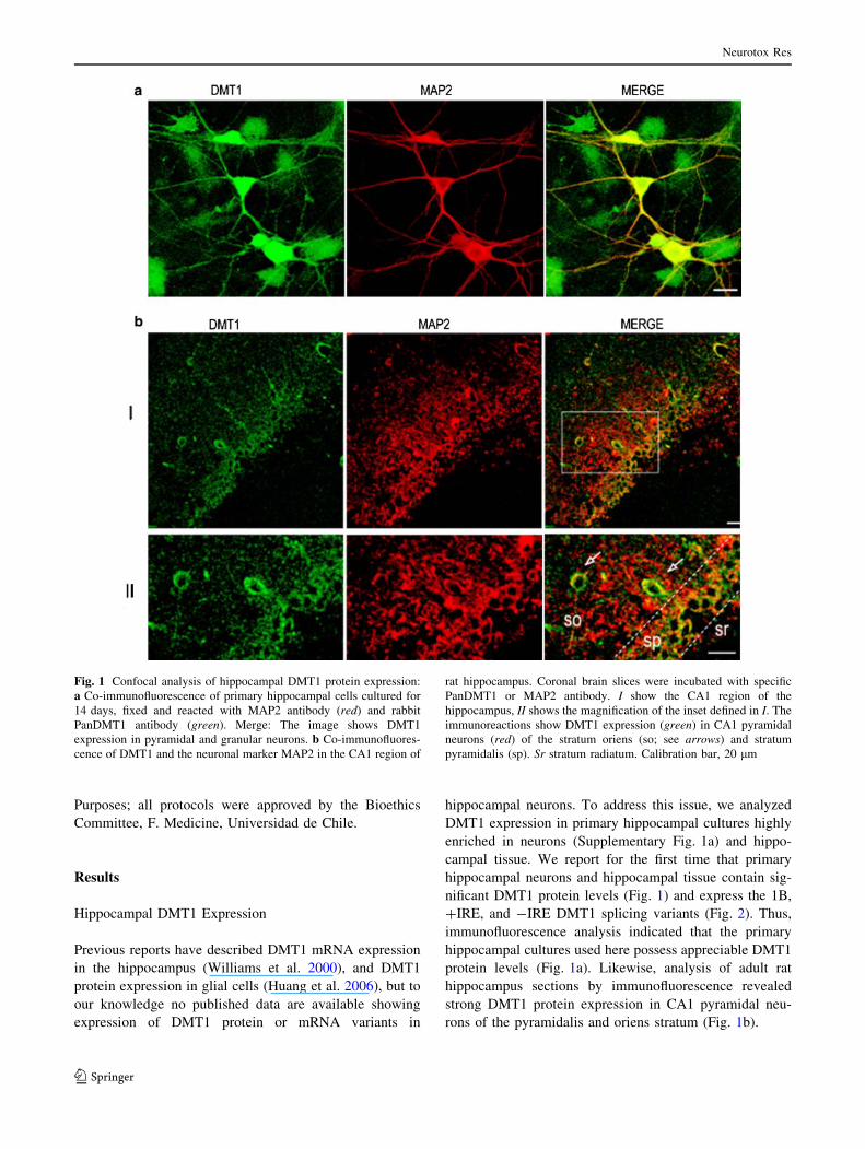
campal tissue. We report for the first time that primary
hippocampal neurons and hippocampal tissue contain sig-
nificant DMT1 protein levels (Fig. 1) and express the 1B,
?IRE, and -IRE DMT1 splicing variants (Fig. 2). Thus,
immunofluorescence analysis indicated that the primary
hippocampal cultures used here possess appreciable DMT1
protein levels (Fig. 1a). Likewise, analysis of adult rat
hippocampus sections by immunofluorescence revealed
strong DMT1 protein expression in CA1 pyramidal neu-
rons of the pyramidalis and oriens stratum (Fig. 1b).
Fig. 1 Confocal analysis of hippocampal DMT1 protein expression:
a Co-immunofluorescence of primary hippocampal cells cultured for
14 days, fixed and reacted with MAP2 antibody (red) and rabbit
PanDMT1 antibody (green). Merge: The image shows DMT1
expression in pyramidal and granular neurons. b Co-immunofluores-
cence of DMT1 and the neuronal marker MAP2 in the CA1 region of
rat hippocampus. Coronal brain slices were incubated with specific
PanDMT1 or MAP2 antibody. I show the CA1 region of the
hippocampus, II shows the magnification of the inset defined in I. The
immunoreactions show DMT1 expression (green) in CA1 pyramidal
neurons (red) of the stratum oriens (so; see arrows) and stratum
pyramidalis (sp). Sr stratum radiatum. Calibration bar, 20 lm
Neurotox Res
123
NMDA Increases the Expression of the Iron
Transporter DMT1
Activation of hippocampal NMDA receptors promotes the
expression of genes required to maintain synaptic plasticity
for prolonged times (Platenik et al. 2000; Deisseroth et al.
2003; Tabuchi 2008). To our knowledge, no data are
available in the literature describing the effects of NMDA
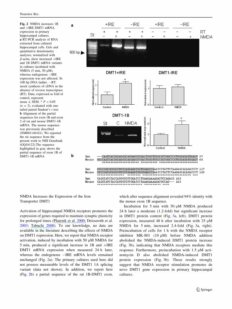
on DMT1 expression. Here, we report that NMDA receptor
activation, induced by incubation with 50 lM NMDA for
5 min, produced a significant increase in 1B and ?IRE
DMT1 mRNA expression when measured 24 h later,
whereas the endogenous -IRE mRNA levels remained
unchanged (Fig. 2a). The primary cultures used here did
not possess measurable levels of the DMT1 1A splicing
variant (data not shown). In addition, we report here
(Fig. 2b) a partial sequence of the rat 1B-DMT1 exon,
which after sequence alignment revealed 94% identity with
the mouse exon 1B sequence.
Incubation for 5 min with 50 lM NMDA produced
24 h later a moderate (1.2-fold) but significant increase
in DMT1 protein content (Fig. 3a, left). DMT1 protein
expression, measured 48 h after incubation with 25 lM
NMDA for 5 min, increased 2.4-fold (Fig. 3a, right).
Preincubation of cells for 1 h with the NMDA receptor
inhibitor MK-801 (10 lM) before NMDA addition
abolished the NMDA-induced DMT1 protein increase
(Fig. 3b), indicating that NMDA receptors mediate this
response. Furthermore, preincubation with 1.5 lM acti-
nomycin D also abolished NMDA-induced DMT1
protein expression (Fig. 3b). These results strongly
suggest that NMDA receptor stimulation promotes de
novo DMT1 gene expression in primary hippocampal
cultures.
Fig. 2 NMDA increases 1B
and ?IRE DMT1 mRNA
expression in primary
hippocampal cultures.
a RT-PCR analysis of RNA
extracted from cultured
hippocampal cells. Gels and
quantitative densitometry
analyses, normalized with
b-actin, show increased ?IRE
and 1B DMT1 mRNA variants
in cultures incubated with
NMDA (5 min, 50 lM),
whereas endogenous -IRE
expression was not affected. St:
100 bp DNA ladder. -RT:
mock synthesis of cDNA in the
absence of reverse transcriptase
(RT). Data, expressed as fold of
control, represent
mean ± SEM. * P \ 0.05
(n = 3), evaluated with one-
tailed paired Student’s t test.
b Alignment of the partial
sequences for exon 1B and exon
2 of rat and mouse DMT1-1B
mRNA. The mouse sequence
was previously described
(NM001146161). We reported
the rat sequence from the
present work to NIH Genebank
(GQ16122).The sequence
highlighted in gray shows the
partial sequence of exon 1B of
DMT1-1B mRNA
Neurotox Res
123

The enhanced expression of DMT1 protein probably
reflects increased expression of the DMT1-1B?IRE iso-
form since the 1A mRNA variant was not detected in the
cultures. The PanDMT1 antibody used does not distinguish
between the different DMT1 protein isoforms, so the
increase in DMT1-1B?IRE content may be higher than
estimated here.
In contrast to the stimulatory effects of NMDA observed
at 24 and 48 h, incubation of primary cultures for 6 h with
BDNF, a neurotrophin functionally stimulated by NMDA
receptor activation (Caldeira et al. 2007), did not increase
expression of the 1B, ?IRE, and -IRE DMT1 variants
when measured right after the incubation period (Fig. 4).
Yet, incubation with BDNF for 6 h significantly stimulates
transcription of several synaptic protein genes (Ring et al.
2006). Accordingly, our results suggest that BDNF does
not stimulate DMT1 expression, or that if it does, this
stimulation occurs at times longer than 6 h.
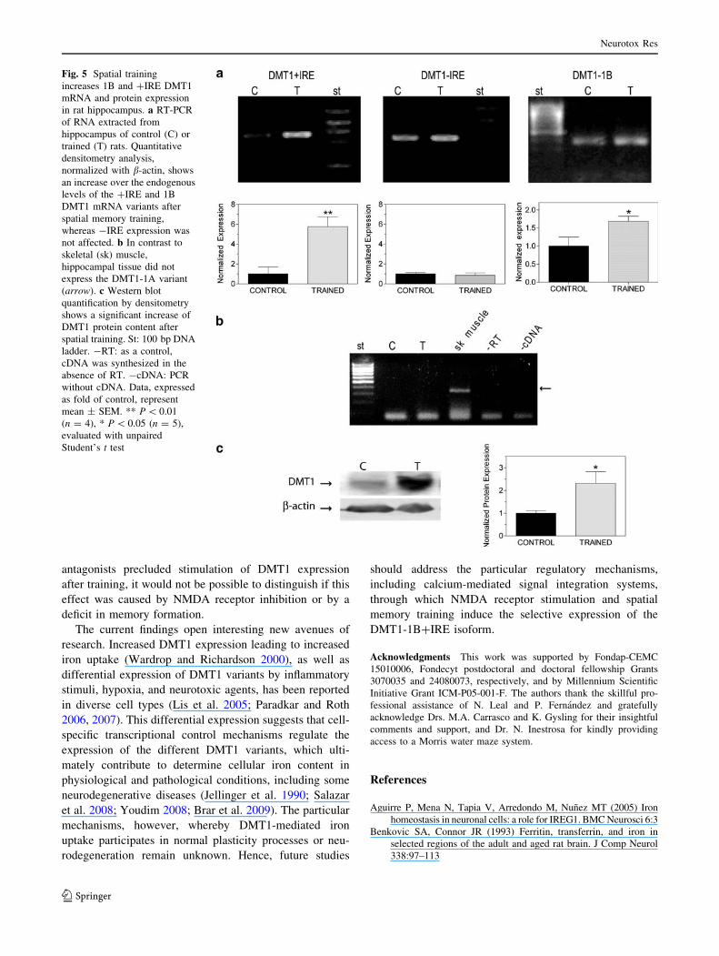
In agreement with the above findings in cultured cells,
the hippocampus of adult rats displayed significant
endogenous levels of DMT1?IRE, DMT1-IRE, and
DMT1-1B mRNA variants (Fig. 5a), whereas DMT1-1A
mRNA was not detected (Fig. 5b). NMDA receptor acti-
vation is a key step of spatial memory formation (Robbins
and Murphy 2006). Accordingly, we investigated if train-
ing in a Morris water maze modified hippocampal DMT1
expression. As observed in NMDA-stimulated primary
cultures, a significant increase in the ?IRE and 1B mRNA
variants occurred following spatial training of rats while
DMT1-IRE mRNA remained unaffected (Fig. 5a).
Immunohistochemistry analysis showed significant DMT1
protein expression in CA1 pyramidal neurons of adult rat
hippocampus (Fig. 1b). Interestingly, after spatial memory
training DMT1 protein content in hippocampal extracts
augmented[2.5-fold, as revealed by western blot analysis
(Fig. 5c).
Discussion
Scant information is available regarding the role of DMT1
in normal or pathological neuronal function. Preliminary
Fig. 3 NMDA increases DMT1
protein levels in primary
hippocampal cultures.
a Hippocampal cells were
incubated for 5 min with 50 or
25 lM NMDA; cell extracts
were collected after 24 or 48 h
of culture, respectively. NMDA
stimulation increased total
DMT1 protein content, as
shown in western blots of cell
extracts obtained 24 and 48 h
after incubation with NMDA.
b-actin was used as loading
control. The number of
experiments is indicated in each
bar. Data are given as
mean ± SEM. Statistical
differences were analyzed by
one-tailed paired Student’s ttest. * P \ 0.05,
*** P \ 0.001. b Previous to
25 lM NMDA stimulation,
hippocampal cells were
incubated for 1 h with the
NMDA receptor antagonist
MK-801 (10 lM) or the
transcription inhibitor
Actinomycin D (Act D, 1.5 lM)
and the extract was obtained
48 h post-NMDA stimulus. The
number of experiments is
indicated in each bar. Statistical
analysis: One-way Anova,
followed by Newman Keuls
post-test
Neurotox Res
123

results from our group1 showed that incubation of primary
cultures with NMDA or spatial memory training of rats in
the MWM increased hippocampal DMT1?IRE mRNA and
protein expression. While this work was in progress, defi-
cient spatial learning in a conditionally targeted DMT1
knockout mouse and increased DMT1 mRNA expression
after spatial memory training was reported (Carlson et al.
2009); however, the DMT1 variants induced by training
were not identified. Expanding on the above results, we
report here that hippocampal cultures briefly exposed for
5 min to the glutamate receptor agonist NMDA displayed
24 h later significantly enhanced expression of the ?IRE
and 1B mRNA variants, coupled with increased DMT1
protein content, which increased even further at 48 h.
Previous reports indicate that NMDA stimulates sig-
naling cascades that enhance iron uptake in cortical neu-
rons (Cheah et al. 2006), and that iron chelation with
desferrioxamine impairs tetanically induced long-term
potentiation at hippocampal CA1 neurons (Hidalgo et al.
2007), a process known to involve NMDA receptors
(Platenik et al. 2000). Hence, the significant up-regulation
of DMT1 expression following NMDA receptor stimula-
tion reported here, suggests that hippocampal neurons
increase DMT1 protein expression after synaptic stimula-
tion to improve their iron uptake capacity, and thus ensure
an adequate iron supply for plasticity processes. Our results
also suggest that enhanced transcription of the 1B variant,
plus selective expression of the ?IRE protein isoform
underlie this increase.
In agreement with the above results in primary hippo-
campal cultures, we show here for the first time that
training rats in a spatial memory task, which entails NMDA
receptor activation (Robbins and Murphy 2006), also
increased DMT1?IRE and DMT1-1B mRNA expression
and increased more than 2.5-fold total DMT1 protein
content in the CA1 hippocampal region. Furthermore, the
increased expression of the DMT1 variants 1B and ?IRE
after spatial memory training strongly suggests that hip-
pocampal neurons up-regulate the expression of the
DMT1-1B?IRE protein isoform during training.
The induction of DMT1 1B and ?IRE mRNA expres-
sion reported here may arise from transcriptional upregu-
lation initiated by NMDA binding to its receptor.
Additionally, NMDA receptor stimulation may activate
signaling pathways that selectively stabilize these particu-
lar mRNA splicing variants. Both pathways would increase
DMT1 protein expression. The DMT1-1B promoter con-
tains an NFjB response element (Paradkar and Roth 2006).
Interestingly, in cerebellar neurons NFjB-dependent tran-
scription is activated by calcium (Lilienbaum and Israel
2003) and by NMDA itself (Lipsky et al. 2001), whereas
many studies have reported activation of NFjB by cellular
ROS (Gloire et al. 2006). Accordingly, the intracellular
increases in calcium (Lerea et al. 1992) and ROS (Kahlert
et al. 2005) produced by NMDA receptor stimulation may
jointly stimulate NFjB-dependent transcription of the
DMT1 gene in hippocampal neurons. Yet, a direct dem-
onstration of NMDA receptor involvement in learning-
induced DMT1 expression is not a trivial matter. NMDA
receptor inhibition induces alterations in spatial mem-
ory formation (Robbins and Murphy 2006; Enomoto
et al. 2008) and provokes schizophrenia-like symptoms
(Tamminga et al. 2003). Thus, if NMDA receptor
Fig. 4 Lack of effect of BDNF on DMT1 mRNA variant expression
in hippocampal cultures. RT-PCR analysis of RNA extracted from
cultured hippocampal cells incubated for 6 h with BDNF (50 ng/ml).
Quantitative densitometry analysis, normalized with b-actin, revealed
that expression of 1B, ?IRE, and -IRE DMT1 mRNA variants did
not undergo significant changes after BDNF incubation. Data,
expressed as fold of control, represent mean ± SEM (n C 3).
Significance was evaluated with one-tailed paired Student’s t test
1 Haeger P., Munoz P., Carrasco M. A., Nunez, M. T. & Hidalgo C.
Increased RyR and DMT1 expression in rat hippocampus after spatial
memory training, 38th annual meeting of the Society for Neurosci-
ence, Washington, USA, November 15–19, 2008.
Neurotox Res
123
antagonists precluded stimulation of DMT1 expression
after training, it would not be possible to distinguish if this
effect was caused by NMDA receptor inhibition or by a
deficit in memory formation.
The current findings open interesting new avenues of
research. Increased DMT1 expression leading to increased
iron uptake (Wardrop and Richardson 2000), as well as
differential expression of DMT1 variants by inflammatory
stimuli, hypoxia, and neurotoxic agents, has been reported
in diverse cell types (Lis et al. 2005; Paradkar and Roth
2006, 2007). This differential expression suggests that cell-
specific transcriptional control mechanisms regulate the
expression of the different DMT1 variants, which ulti-
mately contribute to determine cellular iron content in
physiological and pathological conditions, including some
neurodegenerative diseases (Jellinger et al. 1990; Salazar
et al. 2008; Youdim 2008; Brar et al. 2009). The particular
mechanisms, however, whereby DMT1-mediated iron
uptake participates in normal plasticity processes or neu-
rodegeneration remain unknown. Hence, future studies
should address the particular regulatory mechanisms,
including calcium-mediated signal integration systems,
through which NMDA receptor stimulation and spatial
memory training induce the selective expression of the
DMT1-1B?IRE isoform.
Acknowledgments This work was supported by Fondap-CEMC
15010006, Fondecyt postdoctoral and doctoral fellowship Grants
3070035 and 24080073, respectively, and by Millennium Scientific
Initiative Grant ICM-P05-001-F. The authors thank the skillful pro-
fessional assistance of N. Leal and P. Fernandez and gratefully
acknowledge Drs. M.A. Carrasco and K. Gysling for their insightful
comments and support, and Dr. N. Inestrosa for kindly providing
access to a Morris water maze system.
References
Aguirre P, Mena N, Tapia V, Arredondo M, Nunez MT (2005) Iron
homeostasis in neuronal cells: a role for IREG1. BMC Neurosci 6:3
Benkovic SA, Connor JR (1993) Ferritin, transferrin, and iron in
selected regions of the adult and aged rat brain. J Comp Neurol
338:97–113
Fig. 5 Spatial training
increases 1B and ?IRE DMT1
mRNA and protein expression
in rat hippocampus. a RT-PCR
of RNA extracted from
hippocampus of control (C) or
trained (T) rats. Quantitative
densitometry analysis,
normalized with b-actin, shows
an increase over the endogenous
levels of the ?IRE and 1B
DMT1 mRNA variants after
spatial memory training,
whereas -IRE expression was
not affected. b In contrast to
skeletal (sk) muscle,
hippocampal tissue did not
express the DMT1-1A variant
(arrow). c Western blot
quantification by densitometry
shows a significant increase of
DMT1 protein content after
spatial training. St: 100 bp DNA
ladder. -RT: as a control,
cDNA was synthesized in the
absence of RT. -cDNA: PCR
without cDNA. Data, expressed
as fold of control, represent
mean ± SEM. ** P \ 0.01
(n = 4), * P \ 0.05 (n = 5),
evaluated with unpaired
Student’s t test
Neurotox Res
123
Brar S, Henderson D, Schenck J, Zimmerman EA (2009) Iron
accumulation in the substantia nigra of patients with Alzheimer
disease and parkinsonism. Arch Neurol 66:371–374
Burdo JR, Martin J, Menzies SL, Dolan KG, Romano MA, Fletcher
RJ, Garrick MD, Garrick LM, Connor JR (1999) Cellular
distribution of iron in the brain of the Belgrade rat. Neuroscience
93:1189–1196
Burdo JR, Menzies SL, Simpson IA, Garrick LM, Garrick MD, Dolan
KG, Haile DJ, Beard JL, Connor JR (2001) Distribution of
divalent metal transporter 1 and metal transport protein 1 in the
normal and Belgrade rat. J Neurosci Res 66:1198–1207
Caldeira MV, Melo CV, Pereira DB, Carvalho RF, Carvalho AL,
Duarte CB (2007) BDNF regulates the expression and traffic of
NMDA receptors in cultured hippocampal neurons. Mol Cell
Neurosci 35:208–219
Carlson ES, Tkac I, Magid R, O’Connor MB, Andrews NC, Schallert
T, Gunshin H, Georgieff MK, Petryk A (2009) Iron is essential
for neuron development and memory function in mouse
hippocampus. J Nutr 139:672–679
Cheah JH, Kim SF, Hester LD, Clancy KW, Patterson SE 3rd,
Papadopoulos V, Snyder SH (2006) NMDA receptor-nitric oxide
transmission mediates neuronal iron homeostasis via the GTPase
Dexras1. Neuron 51:431–440
De Ferrari GV, Chacon MA, Barria MI, Garrido JL, Godoy JA,
Olivares G, Reyes AE, Alvarez A, Bronfman M, Inestrosa NC
(2003) Activation of Wnt signaling rescues neurodegeneration
and behavioral impairments induced by beta-amyloid fibrils. Mol
Psychiatry 8:195–208
Deisseroth K, Mermelstein PG, Xia H, Tsien RW (2003) Signaling
from synapse to nucleus: the logic behind the mechanisms. Curr
Opin Neurobiol 13:354–365
Du F, Qian ZM, Zhu L, Wu XM, Yung WH, Tsim TY, Ke Y (2009)
L-DOPA neurotoxicity is mediated by up-regulation of DMT1-
IRE expression. PLoS ONE 4:e4593
Enomoto T, Ishibashi T, Tokuda K, Ishiyama T, Toma S, Ito A (2008)
Lurasidone reverses MK-801-induced impairment of learning
and memory in the Morris water maze and radial-arm maze tests
in rats. Behav Brain Res 186:197–207
Gloire G, Legrand-Poels S, Piette J (2006) NF-kappaB activation by
reactive oxygen species: fifteen years later. Biochem Pharmacol
72:1493–1505
Griffiths PD, Crossman AR (1996) Autoradiography of transferrin
receptors in the human brain. Neurosci Lett 211:53–56
Gunshin H, Mackenzie B, Berger UV, Gunshin Y, Romero MF,
Boron WF, Nussberger S, Gollan JL, Hediger MA (1997)
Cloning and characterization of a mammalian proton-coupled
metal-ion transporter. Nature 388:482–488
Haeger P, Andres ME, Forray MI, Daza C, Araneda S, Gysling K
(2006) Estrogen receptors alpha and beta differentially regulate
the transcriptional activity of the Urocortin gene. J Neurosci
26:4908–4916
Hidalgo C, Nunez MT (2007) Calcium, iron and neuronal function.
IUBMB Life 59:280–285
Hidalgo C, Carrasco MA, Munoz P, Nunez MT (2007) A role for
reactive oxygen/nitrogen species and iron on neuronal synaptic
plasticity. Antioxid Redox Signal 9:245–255
Huang E, Ong WY, Go ML, Connor JR (2006) Upregulation of iron
regulatory proteins and divalent metal transporter-1 isoforms in
the rat hippocampus after kainate induced neuronal injury. Exp
Brain Res 170:376–386
Hubert N, Hentze MW (2002) Previously uncharacterized isoforms of
divalent metal transporter (DMT)-1: implications for regulation
and cellular function. Proc Natl Acad Sci USA 99:12345–12350
Jellinger K, Paulus W, Grundke-Iqbal I, Riederer P, Youdim MB
(1990) Brain iron and ferritin in Parkinson’s and Alzheimer’s
diseases. J Neural Transm Park Dis Dement Sect 2:327–340
Jorgenson LA, Sun M, O’Connor M, Georgieff MK (2005) Fetal iron
deficiency disrupts the maturation of synaptic function and
efficacy in area CA1 of the developing rat hippocampus.
Hippocampus 15:1094–1102
Kahlert S, Zundorf G, Reiser G (2005) Glutamate-mediated influx of
extracellular Ca2? is coupled with reactive oxygen species
generation in cultured hippocampal neurons but not in astro-
cytes. J Neurosci Res 79:262–271
Kwik-Uribe CL, Gietzen D, German JB, Golub MS, Keen CL (2000)
Chronic marginal iron intakes during early development in mice
result in persistent changes in dopamine metabolism and myelin
composition. J Nutr 130:2821–2830
Lerea LS, Butler LS, McNamara JO (1992) NMDA and non-NMDA
receptor-mediated increase of c-fos mRNA in dentate gyrus
neurons involves calcium influx via different routes. J Neurosci
12:2973–2981
Lilienbaum A, Israel A (2003) From calcium to NF-kappa B signaling
pathways in neurons. Mol Cell Biol 23:2680–2698
Lipsky RH, Xu K, Zhu D, Kelly C, Terhakopian A, Novelli A, Marini
AM (2001) Nuclear factor kappaB is a critical determinant in
N-methyl-D-aspartate receptor-mediated neuroprotection. J Neu-
rochem 78:254–264
Lis A, Paradkar PN, Singleton S, Kuo HC, Garrick MD, Roth JA
(2005) Hypoxia induces changes in expression of isoforms of the
divalent metal transporter (DMT1) in rat pheochromocytoma
(PC12) cells. Biochem Pharmacol 69:1647–1655
Lozoff B (2000) Perinatal iron deficiency and the developing brain.
Pediatr Res 48:137–139
Ludwiczek S, Theurl I, Muckenthaler MU, Jakab M, Mair SM, Theurl
M, Kiss J, Paulmichl M, Hentze MW, Ritter M, Weiss G (2007)
Ca2? channel blockers reverse iron overload by a new mech-
anism via divalent metal transporter-1. Nat Med 13:448–454
McEchron MD, Paronish MD (2005) Perinatal nutritional iron
deficiency reduces hippocampal synaptic transmission but does
not impair short- or long-term synaptic plasticity. Nutr Neurosci
8:277–285
Moos T, Oates PS, Morgan EH (1998) Expression of the neuronal
transferrin receptor is age dependent and susceptible to iron
deficiency. J Comp Neurol 398:420–430
Pantopoulos K (2004) Iron metabolism and the IRE/IRP regulatory
system: an update. Ann N Y Acad Sci 1012:1–13
Paradkar PN, Roth JA (2006) Nitric oxide transcriptionally down-
regulates specific isoforms of divalent metal transporter (DMT1)
via NF-kappaB. J Neurochem 96:1768–1777
Paradkar PN, Roth JA (2007) Expression of the 1B isoforms of
divalent metal transporter (DMT1) is regulated by interaction of
NF-Y with a CCAAT-box element near the transcription start
site. J Cell Physiol 211:183–188
Pelizzoni I, Macco R, Zacchetti D, Grohovaz F, Codazzi F (2008)
Iron and calcium in the central nervous system: a close
relationship in health and sickness. Biochem Soc Trans
36:1309–1312
Platenik J, Kuramoto N, Yoneda Y (2000) Molecular mechanisms
associated with long-term consolidation of the NMDA signals.
Life Sci 67:335–364
Ranade SC, Rose A, Rao M, Gallego J, Gressens P, Mani S (2008)
Different types of nutritional deficiencies affect different
domains of spatial memory function checked in a radial arm
maze. Neuroscience 152:859–866
Ring RH, Alder J, Fennell M, Kouranova E, Black IB, Thakker-Varia
S (2006) Transcriptional profiling of brain-derived-neurotrophic
factor-induced neuronal plasticity: a novel role for nociceptin in
hippocampal neurite outgrowth. J Neurobiol 66:361–377
Robbins TW, Murphy ER (2006) Behavioural pharmacology: 40?
years of progress, with a focus on glutamate receptors and
cognition. Trends Pharmacol Sci 27:141–148
Neurotox Res
123
Salazar J, Mena N, Nunez MT (2006) Iron dyshomeostasis in
Parkinson’s disease. J Neural Transm 71(Suppl):205–213
Salazar J, Mena N, Hunot S, Prigent A, Alvarez-Fischer D, Arredondo
M, Duyckaerts C, Sazdovitch V, Zhao L, Garrick LM, Nunez
MT, Garrick MD, Raisman-Vozari R, Hirsch EC (2008) Divalent
metal transporter 1 (DMT1) contributes to neurodegeneration in
animal models of Parkinson’s disease. Proc Natl Acad Sci USA
105:18578–18583
Tabuchi A (2008) Synaptic plasticity-regulated gene expression: a
key event in the long-lasting changes of neuronal function. Biol
Pharm Bull 31:327–335
Tamminga CA, Lahti AC, Medoff DR, Gao XM, Holcomb HH (2003)
Evaluating glutamatergic transmission in schizophrenia. Ann N
Y Acad Sci 1003:113–118
Taneja V, Mishra K, Agarwal KN (1986) Effect of early iron
deficiency in rat on the gamma-aminobutyric acid shunt in brain.
J Neurochem 46:1670–1674
Wardrop SL, Richardson DR (2000) Interferon-gamma and lipopoly-
saccharide regulate the expression of Nramp2 and increase the
uptake of iron from low relative molecular mass complexes by
macrophages. Eur J Biochem 267:6586–6593
Williams K, Wilson MA, Bressler J (2000) Regulation and develop-
mental expression of the divalent metal-ion transporter in the rat
brain. Cell Mol Biol (Noisy-le-grand) 46:563–571
Wu LJ, Leenders AG, Cooperman S, Meyron-Holtz E, Smith S, Land
W, Tsai RY, Berger UV, Sheng ZH, Rouault TA (2004)
Expression of the iron transporter ferroportin in synaptic vesicles
and the blood–brain barrier. Brain Res 1001:108–117
Youdim MB (2008) Brain iron deficiency and excess; cognitive
impairment and neurodegeneration with involvement of striatum
and hippocampus. Neurotox Res 14:45–56
Youdim MB, Green AR, Bloomfield MR, Mitchell BD, Heal DJ,
Grahame-Smith DG (1980) The effects of iron deficiency on
brain biogenic monoamine biochemistry and function in rats.
Neuropharmacology 19:259–267
Neurotox Res
123