Effect of External Electric Field Stress on Gliadin Protein Conformation
Upload
independentCategory
view
0download
0
Article No. jmbi.1999.2934 available online at http://www.idealibrary.com on J. Mol. Biol. (1999) 291, 83±99
Atomic-resolution Crystal Structures of B-DNA RevealSpecific Influences of Divalent Metal Ions onConformation and Packing
George Minasov, Valentina Tereshko and Martin Egli*
Department of MolecularPharmacology and BiologicalChemistry and The DrugDiscovery Program,Northwestern UniversityMedical School, Chicago, IL,60611-3008, USA
E-mail address of the [email protected]
0022-2836/99/310083±17 $30.00/0
Crystal structures of B-form DNA have provided insights into the globaland local conformational properties of the double helix, the solventenvironment, drug binding and DNA packing. For example, structures ofthe duplex with sequence CGCGAATTCGCG, the Dickerson-Drew dode-camer (DDD), established a unique geometry of the central A-tract and ahydration spine in the minor groove. However, our knowledge of thevarious interaction modes between metal ions and DNA is very limitedand almost no information exists concerning the origins of the differenteffects on DNA conformation and packing exerted by individual metalions.
Crystallization of the DDD duplex in the presence of Mg2� and Ca2�
yields different crystal forms. The structures of the new Ca2�-formand isomorphous structures of oligonucleotides with sequencesGGCGAATTCGCG and GCGAATTCGCG were determined at a maxi-mum resolution of 1.3 AÊ . These and the 1.1 AÊ structure of the DDDMg2�-form have revealed the most detailed picture yet of the ionicenvironment of B-DNA. In the Mg2� and Ca2�-forms, duplexes in thecrystal lattice are surrounded by 13 magnesium and 11 calcium ions,respectively.
Mg2� and Ca2� generate different DNA crystal lattices and stabilizedifferent end-to-end overlaps and lateral contacts between duplexes, thususing different strategies for reducing the effective repeat length of thehelix to ten base-pairs. Mg2� crystals allow the two outermost base-pairsat either end to interact laterally via minor groove H-bonds, turning the12-mer into an effective 10-mer. Ca2� crystals, in contrast, unpair the out-ermost base-pair at each end, converting the helix into a 10-mer that canstack along its axis. This reduction of a 12-mer into a functional 10-mer isfollowed no matter what the detailed nature of the 50-end of the chain:C-G-C-G-A- . . . , G-G-C-G-A- . . . , or a truncated G-C-G-A- . . . Rather thanmerely mediating close contacts between phosphate groups, ions are atthe origin of many well-known features of the DDD duplex structure. AMg2� coordinates in the major groove, contributing to kinking of theduplex at one end. While Ca2� resides in the minor groove, coordinatingto bases via its hydration shell, two magnesium ions are located at theperiphery of the minor groove, bridging phosphate groups from oppositestrands and contracting the groove at one border of the A-tract.
# 1999 Academic Press
Keywords: crystal packing; DNA bending; metal ions; hydration; X-raycrystallography
*Corresponding authorIntroduction
X-ray crystallography and solution NMR haveallowed visualization of the remarkable confor-
ing author:
mational versatility and deformability of DNA.Beyond the double-helix families (Kennard &Hunter, 1991; Dickerson, 1992; Egli, 1994), thesetechniques have provided insights on triplexes(Sklenar & Feigon, 1990; Van Meervelt et al., 1995;Vlieghe et al., 1996), tetraplexes (Kang et al., 1992;
# 1999 Academic Press
84 Metal Ions on DNA Conformation and Packing
Laughlan et al., 1994) and parallel-strandedarrangements (Gehring et al., 1993; Chen et al.,1994; Berger et al., 1996). Moreover, proteins oftencause dramatic distortions of the canonical B-formduplex geometry to recognize (Steitz, 1993;reviewed by Allemann & Egli, 1997), process (Kimet al., 1990; Winkler et al., 1993; Klimasauskas et al.,1994) or ef®ciently pack DNA (Luger et al., 1997).Despite the fact that metal ions are a basic com-ponent of all such structural studies, X-ray crystal-lography has only rarely revealed substantialnumbers of ions. High-resolution data were shownto be of crucial importance in this respect, but haveremained the exception with crystals of DNA frag-ments so far (Gessner et al., 1989; Bancroft et al.,1994; Laughlan et al., 1994). Although metal ioncoordination can drastically alter the geometry ofthe DNA double helix (Takahara et al., 1996), theconformational consequences of metal ion coordi-nation can be expected to be rather subtle in manycases and only very precise structures in combi-nation with the corresponding reference data willthen allow their detection.
Most crystallographic studies of B-form DNAwere conducted with dodecamer and decamerduplexes (Grzeskowiak, 1996). The resolutions ofdodecamer structures were generally lower than2.2 AÊ , while the more tightly packed crystals ofdecamer duplexes yielded data with resolutions ofup to 1.3 AÊ . More recent structures of the Dicker-son-Drew dodecamer (DDD) duplex with sequenceCGCGAATTCGCG at resolutions of around 1.5 AÊ
revealed one bound Mg2� per duplex (Berger et al.,1998; Shui et al., 1998). Similarly, single metal ionswere discovered in the lattice of DNA decamercrystals (Prive et al., 1991). The crystal structure ofan octamer duplex with overhanging G residues(sequence GCGAATTCG, comprising the centraloctamer of the DDD) solved to 2 AÊ resolution fea-tured two magnesium ions (Van Meervelt et al.,1995). One of the ions is located in the vicinity ofthe base triples that are formed between terminalC-G base-pairs and the dangling G bases. Usingimproved crystallization protocols and third-gener-ation synchrotron radiation, we recently managedto improve the resolution of the orthorhombicMg2�-form of the DDD duplex to 1.1 AÊ (Tereshkoet al., 1999a). The current resolution of the structureis 0.95 AÊ with 95 % completeness of the data (G.M.,V.T. & M.E., unpublished results). These datafurnished ®ve magnesium ions per crystallographicasymmetric unit and each duplex is thussurrounded by 13 divalent metal ions. Thisstructure will serve as our reference in the analysisof the dependence of DNA conformation andpacking on metal ion coordination and for simpli-city we will refer to it as the CGMg dodecamer.
Crystallization of the same DNA oligomer indifferent crystal lattices can provide informationregarding the intrinsic geometry of a DNA duplexand its tendency to be deformed by the packingforces of a particular lattice. Comparisons betweenthe helix structures of methylated and unmethy-
lated decamers that crystallized in two differentspace groups showed a correlation between theextent of the geometric variations and the degreeof local changes between the crystallographicenvironments (Heinemann & Alings, 1989, 1991;Heinemann & Hahn, 1992). Because the packingarrangements of stacked duplexes in the twodifferent space groups showed close resemblance,the local helix parameters were virtually identical.Conversely, more extensive deviations in the pack-ing modes between two lattices triggered changesin the local helical geometries (Grzeskowiak et al.,1991; Baikalov et al., 1993). Studies that allowed acomparison of the helix geometry of one and thesame B-form DNA duplex in two different spacegroups were conduced for the decamer CGAA-CITTCG (I � inosine) (Lipanov et al., 1993) and forthe decamer CGCAATTGCG (Spink et al., 1995;Wood et al., 1997). In the ®rst study, the differentpackings of duplexes in the monoclinic and trigo-nal crystal lattices led to different local geometriesand demonstrated that some DNA sequences maybe easily deformable. Interestingly, the two crystalforms were obtained by crystallizing the DNA dec-amer in the presence of either magnesium or cal-cium acetate, while the concentrations of the othercrystallization ingredients were virtually identicalin the two cases. The resolutions of the monoclinicCa2�-form and the trigonal Mg2�-form structureswere 1.3 AÊ and 2.2 AÊ , respectively, and only onedivalent metal ion per structure could be found.Therefore, while metal cations are obviously animportant determinant of DNA packing, the detailsof how they affect packing and possibly confor-mation remain to be worked out.
For a number of years, we had observed growthof a new crystal form for the CGCGAATTCGCGoligomer in the presence of Ca2�. For example,crystallization screens for DDD duplexes withincorporated carbocyclic A and T residues hadyielded a rhombohedral Ca2� crystal form of thedodecamer (Egli, 1996; Portmann et al., 1997).However, the standard orthorhombic Mg2� crystalform was not obtained under the familiarmagnesium acetate/spermine conditions. At thetime, these Ca2�-form crystals were not subjectedto a detailed crystallographic investigation, sincethey diffracted to only relatively low resolution(ca 3 AÊ ). Similar observations were made by othersand the structure of the rhombohedral crystal formof the dodecamer at 3 AÊ resolution was reported(Liu et al., 1998). More recently, the nativeCGCGAATTCGCG oligomer and dodecamerscontaining 20-deoxy-20-¯uoro-arabinofuranosylthymine residues (Berger et al., 1998) wereobserved to crystallize in a rhombohedral crystalform (called CGCa here) when magnesium acetatein the crystallization medium was replaced by cal-cium chloride. Initial analysis of the structure indi-cated the lack of electron density for 50-terminalcytidine bases in these crystals. Consequently, the50-terminal C in the DDD sequence was replacedby G to examine whether a different residue would
Metal Ions on DNA Conformation and Packing 85
lead to more ordered duplex ends. Although theresolution of the Ca2�-form crystals of thisGGCGAATTCGCG dodecamer (called GGCa here)was improved relative to the CGCa structure, the50-terminal G residues were not de®ned in the elec-tron density maps. Finally, it was tested whetherthe 11mer GCGAATTCGCG (called GCCa here)would yield Ca2�-form crystals. This was indeedthe case and in the structure re®ned to 1.3 AÊ resol-ution, six Ca2� per crystallographic asymmetricunit were observed. Thus, each duplex is sur-rounded by 11 divalent metal ions in the GCCa lat-tice. The GCCa structure is not strictlyisomorphous with those of CGCa and GGCa,although the arrangement of duplexes in the for-mer displays close similarity to that in the othertwo crystals.
The availability of very high resolution data fortwo different crystal forms of the DDD duplex,1.1 AÊ and 1.3 AÊ , respectively, allows an assessmentof the dependence of its particular features on thecrystal environment. The narrow minor groove inthe A-tract, asymmetric kinking of the duplex,tight contacts between phosphate groups and thehydration spine are of particular interest in thisrespect. The growth of the two different crystalforms is directly related to the use of either Mg2�
or Ca2� in the crystallizations. With more ionsobserved in both lattices than ever before with a B-form DNA, it becomes possible for the ®rst time toexamine at atomic resolution the basis for thestabilization of different lattices by divalent metalions. Because the packing arrangements in the two
Figure 1. Quality of the GCCa structure. The (2Fo ÿ Fc) sutetramer portion of the duplex (green). The view is into the mG from a stacked neighboring duplex (cyan) into the groovebound in the minor groove is drawn with dark blue bonds.
lattices bear some resemblance, in both of themduplexes form semi-continuous rods that arealigned in parallel, differences between the helicalgeometries may be coupled with speci®c ion-DNAinteractions. Here, we report the structure of thenew rhombohedral Ca2�-form of the DDD duplexbased on analyses of the CGCa, GGCa and GCCaoligomers. The ion-DNA interactions in the high-resolution CGMg and GCCa structures are com-pared and the observed differences between thegeometries of the duplex in the two lattices areanalyzed, taking into account the coordinateddivalent metal ions.
Results and Discussion
Geometry of the Ca2�-form helix
Three crystal structures of the[d(CGCGAATTCGCG)]2 duplex (CGCa) andduplexes formed by two related oligomers, onewith a C to G mutation at the 50 terminus (GGCa) ,the other lacking the 50-terminal cytidine base(GCCa), all grown in the presence of Ca2� insteadof Mg2�, were determined at resolutions of 2.2, 1.7and 1.3 AÊ , respectively (see Table 4). Because of itshigher resolution, the ensuing analysis and discus-sion of the Ca2�-form DDD duplex will be basedon the GCCa structure. An example of the qualityof the (2Fo ÿ Fc) sum electron density surroundingthe ®nal GCCa model is depicted in Figure 1. Theuniqueness of this study lies in the availability oftwo crystal forms of the same DNA oligonucleo-
m electron density is contoured at 1.5 s surrounding ainor groove and illustrates the insertion of a 30-terminal
. A Ca2� (Ca3) with seven coordinated water molecules
86 Metal Ions on DNA Conformation and Packing
tide whose structures were determined at high res-olution and have revealed unprecedented details ofthe ionic environment of a B-form DNA. Therefore,the properties of the Ca2�-form structure will bediscussed in the context of the 1.1 AÊ CGMg refer-ence structure throughout the following sections.
In the rhombohedral GCCa crystal, a dimermade up of 22 residues constitutes the crystallo-graphic asymmetric unit. The 11merGCGAATTCGCG forms a decamer duplex with Goverhangs at both 30 termini. To simplify compari-son with the reference dodecamer, we have num-bered the residues in the 11mer from G2 to G12 forstrand 1 and from G14 to G24 in strand 2. The G12and G24 residues are unstacked from the neighbor-ing C11-G14 and G2-C23 base-pairs, respectively,and are inserted into the minor grooves of adjacentduplexes from the same helix column (Figure 1).The geometry of these base-pairs appears onlymoderately affected by the looped out G residues.The G2-C23 pair displays the strongest buckling
(ÿ15 �) and inclination (3.1 �) among all base-pairsand a propeller twist of 8 �, while the C11-G14 pairat the other end remains more or less planar.Selected helical parameters for the GCCa andCGMg structures are shown in Figure 2.
There is no drastic difference in the geometriesof the [d(GCGAATTCGC)]2 decamer portions inthe two duplexes. For example, in both of them theG4-C21 and G16-C9 base-pairs located at the bor-der of the A-tract are characterized by considerablebuckling (ca 12 �). The helical twists between themand the neighboring base-pairs A5-T20 and A17-T8, respectively, are above average, while theirarrangements relative to the other neighbors C3-G22 and C15-G10, respectively, are characterizedby a low twist and a signi®cant slide. A furtherfeature shared by the GCCa and CGMg duplexesis the large propeller twist in the six central base-pairs, which is accompanied by the narrowing ofthe minor groove in that region (Figure 3). Overall,the GCCa duplex displays a higher internal sym-
Figure 2. Helical parameters forthe GCCa (blue) and CGMg (red)duplexes. Rise, slide, x-displace-ment and y-displacement are in AÊ ,and twist, propeller twist, roll andinclination are in degrees. Averagevalues and standard deviations forindividual parameters in bothduplexes are listed at the upperright. The overall helical axis wascalculated based on the central hex-amer duplexes, using C10 and N1and N9 atoms of pyrimidines andpurines, respectively. All par-ameters were calculated with theprogram NEWHELIX93, distribu-ted by Dr Richard E. Dickerson.
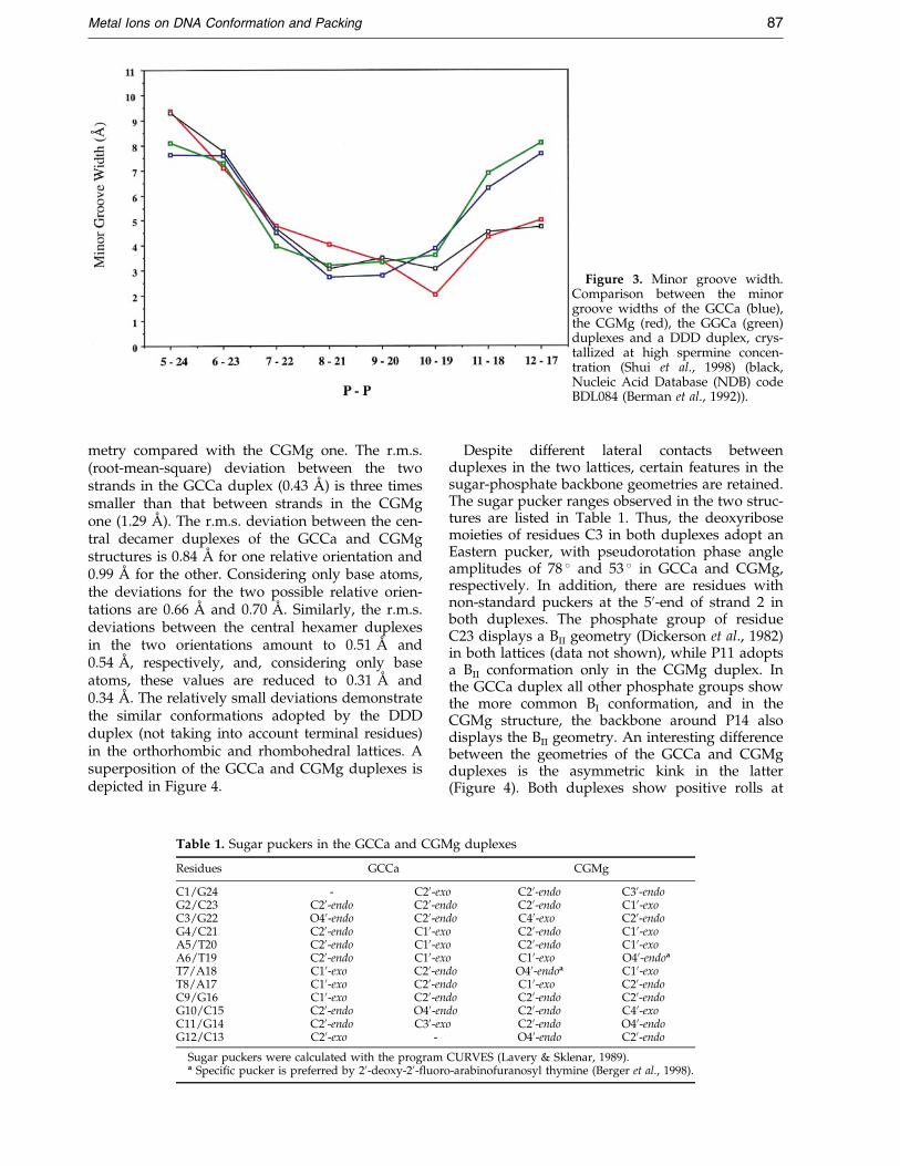
Figure 3. Minor groove width.Comparison between the minorgroove widths of the GCCa (blue),the CGMg (red), the GGCa (green)duplexes and a DDD duplex, crys-tallized at high spermine concen-tration (Shui et al., 1998) (black,Nucleic Acid Database (NDB) codeBDL084 (Berman et al., 1992)).
Metal Ions on DNA Conformation and Packing 87
metry compared with the CGMg one. The r.m.s.(root-mean-square) deviation between the twostrands in the GCCa duplex (0.43 AÊ ) is three timessmaller than that between strands in the CGMgone (1.29 AÊ ). The r.m.s. deviation between the cen-tral decamer duplexes of the GCCa and CGMgstructures is 0.84 AÊ for one relative orientation and0.99 AÊ for the other. Considering only base atoms,the deviations for the two possible relative orien-tations are 0.66 AÊ and 0.70 AÊ . Similarly, the r.m.s.deviations between the central hexamer duplexesin the two orientations amount to 0.51 AÊ and0.54 AÊ , respectively, and, considering only baseatoms, these values are reduced to 0.31 AÊ and0.34 AÊ . The relatively small deviations demonstratethe similar conformations adopted by the DDDduplex (not taking into account terminal residues)in the orthorhombic and rhombohedral lattices. Asuperposition of the GCCa and CGMg duplexes isdepicted in Figure 4.
Table 1. Sugar puckers in the GCCa and CGM
Residues GCCa
C1/G24 - C20-exG2/C23 C20-endo C20-enC3/G22 O40-endo C20-enG4/C21 C20-endo C10-exA5/T20 C20-endo C10-exA6/T19 C20-endo C10-exT7/A18 C10-exo C20-enT8/A17 C10-exo C20-enC9/G16 C10-exo C20-enG10/C15 C20-endo O40-enC11/G14 C20-endo C30-exG12/C13 C20-exo -
Sugar puckers were calculated with the programa Speci®c pucker is preferred by 20-deoxy-20-¯uoro
Despite different lateral contacts betweenduplexes in the two lattices, certain features in thesugar-phosphate backbone geometries are retained.The sugar pucker ranges observed in the two struc-tures are listed in Table 1. Thus, the deoxyribosemoieties of residues C3 in both duplexes adopt anEastern pucker, with pseudorotation phase angleamplitudes of 78 � and 53 � in GCCa and CGMg,respectively. In addition, there are residues withnon-standard puckers at the 50-end of strand 2 inboth duplexes. The phosphate group of residueC23 displays a BII geometry (Dickerson et al., 1982)in both lattices (data not shown), while P11 adoptsa BII conformation only in the CGMg duplex. Inthe GCCa duplex all other phosphate groups showthe more common BI conformation, and in theCGMg structure, the backbone around P14 alsodisplays the BII geometry. An interesting differencebetween the geometries of the GCCa and CGMgduplexes is the asymmetric kink in the latter(Figure 4). Both duplexes show positive rolls at
g duplexes
CGMg
o C20-endo C30-endodo C20-endo C10-exodo C40-exo C20-endoo C20-endo C10-exoo C20-endo C10-exoo C10-exo O40-endoa
do O40-endoa C10-exodo C10-exo C20-endodo C20-endo C20-endodo C20-endo C40-exoo C20-endo O40-endo
O40-endo C20-endo
CURVES (Lavery & Sklenar, 1989).-arabinofuranosyl thymine (Berger et al., 1998).
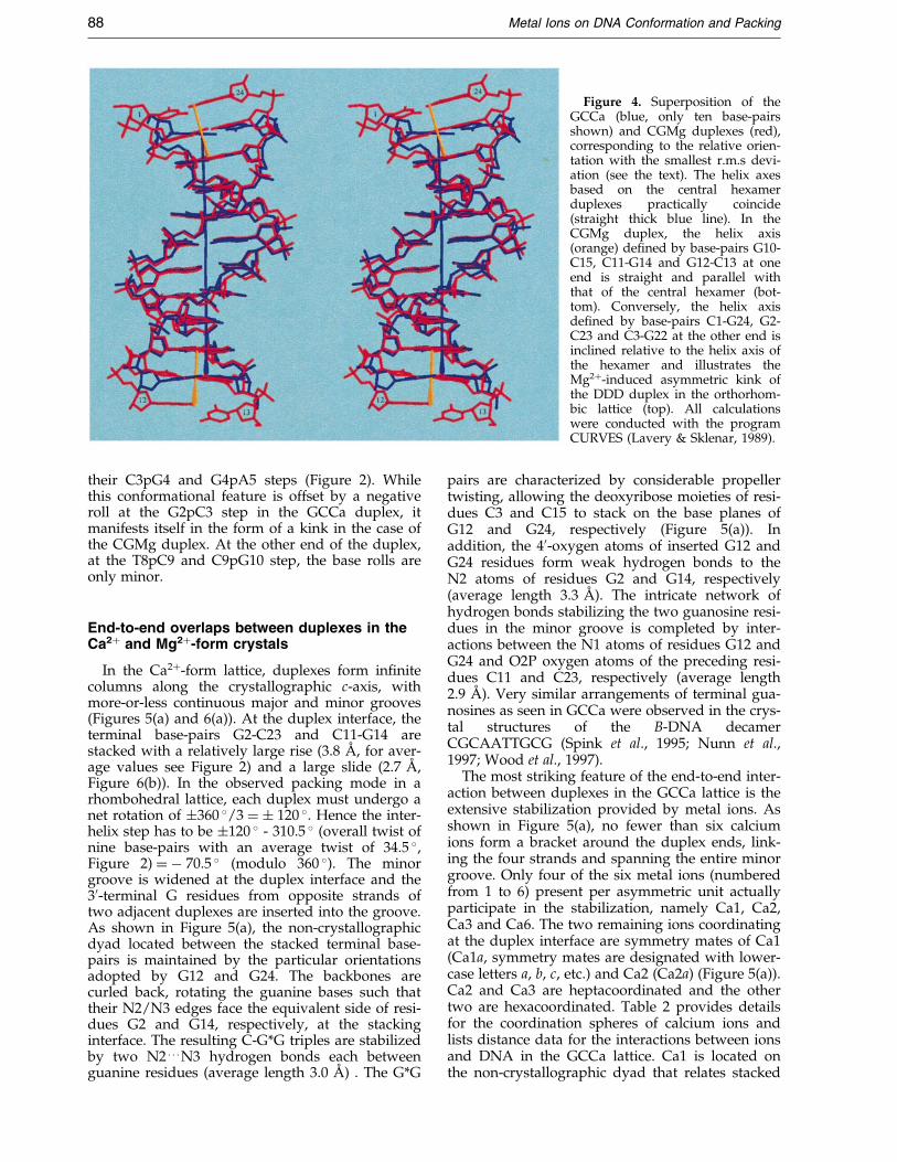
Figure 4. Superposition of theGCCa (blue, only ten base-pairsshown) and CGMg duplexes (red),corresponding to the relative orien-tation with the smallest r.m.s devi-ation (see the text). The helix axesbased on the central hexamerduplexes practically coincide(straight thick blue line). In theCGMg duplex, the helix axis(orange) de®ned by base-pairs G10-C15, C11-G14 and G12-C13 at oneend is straight and parallel withthat of the central hexamer (bot-tom). Conversely, the helix axisde®ned by base-pairs C1-G24, G2-C23 and C3-G22 at the other end isinclined relative to the helix axis ofthe hexamer and illustrates theMg2�-induced asymmetric kink ofthe DDD duplex in the orthorhom-bic lattice (top). All calculationswere conducted with the programCURVES (Lavery & Sklenar, 1989).
88 Metal Ions on DNA Conformation and Packing
their C3pG4 and G4pA5 steps (Figure 2). Whilethis conformational feature is offset by a negativeroll at the G2pC3 step in the GCCa duplex, itmanifests itself in the form of a kink in the case ofthe CGMg duplex. At the other end of the duplex,at the T8pC9 and C9pG10 step, the base rolls areonly minor.
End-to-end overlaps between duplexes in theCa2� and Mg2�-form crystals
In the Ca2�-form lattice, duplexes form in®nitecolumns along the crystallographic c-axis, withmore-or-less continuous major and minor grooves(Figures 5(a) and 6(a)). At the duplex interface, theterminal base-pairs G2-C23 and C11-G14 arestacked with a relatively large rise (3.8 AÊ , for aver-age values see Figure 2) and a large slide (2.7 AÊ ,Figure 6(b)). In the observed packing mode in arhombohedral lattice, each duplex must undergo anet rotation of �360 �/3 � � 120 �. Hence the inter-helix step has to be �120 � - 310.5 � (overall twist ofnine base-pairs with an average twist of 34.5 �,Figure 2) � ÿ 70.5 � (modulo 360 �). The minorgroove is widened at the duplex interface and the30-terminal G residues from opposite strands oftwo adjacent duplexes are inserted into the groove.As shown in Figure 5(a), the non-crystallographicdyad located between the stacked terminal base-pairs is maintained by the particular orientationsadopted by G12 and G24. The backbones arecurled back, rotating the guanine bases such thattheir N2/N3 edges face the equivalent side of resi-dues G2 and G14, respectively, at the stackinginterface. The resulting C-G*G triples are stabilizedby two N2 . . . N3 hydrogen bonds each betweenguanine residues (average length 3.0 AÊ ) . The G*G
pairs are characterized by considerable propellertwisting, allowing the deoxyribose moieties of resi-dues C3 and C15 to stack on the base planes ofG12 and G24, respectively (Figure 5(a)). Inaddition, the 40-oxygen atoms of inserted G12 andG24 residues form weak hydrogen bonds to theN2 atoms of residues G2 and G14, respectively(average length 3.3 AÊ ). The intricate network ofhydrogen bonds stabilizing the two guanosine resi-dues in the minor groove is completed by inter-actions between the N1 atoms of residues G12 andG24 and O2P oxygen atoms of the preceding resi-dues C11 and C23, respectively (average length2.9 AÊ ). Very similar arrangements of terminal gua-nosines as seen in GCCa were observed in the crys-tal structures of the B-DNA decamerCGCAATTGCG (Spink et al., 1995; Nunn et al.,1997; Wood et al., 1997).
The most striking feature of the end-to-end inter-action between duplexes in the GCCa lattice is theextensive stabilization provided by metal ions. Asshown in Figure 5(a), no fewer than six calciumions form a bracket around the duplex ends, link-ing the four strands and spanning the entire minorgroove. Only four of the six metal ions (numberedfrom 1 to 6) present per asymmetric unit actuallyparticipate in the stabilization, namely Ca1, Ca2,Ca3 and Ca6. The two remaining ions coordinatingat the duplex interface are symmetry mates of Ca1(Ca1a, symmetry mates are designated with lower-case letters a, b, c, etc.) and Ca2 (Ca2a) (Figure 5(a)).Ca2 and Ca3 are heptacoordinated and the othertwo are hexacoordinated. Table 2 provides detailsfor the coordination spheres of calcium ions andlists distance data for the interactions between ionsand DNA in the GCCa lattice. Ca1 is located onthe non-crystallographic dyad that relates stacked
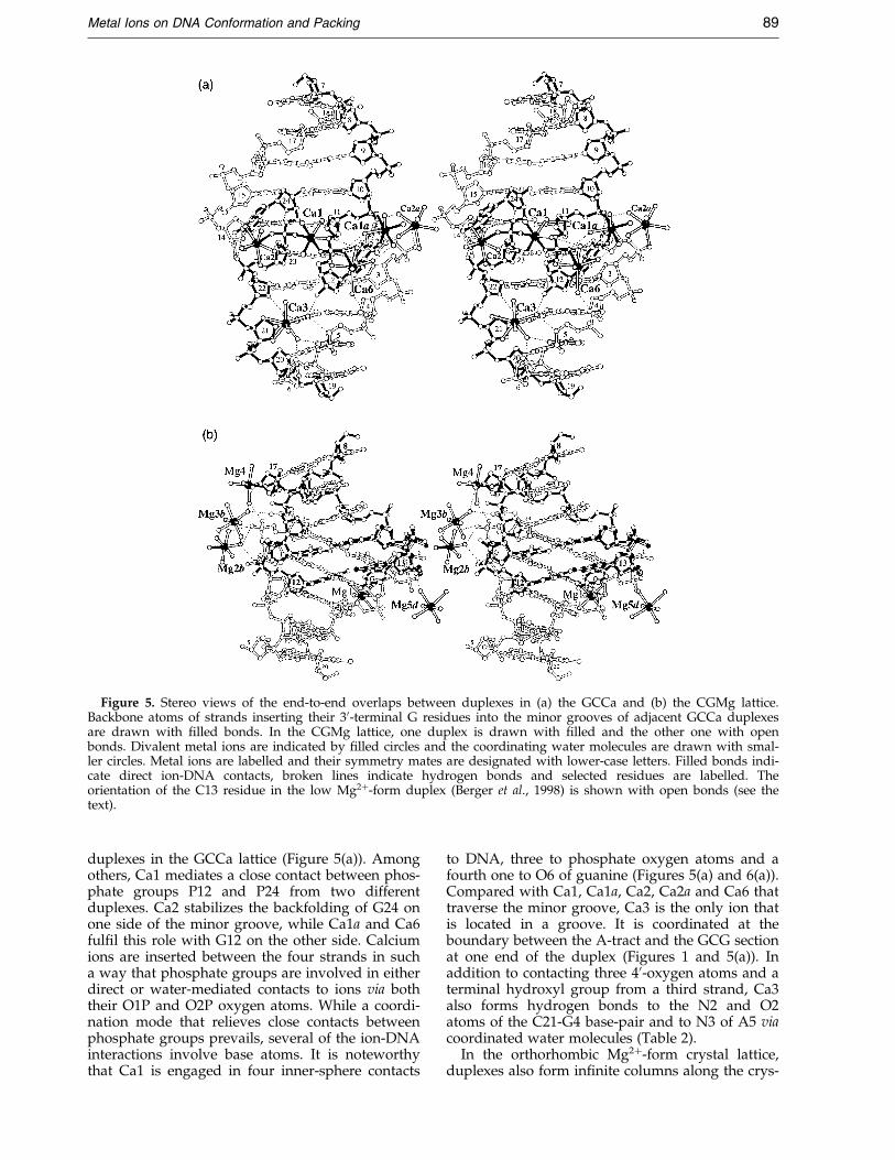
Figure 5. Stereo views of the end-to-end overlaps between duplexes in (a) the GCCa and (b) the CGMg lattice.Backbone atoms of strands inserting their 30-terminal G residues into the minor grooves of adjacent GCCa duplexesare drawn with ®lled bonds. In the CGMg lattice, one duplex is drawn with ®lled and the other one with openbonds. Divalent metal ions are indicated by ®lled circles and the coordinating water molecules are drawn with smal-ler circles. Metal ions are labelled and their symmetry mates are designated with lower-case letters. Filled bonds indi-cate direct ion-DNA contacts, broken lines indicate hydrogen bonds and selected residues are labelled. Theorientation of the C13 residue in the low Mg2�-form duplex (Berger et al., 1998) is shown with open bonds (see thetext).
Metal Ions on DNA Conformation and Packing 89
duplexes in the GCCa lattice (Figure 5(a)). Amongothers, Ca1 mediates a close contact between phos-phate groups P12 and P24 from two differentduplexes. Ca2 stabilizes the backfolding of G24 onone side of the minor groove, while Ca1a and Ca6ful®l this role with G12 on the other side. Calciumions are inserted between the four strands in sucha way that phosphate groups are involved in eitherdirect or water-mediated contacts to ions via boththeir O1P and O2P oxygen atoms. While a coordi-nation mode that relieves close contacts betweenphosphate groups prevails, several of the ion-DNAinteractions involve base atoms. It is noteworthythat Ca1 is engaged in four inner-sphere contacts
to DNA, three to phosphate oxygen atoms and afourth one to O6 of guanine (Figures 5(a) and 6(a)).Compared with Ca1, Ca1a, Ca2, Ca2a and Ca6 thattraverse the minor groove, Ca3 is the only ion thatis located in a groove. It is coordinated at theboundary between the A-tract and the GCG sectionat one end of the duplex (Figures 1 and 5(a)). Inaddition to contacting three 40-oxygen atoms and aterminal hydroxyl group from a third strand, Ca3also forms hydrogen bonds to the N2 and O2atoms of the C21-G4 base-pair and to N3 of A5 viacoordinated water molecules (Table 2).
In the orthorhombic Mg2�-form crystal lattice,duplexes also form in®nite columns along the crys-
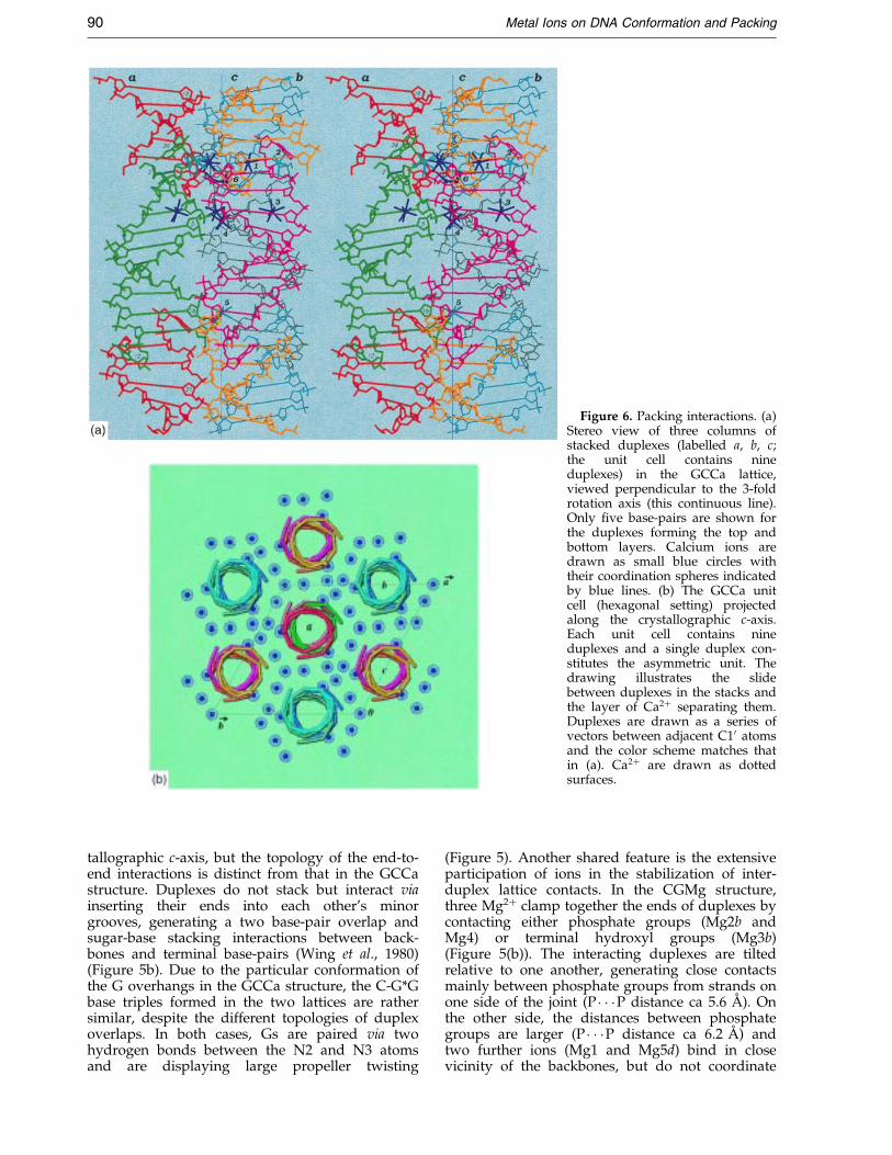
Figure 6. Packing interactions. (a)Stereo view of three columns ofstacked duplexes (labelled a, b, c;the unit cell contains nineduplexes) in the GCCa lattice,viewed perpendicular to the 3-foldrotation axis (this continuous line).Only ®ve base-pairs are shown forthe duplexes forming the top andbottom layers. Calcium ions aredrawn as small blue circles withtheir coordination spheres indicatedby blue lines. (b) The GCCa unitcell (hexagonal setting) projectedalong the crystallographic c-axis.Each unit cell contains nineduplexes and a single duplex con-stitutes the asymmetric unit. Thedrawing illustrates the slidebetween duplexes in the stacks andthe layer of Ca2� separating them.Duplexes are drawn as a series ofvectors between adjacent C10 atomsand the color scheme matches thatin (a). Ca2� are drawn as dottedsurfaces.
90 Metal Ions on DNA Conformation and Packing
tallographic c-axis, but the topology of the end-to-end interactions is distinct from that in the GCCastructure. Duplexes do not stack but interact viainserting their ends into each other's minorgrooves, generating a two base-pair overlap andsugar-base stacking interactions between back-bones and terminal base-pairs (Wing et al., 1980)(Figure 5b). Due to the particular conformation ofthe G overhangs in the GCCa structure, the C-G*Gbase triples formed in the two lattices are rathersimilar, despite the different topologies of duplexoverlaps. In both cases, Gs are paired via twohydrogen bonds between the N2 and N3 atomsand are displaying large propeller twisting
(Figure 5). Another shared feature is the extensiveparticipation of ions in the stabilization of inter-duplex lattice contacts. In the CGMg structure,three Mg2� clamp together the ends of duplexes bycontacting either phosphate groups (Mg2b andMg4) or terminal hydroxyl groups (Mg3b)(Figure 5(b)). The interacting duplexes are tiltedrelative to one another, generating close contactsmainly between phosphate groups from strands onone side of the joint (P � � �P distance ca 5.6 AÊ ). Onthe other side, the distances between phosphategroups are larger (P � � �P distance ca 6.2 AÊ ) andtwo further ions (Mg1 and Mg5d) bind in closevicinity of the backbones, but do not coordinate
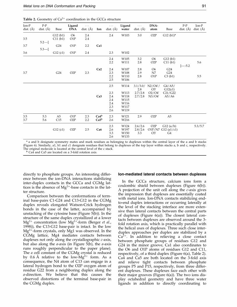
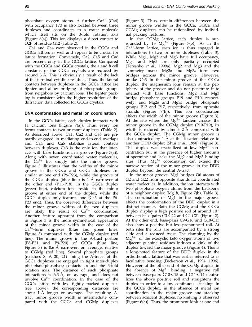
Table 2. Geometry of Ca2� coordination in the GCCa structure
Ion-Pdist. (AÊ )
P-Pdist. (AÊ ) Base
LigandDNA
� � �dist. (AÊ ) Ion
� � �dist. (AÊ )
Ligandwater
� � �dist. (AÊ )
DNA-atom Base
P-Pdist. (AÊ )
Ion-Pdist. (AÊ )
G12 (b1) O6 2.4 2.4 W103 3.0 O2P G12 (b1)a
3.5 C11 (b1) O1P 2.45.2Ð[
3.7 G24 O1P 2.2 Ca15.3Ð[
3.6 G12 (c1) O1P 2.4 2.3 W102
2.4 W105 3.2 O6 G12 (b1)2.2 W111 2.8 O2P C11 (b1) 5.6
]Ð-5.2Ca2 2.4 W107 2.8 O6 G24
3.7 G24 O2P 2.3 2.3 W108 2.9 N7 G242.2 W110 2.8 O1P C3 (b1) 5.52.5 W106
2.5 W114 3.1/3.0/2.8
N2/O40/O30
G4/A5/G12(c1)
2.3 W113 2.7/2.8 O2/O40 C21/G22Ca3 2.4 W118 2.7/2.8 N3/O40 A5/A6
2.4 W1152.4 W1162.3 W1172.6 W119
3.5 5.3 A5 O1P 2.3 Ca4b 2.3 W121 2.9 O2P A53.7 5.6 C15 O2P 2.2 Ca5b 2.6 W216
2.3 W124 2.6/2.6 O2P G12 (a/b) 5.3/5.7G12 (c1) O2P 2.5 Ca6 2.6 W197 2.8/2.6 O1P/N7 G12 (a1/c1)
3.3 W150 3.3 O30 G42.6 W133
a a and b designate symmetry mates and mark residues as belonging to duplexes within the central layer of the a and b stacks(Figure 6). Similarly, a1, b1 and c1 designate residues that belong to duplexes of the top layer within stacks a, b and c, respectively.The original molecule is located at the central level of the c stack.
b Ca4 and Ca5 are located on a 3-fold rotation axis.
Metal Ions on DNA Conformation and Packing 91
directly to phosphate groups. An interesting differ-ence between the ion-DNA interactions stabilizinginter-duplex contacts in the GCCa and CGMg lat-tices is the absence of Mg2�-base contacts in the lat-ter structure.
Comparison between the conformations of term-inal base-pairs C1-G24 and C13-G12 in the CGMgduplex reveals elongated Watson-Crick hydrogenbonds in the case of the latter, accompanied byunstacking of the cytosine base (Figure 5(b)). In thestructure of the same duplex crystallized at a lowerMg2� concentration (low Mg2�-form (Berger et al.,1998)), the C13-G12 base-pair is intact. In the lowMg2�-form crystals, only Mg1 was observed. In theCGMg lattice, Mg4 mediates contacts betweenduplexes not only along the crystallographic c-axis,but also along the a-axis (in Figure 5(b), the a-axisruns roughly perpendicular to the paper plane).The a cell constant of the CGMg crystal is reducedby 0.6 AÊ relative to the low-Mg2� form. As aconsequence, the N4 atom of C13 can engage in alateral hydrogen bond to the O2P oxygen atom ofresidue G22 from a neighboring duplex along thea-direction. We believe that this causes theobserved distortions of the terminal base-pair inthe CGMg duplex.
Ion-mediated lateral contacts between duplexes
In the GCCa structure, calcium ions form acoulombic shield between duplexes (Figure 6(b)).A projection of the unit cell along the c-axis givesthe impression that duplexes are essentially coatedwith metal ions. Ion-DNA contacts stabilizing end-to-end duplex interactions or occurring laterally atthe level of the stacking interface are more exten-sive than lateral contacts between the central partsof duplexes (Figure 6(a)). The closest lateral con-tacts between duplexes are observed around the 3-fold rotation axis, which is practically parallel withthe helical axes of duplexes. Three such close inter-duplex approaches per duplex are stabilized by aCa2�. In addition to relieving a close contactbetween phosphate groups of residues G12 andG24 in the minor groove, Ca1 also coordinates tothe O6 and O1P atoms of residues G12 and C11,respectively, of a third duplex (Figure 6(a), Table 2).Ca4 and Ca5 are both located on the 3-fold axisand relieve tight contacts between phosphategroups P5 and P15, respectively, from three differ-ent duplexes. These duplexes face each other withtheir major grooves (Figure 6(a)). The two ions dis-play octahedral geometry and have three waterligands in addition to directly coordinating to
92 Metal Ions on DNA Conformation and Packing
phosphate oxygen atoms. A further Ca2� (Ca6)with occupancy 1/3 is also located between threeduplexes and coordinates to a water moleculewhich itself sits on the 3-fold rotation axis(Figure 6(a)). This ion displays a direct contact toO2P of residue G12 (Table 2).
Ca1 and Ca4 were observed in the CGCa andGGCa lattices as well and appear to be crucial forlattice formation. Conversely, Ca2, Ca5 and Ca6are present only in the GCCa lattice. Comparedwith the CGCa and GGCa crystals, the a and b cellconstants of the GCCa crystals are reduced byaround 3 AÊ . This is obviously a result of the lackof the terminal cytidine residues. Thus, the lateralcontacts between duplexes in the GCCa lattice aretighter and allow bridging of phosphate groupsfrom neighbors by calcium ions. The tighter pack-ing is consistent with the higher resolution of thediffraction data collected for GCCa crystals.
DNA conformation and metal ion coordination
In the GCCa lattice, each duplex interacts with11 calcium ions (Figure 7(a)). Thus, every ionforms contacts to two or more duplexes (Table 2).As described above, Ca1, Ca2 and Ca6 are pri-marily engaged in mediating end-to-end overlapsand Ca4 and Ca5 stabilize lateral contactsbetween duplexes. Ca3 is the only ion that inter-acts with base functions in a groove (Figure 7(a)).Along with seven coordinated water molecules,the Ca2� ®ts snugly into the minor groove.Figure 3 illustrates that the widths of the minorgroove in the GCCa and GGCa duplexes aresimilar at one end (P6-P23), while the groove ofthe GCCa duplex (blue line) is 1 AÊ narrower atthe other end (P11-P18). In the GGCa duplex(green line), calcium ions reside in the minorgroove at either end of the duplex, while theGCCa duplex only features one (Ca3 at the P6-P23 end). Thus, the observed differences betweenthe minor groove widths of the two duplexesare likely the result of Ca2� coordination.Another feature apparent from the comparisonin Figure 3 is the more symmetrical appearanceof the minor groove width in the case of theCa2�-form duplexes (blue and green lines,Figure 3) compared with the CGMg duplex (redline). The minor groove in the A-tract portion(P8-P21 and P9-P20) of GCCa (blue line,Figure 3) is 0.6 AÊ narrower, on average, relativeto CGMg (red line). Several phosphate groups(residues 8, 9, 20, 21) lining the A-tracts of theGCCa duplexes are engaged in tight inter-duplexphosphate-phosphate contacts around the 3-foldrotation axis. The distance of such phosphateinteractions is 6.3 AÊ , on average, and does notinvolve Ca2� coordination. In the case of theGGCa lattice with less tightly packed duplexes(see above), the corresponding distances areabout 1 AÊ longer on average. The resulting A-tract minor groove width is intermediate com-pared with the GCCa and CGMg duplexes
(Figure 3). Thus, certain differences between theminor groove widths in the GCCa, GGCa andCGMg duplexes can be rationalized by individ-ual packing features.
In the CGMg lattice, each duplex is sur-rounded by 13 Mg2� (Figure 7(b)). As in theCa2�-form lattice, each ion is thus engaged ininteractions to two or more duplexes (Table 3).While Mg1, Mg2 and Mg3 have full occupancy,Mg4 and Mg5 are only partially occupied(Tereshko et al., 1999a). Mg2 and Mg3 and thesymmetry mates Mg2a and Mg3a form twobridges accross the minor groove. However,unlike Ca3 in the minor groove of the GCCaduplex, the magnesium ions remain at the per-iphery of the groove and do not penetrate it tointeract with base functions. Mg2 and Mg3bridge phosphate groups P19 and P10, respect-ively, and Mg2a and Mg3a bridge phosphategroups P12 and P17, respectively, from oppositestrands (Figure 7(b)). This ion coordinationaffects the width of the minor groove (Figure 3).At the site where the Mg2� tandem crosses theminor groove in the CGMg duplex (P10-P19), itswidth is reduced by almost 2 AÊ compared withthe GCCa duplex. The CGMg minor groove isalso contracted by 1 AÊ at this location relative toanother DDD duplex (Shui et al., 1998) (Figure 3).This duplex was crystallized at low Mg2� con-centration but in the presence of higher amountsof spermine and lacks the Mg2 and Mg3 bindingsites. Thus, Mg2� coordination can extend thenarrow section of the minor groove in the DDDduplex beyond the central A-tract.
In the major groove, Mg1 bridges O6 atoms ofG2 and G22 from opposite strands via coordinatedwater molecules. In addition, the ion interacts withtwo phosphate oxygen atoms from the backboneof a neighbor duplex (Mg1b, Figure 7(b), Table 3).The coordination of Mg1 in the major grooveaffects the conformation of the DDD duplex in adistinct manner. Both the CGMg and the GCCaduplex display a high positive roll of around 10 �between base pairs C3-G22 and G4-C21 (Figure 2).At the other end, base-pairs C9-G16 and G10-C15also show a positive but less pronounced roll. Atboth sites the rolls are accompanied by a strongslide and a reduced twist. The clamping by theMg2� of the exocyclic keto oxygen atoms of twoadjacent guanine residues induces a kink of theduplex toward the major groove (Figure 4). This isa long-noted feature of the DDD duplex in theorthorhombic lattice that was earlier referred to asfacultative bending (Dickerson et al., 1994, 1996).However, at the other end of the CGMg duplex, inthe absence of Mg2� binding, a negative rollbetween base-pairs G10-C15 and C11-G14 neutra-lizes the above positive roll and straightens theduplex in order to allow continuous stacking. Inthe GCCa duplex, in the absence of metal ioncoordination in the major groove and stackingbetween adjacent duplexes, no kinking is observed(Figure 6(a)). Thus, the prominent kink at one end
Figure 7. Ionic environment ofindividual duplexes. (a) Stereoview of the GCCa duplex sur-rounded by 11 Ca2� (®lled circles).(b) Stereoview of the CGMg duplexsurrounded by 13 Mg2� (®lled cir-cles). Symmetry mates of ions aredesignated with lower-case letters.Filled bonds indicate direct ion-DNA contacts, broken lines indi-cate hydrogen bonds and terminalresidues are labelled. The orien-tations of the two duplexes provideoptimal views of Ca3 and Mg1coordinated in the minor and themajor groove, respectively, and dif-fer by a rotation of ca 60 � aroundthe vertical.
Metal Ions on DNA Conformation and Packing 93
of the CGMg duplex can be directly attributed tothe presence of a magnesium ion in the majorgroove.
Hydration
The high-resolution structures of the GCCa andCGMg duplexes provide a more complete pictureof the water structure in the minor groove. The
well-known spine of hydration is maintained butthe ordered water structure involves more solventmolecules from higher shells as well (Tereshkoet al., 1999a,b). Rather than just a zig-zag shapedspine winding down the ¯oor of the minor groove(Drew & Dickerson, 1981; Kopka et al., 1983), thewater structure takes on the appearance of a rib-bon composed of four nearly planar fused waterhexagons (Figure 8(b)). In the CGMg structure
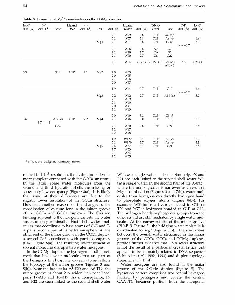
Table 3. Geometry of Mg2� coordination in the CGMg structure
Ion-Pdist. (AÊ )
P-Pdist. (AÊ ) Base
LigandDNA
� � �dist. (AÊ ) Ion
� � �dist. (AÊ )
Ligandwater
� � �dist. (AÊ )
DNA-atom Base
P-Pdist. (AÊ )
Ion-Pdist. (AÊ )
2.1 W29 2.8 O1P A6 (c)a
2.1 W27 2.8 O2P A6 (c) 4.6Mg1 2.1 W31 2.8 O2P T7 (c) 5.3
]± ± ±6.72.1 W26 2.8 N7 G22.1 W28 2.7 O6 G22.1 W30 2.7 O6 G22
2.1 W34 2.7/2.7 O1P/O1P G24 (c)/A18(d)
5.6 4.9/5.4
3.5 T19 O1P 2.1 Mg2 2.0 W332.0 W352.1 W362.0 W37
1.9 W44 2.7 O1P G10 4.6]± ± ±6.2
Mg3 2.2 W42 2.7 O1P A18 (d) 5.22.1 W392.1 W402.0 W412.1 W43
2.0 W49 3.2 O2P C9 (f)3.6 A17 (e) O1P 2.2 Mg4 2.1 W46 3.0 O1P C9 (f) 5.0
5.7± ± ±[G24 2.1 W50 2.8 O2P G24 5.8
2.2 W472.2 W48
1.9 W122 2.7 O1P A5 (c) 5.12.1 W179 2.7 O2P A6 (c) 5.5
Mg5 2.4 W57 2.7 O2P C21 5.82.7 W532.1 W542.2 W55
a a, b, c, etc. designate symmetry mates.
94 Metal Ions on DNA Conformation and Packing
re®ned to 1.1 AÊ resolution, the hydration pattern ismore complete compared with the GCCa structure.In the latter, some water molecules from thesecond and third hydration shells are missing orshow only low occupancy (Figure 8(a)). It is likelythat some of these differences are due to theslightly lower resolution of the GCCa structure.However, another reason for the changes is thecoordination of calcium ions in the minor grooveof the GCCa and GGCa duplexes. The Ca3 ionbinding adjacent to the hexagons distorts the waterstructure only minimally. First shell water mol-ecules that coordinate to base atoms of C-G and T-A pairs become part of its hydration sphere. At theother end of the minor groove in the GGCa duplex,a second Ca2� coordinates with partial occupancy(Ca7, Figure 8(a)). The resulting rearrangement ofsolvent molecules disrupts two water hexagons.
In the CGMg duplex, the hydrogen bonding net-work that links water molecules that are part ofthe hexagons to phosphate oxygen atoms re¯ectsthe topology of the minor groove (Figures 3 and8(b)). Near the base-pairs A5-T20 and A6-T19, theminor groove is about 2 AÊ wider than near base-pairs T7-A18 and T8-A17. As a consequence, P7and P22 are each linked to the second shell water
W10 via a single water molecule. Similarly, P8 andP21 are each linked to the second shell water W30via a single water. In the second half of the A-tract,where the minor groove is narrower as a result ofMg2� coordination (Figures 3 and 7(b)), water mol-ecules from hexagons can directly hydrogen bondto phosphate oxygen atoms (Figure 8(b)). Forexample, W50 forms a hydrogen bond to O1P ofT20 and W7' is hydrogen bonded to O1P of G10.The hydrogen bonds to phosphate groups from theother strand are still mediated by single water mol-ecules. At the narrowest site of the minor groove(P10-P19, Figure 3), the bridging water molecule iscoordinated to Mg2 (Figure 8(b)). The similaritiesbetween the overall water structures in the minorgrooves of the GCCa, GGCa and CGMg duplexesprovide further evidence that DNA water structureis not the result of a particular crystal lattice, butappears to be intimately related to DNA sequence(Schneider et al., 1992, 1993) and duplex topology(Gessner et al., 1994).
Water hexagons are also found in the majorgroove of the CGMg duplex (Figure 9). Thehydration pattern comprises two central hexagons¯anked by pentagons and covers the centralGAATTC hexamer portion. Both the hexagonal
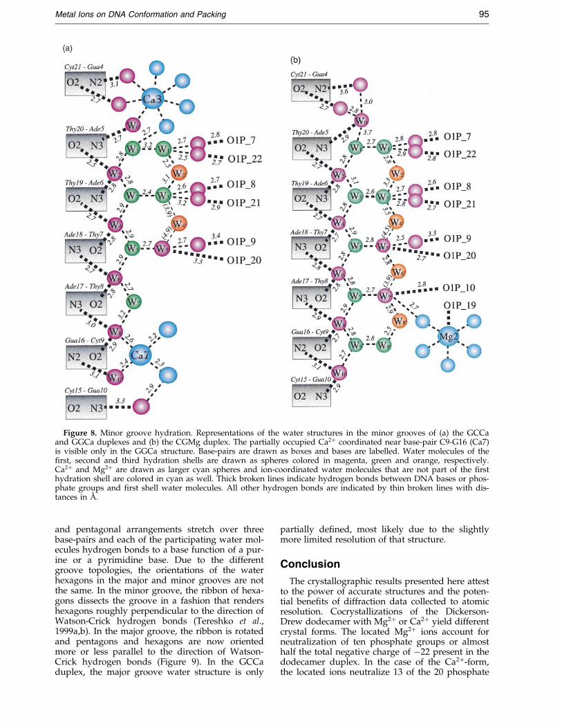
Figure 8. Minor groove hydration. Representations of the water structures in the minor grooves of (a) the GCCaand GGCa duplexes and (b) the CGMg duplex. The partially occupied Ca2� coordinated near base-pair C9-G16 (Ca7)is visible only in the GGCa structure. Base-pairs are drawn as boxes and bases are labelled. Water molecules of the®rst, second and third hydration shells are drawn as spheres colored in magenta, green and orange, respectively.Ca2� and Mg2� are drawn as larger cyan spheres and ion-coordinated water molecules that are not part of the ®rsthydration shell are colored in cyan as well. Thick broken lines indicate hydrogen bonds between DNA bases or phos-phate groups and ®rst shell water molecules. All other hydrogen bonds are indicated by thin broken lines with dis-tances in AÊ .
Metal Ions on DNA Conformation and Packing 95
and pentagonal arrangements stretch over threebase-pairs and each of the participating water mol-ecules hydrogen bonds to a base function of a pur-ine or a pyrimidine base. Due to the differentgroove topologies, the orientations of the waterhexagons in the major and minor grooves are notthe same. In the minor groove, the ribbon of hexa-gons dissects the groove in a fashion that rendershexagons roughly perpendicular to the direction ofWatson-Crick hydrogen bonds (Tereshko et al.,1999a,b). In the major groove, the ribbon is rotatedand pentagons and hexagons are now orientedmore or less parallel to the direction of Watson-Crick hydrogen bonds (Figure 9). In the GCCaduplex, the major groove water structure is only
partially de®ned, most likely due to the slightlymore limited resolution of that structure.
Conclusion
The crystallographic results presented here attestto the power of accurate structures and the poten-tial bene®ts of diffraction data collected to atomicresolution. Cocrystallizations of the Dickerson-Drew dodecamer with Mg2� or Ca2� yield differentcrystal forms. The located Mg2� ions account forneutralization of ten phosphate groups or almosthalf the total negative charge of ÿ22 present in thedodecamer duplex. In the case of the Ca2�-form,the located ions neutralize 13 of the 20 phosphate
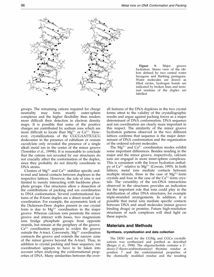
Figure 9. Major groovehydration. Stereo view of the rib-bon de®ned by two central waterhexagons and ¯anking pentagons.Water molecules are drawn as®lled circles, hydrogen bonds areindicated by broken lines and term-inal residues of the duplex arelabelled.
96 Metal Ions on DNA Conformation and Packing
groups. The remaining cations required for chargeneutrality may form mostly outer-spherecomplexes and the higher ¯exibility then rendersmore dif®cult their detection in electron densitymaps. It is possible that some of the positivecharges are contributed by sodium ions which aremore dif®cult to locate than Mg2� or Ca2�. How-ever, crystallizations of the CGCGAATTCGCGdodecamer in the presence of rubidium or cesiumcacodylate only revealed the presence of a singlealkali metal ion in the center of the minor groove(Tereshko et al., 1999b). It is reasonable to concludethat the cations not revealed by our structures donot crucially affect the conformation of the duplex,since they probably do not directly coordinate toDNA atoms.
Clusters of Mg2� and Ca2� stabilize speci®c end-to-end and lateral contacts between duplexes in therespective lattices. However, the role of ions is notlimited to merely interacting with backbone phos-phate groups. Our structures allow a dissection ofthe contributions of packing and ion coordinationto DNA conformation. Several conformational fea-tures of the B-form duplex are a direct result of ioncoordination. For example, the asymmetric kink ofthe Dickerson-Drew duplex present in one crystalform is due to Mg2� coordination in the majorgroove. Whereas calcium ions penetrate the minorgroove and interact with bases, two magnesiumions bridge phosphate groups from oppositestrands, but remain at the periphery of the groove.Ca2� coordination appears to widen the grooveoutside the A-tract. Conversely, Mg2� coordinationcontracts the groove and extends the narrow zoneof the minor groove beyond the A-tract. Thus, inaddition to crystal packing and base sequence, ioncoordination appears to have to be taken intoaccount when analyzing the conformational prop-erties of DNA. Many similarities between the over-
all features of the DNA duplexes in the two crystalforms attest to the validity of the crystallographicresults and argue against packing forces as a majordeterminant of DNA conformation. DNA sequenceand ion coordination are clearly more important inthis respect. The similarity of the minor groovehydration patterns observed in the two differentlattices con®rms that sequence is the major deter-minant of DNA conformation and the organizationof the ordered solvent molecules.
The Mg2� and Ca2� coordination modes exhibitsome important differences. Besides residing in themajor and the minor groove, respectively, calciumions are engaged in more inner-sphere complexes.This is consistent with the lower hydration enthal-py of Ca2� relative to Mg2� (Cowan, 1995). In bothlattices, metal ions mediate contacts betweenmultiple strands, three in the case of Mg2�-formcrystals and four in the case of the Ca2�-form crys-tals. The versatility of the ion-DNA interactionsobserved in the structures provides an indicationfor the important role that ions could play in thestabilization of other DNA structures, for exampletriple-stranded arrangements. In addition, it ispossible that metal ions mediate speci®c contactsbetween DNA and small molecules (minor groovebinding drugs) or proteins. Future high-resolutionstructures of such complexes will shed light onthese aspects.
Materials and Methods
Synthesis, crystallization and data collection
The DDD used for the CGMg and CGCa crystalli-zations was synthesized and puri®ed as described(Berger et al., 1998). The oligonucleotide contains a 20-deoxy-20-¯uoro-arabinofuranosyl thymine residue atposition 7 and the conformational properties ofthe chemically modi®ed residue and the resulting

Metal Ions on DNA Conformation and Packing 97
consequences for the local helix geometry werediscussed in an earlier publication (Berger et al., 1998).The GGCa and GCCa oligomers were obtained formOligos Etc., Wilsonville, OR. Both were puri®ed byRP-HPLC and desalted prior to adjusting the concen-trations of the stock solutions to about 5 mM. Theconditions for growing the CGCa, GGCa and GCCacrystals are summarized in Table 4. All data collec-tions except those with CGCa crystals were conductedon the insertion device beamline (5-ID-B) of theDuPont-Northwestern-Dow Collaborative Access Team(DND-CAT) of the Advanced Photon Source (APS) atArgonne National Laboratory, Argonne, IL, using aMARCCD detector. A summary of the crystal data,data resolutions and re¯ection statistics is provided inTable 4. The data were processed and scaled in theDENZO/SCALEPACK suite (Otwinowski & Minor,1997).
Structure solution and refinement
The crystal structure of the Ca2�-form of the DDDduplex was initially determined based on data collectedwith CGCa crystals using the Molecular Replacementmethod (Navaza, 1994). The resulting model was thenre®ned in combination with the nearly isomorphousGGCa and GCCa data, both of higher resolution thanthe CGCa data. The initial re®nements were conductedwith the program CNS (BruÈ nger, 1998), using the mostrecent parameter ®les (Parkinson et al., 1996). For thehigh-resolution GCCa structure (1.3 AÊ ), all subsequentre®nements were carried out with the program SHELX-97 (Sheldrick & Schneider, 1997), using standardrestraints and setting 10 % of the data aside to calculate
Table 4. Crystal data and re®nement parameters
Structure GCCa
A. CrystallizationDNA (mM) (single strand)pHCaCl2 (mM)MPD (%)
B. Crystal dataSpace groupa � b (AÊ ) 38.76c (AÊ ) 99.08
C. Data collectionX-ray source/detector Synchrotron-APS/MARCCDTemperatureTotal no. reflections 54,240No. unique reflections 13,911Resolution (AÊ ) 1.30Completeness (%) 99.5Rsym
a (%) 8.9
D. Refinement statisticsNo. of DNA atoms 448No. of water molecules 125No. of Ca2� 6 (Ca1 - Ca6)R.m.s distance (AÊ ) 0.006R.m.s. angles (�) 1.57No. reflections [F > 0] 13,120R-factorb (work set) 0.182R-factorc (test set) 0.220
a Rsym � �hkl�ijI(hkl)i ÿ hI(hkl)iij/�hkl�ihI(hkl)ii.b R-factor � �hkljF(hkl)o ÿ F(hkl)cj/�hklF(hkl)o.c For 10 % of the data.
the Rfree value (BruÈ nger, 1992). Solvent molecules werepicked automatically and the correctness of the assign-ments was then examined by visualizing electron densitymaps on the graphics display. All DNA atoms,metal ions and ordered solvent molecules were treatedanisotropically. The anisotropies were restrained toadopt similar shapes and directions for bonded atoms(DELU and SIMU commands, respectively). Final R-fac-tors for the three structures as well as r.m.s. deviationsfrom standard bond lengths and angles are listed inTable 4.
Database accession numbers
The Nucleic Acid Database accession code for theCGMg structure is BD0007. The coordinates and struc-ture factors for the CGCa, GGCa and GCCa structureshave been deposited in the NDB (entry codes BD0020,BD0019 and BD0018, respectively).
Acknowledgements
This work was supported by the National Institutes ofHealth (grant R01 GM-55237). We thank the referees forhelpful comments and suggestions. The DuPont-North-western-Dow Collaborative Access Team (DND-CAT)Synchrotron Research Center located at Sector 5 of theAdvanced Photon Source at Argonne National Labora-tory, Argonne, IL, is supported by the E.I. DuPont deNemours & Co., The Dow Chemical Company as well asthe U.S. National Science Foundation and the State ofIllinois.
GGCa CGCa
0.8-1.26.9
10-4040
R341.99 42.3199.55 99.68
Synchrotron-APS/MARCCD Rigaku RU-200/R-axis IIc120 K39,012 37,5587,036 3,4571.70 2.2099.4 99.95.0 8.8
456 458151 96
4 (Ca1, Ca3, Ca4, Ca7) 3 (Ca1, Ca3, Ca4)0.009 0.0101.35 1.646228 3,2640.194 0.1970.235 0.262
98 Metal Ions on DNA Conformation and Packing
References
Allemann, R. & Egli, M. (1997). DNA bending and rec-ognition. Chem. Biol. 4, 643-650.
Baikalov, I., Grzeskowiak, K., Yanagi, K., Qunitana, J. &Dickerson, R. E. (1993). The crystal structure of thetrigonal decamer C-G-A-T-C-G-(6Me)A-T-C-G - a B-DNA helix with 10.6 base-pairs per turn. J. Mol.Biol. 231, 768-784.
Bancroft, D., Williams, L. D., Rich, A. & Egli, M. (1994).The low-temperature crystal structure of the pure-spermine form of Z-DNA reveals binding of a sper-mine molecule in the minor groove. Biochemistry,33, 1073-1086.
Berger, I., Egli, M. & Rich, A. (1996). Inter-strand C-H � � �O hydrogen bonds stabilizing four-strandedintercalated molecules: stereoelectronic effects ofO40 in cytosine-rich DNA. Proc. Natl Acad. Sci. USA,93, 12116-12121.
Berger, I., Tereshko, V., Ikeda, H., Marquez, V. E. &Egli, M. (1998). Crystal structures of B-DNA withincorporated 20-deoxy-20-¯uoro-arabino-furanosylthymines: implications of conformational preorgani-zation for duplex stability. Nucl. Acids Res. 26, 2473-2480.
Berman, H. M., Olson, W. K., Beveridge, D. L.,Westbrook, J., Gelbin, A., Demeny, T., Hsieh, S.-H.,Srinivasan, A. R. & Schneider, B. (1992). The nucleicacid database: a comprehensive relational databaseof three-dimensional structures of nucleic acids. Bio-phys. J. 63, 751-759.
BruÈ nger, A. T. (1992). The free R value: a novel statisti-cal quantity for accessing the accuracy of crystalstructures. Nature, 355, 472-475.
BruÈ nger, A. T. (1998). Crystallography & NMR System(CNS), Version 0.3, Yale University, New Haven,CT.
Chen, L., Cai, L., Zhang, X. & Rich, A. (1994). Crystalstructure of a four-stranded intercalated DNA:d(C4). Biochemistry, 33, 13540-13546.
Cowan, J. A. (1995). Introduction to the biological roleof magnesium ion. In The Biological Chemistry ofMagnesium (Cowan, J. A., ed.), pp. 1-23, VCHPublishers Inc., New York, NY.
Dickerson, R. E. (1992). DNA structure from A to Z.Methods Enzymol. 211, 67-111.
Dickerson, R. E., Kopka, M. L. & Drew, H. R. (1982).Structural correlations in B-DNA. In Conformation inBiology (Srinivasan, R. & Sarma, R. H., eds), pp.227-257, Adenine Press, New York, NY.
Dickerson, R. E., Goodsell, D. S. & Neidle, S. (1994).`` . . . the tyranny of the lattice . . . ''. Proc. Natl Acad.Sci. USA, 91, 3579-3583.
Dickerson, R. E., Goodsell, D. & Kopka, M. L. (1996).MPD and DNA bending in crystals and in solution.J. Mol. Biol. 256, 108-125.
Drew, H. R. & Dickerson, R. E. (1981). Structure of a B-DNA dodecamer. III. Geometry of hydration. J. Mol.Biol. 151, 535-556.
Egli, M. (1994). Structural patterns in nucleic acids. InStructure Correlation (BuÈ rgi, H.-B. & Dunitz, J. D.,eds), vol. 2, pp. 705-749, VCH Publishers Inc.,Weinheim, Germany.
Egli, M. (1996). Structural aspects of nucleic acid ana-logues and antisense oligonucleotides. Angew.Chem. Int. Ed. Engl. 35, 1894-1909.
Gehring, K., Leroy, J.-L. & GueÂron, M. (1993). A tetra-meric DNA structure with protonated cytosi-ne �cytosine base pairs. Nature, 363, 561-565.
Gessner, R. V., Frederick, C. A., Quigley, G. J., Rich, A.& Wang, A. H.-J. (1989). The molecular structure ofthe left-handed Z-DNA double helix at 1.0-AÊ atomicresolution - geometry, conformation, and ionicinteractions of d(CGCGCG). J. Biol. Chem. 264, 7921-7935.
Gessner, R. V., Quigley, G. J. & Egli, M. (1994). Com-parative studies of high resolution Z-DNA crystalstructures. I. Comparative hydration patterns ofalternating dC-dG. J. Mol. Biol. 236, 1154-1168.
Grzeskowiak, K. (1996). Sequence-dependent structuralvariation in B-DNA. Chem. Biol. 3, 785-790.
Grzeskowiak, K., Yanagi, K., PriveÂ, G. G. & Dickerson,R. E. (1991). The structure of B-helical C-G-A-T-C-G-A-T-C-G and comparison with C-C-A-A-C-G-T-T-G-G - the effect of base pair reversals. J. Biol. Chem.266, 8861-8883.
Heinemann, U. & Alings, C. (1989). Crystallographicstudy of one turn of G-C-rich B-DNA. J. Mol. Biol.210, 369-381.
Heinemann, U. & Alings, C. (1991). The conformation ofa B-DNA decamer is mainly determined by itssequence and not by crystal environment. EMBO J.10, 35-43.
Heinemann, U. & Hahn, M. (1992). C-C-A-G-G-C-M5C-T-G-G - helical ®ne-structure, hydration and com-parison with C-C-A-G-G-C-C-T-G-G. J. Biol. Chem.267, 7312-7341.
Kang, C. H., Zhang, X., Ratliff, R., Moyzis, R. & Rich, A.(1992). Crystal structure of four-stranded Oxytrichatelomeric DNA. Nature, 356, 126-131.
Kennard, O. & Hunter, W. N. (1991). Single crystal X-ray diffraction studies of oligonucleotides and oligo-nucleotide-drug complexes. Angew. Chem. Int. Ed.Engl. 30, 1254-1277.
Kim, Y., Grable, J. C., Love, R., Greene, P. J. &Rosenberg, J. M. (1990). Re®nement of EcoRI endo-nuclease crystal structure: a revised chain tracing.Science, 249, 1307-1309.
Klimasauskas, S., Kumar, S., Roberts, R. J. & Cheng, X.(1994). HhaI methyltransferase ¯ips its target baseout of the DNA helix. Cell, 76, 357-369.
Kopka, M. L., Fratini, A. V., Drew, H. R. & Dickerson,R. E. (1983). Ordered water structure around a B-DNA dodecamer. A quantitative study. J. Mol. Biol.163, 129-146.
Laughlan, G., Murchie, A. I. H., Norman, D., Moore,M. H., Moody, P. C. E., Lilley, D. M. J. & Luisi, B.(1994). The high-resolution crystal structure of aparallel-stranded guanine tetraplex. Science, 265,520-524.
Lavery, R. & Sklenar, H. J. (1989). De®ning the structureof irregular nucleic acids - conventions and prin-ciples. J. Biomol. Struct. Dyn. 6, 655-667.
Lipanov, A., Kopka, M. L., Kaczor-Grzeskowiak, M.,Quintana, J. & Dickerson, R. E. (1993). Structure ofthe B-DNA decamer C-C-A-A-C-I-T-T-G-G in twodifferent space groups: conformational ¯exibility ofB-DNA. Biochemistry, 32, 1373-1389.
Liu, J., Malinina, L., Huynh-Dinh, T. & Subirana, J. A.(1998). The structure of the most studied DNA frag-ment changes under the in¯uence of ions: a newpacking of d(CGCGAATTCGCG). FEBS Letters, 438,211-214.
Luger, K., Mader, A. W., Richmond, R. K., Sargent, D. F.& Richmond, T. J. (1997). Crystal structure of thenucleosome core particle at 2.8 AÊ resolution. Nature,389, 251-260.
Metal Ions on DNA Conformation and Packing 99
Navaza, J. (1994). AMoRe: an automated package formolecular replacement. Acta Crystallog. sect. A, 50,157-163.
Nunn, C. M., Garman, E. & Neidle, S. (1997). Crystalstructure of the DNA decamer d(CGCAATTGCG)complexed with the minor groove binding drugnetropsin. Biochemistry, 36, 4792-4799.
Otwinowski, Z. & Minor, W. (1997). Processing of X-raydiffraction data collected in oscillation mode.Methods Enzymol. 276, 307-326.
Parkinson, G., Vojtechovsky, J., Clowney, L., BruÈ nger,A. T. & Berman, H. M. (1996). New parameters forthe re®nement of nucleic acid containing structures.Acta Crystallog. sect. D, 52, 57-64.
Portmann, S., Altmann, K.-H., Reynes, N. & Egli, M.(1997). Crystal structures of oligodeoxyribonucleo-tides containing 60-a-methyl and 60-a-hydroxycarbocyclic thymidines. J. Am. Chem. Soc. 119, 2396-2403.
PriveÂ, G. G., Yanagi, K. & Dickerson, R. E. (1991).Structure of the B-DNA decamer C-C-A-A-C-G-T-T-G-G and comparison with isomorphous decamersC-C-A-A-G-A-T-T-G-G and C-C-A-G-G-C-C-T-G-G.J. Mol. Biol. 217, 177-199.
Schneider, B., Cohen, D. & Berman, H. M. (1992).Hydration of DNA bases: analysis of crystallo-graphic data. Biopolymers, 32, 725-750.
Schneider, B., Cohen, D. M., Schleifer, L., Srinivasan,A. R., Olson, W. K. & Berman, H. M. (1993). A sys-tematic method for studying the spatial distributionof water molecules around nucleic acid bases. Bio-phys. J., 65, 2291-2303.
Sheldrick, G. M. & Schneider, T. R. (1997). SHELX-97:high-resolution re®nement. Methods Enzymol. 276,319-343.
Shui, X., McFail-Isom, L., Hu, G. H. & Williams, L. D.(1998). The B-DNA dodecamer at high resolutionreveals a spine of water on sodium. Biochemistry,37, 8341-8355.
Sklenar, V. & Feigon, J. (1990). Formation of a stable tri-plex from a single strand. Nature, 345, 836-838.
Spink, N., Nunn, C. M., Vojtechovsky, J., Berman, H. M.& Neidle, S. (1995). Crystal structure of a DNA dec-amer showing a novel pseudo four-way helix-helixjunction. Proc. Natl Acad. Sci. USA, 92, 10767-10771.
Steitz, T. A. (1993). Structural Studies of Protein-NucleicAcid Interaction: The Sources of Sequence-speci®c Bind-ing, Cambridge University Press, Cambridge, UK.
Takahara, P. M., Frederick, C. A. & Lippard, S. J. (1996).Crystal structure of the anticancer drug cisplatinbound to duplex DNA. J. Am. Chem. Soc. 118,12309-12321.
Tereshko, V., Minasov, G. & Egli, M. (1999a). The Dick-erson-Drew B-DNA dodecamer revisited at atomicresolution. J. Am. Chem. Soc. 121, 470-471.
Tereshko, V., Minasov, G. & Egli, M. (1999b). A``hydrat-ion spine'' in a B-DNA minor groove. J. Am.Chem. Soc. 121, 3590-3595.
Van Meervelt, L., Vlieghe, D., Dautant, A., Gallois, B.,PreÂcigoux, G. & Kennard, O. (1995). High-resolutionstructure of a DNA helix forming (C �G)*G basetriplets. Nature, 374, 742-744.
Vlieghe, D., Van Meervelt, L., Dautant, A., Gallois, B.,PreÂcigoux, G. & Kennard, O. (1996). Parallel andantiparallel (G �GC)2 triple helix fragments in a crys-tal structure. Science, 273, 1702-1705.
Wing, R., Drew, H., Takano, T., Broka, C., Tanaka, S.,Itakura, K. & Dickerson, R. E. (1980). Crystal struc-ture analysis of a complete turn of B-DNA. Nature,287, 755-758.
Winkler, F. K., Banner, D. W., Oefner, C., Tsernoglou,D., Brown, R. S., Heathman, S. P., Bryan, R. K.,Martin, P. D., Petratos, K. & Wilson, K. S. (1993).The crystal structure of EcoRV endonuclease and ofits complexes with cognate and non-cognate DNAfragments. EMBO J. 12, 1781-1795.
Wood, A. A., Nunn, C. M., Trent, J. O. & Neidle, S.(1997). Sequence-dependent crossed helix packingin the crystal structure of a B-DNA decamer yieldsa detailed model for the Holliday junction. J. Mol.Biol. 269, 827-841.
Edited by T. Richmond
(Received 5 February 1998; received in revised form 28 May 1999; accepted 7 June 1999)
Copyright © 2022 FDOKUMEN