Role of corticosterone on sleep homeostasis induced by REM sleep deprivation in rats

Dow
nloa
ded
By:
[Ale
rta -
Chi
le 2
005/
2006
Con
sorti
um] A
t: 22
:13
9 Ju
ly 2
008
ERPs studies of cognitive processing during sleep
Agustın M. Ibanez
Universidad Diego Portales, Santiago de Chile,
Chile and Heidelberg University, Heidelberg,
Germany
Rene San Martın
Universidad de Chile and Universidad Diego
Portales, Santiago de Chile, Chile
Esteban Hurtado
Universidad Diego Portales, Santiago de Chile, and
Pontificia Universidad Catolica de Chile, Chile
Vladimir Lopez
University of California San Diego, USA
I n the last few decades, several works on cognitive processing during sleep have emerged. The study of
cognitive processing with event related potentials (ERPs) during sleep is a topic of great interest, since ERPs
allow the study of stimulation with passive paradigms (without conscious response or behavioural response),
opening multiple research possibilities during different sleep phases. We review ERPs modulated by cognitive
processes during sleep: N1, Mismatch Negativity (MMN), P2, P3, N400-like, N300–N550, among others. The
review shows that there are different cognitive discriminations during sleep related to the frequency, intensity,
duration, saliency, novelty, proportion of appearance, meaning, and even sentential integration of stimuli. The
fascinating results of cognitive processing during sleep imply serious challenges for cognitive models. The studies
of ERPs, together with techniques of neuroimaging, have demonstrated the existence of cognitive processing
during sleep. A fundamental question to be considered is if these cognitive phenomena are similar to processing
that occurs during wakefulness. Based on this question we discussed the existence of possible mechanisms
associated with sleep, as well as the specific cognitive and neurophysiologic differences of wakefulness and sleep.
Much knowledge is still required to even understand the conjunction of dramatic changes in cerebral dynamics
and the occurrence of cognitive processes. We propose some insights based on ERPs research for further
construction of theoretical models for integrating both cognitive processing and specific brain sleep dynamics.
D ans les quelques dernieres decennies, plusieurs travaux sur le traitement cognitif durant le sommeil ont
emerge. L’etude du traitement cognitif avec des potentiels evoques lies a un evenement pendant le sommeil
est un sujet d’un grand interet vu que les ERPs permettent l’etude de la stimulation avec des paradigmes passifs
(sans reponse consciente ou de reponse comportementale), en ouvrant des possibilites de recherche pendant les
differentes phases du sommeil. Nous avons recense des potentiels evoques (ERPs) modules par des processus
cognitifs durant le sommeil: N1, negativite de discordance (MMN), P2, P3, N400-like, N300-N550; entre autres.
La recension indique qu’il existe des discriminations cognitives differentes pendant le sommeil liees a la frequence,
a l’intensite, a la duree, a la saillance, a la nouveaute, a la proportion de l’apparence, au sens et meme a
l’integration sententielle des stimuli. Les fascinants resultats du traitement cognitif pendant le sommeil impliquent
de serieux defis pour des modeles cognitifs. Les etudes des ERPs, ensemble avec les techniques de la neuro-
imagerie, ont demontre l’existence de traitement cognitif durant le sommeil. Une question fondamentale a
considerer est si ces phenomenes cognitifs sont similaires au traitement qui a lieu pendant l’etat d’eveil. En se
fondant sur cette question, nous avons discute de l’existence de possibles mecanismes associes au sommeil ainsi
que de specifiques differences cognitives et neurophysiologiques de l’etat d’eveil et du sommeil. Il est necessaire
d’acquerir davantage de connaissances pour meme comprendre la conjonction des changements dramatiques
dans des dynamiques cerebraux et la survenue des processus cognitifs. Nous avons propose quelques reflexions
# 2008 International Union of Psychological Science
http://www.psypress.com/ijp DOI: 10.1080/00207590802194234
Correspondence should be addressed to Dr A. Ibanez, Laboratory of Neuroscience, Universidad Diego Portales, Santiago de Chile,
Chile. (E-mail: [email protected]).
The authors would like to thank Michele Dufey and Sebastian Bacquet for their participation in a previous version of manuscript.
This work was partially supported by a DAAD Grant (PKZ:A/07/71171) and Universidad Diego Portales Grant (Laboratorio de
Neurociencias) to A.I.
INTERNATIONAL JOURNAL OF PSYCHOLOGY
2008, iFirst Article, 1–15

Dow
nloa
ded
By:
[Ale
rta -
Chi
le 2
005/
2006
Con
sorti
um] A
t: 22
:13
9 Ju
ly 2
008
fondees sur la recherche des ERPs pour construire davantage de modeles theoriques pour integrer a la fois un
traitement cognitif et des dynamiques du sommeil du cerveau.
E n las ultimas decadas se han llevado a cabo varios estudios respecto del procesamiento cognitivo durante el
sueno. El estudio del procesamiento cognitivo durante el sueno con potenciales evocados relacionados a
eventos (ERPs) es un topico de gran interes, ya que los ERPs permiten el estudio a partir de paradigmas de
estimulacion pasiva (sin una respuesta consciente o conductual), lo cual ha abierto multiples oportunidades de
investigacion en las diferentes fases del sueno: N1, Negatividad de disparidad (MMN), P2, P3, N400-similar,
N300–N550, entre otras. La presente revision ha mostrado la presencia de diferentes discriminaciones cognitivas
durante el sueno que estan relacionadas con la frecuencia, la intensidad, la duracion, la particularidad, la
novedad, la proporcion de la aparicion, el significado, e incluso la integracion contextual de los estımulos. Los
fascinantes resultados del procesamiento cognitivo durante el sueno plantean serios desafıos para los modelos
cognitivos. Los estudios con ERPs, junto con tecnicas de neuroimagen, han demostrado la existencia de
procesamiento cognitivo durante el sueno. Una pregunta fundamental a ser considerada es si estos fenomenos
cognitivos son similares al procesamiento que ocurre durante la vigila. Tomando esta pregunta como base, se
discute la existencia de posibles mecanismos asociados con el sueno, ası como las diferencias especıficas tanto
cognitivas como neurofisiologicas entre vigilia y sueno. Mucho conocimiento se requiere aun para entender la
conjuncion de dramaticos cambios en la dinamica cerebral y el desarrollo de procesos cognitivos. Nosotros
proponemos algunas ideas claves basadas en la investigacion con ERPs para la futura construccion de modelos
teoricos que integren el procesamiento cognitivo con las dinamicas cerebrales especıficas del sueno.
Keywords: Cognitive neuroscience of sleep; Consciousness; ERP; Passive paradigms; Postlexical integration; Sleep.
Sleep is a highly complex, essential process for the
biological balance of the mammalian organism
(Benington, 2000). Nevertheless, much contro-
versy exists as to whether sleep is equally relevant
for cognitive processes. On the one hand, drastic
changes in cerebral dynamics that happen during
sleep suggest that the brain does not process
cognitive information in this state in the same way
that it does while awake. Diverse theories about
functional disconnection between the brain and
the external environment have been set forth
(Horne, 1989; Jones, 1991; Pompeiano, 1970).
Nevertheless, other data do not seem to support
this perspective. In the first place, sensorial stimuli
affect sleep differentially, suggesting that stimulus
relevance and saliency are processed during sleep,
and therefore there is not a complete disconnection
(Bonnet, 1982; Bradley & Meddis, 1974; Burton,
Harsh & Badia, 1988). Other studies have shown
that some cognitive phenomena happen during
sleep (Cipolli et al., 2003). Finally, the role of sleep
in the consolidation of cognitive processes, such as
memory at a cerebral level, is a known fact
(Stickgold & Walker, 2005).
The study of cognitive processing with event
related potentials (ERPs) during sleep is a topic of
great interest, since ERPs allow the study of
stimulation with passive paradigms (without con-
scious response or behavioural response), opening
multiple research possibilities during different sleep
stages. Additionally, the technique’s excellent
temporal resolution allows inferences to be made
about the type of processing that is taking place at
a specific moment. Thus, how the brain responds
to different stimuli properties, from variations in
intensity, salience, relevance, and frequency to
semantic aspects of cognition, can be studied.
In the first part of the present review, several ERP
components that have been studied as correlated to
cognitive processing of different types are reviewed:
N1, P1, MMN, P2, P3 family, N400, N300–N550.
Only those potentials that have been sensitive to
manipulations of cognitive aspects of the stimula-
tion are reviewed in this text. Many other compo-
nents of evoked potentials reflecting sensorial
processing have been studied during sleep, but their
significance is clearly beyond the scope of the
present review. In the second part, the role of these
studies in cognitive research is discussed. We
discussed the existence of possible mechanisms
associated with sleep, as well as the specific cognitive
and neurophysiologic differences of wakefulness
and sleep. Finally, we propose some insights based
on ERPs research for further construction of
theoretical models for integrating both cognitive
processing and specific brain sleep dynamics.
ERPS RELATED TO COGNITIVEPROCESSING
N1
Auditory N1 is a negative ERP elicited between 75
and 150 ms following the presentation of an
2 IBANEZ ET AL.

Dow
nloa
ded
By:
[Ale
rta -
Chi
le 2
005/
2006
Con
sorti
um] A
t: 22
:13
9 Ju
ly 2
008
auditory stimulus. It is specifically sensitive to the
flow of auditory stimulation (Loveless & Brunia,
1990). Thus, for example, its amplitude diminisheswhen the same stimulus appears repeatedly; this is
due, supposedly, to the incomplete recovery of
neuronal firing between one presentation and
another (Naatanen, 1992). Its generating sources
are mainly bilaterally located in the supratemporal
auditory cortex, although it seemingly receives
additional contribution from frontal regions
(Woods, 1995).Magnetoencephalographic (MEG) studies
suggest that in N1, neural mechanism is
involved the coding of specific characteristics
of auditory stimuli: frequency, intensity, and
location (Elberling, Bak, Kofoed, Lebech, &
Saermark, 1982); characteristics that would be
stored in a format that Naatanen and Winkler
(1999) call ‘‘sensory feature traces.’’ Based onadditional evidence, Atienza, Cantero, and
Escera (2001) suggest that sensory feature traces
would not be available to the mechanisms
responsible for voluntary sensorial discrimina-
tion, but could activate the mechanism of
involuntary attention, resulting in conscious
perception of the stimulus.
According to several studies, N1 would be verysensitive to the general state of the brain at any
given moment. A reduction in the amplitude of
N1 during sleep is observed, which can be
explained by changes in sleep stages, which affect
the pre-cortical information processing.
Observations of sleep-specific changes in
MERPs (middle-latency auditory ERPs) support
this idea. The amplitude of MERPs has beenreduced during sleep (Deiber, Bastuji, Fischer, &
Mauguiere, 1989), indicating that the previous
processing of specific characteristics of the
stimulus is sensitive to sleep. This difference in
the availability of information for successive
cerebral processes could explain the diminished
amplitude of latter components.
N1 has also shown a differential modulation foreach sleep stage. For example, an increase in latency
and a loss of amplitude have been observed during
non-REM sleep (Paavilainen et al., 1987) and a
slight recovery during REM sleep (Bastuji, Garcia-
Larrea, Franc, & Mauguiere, 1995). Results col-
lected on thalamic neurons of anesthetized guinea
pigs seemed to satisfactorily explain the N1
differential modulation during wakefulness—SlowWave Sleep (SWS) and REM sleep (Edeline,
Manunta, & Hennevin, 2000). To be exact, evoked
response diminished in SWS when compared with
wakefulness and REM sleep. This study suggests
that the information received by the cortex in
non-REM is less than that received in paradoxical
sleep, which would explain the increase in the
amplitude of N1 found in this study.
Mismatch Negativity
Mismatch Negativity (MMN) corresponds to a
negative auditory ERP that appears between 100
and 200 ms following stimulus presentation. It is
usually evoked when the repetition of a standard
sound gives way to one that differs in some
physical characteristic (frequency, duration, inten-
sity, or location). Thus, the MMN is understood
as the result of a perceptual process able to detect
changes in a repetitive sonorous sequence, based
on processes regularity detection (Winkler,
Karmos, & Naatanen, 1996), and automatic
comparison (Naatanen, 1992).
A series of studies has shown that MMN can be
found in stage I (Nittono, Momose, & Hori, 2001),
in stage II of non-REM sleep (Sallinen, Kaartinen,
& Lyytinene, 1994), and in REM sleep (Atienza &
Cantero, 2001). Studies such as these suggest the
pre-attentional change detection mechanism con-
tinues working during sleep, possibly based on the
neural activations constructed upon the analysis of
the physical characteristics of the stimuli, also
preserved as shown by N1 studies.
Most of the research on MMN has discussed its
quality to reveal the operation of the sensorial
memory. A pioneer study from Campbell, Bell,
and Bastien (1992) demonstrates that a great
frequency deviation between auditory stimuli
(1000 versus 200 Hz) can elicit a small MMN in
REM sleep and in stage II of non-REM sleep.
Nevertheless, these results could not be repeated
using the oddball paradigm for the last three stages
of non-REM sleep (Nashida et al., 2000). Sallinen
et al. (1994) achieved a certain level of success by
being able to register MMN wave type during
stage II, but this was evident only when the new
stimulation was followed by a K-complex (Kc
hereafter). Several studies report similar results to
those of Campbell et al. (1992) during REM sleep
under similar conditions. Specifically, in a study by
Nashida et al. (2000), MMN was elicited by a
deviant tone (one octave of difference) during
wakefulness, REM sleep and stage I of non-REM.
In this study the interstimulus interval (ISI) varied
between 450 and 600 ms. When testing slower
rhythms, MMN was not registered in response to
the same stimuli, adding to the idea that rhythm of
stimulation is a variable that determines the degree
of its activation in sensorial memory. Another
representative study is that of Atienza et al. (2001),
COGNITIVE PROCESSING DURING SLEEP 3

Dow
nloa
ded
By:
[Ale
rta -
Chi
le 2
005/
2006
Con
sorti
um] A
t: 22
:13
9 Ju
ly 2
008
with the stimulus train paradigm. In this study,
subjects were tested during wakefulness and REM
sleep, presenting simple tones at a relatively fastrhythm (SOA 5 650 ms) within trains separated by
a silent interval of 3, 6, or 9 s; the deviant stimuli
could on the other hand appear in position 1, 2, 4,
or 6 of each train. It was demonstrated that during
wakefulness, there is an MMN elicited in all
conditions, except the one in which the new
stimulus occupies the first position of the train
after an interval of 9 s. In this case, a context topredict the following acoustic events would not
exist; however, when the first tone corresponds to
at least the routine train, the off track stimulus
appears to contradict the generated expectation.
The results in REM sleep show that MMN was
elicited by the new stimulus, disregarding its
position in the acoustic sequence, but only when
the silence interval was the minimum (3 s). In anycase, the MMN appeared diminished in compar-
ison to the one found in wakefulness, supporting
the idea that storage in sensorial memory lasts less
in REM sleep than in wakefulness.
There are two compatible accounts for the
diminished MMN amplitude during sleep in com-
parison with wakefulness. The first takes a bottom-
up perspective, indicating that since the changedetection process to the base of the MMN rests
mainly in the activity of the ascending connections
of the auditory sensorial system, much of the MMN
amplitude reduction during sleep could be explained
by the inhibition of the routes that process the
auditory stimulus prior to it reaching its generators
in the temporal lobe (Atienza, Cantero, &
Dominguez-Marin, 2002). The second line ofexplanation comes from a top-down perspective,
suggesting that the main activity during sleep would
complicate the synchronization of auditory cortex
after the repetition of stimuli, which would also
affect the change detection process to the base of the
MMN (Atienza et al., 2002). In addition, the MMN
would be attenuated by deactivation during sleep,
revealed by PET (Maquet et al., 1996) and studies offMRI (Portas et al., 2000) of the prefrontal areas
that contribute to their elicitation.
Finally, it is worth pointing out that the MMN
generating system not only accedes to information
of the sensorial memory, but also to other more
lasting forms of memory. Atienza and Cantero
(2001) demonstrate that MMN can be elicited
during REM sleep by changes in complex auditorypatterns, which have previously been distinguished
through learning while awake. Prior to the
training, they found that none of the participants
presented MMN, not even in wakefulness; never-
theless, after being taught, an MMN was elicited
as much in wakefulness as in REM sleep, and with
similar amplitude. These results suggest that
initiated neural changes during wakefulness would
be accessible during REM sleep, even 2 days after
the training (Atienza et al., 2002).
P2 (P210–P220)
Certain findings suggest that during sleep it is
possible to obtain a wave sensitive to the novelty
and saliency of the auditory stimuli, similar to the
P3a of wakefulness, around 200 ms after stimulus
presentation. This wave would be distributed in the
frontal cortical region (Nittono et al., 2001).
Nielsen-Bohlman, Knight, Woods, and Woodward
(1991) found a wave of this type after 200 ms of the
presentation of novel and salient stimuli during
stage II. This study showed the amplitude of the
wave being greater in stage II than in wakefulness.
This wave was accompanied by a decrease in N1 and
MMN amplitude, which, according to Nielsen-
Bohlman et al., would indicate the improbability
that this wave corresponds to a P3a of sleep. Winter,
Kok, Kenemans, and Elton (1995) found a similar
wave, P210, in stages of somnolence and during
stage II, elicited as much by standard tones as by
frequency-deviant tones. Winter et al. indicate that
at the base of the P210 amplitude modulation there
would be different refractory patterns from those
elements involved in processing standard and
deviant tones.
P300 family
The P300 component has been described to engage
higher-order cognitive operations related to selec-
tive attention and resource allocation (Donchin &
Coles, 1988), and the amplitude of the P300 is
proportional to the amount of attentional
resources engaged in processing a given stimulus
(Johnson, 1988). There are some intrinsic difficul-
ties in obtaining a canonical P300 during sleep,
especially during stage II. One of them is the fact
that P300 is primarily sensitive to attentional
manipulations. Attentional resources are often
required to elicit this component in wakefulness
(Atienza et al., 2001). P300 has also been related to
a post-decisional ‘‘cognitive closure’’ mechanism
(Verleger, 1998); and to the access of information
by consciousness (Picton, 1992).
Nevertheless, some authors have successfully
used the P300 passive response paradigm during
sleep. For example, positive deflections similar to
P3b have been elicited using an oddball paradigm
in sleep (Bastuji et al., 1995; Nielsen-Bohlman et
4 IBANEZ ET AL.

Dow
nloa
ded
By:
[Ale
rta -
Chi
le 2
005/
2006
Con
sorti
um] A
t: 22
:13
9 Ju
ly 2
008
al., 1991; Pratt, Berlad, & Lavie, 1999). In stage
II, there is some evidence that suggests a positive
wave similar to P3a in wakefulness fulness
(Nielsen-Bohlman et al., 1991; Perrin, Bastuji,
Mauguiere, & Garcia-Larrea, 2000). Even today,
these results are subject to some degree of
controversy. Bastuji et al. (1995) found a P300
in stage I, which was not present in stage II.
Later, Bastuji and Garcia-Larrea (1999) found an
‘‘S II–P3’’ in stage II, close to 600 ms (different
from the Ps–P3 observed in REM and more similar
to wakefulness). There is some research which has
homologated a positivity around the 400 ms post-
stimulus presentation (P400) during stage I and II
with the P300 of wakefulness (Nielsen-Bohlman
et al., 1991); nevertheless, this homologation has
been criticized, suggesting that AEPs induced by
a rare stimulus in the tasks used do not
distinguish between those trials with and without
Kc (Niiyama, Fushimi, Sekine, & Hishikawa,
1995). In this sense, P400 has been categorized as
an important Kc component, more than an
analogue of the P300 of wakefulness, and, more-
over, it has been suggested that during non-REM
sleep, there is no presence of P3 (see a review by
Kotchoubey, 2005). The absence of P300 in stage
II, reported in several studies, could be explained
not only by functional changes in the thalamo-
cortical network in this stage, but also by the
presence, during stage II exclusively, of ‘‘N3’’ and
‘‘P4’’ potentials (Perrin et al., 2000), in the same
temporal window as P300. During REM sleep,
the latency and scalp topography of P300 is
similar to wakefulness (Atienza et al., 2001).
More recently, electrophysiological methods have
been used to demonstrate specific human brain
responses to semantic stimulation during sleep
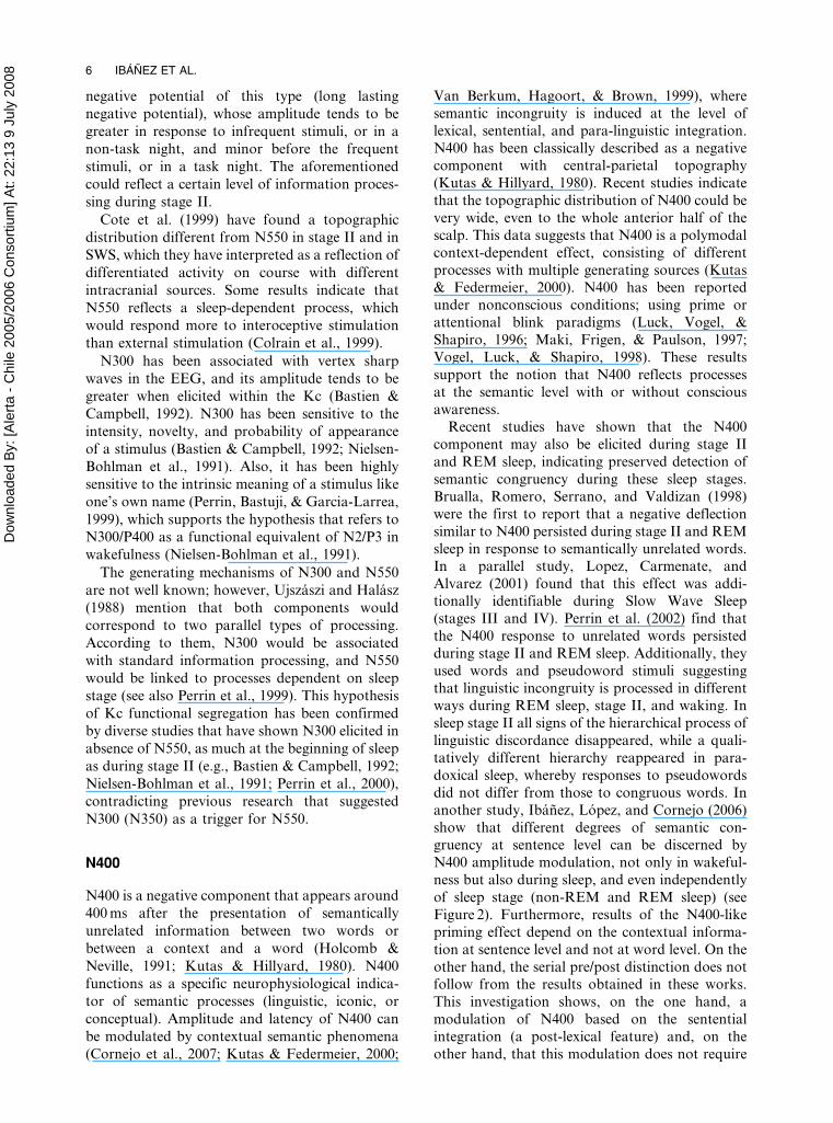
related to P300. For example, two studies (Perrin,
Garcia-Larrea, Mauguiere, & Bastuji, 1999; Pratt,
Berlad, & Lavie, 1999) have, so far, recorded
ERPs to participants’ own names during sleep.
The ERP during wakefulness and REM sleep
(rapid eye movement sleep or paradoxical sleep)
was very similar in latency, amplitude, and scalp
topography related to the cognitive ‘‘P300’’
component recorded in target detection tasks. In
both cases a late positive wave at 400–600 ms
(pseudo P300) was selectively evoked by the
subject’s own name, with maximum amplitude
over the posterior scalp (Figure 1). The persis-
tence of a differential response to the subject’s
own name during REM sleep, relative to any
other proper name, suggests that the brain
remains able to discriminate an intrinsically
relevant word during these sleep stages.
N300 and N550
N300 and N550 are two negative ERPs typically
registered during stages I and II of sleep, and also
during SWS (Cote, de Lugt, Langley, & Campbell,
1999). N550 has been linked to the appearance of
Kc (Bastien & Campbell, 1992), and it has been
found that it is affected mainly by contextual
characteristics of the stimuli (Atienza et al., 2001),
as well as its novelty, saliency (Bastien &
Campbell, 1992), and stimulation proportion
(Colrain, Webster, & Hirst, 1999). Colrain, Di
Parsia, and Gora (2000) have discussed two factors
that make the interpretation of these results
difficult: (1) on the one hand, there is a prevalence
of P300 before new stimuli during wakefulness,
which limits the attribution to the target of a
special meaning for the subject, and (2) on the
other hand, research results do not systematically
discriminate between trials with and without Kc;
so it is difficult to evaluate if the N550 modulation
must be due to the Kc amplitude, to the
probability of eliciting it, or to both factors. A
late negative wave (LNW), with a peak between
500 and 650 ms, is observable in the form of the Kc
average wave; this wave is improved before
infrequent tones during REM sleep (Nordby
et al., 1996). Niiyama et al. (1995) have related
the functional value of N550 within the Kc to a
Figure 1. ERPs in wakefulness and paradoxical sleep inresponse to the subject’s first name and other firstnames, recorded from Fz, Cz and Pz. Positive voltagesare plotted down. Reproduced from Bastuji et al. (2002),with permission from Elsevier # 2002.
COGNITIVE PROCESSING DURING SLEEP 5
Dow
nloa
ded
By:
[Ale
rta -
Chi
le 2
005/
2006
Con
sorti
um] A
t: 22
:13
9 Ju
ly 2
008
negative potential of this type (long lasting
negative potential), whose amplitude tends to be
greater in response to infrequent stimuli, or in a
non-task night, and minor before the frequent
stimuli, or in a task night. The aforementioned
could reflect a certain level of information proces-
sing during stage II.
Cote et al. (1999) have found a topographic
distribution different from N550 in stage II and in
SWS, which they have interpreted as a reflection of
differentiated activity on course with different
intracranial sources. Some results indicate that
N550 reflects a sleep-dependent process, which
would respond more to interoceptive stimulation
than external stimulation (Colrain et al., 1999).
N300 has been associated with vertex sharp
waves in the EEG, and its amplitude tends to be
greater when elicited within the Kc (Bastien &
Campbell, 1992). N300 has been sensitive to the
intensity, novelty, and probability of appearance
of a stimulus (Bastien & Campbell, 1992; Nielsen-
Bohlman et al., 1991). Also, it has been highly
sensitive to the intrinsic meaning of a stimulus like
one’s own name (Perrin, Bastuji, & Garcia-Larrea,
1999), which supports the hypothesis that refers to
N300/P400 as a functional equivalent of N2/P3 in
wakefulness (Nielsen-Bohlman et al., 1991).
The generating mechanisms of N300 and N550
are not well known; however, Ujszaszi and Halasz
(1988) mention that both components would
correspond to two parallel types of processing.
According to them, N300 would be associated
with standard information processing, and N550
would be linked to processes dependent on sleep
stage (see also Perrin et al., 1999). This hypothesis
of Kc functional segregation has been confirmed
by diverse studies that have shown N300 elicited in
absence of N550, as much at the beginning of sleep
as during stage II (e.g., Bastien & Campbell, 1992;
Nielsen-Bohlman et al., 1991; Perrin et al., 2000),
contradicting previous research that suggested
N300 (N350) as a trigger for N550.
N400
N400 is a negative component that appears around
400 ms after the presentation of semantically
unrelated information between two words or
between a context and a word (Holcomb &
Neville, 1991; Kutas & Hillyard, 1980). N400
functions as a specific neurophysiological indica-
tor of semantic processes (linguistic, iconic, or
conceptual). Amplitude and latency of N400 can
be modulated by contextual semantic phenomena
(Cornejo et al., 2007; Kutas & Federmeier, 2000;
Van Berkum, Hagoort, & Brown, 1999), where
semantic incongruity is induced at the level of
lexical, sentential, and para-linguistic integration.N400 has been classically described as a negative
component with central-parietal topography
(Kutas & Hillyard, 1980). Recent studies indicate
that the topographic distribution of N400 could be
very wide, even to the whole anterior half of the
scalp. This data suggests that N400 is a polymodal
context-dependent effect, consisting of different
processes with multiple generating sources (Kutas& Federmeier, 2000). N400 has been reported
under nonconscious conditions; using prime or
attentional blink paradigms (Luck, Vogel, &
Shapiro, 1996; Maki, Frigen, & Paulson, 1997;
Vogel, Luck, & Shapiro, 1998). These results
support the notion that N400 reflects processes
at the semantic level with or without conscious
awareness.Recent studies have shown that the N400
component may also be elicited during stage II
and REM sleep, indicating preserved detection of
semantic congruency during these sleep stages.
Brualla, Romero, Serrano, and Valdizan (1998)
were the first to report that a negative deflection
similar to N400 persisted during stage II and REM
sleep in response to semantically unrelated words.In a parallel study, Lopez, Carmenate, and
Alvarez (2001) found that this effect was addi-
tionally identifiable during Slow Wave Sleep
(stages III and IV). Perrin et al. (2002) find that
the N400 response to unrelated words persisted
during stage II and REM sleep. Additionally, they
used words and pseudoword stimuli suggesting
that linguistic incongruity is processed in differentways during REM sleep, stage II, and waking. In
sleep stage II all signs of the hierarchical process of
linguistic discordance disappeared, while a quali-
tatively different hierarchy reappeared in para-
doxical sleep, whereby responses to pseudowords
did not differ from those to congruous words. In
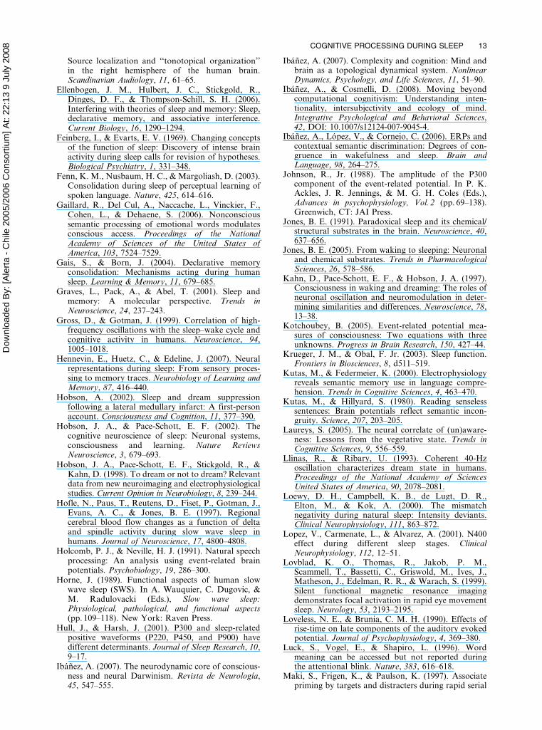
another study, Ibanez, Lopez, and Cornejo (2006)
show that different degrees of semantic con-gruency at sentence level can be discerned by
N400 amplitude modulation, not only in wakeful-
ness but also during sleep, and even independently
of sleep stage (non-REM and REM sleep) (see
Figure 2). Furthermore, results of the N400-like
priming effect depend on the contextual informa-
tion at sentence level and not at word level. On the
other hand, the serial pre/post distinction does notfollow from the results obtained in these works.
This investigation shows, on the one hand, a
modulation of N400 based on the sentential
integration (a post-lexical feature) and, on the
other hand, that this modulation does not require
6 IBANEZ ET AL.

Dow
nloa
ded
By:
[Ale
rta -
Chi
le 2
005/
2006
Con
sorti
um] A
t: 22
:13
9 Ju
ly 2
008
conscious activity (a pre-lexical feature). In short,
these results contradict a serial model in which the
lexical processing precedes the post-lexical proces-
sing (but perhaps not a parallel model).
The negativity described in sleep studies is
congruent with the so-called N400-like effect that
has a slightly different spatial distribution across
the scalp (Van Petten & Luka, 2006). From this
perspective, the process reflected by N400 could be
understood as a stage in a wider process for
conflict or incongruence detection and defined as a
member of a large family of negativities. A variety
of negative deflections in ERP, in different time
windows, have been associated with conflict
detection or task complexity, even during sleep
(see Ibanez et al., 2006). This fact promotes the
consideration of an N400-like component during
sleep as a member of the automatic conflict
detection family of negativities.
DISCUSSION: THE RELEVANCE OF ERPSLEEP STUDIES ON COGNITIVE
PHENOMENA
The review conducted in the previous sections
shows that there are different cognitive discrimina-
tions during sleep related to the frequency,
intensity, duration, saliency, novelty, proportion
of appearance, meaning, and even sentential
integration of stimuli. The fascinating results of
cognitive processing during sleep imply serious
challenges for cognitive models. Not only basic
cognitive processes such as the detection ofinfrequent or novel stimuli, but also semantic
stimuli, produce differences in ERP components.
The participant’s name is distinguished electro-
physiologically and, moreover, the brain discrimi-
nates pseudowords and degrees of incongruence
during sleep at the semantic integration level of a
sentence. Without a doubt, several questions
regarding the relevance of sleep in cognitiveprocessing (i.e., memory, semantic integration,
learning) have to be considered, and call for
further research. For example, it is commonly
accepted that a process of post-lexical integration
(i.e., the integration of the semantic properties of a
sentence) requires conscious activity, since working
memory is required to maintain the different
semantic and lexical properties of each word, inorder to be able to determine the global property
of the sentence (i.e., different degrees of sentential
incongruence). The fact that post-lexical semantic
integration occurs during sleep suggests that an
analogous aspect of the waking state working
memory must be operational during sleep. Perhaps
this semantic working memory process operates
based on nonsemantic heuristics that can resolvethe degrees of incongruence presented in this
design (i.e., a general sequence processing cap-
ability that encodes sequential information and,
when combined with past elements in the sequence,
allows the prediction of successor elements).
Nevertheless, there is no model that explains how
this phenomenon takes place.
At a physiological level, equivalent questionsarise. It has been suggested that the fundamental
difference between sleep and wakefulness is the
activation of frontal lobes during wakefulness. In
general terms, a decrease in the prefrontal activity
exists during sleep, which would make the type of
processes that require prefrontal operation diffi-
cult (i.e., working memory, post-lexical integra-
tion). Although it is well known that certainprefrontal reactivation in presence of stimuli
during sleep exists, it is really difficult to conceive
a model that integrates functional cerebral changes
during sleep and the cognitive processes that have
been reported. A model is required that can
equally account for cognitive processing during
the different phases of sleep and the functional
changes at the electrophysiological and neuro-chemical levels. To our knowledge, such a general
model does not exist and the functions of sleep
remain unclear (Siegel, 2005), although some
partial explicative models related to cognitive
processing have been proposed (i.e., Activation
Figure 2. Different degrees of congruence elicit N400-like amplitude modulation in REM sleep. (A) Scalptopography of N400 for each category. (B) ERPwaveforms at Left Frontal Region (ROI centred overF3 electrode) showing N400 amplitude in each condi-tion. The bar and asterisk at the bottom represent thetime interval at which N400 amplitude exhibitedsignificant statistical differences between categories.Negative voltages are plotted down. Reproduced fromIbanez et al. (2006), with permission from Elsevier #
2006. To view this figure in colour, please visit the onlineversion of this issue.
COGNITIVE PROCESSING DURING SLEEP 7

Dow
nloa
ded
By:
[Ale
rta -
Chi
le 2
005/
2006
Con
sorti
um] A
t: 22
:13
9 Ju
ly 2
008
Syntheses Model, A. Hobson, 2002; the ‘‘reverse
learning’’ of Crick & Mitchison, 1983; and the
memory reactivation and consolidation model of
Wilson, 2002).
Without the intention of proposing an ERP
model of cognitive processing during sleep, we will
discuss two topics that must be considered in any
proposal: the effect of different neurophysiologic
states on ERPs and the nature of cognitive
phenomena during sleep.
Changes of neurophysiologic states andERPs
The usual distinction is between the two great
sleep stages which have been discussed in this
revision: REM sleep (also known as paradoxical
sleep) and non-REM sleep. Non-REM sleep is
subdivided into a continuum of four stages
reflecting the depth of sleep: stage I—sleep onset
period, characterized by decreasing EEG fre-
quency (from 15–60 Hz in waking state to 4–8 Hz
in stage I) and increasing amplitude (from ,30 mV
in waking state to 50–100 mV in stage I); stage II—
light sleep, characterized by 10–12 Hz oscillations
(50–150 mV) called spindles, which occur periodi-
cally and last for a few seconds; and SWS, which
consists of stage III—characterized by slower
waves at 2–4 Hz (100–150 mV); and stage IV—
defined by slow waves (also called delta waves) at
0.5–2 Hz (100–200 mV).
Non-REM sleep and ERPs
Unlike wakefulness and REM sleep, non-REM
sleep is characterized by low frequencies and
higher amplitude waves, oscillations reflecting
huge neuronal activity synchronization in the
thalamocortical network. These neuronal patterns
in the thalamocortical system, a burst-silence
mode during non-REM sleep versus a sustained
single-spike activity during waking and REM, are
under the control of generalized modulatory
systems originating in the brainstem, the hypotha-
lamus, and the basal forebrain (review in Jones,
2005; Pace-Schott & Hobson, 2002; Steriade, 2003;
Steriade & McCarley, 1990).
The ERPs during non-REM sleep differ greatly
from those found during wakefulness, as much
with respect to their morphology as to their
topography. These factors are seen to be strongly
influenced by sleep-specific cortical responses,
where the Kc stand as being the most prominent
cortical responses to the stimulation during non-
REM sleep. The Kc are characterized by a
negativity, whose latency varies between 500 and
600 ms and whose amplitude surpasses even
200 mV (Crowley, Trinder, & Colrain, 2004).
Importantly, Kc in stage II of non-REM sleep
are sensitive to the habituation by stimuli repeti-
tion (Bastien & Campbell, 1994) and have a larger
amplitude to rare stimuli than to frequent ones
(Bastuji et al., 1995; Pratt et al., 1999).
Certain exogenous components are registered in
non-REM sleep. N1 is greater in stage I, smaller in
SWS, and even smaller in stage II during spindles.
Also MMN has been observed in stage I–II of
non-REM sleep, although in the second case only
when a stimulus also elicited a Kc, and without
being able to respond to these results. Several
studies show a substantial decrease or even
disappearance of the MMN during drowsiness or
in stage I. This disparity of results shows that the
research around the MMN in non-REM sleep is
especially sensitive to methodological aspects such
as filtering, presentation rate, and modality.
The most consistent finding in non-REM sleep
is the lack of P3 (Afifi, Guilliminault, & Colrain,
2003; Bastuji et al., 1995; Cote & Campbell, 1999;
Hull & Harsh, 2001; Kotchoubey, 2005; Voss &
Harsh, 1998; Winter et al., 1995). There are other
positive components, possibly related to the Kc,
that inversely correlate with the probability of
stimuli as the P3 of wakefulness; nevertheless they
do not have the typical posterior topography and,
even in stage I, when subjects still produce
behavioural responses, they can have larger
amplitudes to nontargets than targets (Hull &
Harsh, 2001). As already indicated in the revision,
the absence of P3 in non-REM sleep could be
explained not only by functional changes in the
thalamocortical network in this stage, but also by
the presence, during stage II exclusively, of N3 and
P4 potentials in the same temporal window.
Semantic processing in stage II sleep may be
more similar to that in wakefulness. Thus, for
example, semantically inappropriate stimuli
(semantic mismatch) may be elicited N400 during
stage II and in SWS, and even this effect is related
to different degrees of semantic congruency at the
sentence level. Those results suggest a more
automatic processing of semantic stimuli, in agree-
ment with other N400 paradigms of priming, or
attentional blink. Neurophysiologic changes present
in non-REM sleep entails changes in the ERPs of
their classic properties, i.e., latency and amplitude,
and topographic distribution. Some typical ERP
components are difficult to obtain during non-REM
sleep. In stage II it seems that the cortex is able to
respond according to stimulus probability and
8 IBANEZ ET AL.

Dow
nloa
ded
By:
[Ale
rta -
Chi
le 2
005/
2006
Con
sorti
um] A
t: 22
:13
9 Ju
ly 2
008
according to semantic appropriateness of words, a
situation rather less clear in SWS.
REM sleep and ERPs
REM sleep is a neurophysiologic state that
varies greatly from non-REM; it is more similar to
wakefulness (J. A. Hobson & Pace-Schott, 2002;
Pace-Schott & Hobson, 2002), which is also
reflected in the studies of ERPs. For example,
the N1 component, besides being present, behaves
as a component of the orienting response, beinglarger in the first stimulus than in the following of
a stimuli run. The MMN shows a great frequency
deviation (1000 versus 200 Hz). Several studies
reported in this review suggest a significant P3 to
rare stimuli in REM sleep, although its magnitude
was reduced as compared to wakefulness, and its
elicitation required stimuli of higher intensity. The
latency and scalp topography of P300 is similar towakefulness. In wakefulness unexpected words
elicited an N400 greater than nonwords did.
Although N400 is also elicited in stage II, the
difference between unexpected words and non-
words disappeared. In REM sleep nonwords (in
contrast to unexpected words) no longer produced
N400, more similar to when N400 occurs during
wakefulness.
Unlike non-REM sleep, ERP effects in REM
sleep and wakefulness are more similar(Kotchoubey, 2005), but in REM sleep they are
usually reduced and delayed, and require stronger
and more salient stimuli than during wakefulness.
Both non-REM and REM studies of ERPs suggest a
basic cognitive processing during sleep, maybe with
more automatic processing. In order to discuss this
possibility, we now introduce some ERPs studies in
anesthesia, vegetative, and comatose states.
Anesthesia
The anesthetic state is composed of uncon-
sciousness, amnesia, analgesia, immobility, andreduction of autonomic nervous system response.
Despite a century of active research, its molecular
mechanisms are not well known (Barash, Cullen,
& Stoelting, 2006). According to Barash et al.,
plausible sites of action of general anesthetics
include the spinal cord, which does not explain
amnesia or unconsciousness; the brainstem, sup-
ported by increased latency and decreased ampli-tude of somatosensory evoked potentials; and the
cerebral cortex, which shows consistent changes in
surface EEG during anesthesia, possibly caused by
a blockage of thalamocortical communication due
to inhalation anesthetics.
Because of technical and ethical issues, ERP
studies in anesthesia are less common than in
sleep. Studies assessing cortical information pro-
cessing during general anesthesia, sleep, coma, and
vegetative state show that N1 and P1 components
are more likely to remain in these conditions than
endogenous components (Kotchoubey, 2005).
During an auditory oddball task, patients under-
going cardiac surgery exhibit a delayed P1–N1–P2
complex and larger P1–N1 and N1–P2 amplitudes
for infrequent stimuli. Both facts are interpreted as
a decreased, but still present, ability to process
information (Van Hoof et al., 1997).
While Van Hoof et al. (1997) suggest that the
mechanisms underlying auditory processing in
sleep and anesthesia are similar, Hennevin,
Huetz, and Edeline (2007) argue that what occurs
during the anesthetic state is different from what
occurs during natural sleep. Young, Ropper, and
Bolton (1998) support the latter view, stating that
electrophysiologic differences among wakefulness,
coma, sleep, and anesthesia imply that there are
basic differences in the underlying neural activities.
Coma and vegetative state
Coma is characterized by an impairment of the
arousal system, preventing the patient from becom-
ing conscious. The inability to reach wakefulness
makes it difficult to clinically assess higher mental
functions. On the other hand, patients in persistent
vegetative state (PVS) have wake and sleep cycles,
but show no evidence of cognition or awareness of
self or the environment (Young et al., 1998).
Individual case ERPs have been studied on
patients in coma and vegetative state. A single
criterion does not exist for establishing an ERP
component as being valid for an individual case.
Despite this fact, N1, mismatch negativity, and P3 are
consistently found in such patients, and the presence
of late ERP components is related to the severity and
outcome of the coma (Kotchoubey, 2005).
As a conclusion, evidence found by ERP studies
on anesthesia, coma, and vegetative state shows
that basic cognitive processing capabilities would
be present under those states, even though
important differences appear among them and in
relation to wakefulness. This supports the possi-
bility of automatic cognitive processing in states
different to wakefulness.
Cognitive activity during sleep
The studies of ERPs, together with neuroimage
techniques, have demonstrated the existence of
COGNITIVE PROCESSING DURING SLEEP 9

Dow
nloa
ded
By:
[Ale
rta -
Chi
le 2
005/
2006
Con
sorti
um] A
t: 22
:13
9 Ju
ly 2
008
cognitive processes during sleep. The fundamental
question that must be considered is whether these
cognitive phenomena are the same processes thathappen during wakefulness. To this end we will
discuss the cognitive and neurophysiologic exis-
tence of possible mechanisms associated with
sleep, and specific differences of cognition in
wakefulness and sleep.
The existence of online and offline mechanisms
associated to the cognitive phenomena during sleep.
First, there is indirect evidence on the role that
sleep has in the processes associated to memory.
Several studies have shown reactivated neuronal
populations (previously engaged in a learning
task) during sleep; and that this reactivation is a
key process for the consolidation of memory traces
during sleep (Hennevin et al., 2007). This demon-
strates an offline processing of cognitive phenom-ena during sleep associated to sleep-dependent
memory consolidation (Stickgold, 2005), present
in various cognitive processes such as motor-
sequence learning (Cohen, Pascual-Leone, Press,
& Robertson, 2005; Walker et al, 2002); visual-
discrimination learning (Stickgold, James, &
Hobson, 2000), perceptual learning of language
(Fenn, Nusbaum, & Margoliash, 2003), ordeclarative memory (Ellenbogen et al. 2006;
Stickgold, 2005). Those reports are complemented
by cellular and molecular models of sleep-depen-
dent plasticity (Cirelli & Tononi, 2001; Graves,
Pack, & Abel, 2001). Additionally, behavioural,
electrophysiological, and neuroimage studies
(Durmer & Dinges, 2005; Gais & Born, 2004;
Stickgold & Walker, 2005; Tassi et al., 2006) showthat the suppression of sleep produces deficits in
cognitive processing during wakefulness. Similar
alterations of cognition have been reported in
sleep disorders (Verstraeten & Cluydts, 2004).
Those sources of evidence suggest a specific and
differential role from the offline cognitive proces-
sing during sleep.
With respect to the evidence reviewed in thispaper, on the cognitive online processing some
considerations can be assumed. First, selective
changes in an ERP component seem to reveal
specific changes in certain cognitive processes.
Many basic cognitive processes seem to be processed
through a system similar to wakefulness.
Phenomena of infrequency detection, meaningful
stimuli, or the detection of semantic incongruencecould be explained on the basis of well-known
neurophysiologic processes, such as selective
activation (Hofle et al., 1997) and intermittent
gamma activity (J. A. Hobson, Pace-Schott,
Stickgold, & Kahn, 1998) during non-REM; or
gamma oscillations (Kahn, Pace-Schott, & Hobson,
1997; Steriade, 1996) and the increasing of certain
areas’ activation (Braun et al., 1997; Maquet et al.,1996; Nofzinger et al., 1997) during REM.
Additionally, basic cognitive processes that happen
during sleep could be processed in a similar way to
that of wakefulness, but with nonidentical neuro-
physiologic substrata. Finally, the existence of basic
cognitive processes during sleep does not necessarily
imply that wakefulness and sleep have similar
psychophysiological global states.
Specificity and psychophysiological differentia-
tion of cognitive processes during wakefulness and
sleep. Although basic cognitive processes seem to
be processed similarly during wakefulness and
sleep, evidence suggests that, globally, they are
psychologically and neurophysiologically different
processes. The cognitive processing during sleepseems to have a certain specificity. The processes
associated with memory and learning seems to
have unique dynamics. On the other hand, specific
sleep ERPs with possible specific functions have
also been reported (Colrain & Campbell 2007).
Additionally, cognitive phenomena during sleep
seemed to have differential characteristics related
to basic processes (during non-REM sleep) or abizarre cognition (during REM). Both character-
istics of the cognitive processing during sleep
demonstrate a greater difference with wakefulness
processing: The capacity of intentionally orches-
trating different cognitive activities around a
global cognitive process. This aspect of wakeful-
ness coincides with one of the multiple conceptua-
lizations of consciousness (‘‘consciousness as thewaking state’’: Zeman, 2001). The same is empha-
sized in various neurocognitive theories of the
consciousness such as the global workspace theory
(Baars, 2005): the neurodynamic core of con-
sciousness (Ibanez, 2007; Seth & Baars, 2005) or
neurophenomenology (Petitmengin, Navarro, &
Quyen Mle, 2007). Despite the specific differences
between those theories, all assume a globalcoordinating system, which comprises several
basic cognitive processes in a dynamic and
transient pattern. Although some degree of con-
sciousness cannot be discounted during sleep; if
conscious activity implies global coordination of
meaningful cognitive processing (Cosmelli &
Ibanez, 2008; Ibanez & Cosmelli, 2008), non-
conscious processing should be a consequence ofthe sleeping brain (Krueger & Obal, 2003).
Complex stimulus analysis such as semantic
processing can be carried out without conscious-
ness (Gaillard et al., 2006). These automatic
processes can occur too during sleep, processing
10 IBANEZ ET AL.

Dow
nloa
ded
By:
[Ale
rta -
Chi
le 2
005/
2006
Con
sorti
um] A
t: 22
:13
9 Ju
ly 2
008
the incoming information without conscious con-
trol (Hennevin et al., 2007); and the ERPs are a
correlation of such unconscious processing
(Campbell & Colrain, 2002; Colrain & Campbell,
2007). Similarly, residual cognitive functions in
comatose and vegetative states seem limited to
low-level cognition, and do not imply areas of
high-order integration that are considered neces-
sary for conscious perception (Laureys, 2005). The
waking state, REM sleep, and non-REM sleep
exhibit considerable differences with regard to
sensations, perceptions, thoughts, and movements,
as well as physiological and neurological signs
(Coenen, 1995). Therefore it can be stated that
there is specific and differential phenomena in
sleep and wakefulness.
The aforementioned differences also exist at a
neurophysiologic level, since those global states
of the brain during wakefulness and sleep stages
are different. Most neurons reduce their dis-
charge during sleep transition (Steriade, 2001).
During non-REM sleep, thalamocortical neurons
are globally inhibited. In sleep, ultradian rhythms
replace circadian ones (Borbely & Achermann,
1999; Czeisler& Khalsa, 2000). Loss of synchrony
in oscillations of gamma frequencies between
frontal and posterior cortex has been reported
during REM sleep (Gross & Gotman, 1999;
Perez-Garcıa et al., 2001). The presentation of
stimuli acted to reset the oscillation in wakeful-
ness but not in REM (Llinas & Ribary, 1993). In
this stage, a relative deactivation of the dorso-
lateral frontal cortex is observed, compared to
wakefulness (Braun et al., 1997; Maquet et al.,
1996). There is also evidence from fMRI
(Lovblad et al., 1999) indicating that activity in
limbic and paralimbic regions is increased during
REM sleep (Braun et al., 1997; Maquet et al.,
1996; Nofzinger et al., 1997). During non-REM
sleep, the executive areas (dorsolateral prefrontal
cortex) are deactivated (Hofle et al., 1997;
Maquet et al., 1996), and they are not reactivated
during REM sleep (Braun et al., 1997, 1998;
Maquet et al., 1996).
Summarizing the above, an explanatory model
of the cognitive processes during sleep would have
to consider certain heuristics:
1. That the existence of basic cognitive processes
during sleep is a fact, and that some of these basic
processes can be relatively well explained by well-
known neurophysiological mechanisms.
2. That there are specific and differential cogni-
tive phenomena of sleep with characteristics
different from wakefulness.
3. That the conscious coordination of different
cognitive processes (i.e., executive functions, con-
sciousness, working memory) is a fundamental
characteristic of wakefulness and, thus far, it has
not been clearly established in sleep. Many
cognitive processes during sleep could be executed
automatically and without conscious control.
4. That certain neurophysiologic differences
between wakefulness and sleep endorse this differ-
entiation.
Future studies, and the progressive integration of
electrophysiological techniques of neuroimagery
and behaviour will be able to establish the scope
and specificity of such heuristics.
Perhaps because sleep is associated with one
massive gating of stimulus input (thalamocortical
inhibitory loop) and to a reduction of the frontal
activity and areas of association, the platonic idea
that sleep is a partial death (Mansfield, Goddard,
& Moldofsky, 2003) has implicitly survived for a
long time, making it difficult to accept the concept
of an active mind during sleep (Feinberg & Evarts,
1969). Nowadays, the idea that little or no mental
activity occurs during sleep, endorsed by an
implicit identification between conscience and
cognition, cannot be accepted. It is time for an
active search of an unconscious cognition theory
during sleep. The investigation using ERPs repre-
sents a direct means of reaching the integration of
cognitive and physiological levels of this research
agenda.
Manuscript received October 2007
Revised manuscript accepted April 2008
First published online month/year
REFERENCES
Afifi, L., Guilliminault, C., & Colrain, I. M. (2003).Sleep and respiratory stimulus specific dampening ofcortical responsiveness in OSAS. RespiratoryPhysiology & Neurobiology, 136, 221–234.
Atienza, M., & Cantero, J. L. (2001). Complex soundprocessing during human REM sleep by recoveringinformation from long-term memory as revealed bythe mismatch negativity (MMN). Brain Research,901, 151–160.
Atienza, M., Cantero, J., & Escera, C. (2001). Auditoryinformation processing during human sleep asrevealed by event-related brain potentials. ClinicalNeurophysiology, 112, 2031–2045.
Atienza, M., Cantero, J. L., & Dominguez-Marin, E.(2002). Mismatch negativity (MMN): An objectivemeasure of sensory memory and long-lasting mem-ories during sleep. International Journal ofPsychophysiology, 46, 215–225.
Baars, B. J. (2005). Global workspace theory ofconsciousness: Toward a cognitive neuroscience of
COGNITIVE PROCESSING DURING SLEEP 11

Dow
nloa
ded
By:
[Ale
rta -
Chi
le 2
005/
2006
Con
sorti
um] A
t: 22
:13
9 Ju
ly 2
008
human experience. Progress in Brain Research, 150,45–53.
Barash, P. G., Cullen, B. F., & Stoelting, R. K. (2006)Clinical anesthesia (5th ed.), Philadelphia, PA:Lippincott, Williams & Wilkins.
Bastien, C., & Campbell, K. (1992). The evoked K-complex: All-or-none phenomenon? Sleep, 15,236–245.
Bastien, C., & Campbell, K. B. (1994). Effect of rate oftone-pip stimulation on the evoked K-complex.Journal of Sleep Research, 3, 65–72.
Bastuji, H., & Garcia-Larrea, L. (1999). Evokedpotentials as a tool for the investigation of humansleep. Sleep Medicine Reviews, 3, 23–45.
Bastuji, H., Garcia-Larrea, L., Franc, C., & Mauguiere, F.(1995). Brain processing of stimulus deviance duringslow-wave and paradoxical sleep: A study of humanauditory evoked responses using the oddball paradigm.Journal of Clinical Neurophysiology, 12, 155–167.
Bastuji, H., Perrin, F., & Garcia-Larrea, L. (2002).Semantic analysis of auditory input during sleep:Studies with event related potentials. InternationalJournal of Psychophysiology, 46, 243–255.
Benington, J. H. (2000). Sleep homeostasis and thefunction of sleep. Sleep, 23, 959–966.
Bonnet, M. H. (1982). Performance during sleep. In W.Webb (Ed.), Biological rhythms, sleep and perfor-mance (pp. 205–237). Chichester, UK: Wiley.
Borbely, A. A., & Achermann, P. (1999). Sleep home-ostasis and models of sleep regulation. Journal ofBiological Rhythms, 14, 557–568.
Bradley, C., & Meddis, R. (1974). Arousal threshold indreaming sleep. Physiological Psychology, 2,109–110.
Braun, A. R., Balkin, T. J., Wesensten, N. J., Carson, R.,Varga, M., Baldwin, P., Selbie, S., Belenki, G., &Herscovitch, P. (1997). Regional cerebral blood flowthroughout the sleep–wake cycle. Brain, 120,1173–1197.
Braun, A., Balkin, T., Wesensten, N., Gwadry, F.,Carson, R., Varga, M., Baldwin, P., Belenky, G., &Herscovitch, P. (1998). Dissociated pattern of activityin visual cortices and their projections during humanrapid eye-movement sleep. Science, 279, 91–95.
Brualla, J., Romero, M., Serrano, M., & Valdizan, J.(1998). Auditory event-related potentials to semanticpriming during sleep. ElectroencephalographyClinical Neurophysiology, 108, 283–290.
Burton, S., Harsh, J., & Badia, P. (1988). Cognitiveactivity in sleep and responsiveness to externalstimuli. Sleep, 11, 61–68.
Campbell, K., Bell, I., & Bastien, C. (1992). Evokedpotential measures of information processing duringnatural sleep. In R. J. Broughton & R. D. Ogilvie(Eds.), Sleep, arousal, and performance (pp. 88–116).Boston, MA: Birkhauser.
Campbell, K. B., & Colrain, I. M. (2002). Event-relatedpotential measures of the inhibition of informationprocessing: II. The sleep onset period. InternationalJournal of Psychophysiology, 46, 197–214.
Cipolli, C., Cicogna, P. C., Mattarozzi, K., Mazzetti, M.,Natale, V., & Occhionero, M. (2003). Continuity ofthe processing of declarative knowledge duringhuman sleep: Evidence from interrelated contents ofmental sleep experiences. Neuroscience Letters, 342,147–150.
Cirelli, C., & Tononi, G. (2001). The search for themolecular correlates of sleep and wakefulness. SleepMedicine Reviews, 5, 397–408.
Coenen, A. M. L. (1995). Neuronal activities underlyingthe electroencephalogram and evoked potentials ofsleeping and waking: Implications for informationprocessing. Neuroscience and Biobehavioral Reviews,19, 447–463.
Cohen, D. A., Pascual-Leone, A., Press, D. Z., &Robertson, E. M. (2005). Off-line learning of motorskill memory: A double dissociation of goal andmovement. Proceedings of the National Academy ofSciences United States of America, 102, 18237–18241.
Colrain, I. M., & Campbell, K. B. (2007). The use ofevoked potentials in sleep research. Sleep MedicineReviews, 11, 277–293.
Colrain, I. M., Di Parsia, P., & Gora, J. (2000). Theimpact of prestimulus EEG frequency on auditoryevoked potentials during sleep onset. CanadianJournal of Experimental Psychology, 54, 243–245.
Colrain, I. M., Webster, K. E., & Hirst, G. (1999). TheN550 component of the evoked K-complex: Amodality non-specific response? Journal of SleepResearch, 8, 273–280.
Cornejo, C., Simonetti, F., Aldunate, N., Ibanez, A.,Lopez, V., & Melloni, L. (2007). Electrophysiologicalevidence of different interpretive strategies in ironycomprehension. Journal of Psycholinguistic Research,36, 411–430.
Cosmelli, D., & Ibanez, A. (2008). Human cognition incontext: On the biologic, cognitive and social reconsi-deration of meaning. Integrative Psychological andBehavioral Sciences. DOI: 10.1007/s12124-008-9060-0.
Cote, K. A., & Campbell, K. B. (1999). The effect ofvarying stimulus intensity on P300 during sleep.Neuroreport, 10, 2313–2318.
Cote, K. A., de Lugt, D. R., Langley, S. D., &Campbell, K. B. (1999). Scalp topography of theauditory evoked K-complex in stage 2 and slow wavesleep. Journal of Sleep Research, 8, 263–272.
Crick, F., & Mitchison, G. (1983). The function ofdream sleep. Nature, 304, 111–114.
Crowley, K., Trinder, J., & Colrain, I. M. (2004).Evoked K-complex generation: The impact of sleepspindles and age. Clinical Neurophysiology, 115,471–476.
Czeisler, C. A., & Khalsa, S. B. (2000). The humancircadian timing system and sleep–wake regulation.In M. Kryger, T. Roth, & W. Dement (Eds.).Principles and practice of sleep medicine (3rd ed.,pp. 353–375), Philadelphia, PA: W. B. Saunders.
Deiber, M. P., Bastuji, H., Fischer, M. D., &Mauguiere, F. (1989). Changes of middle latencyauditory evoked potentials during natural sleep inhumans. Neurology, 39, 806–813.
Donchin, E., & Coles, M. G. H. (1988). Is the P300component a manifestation of context updating?Behavioral and Brain Science, 11, 355–372.
Durmer, J. S., & Dinges, D. F. (2005). Neurocognitiveconsequences of sleep deprivation. Seminars inNeurology, 25, 117–129.
Edeline, J.-M., Manunta, Y., & Hennevin, E. (2000).Auditory thalamus neurons during sleep: Changes infrequency selectivity, threshold, and receptive fieldsize. Journal of Neurophysiology, 4, 934–952.
Elberling, C., Bak, C., Kofoed, B., Lebech, J., &Saermark, K. (1982). Auditory magnetic fields:
12 IBANEZ ET AL.
Dow
nloa
ded
By:
[Ale
rta -
Chi
le 2
005/
2006
Con
sorti
um] A
t: 22
:13
9 Ju
ly 2
008
Source localization and ‘‘tonotopical organization’’in the right hemisphere of the human brain.Scandinavian Audiology, 11, 61–65.
Ellenbogen, J. M., Hulbert, J. C., Stickgold, R.,Dinges, D. F., & Thompson-Schill, S. H. (2006).Interfering with theories of sleep and memory: Sleep,declarative memory, and associative interference.Current Biology, 16, 1290–1294.
Feinberg, I., & Evarts, E. V. (1969). Changing conceptsof the function of sleep: Discovery of intense brainactivity during sleep calls for revision of hypotheses.Biological Psychiatry, 1, 331–348.
Fenn, K. M., Nusbaum, H. C., & Margoliash, D. (2003).Consolidation during sleep of perceptual learning ofspoken language. Nature, 425, 614–616.
Gaillard, R., Del Cul, A., Naccache, L., Vinckier, F.,Cohen, L., & Dehaene, S. (2006). Nonconscioussemantic processing of emotional words modulatesconscious access. Proceedings of the NationalAcademy of Sciences of the United States ofAmerica, 103, 7524–7529.
Gais, S., & Born, J. (2004). Declarative memoryconsolidation: Mechanisms acting during humansleep. Learning & Memory, 11, 679–685.
Graves, L., Pack, A., & Abel, T. (2001). Sleep andmemory: A molecular perspective. Trends inNeuroscience, 24, 237–243.
Gross, D., & Gotman, J. (1999). Correlation of high-frequency oscillations with the sleep–wake cycle andcognitive activity in humans. Neuroscience, 94,1005–1018.
Hennevin, E., Huetz, C., & Edeline, J. (2007). Neuralrepresentations during sleep: From sensory proces-sing to memory traces. Neurobiology of Learning andMemory, 87, 416–440.
Hobson, A. (2002). Sleep and dream suppressionfollowing a lateral medullary infarct: A first-personaccount. Consciousness and Cognition, 11, 377–390.
Hobson, J. A., & Pace-Schott, E. F. (2002). Thecognitive neuroscience of sleep: Neuronal systems,consciousness and learning. Nature ReviewsNeuroscience, 3, 679–693.
Hobson, J. A., Pace-Schott, E. F., Stickgold, R., &Kahn, D. (1998). To dream or not to dream? Relevantdata from new neuroimaging and electrophysiologicalstudies. Current Opinion in Neurobiology, 8, 239–244.
Hofle, N., Paus, T., Reutens, D., Fiset, P., Gotman, J.,Evans, A. C., & Jones, B. E. (1997). Regionalcerebral blood flow changes as a function of deltaand spindle activity during slow wave sleep inhumans. Journal of Neuroscience, 17, 4800–4808.
Holcomb, P. J., & Neville, H. J. (1991). Natural speechprocessing: An analysis using event-related brainpotentials. Psychobiology, 19, 286–300.
Horne, J. (1989). Functional aspects of human slowwave sleep (SWS). In A. Wauquier, C. Dugovic, &M. Radulovacki (Eds.), Slow wave sleep:Physiological, pathological, and functional aspects(pp. 109–118). New York: Raven Press.
Hull, J., & Harsh, J. (2001). P300 and sleep-relatedpositive waveforms (P220, P450, and P900) havedifferent determinants. Journal of Sleep Research, 10,9–17.
Ibanez, A. (2007). The neurodynamic core of conscious-ness and neural Darwinism. Revista de Neurologıa,45, 547–555.
Ibanez, A. (2007). Complexity and cognition: Mind andbrain as a topological dynamical system. NonlinearDynamics, Psychology, and Life Sciences, 11, 51–90.
Ibanez, A., & Cosmelli, D. (2008). Moving beyondcomputational cognitivism: Understanding inten-tionality, intersubjectivity and ecology of mind.Integrative Psychological and Behavioral Sciences,42, DOI: 10.1007/s12124-007-9045-4.
Ibanez, A., Lopez, V., & Cornejo, C. (2006). ERPs andcontextual semantic discrimination: Degrees of con-gruence in wakefulness and sleep. Brain andLanguage, 98, 264–275.
Johnson, R., Jr. (1988). The amplitude of the P300component of the event-related potential. In P. K.Ackles, J. R. Jennings, & M. G. H. Coles (Eds.),Advances in psychophysiology, Vol. 2 (pp. 69–138).Greenwich, CT: JAI Press.
Jones, B. E. (1991). Paradoxical sleep and its chemical/structural substrates in the brain. Neuroscience, 40,637–656.
Jones, B. E. (2005). From waking to sleeping: Neuronaland chemical substrates. Trends in PharmacologicalSciences, 26, 578–586.
Kahn, D., Pace-Schott, E. F., & Hobson, J. A. (1997).Consciousness in waking and dreaming: The roles ofneuronal oscillation and neuromodulation in deter-mining similarities and differences. Neuroscience, 78,13–38.
Kotchoubey, B. (2005). Event-related potential mea-sures of consciousness: Two equations with threeunknowns. Progress in Brain Research, 150, 427–44.
Krueger, J. M., & Obal, F. Jr. (2003). Sleep function.Frontiers in Biosciences, 8, d511–519.
Kutas, M., & Federmeier, K. (2000). Electrophysiologyreveals semantic memory use in language compre-hension. Trends in Cognitive Sciences, 4, 463–470.
Kutas, M., & Hillyard, S. (1980). Reading senselesssentences: Brain potentials reflect semantic incon-gruity. Science, 207, 203–205.
Laureys, S. (2005). The neural correlate of (un)aware-ness: Lessons from the vegetative state. Trends inCognitive Sciences, 9, 556–559.
Llinas, R., & Ribary, U. (1993). Coherent 40-Hzoscillation characterizes dream state in humans.Proceedings of the National Academy of SciencesUnited States of America, 90, 2078–2081.
Loewy, D. H., Campbell, K. B., de Lugt, D. R.,Elton, M., & Kok, A. (2000). The mismatchnegativity during natural sleep: Intensity deviants.Clinical Neurophysiology, 111, 863–872.
Lopez, V., Carmenate, L., & Alvarez, A. (2001). N400effect during different sleep stages. ClinicalNeurophysiology, 112, 12–51.
Lovblad, K. O., Thomas, R., Jakob, P. M.,Scammell, T., Bassetti, C., Griswold, M., Ives, J.,Matheson, J., Edelman, R. R., & Warach, S. (1999).Silent functional magnetic resonance imagingdemonstrates focal activation in rapid eye movementsleep. Neurology, 53, 2193–2195.
Loveless, N. E., & Brunia, C. M. H. (1990). Effects ofrise-time on late components of the auditory evokedpotential. Journal of Psychophysiology, 4, 369–380.
Luck, S., Vogel, E., & Shapiro, L. (1996). Wordmeaning can be accessed but not reported duringthe attentional blink. Nature, 383, 616–618.
Maki, S., Frigen, K., & Paulson, K. (1997). Associatepriming by targets and distracters during rapid serial
COGNITIVE PROCESSING DURING SLEEP 13

Dow
nloa
ded
By:
[Ale
rta -
Chi
le 2
005/
2006
Con
sorti
um] A
t: 22
:13
9 Ju
ly 2
008
visual presentation: Does word meaning survive theattentional blink? Journal of ExperimentalPsychology: Human Perception and Performance,23, 1014–1034.
Mansfield, R., Goddard, S., & Moldofsky, H. (2003).When the external fire departs: Sleep theories ofPlato and Aristotle and their relevance to modernsleep research. University of Toronto Medical Journal,81, 58–62.
Maquet, P., Peters, J. M., Aerts, J., Delfiore, G.,Degueldre, C., Luxen, A., & Franck, G. (1996).Functional neuroanatomy of human rapid-eye-movement sleep and dreaming. Nature, 383, 163–166.
Naatanen, R. (1992). Attention and brain function.Hillsdale, NJ: Lawrence Erlbaum Associates Inc.
Naatanen, R., & Winkler, I. (1999). The concept ofauditory stimulus representation in cognitive neu-roscience. Psychological Bulletin, 125, 826–859.
Nashida, T., Yabe, H., Sato, Y., Hiruma, T., Sutoh, T.,Shinozaki, N., & Kaneko, S. (2000). Automaticauditory information processing in sleep. Sleep, 23,821–828.
Nielsen-Bohlman, L., Knight, R. T., Woods, D. L., &Woodward, K. (1991). Differential auditory proces-sing continues during sleep. Electroencephalographyand Clinical Neurophysiology, 79, 281–290.
Niiyama, Y., Fushimi, M., Sekine, A., & Hishikawa, Y.(1995). K-complex evoked in REM sleep is accom-panied by a slow negative potential related tocognitive process. Electroencephalography andClinical Neurophysiology, 95, 27–33.
Nittono, H., Momose, D., & Hori, T. (2001). Thevanishing point of the mismatch negativity at sleeponset. Electroencephalography and ClinicalNeurophysiology, 112, 732–739.
Nofzinger, E., Mintun, M., Wiseman, M., Kupfer, D., &Moore, R. (1997). Forebrain activation in REMsleep: An FDG PET study. Brain Research, 770,192–201.
Nordby, H., Hugdahl, K., Stickgold, R., Bronnick, K.S., & Hobson, J. A. (1996). Event-related potentials(ERPs) to deviant auditory stimuli during sleep andwaking. NeuroReport, 7, 1082–1086.
Paavilainen, P., Cammann, R., Alho, K., Reinikainen, K.,Sams, M., & Naatanen, R. (1987). Event-relatedpotentials to pitch change in an auditory stimulussequence during sleep. In R. Johnson, J. W.Rohrbaugh, & R. Parasuraman (Eds.), Current trendsin event-related potential research, EEG Suppl 40(pp. 246–255). Amsterdam: Elsevier.
Pace-Schott, E. F., & Hobson, J. A. (2002). Theneurobiology of sleep: Genetics, cellular physiologyand subcortical networks. Nature ReviewsNeuroscience, 3, 591–605.
Perez-Garcıa, E., del Rio-Portilla, Y., Guevara, M.,Arce, C., & Corsi-Cabrera, M. (2001). Paradoxicalsleep is characterized by uncoupled gamma activitybetween frontal and perceptual cortical regions.Sleep, 24, 118–126.
Perrin, F., Bastuji, H., & Garcia-Larrea, L. (2002).Detection of verbal discordances during sleep.Neuroreport, 13, 1345–1349.
Perrin, F., Bastuji, H., Mauguiere, F., & Garcia-Larrea, L. (2000). Functional dissociation of theearly and late portions of human K-complexes.Neuroreport, 11, 1637–1640.
Perrin, F., Garcıa-Larrea, L., Mauguiere, F., &Bastuji, H. (1999). A differential brain response tothe subject’s own name persists during sleep. ClinicalNeurophysiology, 110, 2153–2164.
Petitmengin, C., Navarro, V., & Quyen Mle, V. (2007).Anticipating seizure: Pre-reflective experience at thecenter of neuro-phenomenology. Consciousness andCognition, 16, 746–764.
Picton, T. W. (1992). The P300 wave of the humanevent-related potential. Clinical Neurophysiology, 9,456–479.
Pompeiano, O. (1970). Mechanisms of sensory-motorintegration during sleep. Progress in Physiology andPsychology, 3, 1–179.
Portas, C. M., Krakow, K., Allen, P., Josephs, O.,Armony, J. L., & Frith, C. D. (2000). Auditoryprocessing across the sleep–wake cycle: SimultaneousEEG and fMRI monitoring in humans. Neuron, 28,991–999.
Pratt, H., Berlad, I., & Lavie, P. (1999). ‘‘Oddball’’event-related potentials and information processingduring REM and non-REM sleep. ClinicalNeurophysiology, 110, 53–61.
Sallinen, M., Kaartinen, J., & Lyytinene, H. (1994). Is theappearance of mismatch negativity during stage 2sleep related to the elicitation of K-complex?Electroencephalography and Clinical Neurophysiology,91, 140–148.
Seth, A. K., & Baars, B. J. (2005). Neural Darwinismand consciousness. Consciousness and Cognition, 14,140–168.
Siegel, J. M. (2005). Clues to the functions ofmammalian sleep. Nature, 437, 1264–1271.
Steriade, M. (1996). Awakening the brain. Nature, 383,24–25.
Steriade, M. (2001). Active neocortical processes duringquiescent sleep. Archives Italiennes de Biologie, 139,37–51.
Steriade, M. (2003). The corticothalamic system in sleep.Frontiers in Bioscience, 8, 878–899.
Steriade, M., & McCarley, R. W. (1990). Brainstemcontrol of wakefulness and sleep. New York: PlenumPress.
Stickgold, R. (2005). Sleep-dependent memory consoli-dation. Nature, 437, 1272–1278.
Stickgold, R., James, L., & Hobson, J. A. (2000). Visualdiscrimination learning requires sleep after training.Nature Neuroscience, 3, 1237–1238.
Stickgold, R., & Walker, M. P. (2005). Sleep andmemory: The ongoing debate. Sleep, 28, 1225–1227.
Tassi, P., Bonnefond, A., Engasser, O., Hoeft, A.,Eschenlauer, R., & Muzet, A. (2006). EEG spectralpower and cognitive performance during sleepinertia: The effect of normal sleep duration andpartial sleep deprivation. Physiology & Behavior, 87,177–184.
Ujszaszi, J., & Halasz, P. (1988). Long latency evokedpotential components in human slow wave sleep.Electroencephalography and Clinical Neurophysiology,69, 516–522.
Van Berkum, J., Hagoort, P., & Brown, C. (1999).Semantic integration in sentences and discourse:Evidence from the N400. Journal of CognitiveNeuroscience, 11, 657–671.
Van Hoof, J. C., De Beer, N. A. M., Brunia, C. H. M.,Cluitmans, P. J. M., & Korsten, H. H. M.
(1997). Event-related potential measures of
14 IBANEZ ET AL.

Dow
nloa
ded
By:
[Ale
rta -
Chi
le 2
005/
2006
Con
sorti
um] A
t: 22
:13
9 Ju
ly 2
008
information processing during general anesthesia.Electroencephalography and Clinical Neurophysiology,103, 268–281.
Van Petten, C., & Luka, B. (2006). Neural localizationof semantics contexts effects in electromagnetic andhemodynamic studies. Brain and Language, 97,297–293.
Verleger, R. (1998). Toward an integration of P3research with cognitive neuroscience. Behavioral andBrain Sciences, 21, 150–152.
Verstraeten, E., & Cluydts, R. (2004). Executive controlof attention in sleep apnea patients: Theoreticalconcepts and methodological considerations. SleepMedicine Reviews, 8, 257–267.
Vogel, E., Luck, S., & Shapiro, K. (1998).Electrophysiological evidence for a postperceptuallocus of suppression during the attentional blink.Journal of Experimental Psychology, 24, 1656–1674.
Voss, U., & Harsh, J. (1998). Information processingand coping style during the wake/sleep transition.Journal of Sleep Research, 7, 225–232.
Walker, M. P., Brakefield, T., Morgan, A., Hobson, J.A., & Stickgold, R. (2002). Practice with sleep makes
perfect: Sleep-dependent motor skill learning.Neuron, 35, 205–211.
Wilson, M. A. (2002). Hippocampal memory formation,plasticity, and the role of sleep. Neurobiology ofLearning and Memory, 78, 565–569.
Winkler, I., Karmos, G., & Naatanen, R. (1996). Adaptivemodeling of the unattended acoustic environmentreflected in the mismatch negativity event-relatedpotential. Brain Research, 742, 239–252.
Winter, O., Kok, A., Kenemans, J. L., & Elton, M.(1995). Auditory event-related potentials to deviant
stimuli during drowsiness and stage 2 sleep.Electroencephalography and. Clinical Neurophysiology,96, 398–412.
Woods, D. L. (1995). The component structure of the N1wave of the human auditory evoked potential.Electroencephalography and Clinical Neurophysiology,44, 102–109.
Young, G. B., Ropper, A. H., & Bolton, C. F. (1998).Coma and impaired consciousness: A clinical perspec-tive. New York: McGraw-Hill.
Zeman, A. (2001). Consciousness. Brain, 124,1263–1289.
COGNITIVE PROCESSING DURING SLEEP 15
Copyright © 2022 FDOKUMEN