Quality improvement of saliva by chewing tapioca pearls in ...
The effects of chewing versus caffeine on alertness, cognitive
performance and cardiac autonomic activity during sleepdeprivation
MARK KOHLER , ALAN PAVY and CAMERON VAN DEN HEUVELCentre for Sleep Research, University of South Australia, Adelaide, SA, Australia
Accepted in revised form 6 July 2006; received 24 November 2005
SUMMARY Chewing has been shown to alleviate feelings of sleepiness and improve cognitive
performance during the day. This study investigated the effect of chewing on alertness
and cognitive performance across one night without sleep as well as the possible
mediating role of cardiac autonomic activity. Fourteen adults participated in a
randomized, counterbalanced protocol employing a chewing, placebo and caffeine
condition. Participants completed tasks assessing psychomotor vigilance, tracking,
grammatical reasoning, alertness and sleepiness each hour across the night. All
participants received either placebo or caffeine (200 mg), while the chewing condition
also chewed on a tasteless and odorless substance for 15 min each hour. Heart rate
(HR), root mean square of the successive differences in R-R intervals on the ECG
(RMSSD), and preejection period (PEP) were simultaneously recorded. Alertness and
cognitive performance amongst the chewing condition did not differ or were in fact
worse when compared with placebo. Similarly, measures of HR and RMSSD remained
the same between these two conditions; however, PEP was reduced in the later part of
the night in the chewing condition compared with a relative increase for placebo.
Caffeine led to improved speed and accuracy on cognitive tasks and increased alertness
when compared with chewing. Relative increases in RMSSD and reductions in HR
were demonstrated following caffeine; however, no change in PEP was seen. Strong
associations between cardiac parasympathetic activity and complex cognitive tasks, as
well as between subjective alertness and simpler cognitive tasks, suggest a differential
process mediating complex versus simple cognitive performance during sleep depriva-
tion.
k e y w o r d s caffeine, chewing, cognition, heart rate variability, sleepiness
INTRODUCTION
It has long been claimed that the chewing of gum facilitates
concentration, alertness and performance on cognitive tasks.
Hollingworth (1939) had described increases in relaxation and
improved test performance while chewing. Few studies have
since investigated the effectiveness of chewing as an effective
and convenient countermeasure to the cognitive impact of a
disrupted sleep–wake schedule.
Hodoba (1999) demonstrated that chewing commercially
available gum continuously during one night of total sleep
deprivation significantly reduced subjective sleepiness in a
controlled environment, and that sleepiness is reduced follow-
ing 15 min of chewing a similar gum in shift workers during a
routine night shift. The potential benefits of chewing in terms
of cognitive performance have not been tested under condi-
tions of restricted sleep. Evidence of the influence chewing may
have on cognition is only available from a very small number
of studies conducted under non-sleep-deprived protocols. Such
studies have produced mixed results for varying areas of
cognitive performance. On one hand, the effects chewing has
on memory performance appear to be in the same general
Correspondence: Mr Mark Kohler, Centre for Sleep Research,
University of South Australia, Level 7, Playford Building, City East
Campus, Frome Road, Adelaide, SA 5000, Australia. Tel.: +61 8
83026624; fax: +61 8 83026623; e-mail: [email protected]
J. Sleep Res. (2006) 15, 358–368
358 � 2006 European Sleep Research Society

direction – suggesting enhanced immediate and delayed
episodic recall and working memory (Baker et al., 2004;
Stephens and Tunney, 2004; Wilkinson et al., 2002). This
effect seems most apparent when information is not presented
verbally (Tucha et al., 2004). On the other hand, the effect of
chewing on other aspects of cognition known to be inhibited
by reduced sleep (Dinges and Kribbs, 1991; Wesensten et al.,
2002), such as attention, vigilance and reasoning ability, have
been less studied.
Wilkinson et al. (2002) compared measures of attention and
concentration between gum-chewing, mimicked chewing and
non-chewing control conditions. Simple reaction time measures
were improved in the sham chewing condition only, compared
with controls. The authors suggest this result may reflect a
distraction caused by performing an unfamiliar task, however,
related measures including choice reaction time and vigilance
measures were unchanged across all conditions. Also, it is
unclear from this study as to whether any enhancement of
cognitive functioning as a result of chewing is sustained beyond
the period of chewing itself or across extended periods of
chewing. Results by Tucha et al. (2004) suggest chewing
differentially impacts specific aspects of attention such that
sustained attention is improved by flavored gum while alertness
and flexibility are adversely affected by chewing in general.
These results point to the effects of gum flavor and fragrance as
distinct from chewing itself. Using a wider battery of tests and
within-subjects design, Stephens andTunney (2004) assessed the
impact of chewing during assessment of cognitive function
following ingestion of a glucose load. They conclude that both
chewing and glucose independently enhanced language-based
attention and processing speed, however no improvement in
non-language-based tests of attention andprocessing speedwere
found. The authors propose that their results are consistent with
a theory of enhanced glucose delivery to regional brain areas
during chewing. However, increases in heart rate (HR) (Wil-
kinson et al., 2002) and either enhancement of sympathetic or
suppression of parasympathetic cardiac activity (Shiba et al.,
2002) by chewing indicate glucose independent increases in
arousal may play an important role in any enhancement of
cognition.
Indeed, in as much as the cognitive impact of chewing is
undefined, the mechanisms underlying any enhancement of
cognitive performance by chewing are not known. Farella
et al. (1999) investigated cardiovascular responses to 20 min of
sustained chewing of a commercially available chewing gum.
They found that mean blood pressure and HR of resting
healthy young adults rose significantly following chewing.
Similarly, the low-frequency band to high frequency band
spectral power ratio of the R-R intervals in the electrocardio-
gram (ECG) of healthy adults has been found to be increased
by chewing (Shiba et al., 2002), suggesting enhancement of
cardiac sympathetic nervous activity (SNA) or suppression of
cardiac parasympathetic nervous activity (PNA). Wilkinson
et al. (2002) demonstrated an increase in HR in their chewing
condition compared with controls and suggested the enhance-
ment of memory functioning previously described during
chewing may be mediated by changes in cranial blood flow.
Changes in autonomic activity, such as measured by the
analysis of HR variability, provide one target mechanism for
the possible enhancement of cognitive functioning due to
chewing.
An important limitation to previous studies of the effects of
chewing on cognitive function is the widespread use of
commercially available flavored chewing gum. There is
evidence that the flavors and odors incorporated into chewing
gums may themselves alter brain function, and previous
studies have indicated that exposure to the fragrances and
odors of commercially available chewing gums increase the
arousal state of an individual, not the chewing of the gum base
itself (Baron and Kalsher, 1998; Masumoto et al., 1999;
Morinushi et al., 2000; Yagyu et al., 1998).
If chewing does induce increased alertness and improved
cognitive performance in sleep-deprived individuals, it is
valuable to know the extent to which it does so compared
with products already widely used to achieve this same end.
Caffeine is considered the most widely used and readily
available countermeasure to the effects of sleep deprivation,
most commonly taken in products such as coffee, tea, cola
drinks, confectionaries and over-the-counter formulations
(Nehlig, 1999). Caffeine is an established psychostimulant that
has been shown to effectively increase alertness and cognitive
performance (Penetar et al., 1993; Rees et al., 1999; Van
Dongen et al., 2001; Wesensten et al., 2001). When compared
with chewing alone, chewing combined with bi-hourly admin-
istration of 200 mg caffeine has significantly increased vigil-
ance and maintained performance at baseline levels across one
night without sleep. Subjective measures of sleepiness however,
were not different between groups in the same study (Kami-
mori et al., 2005). As with many pharmacological fatigue
countermeasures however, caffeine has been associated with a
number of side effects including agitation, anxiety, insomnia,
and transient hypertension (Boutrel and Koob, 2004).
The limited research available, combinedwith the discrepancy
in results, highlights the need for assessment of the effect of
chewing on various cognitive functions. To date, no study has
investigated the effect of chewing alone on cognition, either
compared with control data or a known psychostimulant,
during a period of sleep deprivation. The present study aimed to
determine whether intermittent chewingwas able to alleviate the
typical decrement in cognitive performance and alertness that is
experienced during periods of sleep deprivation, and to establish
whether such effects were mediated by changes in cardiac
autonomic activity. Further, the results are compared with the
effects of caffeine, commonly used in the workplace and known
to have alerting effects in sleep-deprived individuals.
METHOD
Participants
Fifteen participants were initially recruited for this study from
respondents to advertisements posted at local universities,
Chewing versus caffeine during sleep deprivation 359
� 2006 European Sleep Research Society, J. Sleep Res., 15, 358–368
however one withdrew before completing all test conditions.
Of the remaining 14, seven participants were male and seven
were female, aged 18–36 years (M ¼ 24.7, SD ¼ 6.6). All
participants completed a general health questionnaire and a
7-day sleep/wake diary prior to the study to confirm that they
had no current health problems, psychiatric and/or sleep
disorders and that none used medications known to effect
sleep or psychomotor performance or had undertaken trans-
meridian travel in the past three months. No subjects were
smokers, shift workers, or had a body mass index (kg m)2)
>30. All consumed caffeine at doses of <400 mg day)1.
Participants were required to abstain from caffeine and
alcohol and maintain their normal sleep/wake patterns 24 h
prior to the commencement of the study. All subjects received
compensation of $225 on completion of the study for any
inconvenience associated with their participation. The study
was approved by the relevant institutes� ethics and human
research committees.
Materials
Chewing substance
Participants were given a new 2 cm · 5 cm piece of Parafilm
(American National Can, Greenwich, CT, USA) for each
chewing period. Parafilm is an odorless, tasteless sheet-form of
paraffin wax with no nutritional value. Parafilm has been used
in previous studies to stimulate salivation via chewing (Rogers
et al., 1998; Voultsios et al., 1997).
Caffeine and placebo
Capsules, each containing 100 mg of anhydrous caffeine from
a commercially available non-prescription product (No-Doz;
Key Pharmaceuticals, Rhodes, NSW, Australia), and identical
lactose containing placebo capsules were prepared by The
Queen Elizabeth Hospital Pharmacy Production group.
Cognitive performance
Grammatical reasoning. Grammatical reasoning was assessed
using one task from a computerized cognitive test battery
(Worksafe Australia, Sydney, NSW, Australia), the design of
which is based on a test previously developed by Baddeley
(1968). The task involves presentation of 32 statements in
random order on a standard computer monitor. Participants
were required to hold the index finger of their dominant hand
on a �home� button of a response box until they had decided
whether the statement was true or false, at which time they
were to use the same index finger to press one of two other
buttons corresponding to a �true� or �false� response. The
grammatical reasoning task provides a measure of accuracy
(% correct) and response time (the time between statement
appearance and the participant pressing the true or false
response button in seconds).
Psychomotor vigilance. The ability to maintain vigilance was
measured using the psychomotor vigilance task (PVT)
developed by Dinges and Powell (1985). This computerized
task required subjects to watch a blank time display.
Subjects were instructed to press a response button as soon
as numbers appeared in the time display. The numbers,
beginning at �000�, would increment as soon as they
appeared and a response by the subject would stop the
time display and initiate the next trial which was randomly
delayed by 2, 4, 6, 8, or 10 s. Subjects were also instructed
to use their dominant hand to press the response button and
to use the same finger for each trial. Data from the PVT
were used to assess number of lapses (reaction time
>500 ms) and increases in duration of response rate, as
measured by the reciprocal of reaction time (ms) for a given
trial. Task duration was 10 min.
Tracking. Participants also completed a compensatory track-
ing task (Occupational Safety Performance Assessment Tech-
nology; OSPAT version 4, Romtech, Perth, Australia).
Participants were required to use a large tracking ball to
manipulate a cross appearing on a standard computer mon-
itor, maintaining the cross as close as possible to the center of a
target for the 30 s duration of the test. The cross moved
independently of the participant’s manipulations, forcing the
participant to compensate for these independent, random
deviations. The OSPAT generates one score for each admin-
istration of the task, which is an index of performance based
on arbitrary units algorithmically. Scores typically fall
between 10 and 20 with higher scores indicating better
performance.
No feedback was provided to participants in regard to
their performance on any task throughout the duration of the
study.
Subjective alertness
Sleepiness and subjective alertness was assessed using the
Stanford Sleepiness Scale (SSS; Hoddes et al., 1972, 1973) and
via a Visual Analogue Scale (VAS; Bond and Lader, 1974;
Folstein and Luria, 1973). The SSS is a pencil and paper form
including seven points ranging from 1 (�feeling active and
vital; alert; wide awake�) to 7 (�almost in reverie; sleep onset
soon; lost struggle to remain awake�). Participants write the
number best describing their current state on the form. The
SSS is a commonly used (Curcio et al., 2001) and sensitive
measure of sleepiness (Babkoff et al., 1991). Also a pen and
paper questionnaire, the VAS contained a linear 100 mm
blank line which represented a continuum of alertness ranging
from �alert and wide awake� at one pole or �struggling to
remain awake� at the other pole. Participants marked on the
line the extent to which they perceive themselves as closer to
or further away from one or the other pole statements. A
score of alertness was made by measuring the distance at
which a mark was made in millimeters from the �struggling to
remain awake� pole.
360 M. Kohler et al.
� 2006 European Sleep Research Society, J. Sleep Res., 15, 358–368

Cardiac autonomic activity
Root mean square of the successive differences in R-R
intervals (RMSSD) in normal inter-beat intervals is a measure
of the rapid fluctuations in autonomic activity that accompany
respiration and has been recommended by the Task Force of
the European Society of Cardiology and the North American
Society of Pacing and Electrophysiology (1996) as a reliable
measure of cardiac PNA. This validation has largely come
about from the repeated observation that RMSSD almost
perfectly correlates with another well-validated cardiac PNA
measure, respiratory sinus arrhythmia (Bigger et al., 1992;
Vrijkotte et al., 2001). Assessment of ECG to determine HR
and RMSSD was obtained using pregelled Ag-AgCl ECG spot
electrodes (Meditrace; 3M, St Paul, MN, USA) that were
placed in three positions on each subject: the jugular notch of
the sternum, 4 cm below the left nipple, and at the right lateral
side between the lower two ribs. All electrodes were connected
to a Vrije Universiteit ambulatory monitoring system (VU-
AMS, version 4.6, TD-FPP, Vrije Universiteit, Amsterdam,
The Netherlands). The VU-AMS recorded ECG by using an
amplifier with a time constant of 0.3 s, 1 MX impedance, and a
low-pass software filter of 17 Hz. Each R peak was detected
and from the R-peak time series, an average value for HR was
obtained for each 30-s period. The VU-AMS device from the
raw beat-to-beat intervals calculated RMSSD automatically.
An average value for RMSSD was also obtained for each 30-s
period.
One of the most widely validated non-invasive measures of
SNA is preejection period (PEP) (Berntson et al., 1994;
Cacioppo et al., 1994). PEP is a reflection of ventricular
contraction and, as such, is considered a relatively �pure�measure of SNA (Sherwood, 1993). PEP is inversely related to
cardiac SNA such that as cardiac SNA increases, contraction
of the heart is faster and PEP shortens. Assessment of
impedance cardiogram, to determine PEP was obtained with
the attachment of three similar spot electrodes. One electrode
was placed on the base of the neck over vertebrae C3–C4, a
second on the xiphoid process of the sternum, and the third
over the vertebrae below the line connecting the tips of the
shoulder blades, and with regard to the front, at least 3 cm
below the electrode over the xiphoid. To obtain measures of
PEP a constant current of 350-lA at 50 kHz was applied
through the electrodes on the neck and back and the resulting
impedance was recorded via the electrodes on the jugular
notch and xiphoid. The change in impedance with time (dZ/dt)
signal was time locked to the R wave on the ECG signal and
30 s ensemble averages of the dZ/dt derived. PEP was later
determined as the time period between the R wave on the ECG
signal and the upstroke on the ensemble-averaged dZ/dt
signal.
Procedure
Each participant undertook training in each of the cognitive
tests within 7–14 days prior to commencing the study. Train-
ing occurred on a single day from 14:00 until 20:00 hours and
involved 10 trials of the OSPAT and grammatical reasoning
tasks beyond reaching a performance asymptote and a
minimum of five PVT trials. Each participant completed one
night of assessments for each of the conditions (chewing,
caffeine, and control) in a randomized crossover fashion, with
7 days between trials in groups of three people. Participants
were blinded to the status of their pharmacological condition
(caffeine versus placebo) for the duration of the study.
On the day of each test condition subjects were instructed to
awake and rise between 06:30 and 08:00 hours. Subjects
attended the testing location from 19:30 hours. From this
time until the beginning of data collection participants
completed a practice trial of each of the cognitive tests.
Electrodes were then attached to subjects and connected to a
VU-AMS. ECG and ICG signals were confirmed before
measurements began at 21:30 hours. All subjects remained
awake for the entire testing procedure, which concluded at
06:30 hours the following morning.
During each condition participants completed a VAS and
SSS immediately prior to completing the three computer-based
cognitive tests at the beginning of each hour across the testing
period. Participants completed each of the three cognitive tests
in a crossover fashion under the supervision of one examiner
such that each completed a different test at a given time. The
order of test administration was balanced within an individual
and across conditions over the entire study. Cognitive tests
were conducted in an adjacent room and results were digitally
stored in an automated fashion until after completing each
study night. Participants remained seated when not moving to
the assessment room and were allowed to engage in quiet
activities, including listening to quiet music, watching non-
arousing videos or television programs, talking, or reading.
Visits to the toilet were allowed ad libitum. No vigorous
movement, exercise, or napping was permitted. All partici-
pants were administered two capsules at 24:00 hours contain-
ing a total 200 mg dose of caffeine (Caffeine condition) or
placebo (Control and Chewing condition). In the Chewing
condition, participants were required to chew Parafilm con-
tinuously for 15 min each hour prior to completing the VAS,
SSS, and cognitive tests. Participants were told to chew
steadily and constantly, but the rate was not otherwise
controlled. All participants were given a light snack at 24:35
and 03:35 hours and water was freely accessible. The ambient
temperature for the testing location was set at 25 ± 1 �C and
light was kept below 50 lux at the angle of subjects� gaze.
Data analysis
Experimenters were blinded to the group status of participants
during the entire study and analysis. Cardiac data recorded
during the 15-min period prior to completing the VAS, SSS,
and cognitive assessments each hour were used for analysis
and an average score for each hour was derived from these
data. Measures of HR, RMSSD, and PEP were automatically
calculated for every 30-s period during recording using the
Chewing versus caffeine during sleep deprivation 361
� 2006 European Sleep Research Society, J. Sleep Res., 15, 358–368

AMS software. Because baseline cardiac activity can demon-
strate large inter-individual variation, measures of HR,
RMSSD, and PEP were recalculated for each participant
relative to their individual baseline values. The cardiac data
from two participants were not included in analysis, one due to
signal loss and the second due to a large variation from the
group mean beyond 2 standard deviations.
Statistical analysis
Statistical analysis was conducted using SPSS version 12.0 for
Windows (Chicago, IL, USA). Because of the categorical
nature of questionnaires used and a failure of the majority of
measures to fit a normal distribution, non-parametric proce-
dures were used for between condition analyses. All between
condition analyses were conducted separately for precaffeine/
placebo administration and postcaffeine/placebo administra-
tion, as caffeine and placebo conditions are effectively equiv-
alent before capsule administration. The Friedman test for
repeated measures was used to assess changes in variables
between conditions across the night. Post hoc pair-wise
analysis was conducted using the Wilcoxon signed-ranks test
for paired samples. Linear regression was performed to assess
the association of subjective alertness and cognitive perform-
ance measures on each of the cardiac variables as well as to
determine the predictive value of subjective measures in
estimating cognitive performance during sleep deprivation.
Statistical significance was determined at a ¼ 0.05. Data are
presented as mean ± standard deviation unless otherwise
stated.
RESULTS
Precapsule administration
Precapsule administration, the Friedman test for repeated
measures showed no effect of condition for any of the
measures obtained during the study except for the tracking
task (see Table 1). Post hoc Wilcoxon tests showed that when
in the chewing condition, participants tended to perform
better on the tracking task prior to capsule administration
when compared with either of the other two conditions
(P < 0.05).
Postcapsule administration
Across the night following administration of the capsules at
midnight an overall effect of condition was seen on the
Friedman test for all cardiac autonomic variables (HR,
RMSSD, and PEP), for both measures of subjective alertness
(SSS and VAS), and for response time and number of lapses as
measured on the PVT (see Table 2). In contrast to results of
precapsule administration, no effect of condition was seen for
tracking performance. While effects of condition on accuracy
and response speed as measured on the Grammatical Reason-
ing tasks were not significant overall, chi-square values
demonstrated a trend with P ¼ 0.08 and 0.05, respectively.
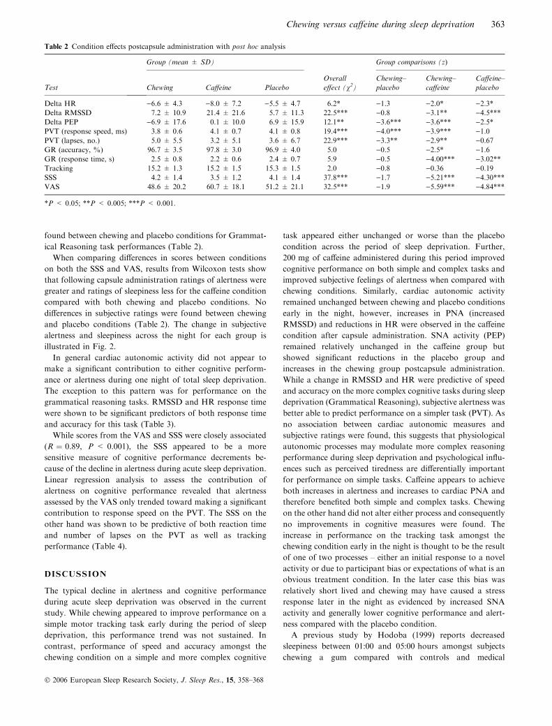
Post hoc analyses using the Wilcoxon test demonstrated a
significant reduction in HR and rise in RMSSD (increase in
PNA) compared with baseline in the caffeine condition
compared with both chewing and placebo conditions. No
difference was seen between chewing and placebo conditions
for these measures. PEP significantly increased relative to
baseline (reduction in SNA) in the placebo condition com-
pared with both chewing and caffeine conditions, with caffeine
showing almost no change to baseline and chewing showing a
significant reduction in PEP relative to baseline (increase in
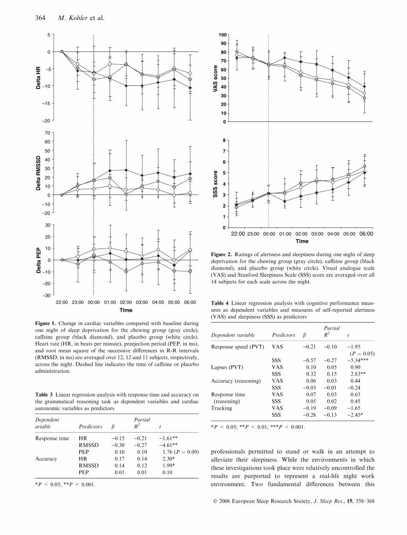
SNA) and compared with both other conditions. Fig. 1
displays mean cardiac activity for each hour across the night
in the three conditions.
Post hoc analyses of performance on the PVT demonstrated
that response speed significantly slowed and lapses were more
frequent in the chewing condition compared with both caffeine
and placebo conditions across the night postcapsule adminis-
tration; however, no difference was seen between performances
in the caffeine versus placebo conditions for either measure
(Table 2). Despite there being only a trend for overall
differences between conditions on the Grammatical Reasoning
task, post hoc tests were conducted and revealed that
performances in the caffeine condition were both faster and
more accurate than the chewing condition, and faster when
compared with the placebo condition. No differences were
Table 1 Condition effects precapsule
administration
Test
Condition (mean ± SD)Overall
effect (v2)Chewing Caffeine Placebo
Delta HR )2.1 ± 2.7 )2.5 ± 4.7 )1.8 ± 3.8 2.4
Delta RMSSD 2.9 ± 5.2 4.4 ± 8.1 2.9 ± 5.9 0.6
Delta PEP )2.3 ± 12.0 )0.4 ± 4.1 1.3 ± 5.2 2.8
PVT (response speed, ms) 4.3 ± 0.5 4.2 ± 0.7 4.4 ± 0.6 0.3
PVT (lapses, no.) 1.5 ± 1.7 2.9 ± 5.4 1.5 ± 2.1 0.4
GR (accuracy, %) 97.3 ± 3.6 97.7 ± 3.1 97.0 ± 3.1 0.5
GR (response time, s) 2.3 ±0.9 2.4 ±0.7 2.4 ± 0.8 1.4
Tracking 16.1 ± 1.3 15.0 ± 1.3 15.4 ± 1.2 7.6*
SSS 2.1 ± 0.7 2.3 ± 0.6 2.3 ± 0.9 1.7
VAS 76.2 ± 10.9 74.0 ± 11.3 75.0 ± 14.6 1.4
*P < 0.05.
362 M. Kohler et al.
� 2006 European Sleep Research Society, J. Sleep Res., 15, 358–368
found between chewing and placebo conditions for Grammat-
ical Reasoning task performances (Table 2).
When comparing differences in scores between conditions
on both the SSS and VAS, results from Wilcoxon tests show
that following capsule administration ratings of alertness were
greater and ratings of sleepiness less for the caffeine condition
compared with both chewing and placebo conditions. No
differences in subjective ratings were found between chewing
and placebo conditions (Table 2). The change in subjective
alertness and sleepiness across the night for each group is
illustrated in Fig. 2.
In general cardiac autonomic activity did not appear to
make a significant contribution to either cognitive perform-
ance or alertness during one night of total sleep deprivation.
The exception to this pattern was for performance on the
grammatical reasoning tasks. RMSSD and HR response time
were shown to be significant predictors of both response time
and accuracy for this task (Table 3).
While scores from the VAS and SSS were closely associated
(R ¼ 0.89, P < 0.001), the SSS appeared to be a more
sensitive measure of cognitive performance decrements be-
cause of the decline in alertness during acute sleep deprivation.
Linear regression analysis to assess the contribution of
alertness on cognitive performance revealed that alertness
assessed by the VAS only trended toward making a significant
contribution to response speed on the PVT. The SSS on the
other hand was shown to be predictive of both reaction time
and number of lapses on the PVT as well as tracking
performance (Table 4).
DISCUSSION
The typical decline in alertness and cognitive performance
during acute sleep deprivation was observed in the current
study. While chewing appeared to improve performance on a
simple motor tracking task early during the period of sleep
deprivation, this performance trend was not sustained. In
contrast, performance of speed and accuracy amongst the
chewing condition on a simple and more complex cognitive
task appeared either unchanged or worse than the placebo
condition across the period of sleep deprivation. Further,
200 mg of caffeine administered during this period improved
cognitive performance on both simple and complex tasks and
improved subjective feelings of alertness when compared with
chewing conditions. Similarly, cardiac autonomic activity
remained unchanged between chewing and placebo conditions
early in the night, however, increases in PNA (increased
RMSSD) and reductions in HR were observed in the caffeine
condition after capsule administration. SNA activity (PEP)
remained relatively unchanged in the caffeine group but
showed significant reductions in the placebo group and
increases in the chewing group postcapsule administration.
While a change in RMSSD and HR were predictive of speed
and accuracy on the more complex cognitive tasks during sleep
deprivation (Grammatical Reasoning), subjective alertness was
better able to predict performance on a simpler task (PVT). As
no association between cardiac autonomic measures and
subjective ratings were found, this suggests that physiological
autonomic processes may modulate more complex reasoning
performance during sleep deprivation and psychological influ-
ences such as perceived tiredness are differentially important
for performance on simple tasks. Caffeine appears to achieve
both increases in alertness and increases to cardiac PNA and
therefore benefited both simple and complex tasks. Chewing
on the other hand did not alter either process and consequently
no improvements in cognitive measures were found. The
increase in performance on the tracking task amongst the
chewing condition early in the night is thought to be the result
of one of two processes – either an initial response to a novel
activity or due to participant bias or expectations of what is an
obvious treatment condition. In the later case this bias was
relatively short lived and chewing may have caused a stress
response later in the night as evidenced by increased SNA
activity and generally lower cognitive performance and alert-
ness compared with the placebo condition.
A previous study by Hodoba (1999) reports decreased
sleepiness between 01:00 and 05:00 hours amongst subjects
chewing a gum compared with controls and medical
Table 2 Condition effects postcapsule administration with post hoc analysis
Test
Group (mean ± SD)
Overall
effect (v2)
Group comparisons (z)
Chewing Caffeine Placebo
Chewing–
placebo
Chewing–
caffeine
Caffeine–
placebo
Delta HR )6.6 ± 4.3 )8.0 ± 7.2 )5.5 ± 4.7 6.2* )1.3 )2.0* )2.3*Delta RMSSD 7.2 ± 10.9 21.4 ± 21.6 5.7 ± 11.3 22.5*** )0.8 )3.1** )4.5***Delta PEP )6.9 ± 17.6 0.1 ± 10.0 6.9 ± 15.9 12.1** )3.6*** )3.6*** )2.5*PVT (response speed, ms) 3.8 ± 0.6 4.1 ± 0.7 4.1 ± 0.8 19.4*** )4.0*** )3.9*** )1.0PVT (lapses, no.) 5.0 ± 5.5 3.2 ± 5.1 3.6 ± 6.7 22.9*** )3.3** )2.9** )0.67GR (accuracy, %) 96.7 ± 3.5 97.8 ± 3.0 96.9 ± 4.0 5.0 )0.5 )2.5* )1.6GR (response time, s) 2.5 ± 0.8 2.2 ± 0.6 2.4 ± 0.7 5.9 )0.5 )4.00*** )3.02**Tracking 15.2 ± 1.3 15.2 ± 1.5 15.3 ± 1.5 2.0 )0.8 )0.36 )0.19SSS 4.2 ± 1.4 3.5 ± 1.2 4.1 ± 1.4 37.8*** )1.7 )5.21*** )4.30***VAS 48.6 ± 20.2 60.7 ± 18.1 51.2 ± 21.1 32.5*** )1.9 )5.59*** )4.84***
*P < 0.05; **P < 0.005; ***P < 0.001.
Chewing versus caffeine during sleep deprivation 363
� 2006 European Sleep Research Society, J. Sleep Res., 15, 358–368
professionals permitted to stand or walk in an attempt to
alleviate their sleepiness. While the environments in which
these investigations took place were relatively uncontrolled the
results are purported to represent a real-life night work
environment. Two fundamental differences between this
–20
–15
–10
–5
0
5D
elta
HR
–20
–10
0
10
20
30
40
50
60
70
Del
ta R
MS
SD
–30
–20
–10
0
10
20
30
22:00 23:00 00:00 01:00 02:00 03:00 04:00 05:00 06:00
Time
Del
ta P
EP
Figure 1. Change in cardiac variables compared with baseline during
one night of sleep deprivation for the chewing group (gray circle),
caffeine group (black diamond), and placebo group (white circle).
Heart rate (HR, in beats per minute), preejection period (PEP, in ms),
and root mean square of the successive differences in R-R intervals
(RMSSD, in ms) are averaged over 12, 12 and 11 subjects, respectively,
across the night. Dashed line indicates the time of caffeine or placebo
administration.
0
10
20
30
40
50
60
70
80
90
100
VAS
sco
re
0
1
2
3
4
5
6
7
8
22:00 23:00 00:00 01:00 02:00 03:00 04:00 05:00 06:00Time
SS
S s
core
Figure 2. Ratings of alertness and sleepiness during one night of sleep
deprivation for the chewing group (gray circle), caffeine group (black
diamond), and placebo group (white circle). Visual analogue scale
(VAS) and Stanford Sleepiness Scale (SSS) score are averaged over all
14 subjects for each scale across the night.
Table 3 Linear regression analysis with response time and accuracy on
the grammatical reasoning task as dependent variables and cardiac
autonomic variables as predictors
Dependent
ariable Predictors bPartial
R2 t
Response time HR )0.15 )0.21 )1.61**RMSSD )0.30 )0.27 )4.61**PEP 0.10 0.10 1.76 (P ¼ 0.09)
Accuracy HR 0.17 0.14 2.30*
RMSSD 0.14 0.12 1.99*
PEP 0.01 0.01 0.10
*P < 0.05; **P < 0.001.
Table 4 Linear regression analysis with cognitive performance meas-
ures as dependent variables and measures of self-reported alertness
(VAS) and sleepiness (SSS) as predictors
Dependent variable Predictors bPartial
R2 t
Response speed (PVT) VAS )0.21 )0.10 )1.95(P ¼ 0.05)
SSS )0.57 )0.27 )5.34***Lapses (PVT) VAS 0.10 0.05 0.90
SSS 0.32 0.15 2.83**
Accuracy (reasoning) VAS 0.06 0.03 0.44
SSS )0.03 )0.01 )0.24Response time
(reasoning)
VAS 0.07 0.03 0.63
SSS 0.05 0.02 0.45
Tracking VAS )0.19 )0.09 )1.65SSS )0.28 )0.13 )2.45*
*P < 0.05; **P < 0.01; ***P < 0.001.
364 M. Kohler et al.
� 2006 European Sleep Research Society, J. Sleep Res., 15, 358–368

previous study and ours were that in the previous study (1)
chewing was continuous throughout the study, and (2) the
chewing substance was a commercially available gum contain-
ing added flavor and aroma. A second study (Wilkinson et al.,
2002) demonstrated little improvement on measures of reac-
tion time and vigilance during periods of chewing. Again,
chewing was of a commercially available gum and chewing was
continuous during task assessment. This study was conducted
during the day using, presumably, non-sleep-deprived individ-
uals. This final point is an important consideration as there
may have been little room for improvement of performance on
measures of simple cognitive tasks such as reaction time and
vigilance tests. More recently Tucha et al. (2004), incorpor-
ating a repeated measures design similar to our own, found
sustained chewing of a flavored chewing gum, neutral gum or
mimicked chewing did not change performance in tasks
assessing short-term attention or vigilance when compared
with controls, and that all chewing groups demonstrated
longer reaction times and reduced alertness and mental
flexibility when compared with controls. The one exception
amongst the chewing groups of this study was that the flavored
gum group demonstrated improved sustained attention com-
pared with controls. This study was performed during the day
and performance assessed during a period of sustained
chewing. Stephens and Tunney’s (2004) assessment of cogni-
tive function during chewing following ingestion of a glucose
load indicates chewing may enhance language-based attention
and processing speed. Such a result would be consistent with
our own if cardiac autonomic changes were evident during
periods of chewing itself, unfortunately however, these data
are not available for comparison. Again, Stephens and
Tunney’s study was conducted during the day in non-sleep-
deprived individuals.
Previous studies provide some concordance as to the
potential ability of chewing to improve alertness and speed
and accuracy on cognitive tasks and suggest little enhancement
is probably achieved. The presumption that chewing benefits
cognition in general may possibly be due to a generalization of
reported effects on simple memory tasks. As to the ability of
chewing to counter the alerting and cognitive decrements seen
during sleep restriction, this study indicates, as for Tucha et al.,
that chewing on its own does not alleviate any decrement in
alertness and �non-memory� cognitive tasks. The strength of
the current study in its applicability to real life situations is
that the testing environment involved a location and activity
not dissimilar to those undertaken by night office staff, such as
administrative positions, call-center customer service employ-
ees or security staff monitoring surveillance video images,
while controlling for other factors such as light, movement,
noise, food intake, and ambient temperature. Also, chewing
was carried out intermittently, for 15 min every hour, much
like someone would self-administer standard flavored chewing
gum. Herein also lays a possible reason for the discrepancy
between our results and others. All previous studies assessed
performance during periods of sustained chewing while tasks
in the present study were completed immediately after a period
of chewing. It may be that chewing activates processes of
cognitive enhancement and alertness only while the act of
mastication is occurring, and that any effects are extremely
short lived. Given the very short period of time between
cessation of chewing and completion of an individual task we
believe such effects are unlikely but need to be investigated.
As expected, caffeine delayed the decline in alertness
commonly seen during sleep deprivation (Dinges et al., 1997;
Dorrian et al., 2000). Ratings of alertness and sleepiness were
improved in the caffeine group compared with chewing and
placebo conditions during a single night without sleep. These
results are consistent with previous studies. Penetar et al.
(1993) administered caffeine doses of around 150, 300 and
600 mg, and placebo to 50 adults after 49 h of total sleep
deprivation. Irrespective of dose, caffeine reduced ratings of
sleepiness on the SSS and increased feelings of alertness as
measured on a VAS for 2 h following administration. The
shorter duration of effect in this previous study may be due to
the fact that caffeine was administered at a time of greater
reported sleepiness and following a longer period of sleep
deprivation than in the present study. Alternatively, the fact
that caffeine was administered at around 08:00 hours in this
previous study may have left little time before circadian driven
increases in alertness were apparent in the placebo group.
Wesensten et al. (2001) report a significant decrease in
sleepiness as reported on the SSS for 2 h following a 600 mg
dose of caffeine at midnight (as in the present study) amongst
10 subjects compared with 10 subjects receiving a placebo.
Caffeine was administered after 41.5 h of total sleep depriva-
tion in this study, suggesting the increased sleep debt in both
previous studies may be the factor shortening the drugs
effectiveness. The study by Wesensten et al. also included
bihourly PVT recordings. Unlike in the present study, response
speed was maintained at predrug, presleep deprivation levels
from 2 h postcaffeine administration for up to 9 h postcaffeine.
This compared with a decline in performance amongst the
placebo group consistent with typical circadian rythmicity.
Subjects in the study were informed they would receive a
substantial bonus monetary reward if performance was main-
tained above a set (but undisclosed) of criteria. It is possible
that the effects of caffeine were attenuated and extended by
motivational forces acting as a mediator to PVT performance.
Further, the caffeine dose was three times that in the present
study, and while dose affects do not seem to occur for ratings
of sleepiness (Penetar et al., 1993) the same may not hold true
for cognitive performance during periods of sleep deprivation.
Strong associations were found between HR and RMSSD
with response time and accuracy on the grammatical reasoning
task. This may suggest that tasks requiring higher cognitive
processing are sensitive to increases in PNA across the night.
The subjective reports of alertness and sleepiness from the
VAS and SSS were found to be predictive of psychomotor
vigilance and tracking performance. Such �simpler� cognitivetasks are perhaps sensitive to subjective states of arousal.
Previous studies have found similar results in that tasks
requiring higher cognitive skills are relatively unaffected by
Chewing versus caffeine during sleep deprivation 365
� 2006 European Sleep Research Society, J. Sleep Res., 15, 358–368

sleep deprivation compared with tasks requiring less cognitive
demand (Harrison and Horne, 2000; Horne, 1985). Borbely
(1986) stated that �it is amazing to see how well sleep-deprived
persons perform tasks needing only brief concentration� (p.158). This is an important consideration given that the
grammatical reasoning tasks lasted for only 3 min compared
with the 10 min of a PVT trial. However, associations between
sleepiness and tracking were also found while each trial of the
tracking task only lasted 30 s.
No association between indices of cardiac autonomic
activity and alertness or cognitive performance was found in
the present study. However, the typical rise in PNA during the
night and concurrent decline in HR, as seen during a normal
sleep/wake cycle (Furlan et al., 1990; Huikuri et al., 1994), was
observed across the night. In general such results suggest a
circadian influence on cardiac PNA activity and similar results
have been previously reported (Burgess et al., 1997). The
comparative increase in RMSSD, reduction in HR, and
inferred increase in overall PNA amongst the caffeine group
following ingestion is also consistent with previous studies
(Richardson et al., 2004; Yeragani et al., 2005), however
contrary results have been reported (Sondermeijer et al.,
2002). Barry et al. (2005) have demonstrated that caffeine is
associated with increased skin conductivity, EEG alpha
frequency and reduced EEG alpha power, despite no effect
on measures of cardiovascular function including HR, blood
pressure and respiration. The authors suggest caffeine induces
a state of arousal, and such results may explain the reported
increase in subjective alertness following caffeine ingestion in
the present study. Cardiac SNA, inferred from PEP, remained
relatively stable across the night. This is in contrast to the
typical decline in activity from a daytime peak across the
normal sleep/wake cycle (Furlan et al., 1990; Huikuri et al.,
1994). Holmes et al. (2002) observed a similar plateau of PEP
as wakefulness was extended beyond subjects� normal sleep
onset time; however, other studies have argued for a circadian
dependency on cardiac SNA (Shiels et al., 2002; Trinder et al.,
2000) and this discrepancy in results remains unresolved. The
relative decrease in PEP amongst the chewing condition
compared with placebo condition increased cardiac sympa-
thetic activity during the early morning period around
02:00 hours. This increase in PEP may suggest induction of a
mild state of stress and/or fatigue due to chewing across the
night. In support of this, Farella et al. (1999) have demon-
strated increases in HR and blood pressure during chewing
gums of differing hardness. Cardiovascular and masticatory
muscle activity were found to be proportional to gum hardness
and perceived fatigue proportional to the level of muscle
activity. It is not unreasonable to expect that chewing
intermittently across an entire night while deprived of sleep
would lead to similar fatigue. Anecdotally, this is consistent
with our observations of subjects� displeasure in chewing the
Parafilm during the later part of the night and this in itself may
present a possible limitation to the study if the chewing of this
unfamiliar substance impacted on mood. Future studies
should endeavor to either obtain as soft as possible gum-base
for chewing or take measures of mood across any extended
period, particularly if participants are sleep deprived and
therefore possibly less resistant to the cognitive impact of a
potentially unpleasant stimuli.
While Wilkinson et al. (2002) observed increases in HR in
their chewing condition and subsequent improvement in
various aspects of short-term memory function, we are unsure
whether any association between the two was apparent. Their
results were explained in terms of possible increased cerebral
blood flow (Momose et al., 1997) and insulin release. In
support of their findings it has been shown that spectral
analysis of HR during chewing of a gum-base increased the
low-frequency band and decreased the high-frequency band
relative to baseline. The ratio of high- to low-frequency power
was markedly increased during chewing, indicating either
enhancement of cardiac SNA or suppression of cardiac PNA
(Shiba et al., 2002). While Tucha et al. (2004) did not observe
differences in pulse rate between chewing groups and control
no differences in measures of memory and attention were
similarly found, still allowing the proposal by Wilkinson et al.
to hold true. The present study is unique in that comprehensive
cardiovascular measures were taken, differences between
conditions were observed, yet no corresponding change in
cognitive performance was found. The decrease in PEP, a
marker of cardiac SNA, is consistent with previous results
however our interpretation differs. Indeed, both interpreta-
tions may still hold true given the differences in time of day of
testing.
Using a 19-channel EEG recording to determine the change
in strength of five frequency bands from prechewing to
chewing, Yagyu et al. (1998) demonstrated that chewing of a
commercially available gum containing flavor and aroma led
to increased power of high-frequency alpha and low-frequency
beta bands with trends for higher delta and theta power when
compared with chewing of unflavored gum base. VAS meas-
ures made during chewing indicated subjects chewing the
commercial gum felt more refreshed and comfortable com-
pared with their gum-base chewing counter-parts. Such results
have been replicated and extended to show that significant
increases in brain-state arousal is primarily due to the aromas
and/or flavors which are constituents in commercially avail-
able gums, and not the chewing of the gum per se (Masumoto
et al., 1999; Morinushi et al., 2000). These results are consis-
tent with the results of the present study and suggest chewing
on its own does not improve alertness or cognitive perform-
ance during periods of acute sleep deprivation. However,
chewing-gum containing added flavors and aromas may
increase alertness and cognitive performance. Such hypotheses
are yet to be thoroughly tested and no conclusions can be
drawn from the present study as constituents of commercial
chewing gum were not included for comparison and no
measures of brain activation were made.
The smaller sample size and fact that caffeine consumption
was not adjusted for body weight of individuals was in part
addressed by the counterbalanced design of this study and
within-subjects analyses. The group n was not unlike previous
366 M. Kohler et al.
� 2006 European Sleep Research Society, J. Sleep Res., 15, 358–368

studies which range from 14 to 30 and generally incorporate an
independent sample design (Baker et al., 2004; Stephens and
Tunney, 2004; Tucha et al., 2004; Wilkinson et al., 2002). Such
samples would be sufficient to detect a �medium� effect with
moderate power (Rosenthal and Rosnow, 1991). Of interest, a
measure of memory performance would have provided valu-
able information and acted as a type of positive control for
chewing effects. Unfortunately, this study was conducted
before the majority of research on memory and chewing was
published and therefore these effects not considered. Further,
the schedule of activities each hour across the night did not
allow for addition testing. Despite such limitations the present
results are very informative given the paucity of research
investigating the cognitive impact of chewing during sleep
deprivation.
The present study is only the second attempt in the literature
to determine the efficacy of chewing in alleviating the increase
in feelings of sleepiness experienced during periods of sleep
deprivation. It is also the first study published which attempts
to look at the efficacy of chewing in countering cognitive
decrements during sleep deprivation while comparing such
effects to another countermeasure for sleepiness, and exploring
possible mechanisms which mediate any effects. This study
suggests chewing per se does not increase alertness or improve
cognitive performance during such periods of sleep loss, and
may in fact induce a state of mild stress or fatigue if continued
over extended periods. Future studies need to address the
influence that flavors and aromas of commercially available
chewing gum may have on measures of alertness and cognition
during sleep deprivation, as such factors may still provide an
effective short-term counter-fatigue alternative.
ACKNOWLEDGEMENTS
The authors thank Dr Alexandra Holmes for her assistance
with analysis of cardiac data and Dr Nicole Lamond for her
assistance with analysis of cognitive performance measures.
REFERENCES
Babkoff, H., Caspy, T. and Mikulincer, M. Subjective sleepiness
ratings: the effects of sleep deprivation, circadian rhythmicity and
cognitive performance. Sleep, 1991, 14: 534–539.
Baddeley, A. A 3 min reasoning test based on grammatical transfor-
mation. Psychon. Sci., 1968, 10: 341–342.
Baker, J., Bezance, J., Zellaby, E. and Aggleton, J. Chewing gum can
produce context-dependent effects upon memory. Appetite, 2004, 43:
207–210.
Baron, R. and Kalsher, M. Effects of a pleasant ambient fragrance on
simulated driving performance: the sweet smell of …safety? Environ.
Behav., 1998, 30: 535–552.
Barry, R., Rushby, J., Wallace, M., Clarke, A., Johnstone, S. and
Zlojutro, I. Caffeine effects on resting-state arousal. Clin. Neuro-
physiol., 2005, 116: 2693–2700.
Berntson, G. G., Cacioppo, J. T. and Quigley, K. S. Autonomic
cardiac control. I. Estimation and validation from pharmacological
blockades. Psychophysiology, 1994, 31: 572–585.
Bigger, J. T. Jr, Fleiss, J. L., Steinman, R. C., Rolnitzky, L. M.,
Kleiger, R. E. and Rottman, J. N. Correlations among time and
frequency domain measures of heart period variability two weeks
after acute myocardial infarction. Am. J. Cardiol., 1992, 69: 891–
898.
Bond, A. and Lader, M. The use of the analogue scales in rating
subjective feelings. Br. J. Med. Psychol., 1974, 4: 211–218.
Borbely, A. Secrets of Sleep. (D. Schneider, Trans.). Basic Books, New
York, 1986.
Boutrel, B. and Koob, G. What keeps us awake: the neuropharma-
cology of stimulants and wakefulness-promoting medications. Sleep,
2004, 27: 1181–1194.
Burgess, H., Trinder, J., Kim, Y. and Luke, D. Sleep and the circadian
influences on cardiac autonomic nervous system activity. Am. J.
Physiol. Heart Circ. Physiol., 1997, 273: H1761–H1768.
Cacioppo, J. T., Berntson, G. G., Binkley, P. F., Quigley, K. S.,
Uchino, B. N. and Fieldstone, A. Autonomic cardiac control. II.
Noninvasive indices and basal response as revealed by autonomic
blockades. Psychophysiology, 1994, 31: 586–598.
Curcio, G., Casagrande, M. and Bertini, M. Sleepiness: evaluating and
quantifying methods. Int. J. Psychophysiol., 2001, 41: 251–263.
Dinges, D. and Kribbs, N. Performing while sleepy: effects of
experimentally induced sleepiness. In: T. H. Monk (Ed.) Sleep,
Sleepiness, and Performance. Wiley, New York, 1991: 97–128.
Dinges, D. and Powell, J. Microcomputer analyses of performance on
a portable, simple visual RT task during sustained operations.
Behav. Res. Methods Instrum. Comput., 1985, 17: 652–655.
Dinges, D., Pack, F., Williams, K., Gillen, K., Powell, J., Ott, G.,
Aptowicz, C. and Pack, A. Cumulative sleepiness, mood distur-
bance, and psychomotor vigilance performance decrements during a
week of sleep restricted to 4–5 h per night. Sleep, 1997, 20: 267–277.
Dorrian, J., Lamond, N. and Dawson, D. The ability to self-monitor
performance when fatigued. J. Sleep Res., 2000, 9: 137–144.
Farella, M., Bakke, M., Michelotti, A., Marotta, G. and Martina, R.
Cardiovascular responses in humans to experimental chewing of
gums of different consistencies. Arch. Oral. Biol., 1999, 44: 835–842.
Folstein, M. and Luria, R. Reliability, validity, and clinical application
of the visual analogue mood scale. Psychol. Med., 1973, 3: 479–486.
Furlan, R., Guzzetti, S., Crivellaro, W., Dassi, S., Tinelli, M., Baselli,
G., Cerutti, S., Lombardi, F., Pagani, M. and Malliani, A.
Continuous 24-h assessment of the neural regulation of systemic
arterial pressure and RR variabilities in ambulant subjects. Circu-
lation, 1990, 81: 537–547.
Harrison, Y. and Horne, J. Sleep loss and temporal memory. Q. J.
Exp. Psychol., 2000, 53A: 271–279.
Hoddes, E., Dement, W. and Zarcone, V. The development and use of
the Stanford Sleepiness Scale (SSS). Psychophysiology, 1972, 9: 150.
Hoddes, E., Zarcone, V., Smythe, H., Phillips, R. and Dement, W.
Quantification of sleepiness: a new approach. Psychophysiology,
1973, 10: 431–436.
Hodoba, D. Chewing can relieve sleepiness in a night of sleep
deprivation. Sleep Res. Online, 1999, 2: 101–105.
Hollingworth, H. Chewing as a technique of relaxation. Science, 1939,
90: 385–387.
Holmes, A., Burgess, H. and Dawson, D. Effects of sleep pressure on
endogenous cardiac autonomic activity and body temperature. J.
Appl. Physiol., 2002, 92: 2578–2584.
Horne, J. Sleep function, with particular reference to sleep deprivation.
Ann. Clin. Res., 1985, 17: 199–208.
Huikuri, H. V., Niemela, M. J., Ojala, S., Rantala, A., Ikaheimo, M. J.
and Airaksinen, K. E. Circadian rhythms of frequency domain
measures of heart rate variability in healthy subjects and patients
with coronary artery disease. Effects of arousal and upright posture.
Circulation, 1994, 90: 121–126.
Kamimori, G., Johnson, D., Thorne, D. and Belenky, G. Multiple
caffeine doses maintain vigilance during early morning operations.
Aviat. Space Environ. Med., 2005, 76: 1046–1050.
Masumoto, Y., Morinushi, T., Kawasaki, H., Ogura, T. and
Takigawa, M. Effects of three principal constituents in chewing
Chewing versus caffeine during sleep deprivation 367
� 2006 European Sleep Research Society, J. Sleep Res., 15, 358–368
gum on electroencephalographic activity. Psychiatry Clin. Neurosci.,
1999, 53, 17–23.
Momose, T., Nishikawa, J., Watanabe, T., Sasaki, Y., Senda, M.,
Kubota, K., Funakoshi, M. and Minakuchi, S. Effect of mastication
on regional cerebral blood flow in humans examined by positron-
emission tomography with 15O-labelled water and magnetic reson-
ance imaging. Arch. Oral Biol., 1997, 42: 57–61.
Morinushi, T., Masumoto, Y., Kawasaki, H. and Takigawa, M. Effect
on electroencephalogram of chewing flavored gum. Psychiatry Clin.
Neurosci., 2000, 54: 645–651.
Nehlig, A. Are we dependent upon coffee and caffeine? A review on
human and animal data. Neurosci. Biobehav. Rev., 1999, 24: 563–
576.
Penetar, D., McCann, U., Thorne, D., Kamimori, G., Galinski, C.,
Sing, H., Thomas, M. and Belenky, G. Caffeine reversal of sleep
deprivation effects on alertness and mood. Psychopharmacology
(Berl.), 1993, 112: 359–365.
Rees, K., Allen, D. and Lader, M. The influence of age and caffeine on
psychomotor and cognitive function. Psychopharmacology, 1999,
145: 181–188.
Richardson, T., Rozkovec, A., Thomas, P., Ryder, J., Meckes, C. and
Kerr, D. Influence of caffeine on heart rate variability in patients
with longstanding type 1 diabetes. Diabetes Care, 2004, 27: 1127–
1131.
Rogers, N., Phan, O., Kennaway, D. and Dawson, D. Effect of
daytime oral melatonin administration on neurobehavioral per-
formance in humans. J. Pineal Res., 1998, 25: 53–57.
Rosenthal, R. and Rosnow, R. Essentials of Behavioral Research:
Methods and data Analysis, 2nd edn. McGraw-Hill, New York,
1991.
Sherwood, L. Human Physiology: From Cells to Systems, 2nd edn.
West Publishing Company, St Paul, MN, USA, 1993.
Shiba, Y., Nitta, E., Hirono, M., Sugita, M. and Iwasa, Y. Evaluation
of mastication-induced change in sympatho-vagal balance through
spectral analysis of heart rate variability. J. Oral Rehabil., 2002, 29:
956–960.
Shiels, S. A., Hilton, M. F., Weiner, M. E. and Shea, S. A. An
endogenous circadian rhythm to cardiac sympathetic tone in
humans. Sleep, 2002, 25 (Abstract Suppl.): A122–A123.
Sondermeijer, H., van Marle, A., Kamen, P. and Krum, H. Acute
effects of caffeine on heart rate variability. Am. J. Cardiol., 2002, 90:
906–907.
Stephens, R. and Tunney, R. Role of glucose in chewing gum-related
facilitation of cognitive function. Appetite, 2004, 43: 211–213.
Task Force of the European Society of Cardiology and the North
American Society of Pacing and Electrophysiology. Heart rate
variability: standards of measurement, physiological interpretation
and clinical use. Circulation, 1996, 93: 1043–1065.
Trinder, J., Carrington, M., Breen, S., Kleiman, J., Kim, Y. and Tan,
N. Sleep and Circadian control of sleep period cardiac activity.
Sleep, 2000, 23(Suppl. 2): A147.
Tucha, O., Mecklinger, L., Maier, K., Hammerl, M. and Lange, K.
Chewing gum differentially affects aspects of attention in healthy
subjects. Appetite, 2004, 42: 327–329.
Van Dongen, H., Price, N., Mullington, J., Szuba, M., Kapoor, S. and
Dinges, D. Caffeine eliminates psychomotor vigilance deficits from
sleep inertia. Sleep, 2001, 24: 813–819.
Voultsios, A., Kennaway, D. and Dawson, D. Salivary melatonin as a
circadian phase marker: validation and comparison with plasma
melatonin. J. Biol. Rhythms, 1997, 12: 457–466.
Vrijkotte, T., Snaar, T., Bernards, C. and de Geus, E. Comparison of
time- and frequency-domain analysis of heart rate variability from
ambulatory recordings. 2001, http://www.psy.vu.nl/ams/research/
posters/comp.of.time.freq.domain.html
Wesensten, N. J., Belenky, G., Kautz, M. A., Thorne, D. A.,
Reichardt, R. M. and Balkin, T. J. Maintaining alertness and
performance during sleep deprivation: modafinil versus caffeine.
Psychopharmacology (Berl.), 2002, 159: 238–247.
Wilkinson, L., Scholey, A. and Wesens, K. Chewing gum selectively
improves aspects of memory in healthy volunteers. Appetite, 2002,
38: 235–236.
Yagyu, T., Kondakor, I., Kochi, K., Koenig, T., Lehmann, D.,
Kinoshita, T., Hirota, T. and Yagyu, T. Smell and taste of chewing
gum affect frequency domain EEG source localizations. Int. J.
Neurosci., 1998, 93: 205–216.
Yeragani, V., Krishnan, S., Engels, H. and Gretebeck, R. Effects of
caffeine on linear and nonlinear measures of heart rate variability
before and after exercise. Depress. Anxiety, 2005, 21: 130–134.
368 M. Kohler et al.
� 2006 European Sleep Research Society, J. Sleep Res., 15, 358–368
Copyright © 2022 FDOKUMEN