EPR−ENDOR of the Cu(I)NO Complex of Nitrite Reductase
11
Subscriber access provided by INST COAL AND COAL CHEMISTRY Journal of the American Chemical Society is published by the American Chemical Society. 1155 Sixteenth Street N.W., Washington, DC 20036 Article EPR−ENDOR of the Cu(I)NO Complex of Nitrite Reductase Oleg M. Usov, Yan Sun, Vladimir M. Grigoryants, James P. Shapleigh, and Charles P. Scholes J. Am. Chem. Soc., 2006, 128 (40), 13102-13111 • DOI: 10.1021/ja056166n Downloaded from http://pubs.acs.org on December 14, 2008 More About This Article Additional resources and features associated with this article are available within the HTML version: • Supporting Information • Links to the 7 articles that cite this article, as of the time of this article download • Access to high resolution figures • Links to articles and content related to this article • Copyright permission to reproduce figures and/or text from this article
-
Upload
independent -
Category
Documents
-
view
0 -
download
0
Transcript of EPR−ENDOR of the Cu(I)NO Complex of Nitrite Reductase
Subscriber access provided by INST COAL AND COAL CHEMISTRY
Journal of the American Chemical Society is published by the American ChemicalSociety. 1155 Sixteenth Street N.W., Washington, DC 20036
Article
EPR−ENDOR of the Cu(I)NO Complex of Nitrite ReductaseOleg M. Usov, Yan Sun, Vladimir M. Grigoryants, James P. Shapleigh, and Charles P. Scholes
J. Am. Chem. Soc., 2006, 128 (40), 13102-13111 • DOI: 10.1021/ja056166n
Downloaded from http://pubs.acs.org on December 14, 2008
More About This Article
Additional resources and features associated with this article are available within the HTML version:
• Supporting Information• Links to the 7 articles that cite this article, as of the time of this article download• Access to high resolution figures• Links to articles and content related to this article• Copyright permission to reproduce figures and/or text from this article
EPR-ENDOR of the Cu(I)NO Complex of Nitrite Reductase
Oleg M. Usov,§ Yan Sun,§ Vladimir M. Grigoryants,§ James P. Shapleigh,# andCharles P. Scholes*,§
Contribution from the Center for Biochemistry and Biophysics, Department of Chemistry,UniVersity at Albany, State UniVersity of New York, Albany, New York 12222, and Department
of Microbiology, Wing Hall, Cornell UniVersity, Ithaca, New York 14853
Received September 7, 2005; E-mail: [email protected]
Abstract: With limited reductant and nitrite under anaerobic conditions, copper-containing nitrite reductase(NiR) of Rhodobacter sphaeroides yielded endogenous NO and the Cu(I)NO derivative of NiR. 14N- and15N-nitrite substrates gave rise to characteristic 14NO and 15NO EPR hyperfine features indicating NOinvolvement, and enrichment of NiR with 63Cu isotope caused an EPR line shape change showing copperinvolvement. A markedly similar Cu(I)NONiR complex was made by anaerobically adding a little endogenousNO gas to reduced protein and immediately freezing. The Cu(I)NONiR signal accounted for 60-90% ofthe integrated EPR intensity formerly associated with the Type 2 catalytic copper. Analysis of NO and Cuhyperfine couplings and comparison to couplings of inorganic Cu(I)NO model systems indicated ∼50%spin on the N of NO and ∼17% spin on Cu. ENDOR revealed weak nitrogen hyperfine coupling to one ormore likely histidine ligands of copper. Although previous crystallography of the conservative I289V mutanthad shown no structural change beyond the 289 position, this mutation, which eliminates the Cδ1 methylof I289, caused the Cu(I)NONiR EPR spectrum to change and proton ENDOR features to be significantlyaltered. The proton hyperfine coupling that was significantly altered was consistent with a dipolar interactionbetween the Cδ1 protons of I289 and electron spin on the NO, where the NO would be located 3.0-3.7 Åfrom these protons. Such a distance positions the NO of Cu(I)NO as an axial ligand to Type 2 Cu(I).
Introduction
Nitrite reductase (NiR) catalyzes the defining reaction ofdenitrification, which is the one-electron reduction of nitrite tonitric oxide:
Copper-containing NiR possesses a Type 2 copper catalyticcenter, where nitrite is bound and converted to nitric oxide, anda Type 1 copper center, where electrons are accepted andreductively shuttled to the nearby Type 2 copper. NiR is atrimeric protein, for which each subunit contains a Type 1 anda Type 2 copper center. Scheme 1 shows the proximity of thetwo copper centers, the location of their direct copper ligands,and the location of catalytically important, non-liganding aminoacids at the Type 2 center.1-4 These latter catalytically importantamino acids are a highly conserved Asp (Asp129 here) and His(His287 here) which connect through hydrogen bonds to the
oxygens of the nitrite substrate for the purpose of substrateorientation when nitrite is bound at Type 2 copper. Additionally,there is a conserved Ile (Ile 289 here) whose bulky side chainoccludes and helps define the ligand binding pocket.
§ SUNY at Albany.# Cornell University.
(1) Godden, J. W.; Turley, S.; Teller, D. C.; Adman, E. T.; Liu, M. Y.; Payne,W. J.; LeGall, J.Science1991, 253, 438-442.
(2) Adman, E. T.; Godden, J. W.; Turley, S.J. Biol. Chem.1995, 270, 27458-27474.
(3) Murphy, M. E. P.; Turley, S.; Kukimoto, M.; Nishiyama, M.; Horinouchi,S.; Sasaki, H.; Tanokura, M.; Adman, E. T.Biochemistry1995, 34, 12107-12117.
(4) Murphy, M. E. P.; Turley, S.; Adman, E. T.J. Biol. Chem.1997, 272,28455-28460.
(5) Tocheva, E. I.; Rosell, F. I.; Mauk, A. G.; Murphy, M. E.Science2004,304, 867-870.
NO2j + 2H+ + ej f NO + H2O
Scheme 1. The Locale of the Type 1 Blue-GreenElectron-Transfer Center and the Type 2 Catalytic Center in NiRa
a As ligands Type 1 copper has His126, His177, Cys167, and Met182.Type 2 copper has His166, His131, His338, and an axial ligand shown asNO here. Conserved amino acids Asp129, His287, and Ile289 near the Type2 catalytic center play a critical role in activity. Structure after Tocheva etal.;5 PDB file 1SNR of NiR fromAlcaligenes faecalisbut withRhodobactersphaeroidesnumbers. TheA. faecalisamino acids His95, His145, Cys136,and Met150 at the Type 1 center respectively correspond to His126, His177,Cys167, and Met182 ofR. sphaeroides; theA. faecalisamino acids His100,His135, His306, Asp98, His255, and Ile257 at the Type 2 center respectivelycorrespond to His131, His166, His338, Asp129, His287, and Ile289 ofR.sphaeroides.
Published on Web 09/15/2006
13102 9 J. AM. CHEM. SOC. 2006 , 128, 13102-13111 10.1021/ja056166n CCC: $33.50 © 2006 American Chemical Society

Interest in the mechanism of NiR has extended to understand-ing what becomes of the NO itself. It has been determined thatNiR can be run backward using NO as a substrate to producenitrite and reducing equivalents.6 X-ray crystallography has beenrecently performed on a construct of NiR in which initiallyreduced NiR was reacted with externally supplied NO gas, andthe structure indicated NO to be ligated in a side-on fashion sothat both the N and the O were directly bound to the Type 2copper.5 On the basis of EPR spectroscopy (not actuallyperformed on the crystals themselves), the adduct was assignedas a cupric complex.5 In a separate crystallographic study,7 theside-on coordination of NO to Type 2 copper was observed asone of several axial coordination modes taken by water, nitrite,and NO, even from within the same batch of crystals and evenwithout the addition of exogenous NO.
A nitrogenous biomimetic for NO-bound Type 2 copper ofNiR has been prepared under reducing conditions that discrimi-nated it from cupric copper, and it was definitively character-ized: first, by EPR as a paramagnetic Cu(I)NO complexshowing electron spin density on Cu and NO, and second, byX-ray crystallography that indicated nitrogen end-on bindingof the NO to Cu(I).8 Recent modeling and density functionaltheory (DFT) studies on NO at the active site of Type 2 copperin NiR have pointed to a stable paramagneticS) 1/2 {CuNO}11
oxidation level having a Cu(I)-NO• configuration with∼90%of the spin on the NO and∼10% on the Cu;9 the conformationof this NO when oriented for side-on copper ligation waspredicted to be marginally more stable than the end-on formbound through N.
The mechanism and underlying structure of cupric NiR havebeen previously studied through application of electron nucleardouble resonance (ENDOR).10-12 Recent studies upon wild-type and mutant forms of NiR combined enzymatic measure-ments with ENDOR; they detected exchangeable proton featuresassociated with the Asp129 and His287 (shown in Scheme 1)and correlated changes in these proton features with loss ofenzymatic activity.12 These critical Asp and His side chainsdetermined the location of protons that would hydrogen bondto a nitrite oxygen and facilitate conversion of nitrite to NOand water upon reduction.12 Mutation of I289 to V289 causedminor perturbation to enzyme activity, but the I289V mutationallowed binding to the Type 2 copper of a small molecule,formate, whose proton features were detected by ENDOR.12
Such formate binding was not observed with wild-type protein,and we suggested that at least one purpose of I289 was to blockunwanted binding of small molecules other than water and nitritewithin the Type 2 copper pocket. Subsequent X-ray study ontheAlcaligenes faecalishomologue of theRhodobacter sphaeroi-desI289V mutant indicated that “no global structure rearrange-ment had resulted from the mutation” and that the “positionsof the side chains of the active site residues that lay within 8 Å
of the copper (with the exception of the isoleucine itself) wereequally well conserved conformationally”.13 In addition, theenzyme activity was actually slightly larger for the I289Vmutant.13 Thus, for the I289V mutant there is only localperturbation to enzyme structure from the mutation and a slightincrease in NiR activity, whereas for the I289A mutant there ismore perturbation to structure and a decrease in enzymeactivity.13
Over the past 30 years, a considerable literature of spectro-scopic,14,15 crystallographic,16 and theoretical17 understandinghas emerged on heme-NO systems. Over the past 10 years,NO has gained intrinsic interest as a small molecule of signalingand sensing, for example, in soluble guanylate cyclase.18 Aheme-containing nitrite reductase generates NO for the metabo-lism of Pseudomonas aeruginosa.19 In ref 20, we used EPRand nitrogen and proton ENDOR to probe the electronicstructure of NO-bound cytochromec′ of R. sphaeroides2.4.3.We determined electronic evidence of spin density on the NOof a heme protein whose function, when reduced, is to transportand store NO21,22 in the sameR. sphaeroidesorganism whereNiR produces the NO.
There is not, at this time, the spectroscopic, crystallographic,and theoretical underpinning for copper-NO complexes thatthere is for heme-NO systems. NiR is an unusual enzymewhose purpose in the metabolism of the denitrifying bacteria isto produce large amounts of NO. Binding of NO to the veryType 2 copper catalytic site that produces the NO in the firstplace is unprecedented, even if the NO binding that we observeturns out to be product inhibition. Furthermore, the enzyme hasbeen made to run in reverse by binding NO to create nitriteand reducing equivalents. For an NO complex of the samecopper protein which catalytically produces NO in largequantities, a motivation for this present work is to providespectroscopic underpinnings on the local metallo-NO electronicstructure. We do this for comparison with the theoreticalpredictions on the electronic structure and spin distribution inmodel Cu(I)NO complexes9 and for comparison with thespectroscopic and crystallographic results of biomimetic Cu(I)-NO complexes.8
Materials and Methods
Materials. Using the plasmid pET17b-nirK, which contains ampi-cillin resistance and theNir gene, overexpression, purification, activity,and copper content assay of wild-type NiR and its I289V and I289Amutants were carried out as described previously.12 Following over-expression of NiR, the protocol was to add∼1 mL of 0.1 M CuCl2 tothe supernatant containing Cu-free NiR in order to replenish the copperin the NiR.23 For preparation of NiR with isotopic enrichment,63CuCl2(99.8% in 63Cu from Isotec, Inc.) was used. (Natural abundance Cu
(6) Wijma, H. J.; Canters, G. W.; de Vries, S.; Verbeet, M. P.Biochemistry2004, 43, 10467-10474.
(7) Antonyuk, S. V.; Strange, R. W.; Sawers, G.; Eady, R. R.; Hasnain, S. S.Proc. Natl. Acad. Sci. U.S.A.2005, 102, 12041-12046.
(8) Ruggiero, C. E.; Carrier, S. M.; Antholine, W. E.; Whittaker, J. W.; Cramer,C. J.; Tolman, W. B.J. Am. Chem. Soc. 1993, 115, 11285-11298.
(9) Wasbotten, I. H.; Ghosh, A.J. Am. Chem. Soc.2005, 127, 15384-15385.(10) Howes, B. D.; Abraham, Z. H. L.; Lowe, D. J.; Bruser, T.; Eady, R. R.;
Smith, B. E.Biochemistry1994, 33, 3171-3177.(11) Veselov, A.; Olesen, K.; Sienkiewicz, A.; Shapleigh, J. P.; Scholes, C. P.
Biochemistry1998, 37, 6095-6105.(12) Zhao, Y.; Lukoyanov, D. A.; Toropov, Y. V.; Wu, K.; Shapleigh, J. P.;
Scholes, C. P.Biochemistry2002, 41, 7464-7474.
(13) Boulanger, M. J.; Murphy, M. E.Protein. Sci.2003, 12, 248-256.(14) Dickinson, L. C.; Chien, J. C.J. Am. Chem. Soc.1971, 93, 5036-5040.(15) Yonetani, T.; Yamamoto, H.; Erman, J. E.; Leigh, J. S., Jr.; Reed, G. H.J.
Biol. Chem.1972, 247, 2447-2455.(16) Scheidt, W. R.; Piciulo, P. L.J. Am. Chem. Soc.1976, 98, 1913-1919.(17) Zhang, Y.; Gossman, W.; Oldfield, E.J. Am. Chem. Soc.2003, 125, 16387-
16396.(18) Denninger, J. W.; Marletta, M. A.Biochim. Biophys. Acta1999, 1411,
334-350.(19) Silvestrini, M. C.; Tordi, M. G.; Musci, G.; Brunori, M.J. Biol. Chem.
1990, 265, 11783-11787.(20) Usov, O. M.; Choi, P. S.-T.; Shapleigh, J. P.; Scholes, C. P.J. Am. Chem.
Soc.2006, 128, 5021-5032.(21) Mayburd, A. L.; Kassner, R. J.Biochemistry2002, 41, 11582-11591.(22) Choi, P. S.; Grigoryants, V. M.; Abruna, H. D.; Scholes, C. P.; Shapleigh,
J. P.J. Bacteriol.2005, 187, 4077-4085.(23) Olesen, K.; Veselov, A.; Zhao, Y.; Wang, Y.; Danner, B.; Scholes, C. P.;
Shapleigh, J. P.Biochemistry1998, 37, 6086-6094.
Cu(I)NO Complex of Nitrite Reductase A R T I C L E S
J. AM. CHEM. SOC. 9 VOL. 128, NO. 40, 2006 13103
contains 69%63Cu and 31%65Cu. 65Cu and63Cu both have the sameI ) 3/2 nuclear spin state, and65Cu has a magnetic moment which isabout 8% higher than that of63Cu.) Samples to be deuterated wereexchanged and concentrated twice with a microfuge concentrator(Microcon) versus D2O (99.9% isotopic enrichment, Cambridge IsotopesLaboratory, CIL) buffer at pD 7 to give an approximate 95% D2Oenrichment; for these samples, perdeuterated glycerol (CIL) was usedas a glassing agent.12,24
Methods. (a) Sample Preparation.For most of the spectra reportedhere, a method was used which enzymatically generated endogenousNO within the small volume of the reduced, deaerated sample. In thismethod, NiR samples having a subunit concentration of 0.5 mM, 50mM pH 7.2 phosphate buffer, a 2-fold molar excess of nitrite, 40%glycerol (for rapid freezing), and 10µM phenazine methosulfate (PMS,Sigma) mediator were first deaerated by argon exchange for 5 minwith gentle agitation under a continuously flowing, scrubbed argonatmosphere in a 7 mLseptum vial.25 Samples were then transferred byan argon-flushed Hamilton gastight syringe to argon-flushed EPR tubes(3.0 mm i.d., 4.0 mm o.d. tubes for X-band EPR; 2.0 mm i.d., 2.4 mmo.d. tubes for Q-band ENDOR). A 2-fold molar excess of deaerated,argon-exchanged NADH (Sigma) reductant was added by Hamiltongastight syringe and thoroughly mixed by the Hamilton needle tip withinthe EPR tube for∼1 min until the sample bleached. This is the stagewhere enzymatically produced NO would be released by NiR itself.Samples were rapidly frozen by plunging into liquid nitrogen as theEPR tube above the sample was continuously flushed with argon. TheX-band samples were∼0.2 mL in volume, and the Q-band sampleswere ∼0.05 mL in volume. Samples were prepared with14N-nitrite(14NO2
-, Sigma) and15N-nitrite (15NO2-, 98% isotopic enrichment,
CIL). A different method, using a small amount of exogenous NO,also provided a signal markedly similar to that obtained from NOgenerated endogenously by enzyme action. With this method, wecarefully reduced our argon-exchanged, deaerated sample by using a2-fold excess of NADH within the EPR tube under argon, bubbledinto it a small (∼50µL) aliquot of freshly scrubbed NO gas, and quicklyfroze the sample within∼1 min of the introduction of NO. The EPRsignal, which is shown in Figure 1C, was remarkably similar to theEPR signal (Figure 1A) produced by endogenous production of NO.
The methods which Tocheva et al.5 used to generate diffraction-quality crystals with a Cu(II) species as the final product had employedlong exposure to exogenous NO. Tocheva et al.5 also generated EPRsignals from liquid samples of reduced NiR subject to long incubationof up to 20 min under exogenous NO. When we reduced our samplewithin a 7 mLvial under argon, added several milliliters of scrubbedNO (14NO, 99% purity, Air Products), and then after a 5 min incubationtransferred the sample anaerobically to the EPR tube where the samplewas frozen, we obtained a Cu(II) signal similar to that reported byTocheva et al.5 This signal is shown in Figure 1D. In our own efforts,we found that details of the cupric Type 2 complex changed over theperiod of NO incubation, possibly because of the re-emergence ofresting cupric NiR, as reported by Tocheva et al. in their SupportingInformation.5
(b) X- and Q-Band EPR.X-band EPR (9.52 GHz) was carried outwith an ER-200 IBM Bruker X-band spectrometer equipped with astandard TE102 EPR cavity and an APD Cryogenics LTR-3 Helitransystem (Allentown, PA) operated at 15 K. X-band EPR data werecollected in a personal computer using the EW Software routines(Scientific Software Sales, Plymouth, MI). The program SIMPIPM was
used for fitting of EPR spectra to obtain estimates of hyperfine couplingsandg-values.26 The Q-band EPR (34.1 GHz) system operates below 2K with pumped helium and is generally used in a dispersion, rapidpassage mode because the dispersion, rapid passage signal is highlysensitive to nuclear spin transitions of the ENDOR process. Dispersion,rapid passage EPR signals with slow electron spin relaxation have theappearance of inverted absorption signals, although spin relaxation cancause distortion of the rapid passage line shape. An EPR signal withthe apparent line shape of a standard first derivative absorption EPRsignal can be obtained by numerically taking a first derivative of therapid passage signal.
Q-band ENDOR measurements were performed under dispersion(ø′), rapid passage field-modulated conditions at 2 K with thecryogenically tunable TE011 Q-band resonator,27 as previously re-ported.11,12,28In doing ENDOR, we monitor the radio frequency (RF)-induced change in the rapid passage, 100 kHz field-modulateddispersion EPR signal as we sweep the frequency of the RF field.
(c) ENDOR Theory: Protons.The frequencies of proton ENDORfeatures,PνENDOR, center to first order at the free proton nuclear Zeemanfrequency,νP. TakingA as the hyperfine coupling, one finds the protonfrequencies are split away fromνP by (1/2 A for protons coupled tothe electron spin1/2 doublet.12,20,24Proton ENDOR frequencies, occur-ring as “+” or as “-” Zeeman branches, are29
First-order expresions hold whenνP . A/2, as is the case here with amagnetic field of∼1.2 T and νP in the 50 MHz range. Detaileddescriptions of the proton hyperfine tensor and dipolar contributionsto it are provided in the Methods Sections of refs 20 and 24.
Distant protons such as those on I289 typically have only dipolarcouplings. The point dipolar couplings,ADip, of protons coupled toelectron spin localized at a particular center would be24
wheref is the fraction of an unpaired electron on a particular center,geff is the electronicg-value where the dipolar coupling is measured,gn is the nuclearg-value () 5.585 for a proton),R is the distance fromthe proton to the localized center of electron spin, andθ is the anglebetween the vectorR and the external magnetic field. The point dipolarHamiltonian will be axial with respect to theR direction. The turningpoints in the powder ENDOR spectra occurring whereθ ) 0° or 90°typically provide the best resolved ENDOR, and the 90° turning pointis spatially the most likely.29
(d) Nitrogen ENDOR. The first-order expressions for spin 114NENDOR frequencies of heme, histidine, and NO are
where 14A is the hyperfine coupling,P is the quadrupolar coupling,and14ν () 3.75 MHz at 1.218 T) is the14N nuclear Zeeman frequency.(If quadrupolar splitting is resolved, the quadrupolar splitting will be3|P|.) The14ν+
ENDOR branch is often the only one observable with rapid
(24) Usov, O. M.; Choi, P. S.-T.; Shapleigh, J. P.; Scholes, C. P.J. Am. Chem.Soc.2005, 127, 9485-9494.
(25) Three cycles of freeze-pump-thaw were also tried for rigorous removalof oxygen. Freeze-pump-thaw gave Cu(I)NONiR spectra essentiallyunchanged from those obtained by the argon-exchange method describedin the Methods section that did not involve repetitive freezing of enzyme.A sample was also prepared with no attempt to remove oxygen fromsolution, and its Cu(I)NONiR features were very similar to those of theanaerobic samples; however, it also gave evidence for oxidized cupriccopper.
(26) SIMPIPM is an extension of PIP, which is an extension of QPOW. Nilges,M. J.; Belford, R. L.; Francesconi, L. C. Simulation of strain in EPR spectrausing the method of gradients. Presented at the 40th Rocky MountainConference on Analytical Chemistry, Denver, CO, July 1998. For QPOW,see: Nilges, M. J. Thesis, University of Illinois, Urbana, 1979.
(27) Sienkiewicz, A.; Smith, B. G.; Veselov, A.; Scholes, C. P.ReV. Sci. Instrum.1996, 67, 2134-2138.
(28) Veselov, A. V.; Osborne, J. P.; Gennis, R. B.; Scholes, C. P.J. Am. Chem.Soc.2000, 122, 8712-8716.
(29) Hoffman, B. M.; DeRose, V. J.; Doan, P. E.; Gurbiel, R. J.; Houseman, A.L. P.; Telser, J. InBiological Magnetic Resonance, Vol. 13: EMR ofParamagnetic Molecules; Berliner, L. J., Reuben, J., Eds.; Plenum: NewYork, 1993.
Pν(ENDOR ) |νP ( A/2| (1)
ADip ) {fgeffgnâeân/hR3}(3 cos2 θ - 1) ) ADip (3 cos2 θ - 1)
) (39.5fgeff/R3)(3 cos2 θ - 1) (MHz) (2)
14ν+ENDOR ) |14A/2 ( 3/2 P + 14ν|
and 14νjENDOR ) |14A/2 ( 3/2 P - 14ν| (3)
A R T I C L E S Usov et al.
13104 J. AM. CHEM. SOC. 9 VOL. 128, NO. 40, 2006
passage Q-band ENDOR for nitrogen features from heme and histidinewith couplings less than 30 MHz.24,28
Results
Characterization of the Cu(I)NONiR Complex Resultingfrom the Reaction of Reduced NiR with NO. Figure 1provides a comparison of the X-band EPR spectrum (1A) ofNiR reduced in the presence of14N-nitrite to the spectrum (1B)of oxidized, resting wild-type NiR, which has both a cupric Type1 and Type 2 center. The samples for spectra 1A and 1B werefrom the same enzyme preparation and were equal in concentra-tion. Spectrum 1A, unlike Cu(II) EPR spectra but like those ofthe {CuNO}11 models,8 showed considerable hyperfine detailnearg ) 2.00 and features whoseg-values were less than 2.00.These latter features show Cu hyperfine coupling. Spectrum 1A,again unlike Cu(II) EPR spectra but like those of the{CuNO}11
models,8 could only be observed below liquid nitrogen tem-perature because of its fast spin relaxation. A double integrationof the signal from NiR reduced in the presence of nitrite, anda comparison of that double integral to the double integral ofthe original resting cupric NiR signal, indicated that spectrum1A accounted for 30-45% of the integrated intensity of thetotal resting cupric signals from Type 1 and Type 2 copper inspectrum 1B. If the signal from the NiR reduced in the presenceof nitrite arises from a Type 2 copper center, the implication isthat a spin count of 60-90% of the available Type 2 coppercenters of NiR is providing that signal. Integration of thebiomimetic model Cu(I)NO EPR signals reported in ref 8indicated that{CuNO}11 signals from the model correspondedto about∼70% of the available copper. Spectrum 1C is theEPR spectrum of reduced NiR to which a small volume ofexogenous NO gas was anaerobically added directly in situ withsubsequent quick freezing. Spectrum 1C shows that a reducedNiR sample protected under argon will initially bind NO so as
to give an EPR signal remarkably similar to that obtained whenreduced NiR reacts with its own nitrite-derived NO. We focusthe work reported here on the initially bound NO species, whichwe characterize in this paper and beyond this point designateas Cu(I)NONiR. A markedly different spectrum is shown inFigure 1D from a sample which was obtained by reactingreduced NiR for∼5 min with a large volume excess ofexogenous NO, as described in the Materials section, beforetransferring the sample to the EPR tube and freezing. Thisspecies cannot be the initial EPR species from the reaction ofNO with reduced NiR since we have already seen in spectra1A and 1C that there is an earlier species, viz. Cu(I)NONiR,prepared by two separate methods. Spectrum 1D showed cupriccharacter with well-resolved large copper hyperfine couplinghavingA| ) 115 G andg| ) 2.307, similar to that reported byTocheva et al.5 This Cu(II)NO construct, like that reported byTocheva et al.,5 hasg-values and hyperfine spectra (Figure 1D)different from those of the Cu(I)NONiR complex. Our workfocuses on Cu(I)NONiR prepared by enzymatic action of NiRunder anaerobic conditions in a small, anaerobically isolatedvolume with limited, biologically relevant reductant and limitednitrite. These limited reducing and nitrite conditions are plausibleconditions that would be encountered when NiR is producingor consuming6 its own NO.
The spectra shown in Figure 2A,B are the X-band firstderivative EPR spectra of63Cu-enriched NiR reduced respec-tively in the presence of14N-nitrite (A) and15N-nitrite (B). Thedifference between14N and15N isotopes is evident in the detailsof hyperfine structure, especially nearg ) 2.00, and theimplication is that NO originating from nitrite contributes tothe EPR signal. Spectrum 2C overlays the spectra of Cu-(I)14NONiR samples prepared from63Cu-enriched (solid) andnaturally abundant copper (dotted). Given the magnetic similar-ity of the 63Cu and 65Cu isotopes, the spectral differencesbetween samples with natural abundance Cu and with enriched
Figure 1. X-band EPR spectra of (A) Cu(I)14NONiR with NO derivedenzymatically from14N-nitrite, (B) resting cupric NiR (signal multipliedby 0.4), (C) Cu(I)14NONiR prepared from reduced NiR by injecting∼50µL of NO gas into the anaerobic sample, and (D) Cu(II)NiR made fromreduced NiR and excess exogenous NO with∼5 min incubation. Thesplitting between the Cu hyperfine features centered atg ) 2.307 is 115G. All samples were from wild-type NiR, 0.5 mM in NiR subunits, withCu in natural isotopic abundance. The spectra were recorded atT ) 15 K,0.6 mT field modulation, 100 s signal averaging over a 0.140 T sweep, 2mW microwave power, EPR frequency) 9.525 GHz.
Figure 2. X-band EPR spectra of (A)63Cu(I)14NONiR prepared from14N-nitrite and (B)63Cu(I)15NO NiR prepared with15N-nitrite. The purpose isshow that NO originating from nitrite contributes to the EPR signal. SpectraC provide an overlay of 99%63Cu-enriched63Cu(I)14NONiR (solid line)with Cu(I)NONiR containing63Cu:65Cu in the natural isotopic 69:31 ratio(dotted line). These spectra were recorded atT ) 15 K, 0.3 mT fieldmodulation, 100 s signal averaging with a 0.070 T sweep, 2 mW microwavepower, EPR frequency) 9.525 GHz.
Cu(I)NO Complex of Nitrite Reductase A R T I C L E S
J. AM. CHEM. SOC. 9 VOL. 128, NO. 40, 2006 13105
63Cu are not large, but the EPR features are sharper in thepresence of enriched63Cu. This spectral difference indicatesthat Cu must be involved with the signal originating from NiRthat has been reduced in the presence of nitrite, thus providingan additional rationale for calling the signal Cu(I)NONiR. TheQ-band spectrum of Cu14NONiR obtained by dispersion, rapidpassage EPR at 1.8 K is shown in the Supporting Information,Figure 1S. Although the passage signal was broader than theX-band spectra and showed distortion at its low-field extremum,the derivative of this spectrum provided additional confirmationof g-values, which were estimated asgx ) 2.044( 0.003,gy )1.998( 0.002, andgz ) 1.923( 0.005, and evidence neargz
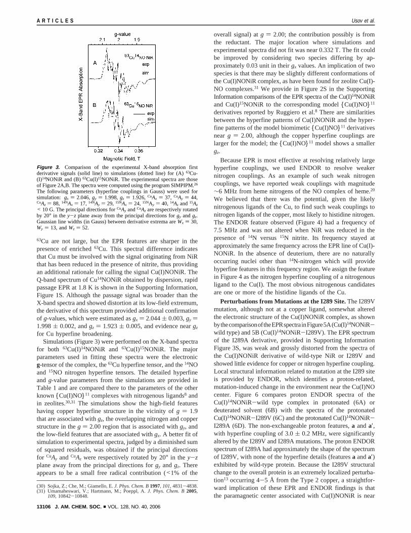
for Cu hyperfine broadening.Simulations (Figure 3) were performed on the X-band spectra
for both 63Cu(I)14NONiR and 63Cu(I)15NONiR. The majorparameters used in fitting these spectra were the electronicg-tensor of the complex, the63Cu hyperfine tensor, and the14NOand 15NO nitrogen hyperfine tensors. The detailed hyperfineand g-value parameters from the simulations are provided inTable 1 and are compared there to the parameters of the otherknown {Cu(I)NO}11 complexes with nitrogenous ligands8 andin zeolites.30,31 The simulations show the high-field featureshaving copper hyperfine structure in the vicinity ofg ) 1.9that are associated withgz, the overlapping nitrogen and copperstructure in theg ) 2.00 region that is associated withgy, andthe low-field features that are associated withgx. A better fit ofsimulation to experimental spectra, judged by a diminished sumof squared residuals, was obtained if the principal directionsfor CuAy andCuAz were respectively rotated by 20° in the y-zplane away from the principal directions forgy andgz. Thereappears to be a small free radical contribution (<1% of the
overall signal) atg ) 2.00; the contribution possibly is fromthe reductant. The major location where simulations andexperimental spectra did not fit was near 0.332 T. The fit couldbe improved by considering two species differing by ap-proximately 0.03 unit in theirgx values. An implication of twospecies is that there may be slightly different conformations ofthe Cu(I)NONiR complex, as have been found for zeolite Cu(I)-NO complexes.31 We provide in Figure 2S in the SupportingInformation comparisons of the EPR spectra of the Cu(I)14NONiRand Cu(I)15NONiR to the corresponding model{Cu(I)NO}11
derivatives reported by Ruggiero et al.8 There are similaritiesbetween the hyperfine patterns of Cu(I)NONiR and the hyper-fine patterns of the model biomimetic{Cu(I)NO}11 derivativesnear g ) 2.00, although the copper hyperfine couplings arelarger for the model; the{Cu(I)NO}11 model shows a smallergz.
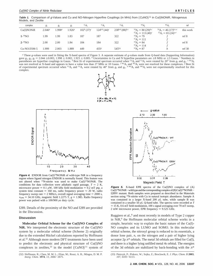
Because EPR is most effective at resolving relatively largehyperfine couplings, we used ENDOR to resolve weakernitrogen couplings. As an example of such weak nitrogencouplings, we have reported weak couplings with magnitude∼6 MHz from heme nitrogens of the NO complex of heme.20
We believed that there was the potential, given the likelynitrogenous ligands of the Cu, to find such weak couplings tonitrogen ligands of the copper, most likely to histidine nitrogen.The ENDOR feature observed (Figure 4) had a frequency of7.5 MHz and was not altered when NiR was reduced in thepresence of14N versus 15N nitrite. Its frequency stayed atapproximately the same frequency across the EPR line of Cu(I)-NONiR. In the absence of deuterium, there are no naturallyoccurring nuclei other than14N-nitrogen which will providehyperfine features in this frequency region. We assign the featurein Figure 4 as the nitrogen hyperfine coupling of a nitrogenousligand to the Cu(I). The most obvious nitrogenous candidatesare one or more of the histidine ligands of the Cu.
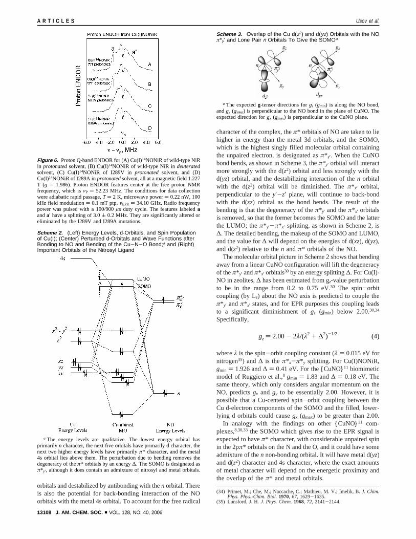
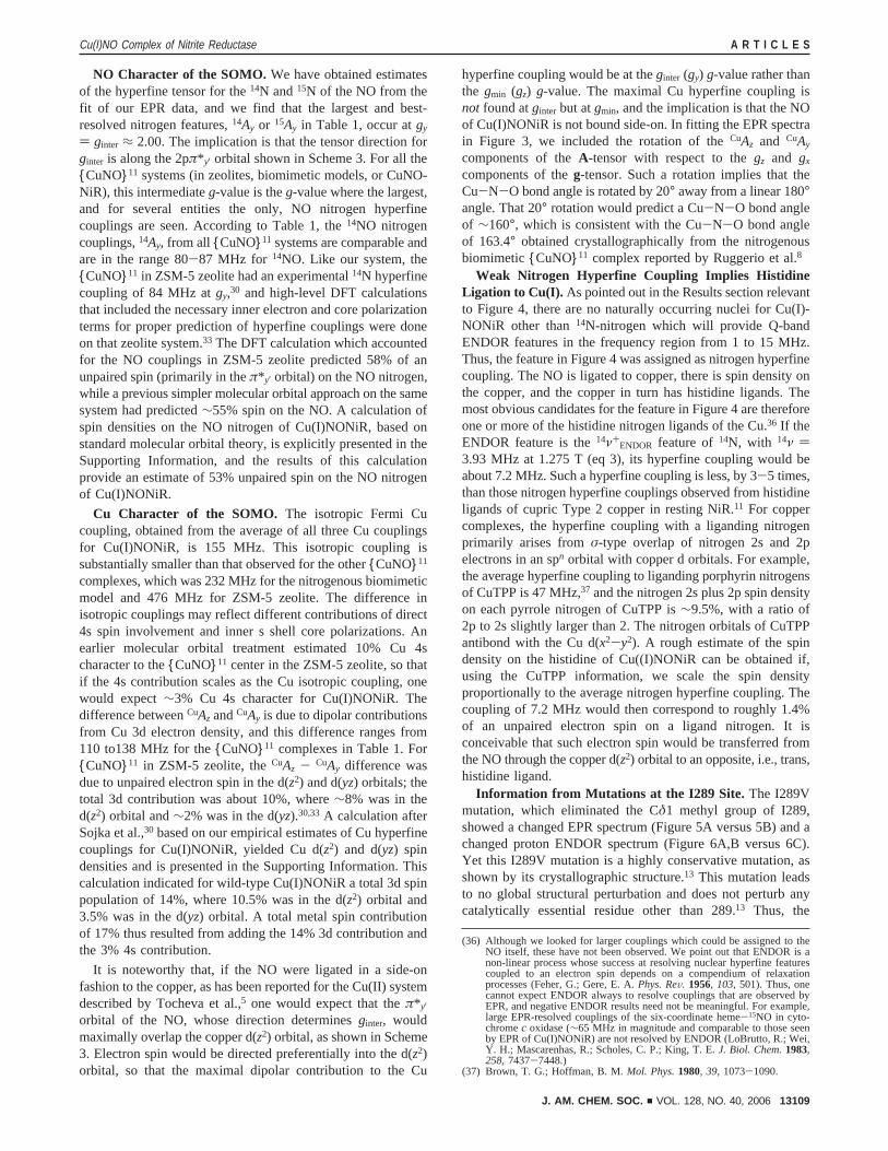
Perturbations from Mutations at the I289 Site.The I289Vmutation, although not at a copper ligand, somewhat alteredthe electronic structure of the Cu(I)NONiR complex, as shownbythecomparisonoftheEPRspectrainFigure5A(Cu(I)14NONiR-wild type) and 5B (Cu(I)14NONiR-I289V). The EPR spectrumof the I289A derivative, provided in Supporting InformationFigure 3S, was weak and grossly distorted from the spectra ofthe Cu(I)NONiR derivative of wild-type NiR or I289V andshowed little evidence for copper or nitrogen hyperfine coupling.Local structural information related to mutation at the I289 siteis provided by ENDOR, which identifies a proton-related,mutation-induced change in the environment near the Cu(I)NOcenter. Figure 6 compares proton ENDOR spectra of theCu(I)14NONiR-wild type complex in protonated (6A) ordeuterated solvent (6B) with the spectra of the protonatedCu(I)14NONiR-I289V (6C) and the protonated Cu(I)14NONiR-I289A (6D). The non-exchangeable proton features,a anda′,with hyperfine coupling of 3.0( 0.2 MHz, were significantlyaltered by the I289V and I289A mutations. The proton ENDORspectrum of I289A had approximately the shape of the spectrumof I289V, with none of the hyperfine details (featuresa anda′)exhibited by wild-type protein. Because the I289V structuralchange to the overall protein is an extremely localized perturba-tion13 occurring 4-5 Å from the Type 2 copper, a straightfor-ward implication of these EPR and ENDOR findings is thatthe paramagnetic center associated with Cu(I)NONiR is near
(30) Sojka, Z.; Che, M.; Giamello, E.J. Phys. Chem. B1997, 101, 4831-4838.(31) Umamaheswari, V.; Hartmann, M.; Poeppl, A.J. Phys. Chem. B2005,
109, 10842-10848.
Figure 3. Comparison of the experimental X-band absorption firstderivative signals (solid line) to simulations (dotted line) for (A)63Cu-(I)14NONiR and (B)63Cu(I)15NONiR. The experimental spectra are thoseof Figure 2A,B. The spectra were computed using the program SIMPIPM.26
The following parameters (hyperfine couplings in Gauss) were used forsimulation: gx ) 2.046,gy ) 1.998,gz ) 1.926,CuAx ) 37, CuAy ) 44,CuAz ) 88, 14NAx ) 17, 14NAy ) 29, 15NAx ) 24, 15NAy ) 40, 14Az and15Az
< 10 G. The principal directions forCuAy andCuAz are respectively rotatedby 20° in the y-z plane away from the principal directions forgy andgz.Gaussian line widths (in Gauss) between derivative extrema areWx ) 30,Wy ) 13, andWz ) 52.
A R T I C L E S Usov et al.
13106 J. AM. CHEM. SOC. 9 VOL. 128, NO. 40, 2006
I289. Details of the proximity of the NO and I289 are providedin the Discussion.
Discussion
Molecular Orbital Scheme for the Cu(I)NO Complex ofNiR. We interpreted the electronic structure of the Cu(I)NOsystem by a molecular orbital scheme (Scheme 2) originallydue to the extended Hu¨ckel calculations reported by Hoffmannet al.32 Although more modern DFT treatments have been usedto predict the electronic and physical structure of Cu(I)NOcomplexes in zeolites,33 in the model{CuNO}11 system of
Ruggiero et al.,8 and most recently in models of Type 2 copperin NiR,9 the Hoffmann molecular orbital scheme works in asimple, heuristic way to explain the basic nature of the Cu(I)-NO complex and its LUMO and SOMO. In this molecularorbital scheme, the nitrosyl group is reduced to its essentials, adonor lone pair,n, on the nitrogen and a pair of higher lyingacceptor 2pπ* orbitals. The metal 3d orbitals are filled for Cu(I),and there is a higher lying unfilled metal 4s orbital. The energiesof the 3d orbitals are stabilized by back-bonding with theπ*
(32) Hoffmann, R.; Chen, M. M. L.; Elian, M.; Rossi, A. R.; Mingos, D. M. P.Inorg. Chem.1974, 13, 2666-2675.
(33) Pietrzyk, P.; Piskorz, W.; Sojka, Z.; Broclawik, E.J. Phys. Chem. B2003,107, 6105-6113.
Table 1. Comparison of g-Values and Cu and NO-Nitrogen Hyperfine Couplings (in MHz) from {CuNO}11 in Cu(I)NONiR, NitrogenousModels, and Zeolite
complex gx gy gzCuAx
CuAyCuAz
NOAyNOAx ref
Cu(I)NONiR 2.046a 1.998a 1.926a 102b (37)c 124b,d (44)c 238b,d (88)b,c 14Ay ) 80 (29)b,c 14Ax ) 46 (17)b,c,e this work15Ay ) 113 (40)c 15Ax ) 65 (24)c,e
1-14NO 1.99 1.99 1.83 187 187 322 14Ay ) 79 f ref 815Ay ) 111 f
2-14NO 2.00 2.00 1.84 184 184 322 14Ay ) 84 f ref 815Ay ) 114 f
Cu-NO/ZSM-5 1.999 2.003 1.889 449 435g 545f,g 14Ay ) 87 h ref 30
a Theseg-values were used in fitting the X-band spectra of Figure 3. A separate estimate ofg-values made from Q-band data (Supporting Information)gavegx, gy, gz ) 2.044(0.003, 1.998( 0.002, 1.923( 0.005.b Uncertainties in Cu and N hyperfine parameters are(8 MHz or (3 Gauss.c Numbers inparentheses are hyperfine couplings in Gauss.d Best fit of experimental spectrum occurred whenCuAz andCuAy were rotated by 20° from gz andgy. e NOAzwas not resolved at X-band and appears to have a value less than 27 MHz or 10 Gauss.f NOAx andNOAz were not resolved for these complexes.g Best fitof experimental spectrum occurred whenCuAz and CuAy were rotated by 40° from gz and gy. h NOAx and NOAz were not experimentally resolved for thiscomplex.
Figure 4. ENDOR from Cu(I)14NONiR of wild-type NiR in a frequencyregion where ligand nitrogen ENDOR is normally found. This feature wasnot altered when15N-nitrite was used to make Cu(I)15NONiR. Theconditions for data collection were adiabatic rapid passage,T ) 2 K,microwave power) 0.1 µW, 100 kHz field modulation) 0.2 mT ptp, asystem time constant) 160 ms, radio frequency power≈ 20 W, radiofrequency sweep rate) 2 MHz/s, overall signal averaging time) 2000 s,νEPR ) 34.10 GHz, magnetic field 1.2275 T,g ) 1.985. Radio frequencypower was pulsed with a 100/900µs duty cycle.
Figure 5. X-band EPR spectra of the Cu(I)NO complex of (A)Cu(I)14NONiR-wildtypeandthecorrespondingcomplexof(B)Cu(I)14NONiR-I289V mutant. Both samples were prepared as described in the Materialssection using14N-nitrite with Cu in natural isotopic abundance. Sample Awas contained in a larger X-band 200µL tube, while sample B wascontained in a smaller 50µL Q-band tube. The spectra were recorded atT) 15 K, 0.6 mT field modulation, 100 s signal averaging over 70 mT sweep,2 mW microwave power, EPR frequency) 9.525 GHz.
Cu(I)NO Complex of Nitrite Reductase A R T I C L E S
J. AM. CHEM. SOC. 9 VOL. 128, NO. 40, 2006 13107
orbitals and destabilized by antibonding with then orbital. Thereis also the potential for back-bonding interaction of the NOorbitals with the metal 4s orbital. To account for the free radical
character of the complex, theπ* orbitals of NO are taken to liehigher in energy than the metal 3d orbitals, and the SOMO,which is the highest singly filled molecular orbital containingthe unpaired electron, is designated asπ* y′. When the CuNObond bends, as shown in Scheme 3, theπ* y′ orbital will interactmore strongly with the d(z2) orbital and less strongly with thed(yz) orbital, and the destabilizing interaction of then orbitalwith the d(z2) orbital will be diminished. Theπ* x′ orbital,perpendicular to they′-z′ plane, will continue to back-bondwith the d(xz) orbital as the bond bends. The result of thebending is that the degeneracy of theπ* y′ and theπ* x′ orbitalsis removed, so that the former becomes the SOMO and the latterthe LUMO; theπ* y′-π* x′ splitting, as shown in Scheme 2, is∆. The detailed bending, the makeup of the SOMO and LUMO,and the value for∆ will depend on the energies of d(xz), d(yz),and d(z2) relative to then andπ* orbitals of the NO.
The molecular orbital picture in Scheme 2 shows that bendingaway from a linear CuNO configuration will lift the degeneracyof theπ*y′ andπ*x′ orbitals30 by an energy splitting∆. For Cu(I)-NO in zeolites,∆ has been estimated from gz-value perturbationto be in the range from 0.2 to 0.75 eV.30 The spin-orbitcoupling (by Lz) about the NO axis is predicted to couple theπ* y′ andπ* x′ states, and for EPR purposes this coupling leadsto a significant diminishment ofgz (gmin) below 2.00.30,34
Specifically,
whereλ is the spin-orbit coupling constant (λ ) 0.015 eV fornitrogen35) and∆ is theπ* x-π* y splitting. For Cu(I)NONiR,gmin ) 1.926 and∆ ) 0.41 eV. For the{CuNO}11 biomimeticmodel of Ruggiero et al.,8 gmin ) 1.83 and∆ ) 0.18 eV. Thesame theory, which only considers angular momentum on theNO, predictsgx and gy to be essentially 2.00. However, it ispossible that a Cu-centered spin-orbit coupling between theCu d-electron components of the SOMO and the filled, lower-lying d orbitals could causegx (gmax) to be greater than 2.00.
In analogy with the findings on other{CuNO}11 com-plexes,8,30,33 the SOMO which gives rise to the EPR signal isexpected to haveπ* character, with considerable unpaired spinin the 2pπ* orbitals on the N and the O, and it could have someadmixture of then non-bonding orbital. It will have metal d(yz)and d(z2) character and 4s character, where the exact amountsof metal character will depend on the energetic proximity andthe overlap of theπ* and metal orbitals.
(34) Primet, M.; Che, M.; Naccache, C.; Mathieu, M. V.; Imelik, B.J. Chim.Phys. Phys.-Chim. Biol.1970, 67, 1629-1635.
(35) Lunsford, J. H.J. Phys. Chem.1968, 72, 2141-2144.
Figure 6. Proton Q-band ENDOR for (A) Cu(I)14NONiR of wild-type NiRin protonatedsolvent, (B) Cu(I)14NONiR of wild-type NiR in deuteratedsolvent, (C) Cu(I)14NONiR of I289V in protonated solvent, and (D)Cu(I)14NONiR of I289A inprotonatedsolvent, all at a magnetic field 1.227T (g ) 1.986). Proton ENDOR features center at the free proton NMRfrequency, which isνP ) 52.23 MHz. The conditions for data collectionwere adiabatic rapid passage,T ) 2 K, microwave power) 0.22 nW, 100kHz field modulation) 0.1 mT ptp,νEPR ) 34.10 GHz. Radio frequencypower was pulsed with a 100/900µs duty cycle. The features labeledaanda′ have a splitting of 3.0( 0.2 MHz. They are significantly altered oreliminated by the I289V and I289A mutations.
Scheme 2. (Left) Energy Levels, d-Orbitals, and Spin Populationof Cu(I); (Center) Perturbed d-Orbitals and Wave Functions afterBonding to NO and Bending of the Cu-N-O Bond;a and (Right)Important Orbitals of the Nitrosyl Ligand
a The energy levels are qualitative. The lowest energy orbital hasprimarily n character, the next five orbitals have primarily d character, thenext two higher energy levels have primarilyπ* character, and the metal4s orbital lies above them. The perturbation due to bending removes thedegeneracy of theπ* orbitals by an energy∆. The SOMO is designated asπ* y′, although it does contain an admixture of nitrosyl and metal orbitals.
Scheme 3. Overlap of the Cu d(z2) and d(yz) Orbitals with the NOπ*y′ and Lone Pair n Orbitals To Give the SOMOa
a The expectedg-tensor directions forgz (gmin) is along the NO bond,andgy (ginter) is perpendicular to the NO bond in the plane of CuNO. Theexpected direction forgx (gmax) is perpendicular to the CuNO plane.
gz ) 2.00- 2λ/(λ2 + ∆2)-1/2 (4)
A R T I C L E S Usov et al.
13108 J. AM. CHEM. SOC. 9 VOL. 128, NO. 40, 2006
NO Character of the SOMO. We have obtained estimatesof the hyperfine tensor for the14N and15N of the NO from thefit of our EPR data, and we find that the largest and best-resolved nitrogen features,14Ay or 15Ay in Table 1, occur atgy
) ginter ≈ 2.00. The implication is that the tensor direction forginter is along the 2pπ* y′ orbital shown in Scheme 3. For all the{CuNO}11 systems (in zeolites, biomimetic models, or CuNO-NiR), this intermediateg-value is theg-value where the largest,and for several entities the only, NO nitrogen hyperfinecouplings are seen. According to Table 1, the14NO nitrogencouplings,14Ay, from all {CuNO}11 systems are comparable andare in the range 80-87 MHz for 14NO. Like our system, the{CuNO}11 in ZSM-5 zeolite had an experimental14N hyperfinecoupling of 84 MHz atgy,30 and high-level DFT calculationsthat included the necessary inner electron and core polarizationterms for proper prediction of hyperfine couplings were doneon that zeolite system.33 The DFT calculation which accountedfor the NO couplings in ZSM-5 zeolite predicted 58% of anunpaired spin (primarily in theπ* y′ orbital) on the NO nitrogen,while a previous simpler molecular orbital approach on the samesystem had predicted∼55% spin on the NO. A calculation ofspin densities on the NO nitrogen of Cu(I)NONiR, based onstandard molecular orbital theory, is explicitly presented in theSupporting Information, and the results of this calculationprovide an estimate of 53% unpaired spin on the NO nitrogenof Cu(I)NONiR.
Cu Character of the SOMO. The isotropic Fermi Cucoupling, obtained from the average of all three Cu couplingsfor Cu(I)NONiR, is 155 MHz. This isotropic coupling issubstantially smaller than that observed for the other{CuNO}11
complexes, which was 232 MHz for the nitrogenous biomimeticmodel and 476 MHz for ZSM-5 zeolite. The difference inisotropic couplings may reflect different contributions of direct4s spin involvement and inner s shell core polarizations. Anearlier molecular orbital treatment estimated 10% Cu 4scharacter to the{CuNO}11 center in the ZSM-5 zeolite, so thatif the 4s contribution scales as the Cu isotropic coupling, onewould expect∼3% Cu 4s character for Cu(I)NONiR. Thedifference betweenCuAz andCuAy is due to dipolar contributionsfrom Cu 3d electron density, and this difference ranges from110 to138 MHz for the{CuNO}11 complexes in Table 1. For{CuNO}11 in ZSM-5 zeolite, theCuAz - CuAy difference wasdue to unpaired electron spin in the d(z2) and d(yz) orbitals; thetotal 3d contribution was about 10%, where∼8% was in thed(z2) orbital and∼2% was in the d(yz).30,33 A calculation afterSojka et al.,30 based on our empirical estimates of Cu hyperfinecouplings for Cu(I)NONiR, yielded Cu d(z2) and d(yz) spindensities and is presented in the Supporting Information. Thiscalculation indicated for wild-type Cu(I)NONiR a total 3d spinpopulation of 14%, where 10.5% was in the d(z2) orbital and3.5% was in the d(yz) orbital. A total metal spin contributionof 17% thus resulted from adding the 14% 3d contribution andthe 3% 4s contribution.
It is noteworthy that, if the NO were ligated in a side-onfashion to the copper, as has been reported for the Cu(II) systemdescribed by Tocheva et al.,5 one would expect that theπ* y′orbital of the NO, whose direction determinesginter, wouldmaximally overlap the copper d(z2) orbital, as shown in Scheme3. Electron spin would be directed preferentially into the d(z2)orbital, so that the maximal dipolar contribution to the Cu
hyperfine coupling would be at theginter (gy) g-value rather thanthe gmin (gz) g-value. The maximal Cu hyperfine coupling isnot found atginter but atgmin, and the implication is that the NOof Cu(I)NONiR is not bound side-on. In fitting the EPR spectrain Figure 3, we included the rotation of theCuAz and CuAy
components of theA-tensor with respect to thegz and gx
components of theg-tensor. Such a rotation implies that theCu-N-O bond angle is rotated by 20° away from a linear 180°angle. That 20° rotation would predict a Cu-N-O bond angleof ∼160°, which is consistent with the Cu-N-O bond angleof 163.4° obtained crystallographically from the nitrogenousbiomimetic{CuNO}11 complex reported by Ruggerio et al.8
Weak Nitrogen Hyperfine Coupling Implies HistidineLigation to Cu(I). As pointed out in the Results section relevantto Figure 4, there are no naturally occurring nuclei for Cu(I)-NONiR other than14N-nitrogen which will provide Q-bandENDOR features in the frequency region from 1 to 15 MHz.Thus, the feature in Figure 4 was assigned as nitrogen hyperfinecoupling. The NO is ligated to copper, there is spin density onthe copper, and the copper in turn has histidine ligands. Themost obvious candidates for the feature in Figure 4 are thereforeone or more of the histidine nitrogen ligands of the Cu.36 If theENDOR feature is the14ν+
ENDOR feature of14N, with 14ν )3.93 MHz at 1.275 T (eq 3), its hyperfine coupling would beabout 7.2 MHz. Such a hyperfine coupling is less, by 3-5 times,than those nitrogen hyperfine couplings observed from histidineligands of cupric Type 2 copper in resting NiR.11 For coppercomplexes, the hyperfine coupling with a liganding nitrogenprimarily arises fromσ-type overlap of nitrogen 2s and 2pelectrons in an spn orbital with copper d orbitals. For example,the average hyperfine coupling to liganding porphyrin nitrogensof CuTPP is 47 MHz,37 and the nitrogen 2s plus 2p spin densityon each pyrrole nitrogen of CuTPP is∼9.5%, with a ratio of2p to 2s slightly larger than 2. The nitrogen orbitals of CuTPPantibond with the Cu d(x2-y2). A rough estimate of the spindensity on the histidine of Cu((I)NONiR can be obtained if,using the CuTPP information, we scale the spin densityproportionally to the average nitrogen hyperfine coupling. Thecoupling of 7.2 MHz would then correspond to roughly 1.4%of an unpaired electron spin on a ligand nitrogen. It isconceivable that such electron spin would be transferred fromthe NO through the copper d(z2) orbital to an opposite, i.e., trans,histidine ligand.
Information from Mutations at the I289 Site. The I289Vmutation, which eliminated the Cδ1 methyl group of I289,showed a changed EPR spectrum (Figure 5A versus 5B) and achanged proton ENDOR spectrum (Figure 6A,B versus 6C).Yet this I289V mutation is a highly conservative mutation, asshown by its crystallographic structure.13 This mutation leadsto no global structural perturbation and does not perturb anycatalytically essential residue other than 289.13 Thus, the
(36) Although we looked for larger couplings which could be assigned to theNO itself, these have not been observed. We point out that ENDOR is anon-linear process whose success at resolving nuclear hyperfine featurescoupled to an electron spin depends on a compendium of relaxationprocesses (Feher, G.; Gere, E. A.Phys. ReV. 1956, 103, 501). Thus, onecannot expect ENDOR always to resolve couplings that are observed byEPR, and negative ENDOR results need not be meaningful. For example,large EPR-resolved couplings of the six-coordinate heme-15NO in cyto-chromec oxidase (∼65 MHz in magnitude and comparable to those seenby EPR of Cu(I)NONiR) are not resolved by ENDOR (LoBrutto, R.; Wei,Y. H.; Mascarenhas, R.; Scholes, C. P.; King, T. E.J. Biol. Chem.1983,258, 7437-7448.)
(37) Brown, T. G.; Hoffman, B. M.Mol. Phys.1980, 39, 1073-1090.
Cu(I)NO Complex of Nitrite Reductase A R T I C L E S
J. AM. CHEM. SOC. 9 VOL. 128, NO. 40, 2006 13109
spectroscopic changes we observed for I289V reflect a localizedstructural change rather than a delocalized distortion that mightextend, for example, to Cu-His ligands. The EPR resultindicates that the Cδ1 methyl group influences the electronicstructure of Cu(I)NONiR, as evidenced byg-values and hyper-fine structure. The proton ENDOR features denoted bya anda′ in Figure 6 A,B occur for wild-type protein but aresignificantly altered or eliminated for the I289V and I289Amutants. The most direct assignment for these features and theirbehavior is the Cδ1 methyl protons of I289, which are closeenough to the NO paramagnetic center to cause the∼3.0 MHzproton coupling of featuresa and a′. For the mutants, thiscoupling is eliminated along with the Cδ1 methyl.
The 3.0 MHz coupling ofa anda′ is associated with spin-proton coupling by using the dipolar formula (eq 2). Here, thespin density in the delocalized Cu(I)NONiR wave function willbe greatest at the nitric oxide N but delocalized over N, O, andCu.38 The dipolar formula includes the angleθ between theapplied magnetic field and the vector from the proton to theelectron spin; ifθ ) 90°, a coupling of 3.0 MHz implies a spin-proton distance of 3.0 Å, and ifθ ) 0°, it implies a spin-proton distance of 3.7 Å. In previously reported structures, thedistance from the axial ligand of Type 2 Cu in NiR to Cδ1methyl and Cγ1 methylene protons of I289 averaged 3.6 Å,with the closest protons 2.8-2.9 Å away. (See details of distancecomputation in footnote 39.) The average distance from the axialligand to the Cγ2 methyl protons is 4.0 Å, with the closest Cγ2proton at 3.2 Å. The Câ proton is 5.3 Å distant. The 3.0-3.7Å distances implied by ENDOR would thus be consistent withthose distances estimated between an axial ligand of Type 2Cu and the Cδ1 methyl, the Cγ1 methylene, or perhaps oneCγ2 proton of I289. The proton ENDOR results are notconsistent with a dipolar coupling between spin localized,instead, on the more distant copper and the protons on I289,which would imply a coupling<1.5 MHz to the I289 protons.
The similarity of the ENDOR line shapes of I289V (Figure6C) and I289A (Figure 6D) implies that the Cγ protons of I289Vhave little effect on the proton ENDOR line shape because thatline shape persists when Val is replaced by Ala. If the Cγprotons of I289 and V289 have the same location, additionalevidence is thereby provided that the Cδ1 methyl protons ofI289, rather than the Cγ protons of I289, will account for thefeaturesa anda′ in Figure 6A,B. The conclusion from EPR isthat non-covalent interaction with the bulky I289 side chain isimportant in establishing the electronic structure of the Cu(I)-NONiR. The conclusion from ENDOR is that the I289 Cδ1methyl proton hyperfine couplings assigned to featuresa anda′ serve to locate the NO, which carries the electron spin. Theylocate the NO as an axial ligand to Type 2 copper in the spacebetween the Cu and the Cδ1 methyl protons of I289.
Conclusions
Copper-containing nitrite reductase endogenously producesits own NO from nitrite under reducing conditions, and thecomparison between the EPR signals that were respectivelyproduced from14N- versus15N-nitrite (spectrum 2A versus 2B)provided evidence that endogenously produced NO is a majorcomponent of the paramagnetic signal of Cu(I)NONiR. Themarked similarity of the signals (spectrum 1A versus 1C) fromendogenously produced14NO and from exogenous14NO gasadded anaerobically in a limited amount was additional evidencethat NO is a component of the paramagnetic signal of Cu(I)-NONiR. Comparison of the well-resolved nitrogen coupling nearg ) 2.00 to those of other{CuNO}11 systems on which DFTtreatments have been performed indicates 50-55% unpairedspin of Cu(I)NONiR exists in the 2p orbitals of the NO nitrogen.The evidence for copper in Cu(I)NONiR is directly shown bythe change in its EPR spectrum (spectrum 2C) when the NiRwas prepared with isotopically enriched63Cu, as opposed toCu in natural abundance. There is a similarity ofg-values fromCu(I)NONiR, notably the uniquegz < 2.00, tog-values fromNO trapped in matrices or absorbed on surfaces,34,35,40but thecopper hyperfine structure associated withgz is another indicatorthat the complex we have designated Cu(I)NONiR does containcopper. Using theory developed by Sojka et al.,30 we haveestimated the total unpaired spin on the copper d and 4s orbitalsto be∼17%. Assuming that the contributions of the Cu, N, andO sum to unity, this would mean that the oxygen of the NOshould have∼30% unpaired spin. There is, in addition, ENDORevidence that Cu(I)NONiR has at least one nitrogenous ligand,most likely histidine, with∼1% unpaired spin. The perturbationto the EPR spectrum (Figure 5) and to the proton ENDORspectrum (Figure 6) from the I289V and I289A mutations, whicheliminate the Cδ1 methyl, is a sign that the paramagnetic NOcenter associated with Cu(I)NONiR is near I289. The protonhyperfine coupling indicated that the electron spin whichinteracts with the protons of I289 is centered 3.0-3.7 Å awayfrom the bulky Cδ1 and Cγ side chains of I289. This distanceis consistent with the electron spin primarily being on the axialNO ligand to Type 2 copper, while the NO abuts the bulkyI289 side chains, most likely the Cδ1 methyl group. In short,the system which we report here and spectroscopically char-acterize is a Cu(I)NO system localized on NO but situatedsufficiently near to the original Type 2 copper that it covalentlyinteracts with that copper. Additionally, the NO experiences anon-covalent perturbation by the bulky side chain of nearbyI289.
Acknowledgment. These studies were partially supportedby the NIH (EB00326929, C.P.S.) and the DOE (95ER2-0206, J.P.S.). We are grateful to Dr. William Antholine forproviding isotopically enriched63Cu and for providing theoriginal spectra of the model Cu(I)NO complexes reportedby Ruggiero et al.8 and preliminary S-band spectra of Cu(I)-NONiR.
Supporting Information Available: Figure 1S, the Q-bandfirst derivative EPR spectrum of Cu(I)14NONiR; Figure 2S,comparing the X-band spectra of Cu(I)14NONiR (2SA) andCu(I)15NONiR (2SB) with the corresponding14NO and15NO
(38) This necessarily imprecise location of the spin does not interfere in ourultimately locating the position of the spin on the NO within 3.0-3.7 Å ofthe Cδ1 and Cγ1 protons of I289.
(39) Since there are no crystal structures of Cu(I)NONiR, we have used thewater oxygen of the NiR aquo derivative (ref 3) or the N of the recentCu(II)NO derivative (ref 5) to estimate distances to protons of I289 fromthe axial ligand positioned on Type 2 copper. The latter Cu(II)NO derivativedescribed by Tocheva et al.5 is not our derivative, but at least it providesa reference point for estimating distances from N of NO to nearby I289protons. Protons were placed on isoleucine carbons with the rectificationtool of ChemPro because protons are not resolved by X-ray crystallography. (40) Lunsford, J. H.J. Catal.1969, 14, 379-385.
A R T I C L E S Usov et al.
13110 J. AM. CHEM. SOC. 9 VOL. 128, NO. 40, 2006
derivatives of the Cu(I)NO model compounds described byRuggiero et al.;8 Figure 3S, comparing the EPR spectra fromCu(I)NONiR prepared from I289V and I289A; a calculation ofspin densities on the NO nitrogen of Cu(I)NONiR based onstandard molecular orbital theory; and a calculation of Cu d(z2)
and d(yz) orbital coefficients via methods of Sojka et al.30 Thismaterial is available free of charge via the Internet athttp://pubs.acs.org.
JA056166N
Cu(I)NO Complex of Nitrite Reductase A R T I C L E S
J. AM. CHEM. SOC. 9 VOL. 128, NO. 40, 2006 13111