Epidemiology of Cardiovascular disease and associated risk ...
215
HAL Id: tel-02560078 https://tel.archives-ouvertes.fr/tel-02560078 Submitted on 1 May 2020 HAL is a multi-disciplinary open access archive for the deposit and dissemination of sci- entific research documents, whether they are pub- lished or not. The documents may come from teaching and research institutions in France or abroad, or from public or private research centers. L’archive ouverte pluridisciplinaire HAL, est destinée au dépôt et à la diffusion de documents scientifiques de niveau recherche, publiés ou non, émanant des établissements d’enseignement et de recherche français ou étrangers, des laboratoires publics ou privés. Epidemiology of Cardiovascular disease and associated risk factors in Gaza Strip- Palestine Amal Jamee Shahwan To cite this version: Amal Jamee Shahwan. Epidemiology of Cardiovascular disease and associated risk factors in Gaza Strip- Palestine. Human health and pathology. Université de Limoges, 2019. English. NNT : 2019LIMO0011. tel-02560078
-
Upload
khangminh22 -
Category
Documents
-
view
1 -
download
0
Transcript of Epidemiology of Cardiovascular disease and associated risk ...

HAL Id: tel-02560078https://tel.archives-ouvertes.fr/tel-02560078
Submitted on 1 May 2020
HAL is a multi-disciplinary open accessarchive for the deposit and dissemination of sci-entific research documents, whether they are pub-lished or not. The documents may come fromteaching and research institutions in France orabroad, or from public or private research centers.
L’archive ouverte pluridisciplinaire HAL, estdestinée au dépôt et à la diffusion de documentsscientifiques de niveau recherche, publiés ou non,émanant des établissements d’enseignement et derecherche français ou étrangers, des laboratoirespublics ou privés.
Epidemiology of Cardiovascular disease and associatedrisk factors in Gaza Strip- Palestine
Amal Jamee Shahwan
To cite this version:Amal Jamee Shahwan. Epidemiology of Cardiovascular disease and associated risk factors in GazaStrip- Palestine. Human health and pathology. Université de Limoges, 2019. English. �NNT :2019LIMO0011�. �tel-02560078�

Amal Jamee Shahwan | Ph.D. Thesis | University of Limoges | 2019 License CC BY-NC-ND 4.0
1
.University of Limoges
ED 615 - Sciences Biologiques et Santé (SBS)
Neuroépidémiologie tropicale
A thesis submitted to University of Limoges In fulfilment of the requirements of the degree of Doctor of Science Public Heath / Epidemiology
Presented and defensed by
Amal Jamee Shahwan
On 30 avril 2019
Thesis supervisor: Professor Philippe LACROIX
Professor Yehia ABED
JURY:
President of jury
Pr. Habiba Ben Romdhane, Tunis University
Reporters Pr Vanina BONGARD, Toulouse University, France Pr Nadine SALEH, Lebanese University, Lebanon
Examiners
Pr. Philippe LACROIX, Limoges University, France
Pr. Yehia ABED, Al Quds University-Gaza, Palestine
Pr. Victor ABOYANS, Limoges University, France
Pr. Nadine SALEH, Lebanese University, Lebanon
Pr. Vanina BONGARD, Toulouse University, France
Epidemiology of Cardiovascular disease and associated risk
factors in Gaza Strip- Palestine
Ph.D. Thesis

Amal Jamee Shahwan | Ph.D. Thesis | University of Limoges | 2019 License CC BY-NC-ND 4.0
2
Au nom d'Allah le tout miséricordieux, le très miséricordieux.
Dieu dit dans son livre saint
Ceux qui disent : « Notre Seigneur est Allah » et qui ensuite se tiennent sur le droit chemin. Ils ne doivent avoir aucune crainte et ne seront point affligés
Sourate AL-AHQAF (verset 13)
à Amal, en hommage à son labeur !
à chaque être humain libre je dédie ce travail de recherche

Amal Jamee Shahwan | Ph.D. Thesis | University of Limoges | 2019 License CC BY-NC-ND 4.0
3
The fountain of hope never ends

Amal Jamee Shahwan | Ph.D. Thesis | University of Limoges | 2019 License CC BY-NC-ND 4.0
4
Acknowledgements
Il me serait très difficile de remercier tout le monde car c’est grâce à de nombreuses personnes
que je suis là et que j’ai pu mener cette thèse à son terme.
Je voudrais tout d’abord remercier profondément mon directeur de thèse le Professeur
Philippe Lacroix, pour toute son aide. Je suis ravie d’avoir travaillé avec lui, grand merci pour
son soutien et ses conseils au cours de la réalisation de ma thèse.
Je souhaite également rendre hommage au Professeur Marc Laskar qui m’a mis en contact
avec Monsieur Lacroix lors de notre rencontre à Tunis.
Je tiens également à remercier le Professeur Yehia Abed de Gaza qui a toujours été présent,
en particulier lors du travail sur le terrain.
J’adresse de sincères remerciements au Professeur Victor Aboyans pour son soutien et sa
patience dans la réponse à mes questions, j’ai beaucoup appris de lui sur la rédaction des
articles aussi d’avoir accepté d’être dans mon jury de thèse, grand merci.
Je remercie chaleureusement le Professeur Pierre-Marie Preux, directeur de UMR-S 1094
NET pour sa grande gentillesse et son soutien moral.
Je tiens à remercie le Dr julien, on a beaucoup travaillé ensemble sur l’analyse statistique et
j’ai bien appris de lui.
Je souhaite particulièrement remercie le Dr Farid Boumediene et Dr Daniel Ajzenberg pour
leur précieuse relecture de la présentation et leurs précieux conseils
J’exprime tous mes remerciements à l’ensemble des membres du jury :
Madame la Professeure Habiba Ben Rhomdhane pour l’honneur qu’elle me fait d’être dans
mon jury de thèse et pour son expérience dans le domaine de la recherche cardiovasculaire,
j’espère que mon travail est à la hauteur de ses exigences.
Je remercie infiniment Madame la Professeure Nadine Salah et Madame la Professeure
Vanina Bongard qui m’ont fait l’honneur d’être membres du jury et rapporteurs de ma thèse et
dont les remarques m’ont permis d’examiner et d’élargir mon regard sur le travail que j’ai
accompli.
Mes remerciements à tous les membres de l’équipe UMR-S 1094 ED 615 de Limoges avec
qui j’ai partagé mes études et d’agréables moments.
Je remercie aussi celles et ceux qui me sont chers et que j’ai été obligée de quitter pendant
des mois et des mois, pour achever ce travail de recherche. Leur attention, encouragements,
confiance et soutien m’ont toujours accompagnée, je suis très redevable à mon cher mari

Amal Jamee Shahwan | Ph.D. Thesis | University of Limoges | 2019 License CC BY-NC-ND 4.0
5
Ahmad, mes très chers enfants Asaad et Shaden, ma famille et mes amis proches de
Palestine, d’Algérie et de France. Enfin, une pensée particulière pour mon père décédé lors
de mon dernier séjour à Limoges.
Mes remerciements vont également à l’équipe qui m’a accompagné pendant mon travail sur
le terrain, on a passé de bon moment ensemble.
Je tiens à remercie les chefs de famille pour leur hospitalité dont ils ont fait preuve envers moi
et la bonne ambiance que j’ai trouvée.
Je remercie celles et ceux qui se sont déplacés pour assister à ma soutenance.
Je souhaite exprimer mes remerciements et sans les nommer individuellement a tous ceux
qui d’une manière ou d’une autre ont su être présents quand j’en ai eu besoin.

Amal Jamee Shahwan | Ph.D. Thesis | University of Limoges | 2019 License CC BY-NC-ND 4.0
6
Rights
This creation is available under a Creative Commons contract :
« Attribution-NonCommercial-NoDerivatives 4.0 International »
online at : http://creativecommons.org/licenses/by-nc-nd/4.0/

Amal Jamee Shahwan | Ph.D. Thesis | University of Limoges | 2019 License CC BY-NC-ND 4.0
7
Table of Contents
Acknowledgements .................................................................................................................. 4
Rights ....................................................................................................................................... 6
Table of Contents ..................................................................................................................... 7
List of Figures ......................................................................................................................... 11
List of Tables .......................................................................................................................... 12
Abbreviations ......................................................................................................................... 13
Introduction ............................................................................................................................ 18
Chapter I. Literature review .................................................................................................... 21
I.1. Cardiovascular health definition ................................................................................... 21
I.2. Cardiovascular disease definitions ............................................................................... 21
I.3. Types of cardiovascular disease .................................................................................. 22
I.3.1. Coronary artery disease ......................................................................................... 22
I.3.1.1. Definition and classification ............................................................................. 22
I.3.1.2. Physiopathology .............................................................................................. 22
I.3.1.3. Acute coronary syndrome ............................................................................... 24
I.3.1.3.1. Clinical presentation of Acute coronary syndrome ................................... 24
I.3.1.3.1.1. Myocardial infraction .......................................................................... 24
I.3.1.3.2. Physical examination findings ................................................................... 25
I.3.1.3.3. Electrocardiograms ................................................................................... 26
I.3.1.3.4. Serum cardiac markers ............................................................................. 26
I.3.1.4. Stable coronary artery disease ........................................................................ 26
I.3.1.4.1. Definition, classification ............................................................................ 26
I.3.1.4.2. Clinical presentation ................................................................................. 27
I.3.1.4.3. Investigations ............................................................................................ 28
I.3.1.4.4. Strategy diagnostic (Figure 5,6) ................................................................ 29
I.3.1.5. Management of coronary artery disease ......................................................... 30
I.3.1.5.1. Invasive coronary revascularization .......................................................... 30
I.3.1.5.2. Anticoagulant agent .................................................................................. 30
I.3.1.5.3. Long term management ............................................................................ 31
I.3.1.5.3.1. Antithrombotic Agents ........................................................................ 31
I.3.1.5.3.2. B-blocker ............................................................................................ 31
I.3.1.5.3.3. Inhibitors of the renin-Angiotensin system ......................................... 31
I.3.1.5.3.4. Statin .................................................................................................. 31
I.3.1.5.3.5. Risk factor correction ......................................................................... 31
I.3.2. Cerebrovascular accident: Stroke .......................................................................... 32
I.3.2.1. Definition and classification ............................................................................. 32
I.3.2.2. Physiopathology .............................................................................................. 32
I.3.2.2.1. Hemorrhagic stroke .................................................................................. 32
I.3.2.2.2. Atherosclerosis and stroke ....................................................................... 33
I.3.2.3. Risk factors ...................................................................................................... 33
I.3.2.4. Diagnosis ......................................................................................................... 33
I.3.2.4.1. Clinical symptoms ..................................................................................... 34
I.3.2.4.2. Physical examination ................................................................................ 34
I.3.2.4.3. Imaging study ........................................................................................... 34
I.3.2.5. Management ................................................................................................... 34
I.3.2.5.1. Acute treatment ........................................................................................ 34
I.3.2.5.1.1. General measures: stroke unit ........................................................... 34
I.3.2.5.1.2. Thrombolytic therapy .......................................................................... 35
I.3.2.5.1.3. Mechanical thrombectomy: ................................................................ 35
I.3.2.5.1.4. Antithrombotic and antiplatelets therapy ............................................ 35
I.3.2.5.1.5. Treatment of Hemorrhagic stroke ...................................................... 35

Amal Jamee Shahwan | Ph.D. Thesis | University of Limoges | 2019 License CC BY-NC-ND 4.0
8
I.3.3. Peripheral artery disease ....................................................................................... 36
I.3.3.1. Definition ......................................................................................................... 36
I.3.3.2. Physiopathology .............................................................................................. 36
I.3.3.3. Clinical presentation ........................................................................................ 36
I.3.3.3.1. Intermittent claudication ............................................................................ 36
I.3.3.4. Physical examination ....................................................................................... 37
I.3.3.5. Investigations .................................................................................................. 38
I.3.3.6. Risk factors for LEAD ...................................................................................... 38
I.3.3.7. Treatment ........................................................................................................ 38
I.3.3.7.1. Pharmacological therapy .......................................................................... 38
I.3.3.7.2. Revascularization ..................................................................................... 39
I.4. Risk factors related to Cardiovascular diseases ........................................................... 39
I.4.1. Definition and classification of cardiovascular risk factors ..................................... 39
I.4.1.1. Modifiable risk factors ...................................................................................... 39
I.4.1.1.1. Hypertension ............................................................................................. 40
I.4.1.1.1.1. Definition, classification ...................................................................... 40
I.4.1.1.2. Obesity ...................................................................................................... 40
I.4.1.1.2.1. Definition, classification ...................................................................... 40
I.4.1.1.3. Diabetes mellitus ...................................................................................... 41
I.4.1.1.3.1. Definition, classification ...................................................................... 41
I.4.1.1.4. Alcohol ...................................................................................................... 42
I.4.1.1.5. Serum lipids .............................................................................................. 42
I.4.1.1.6. Nutrition .................................................................................................... 44
I.4.1.1.7. Physical activity ........................................................................................ 45
I.4.1.1.8. Stress ........................................................................................................ 45
I.4.1.1.9. Socioeconomic status ............................................................................... 46
I.4.1.2. Non-modifiable risk factors .............................................................................. 47
I.4.1.2.1. Age ........................................................................................................... 47
I.4.1.2.2. Gender ...................................................................................................... 47
I.4.1.2.3. Family history ............................................................................................ 48
I.4.1.2.4. Menopause ............................................................................................... 49
I.4.1.3. Novel risk factors ............................................................................................. 49
I.4.1.3.1. Fibrinogen ................................................................................................. 49
I.4.1.3.2. Homocysteine ........................................................................................... 50
I.4.1.3.3. C. Reactive Protein ................................................................................... 50
I.4.1.4. Co-occurrence of risk factors .......................................................................... 50
Chapter II. Epidemiology of Cardiovascular disease and cardiovascular risk factors ............ 52
II.1. Cardiovascular disease in the world ............................................................................ 52
II.2. In the Arab countries ................................................................................................... 52
II.3. Coronary artery disease .............................................................................................. 55
II.4. Stroke .......................................................................................................................... 56
II.5. Lower extremity arteries disease ................................................................................. 57
II.6. Cardiovascular risk factors in the world ....................................................................... 58
II.6.1. Hypertension ......................................................................................................... 58
II.6.1.1. Hypertension and cardiovascular disease ...................................................... 58
II.6.2. Obesity .................................................................................................................. 58
II.6.2.1. Obesity and cardiovascular disease ............................................................... 59
II.6.3. Diabetes ................................................................................................................ 59
II.6.3.1. Diabetes and cardiovascular disease ............................................................. 60
II.6.4. Smoking ................................................................................................................ 60
II.6.4.1. Smoking and cardiovascular disease ............................................................. 61
II.6.5. Alcohol .................................................................................................................. 62
II.6.5.1. Alcohol and cardiovascular disease ............................................................... 62
II.7. International studies .................................................................................................... 63
II.7.1. Framingham study ................................................................................................ 63
Amal Jamee Shahwan | Ph.D. Thesis | University of Limoges | 2019 License CC BY-NC-ND 4.0
9
II.7.2. The Prospective Urban Rural Epidemiology (PURE) study .................................. 63
II.8. Cardiovascular risk scoring models ............................................................................. 64
II.8.1. Framingham Risk Score ....................................................................................... 64
II.8.1.1. Framingham risk score for hard Coronary Heart disease .............................. 64
II.8.1.2. Global Cardiovascular Framingham Risk ....................................................... 64
II.8.2. Systemic Coronary Risk Evaluation (SCORE) ...................................................... 65
II.8.3. WHO / ISH cardiovascular risk prediction charts .................................................. 65
II.8.4. Atherosclerotic Cardiovascular Disease risk calculator (ASCVD Risk) ACC/AHA) ........................................................................................................................................ 65
II.8.5. Coronary Heart Disease risk equivalents .............................................................. 66
II.8.6. Heart age .............................................................................................................. 66
II.8.7. Non-laboratory model ........................................................................................... 66
II.9. Prevention of cardiovascular diseases ........................................................................ 66
II.9.1. Primordial prevention ............................................................................................ 67
II.9.2. Primary prevention ................................................................................................ 68
II.9.3. Secondary prevention ........................................................................................... 68
II.9.4. Tertiary prevention ................................................................................................ 68
II.9.5. How to intervene at the individual level: risk factors interventions ........................ 69
II.9.5.1. Smoking ......................................................................................................... 69
II.9.5.2. Nutrition .......................................................................................................... 70
II.9.5.3. Physical activity .............................................................................................. 71
II.9.5.4. Obesity ........................................................................................................... 71
II.9.5.5. Hypertension .................................................................................................. 71
II.9.5.6. Diabetes ......................................................................................................... 72
II.9.5.7. Dyslipidemias ................................................................................................. 74
II.9.6. Mobile phone interventions for the secondary prevention of CVD ........................ 75
II.9.7. The WHO 25 by 25 vision for chronic disease target ............................................ 76
II.9.8. Best buys for Non-communicable disease prevention .......................................... 78
II.10. The National Health vision and Strategy in Palestine for NCD .................................. 78
Chapter III. Cardiovascular disease and risk factors in Palestine .......................................... 80
III.1. Epidemiology of Cardiovascular disease in Palestine ................................................ 80
III.2. Causes of death in Palestine ...................................................................................... 81
III.3. Justification of the study ............................................................................................. 81
III.3.1. Geography of Gaza .............................................................................................. 82
III.3.2. Demography, culture and economy ..................................................................... 83
III.3.3. Political situation .................................................................................................. 84
III.3.4. Health situation and health centers in Gaza strip ................................................ 84
III.4. Objectives of the study ............................................................................................... 85
III.4.1. General objective ................................................................................................. 85
III.4.2. Specific objectives ............................................................................................... 85
III.5. Methodology and protocol of the study ....................................................................... 86
III.5.1. Target population ................................................................................................. 86
III.5.2. Sample size (Appendixes: 11,12,13,14,15) ......................................................... 87
III.5.3. Selection criteria .................................................................................................. 87
III.5.3.1. Inclusion criteria ............................................................................................ 87
III.5.3.2. Exclusion criteria ........................................................................................... 87
III.5.4. Sample design ..................................................................................................... 88
III.5.5. Study instrument .................................................................................................. 90
III.5.5.1. STEP1 : Questionnaire (Appendix 1) ............................................................ 90
III.5.5.2. STEP2: Measurements ................................................................................. 92
III.5.5.2.1. Anthropometric parameters .................................................................... 92
III.5.5.3. STEP 3 : Blood sample ................................................................................. 93
III.5.6. Data management ............................................................................................... 93
III.5.6.1. Staff recruitment and training ........................................................................ 93
III.5.6.2. Pilot study ...................................................................................................... 94

Amal Jamee Shahwan | Ph.D. Thesis | University of Limoges | 2019 License CC BY-NC-ND 4.0
10
III.5.6.3. Data collection ............................................................................................... 94
III.5.6.4. Data entry ...................................................................................................... 94
III.5.7. Statistical analysis ................................................................................................ 94
III.6. Ethical issues .............................................................................................................. 95
Chapter IV. Results ................................................................................................................ 96
IV.1. Article 1: Epidemiology of coronary artery disease and stroke and associated risk factors in Gaza community- Palestine ................................................................................ 96
IV.2. Article 2: Epidemiology of Lower Extremity Artery Disease in Gaza –Palestine ...... 108
IV.3. Article 3: Epidemiology of the Metabolic Syndrome among the Palestinians in the Gaza Strip ......................................................................................................................... 128
Chapter V. Discussion .......................................................................................................... 147
V.1. Discussion of four articles ......................................................................................... 147
V.2. Limitations and strengths .......................................................................................... 149
V.2.1. Limitation points .................................................................................................. 149
V.2.2. Strengths points .................................................................................................. 150
Conclusion and opportunities ............................................................................................... 151
Bibliography ......................................................................................................................... 153
Annexes ............................................................................................................................... 188
Appendix 1. Questionnaire used during data collection ................................................... 189
Appendix 2. International Physical activity (short English version) ................................... 198
Appendix 3. International Physical activity (short Arabic version) .................................... 200
Appendix 4. Perceived stress scale of Cohen (English version) ...................................... 203
Appendix 5. Perceived Stress Scale of Cohen: (Arabic version) ...................................... 204
Appendix 6. Complete WHO Rose Angina questionnaire (English version) .................... 205
Appendix 7. Complete WHO Rose Angina questionnaire (Arabic version) ...................... 206
Appendix 8. Ethical issue ................................................................................................. 207
Appendix 9. Informed consent English version ................................................................ 208
Appendix 10. Locality in Gaza Governorate and population estimation 2016 .................. 209
Appendix 11. Locality in Khan Yunis Governorate and population estimation 2016 ........ 210
Appendix 12. Locality Rafah Governorate and population estimation 2016 ..................... 211
Appendix 13. Locality Deir al Balah (mid Gaza) Governorate and population estimation 2016 .................................................................................................................................. 212
Appendix 14. Locality North Gaza Governorate and population estimation 2016 ............ 213

Amal Jamee Shahwan | Ph.D. Thesis | University of Limoges | 2019 License CC BY-NC-ND 4.0
11
List of Figures
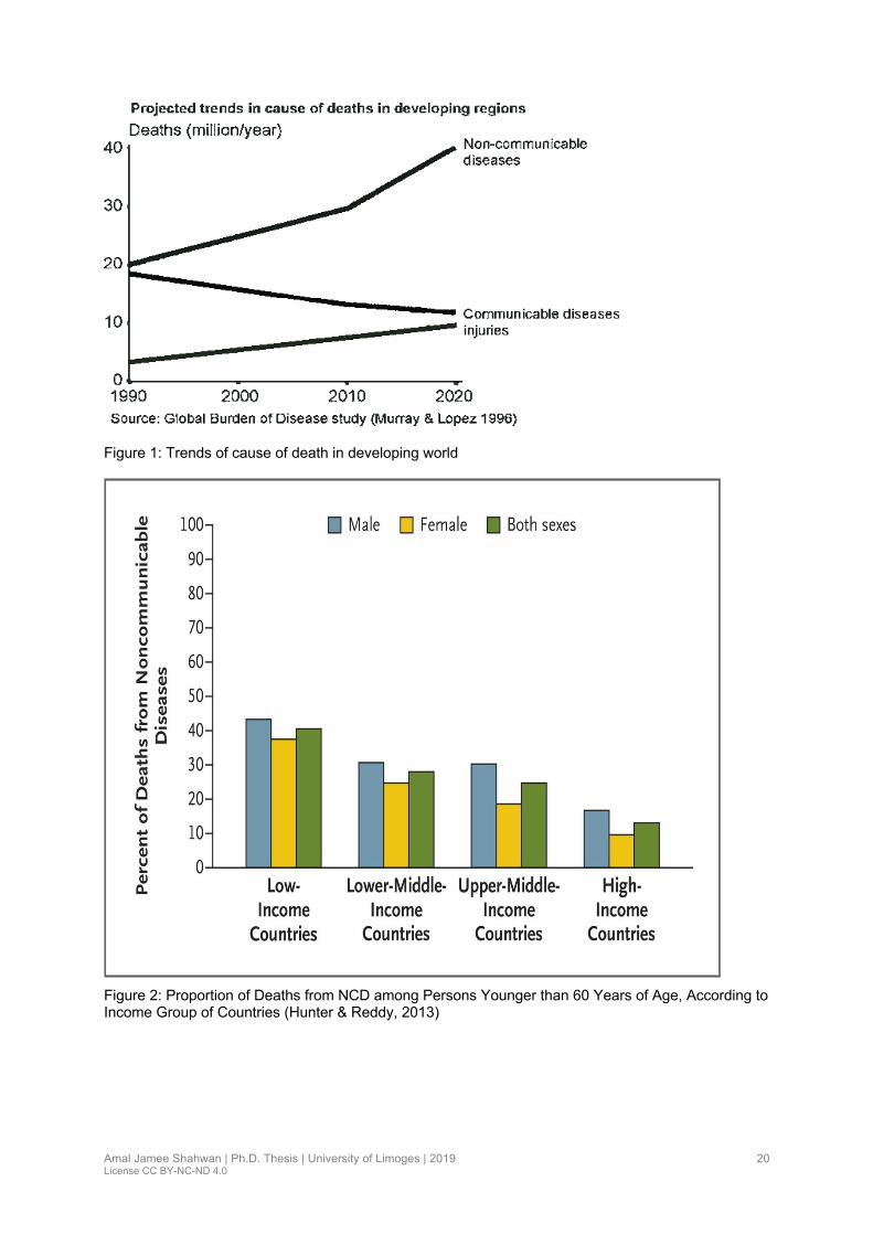
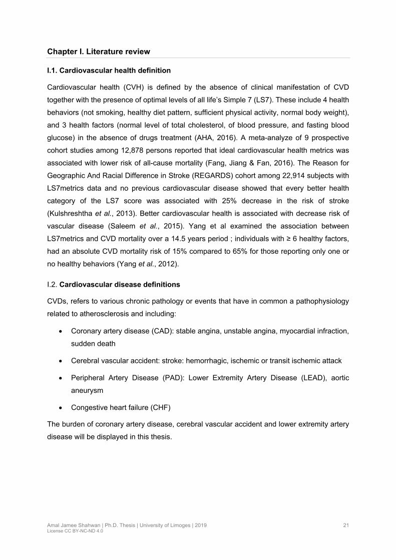
Figure 1: Trends of cause of death in developing world ........................................................ 20
Figure 2: Proportion of Deaths from NCD among Persons Younger than 60 Years of Age, According to Income Group of Countries (Hunter & Reddy, 2013) ........................................ 20
Figure 3: Structure of a normal large artery (Lusis, 2000) ..................................................... 23
Figure 4: (a) Fatty streak with dysfunctional endothelial cells, lipid insudation and the macrophage transformation into foam cells. (b) Stable plaque with extensive calcification and thick fibrous cap overlying foam cells and necrotic core. (c) Acutely ruptured unstable plaque, with a collection of fibrin and platelets forming a thrombus over the disrupted thin fibrous cap (Wang & Butany, 2017). ......................................................................................................... 23
Figure 5: ESC and ACC/AHA recommendations for stress testing and CCTA in the assessment of patients with suspected stable CAD according to pre-test probability of disease (Joseph et al., 2018) ................................................................................................. 29
Figure 6: ESC and ACC/AHA guidance for follow-up assessment of patients with stable CAD according to symptoms (Joseph et al., 2018) ........................................................................ 30
Figure 7: Effects of stress (Nature Reviews cardiology 2018) ............................................... 46
Figure 8: Arab countries Map ................................................................................................. 54
Figure 9: Text message effects on target level of cardiovascular risk factors (Chow et al., 2015) ...................................................................................................................................... 76
Figure 10: 25x25 WHF global CVD roadmap (Grainger-Gasser, Perel, Lagier-Hässig,& Wood, 2017) ........................................................................................................................... 77
Figure 11: Crude Mortality Rate per 1,000 Population, Palestine 1997-2017(Anon, n.d.) ..... 81
Figure 12: Gaza Strip Map ..................................................................................................... 82
Figure 13: Population Pyramid in Gaza Strip(PCBS 2016, n.d.) ............................................ 84
Figure 14: Sampling design study .......................................................................................... 89

Amal Jamee Shahwan | Ph.D. Thesis | University of Limoges | 2019 License CC BY-NC-ND 4.0
12
List of Tables
Table 1: New Universal classification of myocardial infarction (2018)(Thygesen et al., 2019) ............................................................................................................................................... 25
Table 2: Classification and Severity of Angina ....................................................................... 27
Table 3:Traditional clinical classification of chest pain (Diamond, 1983) ............................... 28
Table 4: Stroke Risk factors ................................................................................................... 33
Table 5: Blood pressure classification according to WHO (WHO hypertension 2013) .......... 40
Table 6: Criteria for the diagnosis of diabetes (ADA, 2018) ................................................... 41
Table 7: Type 2 Diabetes Mellitus risk factors (“Risk Factors of Type 2 Diabetes | NIDDK,” 2016) ...................................................................................................................................... 42
Table 8: Classification of elevated TG levels (Jellinger et al., 2017) ...................................... 44
Table 9: LDL-C and non-HDL-C goals in three CHD risk groups by NCEP ATPIII)(Graham et al., 2007) ................................................................................................................................ 44
Table 10: Cardiovascular risk factors (smoking/physical inactivity) in adults aged ≥15 years and overweight and obesity in adults ≥ aged 20 years in Arab countries (Rahim et al., 2014). ............................................................................................................................................... 55
Table 11: Tobacco or secondhand smoke and CVD risk (World Heart Federtion, 2017) ...... 62
Table 12: Characteristics of a heart-healthy population ......................................................... 67
Table 13: Target values for patients with established CVD or DM/ for persons at high CVD risk .......................................................................................................................................... 67
Table 14: Ideal cardiovascular Health, defined by AHA, ‘Life Simple 7’ ................................ 68
Table 15: Prevention of Cardiovascular diseases .................................................................. 69
Table 16: Dietary targets to prevent CVD(Guy De Backer, 2017) ......................................... 70
Table 17: Recommendation regarding BP targets in patients with hypertension (Guy De Backer, 2017; Whelton et al., 2018). ...................................................................................... 72
Table 18:Type 2 diabetes Combination strategies (Giugliano et al., 2018) ........................... 73
Table 19: Criteria for testing for diabetes or prediabetes in asymptomatic adults (ADA, 2018) ............................................................................................................................................... 73
Table 20: Lipid Goals for Patients at risk for Atherosclerotic CVD (Jellinger et al., 2017) ..... 74
Table 21: Goal and treatment for LDL Cholesterol (Guy De Backer, 2017;Hendrani et al., 2016) ...................................................................................................................................... 75
Table 22: "Best Buy" Interventions (“WHO | Scaling up action against NCDs,” n.d.2014) ..... 78
Table 23: National strategic targets to control NCDs in Palestine compared to global targets ............................................................................................................................................... 79
Table 24: Distribution of population in Gaza goverenate areas (PCBS2016) ........................ 83
Table 25: Distribution of population in Gaza strip by age group ............................................ 86
Table 26: Proportional sample selection in Gaza governorates ............................................. 87

Amal Jamee Shahwan | Ph.D. Thesis | University of Limoges | 2019 License CC BY-NC-ND 4.0
13
Abbreviations
ABI Ankle rachial Index
ACC American College of Cardiology
ACE Africa Middle East Cardiovascular Epidemiology study
ACEIs Angiotensin- Converting Enzyme Inhibitors
ACS Acute coronary syndrome
ADA American Diabetes Association
ADF American Diabetes Federation
AGATHA-ME Global Atherothrombosis Assessment -Middle East
AHA American Heart Association
ARBs Angiotensin Receptors blockers
ASA American Stroke Association
ASCVD Atherosclerotic Cardiovascular Disease
BMI Body Mass Index
BP Blood Pressure
CAD Coronary artery disease
CAPRIE Clopidogrel versus Aspirin in Patient at Risk of Ischemic Events
CCS Causative Classification of Stroke
CCTA Coronary Computed Tomography Angiography
CK Creatine Kinase
CK Mb Creatine Kinase MB
CNS Central nervous system
CRP C-Reactive Protein

Amal Jamee Shahwan | Ph.D. Thesis | University of Limoges | 2019 License CC BY-NC-ND 4.0
14
CT Computed Tomography
CVA Cerebrovascular Accident
CVD Cardio Vascular Disease
CVH Cardiovascular Health
CVRF Cardiovascular risk factors
DALYs Disability Adjusted Life Years
DAPT Dual Antiplatelet Therapy
DBP Diastolic Blood Pressure
DM Diabetes mellitus
ECG Electrocardiogram
EMR Eastern Mediterranean Region
ESC European Society of Cardiology
F&V FruitS and Vegetables
FH Family History
FHS Framingham Heart study
FRS Framingham Risk Score
GBD Global burden of disease
GCC Gulf Cooperation Council
GNI Gross National Income
GPS Global Positioning System
GS Gaza Strip
Hamas Palestinian Islamic Movement founded in 1987
HbA1C Glycated Hemoglobin A1c

Amal Jamee Shahwan | Ph.D. Thesis | University of Limoges | 2019 License CC BY-NC-ND 4.0
15
HDL-C High-Density Lipoprotein Cholesterol
HIC High Income Countries
HPFS Health Professionals Follow- Up Study
HRT Hormonal Replacement Therapy
HTN Hypertension
IC Intermittent claudication
ICH Intra Cerebral Hemorrhage
IDF International Diabetes Federation
IDL Intermediate Density Lipoprotein
IHD Ischemic Heart Disease
IPAQ International physical activity questionnaire
ISH International Society of Hypertension
LDL-C Low-density lipoprotein Cholesterol
LEAD Lower Extremities Artery Disease
LMIC Low and Middle-Income Countries
LS7 Life’s Simple 7
MENA Middle East and North Africa
MetS Metabolic Syndrome
MI Myocardial Infarction
MIC Middle Income Countries
min Minute
MOH Ministry of Health
MRA Magnetic Resonance Angiography

Amal Jamee Shahwan | Ph.D. Thesis | University of Limoges | 2019 License CC BY-NC-ND 4.0
16
MRI Magnetic Resonance Imaging
NCDs Non-communicable diseases
NCEP ATP III National Cholesterol Education Program Adult Treatment Panel III
NGO Non-Governmental Organization
NHANES The National Health and Nutrition Examination Survey
NHLBI The National Heart, Lung and Blood Institute
NNT Number Needed to Treat
NRT Nicotine Replacement Therapy
NSTEMI Non-ST Elevation Myocardial Infarction
OR Odds ratio
PAD Peripheral Artery Disease
PCBS Palestinian Central Bureau of Statistics
PCI Percutaneous Coronary Intervention
PSS Perceived Scale Stress
PTP Pretest Probability
PURE Prospective Urbane Rural Epidemiology study
RCT Randomized Controlled Trial
REGARDS The Reasons for Geographic and Racial Differences in Stroke
RQ Rose Questionnaire
SAH Subarachnoid Hemorrhage
SBP Systolic Blood Pressure
SCAD Stable Coronary Artery Disease
SES Socio Economic Status

Amal Jamee Shahwan | Ph.D. Thesis | University of Limoges | 2019 License CC BY-NC-ND 4.0
17
STEMI ST Elevation Myocardial Infarction
TIA Transit Ischemic Attack
TOAST Trial of ORG 10172 in Acute Stroke Treatment
UA Unstable Angina
UK United Kingdom
UNRWA United Nations Relief and Works Agency
USA United State of America
VLDL Very Low-Density Lipoprotein
WB West Bank
WHO World Health Organization
Amal Jamee Shahwan | Ph.D. Thesis | University of Limoges | 2019 License CC BY-NC-ND 4.0
18
Introduction
In 2014, the World Health Organization (WHO) defined the Non-Communicable Diseases
(NCDs) as one of the greatest challenges of 21st century. The NCDs are medical conditions
not related to infectious agents, they may be caused by genetic or behavior factors and have
a slow progression and long duration (WHO 2014, n.d.). The burden of NCDs is growing rapidly
affecting people of all ages and different income levels in all regions of the world mainly in low
and middle-income countries (LMIC). Four risk factors (tobacco use, excessive alcohol
consumption, poor diet and lack of physical activity) are associated with four disease cluster
(cardiovascular disease, cancer, chronic pulmonary disease and diabetes)(Lozano et al.,
2012). Six of the top ten leading causes of death in 2012 were NCDs, including the top three
diseases (ischemic heart disease, stroke and chronic obstructive pulmonary disease) (LUKE,
2017). NCDs are the leading cause of death and disability, killing three in five people worldwide
and responsible for nearly half of the global burden of disease. NCD death increased from
2006 to 2016, rising 16.1%. In 2016, NCDs caused 39.5 million (72.3%) of death worldwide,
and were projected to increase by 15% in 2020 (Bowery, 2015; GBD 2016 Causes of Death
Collaborators, 2017). More than 16 million (42%) of all NCDs deaths were premature deaths
(under age 70 years). A large amount of deaths from NCDs (29%) in LMIC occur among people
younger than 60 years compared with High Income Countries (HIC) (13%). The leading cause
of premature deaths for NCDs were cardiovascular disease (CVD)(45%), malignant neoplasm
(22%), respiratory disease (10%) and diabetes (4%) (WHO 2014). The risk of death between
age 30 and 70 years from any one of the four main NCDs cited, decrease from 23% in 2000
to 19% in 2015 (WHO, 2017b).
The WHO suggests that cardiovascular death occurs 3 times more in LMIC and in the working
age group with an equal rate in males and females (Organization, 2003).
WHO Global Plan Action for the prevention of NCDs 2013-2020 identified seven major risk
factors including harmful use of alcohol, current tobacco smoking, high blood pressure, intake
of salt or sodium, diabetes and obesity, physical inactivity (referred as the 25 by 25 risk factors)
with the goal of reducing premature mortality from NCDs by 25% (WHO/GPA, 2013).
The Eastern Mediterranean Region (EMR) is comprised of 23 countries with a population of
583 million people. Arabs living in this region share language, cultural background as well as
lifestyle. However, they vary in their sociodemographic profile, political, economic situation and
health system, several of these nations have long years of political instability (Mokdad et al.,
2016). Countries in EMR have been classified into three groups of health systems according
to population health outcome, health system performance and health budget levels. Palestine
is part of the second group. Modernization, economic and technologic development advances

Amal Jamee Shahwan | Ph.D. Thesis | University of Limoges | 2019 License CC BY-NC-ND 4.0
19
have led to rapid demographic changes in the Arab world inducing an increase in death rate
(Alwan, Alwan & Jabbour, 2012) . Morbidity related to NCD is accounting for 47% and is
estimated to reach 60% by the year 2020 (Khatib, 2004). More than 2.2 million people in EMR
died from NCDs representing 53% deaths. Thirty five per cent of death from NCDs were in
persons younger than 60 years (WHO EMRO, 2016). The ischemic heart disease was the
leading cause of death in this region accounting for 14.3% of deaths (Mokdad et al., 2014).
Rapid increase in NCDs suggests that the change in disease burden was more affected by
behavioral than genetic factors. Arabs became less physically active and consumed unhealthy
diet (Mehio Sibai et al., 2010). Data collected by STEPS Wise WHO for chronic disease 2010-
2011 among adults aged 15-65 years in Eastern Mediterranean countries showed the high
prevalence of NCDs risk factors. One quarter of adult population was hypertensives, daily
current tobacco smoking exceeds 30% in males, obesity was alarming particularly in women.
Six out ten countries in the world reported the highest prevalence of diabetes with a rate up to
20% (IDF 2009, n.d.).
Epidemiological transition
The epidemiological transition defined as shift from infectious disease to NCDs. In the Arab
world, the situation of health changed dramatically during the few decades driven by rapid
aging of the population, economic development, globalization and technological advances,
westernization of diet (food high in fat, salt and sugar, high intake of fast food, snacks rich in
calories, edible oils, and an increase in consumption of animal source foods). Life style
changes in employment activities, rising obesity and decrease in physical activity (Omran
2005; Islam et al. 2014) are particularly prevalent in the high income countries (HIC) of the
Gulf Cooperation Council (GCC) mainly, in females (Rahim et al., 2014). The rate of
urbanization is increasing from 36.6% of the world population living in urban areas in 1970, to
44.8% in 1994. This proportion is projected to increase to 61.1% by 2025 (Chockalingam et
al., 2000).
Amal Jamee Shahwan | Ph.D. Thesis | University of Limoges | 2019 License CC BY-NC-ND 4.0
20
Figure 1: Trends of cause of death in developing world
Figure 2: Proportion of Deaths from NCD among Persons Younger than 60 Years of Age, According to Income Group of Countries (Hunter & Reddy, 2013)
Amal Jamee Shahwan | Ph.D. Thesis | University of Limoges | 2019 License CC BY-NC-ND 4.0
21
Chapter I. Literature review
I.1. Cardiovascular health definition
Cardiovascular health (CVH) is defined by the absence of clinical manifestation of CVD
together with the presence of optimal levels of all life’s Simple 7 (LS7). These include 4 health
behaviors (not smoking, healthy diet pattern, sufficient physical activity, normal body weight),
and 3 health factors (normal level of total cholesterol, of blood pressure, and fasting blood
glucose) in the absence of drugs treatment (AHA, 2016). A meta-analyze of 9 prospective
cohort studies among 12,878 persons reported that ideal cardiovascular health metrics was
associated with lower risk of all-cause mortality (Fang, Jiang & Fan, 2016). The Reason for
Geographic And Racial Difference in Stroke (REGARDS) cohort among 22,914 subjects with
LS7metrics data and no previous cardiovascular disease showed that every better health
category of the LS7 score was associated with 25% decrease in the risk of stroke
(Kulshreshtha et al., 2013). Better cardiovascular health is associated with decrease risk of
vascular disease (Saleem et al., 2015). Yang et al examined the association between
LS7metrics and CVD mortality over a 14.5 years period ; individuals with ≥ 6 healthy factors,
had an absolute CVD mortality risk of 15% compared to 65% for those reporting only one or
no healthy behaviors (Yang et al., 2012).
I.2. Cardiovascular disease definitions
CVDs, refers to various chronic pathology or events that have in common a pathophysiology
related to atherosclerosis and including:
• Coronary artery disease (CAD): stable angina, unstable angina, myocardial infraction,
sudden death
• Cerebral vascular accident: stroke: hemorrhagic, ischemic or transit ischemic attack
• Peripheral Artery Disease (PAD): Lower Extremity Artery Disease (LEAD), aortic
aneurysm
• Congestive heart failure (CHF)
The burden of coronary artery disease, cerebral vascular accident and lower extremity artery
disease will be displayed in this thesis.

Amal Jamee Shahwan | Ph.D. Thesis | University of Limoges | 2019 License CC BY-NC-ND 4.0
22
I.3. Types of cardiovascular disease
I.3.1. Coronary artery disease
I.3.1.1. Definition and classification
Also, known as ischemic heart disease (IHD) refers to conditions that involve impairment of
coronary artery blood flow that can result in silent ischemia, angina pectoris, acute coronary
syndrome (ACS) or sudden cardiac death. Coronary artery disease (CAD) is a common public
health problem associated with high mortality and increased health cost (He et al., 2017).
I.3.1.2. Physiopathology
Atherosclerosis is a complex progressive chronic multifocal, immune-inflammatory, fibro
proliferative disease, with the accumulation of lipid metabolism, active cellular interaction,
inflammation and matrix remodeling in the large arteries (Brown et al., 2017; Hamm et al.,
2006). Atherosclerosis causes complex, lesions on coronary, cerebrovascular and peripheral
vascular diseases (Tabas, García-Cardeña & Owens, 2015). The anatomy of normal artery is
displayed in Figure 2. The early lesion of atherosclerosis consists of fatty streak comprised of
(cholesterol and macrophage). They are limited to the aorta in the first decade of life, then
extend later to the coronary arteries and peripheral arteries.
The lesion leads to acute occlusion of the artery by a thrombus often related to intimal of
rupture or erosion, clinically present as unstable angina, Myocardial Infarction (MI) or sudden
cardiac death (Lusis, 2000, Anon, 2017a). In Stable Coronary Artery Disease (SCAD),
atherosclerosis lesion progresses slowly, allowing for the development of collateral circulation
(T. Wang and Butany 2017) (figure 3). The atherosclerosis process can be accelerated by the
cardiovascular traditional risk factors such as diabetes, hypertension, obesity, dyslipidemia,
smoking, and genetics factors.

Amal Jamee Shahwan | Ph.D. Thesis | University of Limoges | 2019 License CC BY-NC-ND 4.0
23
Figure 3: Structure of a normal large artery (Lusis, 2000)
A large artery consists of three morphologically distinct layers. The intima, the innermost layer, is bounded by a monolayer of
endothelial cells on the luminal side and a sheet of elastic fibres, the internal elastic lamina, on the peripheral side. The normal
intima is a very thin region (size exaggerated in this figure) and consists of extracellular connective tissue matrix, primarily
proteoglycans and collagen. The media, the middle layer, consists of SMCs. The adventitia, the outer layer, consists of connective
tissues with interspersed fibroblasts and SMCs. Nature. Author manuscript; available in PMC 2010 February 22.
Figure 4: (a) Fatty streak with dysfunctional endothelial cells, lipid insudation and the macrophage
transformation into foam cells. (b) Stable plaque with extensive calcification and thick fibrous cap
overlying foam cells and necrotic core. (c) Acutely ruptured unstable plaque, with a collection of fibrin
and platelets forming a thrombus over the disrupted thin fibrous cap (Wang & Butany, 2017).

Amal Jamee Shahwan | Ph.D. Thesis | University of Limoges | 2019 License CC BY-NC-ND 4.0
24
I.3.1.3. Acute coronary syndrome
Acute coronary syndrome refers to a spectrum of clinical symptoms compatible with acute
myocardial ischemia and includes Unstable Angina (UA), Non-ST Segment Elevation
Myocardial Infarction (NSTEMI), and ST-segment Elevation Myocardial Infraction (STEMI),
thus they share common pathophysiological origins related to coronary plaque progression,
instability, or rupture with or without luminal thrombosis and vasospasm. The main clinical
expression are MI, and sudden cardiac death.
Differentiating ACS from other cardiac chest pain is the primary diagnostic challenge. The
initial assessment requires a good history collection including risk factors analysis, a physical
examination, an Electrocardiogram (ECG) and cardiac biomarkers analysis that help in
determining the differential diagnosis such as aortic dissection, pericarditis, pulmonary
embolism and musculoskeletal pain (Braunwald et al., 1994).
I.3.1.3.1. Clinical presentation of Acute coronary syndrome
Most patients describe diffuse severe pain, which may occur with exertion or at rest localized
in the sub sternal region in typical cases or epigastric discomfort. The pain radiates to the neck,
jaw, left shoulder and left arm or both, not affected by movements. Other symptoms can be
associated such as nausea, vomiting, unexplained fatigue and in rare cases syncope.
Discomfort persists more >20 Minutes (min). Symptoms might be atypical in diabetic patients,
women and elderly persons (Kumar & Cannon, 2009; Thygesen et al., 2012). Five factors
reinforce the diagnosis of acute ischemia due to CAD. They are according their weight: a past
history of CAD, male sex, older age, the characteristics of angina pain and the presence of
cardiovascular risk factors such as HTN, DM, dyslipidemia, cigarette smoking, and family
history of premature CAD (Pryor et al., 1993).
I.3.1.3.1.1. Myocardial infraction
Acute myocardial infarction is a myocardial necrosis due to prolonged ischemia (Thygesen et
al., 2012). The diagnosis is based on biochemical criteria (troponin elevation as a result of
irreversible cell damage), clinical evaluation, ECG findings, invasive and noninvasive imaging
and pathological evaluation (Table1) (Anderson & Morrow, 2017).

Amal Jamee Shahwan | Ph.D. Thesis | University of Limoges | 2019 License CC BY-NC-ND 4.0
25
Table 1: New Universal classification of myocardial infarction (2018)(Thygesen et al., 2019)
Type 1: Spontaneous myocardial infarction
Spontaneous myocardial infarction related to atherosclerotic plaque rupture, ulceration, fissuring, erosion, or dissection with resulting intraluminal thrombus in one or more of the coronary arteries leading to decreased myocardial blood flow or distal platelet emboli with ensuing myocyte necrosis. The patient may have underlying severe CAD but on occasion non-obstructive or no CAD.
Type 2: Myocardial infarction secondary to an ischaemic imbalance
In instances of myocardial injury with necrosis where a condition other than CAD contributes to an imbalance between myocardial oxygen supply and/or demand, e.g. coronary endothelial dysfunction, coronary artery spasm, coronary embolism, tachy-/brady-arrhythmias, anemia, respiratory failure, hypotension, and hypertension with or without LVH.
Type 3: Myocardial infarction resulting in death when biomarker values are unavailable
Cardiac death with symptoms suggestive of myocardial ischemia and presumed new ischemic ECG changes or new LBBB, but death occurring before blood samples could be obtained, before cardiac biomarker could rise, or in rare cases cardiac biomarkers were not collected.
Type 4a: Myocardial infarction related to percutaneous coronary intervention (PCI)
Myocardial infarction associated with PCI is arbitrarily defined by elevation of cTn values >5 x 99th
percentile URL in patients with normal baseline values (£99th percentile URL) or a rise of cTn values >20% if the baseline values are elevated and are stable or falling. In addition, either (i) symptoms suggestive of myocardial ischemia, or (ii) new ischemic ECG changes or new LBBB, or (iii) angiographic loss of patency of a major coronary artery or a side branch or persistent slow- or no-flow or embolization, or (iv) imaging demonstration of new loss of viable myocardium or new regional wall motion abnormality are required.
Type 4b: Myocardial infarction related to stent thrombosis
Myocardial infarction associated with stent thrombosis is detected by coronary angiography or autopsy in the setting of myocardial ischemia and with a rise and/ or fall of cardiac biomarkers values
with at least one value above the 99th percentile URL.
Type 5: Myocardial infarction related to coronary artery bypass grafting (CABG)
Myocardial infarction associated with CABG is arbitrarily defined by elevation of cardiac biomarker
values >10 x 99th percentile URL in patients with normal baseline cTn values (£99th percentile URL). In addition, either (i) new pathological Q waves or new LBBB, or (ii) angiographic documented new graft or new native coronary artery occlusion, or (iii) imaging evidence of new loss of viable myocardium or new regional wall motion abnormality.
I.3.1.3.2. Physical examination findings
Physical examination can vary from normal to hemodynamic instability including ischemic
mitral regurgitation, hypotension, gallop heart sound, jugular venous distension, left ventricular
failure, and cardiogenic shock.

Amal Jamee Shahwan | Ph.D. Thesis | University of Limoges | 2019 License CC BY-NC-ND 4.0
26
I.3.1.3.3. Electrocardiograms
Electrocardiogram provides important information about the presence, extent and severity of
myocardial ischemia in stratifying the patient risk of ACS and determining the treatment
strategy.
I.3.1.3.4. Serum cardiac markers
Elevated cardiac biomarkers especially Troponin (I or T), or creatinine kinase (CK) or its
isoenzyme MB (CK-MB), reflect myocardial cells necrosis. Troponins have higher clinical
sensitivity and specificity than traditional cardiac enzymes. Myoglobin is not specific for the
detection of myocardial cell injury (Eggers et al., 2004). Troponin level should be measured
within 6 first hours of the onset of pain (strength of recommendation), and the elevation stays
for 2 weeks after the onset of myocardial necrosis (Smith et al., 2015). Troponin increase
reflects irreversible myocardial cellular necrosis (Eggers et al., 2004). However cardiac
biomarkers are not specific of acute MI (Thygesen et al., 2012). Clinical conditions such as
pulmonary embolism, heart failure, end stage renal failure, and myocarditis are associated with
of cardiac biomarkers increase (Korff, Katus & Giannitsis, 2006).
I.3.1.4. Stable coronary artery disease
I.3.1.4.1. Definition, classification
Stable coronary artery disease includes all clinical entities that are characterized by coronary
atherosclerosis in the absence of ACS (Abrams, 2005). It is defined as episodes of reversible
myocardial demand /supply mismatch, related to ischemia or hypoxia, which are usually
inducible by exercise, emotion or other stress and, reproducible but, may also be occurring
spontaneously (Task members of ESC et al., 2013). The consequences of ischemia are
according to a predictable temporal chronology:
• Increased H+ and K+ concentration in the venous blood
• Signs of ventricular diastolic and subsequently systolic dysfunction with regional wall
motion abnormalities
• Development of ST-T changes
• Cardiac ischemic pain (angina) (Crea et al., 2010)
In SCAD the symptoms are reversible, repetitive for months to years and relieved by rest or
sublingual nitroglycerin. Some conditions such as anemia, hypertension crisis, thyrotoxicosis
can exacerbate the angina.
The SCAD definition includes:
Amal Jamee Shahwan | Ph.D. Thesis | University of Limoges | 2019 License CC BY-NC-ND 4.0
27
• Patients symptomatic for stable angina pectoris or a symptom like angina (e.g.
dyspnea)
• Patients with a history of obstructive or non-obstructive CAD, who have become
asymptomatic with treatment and need regular follow up
• Patients reporting symptoms for the first time, but already in chronic stable condition
(since several months)
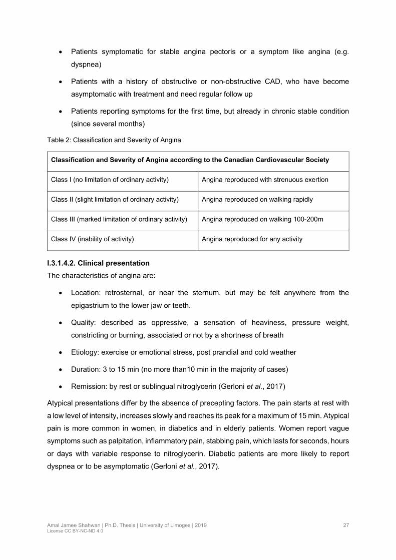
Table 2: Classification and Severity of Angina
Classification and Severity of Angina according to the Canadian Cardiovascular Society
Class I (no limitation of ordinary activity) Angina reproduced with strenuous exertion
Class II (slight limitation of ordinary activity) Angina reproduced on walking rapidly
Class III (marked limitation of ordinary activity) Angina reproduced on walking 100-200m
Class IV (inability of activity) Angina reproduced for any activity
I.3.1.4.2. Clinical presentation
The characteristics of angina are:
• Location: retrosternal, or near the sternum, but may be felt anywhere from the
epigastrium to the lower jaw or teeth.
• Quality: described as oppressive, a sensation of heaviness, pressure weight,
constricting or burning, associated or not by a shortness of breath
• Etiology: exercise or emotional stress, post prandial and cold weather
• Duration: 3 to 15 min (no more than10 min in the majority of cases)
• Remission: by rest or sublingual nitroglycerin (Gerloni et al., 2017)
Atypical presentations differ by the absence of precepting factors. The pain starts at rest with
a low level of intensity, increases slowly and reaches its peak for a maximum of 15 min. Atypical
pain is more common in women, in diabetics and in elderly patients. Women report vague
symptoms such as palpitation, inflammatory pain, stabbing pain, which lasts for seconds, hours
or days with variable response to nitroglycerin. Diabetic patients are more likely to report
dyspnea or to be asymptomatic (Gerloni et al., 2017).

Amal Jamee Shahwan | Ph.D. Thesis | University of Limoges | 2019 License CC BY-NC-ND 4.0
28
Table 3:Traditional clinical classification of chest pain (Diamond, 1983)
Typical angina
(Definite)
Meets all three of the following characteristics
Substernal chest discomfort of characteristic quality and duration
Provoked by exertion or emotional stress
Relived by rest and /or nitrates within minutes
Atypical angina
(probable)
Meets two of these characteristics
Non-anginal chest pain Lacks or meets only one or none of the characteristics
I.3.1.4.3. Investigations
Physical examination is poor and has low sensitivity. A normal ECG does not exclude the
diagnosis, but an abnormal resting one increases the diagnosis probability. Routine laboratory
tests are recommended to determine the severity of factors. Hyperglycemia, dyslipidemia,
thyroid disorder and renal failure should be evaluated in every patient with suspected CAD
(Task members of ESC et al., 2013). Plasma cardiac troponin levels are below the normal.
According to ESC and Canadian guidelines echocardiography should be performed in all
patients with SCAD to identify left ventricular function, kinetic segments, valvular lesion (mainly
mitral regurgitation).
• The Multidetector row CT permits the detection of coronary calcification. The
measurement of calcium scoring is calculated as the coronary calcium area by maximal
plaque density (in Hounsfield units) and calcified lesions are quantified using (Agatston
score). Calcium scoring helps to evaluate the atherosclerosis burden (Omland et al.,
2009; Fihn et al., 2012)
• Coronary computed tomography angiography (CTA): is indicated in patients with low-
intermediate risk of obstructive CAD. CTA can visualize the coronary arteries after
intravenous injection of contrast agent. CTA is helpful in patients without severe
obesity, favorable calcium score (Agatston score < 400) and with heart rate ≤ 65 beats
per minute.
• Exercise ECG: with a high specificity > 90% and a low sensitivity < 50% (high
probability of false positive mainly in females). The test is usually carried out on a
treadmill from rest to maximum exertion according the Bruce protocol. ST segment
modification are analyzed (Ashley & Niebauer, 2004).

Amal Jamee Shahwan | Ph.D. Thesis | University of Limoges | 2019 License CC BY-NC-ND 4.0
29
• Stress echocardiography: the predictive value is higher than treadmill test. It detects
the difference in wall motion between ischemic and non-ischemic myocardium, and
provides information on hibernating myocardium
• Magnetic resonance imaging: detects wall motion abnormalities
• Cardiac Coronary angiography: remains the gold standard. ACS or Positive stress test
induced large wall motion abnormality, or poor answer to medical treatment are the
main indications. Location and number of lesions, type and degree of stenosis are
described (Ashley & Niebauer, 2004).
I.3.1.4.4. Strategy diagnostic (Figure 5,6)
A Pre-Test Probability (PTP) evaluation prior to non-invasive testing with the Duke clinical
score and Diamond Forrest model is recommended in The European and American guidelines
(Joseph et al., 2018). PTP of CAD is based upon age, gender and symptoms. It is defined by
ESC guidelines as: Low <15%, intermediate 15-85% and high >85%. The intermediate group
is classified further into (a) 15-65%, and (b) 66-85% (Montalescot et al., 2013). In the ESC
GUIDELINES, CCTA is recommended in patients with a low -intermediate PTP of CAD (15-
50%). Pharmacological stress MRI is a class IIa indication according to the ACC/AHA
guidelines*.
CCTA*Coronary Computed Tomography Angiography ACC/AHA guidelines*: American college of cardiology/American Heart Association guidelines
Figure 5: ESC and ACC/AHA recommendations for stress testing and CCTA in the assessment of patients with suspected stable CAD according to pre-test probability of disease (Joseph et al., 2018)
Amal Jamee Shahwan | Ph.D. Thesis | University of Limoges | 2019 License CC BY-NC-ND 4.0
30
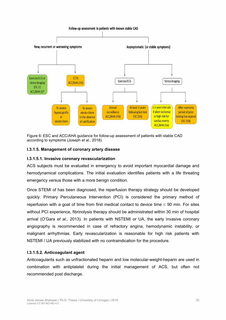
Figure 6: ESC and ACC/AHA guidance for follow-up assessment of patients with stable CAD according to symptoms (Joseph et al., 2018)
I.3.1.5. Management of coronary artery disease
I.3.1.5.1. Invasive coronary revascularization
ACS subjects must be evaluated in emergency to avoid important myocardial damage and
hemodynamical complications. The initial evaluation identifies patients with a life threating
emergency versus those with a more benign condition.
Once STEMI of has been diagnosed, the reperfusion therapy strategy should be developed
quickly. Primary Percutaneous Intervention (PCI) is considered the primary method of
reperfusion with a goal of time from first medical contact to device time £ 90 min. For sites
without PCI experience, fibrinolysis therapy should be administrated within 30 min of hospital
arrival (O’Gara et al., 2013). In patients with NSTEMI or UA, the early invasive coronary
angiography is recommended in case of refractory angina, hemodynamic instability, or
malignant arrhythmias. Early revascularization is reasonable for high risk patients with
NSTEMI / UA previously stabilized with no contraindication for the procedure.
I.3.1.5.2. Anticoagulant agent
Anticoagulants such as unfractionated heparin and low molecular-weight-heparin are used in
combination with antiplatelet during the initial management of ACS, but often not
recommended post discharge.

Amal Jamee Shahwan | Ph.D. Thesis | University of Limoges | 2019 License CC BY-NC-ND 4.0
31
I.3.1.5.3. Long term management
I.3.1.5.3.1. Antithrombotic Agents
Antiplatelet therapy reduces the risk of thrombosis by interfering with platelet release and
aggregation and reduces the risk of thrombosis. The most used therapies in the management
of CAD were Aspirin, adenosine diphosphate P2Y12 receptor antagonist (Clopidogrel,
prasugrel and ticagrelor), and glycoprotein II b III a (abciximab and eptifibatide). Aspirin should
be introduced immediately after ACS diagnosis, with a dose of 160-325 mg, and maintained
on long-time at dose (81-100mg (Smith et al., 2015; Mehta et al., 2010). Oral P2y12 inhibitor is
indicated for patients undergoing primary PCI. A loading dose should be administrated before
PCI then maintained during one year (O’Gara et al., 2013). In NSTEMI / UA a Dual Antiplatelet
Therapy as aspirin plus clopidogrel (DAPT) has been demonstrated to reduce death from
cardiovascular events (nonfatal MI or stroke) by a relative risk of 20% ( Yusuf et al. 2004). In
STEMI, DAPT reduced the re-occlusion infarct-related artery after PCI, death, and recurrent
MI before angiography by 36% (Sabatine et al., 2005). In SCAD, the use of aspirin at a dose
of 81-150 mg daily reduced cardiovascular mortality and morbidity by 20-25% (O’Gara et al.,
2013).
I.3.1.5.3.2. B-blocker
These drugs decrease myocardial oxygen consumption, decrease heart rate, blood pressure,
and reduce myocardial contractility. They are recommended in patients with ACS within 24
hours except in cases of heart failure or cardiogenic shock (Smith et al., 2015).
I.3.1.5.3.3. Inhibitors of the renin-Angiotensin system
The angiotensin converting enzyme inhibitors (ACEIs) or angiotensin receptors blockers
(ARBs) should be initiated in the first 24 hours of ACS patients with heart failure, or large zone
of infarction (massive MI), or left ventricular EF<40%. These drugs reduce mortality and
morbidity in select patients. With ACE, or ARBs, monitoring of serum creatinine, potassium
level and blood pressure is mandatory.
I.3.1.5.3.4. Statin
Statin are recommended in all patients presenting with CAD. Starting with high intensity dose
of statin therapy in ACS confers an absolute risk reduction of 3.9% for death from any cause,
recurrent MI, re-hospitalization and stroke (Pedersen et al., 2004; Stone et al., 2013).
I.3.1.5.3.5. Risk factor correction
Stopping smoking is probably the most-effective preventive measure. Diabetes, Hypertension
and dyslipidemia controls are mandatory.

Amal Jamee Shahwan | Ph.D. Thesis | University of Limoges | 2019 License CC BY-NC-ND 4.0
32
I.3.2. Cerebrovascular accident: Stroke
I.3.2.1. Definition and classification
Stroke or cerebrovascular Accident (CVA) is a neurological deficit due to an acute focal injury
of the central nervous system (CNS) by a vascular origin. Two process, are described, first
brain ischemia due to thrombosis, embolism, or systemic hypo perfusion, second brain
hemorrhage due to intra Cerebral Hemorrhage (ICH), or subarachnoid hemorrhage (SAH)
(Sacco et al., 2013). Roughly 80% of strokes in HIC are due to ischemic cerebral infarction
and 20% to brain hemorrhage (Sacco et al., 2013; Louis R & Scot E, 2018).
Transit Ischemic Attack (TIA) was defined in 2009 by the expert committee of the AHA/ASA as
“a transit episode of neurological dysfunction caused by focal brain, spinal cord or retinal
ischemia without acute infarction”. This definition is based on tissue pathophysiology rather
than symptom duration (Easton et al., 2009). TIA results from the same mechanisms as
ischemic stroke and causes temporary neurologic deficit. Eighty per cent of TIA resolve within
60 min. TIA might preceded stroke.
The Trial of Org10172 in Acute Stroke Treatment (TOAST) classification, is the most used one.
In order to identify the etiology the severity and the localization of the ischemic stroke TOAST
distinguishes 5 etiological subtypes: atherothrombotic stroke (due to large artery
atherosclerosis), cardio embolic stroke, lacunar stroke due to arterial (dissection, vasculitis,
vasospasm), stroke of other determined etiology, and stroke of unknown origin (Ay et al.,
2005). The last subtype includes all cases with an incomplete evaluation or non-determined
etiology. SSS-TOAST is the Modified definition of the TOAST based on the clinical and imaging
criteria, and divides each of the original TOAST into three sub categories as “evident, probable,
possible”. An algorithm computerized version: the Causative Classification of Stroke system
(CCS) is available in multiple centers (Ay et al., 2007).
I.3.2.2. Physiopathology
Three major etiologies are reported for ischemic stroke; hypo-perfusion, embolism and
thrombosis (Ojaghihaghighi et al., 2017). Normal cerebral blood flow in humans is 50 to 60 ml
/100 g of brain tissue per min. When flow decreases to 20-40 ml /100 mg per min, neuronal
dysfunction occurs, and an irreversible tissue damage occurs when it is less than 10 to 15
ml/100 mg per min.
I.3.2.2.1. Hemorrhagic stroke
In this condition, stroke arises due to rupture of blood vessel in the brain. Its harmful effects
are a result of hypoxia, direct effect of released blood on brain parenchyma and and increase
of intra cerebral pressure. ICH condition is often due to hypertension, trauma, toxic drugs
Amal Jamee Shahwan | Ph.D. Thesis | University of Limoges | 2019 License CC BY-NC-ND 4.0
33
(cocaine) and vascular malformations. Rupture of aneurysms from vascular malformation may
lead to SAH (Escudero Augusto, Marqués Alvarez & Taboada Costa, 2008).
I.3.2.2.2. Atherosclerosis and stroke
Atherosclerosis development is increased by major risk factors such as hypertension,
diabetes, obesity, chronic inflammation and increase concentration of oxidized lipoprotein.
Hemorrhage in the plaque contribute to the progressive narrowing of the lumen.The endothelial
integrity become vulnerable and unstable leading to platelets adhesion and aggregation and
thrombus formation (Flemming, 2015).
I.3.2.3. Risk factors
The relative risk associated with the most important risk factors for stroke are displayed in table
4 (Markus, 2016)
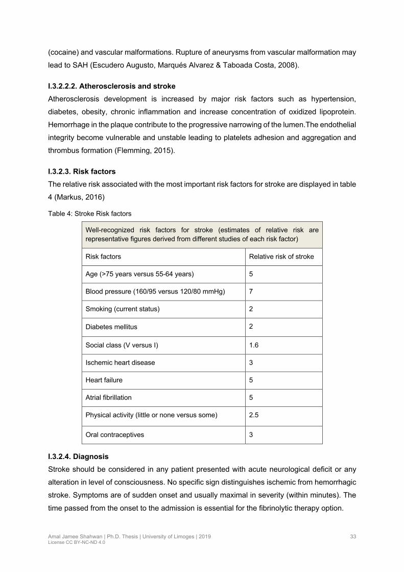
Table 4: Stroke Risk factors
Well-recognized risk factors for stroke (estimates of relative risk are representative figures derived from different studies of each risk factor)
Risk factors Relative risk of stroke
Age (>75 years versus 55-64 years) 5
Blood pressure (160/95 versus 120/80 mmHg) 7
Smoking (current status) 2
Diabetes mellitus 2
Social class (V versus I) 1.6
Ischemic heart disease 3
Heart failure 5
Atrial fibrillation 5
Physical activity (little or none versus some) 2.5
Oral contraceptives 3
I.3.2.4. Diagnosis
Stroke should be considered in any patient presented with acute neurological deficit or any
alteration in level of consciousness. No specific sign distinguishes ischemic from hemorrhagic
stroke. Symptoms are of sudden onset and usually maximal in severity (within minutes). The
time passed from the onset to the admission is essential for the fibrinolytic therapy option.
Amal Jamee Shahwan | Ph.D. Thesis | University of Limoges | 2019 License CC BY-NC-ND 4.0
34
I.3.2.4.1. Clinical symptoms
More likely the symptoms occur in combination: hemiparesis, hemi sensory deficit, perturbation
in vision or sudden vision loss, diplopia, dysarthria, aphasia, facial droop, ataxia and vertigo.
I.3.2.4.2. Physical examination
Medical history for patients helps to identify risk factors for atherosclerosis and cardiac causes.
General and neurological examination confirm the presence of a stroke and evaluate the stroke
severity. The presence of neck bruit suggests carotid stenosis; atrial fibrillation indicates cardio
embolism origin. Absence of pulses in lower extremities suggests the diagnosis of
atherosclerosis. Neuroimaging is mandatory. Few clinical findings are in favor of hemorrhagic
stroke such as coma, neck stiffness, seizures, high blood pressure, headache and vomiting
(Runchey & McGee, 2010).
I.3.2.4.3. Imaging study
Urgent brain imaging is required in all stroke patients, to distinguish ischemic stroke from
intracerebral hemorrhage and to detect aneurysm or malformations.
• Non-contrast computed tomography (CT) scanning is essential to rule out cerebral
hemorrhage or other causes. CT may be normal in the initial hours after ischemic stroke
• Magnetic Resonance Imaging (MRI) is the cornerstone imaging which provides
structural details, detects early cerebral oedema, and identifies old hemorrhage
(“Stroke in the Acute Setting” 2017)
• Carotid duplex scanning detects carotid stenosis and its severity
I.3.2.5. Management
Stroke is an emergency requiring immediate intervention in order to minimize the size of the
infarct, and the rate of neurological impairment. In addition, differentiating the type of CVA
plays decisive role in the management. TIA requires serious evaluation as 30 days stroke risk
is around 10% (Anon, 2017b; Muir, 2001).
I.3.2.5.1. Acute treatment
I.3.2.5.1.1. General measures: stroke unit
Stroke units improve outcome and avoids complications (Turner et al., 2015). Patients are
monitored continuously, to detect and to correct factors that may worsen the acute ischemic
stroke. General care is effective to improve the prognosis such as correct electrolytes
imbalance, severe hypertension, hyperglycemia, hydration disorders and malnutrition
(Powers, 1993).

Amal Jamee Shahwan | Ph.D. Thesis | University of Limoges | 2019 License CC BY-NC-ND 4.0
35
I.3.2.5.1.2. Thrombolytic therapy
In Ischemic stroke, “Time is brain” and early treatment is a positive point for successful
thrombolysis. Intravenous thrombolytic with rt-AP initiated within 4.5 hours of symptoms onset
is associated with high benefit (Emberson et al., 2014). The number-Needed to Treat (NNT)
for good outcome is 5 for patients treated within 90 min, 9 if treated within 90-180 min and 14
if within 181-270 min (“Stroke in the Acute Setting” 2017).The National Institute for
Neurological Disease and Stroke (NINDS) study showed that the use of rt-PA in the ideal time
increases 11% to 13% of the number of patients with an excellent outcome and 30% higher
possibility for full recovery or minimal disability 3 months after stroke, compared with patients
who received placebo. The benefit is associated with high risk of hemorrhage (National
Institute of Neurological Disorders and Stroke rt-PA Stroke Study Group, 1995).
Intra-arterial thrombolysis is used in the treatment of basilar or vertebral occlusive stroke
within 24 hours of symptom onset (Howard, 2016).
I.3.2.5.1.3. Mechanical thrombectomy:
Multiple studies evaluating Mechanical thrombectomy demonstrated favorable effect and
specific sub groups (Powers William J. et al., 2018).
I.3.2.5.1.4. Antithrombotic and antiplatelets therapy
• Antiplatelet therapy: aspirin (150-300 mg daily), given within 48 hours of onset of stroke
prevents early recurrent stroke by (0.7%), reduces death and disability at 3-6 months
by (1.3%), and increases the proportion of patients who fully recover (Muir, 2001). The
addition of clopidogrel can improve the benefit (Howard, 2016).
• For patients with cardio-embolic stroke the initiation of oral anticoagulation is
considered after the evaluation of the bleeding risk and the severity of the stroke
according the NIHSS score. In case of TIA related to non-valvular AF oral anticoagulant
is started one day after the acute event (Kirchhof et al., 2016)
I.3.2.5.1.5. Treatment of Hemorrhagic stroke
Surgical treatment is considered in case of cerebral hematoma > 3 cm, with trunk compression
and hydrocephalus, early surgical drainage is indicated. In SAH, the placement of coils to
repair the ruptured aneurysm is recommended (Broderick et al., 2007; Ustrell-Roig & Serena-
Leal, 2007).

Amal Jamee Shahwan | Ph.D. Thesis | University of Limoges | 2019 License CC BY-NC-ND 4.0
36
I.3.3. Peripheral artery disease
I.3.3.1. Definition
Peripheral artery disease (PAD) includes all atherosclerosis disorders located in all arterial
beds except the coronary arteries. In case of disorders limited to the lower limbs the
abbreviation LEAD is used.
Ankle Brachial Index (ABI) is a standard screening exam, inexpensive and non-invasive. It
detects LEAD at all stages and can be used to stratify severity of the disease and guide
treatment (Allison et al., 2007). The cut-off point for diagnosing LEAD is ≤ 0.90 at rest (Mehta,
Ogendo & Awori, 2017). This cut-off value is commonly used in both clinical practice and
epidemiological research. An ABI value < 0.90 is strongly associated with CVD morbidity and
mortality (Selvin & Erlinger, 2004) and can be considered as a marker for atherosclerosis
(Zheng et al., 2005). The test of ABI ≤0.90 has a specificity of (83.3%-99%) and a sensitivity
of (72.1%-89.2%) (Xu et al., 2010).
I.3.3.2. Physiopathology
LEAD share the same pathogenesis with CAD, namely atherosclerotic changes with its three
stages: initiation of the lesion, progression of the lesion and plaque complication leading to
stenosis of the artery. Depending on the severity of stenosis, blood viscosity and flow velocity,
resistance in the vascular system increases with exercise and reduces oxygenation of the
muscles. With the decrease of blood flow, intermittent claudication (IC) appears. The formation
of thrombus may occlude the vessel partially or completely leading to ischemia, claudication
and rest pain or ulceration (Ouriel, 2001). Chronic ischemia leads to the buildup of lactate and
acylcarnitine in the lower limbs causing muscle deterioration, denervation and atrophy (Levy,
2002).
I.3.3.3. Clinical presentation
I.3.3.3.1. Intermittent claudication
It is a primary symptom of LEAD. Most of subject are unaware of the disease, one third
considered this symptom as a natural manifestation of the aging process or hard exercise. The
leg pain is associated with walking and relieved by rest. Few questionnaires were developed,
the first one was the WHO Rose questionnaire and the most used was the San Diego
Claudication Questionnaire. Intermittent claudication (IC) has a low sensitivity but a high
specificity for an abnormal ABI. The clinical presentation is related to the severity of underlying
disease and associated comorbidity. Approximately 10% of individuals with LEAD described
typical symptoms and up to two-third are asymptomatic, or presenting atypical symptoms in
this case the diagnosis is based on ABI <0.90 (Mozaffarian et al., 2016).
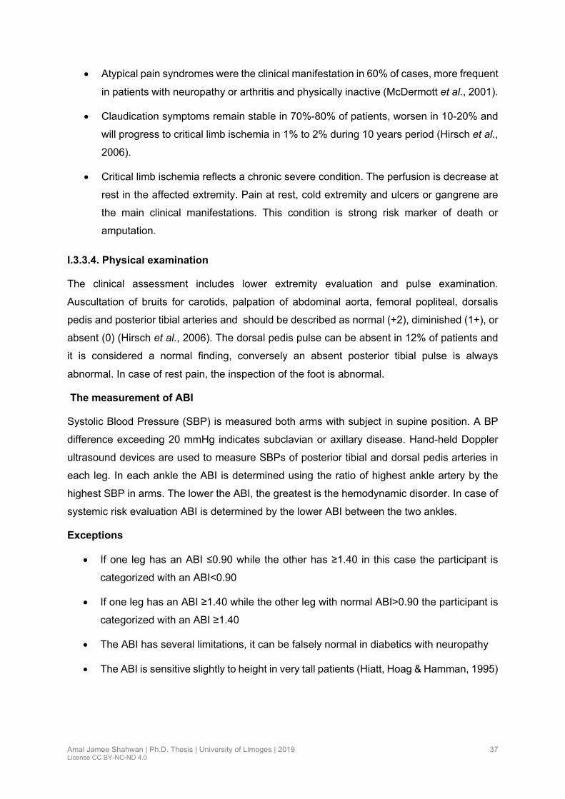
Amal Jamee Shahwan | Ph.D. Thesis | University of Limoges | 2019 License CC BY-NC-ND 4.0
37
• Atypical pain syndromes were the clinical manifestation in 60% of cases, more frequent
in patients with neuropathy or arthritis and physically inactive (McDermott et al., 2001).
• Claudication symptoms remain stable in 70%-80% of patients, worsen in 10-20% and
will progress to critical limb ischemia in 1% to 2% during 10 years period (Hirsch et al.,
2006).
• Critical limb ischemia reflects a chronic severe condition. The perfusion is decrease at
rest in the affected extremity. Pain at rest, cold extremity and ulcers or gangrene are
the main clinical manifestations. This condition is strong risk marker of death or
amputation.
I.3.3.4. Physical examination
The clinical assessment includes lower extremity evaluation and pulse examination.
Auscultation of bruits for carotids, palpation of abdominal aorta, femoral popliteal, dorsalis
pedis and posterior tibial arteries and should be described as normal (+2), diminished (1+), or
absent (0) (Hirsch et al., 2006). The dorsal pedis pulse can be absent in 12% of patients and
it is considered a normal finding, conversely an absent posterior tibial pulse is always
abnormal. In case of rest pain, the inspection of the foot is abnormal.
The measurement of ABI
Systolic Blood Pressure (SBP) is measured both arms with subject in supine position. A BP
difference exceeding 20 mmHg indicates subclavian or axillary disease. Hand-held Doppler
ultrasound devices are used to measure SBPs of posterior tibial and dorsal pedis arteries in
each leg. In each ankle the ABI is determined using the ratio of highest ankle artery by the
highest SBP in arms. The lower the ABI, the greatest is the hemodynamic disorder. In case of
systemic risk evaluation ABI is determined by the lower ABI between the two ankles.
Exceptions
• If one leg has an ABI ≤0.90 while the other has ≥1.40 in this case the participant is
categorized with an ABI<0.90
• If one leg has an ABI ≥1.40 while the other leg with normal ABI>0.90 the participant is
categorized with an ABI ≥1.40
• The ABI has several limitations, it can be falsely normal in diabetics with neuropathy
• The ABI is sensitive slightly to height in very tall patients (Hiatt, Hoag & Hamman, 1995)

Amal Jamee Shahwan | Ph.D. Thesis | University of Limoges | 2019 License CC BY-NC-ND 4.0
38
I.3.3.5. Investigations
• Arterial duplex ultrasound is non-invasive approach used to define the anatomy, the
hemodynamic, the extent of LEAD and the lesion morphology. The sensitivity and
specificity were reported to 95% and 99% respectively (Whelan, Barry & Moir, 1992).
• Peripheral angiography or computed tomography angiography defines the location and
the extent of the lesions. These tests are recommended when surgery is mandatory
I.3.3.6. Risk factors for LEAD
LEAD shares the same risk factors with another CVD such as CAD and stroke. Smoking is the
strongest risk factors for LEAD and the only traditional cardiovascular risk factor for which the
Odds ratio differ between LEAD and CAD or stroke (Criqui & Aboyans, 2015). The increase in
risk is related to the number of cigarettes smoked (Bainton et al., 1994). In the majority of
studies, current smoking versus non-smoking double the odds of LEAD (Fowkes et al., 2013;
Criqui & Aboyans, 2015). In LMIC this hypothesis is significant in males more than females
due to conservative cultural and social values. Diabetes mellitus was found to be associated
with LEAD after multivariate adjustment with Odds ratio varying from 1.50-4.05 (Criqui &
Aboyans, 2015). Hypertension demonstrated an association to LEAD with Odds ratios ranging
from of 1.50 to 2.20. In the Framingham Study, the risk of IC increases by 30% in the population
with blood pressure exceeding 160/100 mmHg (Murabito et al., 1997). In Health Professionals
Follow-up Study (HPFS), hypertension was reported up to 41% in persons with LEAD, in
addition the risk in hypertensive males is 2.5-fold and 3.9-fold in females (Joosten et al., 2012;
Ingolfsson et al., 1994). Total cholesterol, High density lipoprotein cholesterol (HDL ch) and
triglycerides, all appear to be potential risk factors for LEAD (Fowkes et al., 1992; Curb et al.,
1996). Obesity seems to be the cause of other risk factors for LEAD such as diabetes,
hypertension and dyslipidemia. Many large studies have failed to find a significant association
between obesity and LEAD or IC after multivariate adjustment regression (Hooi et al., 2001;
Ness, Aronow & Ahn, 2000).
I.3.3.7. Treatment
I.3.3.7.1. Pharmacological therapy
The main goals of treatment for LEAD include reduction in the risk of cardiovascular event
such as myocardial infarction and stroke, and improvement of symptoms and quality of life.
• Statins is recommended as primary and secondary prevention (Aboyans et al., 2018).
Statin reduced cardiac event, mortality post revascularization mortality and improved
painless walking distance (Aung et al., 2007; Ramos et al., 2016)
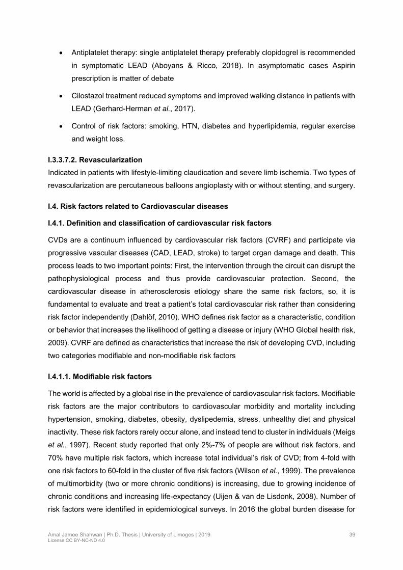
Amal Jamee Shahwan | Ph.D. Thesis | University of Limoges | 2019 License CC BY-NC-ND 4.0
39
• Antiplatelet therapy: single antiplatelet therapy preferably clopidogrel is recommended
in symptomatic LEAD (Aboyans & Ricco, 2018). In asymptomatic cases Aspirin
prescription is matter of debate
• Cilostazol treatment reduced symptoms and improved walking distance in patients with
LEAD (Gerhard-Herman et al., 2017).
• Control of risk factors: smoking, HTN, diabetes and hyperlipidemia, regular exercise
and weight loss.
I.3.3.7.2. Revascularization
Indicated in patients with lifestyle-limiting claudication and severe limb ischemia. Two types of
revascularization are percutaneous balloons angioplasty with or without stenting, and surgery.
I.4. Risk factors related to Cardiovascular diseases
I.4.1. Definition and classification of cardiovascular risk factors
CVDs are a continuum influenced by cardiovascular risk factors (CVRF) and participate via
progressive vascular diseases (CAD, LEAD, stroke) to target organ damage and death. This
process leads to two important points: First, the intervention through the circuit can disrupt the
pathophysiological process and thus provide cardiovascular protection. Second, the
cardiovascular disease in atherosclerosis etiology share the same risk factors, so, it is
fundamental to evaluate and treat a patient’s total cardiovascular risk rather than considering
risk factor independently (Dahlöf, 2010). WHO defines risk factor as a characteristic, condition
or behavior that increases the likelihood of getting a disease or injury (WHO Global health risk,
2009). CVRF are defined as characteristics that increase the risk of developing CVD, including
two categories modifiable and non-modifiable risk factors
I.4.1.1. Modifiable risk factors
The world is affected by a global rise in the prevalence of cardiovascular risk factors. Modifiable
risk factors are the major contributors to cardiovascular morbidity and mortality including
hypertension, smoking, diabetes, obesity, dyslipedemia, stress, unhealthy diet and physical
inactivity. These risk factors rarely occur alone, and instead tend to cluster in individuals (Meigs
et al., 1997). Recent study reported that only 2%-7% of people are without risk factors, and
70% have multiple risk factors, which increase total individual’s risk of CVD; from 4-fold with
one risk factors to 60-fold in the cluster of five risk factors (Wilson et al., 1999). The prevalence
of multimorbidity (two or more chronic conditions) is increasing, due to growing incidence of
chronic conditions and increasing life-expectancy (Uijen & van de Lisdonk, 2008). Number of
risk factors were identified in epidemiological surveys. In 2016 the global burden disease for

Amal Jamee Shahwan | Ph.D. Thesis | University of Limoges | 2019 License CC BY-NC-ND 4.0
40
risk profiles in Middle East and North Africa (MENA) stranded out these risk factors by order
of priority: high blood pressure ranked as the first, followed by obesity, diabetes then smoking
and dyslipidemia (Forouzanfar et al., 2016).
I.4.1.1.1. Hypertension
I.4.1.1.1.1. Definition, classification
Hypertension constitutes a major public health challenge in the world. HTN is defined as a
Systolic Blood Pressure (SBP) ≥140 mmHg and /or Diastolic Blood Pressure (DBP) ≥ 90
mmHg or taking antihypertensive drugs or having been told at least twice by a healthcare
professional as having HTN (Table 5). Normal levels of both systolic and diastolic blood
pressure are essential for the function of vital organs such as the heart, brain, kidney, for
overall health and wellbeing (WHO, 2013).
Table 5: Blood pressure classification according to WHO (WHO hypertension 2013)
Normal Systolic: less than 120 mmHg
Diastolic: less than 80 mmHg
A risk (prehypertension)
Systolic: 120–139 mmHg
Diastolic: 80–89 mmHg
High Systolic: 140 mmHg or higher
Diastolic: 90 mmHg or higher
I.4.1.1.2. Obesity
I.4.1.1.2.1. Definition, classification
Obesity is defined as excess amount of body fat, which leads to an ill health and risk of the
development of type 2 diabetes and cardiovascular event. It is related to the epidemiological
transition of NCDs. Overall, obesity ranked as the sixth leading cause of Disability Adjusted
Life Years (DALYs). Overweight causes almost 3 million deaths worldwide each year (WHO
Global health risk, 2009). WHO and National heart, Lung, and blood institute (NHLBI) define
weight categories for adults as follows: Body Mass Index (BMI): normal weight (25.0 kg/m2 ≤
BMI ≤ 29.9 kg/m2 ), obese class I (BMI 30-35 kg/m2), class II (BMI >35 to 39.9 kg/m2), and
class III (BMI ≥40 kg/m2) (WHO, 2017a).

Amal Jamee Shahwan | Ph.D. Thesis | University of Limoges | 2019 License CC BY-NC-ND 4.0
41
I.4.1.1.3. Diabetes mellitus
I.4.1.1.3.1. Definition, classification
Diabetes mellitus is a condition defined by an elevated level of blood glucose. Glycated
haemoglobin A1c (Hb A1c) has been recommended as diagnostic test for DM. The threshold
of ≥6.5% was adopted to diagnose diabetes.
The classification based on recommendation of the WHO and the American Diabetes
Federation (ADF) includes four classes (ESC et al., 2013).
• Type 1 diabetes: results from B-cells destruction leading to insulin deficiency
• Type 2 diabetes: the most common, accounts for 90-95% of all cases. It is the
consequence of a progressive insulin secretory defect, in association with obesity,
sedentary lifestyle, and insulin resistance
• Gestational diabetes: diagnosed during pregnancy, after delivery, most of them return
to a euglycemic state but they are at increased risk for T2DM in the future. A large
Canadian study estimates the probability to develop diabetes after 9 months of delivery
was 4 % and 19 % after 9 years (Feig et al., 2008)
• Other specific types of diabetes due to other cause such as genetic disorder, diabetes
secondary to pathological conditions or disease (pancreatitis, trauma, or surgery of the
pancreas), drugs induced diabetes (HIV/AIDS or after organ transplantation)
Table 6: Criteria for the diagnosis of diabetes (ADA, 2018)
FPG ≥126 mg/dL (7.0 mmol/L). Fasting is defined as no caloric intake for at least 8 h*.
OR
2-h PG ≥200 mg/dL (11.1 mmol/L) during OGTT. The test should be performed as described by the WHO, using a glucose load containing the equivalent of 75-g anhydrous glucose dissolved in water.*
OR
A1C ≥6.5% (48 mmol/mol). The test should be performed in a laboratory using a method that is NGSP certified and standardized to the DCCT assay.*
OR
In a patient with classic symptoms of hyperglycemia or hyperglycemic crisis, a random plasma glucose ≥200 mg/dL (11.1 mmol/L).
* In the absence of unequivocal hyperglycemia, results should be confirmed by repeat testing.

Amal Jamee Shahwan | Ph.D. Thesis | University of Limoges | 2019 License CC BY-NC-ND 4.0
42
Table 7: Type 2 Diabetes Mellitus risk factors (“Risk Factors of Type 2 Diabetes | NIDDK,” 2016)
1. Age ≥45years
2. Overweight or obese.
3. Sedentary lifestyle.
4. Family history with diabetes mellitus.
5. History of delivering a baby weighing >9 pounds.
6. Polycystic vary syndrome; and history of diabetes mellitus during pregnancy.
7. Elevated blood pressure (≥140/90 mmHg) or being treated for hypertension.
8. Levels of major lipids: High-density lipoprotein below 35 mg/dL, or TG above 250 mg/dL.
9. Prediabetes HbA1C level of 5.7 to 6.4 %; an elevated FPG test result of 100–125 mg/dL; or a two-hour oral glucose tolerance test result of 140–199 mg/dL.
10. Acanthosis nigricans presenting with a dark, velvety rash around the neck or armpits.
11. Current or prior history of CVDs
I.4.1.1.4. Alcohol
Epidemiologic studies define “heavy” drinking as ≥ 3 standard drinks per day, and lesser
amounts “light” or “moderate” drinking. According to WHO harmful use of alcohol is defined by
6.2 liters of pure alcohol per year which translates to 13.5 grams of pure alcohol per day for
persons ≥ 15 years of age (Anon, 2014).
I.4.1.1.5. Serum lipids
Dyslipidemia is defined by the elevation or attenuation of serum lipids. Cholesterol and
triglycerides are the major lipoproteins. To date, there is no evidence that fasting is superior to
non-fasting in evaluating a lipid profile for cardiovascular risk prediction, Many countries are
currently in the process of modifying their guidelines for measuring a lipid profile in the non-
fasting state, which facilitates blood collection for patients, laboratory technicians and clinician
(Langsted & Nordestgaard, 2019; Nordestgaard & Varbo, 2014).
All lipoproteins have a common basic structure but they differs in their size, density composition
and chemical proprieties (Yusuf et al., 2004). The different lipoproteins are including
chylomicrones, Intermediate Density Lipoprotein (IDL), Very Low-Density Lipoprotein (VLDL),
LDL, HDL, and apolipoproteins such as (Apo A, apo B, apo C and apo E). Lipids disorders are
defined as the total cholesterol, Low density Lipoprotein Cholesterol (LDL-C), High density
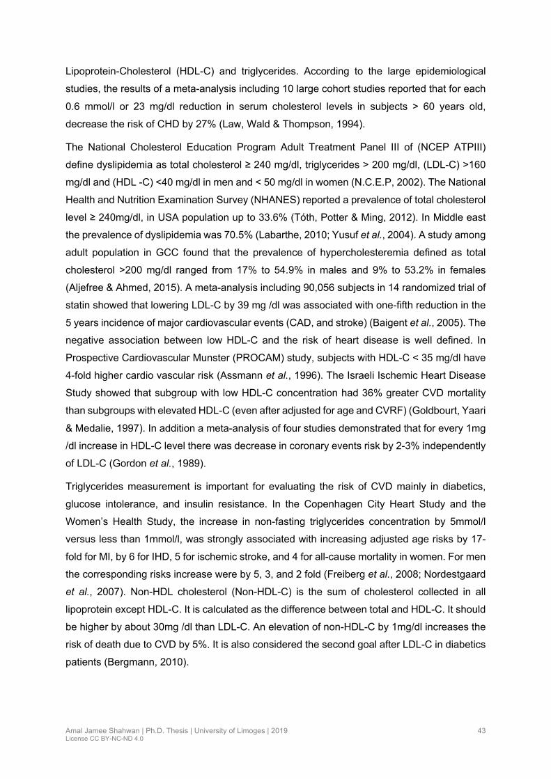
Amal Jamee Shahwan | Ph.D. Thesis | University of Limoges | 2019 License CC BY-NC-ND 4.0
43
Lipoprotein-Cholesterol (HDL-C) and triglycerides. According to the large epidemiological
studies, the results of a meta-analysis including 10 large cohort studies reported that for each
0.6 mmol/l or 23 mg/dl reduction in serum cholesterol levels in subjects > 60 years old,
decrease the risk of CHD by 27% (Law, Wald & Thompson, 1994).
The National Cholesterol Education Program Adult Treatment Panel III of (NCEP ATPIII)
define dyslipidemia as total cholesterol ≥ 240 mg/dl, triglycerides > 200 mg/dl, (LDL-C) >160
mg/dl and (HDL -C) <40 mg/dl in men and < 50 mg/dl in women (N.C.E.P, 2002). The National
Health and Nutrition Examination Survey (NHANES) reported a prevalence of total cholesterol
level ≥ 240mg/dl, in USA population up to 33.6% (Tóth, Potter & Ming, 2012). In Middle east
the prevalence of dyslipidemia was 70.5% (Labarthe, 2010; Yusuf et al., 2004). A study among
adult population in GCC found that the prevalence of hypercholesteremia defined as total
cholesterol >200 mg/dl ranged from 17% to 54.9% in males and 9% to 53.2% in females
(Aljefree & Ahmed, 2015). A meta-analysis including 90,056 subjects in 14 randomized trial of
statin showed that lowering LDL-C by 39 mg /dl was associated with one-fifth reduction in the
5 years incidence of major cardiovascular events (CAD, and stroke) (Baigent et al., 2005). The
negative association between low HDL-C and the risk of heart disease is well defined. In
Prospective Cardiovascular Munster (PROCAM) study, subjects with HDL-C < 35 mg/dl have
4-fold higher cardio vascular risk (Assmann et al., 1996). The Israeli Ischemic Heart Disease
Study showed that subgroup with low HDL-C concentration had 36% greater CVD mortality
than subgroups with elevated HDL-C (even after adjusted for age and CVRF) (Goldbourt, Yaari
& Medalie, 1997). In addition a meta-analysis of four studies demonstrated that for every 1mg
/dl increase in HDL-C level there was decrease in coronary events risk by 2-3% independently
of LDL-C (Gordon et al., 1989).
Triglycerides measurement is important for evaluating the risk of CVD mainly in diabetics,
glucose intolerance, and insulin resistance. In the Copenhagen City Heart Study and the
Women’s Health Study, the increase in non-fasting triglycerides concentration by 5mmol/l
versus less than 1mmol/l, was strongly associated with increasing adjusted age risks by 17-
fold for MI, by 6 for IHD, 5 for ischemic stroke, and 4 for all-cause mortality in women. For men
the corresponding risks increase were by 5, 3, and 2 fold (Freiberg et al., 2008; Nordestgaard
et al., 2007). Non-HDL cholesterol (Non-HDL-C) is the sum of cholesterol collected in all
lipoprotein except HDL-C. It is calculated as the difference between total and HDL-C. It should
be higher by about 30mg /dl than LDL-C. An elevation of non-HDL-C by 1mg/dl increases the
risk of death due to CVD by 5%. It is also considered the second goal after LDL-C in diabetics
patients (Bergmann, 2010).
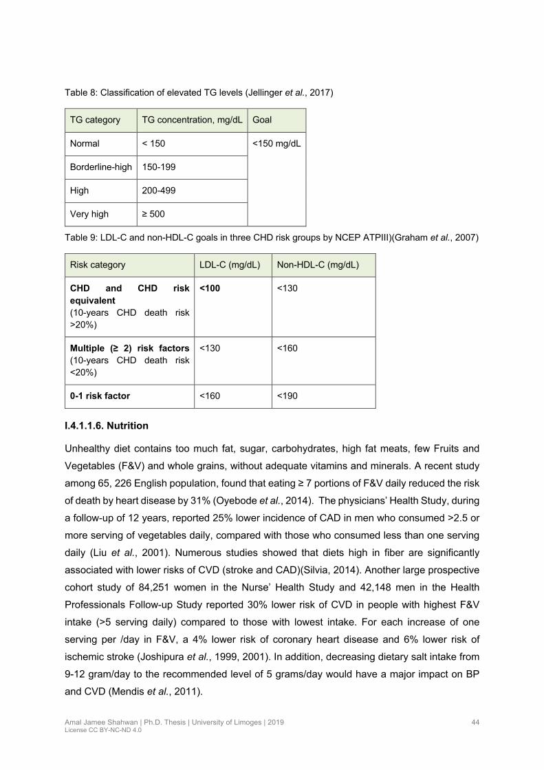
Amal Jamee Shahwan | Ph.D. Thesis | University of Limoges | 2019 License CC BY-NC-ND 4.0
44
Table 8: Classification of elevated TG levels (Jellinger et al., 2017)
TG category TG concentration, mg/dL Goal
Normal < 150 <150 mg/dL
Borderline-high 150-199
High 200-499
Very high ≥ 500
Table 9: LDL-C and non-HDL-C goals in three CHD risk groups by NCEP ATPIII)(Graham et al., 2007)
Risk category LDL-C (mg/dL) Non-HDL-C (mg/dL)
CHD and CHD risk
equivalent (10-years CHD death risk >20%)
<100
<130
Multiple (≥ 2) risk factors (10-years CHD death risk <20%)
<130 <160
0-1 risk factor <160 <190
I.4.1.1.6. Nutrition
Unhealthy diet contains too much fat, sugar, carbohydrates, high fat meats, few Fruits and
Vegetables (F&V) and whole grains, without adequate vitamins and minerals. A recent study
among 65, 226 English population, found that eating ≥ 7 portions of F&V daily reduced the risk
of death by heart disease by 31% (Oyebode et al., 2014). The physicians’ Health Study, during
a follow-up of 12 years, reported 25% lower incidence of CAD in men who consumed >2.5 or
more serving of vegetables daily, compared with those who consumed less than one serving
daily (Liu et al., 2001). Numerous studies showed that diets high in fiber are significantly
associated with lower risks of CVD (stroke and CAD)(Silvia, 2014). Another large prospective
cohort study of 84,251 women in the Nurse’ Health Study and 42,148 men in the Health
Professionals Follow-up Study reported 30% lower risk of CVD in people with highest F&V
intake (>5 serving daily) compared to those with lowest intake. For each increase of one
serving per /day in F&V, a 4% lower risk of coronary heart disease and 6% lower risk of
ischemic stroke (Joshipura et al., 1999, 2001). In addition, decreasing dietary salt intake from
9-12 gram/day to the recommended level of 5 grams/day would have a major impact on BP
and CVD (Mendis et al., 2011).
Amal Jamee Shahwan | Ph.D. Thesis | University of Limoges | 2019 License CC BY-NC-ND 4.0
45
I.4.1.1.7. Physical activity
Physically active means that you are protected from numerous risk factors, thus less exposed
to CVD. Additionally physical activity is fundamental to energy balance and weight control
(WHO, 2010). Morris et al found that civil servant post men who walked or cycled while
delivering mail had lower rates of heart disease compared to postal clerks who had sedentarily
jobs (Morris & Crawford, 1958). The sedentary life was a predominant factor in industrialized
nations (Archer & Blair, 2011). The rise in sedentarism has been an epidemic for chronic
disease mainly obesity. Regular physical activity has a protective effect on cardiovascular risk
by the deceleration of atherosclerosis progression, improvement of endothelial dysfunction,
decreased systemic inflammation and controlling major CVRF such as HTN, diabetes and
obesity (Cheng et al., 2013). The prevalence of inactivity was higher among the younger
population in GCC ranging from 24.3% to 93.9% in males and from 50% to 98.1% in females.
Saudi Arabia keeps the higher rate (Aljefree & Ahmed, 2015). In a meta-analysis of data from
32 studies comparing inactive persons and those who practiced 150 min /week of moderate -
intensity, physical activity had a 14% decrease in CAD, and those who practiced the equivalent
of 300 min /week of moderate-intensity physical activity had 20% lower CAD (Sattelmair et al.,
2011). In another meta-analysis including 23 studies the risk of stroke (ischemic or
hemorrhagic) was 27% in individuals with a moderate activity vs 20% in high physical activity
(Lee, Folsom & Blair, 2003).
I.4.1.1.8. Stress
Everyone feels stress in different ways and reacts to it in different ways. How much stress you
experience and how you react to it can lead to a wide variety of health problems (AHA 2014,
2014). The concept of psychological stress produces a physiological change by the activation
of the hypothalamic-pituitary-adrenocortical and sympathetic nervous system, which triggers
pathophysiological mechanisms that include inflammation, hemostasis, and dysfunction of
metabolic and cardiac autonomic control (Brotman, Golden & Wittstein, 2007). Personality
types (type A and type D) lead to unhealthy response to daily stressors. The type D (distressed)
personality is a negative affectivity characterized by a combination of pessimistic emotions,
depressed mood, anxiety, anger, worried and hostile feelings. This personality is almost
related to social phobia and panic disorder (Sher, 2005). By contrast, the type A personality is
characterized by anxiety, intense time urgency, intense competitiveness, hypervigilance, and
sometimes hostile behaviors (Ragland & Brand, 1988). In the general population, adults with
work stress or private-life stress have 1.1-1.6-fold increased risk of CAD and stroke (Kivimäki
& Steptoe, 2018). Chronic stress plays a role as a disease trigger in individuals with high
atherosclerotic plaque leading to cardiovascular events such as hypertension, insulin
resistance, arrhythmia, myocardial ischemia, cardiac failure and stroke (Kivimäki & Steptoe,

Amal Jamee Shahwan | Ph.D. Thesis | University of Limoges | 2019 License CC BY-NC-ND 4.0
46
2018). Emotional stress is involved in 3.9% of acute cardiac events (Nawrot et al., 2011).The
association with stroke was uncertain (Truelsen & Nielsen, 2003). Anxiety was reported to be
high in patients with a history of ACS. The influence lasts for a long period time in 20% to 25%
of patients after the first event (Moser et al., 2007).
Figure 7: Effects of stress (Nature Reviews cardiology 2018)
I.4.1.1.9. Socioeconomic status
Socioeconomic Status (SES) is a complex concept affecting health and known as powerful
predictor of CVDs and death (Stokols, Pelletier & Fielding, 1996). It is measured as a
combination of education, income, and occupation but may include age, sex, ethnicity and
marital status. The available data showed that the association between SES and CVD depends
on the socioeconomic development context, and the stage of the demographic,
epidemiological and nutritional transition of the population (Mestral & Stringhini, 2017). In the
HIC there is an inverse association between SES and CVD and CVRFs. The SES was
measured via education, occupation, or income. However, in LMIC the relation between SES

Amal Jamee Shahwan | Ph.D. Thesis | University of Limoges | 2019 License CC BY-NC-ND 4.0
47
and CVD or CDVRFs shows a positive association (Mestral & Stringhini, 2017), because
people with lower SES tend to have higher levels of traditional cardiovascular risk factors such
as higher BP, smoking, and obesity (Yu et al., 2000). Recent data confirms that SES is closely
related with the quality of diet (Turrell et al., 2003). Low SES people prefer white bread,
potatoes rice, and refined cereals compared to those with high SES, who prefer whole grain
products with lower glycemic index, consumption of vegetables and fruit (Cronin et al., 1982;
Shimakawa et al., 1994; Smith & Baghurst, 1992). The behavioral factors, such as physical
inactivity, smoking and alcohol intake, explain only 13%-60% of the SES differences in CVD
morbidity and 19%-55% of CVD mortality (Méjean et al., 2013).
I.4.1.2. Non-modifiable risk factors
I.4.1.2.1. Age
By 2030 20% of the population will be aged > 65 years. In this age group CVD will result in
40% of all deaths and will be the leading cause (North & Sinclair, 2012). The cardiovascular
system is strongly affected by the ageing process leading to progressive deterioration in
structure and function of the heart and vasculature that contribute to the development of CVD
(Costa et al., 2015). Epidemiologic studies revealed that at any age the risk of cardiovascular
events varies widely (4-5-fold) depending on the associated risk factors. Studies indicate that
the chances of surviving to age 85 years have decreased significantly with cumulative risk
factors, from 37% for men without risk factors to 2% with five risk factors and from 65% for
women without risk factors to 14% with five risk factors (Kannel & Vasan, 2009).
I.4.1.2.2. Gender
For many years, CVD was considered as a male disease. However, in the European population
38% of cardiovascular deaths before the age of 75 years were in women and 37% in men
(European Cardiovascular Disease Statistics, 2012). The difference between male and female
was previously described in epidemiology, pathophysiology, clinical manifestation and
management of CVD (Anon, 2012c). The sexual hormones drive differences in gene
expression and the function of cardiovascular system (Regitz-Zagrosek et al., 2016). In male
sex, cardiovascular risk increases over time, as well as atherosclerosis process continues. In
contrast women are protected from atherosclerosis during the fertile age by estrogens that
exert favorable effect on cardiovascular system. The effect disappears after menopause.
Women and men manifest a similar cardiovascular profile with a difference of 10 years of age
(Perk et al., 2012). In 2004, the WHO reported a total cardiovascular mortality of 55% in women
and 43% in men. IHD, stroke and other CVD represent 23%, 18% and 15% respectively in
women and 21%,11% and 11% respectively in men (Stramba-Badiale et al., 2006).

Amal Jamee Shahwan | Ph.D. Thesis | University of Limoges | 2019 License CC BY-NC-ND 4.0
48
Over 5 decades in the Framingham study cohort the occurrence of CAD or stroke were up to
47%-31% and 15%-18% respectively in males and females. The higher risk of stroke in female
was related to longer life expectancy (Lloyd-Jones et al., 1999; Seshadri et al., 2006). Gender
difference for traditional cardiovascular risk factors associated with CVD were also
documented. Age, hypertension, total cholesterol and LDL-C had a great influence in men,
while menopause, systolic arterial hypertension, smoking, diabetes, triglycerides and HDL-C
were the main actors in women (Leonarda & Gabriella, 2015). Every 10 mmHg of SBP was
associated with15% increased risk of CAD and 25% increased risk of stroke in both sexes
(Peters, Huxley & Woodward, 2013). Diabetes increased cardiovascular risk of 3-7 fold in
women and 2-3 fold in men (Manson, 1996).Two large meta-analyses reported that the risk of
CAD and stroke were increased by 44% and 27% in diabetic women (Huxley, Barzi &
Woodward, 2006; Peters, Huxley & Woodward, 2014). The prevalence of overweight in men
and women differs according to the level of the development of the country. Higher BMI is
more prevalent in men than women in HIC, conversely in LMIC mainly in Arab countries a
female predominance was described (Anon, 2015).
Total cholesterol confers the same risk of cardiovascular in both sexes. However LDL-C
increases cardiovascular risk in men more than women (Manolio et al., 1992). Low HDL-C
represents equal risk for CAD in both sexes, mainly young age, but predicts CAD mortality in
women more than in men. In addition, triglycerides are a part of metabolic syndrome which is
higher in women. Smoking increases risk of cardiovascular events by 3.6 in women and 2.4 in
men (Willett et al., 1987). A meta-analysis from 74 prospective cohort studies show that women
who smoke had a 25% greater relative risk of CAD than men (Huxley & Woodward, 2011).
I.4.1.2.3. Family history
Represents one of the main risk factors for CVD, especially in the younger population with a
first-degree relative disorder: (men below the age of 55 years and women below 65 years)
(Choudhury & Marsh, 1999; Elis & Lishner, 2004). Family history helps to define the small
subset of families that account for the majority of prevalent cases in the population (Hunt,
Gwinn & Adams, 2003). The risk for CVD and stroke among subjects with positive family
history ranges from 2 to 9 and 1.5 to 2, respectively (Kardia, Modell & Peyser, 2003; Liao et
al., 1997). Family history represents the interaction between genetic, environmental and
behavioral factors (Elis et al., 2008). Even a non-premature parental history increases the risk
of CVD in offspring (Sesso et al., 2001).The history of heart attack in both parents increases
the risk of CAD mainly when 1 parent has a premature coronary event before 50 years of age
(Chow et al., 2011). Sibling history of CVD has been shown to increase the odds of CVD in
males and females by 45% (Murabito et al., 2005). In a recent study of patients with premature
Amal Jamee Shahwan | Ph.D. Thesis | University of Limoges | 2019 License CC BY-NC-ND 4.0
49
ACS (age ≤ 55 years), 28% of the females and 20% of the males had a family history of CAD.
Patients with family history of CAD had a higher prevalence of traditional CVD risk factors
(HTN, DM, dyslipidemia and obesity) (Choi et al., 2014; Hunt, Gwinn & Adams, 2003; Yoon et
al., 2002). In monozygotic twins the risk of death from CAD increased 3.8 to 15 times if a
sibling died of CAD before age 75 (Marenberg et al., 1994). In addition a large international
case-control study reported a rise in the risk of MI if one parent had MI (OR=1.67), or one
parent had MI before age 50 (OR=2.36), or both parents had MI (OR=2.90) and if both parent
had MI before age 50 (OR=6.56) (Anderson et al., 2013). The prevalence of a positive family
history ranges from 14% to 35% in the general population, 75% of those with premature heart
disease have a positive family history highlighting the opportunities for prevention (Hawe et
al., 2003).
I.4.1.2.4. Menopause
Menopause is an indicator of the transition from reproductive to non-reproductive life and is
associated with biological and hormonal changes. The risk is related to post menopause is
due to a sudden decrease of estrogen hormone, which has protective effects on lipid, glycemic
metabolism and vessels (Rossi et al., 2002). Menopausal status and estrogen deficiency were
frequently associated with hypertension due to increase in BMI, insulin-resistance, sodium
retention, and with increased smooth muscle cell proliferation leading to an increase in
systemic vascular resistance. The role of Hormonal Replacement Therapy (HRT) in CVD is
controversial. In contrast Women’s Health Initiative study described an increased risk of CAD
and breast cancer in users of HRT including estrogens and a synthetic progestin (Rossouw et
al., 2007; The Writing Group on behalf of the Workshop Consensus Group, 2009).
I.4.1.3. Novel risk factors
I.4.1.3.1. Fibrinogen
Plasma fibrinogen as a coagulation factor is a heterogeneous mixture of many different
molecular forms (Gordon et al., 1989). Many studies reported the strong relationship between
plasma fibrinogen concentration and cardiovascular disease mainly CAD, stroke and LEAD
(Lowe, 1995). Fibrinogen concentration raises the risk of atherogenesis, thrombogenesis and
ischemia (Danesh et al., 2000). Recently fibrinogen was introduced as risk factor for premature
CAD in subjects <55 years (Pineda et al., 2009; Shojaie et al., 2009). The fibrinogen is a
pathway by which traditional risk factors exert their effect. For example, fibrinogen levels
increased risk of CVD associated with smoking and essentially with the number of cigarettes
smoked. The level decreases after smoking cessation (Fogari et al., 1994). The risk of CVD
associated with obesity might be driven by the fibrinogen. A loss of weight after low calories
diet leads to fall in fibrinogen levels (Ditschuneit, Flechtner-Mors & Adler, 1995). The

Amal Jamee Shahwan | Ph.D. Thesis | University of Limoges | 2019 License CC BY-NC-ND 4.0
50
Prospective Cardiovascular Munster (PROCAM) study found that subjects with both high level
of LDL-C and fibrinogen had a 6.1-fold increase in CAD compared with those with lower or
normal levels (Heinrich et al., 1994; Thompson et al., 1995). The Gothenburg and Framingham
Studies reported that plasma fibrinogen levels represent an independent risk factor for MI and
stroke and strong risk factor for cardiac sudden death in patients with CAD (Thompson et al.,
1995). Also, platelets hyperactivity due to high fibrinogen concentration was found in diabetic
patients (Stec et al., 2000).
I.4.1.3.2. Homocysteine
Homocysteine concentration is higher in men than women and increases with age.The
difference becomes apparent in puberty and it is related to hormonal factors, lifestyle, nutrition,
and vitamins. Thromboembolic events were present in 50% of untreated persons with high
level of homocysteine, and 20% die before the age of 30 years (Nygård et al., 1999). Numerous
clinical studies demonstrated a relationship between total homocysteine levels and CAD,
LEAD, stroke or venous thrombosis (Boushey et al., 1995; Verhoef & Stampfer, 1995).The
homocysteine affects the coagulation system and the resistance of the endothelium to
thrombosis and may interfere with the vasodilator and antithrombotic effects of nitric oxide
(Stamler & Slivka, 1996).
I.4.1.3.3. C. Reactive Protein
C-Reactive Protein (CRP) is a marker of inflammation and a hepatically derived pentraxin that
plays a role in the immune response. CRP has a long plasma half-life. Numerous
epidemiological studies have demonstrated the role of CRP in the occurrence of MI, stroke,
LEAD, sudden cardiac death and it plays a role in almost all process associated with metabolic
syndrome (Ridker, 2003). CRP seems to be a stronger predictor of cardiovascular events than
LDL ch (Mendall et al., 2000). CRP levels <1, 1 to 3, and >3 mg/dl correspond to low-moderate,
and high risk. Subjects with LDL ch <130 mg/dl and CRP >3 mg /dl represent a high-risk group
(Ridker, 2003). CRP was a strong predictor of risk even 20 years after initial blood samples
were obtained (Sakkinen et al., 2002).
I.4.1.4. Co-occurrence of risk factors
A number of studies have demonstrated that five modifiable CVRF such as cigarette smoking,
overweight or obesity, hypertension, diabetes and dyslipidemia can be eliminated by
management. The risk of CVD increases with increasing number of risk factors (Yusuf et al.,
1998). Data from the first National Health and Nutrition Examination Surveys Epidemiologic
Follow-up study showed that the risk for CVD increased with each additional risk factor. More
than 50% of the incidence of CAD, stroke and all-cause mortality was due to having one risk

Amal Jamee Shahwan | Ph.D. Thesis | University of Limoges | 2019 License CC BY-NC-ND 4.0
51
factor and the risk increases up to 70% for individuals with three risk factors (Yusuf et al.,
1998). Primary prevention and control risk factors may not reduce the risk of CVD to the
equivalent of never having a risk factor. Individuals who control their hypertension are at high
risk for CVD compared with those who never develop HTN (N.H.BP.E.P, 1993). Furthermore,
the Metabolic Syndrome (MetS) was identified by several criteria and defined as an
asymptomatic, pathophysiological state of chronic inflammation, and a cluster of the most
harmful risk factors, such as obesity, insulin resistance, hypertension, hyperglycemia and
dyslipidemia (Kaur, 2014). The International Diabetes Federation (IDF) estimates that a
quarter of the world’s adult population has MetS (O’Neill & O’Driscoll, 2015). The rate varies
depending on age, ethnicity, and gender of the population (Kaur, 2014). MetS confers a 5-fold
increase in the risk of type 2 diabetes mellitus and 2-fold the risk of developing CVD over the
next 5 to 10 years (Alberti et al., 2009). In addition individuals with MetS are at 2- 4 fold the
risk of stroke and 3-4 fold the risk of myocardial infraction (Alberti, Zimmet & Shaw, 2005). The
early identification and control of MetS components prevents the development of the syndrome
and reduces CVD events.

Amal Jamee Shahwan | Ph.D. Thesis | University of Limoges | 2019 License CC BY-NC-ND 4.0
52
Chapter II. Epidemiology of Cardiovascular disease and cardiovascular risk
factors
II.1. Cardiovascular disease in the world
CVD is a major public health burden and is the leading cause of total deaths in all regions of
the world except Sub Saharan Africa. In 2016 the number of people dying from CVD increased
by 15% accounting for 17.6 million deaths per years, due to population ageing and growth
(GBD 2016 Causes of Death Collaborators, 2017), and projected to increase to 23.6 million by
2030 (WHO 2014, n.d.) with total cost of $863 billion (Bloom et al., 2012). Ischemic heart
disease and stroke combined account for more than 85.1% of all CVD death in 2016 (GBD
2016 Causes of Death Collaborators, 2017). Coronary artery disease was the most common
45.1%, followed by stroke 16.5% (CDC, 2016a). In USA adults’ population 92.1 million have
at least one type of CVD (Benjamin et al., 2017). By 2030, 43.9% of USA population is
projected to have some form of CVD (Heidenreich et al., 2011). The prevalence of CVD in USA
population >25 of age in 2011-2014 was 36.6% (Benjamin Emelia J. et al., 2018). In Europe
CVD account for 45% of all deaths in Europe and 37% of all death in the European Union
mainly (Wilkins et al., 2017). In United kingdom in 2012, CVD represented a second cause of
deaths (28%), 46% were from CAD and 26% were from stroke (Bhatnagar et al., 2016). Major
CVD event rates, CVD death rates, and all cause death rate were lowest in HIC and highest
in LMIC accounting for 70% of CVD death (WHO 2014, n.d.).
II.2. In the Arab countries
The EMR represents a mosaic group of 23 countries (Figure8), including Arab states located
in North east African nations comprise (Djibouti, Egypt, Somalia, Sudan) and North west
African include (Libya, Morocco, Tunisia and Algeria). Gulf Cooperation Council with (Bahrain,
Kuwait, Oman, Qatar, Saudi Arabia, United Arab Emirates and Yemen). Another group which
include (Iraq, Jordan, Lebanon, Palestine, and Syria), Non-Arab countries included in EMR
were (Afghanistan, Iran and Pakistan). Each country has a unique historical, geopolitical,
social, cultural, and economic profile (Mandil, Chaaya & Saab, 2013). Arab countries were
also, divided according to their Gross National Income (GNI) per person.
• Low income countries (LIC) (Djibouti, Mauritania, Yemen, and Somalia) had a mean
GNI per person of $523.
• Middle income countries (MIC) (Algeria, Egypt, Iraq, Lebanon, Libya, Morocco,
Palestine Sudan, Syria and Tunisia) had a GNI of $3251.
• HIC (Bahrain, Saudi Arabia, Kuwait, Oman, Qatar and United Arab Emirates) had GNI
of $39 688 (Mokdad et al., 2014).
Amal Jamee Shahwan | Ph.D. Thesis | University of Limoges | 2019 License CC BY-NC-ND 4.0
53
The estimated median life expectancy in EMR population is 72 years, higher in GCC (78
years), ranging from 48-59 years in Somalia, Afghanistan and Sudan. In The Other
countries, life expectancy interval is between 63-74 years (Anon, n.d.). Arab Middle East
Countries which have a predominance of young population ranging in middle age from 17-
28 years, have undergone rapid socioeconomic changes, political conflicts, poverty,
intermediate level of economic development, instability and epidemiologic transition
(Ramahi, 2010). The Arab countries (Iraq, Syria, Lebanon, Jordan, Egypt, Yemen and
Turkey) have a highest CVD mortality death ranging from 145 per 100000 in Qatar to 548
per 100000 in Yemen, with significant variation between urban and rural areas (Roth et al.,
2015). Very few community-based studies were conducted in these countries. Conversely,
more data in CVD are available for Arab Gulf states. These countries have undergone rapid
modernization and socioeconomic development. Ischemic heart disease and stroke were
two of the top five causes of deaths in Arab world. Ischemic heart disease accounting for
90.3 deaths per 100 000 people (Mokdad et al., 2016). The diabetes prevalence is up to
9.3% (ranging from 7 % to 26% among adult aged 15-65 years; it is the second highest
among all WHO regions (Jabbour & Yamout, 2012). The global Adult tobacco survey
showed that in 14 MICs 48.6% of men and 11.3% of women were tobacco users (Giovino
et al. 2012). In nine Arab countries (Bahrain, Egypt, Libya, Jordan, Kuwait, Lebanon,
Palestine, Tunisia and Syria) the prevalence of daily tobacco use exceeds 30% in men
(Rahim et al., 2014). Water pipe smoking is increasing in young Arabs with prevalence
estimates between 6% and 34% in age group 13-15 years (Maziak, 2011). Data from
regional STEPS survey shows that 79%-90% of adults in most Arab countries reported
eating less than the 5 serving of fruits and vegetables (Anon, 2012b). Obesity continues to
rise in Arab population with alarming rate in female more than male. LMICs accounted for
7% of the world’s overweight individuals (Boyko et al., 2000). Also, physical inactivity
represents high prevalence level particularly in Gulf cooperation which accounts for 70%.
(Rahim et al., 2014) .

Amal Jamee Shahwan | Ph.D. Thesis | University of Limoges | 2019 License CC BY-NC-ND 4.0
54
Figure 8: Arab countries Map

Amal Jamee Shahwan | Ph.D. Thesis | University of Limoges | 2019 License CC BY-NC-ND 4.0
55
Table 10: Cardiovascular risk factors (smoking/physical inactivity) in adults aged ≥15 years and overweight and obesity in adults ≥ aged 20 years in Arab countries (Rahim et al., 2014).
II.3. Coronary artery disease
CAD remains the main cause of death up to 13.2% of total deaths worldwide (Usta & Bedel,
2017). In the United State of America (USA), it accounts for one quarter of all deaths. In the
European countries 27%-34% of people with CAD are over 75 years (Members et al., 2002).
In United kingdom (UK), CAD was responsible for 16% of all male deaths and 10% of all female
deaths (Bhatnagar et al., 2015). The mortality rate has declined in HIC, in the SWEDEHEART
registry the mortality related to ACS has decreased over the last three decades (Szummer et
al., 2017). Total CAD prevalence was 6.3% in USA adults ≥ 20 years of age (7.4% in males

Amal Jamee Shahwan | Ph.D. Thesis | University of Limoges | 2019 License CC BY-NC-ND 4.0
56
and 5.3% in females) (Benjamin Emelia J. et al., 2018). There is a strong relation between
sudden death and coronary disease. Post mortem studies and death certificates revealed that
62-85% of patients who died out of hospital have past history of CAD (Sanchis-Gomar et al.,
2016). According to data from NHANES 2011 to 2014, myocardial infraction has a prevalence
of 3.0% (3.3% for males and 2.3% in females) in adult USA population (Benjamin et al., 2017).
Approximatively every 40 seconds, an American will have an MI. The average age at first MI
in American population is 65.6 years for males and 72.0 years for females (Benjamin Emelia
J. et al., 2018)). In Middle East the rate of death from CAD was higher compared to western
countries United Kingdom (UK), Germany, and the USA. The INTERHEART study (an
international case-control analysis of the risk factors for the first MI carried out in 52 countries)
found that the median age at MI onset was 51 years in the Middle East population, and was
12 years lower than the median age at presentation in western countries (Gehani et al., 2014).
II.4. Stroke
In 2013, strokes were the second cause of death worldwide (11.8%) after IHD, the third
common cause of disability adjusted life years from all causes, the second cause of dementia
and the most probable cause of epilepsy in elderly (Markus, 2016). Stroke leads to morbidity
and major disability in 15%-30% of survivors, 20% of them need rehabilitation (Marx,
Hockberger & Walls, 2006). Globally the incidence of stroke due to ischemia was 68% and
32% was due to hemorrhagic stroke (Krishnamurthi et al., 2013). Hemorrhagic stroke occurs
most frequently in patients under 40 years of age and 2-3 times higher in blacks and Asians
(Foulkes et al., 1988). China has the greatest mortality rate of stroke in the world (Wang et al.,
2017) . In USA, stroke accounts for 6% (Bhatnagar et al., 2015), while the proportion of
ischemic stroke is 87%, ICH 10% and SAH 3% (Benjamin Emelia J. et al., 2018). Two third of
all stroke occur in persons less than 70 years of age, probably related to increasing prevalence
of metabolic risk factors (Kissela et al., 2012; Giang et al., 2013; Feigin, Norrving & Mensah,
2017). In addition, 15%-30% of all cerebral infarction are preceded by TIA (Hankey & Warlow,
1999),17% in the same day,9% on the previous day and 43% on the preceding week (Rothwell
& Warlow, 2005). Hemorrhagic stroke accounts for 15% to 20% of all stroke, with 30-day
mortality of 35% to 52% of them and 50% of death occurring in the first 48 hours (Counsell et
al., 1995).
During 1970 and 2008 period, there was a 42% decrease in stroke incidence in HIC and more
than 100% increase in LMIC. The global incidence in LMIC has for the first time exceeded the
stroke incidence in HIC by 20%, with higher proportion of hemorrhagic stroke accompanied
with high mortality rate accounting for 85% of stroke death globally (Feigin et al., 2016;
Lackland et al., n.d.). In the Arab world the trends of stroke are not precisely established
(Rahim et al., 2014). Epidemiological data in Arab word suffers from important limits. First,
Amal Jamee Shahwan | Ph.D. Thesis | University of Limoges | 2019 License CC BY-NC-ND 4.0
57
most of the data was hospital based, second the modern imaging technic (CT and MRI) were
not available all the time. The Stroke incidence rate is increasing in Arab countries with
increasing in the prevalence of cardiovascular disease. The review of Benamer on stroke
epidemiology in Arab countries shows a prevalence of 0.04-0.07% in ten Arab countries with
higher prevalence in males than females, and ischemic type was the commonest type ranging
from 55% to 87% while cerebral hemorrhage from 6.3% to 41.3% (Benamer & Grosset, 2009).
Hypertension is the most common risk factor weather in HIC or LMIC.
II.5. Lower extremity arteries disease
LEAD is the third leading cause of atherosclerosis morbidity after CAD and stroke. In 2010 it
was estimated that 202 million individuals have LEAD, 69.7% of them in LMIC. The prevalence
rate increased by 28.7% in LMIC and 13.1% in HIC (Fowkes et al., 2013).
According to the AHA during the period of 1970-2000, 6.5 million Americans aged ≥40 years
(5.5%) have LEAD (ABI<0.90), and one quarter of them have severe LEAD (ABI<0.70) (Allison
et al., 2007; Benjamin et al., 2017). The prevalence of LEAD increases with age. It affects 7%
of individuals aged 60-69 years,12.5% for those aged 70-79 years, and 23.2% of those ≥ 80
years (Ostchega et al., 2007). The rate increases as the population ages and becomes more
obese and suffers from diabetes (Teodorescu, Vavra & Kibbe, 2013). The generalized nature
of atherosclerosis was well documented by the Clopidogrel versus Aspirin in Patients at Risk
of Ischemic Events (CAPRIE) data, where 41 % of patients with LEAD had CAD or CBVD and
8.6% had disease in all three territorries (Ouriel, 2001). Patients with LEAD have a five-fold to
six-fold increased risk of death or morbidity from other atherosclerotic disease process such
as stroke or CAD (Hirsch et al., 2001). Nearly 42.8% of those with LEAD had CAD (Aboyans
et al., 2010). Also, the risk of death is higher in those with LEAD compared to those without,
and in both symptomatic or asymptomatic patients (Diehm et al., 2009). The Framingham
Heart Study reports that people with LEAD were 5.9 times at higher risk for death by CVD and
6.6 times by CAD (Aponte, 2012). Most studies on LEAD suggest that the prevalence rate and
associated risk factors may differ between male and female (Fowkes et al., 2013). The
prevalence of females with LEAD is slightly higher in LMIC at all ages. In HIC the prevalence
is higher in males (Fowkes et al., 2013). To date there are no published data about LEAD in
the Arab countries’ population. Available little studies are hospital based. The most important
of them is the Global Atherothrombosis Assessment-Middle East (AGATHA-ME) conducted in
five Arab countries of Middle East which recruited 1341 patients from tertiary care. Patients
were classified into two groups: with CVD group and at-risk group. The prevalence of PAD was
higher in two groups with predominance in at risk group (31.5% vs 28.2%) (El-Menyar, Al
Suwaidi & Al-Thani, 2013).

Amal Jamee Shahwan | Ph.D. Thesis | University of Limoges | 2019 License CC BY-NC-ND 4.0
58
II.6. Cardiovascular risk factors in the world
II.6.1. Hypertension
Approximately one billion of people worldwide have HTN which corresponds to more than
40% (Mozaffarian et al., 2016) and projected to increase by 30% in 2025 (Kearney et al., 2005).
In all world regions the prevalence is similar in males and females and rises with increasing
age and BMI. Globally HTN is the major cause of mortality responsible for over 7.5 million
deaths annually (WHO Global health risk, 2009). The NHANES 2011-2014 estimates the
prevalence of USA adults aged ≥ 20 years was up to 34%, ranging from 11.6% in those aged
20-39 years to 67.2% among those aged ≥ 60 years (Benjamin Emelia J. et al., 2018). In LMIC
1 in 3 persons have HTN, with higher rate in elderly and overweight or obese person (Sarki et
al., 2015). In Middle East the prevalence of HTN is 30% with slight difference between sexes
(30.7% in male and 29.1% in female) (Anon, 2012a). In North Africa and Middle East, HTN
was among the three leading risk factors for disease burden in 2015 as well as in Palestine
(Forouzanfar et al., 2016). In the Africa Middle East Cardiovascular Epidemiology study (ACE),
conducted in 14 countries of Middle east and Africa (2012), the prevalence of HTN ranged
from 25% in Tunisia to 53% in South Africa (Alsheikh-Ali et al., 2014).
II.6.1.1. Hypertension and cardiovascular disease
Several studies have reported the association between either SBP or DBP and the increase
CVD risk. There is no threshold at which the risk becomes apparent. Stroke, CAD, left
ventricular hypertrophy, LEAD and chronic kidney disease are the main complications of HTN.
An increase of 20 mmHg in SBP or a 10 mmHg increase in DBP was associated with a 2-fold
increased risk of death from stroke, heart disease or other vascular disease (Rapsomaniki et
al., 2014). Individuals with high-normal BP (130-139/85-89mm Hg) have a 3-fold greater risk
of progression to HTN and 2-fold increase risk of CVD (Julius et al., 2006).The risk of CVD
associated with hypertension is observed from 30 years to 80 years of age (Whelton et al.,
2017). In the Framingham Heart Study during 36-years of follow up HTN was associated with
a 2 to 4 fold increase of cardiovascular events in men and women equally (Kannel & Wilson
PWf, 2003). HTN is closely associated with the risk of stroke, and it is the commonest factor
for end stage renal disease. Controlling BP alone decreases the risk of stroke by 30% and MI
by 20%-25% (Tailakh et al., 2014).
II.6.2. Obesity
In 2016, 39% of adults aged 18 years (39% of men and 40% of women) were overweight and
13% of the world’s adult population (11% of men and 15% of women) were obese (WHO,
2017a). The prevalence of obesity in 2015-2016 among American adults was 39.6% and 4 in
10 adults were obese (Hales et al., 2017).
Amal Jamee Shahwan | Ph.D. Thesis | University of Limoges | 2019 License CC BY-NC-ND 4.0
59
The Middle East region is affected by alarming increase in the prevalence of obesity at all
ages, mainly in the Arab countries (Ali et al., 2013; Ng et al., 2011), where the prevalence is
close to that found in western countries. The areas with the higher rate were Jordan (49.7%),
Palestine (41.5%), Qatar (40.8%),Tunis (34%) and Oman (30.8%) (Elasmi et al., 2009;
Motlagh, O’Donnell & Yusuf, 2009).
II.6.2.1. Obesity and cardiovascular disease
Overweight or abdominal obesity causes or exacerbates other cardiovascular metabolic risk
factors including hypertension, diabetes, dyslipidemia. These risk factors in turn, increase the
likelihood of morbidity and mortality from CVD and contribute to increased health care costs
(Cannon, 2008; Tangalos, Cota & Fujioka, 2006). Adiposity is the result of the balance between
energy intake and energy expenditure. The rapid rise in the rate of obesity is driven by
increased total energy intake, sedentary life or both (Canoy & Buchan, n.d.). Higher body mass
index (BMI) was associated with premature mortality. Non-smokers who were obese at age 40
years died 6-7 years earlier than no obese (Peeters et al., 2003). Another important factor
associated with obesity is the socio-economic status (SES). In developing countries obesity is
more prevalent in women of higher SES, but the epidemic affects lower SES when high fat
diet becomes more affordable (Prentice, 2006). Higher levels of education are associated with
lower rates of obesity in HIC (Mitchell & Shaw, 2015). Physical inactivity and sedentarily
lifestyle are added to the above risk for the development of obesity (Yusuf et al., 2004). Obesity
is associated with increase prevalence of type 2 DM, HTN, dyslipidemia, sleep-disorder
breathing, CAD, stroke, atrial fibrillation and dementia (Benjamin et al., 2017). In USA
population data from NHANES showed that DM type 2 was 18.5% in obese adults, 8.2% in
those who were overweight, and 5.4% in normal weight. The prevalence of HTN were 35.7%,
26.4% and 19.8% respectively. Prevalence of dyslipidemia was 49.7% in obese adults, 44.2%
in overweight, and 28.6% in normal weight (Saydah et al., 2014). Cardiovascular risks was
higher with class III obesity than with class I obesity (McTigue et al., 2014) .
II.6.3. Diabetes
According to The WHO, diabetes is the leading cause of death in the world and the similar
trend was available from the Arab world (Abuyassin & Laher, 2016). Data from (IDF) estimated
that 415 million adults aged 20-79 years have DM in 2015 and the number will reach 642
million in 2040 with a rise in prevalence from 8.8% to 10.4. This prevalence accounts for 3.3%
in Africa,7.3% in Europe, and 10.7% in Middle East and North Africa (Fan, 2017). In USA 9.4%
of adult population had diabetes and 25.2% among those ≥ 65 years of age (CDC, 2018).
WHO estimates that 58% of diabetes mellitus occurs in individual with BMI >21kg/m2 (Mokdad
et al., 2000). The prevalence of diabetes in the GCC ranged from 6% to 23.7% (Aljefree &
Ahmed, 2015). In the MENA region (2015), four out of ten adults with diabetes are
Amal Jamee Shahwan | Ph.D. Thesis | University of Limoges | 2019 License CC BY-NC-ND 4.0
60
undiagnosed, and approximatively 9.3% (6.3-12.2%) of adults aged 20-79 years are living with
diabetes, over 40.6% of them are undiagnosed (Majeed et al., 2014). For Arab countries, the
prevalence of type 2 diabetes was 25.4%,17.9%,19.7% and 15.1% in Arabia Saudi, Kuwait,
Iraq and Tunis respectively (Alarouj et al., 2013; Al-Rubeaan et al., 2015; Ben Romdhane et
al., 2014; Mansour et al., 2014). The countries with the largest number of adults with diabetes
are Egypt, Pakistan and Iran (7.8,7.0 and 4.6 million)(IDF 2015, 2015). In 2015 a Tunisian
study revealed that the prevalence of type 2 DM will reach 26.6% in 2027 (Saidi et al., 2015).
And the Saudi study indicates that the prevalence of type 2 DM in Saudi Arabia will increase
to 44.1% in 2022 (Al-Quwaidhi et al., 2014). This means that serious and effective action on
obesity and other risk factors must be taken. The IDF evaluates a total health care cost for
diabetes in many countries for 5-10% of the total budget (Alberti, Zimmet & Shaw, 2007).
II.6.3.1. Diabetes and cardiovascular disease
CVD is the most prevalent cause of mortality and morbidity in diabetic populations (Matheus
et al., 2013). The relative risk in adults with diabetes ranges from 1 to 3 in men and from 2 to
5 in women compared to those without diabetes (Rivellese, Riccardi & Vaccaro, 2010).
Diabetic subjects have twice to four times risk of death from heart disease, 68% in age > 65
years die from CAD and 16% from stroke (AHA 2018, n.d.). The death accounts for 44% in
type 1 DM and 52% in type 2 DM (Morrish et al., 2001). Actually AHA/ACC and ESC consider
diabetes as a CAD risk equivalent (Piepoli et al., 2016; Stone et al., 2013). Cardiovascular risk
factors such as obesity, hypertension and dyslipidemia are common in diabetic patients (Leon
& Maddox, 2015). Several studies reported that increased factor like oxidative stress,
coagulability, endothelial dysfunction and autonomic neuropathy are associated with DM and
lead to the development of CVD (Matheus et al., 2013). Individuals with diabetes and with poor
control suffer from microvascular and macrovascular complications (Matheus et al., 2013)
II.6.4. Smoking
The most common form of tobacco use are cigarette smoking, electronic cigarette (e-cigarette)
involving the inhalation of a vaporized liquid that includes nicotine, solvents, and flavoring
cigarillos, water pipe and hookahs (CDC, 2016b). This addictive practice is a well-known cause
of cancers, cardiovascular, and respiratory diseases (Office of the Surgeon General (US) &
Office on Smoking and Health (US), 2004). Cigarette smoking increases inflammation and
thrombosis leading to oxidative stress manifestation, prothrombotic activity, platelet
aggregation, leukocyte activation, lipids peroxidative and smooth muscle proliferation
(Ambrose & Barua, 2004). Nicotine affects the cardiovascular system by increasing systolic
and diastolic blood pressure, heart rate and cardiac output (Filion & Luepker, 2013). The WHO
estimates the mortality rate associated with tobacco smoking to be seven million people per
annum projected to increase by eight million in 2030, while around 890,000 are the result of

Amal Jamee Shahwan | Ph.D. Thesis | University of Limoges | 2019 License CC BY-NC-ND 4.0
61
non-smokers being exposed to second-hand smoke. Nowadays around 80% of smokers live
in LMIC (WHO,Tobacco 2018, n.d.). The rate is 5 times higher in men than in women (48%
vs.10%) (Hitchman & Fong, 2011). In the EMR, the prevalence of smoking reported in 21
studies was 15.6% and still more common in men than women (28.8% vs.2.9% respectively)
(Motlagh, O’Donnell & Yusuf, 2009). Jordan and Tunisia have the highest age standardized
tobacco use (36% and 26% respectively), where Oman has the lowest (11%) (Mandil, Chaaya
& Saab, 2013). In many Arab countries half of the male population smokes cigarette with a
gender based ratio of 10:1 (Eriksen, Mackay & Ross, 2013). This inequality is due to the
unacceptability of smoking among females in the culture of Arab countries (Tamim et al.,
2007). In contrast women and youth group use water pipe smoker (Maziak et al., 2014). In a
longitudinal study smoking behavior among youth in the region, water pipe smoking prevalence
was more than double that of cigarette at baseline (13.3% vs.5.3%) and increased by 40%
within 2 years of follow-up from (13.3% to 18.9%) (Mzayek et al., 2012). In the Gulf Cooperation
Council the rate of smoking ranged from 13.4% to 37.4% in males and from 0.5% to 20.7% in
females (Aljefree & Ahmed, 2015).
II.6.4.1. Smoking and cardiovascular disease
Cigarette smoking is the major cause of CVD. It influences other cardiovascular risk factors
and predisposes individuals to different clinical atherosclerosis syndrome such as CAD, stroke
and LEAD, but the relative risk for each disease varies with the vascular bed. The risk is
greatest for LEAD, lower for stroke and intermediate for CAD (Health, 2014). The European
data indicates that smoking doubles the 10 years CVD mortality rate. The presence of smoking
alone doubles the level of risk. The addition of other major risk factors with smoking results in
approximately a 4-fold (2x2) increase in risk and the presence of 2 other risk factors together
with smoking leads to an 8-fold (2x2x2) increase risk (Burns, 2003). Cigarette smoking along
with diabetes are well known as the major risk factors for symptomatic and asymptomatic
LEAD. In addition cigarette smoking has been associated with progression of LEAD over a 4-
years interval (Hooi et al., 1998; Palumbo et al., 1991).

Amal Jamee Shahwan | Ph.D. Thesis | University of Limoges | 2019 License CC BY-NC-ND 4.0
62
Table 11: Tobacco or secondhand smoke and CVD risk (World Heart Federtion, 2017)
• For every cigarette smoked, the risk of a non-fatal heart attack increases by 5.6%.
• The risk of heart attack is likely to be more than double by chewing tobacco.
• Breathing secondhand smoke increases in non-smokers the risk of developing a CVD by 25–30%.
• Secondhand smoke contributes to 600,000 deaths annually, of which 28% are children
• The risk of a heart attack is almost doubled by frequent expo-sure to tobacco smoke at workplace
or home.
II.6.5. Alcohol
In 2012, 5.9% of all global deaths were attributable to alcohol intake with significant sex
differences (7.6% and 4%) among males and females, respectively (Anon, 2014). Alcohol
dependence is a major health and social issue in the European union which is the heaviest
drinking region in the world (Anon, 2014). Excessive drinking is the third leading cause of
premature death after smoking and obesity and approximately 30% of the US population are
excessive drinkers (Stahre et al., 2014). Heavy drinking doubles mortality rate, and
consumption of 3-5 drinks is associated with a 50% higher mortality rate compared with non-
drinkers (Mukamal et al., 2010). Alcohol consumption is associated with an increase in HDL-
C, a decrease in Low-density lipoprotein Cholesterol (LDL-C), and fibrinogen levels, which thus
reduces platelet aggregability (Brien et al., 2011; Movva & Figueredo, 2014). Arab countries
have a strict regulation on the sale and consumption of alcohol.
II.6.5.1. Alcohol and cardiovascular disease
Confusion and controversy are seen in numerous studies about the role played by alcohol
consumption in the etiology and prognosis of cardiovascular events (Klatsky, 2015). In the
INTERSTROKE study, including 13,447 stroke cases and 13,472 controls from 32 countries,
the risk for stroke in low-moderate alcohol use (≤14 drinks per week in women and ≤ 21 drinks
per week in men) were (1.14 and 1.43) in men and women respectively. In high alcohol intake
(>14 drinks per week in women and >21 drinks per week in men) the risks were 2.09 and 2.44
respectively (O’Donnell et al., 2016). However in Prospective Urban Rural Epidemiology
(PURE) study which involved 114,970 adults from high, middle, and low income countries
showed a neutral relationship between alcohol use and stroke risk (Smyth et al., 2015). Taken
together these two large epidemiologic studies indicate that even small amount of alcohol do
not protect against stroke, or cardiac event (Toma, Paré & Leong, 2017). Also, in the Health
Professionals Follow up Study during 16 years, including 8867 physicians men free of major

Amal Jamee Shahwan | Ph.D. Thesis | University of Limoges | 2019 License CC BY-NC-ND 4.0
63
illness, moderate alcohol intake was associated with lower risk of MI (Mukamal, Chiuve &
Rimm, 2006). Keeping alcohol consumption less than 46 g /day in men and 23 g /day in women
appears to minimize the risk of mortality in Japanese population (Inoue et al., 2012). In Second
Manifestation of ARTerial (SMART) study, moderate alcohol intake (1-2drinks/day) was
associated with decrease risk of vascular, non-fatal events from CAD, stroke, amputation and
all-causes of death (Beulens et al., 2010).
II.7. International studies
II.7.1. Framingham study
The Framingham Heart Study (FHS) began in 1948. It was named for Framingham, a town in
eastern Massachusetts that was selected as the site of the study. The project was initiated
under the direction of the National Heart, lung and Blood Institute and with the collaboration
with the Boston University School of Medicine. The goal of the study was to identify common
risk factors or characteristics that contribute to CVD. The study included two-third of the adult
population (more than 5,200 residents of Framingham city) with ages ranging from 30-62
years. In 1972 more than 5,120 individuals (offspring) of original study participants and their
spouses were added in the cohort. In 2001 a third-generation cohort, consisting of
grandchildren of the original cohort was added to explore genetic factors to deepen the
research in CVD. Additionally, the study recruited the OMNI1 and OMNI2 cohort in1994 and
2003, respectively aimed to reflect the racial and ethnic diversity of the town of Framingham.
Every two years persons enrolled in the study were submitted to medical exams and detailed
questionnaire about their lifestyle (Tsao & Vasan, 2015). In 1961, Dr William Kannel, director
of FHS published the first report on coronary heart disease risk associated with age, male
gender, HTN, high cholesterol, DM and electrocardiographic left ventricular hypertrophy
(Kannel et al., 1961). Also, reports on HTN and its relation to CVD, studies on lipids and relation
to CAD, and life style factors and their implications for CVD were published. All this collective
data constitute a foundational knowledge base to guide public health efforts on CVD prevention
(Doyle et al., 1962; Hubert et al., 1983; Kannel & Sorlie, 1979; Pencina et al., 2009). Also,
from FHS numerous scores were established.
II.7.2. The Prospective Urban Rural Epidemiology (PURE) study
The PURE study is a large epidemiological study that involved 150,000 adults aged 35-70
years residing in approximately 600 communities selected from 17 countries: 3 HIC, 7 MIC and
7 LIC, around the world. Within this context, the PURE study was designed to collect data on
socioeconomical status, medical history, lifestyle behaviors (smoking, physical activity, diet)
anthropometrics measure, biological and genetic factors (Teo et al., 2009). Also, PURE looks
Amal Jamee Shahwan | Ph.D. Thesis | University of Limoges | 2019 License CC BY-NC-ND 4.0
64
at countries grouped by socioeconomic status and the differences between rural and urban
communities (Spencer, 2014). The main results revealed:
• That major CVD, fatal CVD and death from any cause are higher in LIC than in HIC
• PURE confirm that the burden of total CVD is similar in HIC, MIC and LIC
• Regarding the difference between rural and urban areas, PURE found that the burden of
CVD was lower in urban communities, but the rate of non-major cardiovascular events was
higher in urban areas. This is probably due to the high availability of hospitals in urban
areas and better access to health care which explains the lower cardiovascular mortality
rate than in rural areas
• Better control of HTN in HIC compared with LIC (Chow et al., 2013)
• PURE indicates that secondary preventive drugs (antiplatelets drugs, B blockers, ACEIs,
ARBs and statin) even at low-cost are less likely to be used in LIC than in HIC (Yusuf et
al., 2011)
II.8. Cardiovascular risk scoring models
The cardiovascular risk scoring gives an estimate “of the probability that a person will develop
cardiovascular disease within a specified amount of time” (Wikipedia, 2018). Because these
scores give an estimation of the risk of developing CVD, they also indicate who is most likely
to benefit from prevention.
II.8.1. Framingham Risk Score
II.8.1.1. Framingham risk score for hard Coronary Heart disease
The Framingham Risk Score (FRS) is an older popular tool widely used in clinical practice and
research studies. It calculates the risk of CAD events (angina, myocardial infarction, and
coronary death) over a 10 years period in asymptomatic patients. Risk factors used in
Framingham scoring include age, sex, total cholesterol, HDL cholesterol, smoking, and blood
pressure. The score has been validated in many populations, such as Caucasian Americans
and African- Americans. This score was implemented in several guidelines for CVD prevention
and has been used to guide treatment of risk factors (Berger et al., 2010; D’Agostino et al.,
2001).
II.8.1.2. Global Cardiovascular Framingham Risk
Recently, the Framingham investigators presented a modified score for the estimation of the
global CVD including (CVD death, general CAD, ischemic stroke, hemorrhagic stroke,
transient ischemic attack, peripheral artery disease and congestive heart failure), based on
age, diabetes, smoking, SBP, treated and untreated BP, total cholesterol and HDL cholesterol;
Amal Jamee Shahwan | Ph.D. Thesis | University of Limoges | 2019 License CC BY-NC-ND 4.0
65
it applied to both sexes without previous history of CVD (D’Agostino et al., 2008). A10-year
risk score can be derived as percentage which can be used to inform the decision initiating
lipids lowering drugs for primary prevention. The risk is considered low if the score is <10%,
moderate (10%-20%) and high ≥ 20% (D’Agostino et al., 2013).
II.8.2. Systemic Coronary Risk Evaluation (SCORE)
SCORE was derived from 12 different European cohort studies, with a large number of
participants (250,000). The SCORE system predicts the10-year risk of a first fatal CVD event
including heart attack, stroke or aortic aneurysm. Risk factors used in this score system include
age, gender, total cholesterol to HDL cholesterol ratio, SBP, and smoking (Piepoli et al., 2016).
Two charts are proposed considering the overall characteristics of the population (high or low
risk).
II.8.3. WHO / ISH cardiovascular risk prediction charts
The WHO/ISH risk prediction charts indicate 10-year risk of a fatal or nonfatal major
cardiovascular event (Myocardial infraction or stroke), help to identify those at high
cardiovascular risk. High risk score motivates patients to change behavior and to take
antihypertensive drugs, lipid lowering drugs and aspirin in people without CAD, stroke or other
atherosclerotic disease. The score is based on age, sex, blood pressure, smoking, total
cholesterol and the presence or absence of diabetes mellitus for 14 WHO regions, mainly in
LMIC populations. There are two types of charts: one with lab test where the cholesterol can
be measured, the other one without lab test in which blood cholesterol cannot be measured
(Anon, 2007). The color of the cell indicates the 10-year risk as shown: Green (<10%), Yellow
(10% to < 20%), Orange (20% to < 30%), Red (30% to < 40%), Deep Red (≥ 40%).
II.8.4. Atherosclerotic Cardiovascular Disease risk calculator (ASCVD Risk) ACC/AHA)
The ASCVD is defined as nonfatal myocardial infarction, coronary heart disease, or stroke.
The Pooled Cohort Equation (PCE) estimates the 10-year primary risk of ASCVD event
developed by the American College of Cardiology /American Health Association. It was
validated among Caucasian and African American and used only for adult patients without
clinical ASCVD in age between 40-79 years (Goff et al., 2014). The risk factors used in this
model were age, gender, race, total cholesterol, HDL cholesterol, SBP, treatment of BP,
diabetes mellitus and smoking. Patients are considered to be at elevated risk if the pooled
cohort equation predicts a risk of >7.5%, in this condition the 2013 ACC/AHA guidelines
recommended the use of statin (Stone et al., 2013).

Amal Jamee Shahwan | Ph.D. Thesis | University of Limoges | 2019 License CC BY-NC-ND 4.0
66
II.8.5. Coronary Heart Disease risk equivalents
Defined as patients with a 10 -years risk for MI or coronary death >20%. The diseases cited
by the National Cholesterol Education Program were:
• Diabetes mellitus
• Clinical coronary artery disease
• Symptomatic carotid artery disease
• Lower extremity artery disease
• Abdominal aortic aneurysm
• Chronic kidney Disease
II.8.6. Heart age
Also, called vascular age and defined as the age that corresponds to a person with normal risk
factors and the same 10-year absolute risk. It is possible for a person to have a low 10-year
risk but have a vascular age much older than their chronological age. For example a 40 year -
old person with high levels of some risk factors may have the risk age of a 60-year old, because
the risk equals that of a 60 year old with ideal risk factors levels (i.e. non-smoking, total
cholesterol of 4mm/l and BP of12mmHg) (Cooney et al., 2012; D’Agostino Sr. et al., 2013).
II.8.7. Non-laboratory model
The non-laboratory-based model used the same risk factors from the FRS but excluded HDL-
cholesterol and total cholesterol and were replaced by BMI. This approach can predict CVD
outcomes as accurately as one that requires laboratory testing. Further this model can ensure
a rapid start of treatment without the added cost of laboratory tests (Gaziano et al., 2008).
II.9. Prevention of cardiovascular diseases
The most CVD symptoms occur in middle age. Acute coronary events and cerebrovascular
accident occur frequently suddenly and are often fatal before medical procedures can be
performed. Prevention aims to improve both quality of life and life expectancy for people with
established CVD as well as in those who are at high cardiovascular risk for developing CVD
due to one or more risk factors. The cardiovascular prevention provides three types of
prevention (primary, secondary and tertiary). The characteristics of healthy population are
illustrated in table 8 and the target values for the prevention in table 9 (Perk, 2009).
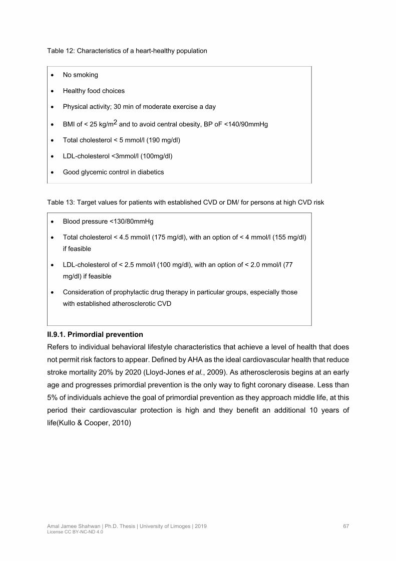
Amal Jamee Shahwan | Ph.D. Thesis | University of Limoges | 2019 License CC BY-NC-ND 4.0
67
Table 12: Characteristics of a heart-healthy population
Table 13: Target values for patients with established CVD or DM/ for persons at high CVD risk
II.9.1. Primordial prevention
Refers to individual behavioral lifestyle characteristics that achieve a level of health that does
not permit risk factors to appear. Defined by AHA as the ideal cardiovascular health that reduce
stroke mortality 20% by 2020 (Lloyd-Jones et al., 2009). As atherosclerosis begins at an early
age and progresses primordial prevention is the only way to fight coronary disease. Less than
5% of individuals achieve the goal of primordial prevention as they approach middle life, at this
period their cardiovascular protection is high and they benefit an additional 10 years of
life(Kullo & Cooper, 2010)
• No smoking
• Healthy food choices
• Physical activity; 30 min of moderate exercise a day
• BMI of < 25 kg/m2 and to avoid central obesity, BP oF <140/90mmHg
• Total cholesterol < 5 mmol/l (190 mg/dl)
• LDL-cholesterol <3mmol/l (100mg/dl)
• Good glycemic control in diabetics
• Blood pressure <130/80mmHg
• Total cholesterol < 4.5 mmol/l (175 mg/dl), with an option of < 4 mmol/l (155 mg/dl)
if feasible
• LDL-cholesterol of < 2.5 mmol/l (100 mg/dl), with an option of < 2.0 mmol/l (77
mg/dl) if feasible
• Consideration of prophylactic drug therapy in particular groups, especially those
with established atherosclerotic CVD

Amal Jamee Shahwan | Ph.D. Thesis | University of Limoges | 2019 License CC BY-NC-ND 4.0
68
Table 14: Ideal cardiovascular Health, defined by AHA, ‘Life Simple 7’
1.Not smoking or quitting over 1 year ago.
2.A body mass index ,25 kg/m2.
3.exercising at a moderate intensity $150 minutes (or 75 minutes at vigorous intensity) each week.
4. Eating a healthy diet: adhering to four to five important dietary components •sodium intake ,1.5 g/day; •sugar-sweetened beverage intake ,36 oz weekly; • 4.5 cups of fruits and vegetables/day; • three 1 oz servings of fiber-rich whole grains/day; • two 3.5 oz servings of oily fish/week.
5.Maintaining total cholesterol (TC) ,200 mg/dL.
6.Keeping blood pressure ,120/80 mmHg.
7.Keeping fasting blood glucose ,100 mg/dL
Notes: *recommendations include $ four servings of nuts, legumes and seeds/week; # less servings of processed meats/week; less than 7% total energy intake as saturated fat
II.9.2. Primary prevention
Concerning people with risk factors but without any clinical manifestation of cardiovascular
disease. The objective of the primary prevention is to take measures by arming communities
with information on healthy life styles, detecting cardiovascular risk factors, and providing
healthy diet. It is the most cost-effective method of ensuring the health of the community by
preventing diseases and limiting or delaying the occurrence of CVD.
II.9.3. Secondary prevention
Concerning people with CVD (CAD, stroke, LEAD) and its objective is to ovoid the occurrence
of complications and recurrences. Early treatment can significantly alter the development and
manifestation of chronic conditions.
II.9.4. Tertiary prevention
The goal of tertiary prevention is to improve quality of life and extend overall life expectancy
by preventing complications in the future. Tertiary prevention is the heaviest tax burden on the
health care system due to considerable costs of surgery and lifelong management of chronic
disease via medications and rehabilitation.
The primary and secondary prevention are based on different effective interventions involving
behavior factors and life style changes such as smoking cessation, regular physical activity,
healthy diet and pharmaceutical treatments such as anti-hypertensive drugs, anti-diabetes,
antiplatelet agent and cholesterol lowering drugs. The choice of the type of intervention for
each individual can be based on two approaches: a risk factor approach and another one
involving all risk factors (Global cardio-vascular risk).

Amal Jamee Shahwan | Ph.D. Thesis | University of Limoges | 2019 License CC BY-NC-ND 4.0
69
The WHO/ISH (International Society of Hypertension) risk prediction charts indicate 10-year
risk of a fatal or non-fatal major cardiovascular event according to age, sex, blood pressure,
smoking status, total blood cholesterol and presence or absence of diabetes mellitus (Table10)
for 14 WHO epidemiological sub regions (Africa, The Americas, Eastern Mediterranean,
Europe South-East Asia, Western Pacific) and define the strategy according to risk evaluation
There are two sets of charts: one set of charts when lab test is available and the other one if
the blood cholesterol is not available (WHO, 2007).
Table 15: Prevention of Cardiovascular diseases
Individual total risk a: define as10 -year risk of cardiovascular event low risk (<10%), moderate risk (10
to < 20%), high risk (20 to < 30%), very high risk (≥30%).
When resources are limited, individual counselling and provision of care may have to be
prioritized according to cardiovascular risk.
Risk <10% Individuals in this category are at low risk. Low risk does not mean “no” risk
Conservative management focusing on lifestyle interventions is suggested
Risk 10% to <20%
Individuals in this category are at moderate risk of fatal or non-fatal vascular
events.
Monitor risk profile every 6–12 months
Risk 20% to <30%
Individuals in this category are at high risk of fatal or non-fatal vascular
events.
Monitor risk profile every 3–6 months
Risk ≥30%
Individuals in this category are at very high risk of fatal or non-fatal vascular
events.
Monitor risk profile every 3–6 months
a Excluding people with established CAD, CVA and PVD
II.9.5. How to intervene at the individual level: risk factors interventions
II.9.5.1. Smoking
Smoking is the major risk factor for CVD. Smoking cessation is the single most cost-effective
intervention in CVD prevention (Eckel et al., 2014). High taxes on tobacco products are
effective policies to reduce smoking in young people. In a systematic metanalysis of 20
prospective cohort studies, quitting smoking was associated with 36% decrease in risk of all-
cause mortality among patients with CAD (Critchley & Capewell, 2003). Pharmacological

Amal Jamee Shahwan | Ph.D. Thesis | University of Limoges | 2019 License CC BY-NC-ND 4.0
70
support and professional assistance are effective. Drug therapy including nicotine replacement
therapy (NRT), bupropion or varenicline should be considered early (Cahill et al., 2013).
Electronic cigarette can provide high concentration of nicotine, so it is recommended as a
measure to help cessation of smoking of regular cigarettes (Chetty et al., 2016). It is also
recommended to avoid passive smoking to prevent CVD. Within 15 years, the risk of CVD
becomes nearly the same as someone who has never smoked. Also, quitting smoking at age
66 years, men gained up to two years of life and women gained up to 3.7 years of life (World
Heart Federtion, 2017).
II.9.5.2. Nutrition
There is a strong relationship between unhealthy nutrition and the occurrence of CVD.
However, nutritional counselling is an essential tool for cardiovascular prevention. Patients
with CVD or at high risk, should receive recommendations on foods and dietary pattern to
reduce the risk.
At the individual level varied and energy-balanced regimen is essential for the preservation of
good health.
Table 16: Dietary targets to prevent CVD(Guy De Backer, 2017)
• Consume more fruit, nuts, seeds, vegetables; 2 to 3 servings of each per day.
• Limit the consumption of saturated fatty acids to <10% of total energy through replacement
by poly-unsaturated fatty acids (PUFA).
• Use vegetable oils rich in PUFA and soft spreads based on e.g., soybean oil, canola oil
and extra-virgin olive oil.
• Limit the consumption of refined grains and sugar; aim at 30-45 gr of fiber per day,
preferably from wholegrain products.
• Consume 1 or 2 servings of fish per week, preferably oily fish such as sardines, herring,
tuna, salmon, mackerel, trout.
• Don’t eat processed meat; limit the consumption of fresh red meat to 2-3 servings per
week.
• Avoid foods made with partially hydrogenated vegetable oils aiming at a zero consumption
of trans unsaturated fatty acids.
• Avoid drinking sugar-sweetened beverages.
• Limit the intake of sodium aiming at <5 gr of salt per day.
• If alcohol is consumed it should be limited to 2 glasses per day (20 gr alcohol) in men and
to 1 glass per day (10 gr alcohol) in women
Amal Jamee Shahwan | Ph.D. Thesis | University of Limoges | 2019 License CC BY-NC-ND 4.0
71
II.9.5.3. Physical activity
Physical exercise is a fundamental issue in all strategies of CVD prevention, while a sedentary
life increases the risk of cardiovascular risk factors, such as hypertension, high triglyceride
diabetes and obesity. WHO recommended for all individuals to undertake at least 150 min a
week of moderate aerobic physical activity (30 min for 5 days / week) or 75 min / week of
vigorous physical activity (15 min for 5 days / week) or a combination of these two elements
(Guy De Backer, 2017, Anon, n.d.). Also, in old adults aged ≥ 65 years, the WHO recommends
participation in muscle training activities at least two days a week (Guy De Backer, 2017).
II.9.5.4. Obesity
Weight reduction is strongly recommended for obese persons with a BMI ≥ 30 or ≥ 25 kg/m2
in the presence of cardiovascular risk factors. Weight loss is achieved by combination of a
reduction in caloric intake and increase in physical exercise. Different diets have been
proposed for the treatment of obesity, a low fat diet is still considered the standard approach
to weight reduction (Perk et al., 2012).
II.9.5.5. Hypertension
Raised BP is one of the most powerful modifiable risk factors for CVD. BP lowering can be
achieved by two strategies: lifestyle changes and drug therapy. Numerous meta-analyses have
shown that a 10-mmHg reduction in SBP or 5 mmHg reduction in DBP is associated with
significant reduction in all major cardiovascular events by ~20%, all-cause mortality by 10-
15%, stroke by 35%, coronary events by 20% and heart failure by 40% (Ettehad et al., 2016;
Thomopoulos, Parati & Zanchetti, 2014).
The ESC/ESH and USA guidelines both advise use of home BP monitoring and ambulatory
BP monitoring to confirm the diagnosis, detect white coat and masked hypertension, and
monitor BP control for medication. In patients at very high risk as is the case in the presence
of CVD, lowering BP may require more intensive strategies than those currently recommended
in the guidelines.

Amal Jamee Shahwan | Ph.D. Thesis | University of Limoges | 2019 License CC BY-NC-ND 4.0
72
Table 17: Recommendation regarding BP targets in patients with hypertension (Guy De Backer, 2017;
Whelton et al., 2018).
Use of BP-lowering medications is recommended
• for secondary prevention of: recurrent CVD events in patients with clinical CVD and SBP ≥ 130 mm Hg or DBP ≥ 80 mm Hg
• for primary prevention in adults with an estimated 10-year (ASCVD) risk of ≥10% and an SBP ≥130 mm Hg or DBP ≥ 80 mm Hg.
Use of BP-lowering medication is recommended
• for primary prevention CVD in adults with no history of CVD and with an estimated 10-year ASCVD risk < 10% and SBP ≥ 140 mm Hg or DBP≥ of 90 mm Hg.
Nonpharmacologic therapy and repeat BP evaluation within 3 to 6 months.
• should be recommended In adults with an elevated BP or stage 1 hypertension who have an estimated 10-year ASCVD risk < 10%
Combination of nonpharmacologic and antihypertensive drug therapy and repeat BP evaluation in 1 month
• should be recommended in adults with stage 1 hypertension who have an estimated 10-year ASCVD risk of ≥ 10%
Evaluation followed by prompt antihypertensive drug treatment
• is recommended in l adults with SBP ≥180 mm Hg or DBP ≥110 mm Hg
Two or more antihypertensive medications
• are recommended in most adults with HTN, especially in black adults to achieve a BP target < 130/80 mm Hg
Physicians should consider starting or increasing drug therapy to reduce the risk for stroke and cardiac events
• In patients ≥ 60 years who have a history of stroke or transient ischemic attack or have high cardiovascular risk, to achieve an SBP < 140 mm Hg
Blood pressure targets diabetes mellitus
• are recommended in type 2 diabetes to be <140/85 mmHg but a lower target of <130/80 mmHg is recommended in selected patients (younger patients at elevated risk for specific complications) for additional gains on stroke, retinopathy and albuminuria risk.
• in type 1 diabetes mellitus recommended blood pressure target is <130/80 mmHg
In individuals >80 years old and with initial SBP ≥160 mmHg
• it is recommended to reduce SBP to between 150 and 140 mmHg provided they are in good physical and mental condition
II.9.5.6. Diabetes
In most Randomized Controlled Trials (RCTs), intensive glucose control in patients with type
2 diabetes leads to 9% reduction in major cardiovascular events (MACE), 20% reduction of

Amal Jamee Shahwan | Ph.D. Thesis | University of Limoges | 2019 License CC BY-NC-ND 4.0
73
kidney events and 13% of eye events however, residual microvascular and macrovascular risk
remains high (Giugliano et al., 2018). In addition, the equation “diabetes equal coronary artery
disease” has accelerated the implementation of preventive therapy for diabetes risk factors. In
a real-world population among 93,866 Danish patients of whom 13.4% were diabetics, followed
for a median of 4.1 years, reported that in the absence of significant lesion, the diabetics group
with a preventive treatment of statin and aspirin had the same risk of cardiovascular events as
in group without diabetes (Olesen et al., 2017).
Table 18:Type 2 diabetes Combination strategies (Giugliano et al., 2018)
• Intensive glycemic control is important for the prevention of vascular complications in adults
with type 2 diabetes, but residual micro- and macro-vascular risk remains high
• Newer diabetes drugs (empagliflozin, canagliflozin, liraglutide, and semaglutide) may
decrease the incidence of MACE* and nephropathy; depending on the drug used,
cardiovascular mortality and heart failure may also be reduced
• The combination of intensive glycemic control and newer diabetes drugs may have
beneficial, additive effects on diabetic vascular complications
• Statin and aspirin should also be used for preventing and fighting cardiovascular
complications in type 2 diabetes
MACE*: major cardiovascular events
Testing for prediabetics and risk for future diabetes in asymptomatic people should be
considered in adults of any age who are overweight or obese (BMI≥25kg/m2 in Asian American)
and in individual who have one or more additional risk factors for diabetes (Table14)
Table 19: Criteria for testing for diabetes or prediabetes in asymptomatic adults (ADA, 2018)
1. Testing should be considered in overweight or obese (BMI ≥25 kg/m2 or ≥23 kg/m2 in Asian Americans)
2. Adults who have one or more of the following risk factors:
• First-degree relative with diabetes
• High-risk race/ethnicity (e.g., African American, Latino, Native American, Asian American, Pacific Islander)
• History of CVD
• Hypertension (≥140/90 mmHg or on therapy for hypertension)
• HDL cholesterol level <35 mg/dL (0.90 mmol/L) and/or a triglyceride level >250 mg/dL
• Women with polycystic ovary syndrome
• Physical inactivity

Amal Jamee Shahwan | Ph.D. Thesis | University of Limoges | 2019 License CC BY-NC-ND 4.0
74
• Other clinical conditions associated with insulin resistance (e.g., severe obesity, acanthosis nigricans)
2. Patients with prediabetes (A1C ≥5.7% [39 mmol/mol], IGT, or IFG) should be tested yearly, treat other cardiovascular risk factors.
3. Women who were diagnosed with gestational diabetes mellites should have lifelong testing at least every 3 years.
4. For all other people, testing should begin at age 45 years.
5. If results are normal, testing should be repeated at a minimum of 3-year intervals, with consideration of more frequent testing depending on initial results and risk status.
II.9.5.7. Dyslipidemias
Randomized controlled trials have demonstrated that lowering LDL cholesterol level with
healthy diet and or lipid lowering drugs (statin) can reduce the risk of CVD events (Kotseva et
al., 2016). Lipid lowering therapy with statins reduces relative cardiovascular risk by 20 % to
30% whatever the baseline LDL-Ch is. The treatment goals for LDL-Ch depend on the total
CVD risk of the individual and of the baseline LDL-Ch level. Measuring LDL-Ch every six weeks
after treatment initiation and every 12 months to assess the compliance with treatment and
diet is recommended (Table 19, 20).
Table 20: Lipid Goals for Patients at risk for Atherosclerotic CVD (Jellinger et al., 2017)
Lipid parameter Goal (mg/dL)
TC • <200
LDL-C • <130 (low risk) <100 (moderate risk) <100 (high risk)
• <70 (very high risk) <55 (extreme risk)
Non-HDL-C • 30 above LDL-C goal
• 25 above LDL-C goal (extreme risk patients)
TG • <150
Apo B
• <90 (patients at high risk of ASCVD, including those with diabetes
• <80 (patients at very high risk with established ASCVD or diabetes plus ≥1 additional risk factor)
• <70 (patients at extreme risk)
Abbreviations: apo = apolipoprotein; ASCVD = atherosclerotic cardiovascular disease; HDL-C = high- density lipoprotein cholesterol; LDL-C = low-density lipoprotein cholesterol; TC = total cholesterol; TG = triglycerides

Amal Jamee Shahwan | Ph.D. Thesis | University of Limoges | 2019 License CC BY-NC-ND 4.0
75
Table 21: Goal and treatment for LDL Cholesterol (Guy De Backer, 2017;Hendrani et al., 2016)
Target Therapeutic option
Very high CVD risk LDL-Ch <1.8mmol/L (70mg/dl) or a
reduction of 50% if the LDL Ch is between
1.8-3.5 mmol/L (70-135 mg/dl)
• Dietary intervention
• Drug treatment (statin
+Ezetimibe)
High CVD risk LDL-Ch <2.6mmol/L (100mg/dl) or a
reduction of 50% if the LDL Ch is between
2.6-5.2 mmol/L (100-200mg/dl)
• Dietary intervention
• Drug treatment
Moderate risk LDL-Ch <3.0 mmol/L (115mg/dl) • Dietary intervention
• Drug treatment?
II.9.6. Mobile phone interventions for the secondary prevention of CVD
Unlike traditional approaches of secondary prevention of CVD, a quantitative review of
database from 2002-2016 through numerous studies revealed the use of mobile health
(mHealth) as new area of health care. The WHO announced in their report 2011 that, “the use
of mobile and wireless technologies to support the achievement of health objectives has the
potential to transform the face of health service delivery across the globe” (WHO Global
Observatory for eHealth, 2011). mHealth technologies include text messaging, mobile
applications, Global Positioning System (GPS), Bluetooth technologies, camera, and more
recently the use of sensors that track heart rate, steps walk, sleep cycle and transmit feedback
to clinicians (Chow et al., 2016). Mobile phone technology is a personalized, easily accessible
and inexpensive tool for communication with patients, tracking personal health data, updating
information and reminders for health behaviors (Hamilton et al., 2018, Anon, 2016a). The use
of mobile applications for tracking needs to be combined with text messaging for improving
outcomes (Martin et al., 2015). However, smartphone interventions were limited by cost, for
people with lower socio-economic status.
mHealth applications were divided into two categories:
• Those used in primary prevention to facilitate wellness such as physical activity (e.g.
My Fitness Pal), weight loss (e.g. Weight Watchers), stress, diet, nutrition, blood
pressure tracking (e.g. Blood Pressure Log) and quit smoking (e.g. Quit START). The
Secondary prevention accounts for a quarter of mHealth applications (AARON, 2015).
• Cardiac applications have been used in cardiac rehabilitation services. The National
Heart Foundation has launched “My Heart, My Life” application, which helps users to

Amal Jamee Shahwan | Ph.D. Thesis | University of Limoges | 2019 License CC BY-NC-ND 4.0
76
track medications, obtain personal health statistics, search for recipes, provides
information and video on heart attack symptoms (Foundation, n.d.; Neubeck et al.,
2015). Also, the Canadian Heart and Stroke Foundation offers a 30-day application,
that guides the user to establish goals to reduce their CVD risk (Heart and Stroke
Foundation of Canada, 2014). Cardiologists use mobile applications to obtain real time
information like drugs lists, patients heart rate monitoring, ECG, blood pressure,
calculate risk Algorithm and screen patients with Atrial fibrillation (Lowres et al., 2014).
Figure 9: Text message effects on target level of cardiovascular risk factors (Chow et al., 2015)
II.9.7. The WHO 25 by 25 vision for chronic disease target
The growing epidemic for NCDs has been well known for several years. WHO has developed
a global strategy for the prevention and control for NCDs. In the 65th World Health Assembly
adopted a resolution to reach a global target of 25% reduction in premature mortality from
NCDs by 2025 (Figure 10). The WHO / WHF set voluntary targets for six risk factors and two
health services goals in order to achieve the 25% reduction in premature mortality from four
major NCDs (CVD, diabetes, cancers and lung disease). In 2015, the 193 Member States of
the United Nations adopted a new Agenda 2030 based on the WHO and WHF “25 by 25 “and
aims for a 30% reduction in NCD by 2030 (Dugani & Gaziano, 2016).

Amal Jamee Shahwan | Ph.D. Thesis | University of Limoges | 2019 License CC BY-NC-ND 4.0
77
Figure 10: 25x25 WHF global CVD roadmap (Grainger-Gasser, Perel, Lagier-Hässig,& Wood, 2017)
When risk factor targets are met, the probability of dying from the four major NCDs between
the ages of 30 and 70 years will decrease by 22% in men and 19% in women between 2010-
2025, compared with a decrease of 11% in males and 10% in females based on current trends
without further action (Kontis et al., 2014).

Amal Jamee Shahwan | Ph.D. Thesis | University of Limoges | 2019 License CC BY-NC-ND 4.0
78
II.9.8. Best buys for Non-communicable disease prevention
Defined as interventions that are cost effective, cheap, feasible and culturally acceptable for
the prevention and control of NCDs (Table 22). A number of criteria enter into such decision,
including the burden of the disease, and political considerations. NCDs cost in LIMIC was
approximatively $500 billion annually. In contrast, the total annual cost of implementing all of
the “Best Buys” to address NCDs is just $11.4 billion, with the added benefit of saving millions
of lives. The implementation of Best Buy requires an organized strategy and a highest level of
political mobilization.(“Best Buy” WHO, 2014).
Table 22: "Best Buy" Interventions (“WHO | Scaling up action against NCDs,” n.d.2014)
Risk factor / disease Interventions
Tobacco use
• Tax increases
• Smoke-free indoor workplaces and public places
• Health information and warnings
• Bans on tobacco advertising, promotion and sponsorship
Harmful alcohol use
• Tax increases
• Restricted access to retailed alcohol
• Bans on alcohol advertising
Unhealthy diet and
physical inactivity
• Reduced salt intake in food
• Replacement of trans fat with polyunsaturated fat
• Public awareness through mass media on diet and physical activity
Cardiovascular
disease and diabetes
• Counselling and multi-drug therapy for people with a high risk of
developing heart attacks and strokes (including those with
established CVD)
• Treatment of heart attacks with aspirin
II.10. The National Health vision and Strategy in Palestine for NCD
The global action plan for the prevention and control of NCDs 2013-2020 has 6 objectives, 25
indicators and 9 voluntary targets (MOH, 2017).

Amal Jamee Shahwan | Ph.D. Thesis | University of Limoges | 2019 License CC BY-NC-ND 4.0
79
Table 23: National strategic targets to control NCDs in Palestine compared to global targets
Target Global National
1 Relative reduction in premature mortality from NCDs 25% 10%
2 Relative reduction in prevalence of insufficient physical activity 10% 5%
3 Relative reduction in mean population intake of salt 30% 30%
4 Relative reduction in prevalence of current tobacco use in persons aged
> 15years
30% 30%
5 Eligible people receive drug therapy and counseling (including glycemic
control) to prevent heart attacks and strokes
50% 50%
6 Availability of the affordable basic technologies and essential medicines,
including generics, required to treat major NCDs in both public and private
facilities
80% 80%
7 Relative reduction in prevalence of raised blood pressure 25% N.C*
8 Diabetes and obesity increase in prevalence 0% N.C
9 Relative reduction in prevalence of harmful alcohol use 10% N.C
N.C*: Not concerning

Amal Jamee Shahwan | Ph.D. Thesis | University of Limoges | 2019 License CC BY-NC-ND 4.0
80
Chapter III. Cardiovascular disease and risk factors in Palestine
III.1. Epidemiology of Cardiovascular disease in Palestine
Palestinian territories or occupied Palestinian territories is a description often used by United
Nation (UN) and other international organizations to describe the west bank including East of
Jerusalem and Gaza strip between 1998 to 2013 in order to refer to area controlled by
Palestinian National authority. In 2012, the United Nations replaced this by the term of state of
Palestine. Palestine is located on the east side of the Mediterranean Sea; the total surface
area is estimated at 20,770 square Kilometer. The number of Palestinians in the world is 12.7
million. The number of Palestinians living in the state of Palestine in 2017 was 4.88 million.
The population distribution shows that 60.1% live in the northern governorates (West Bank)
and 39.9% in the southern governorates (Gaza strip), with 50.9% of males and 49.1% of
females (S. Yusuf et al., 2011). Also, 41.9% of the Palestinian populations are refugee. The
rate of the natural growth is 2.8% and the life expectancy is 73.7 years; 72.1 years for males
and 75.2 years for females (PCBS 2016, n.d.). The population of Palestinian territories is
undergoing a rapid epidemiological transition from communicable diseases to NCDs such as
CVDs, HTN, DM and Cancer. The prevalence of HIV /AIDS is very low, the population is
deemed free of poliomyelitis and immunization program is mostly controlled by WHO
(Giacaman et al., 2009). Like other countries an epidemiological transition has occurred in
Palestine. The leading cause of death in Palestinian community was NCDs which accounted
for 65.7% of all deaths (PHIC, 2018); the incidence is higher in West bank (WB) 57% vs 40%
in Gaza Strip(GS) (Zynia L & Andrew, 2000). CVDs are major cause of illness, disability and
death in Palestine, which cause an increase in personnel community and health cost. There is
no available data to suggest the prevalence or incidence of CVDs; we depend mainly on
mortality data from the different health centers.
Epidemiological studies of Cardiovascular risk factors and CVDs in our country suffer from
underestimates and other limitations. Firstly, few studies have been undertaken with the use
of sampling techniques. Secondly, recent demographic lifestyle changes, cultural, and political
prospective are modifying the prevalence of CVRF. Thirdly, CVRF are often not stratified by
age and sex. The United Nations Relief and Works Agency (UNRWA) report 2016 for
Palestinian refugee, reported a prevalence of 20.1%, 21.9% of hypertension and diabetes
12.9%,15.9% among population aged ≥ 40 years in Gaza and West Bank respectively
(Palestinian Refugees of Syria included) (Seita et al., 2017). A survey in Palestine (G.S/WB
1999-2000) reported the prevalence of overweight 35.5%, 40.3% and obesity 31.5%, 17.5%
respectively in women and men (Abdeen et al., 2012). By the way the epidemiology of CVDs
remains still unclear and the health policy makers do not take in account the results.
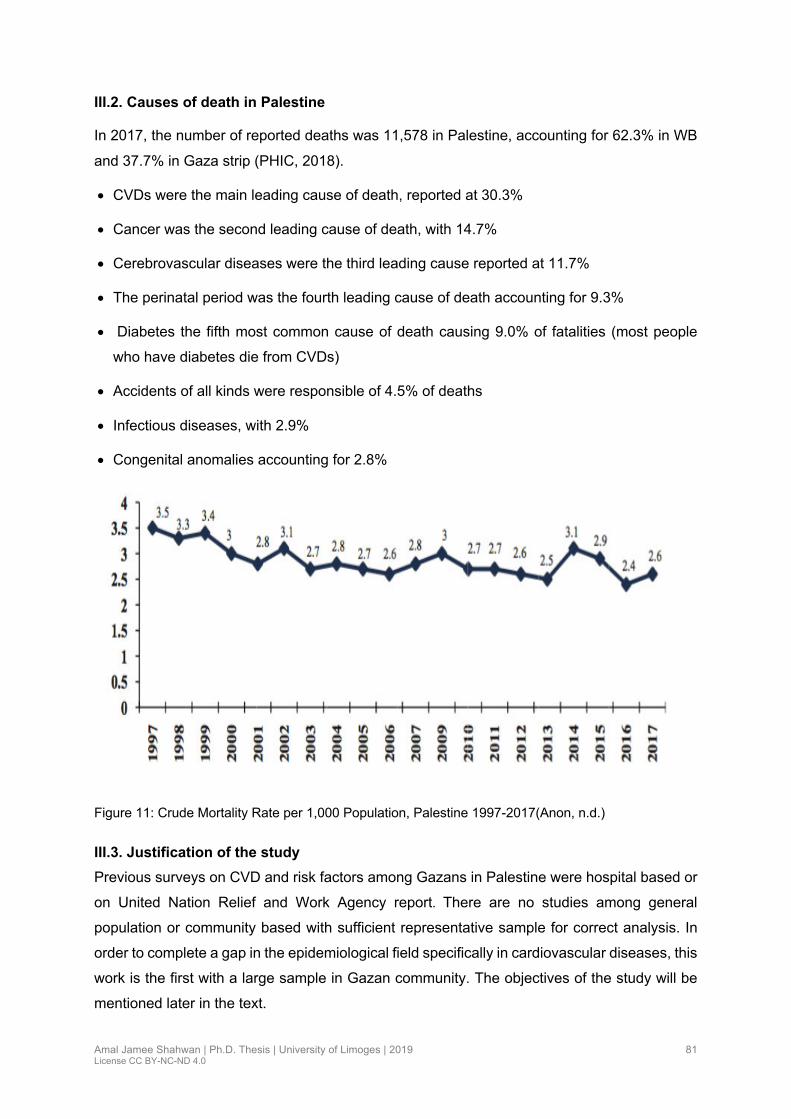
Amal Jamee Shahwan | Ph.D. Thesis | University of Limoges | 2019 License CC BY-NC-ND 4.0
81
III.2. Causes of death in Palestine
In 2017, the number of reported deaths was 11,578 in Palestine, accounting for 62.3% in WB
and 37.7% in Gaza strip (PHIC, 2018).
• CVDs were the main leading cause of death, reported at 30.3%
• Cancer was the second leading cause of death, with 14.7%
• Cerebrovascular diseases were the third leading cause reported at 11.7%
• The perinatal period was the fourth leading cause of death accounting for 9.3%
• Diabetes the fifth most common cause of death causing 9.0% of fatalities (most people
who have diabetes die from CVDs)
• Accidents of all kinds were responsible of 4.5% of deaths
• Infectious diseases, with 2.9%
• Congenital anomalies accounting for 2.8%
Figure 11: Crude Mortality Rate per 1,000 Population, Palestine 1997-2017(Anon, n.d.)
III.3. Justification of the study
Previous surveys on CVD and risk factors among Gazans in Palestine were hospital based or
on United Nation Relief and Work Agency report. There are no studies among general
population or community based with sufficient representative sample for correct analysis. In
order to complete a gap in the epidemiological field specifically in cardiovascular diseases, this
work is the first with a large sample in Gazan community. The objectives of the study will be
mentioned later in the text.

Amal Jamee Shahwan | Ph.D. Thesis | University of Limoges | 2019 License CC BY-NC-ND 4.0
82
Figure 12: Gaza Strip Map
III.3.1. Geography of Gaza
The Gaza strip is a small area of Palestinian territory on the east coast of Mediterranean Sea.
It borders Egypt on the southwest for 11 km and Israel on the east and north a long 51 km
border with an area of 365 square kilometers (Figure12). It is mostly flat and sandy with little
fertile soil. The GS has a temperature climate with mild winter and dry, hot summers. January
is the coldest month with average temperatures ranging from 6 0C to 15 0C and July and August
are the hottest months at 22 0C to 33 0C on average. Gaza strip consists of 5 governorates
with fourteen villages and eight refugee camps (Table 24) (Wikipedia, 2017).

Amal Jamee Shahwan | Ph.D. Thesis | University of Limoges | 2019 License CC BY-NC-ND 4.0
83
Table 24: Distribution of population in Gaza goverenate areas (PCBS2016)
Gaza Governorates Populations % of G. S Area km2
North Gaza Governorate 377,126 20.1% 61
Gaza Governorate 645, 205 34.3% 74
Deir-Al Balah Governorate 273, 390 14.5% 58
Khan-younis Governorate 351,934 18.7% 108
Rafah Governorate 233,480 12.4% 64
Gaza strip (total) 1.881,135 100% 365
III.3.2. Demography, culture and economy
There are three separate Palestinian communities; those living in Israel, those living in West
Bank and those living in Gaza Strip. The latter two groups are the same population separated
by geographic and political boundaries. Spoken languages are Arabic and Hebrew, and
English is widely understood. In 2017 the population of G.S was 1.88 million, 39.9% of total
population of Palestine, and 66.7% were refugees. This large refugee population is supported
by the UNRWA which provides health, educational and social services. Gaza has a high fertility
rate of 4.4 children per woman and has an annual population growth rate of 2.33%, the 13th
highest in the world. The population pyramid shows that the society is young, 44.7% are under
the age of 15 years, 2.5% in age ≥ 65 (Fig 13), and the average of family size is 6.5 (CIA.,
2018). The population is expected to increase to 2.1 million in 2020. The life expectancy rate
is 74.2 years; (72.5 years for males and 75.9 years for females). Around 76.2% of Gazans live
in urban areas (CIA., 2018; PHIC, 2018). The Sunni Muslims make up the predominant part of
the Palestinian population. There is also a Christian minority who live in the Gaza city, most of
them are orthodox. Access restriction, violent attacks, and the Palestinian-Palestinian and
Israeli- Palestinian conflict continue to worsen economic and social conditions in G.S. Gaza
has suffered from rising unemployment and elevated poverty rates. Gaza strip industries are
generally small family businesses that produce textiles, soap, olive-wood and embroidery.
Agriculture products are olives, citrus and flowers (CIA., 2018).

Amal Jamee Shahwan | Ph.D. Thesis | University of Limoges | 2019 License CC BY-NC-ND 4.0
84
Figure 13: Population Pyramid in Gaza Strip(PCBS 2016, n.d.)
III.3.3. Political situation
Palestinians in Gaza are locked in, denied free access to the remainder of the occupied
Palestinian territories and the outside world. Israel maintained severe restrictions on the
movements of people and goods into and out of Gaza since 2009 and intensified them on June
2007, after the takeover of Gaza by Hamas, an Islamic Palestinian organization which came
into office in 2006 through free elections. The situation was exacerbated by Egypt’s closure of
the border most of the time, and the internal Palestinian political divide between authorities in
Ramallah and Gaza. More than ten years of blockade and the isolation of Gaza has worsened
the dire humanitarian conditions such as restricting access to medical care, educational and
economic situation, electricity deficit, and perpetuating unemployment and poverty.
Approximately 70% of Gaza population rely on humanitarian assistance.
III.3.4. Health situation and health centers in Gaza strip
Gaza strip is suffering from triple burden of health status: 1- a poverty related socioeconomic
status, 2- violence related war, 3- increase in life style related NCDs. Years of socioeconomic
decline and the political separation between political parties (Fatah and Hamas) in controlling
each region have affected the health system as a whole. The serious impact of the conflict led
to the destruction of the vital health infrastructure, loss of life, disabilities, shortages of drugs

Amal Jamee Shahwan | Ph.D. Thesis | University of Limoges | 2019 License CC BY-NC-ND 4.0
85
and medical supplies, deterioration of chronic diseases, limitation of tertiary care capacity, and
severe effect on the mental wellbeing of the population. External support from donors, the UN
and Non-Governmental Organization (NGO), supported the Ministry of Health (MOH), mainly
during emergency. In Gaza, health services are delivered by MOH, UNRWA, NGOs, military
medical services and the private sector. There are 160 primary health care centers in Gaza
strip. UNRWA provides 22 primary health care services to the majority of the Palestinian
refugees in Gaza (PHIC, 2018). According to CIA THE WORLD FACTBOOK the physician’s
density was 2.2 physicians /1,000 population in 2014 and the hospital beds 1.32/1,000
population in 2015.
NCDs are responsible for more than 50% of deaths in Gaza. Actually, there are no sufficient
measures to control unhealthy eating and to promote physical activity (Anon, n.d.). NCD care
is available at primary health care in MOH and UNRWA, which provide diagnosis, treatment
and follow up for uncomplicated hypertension, diabetes mellitus, heart disease and chronic
lung disease. Other NGOs such as the Palestinian Medical Relief Society (PMRS), the Union
of Health Care Committees (UHCC) and private sectors provide NCD programs in their clinics.
III.4. Objectives of the study
III.4.1. General objective
The principal objective of our study is to estimate the epidemiology of CVDs (coronary artery
disease, stroke and lower extremity artery disease) in Gazan community and to identify
associated risk factors.
III.4.2. Specific objectives
• To measure the prevalence of each CVD (coronary artery disease, stroke and lower
extremity artery disease) in Gazan community
• To analyze the prevalence of associated risk factors (sociodemographic and
cardiovascular) with CVDs
• To examine the association between sociodemographic variables, and the prevalence
of risk factors
• To evaluate the clustering of risk factors (metabolic syndrome) and its relationship with
CVDs
• To estimate the prevalence of people having high overall 10 years CVD risk using
Framingham Global CV risk score

Amal Jamee Shahwan | Ph.D. Thesis | University of Limoges | 2019 License CC BY-NC-ND 4.0
86
III.5. Methodology and protocol of the study
III.5.1. Target population
The target population includes all adult male and female aged 25 years and more. The target
population is 35.8 % of the total Gaza strip population. It was approximately around 673 523
inhabitants. The participants were selected by proportional stratified cluster sampling
according to geographic population sector
Table 25: Distribution of population in Gaza strip by age group
Age group
Years
Population
Number
Percent %
0-14 804 614 42.8
15-19 210 132
21.3 20-24 190 504
25-29 157 348
29.9%
35.8%
30-34 120 853
35-39 96 112
40-44 76 044
45-49 62 282
50-54 51 711
55-59 39 253
3.5% 60-64 27 249
65-69 18 578
2.5%
70-74 11 949
75-79 7 505
80+ 6 896
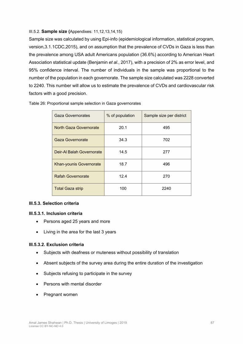
Amal Jamee Shahwan | Ph.D. Thesis | University of Limoges | 2019 License CC BY-NC-ND 4.0
87
III.5.2. Sample size (Appendixes: 11,12,13,14,15)
Sample size was calculated by using Epi-info (epidemiological information, statistical program,
version,3.1.1CDC,2015), and on assumption that the prevalence of CVDs in Gaza is less than
the prevalence among USA adult Americans population (36.6%) according to American Heart
Association statistical update (Benjamin et al., 2017), with a precision of 2% as error level, and
95% confidence interval. The number of individuals in the sample was proportional to the
number of the population in each governorate. The sample size calculated was 2228 converted
to 2240. This number will allow us to estimate the prevalence of CVDs and cardiovascular risk
factors with a good precision.
Table 26: Proportional sample selection in Gaza governorates
Gaza Governorates % of population Sample size per district
North Gaza Governorate 20.1 495
Gaza Governorate 34.3 702
Deir-Al Balah Governorate 14.5 277
Khan-younis Governorate 18.7 496
Rafah Governorate 12.4 270
Total Gaza strip 100 2240
III.5.3. Selection criteria
III.5.3.1. Inclusion criteria
• Persons aged 25 years and more
• Living in the area for the last 3 years
III.5.3.2. Exclusion criteria
• Subjects with deafness or muteness without possibility of translation
• Absent subjects of the survey area during the entire duration of the investigation
• Subjects refusing to participate in the survey
• Persons with mental disorder
• Pregnant women

Amal Jamee Shahwan | Ph.D. Thesis | University of Limoges | 2019 License CC BY-NC-ND 4.0
88
III.5.4. Sample design
The sample design is a cross-sectional study using stratified cluster sample, with the
advantage of covering a wide geographic area, in 5 governorates spanning both urban and
rural spaces between July and October 2017 (Figure 14).
• The Primary cluster selection: In this stage, the five governorates of Gaza Strip were
identified, and the sample size was selected proportional to the size of the population
in each Governorate.
• The Secondary cluster: At this stage, in each governorate, one city was selected
randomly, then, within each city one camp area and 2-3 rural and urban areas were
randomly chosen. The total number of areas was sixteen (five camps and eleven rural
plus urban areas). The sample size in each city was proportional to the size of
population in each city.
• Tertiary Cluster: within each stratum, whether camp, urban, or rural, a sample block (a
group of buildings that form a locality) was selected and a cluster of 15 to 20
households were randomly chosen. The investigators went from door to door, and in
every house, all subjects, male and female, of age group ≥ 25 years were interviewed
and examined.

Amal Jamee Shahwan | Ph.D. Thesis | University of Limoges | 2019 License CC BY-NC-ND 4.0
89
GA
ZA
S
trip
Nor
th G
AZ
A
Jaba
lia
495
Cam
p:
Jab
alia
165
Are
a1:
Al-N
azla
165
Are
a2:
Fal
luja
165
Gaz
a
Gaz
a C
ity
702
Sha
tia
Cam
p 55
Are
a1:
Al-R
mal
233
Are
a2:
Al-D
araj
188
Are
a3:
Sha
jaia
226
Cen
tre
Dei
r-A
l Bal
ah
277
Nus
eira
t
Cam
p
104
Are
a1:
Dei
rcity
173
Sou
th
khan
You
nes
496
Kha
nYou
nes
Cam
p 88
Are
a1:
cent
er 1
34
Are
a2:
Aba
ssan
141
Are
a3:
Mid
-city
134
Ext
rem
e S
outh
Raf
ah
270
Raf
ah
Cam
p 65
Are
a1:
Eas
t R
afah
104
Are
a2:
Wes
t R
afah
10
1
Figure 14: Sampling design study
Amal Jamee Shahwan | Ph.D. Thesis | University of Limoges | 2019 License CC BY-NC-ND 4.0
90
III.5.5. Study instrument
The study was conducted in accordance with WHO’s STEP wise approach to surveillance
chronic disease risk factors (STEPS) (WHO 2001). STEPS involve three primary “steps”: 1)
the use of a structured questionnaire to assess study subjects’ self-reported behavioral and
lifestyle risk factors for chronic diseases, 2) the measurement of subjects’ blood pressure and
anthropometrical parameters, and 3) the collection and biochemical analysis of subjects’ blood
samples. We added Rose angina questionnaire, and Perceived stress scale to the
questionnaire.
III.5.5.1. STEP1 : Questionnaire (Appendix 1)
• Demographic and socio-economic status includes information about age, gender,
address, educational level, occupation, family size and monthly income, each of these
socio demographic variables had two to three categories.
Ö The residential area: was specified by interviewers when they completed the
questionnaire item, (urban, rural and camp).
Ö Education level: All respondents were asked the total numbers of years spent at
school. The responders were categorized into five groups: those with no formal
schooling, primary schooling (completed less than 6 years of school), secondary
school (completed 7 to 12 years), college or university (12-15 years), and post
graduate degree.
Ö Employment status: The original questionnaire includes 7 responses categories.
For this analysis, occupational class was analyzed as four categories:
governmental employee, self-employee, homme worker, retired and no-employee.
Ö Economic status (average income in Dollar per month) participants were classified
as low (less than $150), middle ($150 to $500) and high >$ 500.
Ö Marital status was analyzed as three categories: single, married, divorced or
windowed.
• Physical activity: measured by the use of International physical activity questionnaire
(IPAQ): the Arabic version was used (Appendix 2, 3). An international consensus
developed long and short forms of the IPAQ instrument. During 2000, 14 centers from
12 countries collected reliability and validity data and concluded that IPAQ has
reasonable measurement properties for monitoring population levels of physical activity
among 18- to 69 years old adults in diverse sittings activity but is often used
inappropriately in old age. We used the short IPAQ form in our research, this form
comprises of 7 items covering all domains. The specific types of activity that are

Amal Jamee Shahwan | Ph.D. Thesis | University of Limoges | 2019 License CC BY-NC-ND 4.0
91
assessed are walking, moderate-intensity activities and vigorous-intensity activities.
The physical activity was calculated according to the Guidelines for the data processing
and analysis of the international physical activity questionnaire, and participants were
classified into three groups according to IPAQ Scoring Protocol (Short Forms) as low,
moderate and high activity (Anon, 2005).
• Dietary pattern: measured by Food frequency questionnaire (SFFQ): We use the same
questions as used in STEPS WHO
• Stress and anxiety: Measured by Perceived Stress Scale (PSS 10), the tool developed
in 1983, it is the most widely used psychological instrument of measuring the perception
of stress. Five categories of responses were used: 0. Never,1. almost never, 2.
sometimes, 3. fairly often, 4. very often. Scores on the PSS can range from 0 to 40:
Scores ranging from 0-13 are considered low stress, 14-26 are considered moderate
stress and from 27-40 are considered high perceived stress (Cohen, Kamarck &
Mermelstein, 1983). We used the Arabic version (Appendix 4, 5)
• Rose questionnaire: The WHO Rose Questionnaire (RQ) (Appendix 6,7) has been
widely used since 1962 to detect coronary heart disease in epidemiological research
as standardized method as a screening tool rather than diagnostic (Lampe et al., 1998).
The original RQ angina pectoris was indicated by response to seven questions and
administrated by trained nurse at the interview. Studies reported different sensitivity
and specificity of the RQ, it has high sensitivity (80%-95%) but variable specificity (19%-
83%) (Fischbacher et al., 2001). The test is considered positive if response to Question
1, 2 or 3, is yes, Question 4 Stop or slow down, Question 5: relieve and Question 6: 10
min or less (Biloglav, 2004).
• Medical history of personal chronic disease: these variables had two categories (Yes
or No).
Ö Hypertension: We considered subjects to have HTN if their average systolic blood
pressure in both arms was ≥ 140 mmHg or their average diastolic blood pressure ≥ 90
mmHg, or if they were being treated for HTN (James et al., 2014; WHO hypertension,
2015).
Ö Diabetes Mellitus: Was defined as capillary blood sugar level ≥126 mg/dl if the
participant was fasting or ≥ 200 mg/dl if the participant was non-fasting and or self-
reported as currently taking any diabetes medications (American Diabetes Association
2016, n.d.).

Amal Jamee Shahwan | Ph.D. Thesis | University of Limoges | 2019 License CC BY-NC-ND 4.0
92
• Medical history of familial cardiovascular disease: this variable had two categories (Yes
or No), and it was considered positive if the first-degree relatives, before 55 years of
age in men and 65 years of age in women, had premature CVD or sudden death.
• Smoking: The data on smoking habits were based on participants’ responses in the
tobacco use module of the steps questionnaire. The question was designed to identify
current smokers, past smokers, and never smoker. Current smokers were defined as
those who reported smoking greater than 100 cigarettes in their lifetime (including all
type of cigarettes) and who currently smoke. Past smoker is someone who has smoked
more than 100 cigarettes in their life time but has not smoked in the last 28 days. Never
smoker is someone who has not smoked, or who has smoked less than 100 cigarettes
in his or her life time (CDC, 2017).
• Cardiovascular diseases
Ö Coronary artery disease: Was defined in our study by self-reported history of
hospitalization for angina pectoris, myocardial infarction, procedures
performing percutaneous coronary intervention or coronary bypass graft and
their medical prescription list has been checked.
Ö Stroke was identified with the question that raises the existence, among the
house inhabitants of a person having a history of stroke diagnosed by a
physician (we asked, “have you ever been told by a physician that you suffered
a stroke”?), or presence of neurological deficit.
Ö Lower extremity artery disease: The diagnosis was retained for an ankle
brachial index ≤ 0.90.
III.5.5.2. STEP2: Measurements
III.5.5.2.1. Anthropometric parameters
• Body weight of participants was measured according to the standard procedures, while
they were without shoes and wearing light clothes. to the nearest 10 grams with
electronic scales (Dr Feisher-care) with a capacity from 10 kg to 180 kg.
• The height of participants was measured barefoot, using carpenter’s meter, and the
reading of measurement was made using a ruler placed on the subject’s head
perpendicular to the carpenter’s meter.
• Body mass index was calculated as weight in kilograms divided by height in meters
squared, and the cut-off points of WHO for BMI were used to define BMI of 18.5-24.9

Amal Jamee Shahwan | Ph.D. Thesis | University of Limoges | 2019 License CC BY-NC-ND 4.0
93
as normal; BMI of 25 kg/m2 or higher overweight, and more than 30 kg/m2 as obese
(WHO, 2004).
• Waist circumflex was measured mid-way between the lateral lower rib margin, and the
iliac crest, and according to European value which was used for Middle East Arab
population we defined abnormal waist ≥ 94 cm in men and ≥ 80 cm in women.
• Blood pressure was recorded by aneroid sphygmomanometer with adapted cuff size.
Two readings were taken in each arm, while the participant was in a supine position
after having rest for at least 5 min and the average of the reading was used as measure
of BP. HTN was diagnosed as having a BP of 140/90 mm Hg or greater.
• Heart rate: measured by pulse oximeter.
• Ankle Brachial Index: The ABI was measured by (Summit Doppler LifeDepo 150
vascular system-USA) following the AHA statement standardized protocol (Aboyans et
al., 2012). The leg-specific ABI was calculated as the higher SBP in the posterior tibial
or dorsalis pedis divided by the higher of the 2 arms SBP. ABI has been classified into
3 categories: normal value (0.90-1.4), an ABI value < 0.90 indicates LEAD and an ABI
≥1.40 indicates incompressible arteries.
III.5.5.3. STEP 3 : Blood sample
All participants have finger prick for fasting or random blood sugar which was tested by the
use of glucometer (DIAVUE Prudential). Blood sample for lipids profile were taken by nurses
from antecubital vein, drawn into plain tubes, stored in a cool box, and then sent to the
laboratory for analysis using standard methods. We defined high plasma of total cholesterol ≥
240 mg/dl, plasma triglycerides ≥ 150 mg /dl, high density lipoprotein cholesterol (HDL) ≤ 40
mg/dl in males and ≤ 50 mg/dl in females, and low density lipid cholesterol (LDL) ≥ 160 mg/dl
which was calculated using Modified friedewald equation = “LDL (mg/dl) =Non-HDL X 90%−TG
X 10%” (Chen et al., 2010; N.C.E.P, 2002).
III.5.6. Data management
III.5.6.1. Staff recruitment and training
There were twelve (12) interviewers (nurses) working in groups of two to three, and preferably
be a resident in the same region to ensure acceptance among the interviewed families. The
issue of including a female member within each team was taken into consideration. For all
interviewers, preparatory Stepwise NCD surveillance conference, training in basic interviewing
techniques and standard methods of obtaining physical measurement at the same venue and
time were conducted to ensure standardization of data collected.
Amal Jamee Shahwan | Ph.D. Thesis | University of Limoges | 2019 License CC BY-NC-ND 4.0
94
III.5.6.2. Pilot study
A pilot study was carried out before data collection with 10 cases. The three steps were piloted
in order to detect any errors in design. Blood sample was analyzed in 3 different laboratories
to confirm results. Pilot subjects were included in the study.
III.5.6.3. Data collection
The data was collected by trained interviewers using face to face settings in participants home
or designed place to get the sample of 2240 participants over a period of three months. The
teams conducted household visits to fill the questionnaires, to take the physical measurements
and blood sampling. During the field work we used the Arabic version to interviewing
participants. Venous blood samples were obtained via the antecubital vein lipids profile
including non-fasting serum Total Cholesterol, HDL-C and Triglycerides. The glucose level
was obtained by glycometer. Lipids profile and glucose level were previously defined.
III.5.6.4. Data entry
The data entry was conducted directly in the same day of data collection to improve validity
and reliability of the study. After checking and reviewing all questionnaires. SPSS (Statistical
Package for Social Science) software version 22 was used for data entry and analysis, a code
was attributed to each file of questionnaire, after finishing the data entry process, the data
accuracy was checked by data selecting and checking out a random number of the filled
questionnaires to ensure that all data entered accurately and in an appropriate way. Data
cleaning included checking for duplicates, missing, or illogical responses; and performing
descriptive statistics, through operating frequencies for all the study variables.
III.5.7. Statistical analysis
The collected data was captured and analyzed using SPSS (Statistical Package for Social
Sciences) version 22. The analysis was carried out at both descriptive and inferential analysis.
Recoding of selected variables was done and we kept both continuous and categorical values
for these variables. Categorical data were presented as percentages, and continuous data as
mean and standard deviation. Frequency for both categorical and continuous variables of
study was done and serve the purpose of the descriptive analysis. The prevalence of the CVD
and the risk factors were calculated. The inferential analysis includes cross tabulation between
CVDs as dependent variables and independent variables such as socio demographic variables
and the cardiovascular risk factors included in our study. Statistical relationship between
independent variables and the dependent variables was examined for statistically significant
Chi-Square test and the use of Odds Ratio 95% confidence interval. For comparing means of
the continuous variables t-Student test and ANOVA test was used. Multivariate logistic

Amal Jamee Shahwan | Ph.D. Thesis | University of Limoges | 2019 License CC BY-NC-ND 4.0
95
regression was used to examine the relationship between CVDs and their risk factors. P value
was considered statistically significant when lower than 0.05.
III.6. Ethical issues
An approval letter from Palestinian Helsinki Committees (Appendix 8) was obtained to conduct
the study and to make the necessary analysis. Also, a consent form was obtained from each
participant involved in the study after showing them an explanatory letter that includes
information about the researcher, the proposal, the tests and measurements of the study. It
also included a clear statement to each respondent in the study about the right to refuse
answering any question given in the questionnaire and confidentiality of the information that
was given to the research team (Appendix 9).

Amal Jamee Shahwan | Ph.D. Thesis | University of Limoges | 2019 License CC BY-NC-ND 4.0
96
Chapter IV. Results
IV.1. Article 1: Epidemiology of coronary artery disease and stroke and associated risk factors in Gaza community- Palestine
Contexte de l’étude
Le monde Arabe souffre de plus en plus des maladies cardiovasculaires ceci à cause de la
transition épidémiologique. Les données disponibles en Palestine s’appuient sur des cohortes
hospitalières, et sur les rapports annuels délivrée par UNRWA sur les facteurs de risque
majeurs (hypertension et diabète). La recherche dans la littérature n’a dévoilé aucune étude
sur la prévalence de la maladie coronarienne ou l’accident vasculaire cérébral en Palestine.
L’objectif de l’étude
Déterminer la prévalence de ces maladies cardio-neurovasculaires chez toute personne ≥ 25
ans dans la bande de Gaza, connaître aussi les facteurs de risque cardiovasculaires et
socioéconomiques associés dans la population.
Méthodologie
Une étude transversale a été menée suivant un protocole STEPS (OMS) modifié à l’aide d’un
questionnaire chez des sujets issus de cinq districts de la bande de Gaza. Chaque district
couvre un milieu rural, urbain et camp.
Valorisation de l’étude
L’étude est publiée le 25 Janvier 2019 dans Plos one journal (impact factor 2.7666)
Communications
• Communication orale : ‘8th International Conference of Epidemiology and Public
Health’, September 17-19, 2018, Rome, Italy (abstract mis sur le programme),
communication non faite à cause du blocus sur Gaza
• Communication orale : à la dixième conférence des maladies cardiovasculaires à Gaza
Novembre 2018

Amal Jamee Shahwan | Ph.D. Thesis | University of Limoges | 2019 License CC BY-NC-ND 4.0
97
RESEARCH ARTICLE
Epidemiology of coronary artery disease andstroke and associated risk factors in Gazacommunity –Palestine
Amal Jamee ShahwanID1,2,3*, Yehia Abed4, Ileana Desormais1,2,5, Julien Magne1,2,6, Pierre
Marie Preux1,2, Victor Aboyans1,2,6, Philippe Lacroix1,2,5
1 INSERMUMR 1094, Tropical Neuroepidemiology, Limoges, France, 2 University of Limoges, School ofMedicine, Institute of Neuroepidemiology and Tropical Neurology, CNRS FR 3503 GEIST, Limoges, France,
3 Cardiology department, Ministry of health, Gaza-Palestine, 4 University Al Quods, Gaza, Palestine,5 Department of Thoracic and Vascular Surgery–Vascular Medicine, Dupuytren University Hospital,
Limoges, France, 6 Department of Cardiology, Dupuytren University Hospital, Limoges, France
Abstract
Aim of study
To determine the prevalence of cardiovascular disease and associated risk factors in the
population of Gaza strip in Palestine.
Methods
A cross-sectional stratified cluster sample design was applied in this study. A sample of
2240 participant (1121 males and 1119 females) aged!25 years participated in the study.
For each individual, trained staff administered a questionnaire, where all variables of interest
followedWHO’s STEP wise approach to surveillance chronic disease risk factors (STEPS)
(WHO, 2001). Sociodemographic data, anthropometric measure (body mass index, blood
pressure), and biochemical test (blood sugar and lipids profiles) were measured. Short Inter-
national Physical Activity (IPAQ) questionnaire form was used. Bivariate analysis and logis-
tic regression were used with SPSS (version 22.0) to analyze the data.
Results
The most common condition was coronary artery disease (8.3%), followed by stroke events
(3%). The associated risk factors were obesity (47.8%), hypertension (28.4%), current
smoking account for (23.2%), diabetes mellitus (19.1%), high cholesterol level (8.8%), and
high triglycerides level (40.2%). Additionally, the proportion of being physical active was
found to be low (48.3%); particularly with increasing age. More than 30% of the population
has less than 4 days of consumption of fruit and vegetables per week and 65.9% has less
than 2 servings per day.
Conclusion
The burden of CVDs and their associated risk factors is considerable in Gaza and repre-
sents a major public health concern. Effective strategies in management, education and
PLOSONE | https://doi.org/10.1371/journal.pone.0211131 January 25, 2019 1 / 11
a1111111111a1111111111a1111111111a1111111111a1111111111
OPEN ACCESS
Citation: Jamee Shahwan A, Abed Y, Desormais I,
Magne J, Preux PM, Aboyans V, et al. (2019)
Epidemiology of coronary artery disease and stroke
and associated risk factors in Gaza community –
Palestine. PLoS ONE 14(1): e0211131. https://doi.
org/10.1371/journal.pone.0211131
Editor: Liwei Chen, Clemson University, UNITED
STATES
Received: July 14, 2018
Accepted: January 8, 2019
Published: January 25, 2019
Copyright: © 2019 Jamee Shahwan et al. This is an
open access article distributed under the terms of
the Creative Commons Attribution License, which
permits unrestricted use, distribution, and
reproduction in any medium, provided the original
author and source are credited.
Data Availability Statement: All relevant data are
within the paper and its Supporting Information
files.
Funding: The authors received no specific funding
for this work.
Competing interests: The authors have declared
that no competing interests exist.

Amal Jamee Shahwan | Ph.D. Thesis | University of Limoges | 2019 License CC BY-NC-ND 4.0
98
healthcare centers are required for an accurate management and implementation of preven-
tive measure in this area.
Introduction
In the previous years, there were dramatic changes in the occurrence of the major manifesta-
tions of cardiovascular disease (CVDs), mainly coronary artery disease (CAD) as well as cere-
brovascular disease (CBVD). Cardiovascular diseases are now recognized as the leading cause
of death and disability worldwide [1]. In 2015, it was estimated that 17.7 million people died
from CVD worldwide, representing 31% of all global deaths; out of whom, 7.4 million were
due to CAD, and 6.7 million were due to stroke [2]. In the United States (US), 92.1 million
adults experienced at least one type of CVD. By 2030, 43.9% of the US adult population is pro-
jected to have some form of CVD. However from 2004 to 2014, death rates attributable to
CVD declined by 25.3% in that country [3]. Three quarters of global CVD deaths occur in Low
and Middle-Income Countries (LMIC). In 2015, among 17 million premature deaths (under
the age of 70) was due to Non-Communicable diseases (NCDs); 82% of them took place in
LMIC and 37% of the deaths are caused by CVDs, almost equal in males and females [4–6].
Arab countries in the Middle East have undergone rapid and dramatic socioeconomic
changes. In these countries which have young populations, CVDmortality accounts for 45%
of deaths [7]. The rates of CVDs deaths were up to 42%,38%,32% and 23% respectively in
Saudi Arabia, the United Arabic Emirates, Bahrain and Qatar [8].
Similar to other countries, an epidemiological transition occurred in Palestine. The leading
causes of death in the Palestinian community are NCDs, up to 50% of all deaths. The incidence
is higher in the West Bank (WB) 57% vs 40% in Gaza Strip (GS) [9]. In 2014, CVD was
reported as the first cause of deaths in Palestine accounting for 29.5%, CBVD was the third
common cause corresponding to 11% of all deaths [10]. Most data are from hospital based,
few studies were conducted on prevalence of CVD risk factors in Gazans population. Data
regarding the prevalence or incidence of CAD and CBVD, in this community are lacking.
The aim of this study is to document the prevalence of CVDs (i.e. CAD, Stroke) and associ-
ated risk factors in Gazan community.
Methodology
The Gaza strip is a small 365 square kilometers area of Palestinian occupied territories; it is a
very crowded place as around 1.9 million Palestinians live in. Gaza strip consists of 5 governor-
ates with fourteen villages and eight refugee camps [11]. We conducted a cross-sectional study
using stratified cluster sample, with the advantage of covering a wide geographic area, in 5 gov-
ernorates spanning both urban and rural spaces between July and October 2017. The target
population include 673 523 inhabitants, almost 35.8% of the total Gaza strip population.
The sample size was 2240 calculated by using Epi-info (epidemiological information, statis-
tical program, v 3.1.1, CDC,2015), considering a CVDs prevalence among US adult Americans
population (36.6%) according to American Heart Association statistical update [3]. with a pre-
cision of 2% as error level, and 95% confidential interval, and a cluster effect of ‘1. The number
of individuals in the sample was proportional to the number of the population in each district.
The cluster areas were randomly chosen and each area yielded 15–20 households. The investi-
gators went from door to door, and in every house, all adults aged!25 years were proposed to
participate. Pregnant women and individuals with mental disability were excluded.
Epidemiology of coronary artery disease and stroke and risk factors in Gaza community-Palestine
PLOSONE | https://doi.org/10.1371/journal.pone.0211131 January 25, 2019 2 / 11

Amal Jamee Shahwan | Ph.D. Thesis | University of Limoges | 2019 License CC BY-NC-ND 4.0
99
The study was conducted in accordance with WHO’s STEP wise approach to surveillance
chronic disease risk factors (STEPS, WHO 2001) which involves three primary “steps”. At step
1, we used questionnaire to assess demographic, socioeconomic, reported behavioral, and life-
style risk factors for chronic diseases. In addition, short international physical activity (IPAQ)
form questionnaire was used. At Step 2, blood pressure on both arms and anthropometrical
parameters were collected: height, weight, waist circumference, and Heart rate, by pulse oxim-
eter, were measured. Then at Step 3, 2226 participants underwent a venous puncture for lipids
analysis, fasting or random blood sugar (DIAVUE Prudential). Fourteen participants refused.
Variables definitions
Height was measured with a wall-mounted stadiometer to the nearest 0.1 cm. Weight mea-
sured to the nearest 10 grams with electronic scales (Seca, Hamburg, Germany). Waist circum-
flex measured mid-way between the lateral lower rib margin, and the iliac crest and according
to European data which was used for Eastern Mediterranean population, we defined normal
waist<80 cm in females and< 94 cm in males [12]. Diabetes mellitus(DM) was defined as
capillary blood sugar level!126 mg/dl if the participant was fasting or! 200mg/dl if the par-
ticipant was non-fasting and or self-reported as currently taking any diabetes medications
[13]. For hypertension (HTN), we considered subjects to have HTN if their average systolic
blood pressure (SBP) in both arms was!140 mmHg or their average diastolic blood pressure
(DBP)!90 mmHg, or if they were being treated for HTN [14,15]. Body mass index (BMI) was
calculated as weight in kilograms divided by their height in meters in squared and considered
anyone with a BMI of 25kg/m2 or higher overweight, and more than 30kg/m2 as obese accord-
ing (WHO classification). Low density lipoprotein (LDL cholesterol) was calculated using
modified Friedewald equation [16]. Lipids profiles abnormalities were classified according to
ATPIII Guidelines as total cholesterol levels! 240mg/dl, high density lipoprotein (HDL cho-
lesterol)40 mg/dl in men and50 mg/dl in women, low density lipoprotein(LDL choles-
terol)! 160 mg/dl, and triglycerides!150 mg/dl [17]. CAD was defined in our study by self-
reported history of hospitalization for angina pectoris, myocardial infarction, procedures per-
forming percutaneous coronary intervention or coronary bypass graft and has been check by
their medical prescription list. Stroke was identified with the question that raises the existence,
among the house inhabitants of a person having a history of stroke diagnosed by a physician
(we asked “have you ever been told by a physician that you suffered a stroke”?).
Authorizations
The study protocol was approved by the Ethical of Human Research Committee of Palestinian
Health Research Council, and an individual written or verbal consent was obtained by each
participant.
Data analysis
Data was analyzed using Statistical Package for Social Sciences version 22. Categorical variables
are presented as percentage, and continuous data as the means and standard deviation. We
first compared differences in socio-demographic as well as lifestyle-related factors using chi
square test, Somers’ D test (for ordinal qualitative variables), student’s t-test, or Wilcoxon’s
rank sum test as appropriate. Then we used logistic regression to examine the association
between CVDs and their risk factors with/without adjustment for covariates. Any covariate
with a p-value 0.25 in the age-and gender-adjusted model was considered in multiple logis-
tic regression model. The final model was obtained using a backward stepwise procedure. We
Epidemiology of coronary artery disease and stroke and risk factors in Gaza community-Palestine
PLOSONE | https://doi.org/10.1371/journal.pone.0211131 January 25, 2019 3 / 11
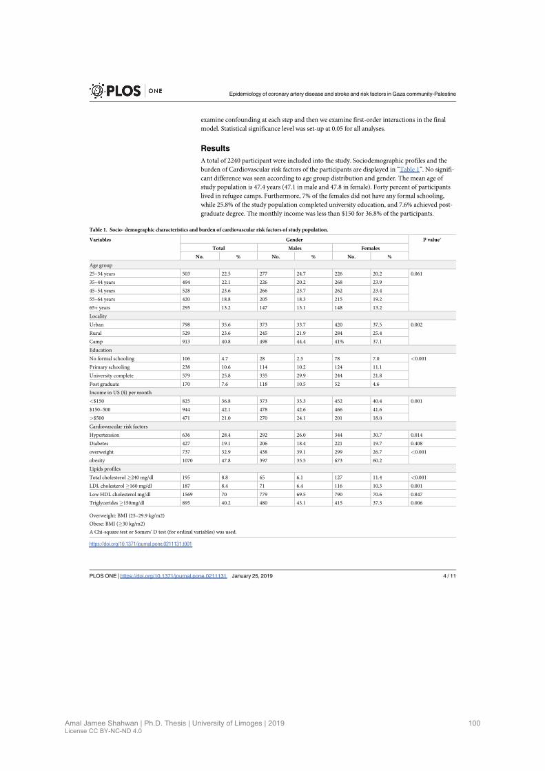
Amal Jamee Shahwan | Ph.D. Thesis | University of Limoges | 2019 License CC BY-NC-ND 4.0
100
examine confounding at each step and then we examine first-order interactions in the final
model. Statistical significance level was set-up at 0.05 for all analyses.
Results
A total of 2240 participant were included into the study. Sociodemographic profiles and the
burden of Cardiovascular risk factors of the participants are displayed in “Table 1”. No signifi-
cant difference was seen according to age group distribution and gender. The mean age of
study population is 47.4 years (47.1 in male and 47.8 in female). Forty percent of participants
lived in refugee camps. Furthermore, 7% of the females did not have any formal schooling,
while 25.8% of the study population completed university education, and 7.6% achieved post-
graduate degree. The monthly income was less than $150 for 36.8% of the participants.
Table 1. Socio- demographic characteristics and burden of cardiovascular risk factors of study population.
Variables Gender P value⇤
Total Males Females
No. % No. % No. %
Age group
25–34 years 503 22.5 277 24.7 226 20.2 0.061
35–44 years 494 22.1 226 20.2 268 23.9
45–54 years 528 23.6 266 23.7 262 23.4
55–64 years 420 18.8 205 18.3 215 19.2
65+ years 295 13.2 147 13.1 148 13.2
Locality
Urban 798 35.6 373 33.7 420 37.5 0.002
Rural 529 23.6 245 21.9 284 25.4
Camp 913 40.8 498 44.4 41% 37.1
Education
No formal schooling 106 4.7 28 2.5 78 7.0 <0.001
Primary schooling 238 10.6 114 10.2 124 11.1
University complete 579 25.8 335 29.9 244 21.8
Post graduate 170 7.6 118 10.5 52 4.6
Income in US ($) per month
<$150 825 36.8 373 33.3 452 40.4 0.001
$150–500 944 42.1 478 42.6 466 41.6
>$500 471 21.0 270 24.1 201 18.0
Cardiovascular risk factors
Hypertension 636 28.4 292 26.0 344 30.7 0.014
Diabetes 427 19.1 206 18.4 221 19.7 0.408
overweight 737 32.9 438 39.1 299 26.7 <0.001
obesity 1070 47.8 397 35.5 673 60.2
Lipids profiles
Total cholesterol "240 mg/dl 195 8.8 65 6.1 127 11.4 <0.001
LDL cholesterol "160 mg/dl 187 8.4 71 6.4 116 10.3 0.001
Low HDL cholesterol mg/dl 1569 70 779 69.5 790 70.6 0.847
Triglycerides"150mg/dl 895 40.2 480 43.1 415 37.3 0.006
Overweight: BMI (25–29.9 kg/m2)
Obese: BMI ("30 kg/m2)
A Chi-square test or Somers’ D test (for ordinal variables) was used.
https://doi.org/10.1371/journal.pone.0211131.t001
Epidemiology of coronary artery disease and stroke and risk factors in Gaza community-Palestine
PLOSONE | https://doi.org/10.1371/journal.pone.0211131 January 25, 2019 4 / 11

Amal Jamee Shahwan | Ph.D. Thesis | University of Limoges | 2019 License CC BY-NC-ND 4.0
101
Regarding CVD risk factors hypertension and diabetes were higher among females. BMI
was! 30kg/m2 in 47.8% of the cases. Obesity was more common in females than males,
60.2%, 35.5% respectively. Conversely The overweight was more common in males than
females 39.1% vs 26.7% (p value<0.001). In addition, 8.8% and 40.2% of study population had
high levels of total cholesterol and triglycerides respectively.
Data on the lifestyle related risk factors are displayed in “Table 2”. Smoking was by far less fre-
quent among women since smoking is not accepted for women in Gaza. The prevalence of smok-
ing among males increased with age until the age of 44 and decreased later. Additionally, 64.3% of
men in age group 25–34 years started to smoke before the age of 18 years. The consumption and
serving of fruits and vegetables was low, with minor differences in gender and age groups.
Overall 218 subjects (9.7%) of participants had CVD 11.5%in males and 8.0% in females
“Table 3”. CAD was present in 185 persons 8.3% (95% CI, 7.14%-9.46%), males reported a
Table 2. Life style-related risk factors in the study population by age for men and women.
Variables Gender Age groups in years % Overall % Age Adjusted % P value
25–34 35–44 45–54 55–64 65+
Current Smoking All 30.2 23.9 22.3 22.4 12.9 23.2 23.6 <0.001
Males 50.5 50.4 42.9 42.4 25.9 44.0 44.5 <0.001
Females 5.3 1.5 1.5 3.3 0.0 2.4 2.61 <0.001
Age of onset of smoking <18y All 61.2 50.0 53.4 47.9 44.7 53.3 52.7 0.05
Males 64.3 49.1 55.3 48.3 44.7 54.4 53.8 0.01
Females 25.3 75.0 0.0 42.0 0.0 33.3 31.3 0.15
Low Physical Activity All 36.4 45.1 49.4 55.2 61.7 48.3 47.3 <0.001
Males 27.1 37.2 40.6 42.9 50.3 38.3 37.7 <0.001
Females 47.8 51.9 58.4 67.0 73.0 58.3 57.1 <0.001
Fruits+ vegetable intake< 4days /week All 26.9 34.0 33.8 28.3 29.2 30.7 30.5 <0.001
Males 24.6 32.0 31.3 26.3 26.5 28.3 28.3 0.04
Females 29.6 35.7 36.4 30.2 31.8 33.1 32.8 0.29
Fruits+ vegetable <2 serving / day All 62.7 70.1 65.6 68.3 61.4 65.9 65.7 0.03
Males 62.0 64.0 63.0 65.0 59.9 63.1 62.8 0.64
Females 63.6 74.8 68.2 70.7 62.8 68.6 68.2 0.07
Data are represented as percent (%)
A Chi-square test was used to compare the difference in risk factors for CAD and stroke across age subgroups for the overall population as well as for each gender.
https://doi.org/10.1371/journal.pone.0211131.t002
Table 3. Prevalence of cardiovascular disease (CAD, stroke) by gender.
Cardiovascular diseases Gender
All Males Females Odds Ratio (95%CI) P value
No. % No. % No. %
CAD 185 8.3 113 10.1 72 6.4 1.63 (1.19–2.21) 0.002
Stroke 67 3.0 39 3.5 28 2.5 1.40 (0.86–2.29) 0.175
CAD or Stroke 184 8.2 106 9.5 78 7.0 1.41 (1.05–1.91) 0.037
CAD and Stroke 34 1.5 23 2.1 11 1.0 2.17 (1.05–4.47) 0.030
CVD 218 9.7 129 11.5 89 8.0 1.51 (1.13–2.01) 0.005
Female gender is the reference group, CAD: Coronary artery disease, CVD: cardiovascular disease
Odds Ratio: male vs female CI: confidence interval 95%
Fisher’s exact method was used
https://doi.org/10.1371/journal.pone.0211131.t003
Epidemiology of coronary artery disease and stroke and risk factors in Gaza community-Palestine
PLOSONE | https://doi.org/10.1371/journal.pone.0211131 January 25, 2019 5 / 11

Amal Jamee Shahwan | Ph.D. Thesis | University of Limoges | 2019 License CC BY-NC-ND 4.0
102
higher prevalence (10.1%) than females (6.4%). Among the 2240 participants we found only 67
cases which reported a history of stroke 3% (95%CI, 2.28%-3.72%) with no difference in gen-
der (3.5% vs.2.5%). The clinical disorders were limited to one territory for 184 participants
(8.2%) involving 9.5% of the males and 7.0% of the females. Two territories were involved in
34 participants (1.5%), with a1.5 higher risk in males.
After adjustment by age and gender, the most important risk factors associated with CVD
were HTN, and DM, p value<0. 001 “Table 4”. Hypertension was associated with a 3-fold
increased risk of CVD and diabetes mellitus a 2.5-fold.
Discussion
Our study is the first to report the prevalence of CVDs among Gazans in Palestine. Previous
surveys were hospital based or on United Nation Relief andWork Agency (UNRWA) report.
The prevalence of CVDs nears 10% of the population above the age of 25years in this area.
Data on the CVD epidemiology in the Middle-East are limited. In the Coronary Artery Dis-
ease in Saudi study (CADISS), a national community-based study conducted in urban and
rural area of Saudi Arabia; 17 232 subjects aged 30–70 years randomly selected were included.
Coronary artery disease was diagnosed on positive questionnaire of angina or history of possi-
ble myocardial infarction. The prevalence of CAD was 5.5% (6.6% in males and 4.4% in
females) [18]. Similar findings have been reported in a Lebanese cross-sectional study using
multistage cluster sample including 1200 subjects!40 years of age. The prevalence of CAD
was 13.4% (17.8% in males, 9.0% in females) [19]. In the Jordanian population, the prevalence
of myocardial infarction was reported at 5.9% in adults over 40 years [20]. Regarding stroke
our results are in line with another Lebanese study including1515 individuals with a mean age
of 57.2 years; stroke and or transit ischemic attack were (3.6%), and the prevalence of any
stroke symptom was up to 12.1% [21]. According to a systemic review in Middle east, the prev-
alence of stroke was estimated between 508 to 777 per 100,000 population [22], more com-
monly in males than females [22,23]. Two hospitals based studies conducted in Palestine,
showed more stroke cases among females than males groups [24,25].
Number of risk factors were identified in epidemiological surveys. In 2016 the global bur-
den disease (GBD) for risk profiles in Middle east and north Africa (MENA) stranded out
these risk factors by order of penetrance, high blood pressure ranked as the first, followed by
obesity, diabetes then smoker [26]. The same grading was retained for Palestine [26].The
Table 4. Multivariate logistic regression (adjusted for age, and gender) with CVD as dependent variable and major risk factors as independent variables.
Variables Univariate analysis Adjusted analysis
Odds Ratio (CI) P value Odds Ratio (CI) P value
Age 0.94 (0.93–0.95) <0.001
Gender 1.51 (1.13–2.01) 0.005
Hypertension 4.73 (3.53–6.31) <0.001 2.51 (1.81–3.50) <0.001
Diabetes 4.49 (3.36–6.01) <0.001 2.22 (1.61–3.11) <0.001
BMI!30kg/m2 1.44 (1.10–1.91) 0.011
Current smoking 1.07(0.77–1.48) 0.686
High cholesterol 1.27(0.80–2.01) 0.303
High triglycerides 1.43 (1.08–1.89) 0.013
BMI: Body mass index
Logistic regression model was used
Odds Ratio: male vs female. CI: confidence interval (95%)
https://doi.org/10.1371/journal.pone.0211131.t004
Epidemiology of coronary artery disease and stroke and risk factors in Gaza community-Palestine
PLOSONE | https://doi.org/10.1371/journal.pone.0211131 January 25, 2019 6 / 11

Amal Jamee Shahwan | Ph.D. Thesis | University of Limoges | 2019 License CC BY-NC-ND 4.0
103
UNRWA 2016 report for Gaza, found 20.1% subjects with hypertension among population
aged! 40 years (Palestinian Refugees of Syria included) [27]; we reported a prevalence of
(28.4%). The literature review revealed a higher prevalence of hypertension in Arab countries
29.5%. The same prevalence was found in the US (29.6%), and (30%) in United Kingdom [28].
Obesity is a common health problem worldwide particularly in Arab world. The prevalence
of obesity and overweight in Arab states ranges from (25% to 81.9%) [29]. The Global burden
disease (GBD) 2016 estimated that high BMI was the leading six risk factors in Arab countries
[26]. The overall obesity in our study was found to be 47.8%, with higher prevalence in females
(60.2%), the mean BMI was 30.35. It is in line with previous study performed in urban Pales-
tinian population of Ramallah, describing an obesity prevalence up to 41% (49% and 30%) in
females and males respectively [30], these results were higher than those in the US population
(36.5%)[31] or in Israel which the National Health and Nutrition 1999–2001 (MABAT) was
conducted among 2782 persons, and showed a prevalence of overweight in 39.3% and obesity
in 22.9% adults population. The Israeli Arab population was more obese than the Jewish one
[32]. Also, our prevalence was higher than among Tunisian males and females (18.2%, 33.5%)
respectively [33].
Diabetes mellitus was identified in our study with a prevalence of 19.1%. WHO report 2015
fromMiddle East Region (Bahrain, Kuwait, Oman, and the United Arab Emirates) estimated
that the prevalence of DM was between 3.5% and 30% [34]. In a study in Saudi Arabia, the
prevalence of type 2 diabetes among 2355 adults was 29.3% [35]. For smokers, our estimated
prevalence was 23.2%. The WHO report on the global tobacco epidemiology (2011) estimated
that Europe had the highest prevalence of smokers (35%); and the lowest was in Africa. The
global Adult tobacco survey showed that in fourteen LMIC, 48.6% of men and 11.3% of
women were tobacco users [36]. Additionally, in our neighbour countries, a cross sectional
study conducted in Lebanon, among 2836 adults!18 years of age, showed that the current
smoking prevalence rate was 34.7% with higher rate in males than females,42.9% and 27.5%
respectively [37]. In the BREATH study, 62 086 subjects aged! 40years in ten countries in
MENA region, were interviewed regarding smoking habits, with equal number of men and
women. The smoking rate was estimated to be 31.2%, ranging from 15.3% in Morocco to
53.9% in Lebanon. The proportion was higher in men (48.0%) than in women (13.8%) [38],
the prevalence of female smokers has traditionally been low due to the Eastern Mediterranean
region’s conservative cultural and social values. Less than one third of our study population
have dyslipidaemia. However, our finding was counterpart with data from ACE (Cardiovascu-
lar Epidemiological) study in United Arab Emirates, the prevalence was 74% [39], while the
high prevalence of dyslipidaemia in our study in young adults (age 25-34years) calls for an ear-
lier screening for dyslipidaemia and other risk factors. Another important factor observed in
our study was the proportion of people with low physical activity, it was estimated that half of
the population 48.3% had low physical activity. Women exercised less than men (58.3%). We
ascribe these phenomena to the fact that women have maternal responsibility and were more
obese. In a study for 163 556 persons in 38 Muslim countries who completed IPAQ, the total
physical inactivity prevalence was 32.8%. The prevalence among Arab countries was 43.7% vs
28.6% in non-Arab countries, and Arab women were more physically inactive [40]. In a cross-
sectional study conducted in Saudi Arabia, physical activity was assessed using the global phys-
ical activity questionnaire among 4758 participants. The prevalence of low activity was 66.6%
(60.1% in males, and 72.9% in females). Similar data were found in the Arabian Gulf region
[41,42].
Nutrition is an important determinant of health; inadequate consumption of fruit and vege-
table is a factor that can play role in morbidity. As other studies in Arab gulf countries our
study population did not consume sufficient quantities of fruits and vegetables. A study
Epidemiology of coronary artery disease and stroke and risk factors in Gaza community-Palestine
PLOSONE | https://doi.org/10.1371/journal.pone.0211131 January 25, 2019 7 / 11

Amal Jamee Shahwan | Ph.D. Thesis | University of Limoges | 2019 License CC BY-NC-ND 4.0
104
analyzed data from 197 373 adult participants from 52 countries taking part in the world health
survey in LIMIC (2002–2003). The prevalence of low consumption of fruit and vegetable was
77.6% in males and 78.4% in females [43].
Strengths and limitations
There are few limitations to our study. First, the cross-sectional nature of this study design lim-
its the interference of causal relation-ships between risk factors and CAD or stroke. Also, the
prevalence of high blood pressure and raised blood glucose have been over estimated because
these two risk factors were evaluated once (no reevaluation during another visit, and no three
consecutive measures of BP according to WHO step). Further, possible bias could have been
introduced since study was conducted at home and data concerning more risk factors and his-
tory of CAD and stroke were self- reported. Also, the wide age range of participants, 25 years
and more is both a strength and a weakness of this study. However, 32% of the population was
aged 55 years and more, who have a greatest prevalence of CVD and risk factors. Even so we
found considerable charge of risk factors in young population. Also, this study has strengths
points: women were well represented, with a male: female ratio of 1, and it was performed in a
large mixed area with representative sample, and good response rate; making it the first study
in Palestine to report a national estimate for CVD prevalence.
Conclusion
This study was the first nationwide endeavor that provides information about the prevalence
of CVDs and the level of cardio vascular risk factors among palestinian community in Gaza.
A rate of CVD has been reported in our population, 10% reflecting a serious health problem
in Gaza strip. Obesity, hypertension and diabetes, were highly prevalent. Increased, effort and
research to monitor and improve strategies and policies for reducing cardiovascular risk are
mandatory.
Supporting information
S1 Dataset. Database of study participants.
(XLSX)
S1 Text. Definition of study variables.
(DOCX)
Acknowledgments
We thank all the nurse’s team for collecting data and we acknowledge Dr Raid Sabah the direc-
tor of United Health Care Center (UHCC) for his support.
Author Contributions
Conceptualization: Amal Jamee Shahwan, Philippe Lacroix.
Data curation: Amal Jamee Shahwan.
Formal analysis: Amal Jamee Shahwan, Yehia Abed.
Investigation: Amal Jamee Shahwan.
Methodology: Amal Jamee Shahwan.
Supervision: Philippe Lacroix.
Epidemiology of coronary artery disease and stroke and risk factors in Gaza community-Palestine
PLOSONE | https://doi.org/10.1371/journal.pone.0211131 January 25, 2019 8 / 11

Amal Jamee Shahwan | Ph.D. Thesis | University of Limoges | 2019 License CC BY-NC-ND 4.0
105
Validation: Amal Jamee Shahwan, Pierre Marie Preux, Victor Aboyans, Philippe Lacroix.
Visualization: Ileana Desormais, Julien Magne, Pierre Marie Preux, Victor Aboyans, Philippe
Lacroix.
Writing – original draft: Amal Jamee Shahwan.
Writing – review & editing: Amal Jamee Shahwan, Philippe Lacroix.
References1. Mendis S, Puska P, Norrving B, OrganizationWH, FederationWH, OrganizationWS. Global atlas on
cardiovascular disease prevention and control [Internet]. Geneva: World Health Organization; 2011[cited 2017 Feb 14]. Available from: http://www.who.int/iris/handle/10665/44701
2. WHO.WHO | Cardiovascular diseases (CVDs) [Internet]. 2017 [cited 2017 Dec 19]. Available from:http://www.who.int/mediacentre/factsheets/fs317/en/
3. Benjamin EJ, Blaha MJ, Chiuve SE, CushmanM, Das SR, Deo R, et al. Heart Disease and Stroke Sta-tistics—2017 Update: A Report From the American Heart Association. Circulation [Internet]. 2017 Jan 1[cited 2017 May 6]; Available from: http://circ.ahajournals.org/content/early/2017/01/25/CIR.0000000000000485
4. WHO | Cardiovascular diseases (CVDs) [Internet]. WHO. [cited 2017 Feb 3]. Available from: http://www.who.int/mediacentre/factsheets/fs317/en/
5. GBD 2013 Mortality and Causes of Death Collaborators. Global, regional, and national age-sex specificall-cause and cause-specific mortality for 240 causes of death, 1990–2013: a systematic analysis forthe Global Burden of Disease Study 2013. Lancet Lond Engl. 2015 Jan 10; 385(9963):117–71.
6. Roth GA, HuffmanMD, Moran AE, Feigin V, Mensah GA, Naghavi M, et al. Global and regional patternsin cardiovascular mortality from 1990 to 2013. Circulation. 2015 Oct 27; 132(17):1667–78. https://doi.org/10.1161/CIRCULATIONAHA.114.008720 PMID: 26503749
7. Husseini A, Abu-Rmeileh NM, Mikki N, Ramahi TM, Ghosh HA, Barghuthi N, et al. Cardiovascular dis-eases, diabetes mellitus, and cancer in the occupied Palestinian territory. The Lancet. 2009 Mar 27;373(9668):1041–9.
8. World Health Statistics 2011 [Internet]. WHO. [cited 2017 Feb 4]. Available from: http://www.who.int/whosis/whostat/2011/en/
9. Zynia L R, Andrew C. THE BURDENOF DISEASE IN THEWEST BANK ANDGAZA AN ASSESS-MENT REPORT. 2000 Feb;
10. WHO. Health conditions in the occupied Palestinian territory, including east Jerusalem, and in the occu-pied Syrian Golan. 2016 May 6;2–59.
11. PCBS 2016. palestinian central bureau of statistic.
12. Report WHO Expert. WHO |Waist circumference and waist–hip ratio [Internet]. WHO. 2008 [cited 2018Feb 15]. Available from: http://www.who.int/nutrition/publications/obesity/WHO_report_waistcircumference_and_waisthip_ratio/en/
13. American Diabetes Association 2016. ADA Diabetes Management Guidelines A1C Diagnosis | NDEI[Internet]. [cited 2018 Jan 22]. Available from: http://www.ndei.org/ADA-diabetes-management-guidelines-diagnosis-A1C-testing.aspx.html
14. James PA, Oparil S, Carter BL, CushmanWC, Dennison-Himmelfarb C, Handler J, et al. 2014 evi-dence-based guideline for the management of high blood pressure in adults: report from the panelmembers appointed to the Eighth Joint National Committee (JNC 8). JAMA. 2014 Feb 5; 311(5):507–20. https://doi.org/10.1001/jama.2013.284427 PMID: 24352797
15. WHO hypertension. WHO | Q&As on hypertension [Internet]. WHO. 2015 [cited 2018 Jan 9]. Availablefrom: http://www.who.int/features/qa/82/en/index.html
16. Chen Y, Zhang X, Pan B, Jin X, Yao H, Chen B, et al. A modified formula for calculating low-density lipo-protein cholesterol values. Lipids Health Dis. 2010 May 21; 9:52. https://doi.org/10.1186/1476-511X-9-52 PMID: 20487572
17. National Cholesterol Education Program (NCEP) Expert Panel on Detection, Evaluation, and Treatmentof High Blood Cholesterol in Adults (Adult Treatment Panel III). Third Report of the National CholesterolEducation Program (NCEP) Expert Panel on Detection, Evaluation, and Treatment of High Blood Cho-lesterol in Adults (Adult Treatment Panel III) final report. Circulation. 2002 Dec 17; 106(25):3143–421.PMID: 12485966
Epidemiology of coronary artery disease and stroke and risk factors in Gaza community-Palestine
PLOSONE | https://doi.org/10.1371/journal.pone.0211131 January 25, 2019 9 / 11

Amal Jamee Shahwan | Ph.D. Thesis | University of Limoges | 2019 License CC BY-NC-ND 4.0
106
18. Al-NozhaMM, Arafah MR, Al-Mazrou YY, Al-Maatouq MA, Khan NB, Khalil MZ, et al. Coronary arterydisease in Saudi Arabia. Saudi Med J. 2004 Sep; 25(9):1165–71. PMID: 15448760
19. Zeidan RK, Farah R, Chahine MN, Asmar R, Hosseini H, Salameh P, et al. Prevalence and correlatesof coronary heart disease: first population-based study in Lebanon. Vasc Health Risk Manag. 2016Mar17; 12:75–84. https://doi.org/10.2147/VHRM.S97252 PMID: 27051290
20. Nsour M, Mahfoud Z, KanaanMN, Balbeissi A. Prevalence and predictors of nonfatal myocardial infarc-tion in Jordan. East Mediterr Health J Rev Sante Mediterr Orient Al-Majallah Al-Sihhiyah Li-Sharq Al-Mutawassit. 2008 Aug; 14(4):818–30.
21. Farah R, Zeidan RK, Chahine MN, Asmar R, Chahine R, Salameh P, et al. Prevalence of stroke symp-toms among stroke-free residents: first national data from Lebanon. Int J Stroke. 2015 Oct 1; 10(SA100):83–8. https://doi.org/10.1111/ijs.12563 PMID: 26178607
22. El-Hajj M, Salameh P, Rachidi S, Hosseini H. The epidemiology of stroke in the Middle East. Eur StrokeJ. 2016 Sep 1; 1(3):180–98.
23. Benamer HT, Grosset D. Stroke in Arab countries: A systematic literature review. J Neurol Sci. 2009Sep 15; 284(1–2):18–23. https://doi.org/10.1016/j.jns.2009.04.029 PMID: 19428027
24. Sawalha A. Characterization of Hospitalized Ischemic Stroke Patients in Palestine. Libyan J Med [Inter-net]. 2009 [cited 2017 Feb 7]; 4(2). Available from: http://www.libyanjournalofmedicine.net/index.php/ljm/article/view/4803
25. SweilehWM, Sawalha AF, Al-Aqad SM, Zyoud SH, Al-Jabi SW. The Epidemiology of Stroke in NorthernPalestine: A 1-Year, Hospital-Based Study. J Stroke Cerebrovasc Dis. 2008 Nov; 17(6):406–11. https://doi.org/10.1016/j.jstrokecerebrovasdis.2008.06.008 PMID: 18984436
26. Forouzanfar MH, Bhutta ZA, Burnett R, Cercy K, Charlson FJ, Cohen AJ, et al. Global, regional, andnational comparative risk assessment of 79 behavioural, environmental and occupational, and meta-bolic risks or clusters of risks, 1990–2015: a systematic analysis for the Global Burden of Disease Study2015. The Lancet. 2016 Oct 8; 388(10053):1659–724.
27. Seita A, Khader A, Shahin Y, HababeehM, ZeidanW, Turki Y, et al. UNRWAHealth Annual Report2016 (published May 2017). 2017.
28. Joffres M, Falaschetti E, Gillespie C, Robitaille C, Loustalot F, Poulter N, et al. Hypertension preva-lence, awareness, treatment and control in national surveys from England, the USA and Canada, andcorrelation with stroke and ischaemic heart diseasemortality: a cross-sectional study. BMJ Open. 2013Aug 1; 3(8):e003423. https://doi.org/10.1136/bmjopen-2013-003423 PMID: 23996822
29. Musaiger AO. Overweight and Obesity in Eastern Mediterranean Region: Prevalence and PossibleCauses. J Obes [Internet]. 2011 [cited 2018 Jan 27];2011. Available from: https://www.ncbi.nlm.nih.gov/pmc/articles/PMC3175401/
30. Abdul-Rahim HF, Abu-Rmeileh NM, Husseini A, Holmboe-Ottesen G, Jervell J, Bjertness E. Obesityand selected co-morbidities in an urban Palestinian population. Int J Obes Relat Metab Disord J IntAssoc Study Obes. 2001 Nov; 25(11):1736–40.
31. CDC. CDCWorks 24/7 [Internet]. Centers for Disease Control and Prevention. 2017 [cited 2017 Feb14]. Available from: https://www.cdc.gov/index.htm
32. Kaluski DN, Berry EM. Prevalence of obesity in Israel. Obes Rev Off J Int Assoc Study Obes. 2005May; 6(2):115–6.
33. Maatoug J, Harrabi I, Hmad S, BelkacemM, Nouira A, GhannemH. Advising Obese Adults about Dietand Physical Activity in Sousse, Tunisia [Internet]. International Scholarly Research Notices. 2013[cited 2018 Jan 27]. Available from: https://www.hindawi.com/journals/isrn/2013/498527/
34. Noncommunicable diseases country profiles 2014-WHO global report [Internet]. 2018 [cited 2018 Feb23]. Available from: http://www.euro.who.int/en/health-topics/noncommunicable-diseases/ncd-background-information/noncommunicable-diseases-country-profiles-2014
35. Khudairy LA, Rees K, Kumar S, Al-Daghri N, Attas OA, Okail MA, et al. Abstract P263: Central Obesityand the Emerging Epidemic of Type 2 Diabetes in Saudi Arabia. Circulation. 2015 Mar 10; 131(Suppl1):AP263–AP263.
36. Giovino GA, Mirza SA, Samet JM, Gupta PC, Jarvis MJ, Bhala N, et al. Tobacco use in 3 billion individu-als from 16 countries: an analysis of nationally representative cross-sectional household surveys. TheLancet. 2012; 380(9842):668–679.
37. Sibai AM, Iskandarani M, Darzi A, Nakkash R, Saleh S, Fares S, et al. Cigarette smoking in a MiddleEastern country and its association with hospitalisation use: a nationwide cross-sectional study. BMJOpen [Internet]. 2016 Apr 8 [cited 2017 Jan 29]; 6(4). Available from: http://www.ncbi.nlm.nih.gov/pmc/articles/PMC4838686/
38. Khattab A, Javaid A, Iraqi G, Alzaabi A, Ben Kheder A, Koniski M-L, et al. Smoking habits in the MiddleEast and North Africa: results of the BREATHE study. Respir Med. 2012 Dec; 106 Suppl 2:S16–24.
Epidemiology of coronary artery disease and stroke and risk factors in Gaza community-Palestine
PLOSONE | https://doi.org/10.1371/journal.pone.0211131 January 25, 2019 10 / 11

Amal Jamee Shahwan | Ph.D. Thesis | University of Limoges | 2019 License CC BY-NC-ND 4.0
107
39. Radaideh G, Tzemos N, Ali TM, Eldershaby Y, Joury J, Abreu P. Cardiovascular Risk Factor Burden inthe United Arab Emirates (UAE): The Africa Middle East (AfME) Cardiovascular Epidemiological (ACE)Study Sub-analysis. Int Cardiovasc Forum J [Internet]. 2017 Jun 3 [cited 2018 Jan 26]; 11(0). Availablefrom: http://icfjournal.org/index.php/icfj/article/view/414
40. Kahan D. Adult physical inactivity prevalence in the Muslim world: Analysis of 38 countries. Prev MedRep. 2015 Jan 1; 2:71–5. https://doi.org/10.1016/j.pmedr.2014.12.007 PMID: 26844051
41. Mabry RM, Reeves MM, Eakin EG, Owen N. Evidence of physical activity participation amongmen andwomen in the countries of the Gulf cooperation council: a review. Obes Rev Off J Int Assoc Study Obes.2010 Jun; 11(6):457–64.
42. Saeed AA, Al-Hamdan NA, Al-Zalabani AH. The prevalence of physical activity and its socioeconomiccorrelates in Kingdom of Saudi Arabia: A cross-sectional population-based national survey. 2015 [cited2018 Mar 10]; Available from: http://repository.taibahu.edu.sa/handle/123456789/14235.
43. Hall JN, Moore S, Harper SB, Lynch JW. Global variability in fruit and vegetable consumption. Am JPrev Med. 2009 May; 36(5):402–409.e5. https://doi.org/10.1016/j.amepre.2009.01.029 PMID:19362694
Epidemiology of coronary artery disease and stroke and risk factors in Gaza community-Palestine
PLOSONE | https://doi.org/10.1371/journal.pone.0211131 January 25, 2019 11 / 11

Amal Jamee Shahwan | Ph.D. Thesis | University of Limoges | 2019 License CC BY-NC-ND 4.0
108
IV.2. Article 2: Epidemiology of Lower Extremity Artery Disease in Gaza –Palestine
Contexte de l’étude
L’Artériopathie oblitérante des membres inférieurs (AOMI) est une des expressions de
l’athérosclérose, L’AOMI est bien définie par la mesure de l’index de pression systolique.
Concernant les pays du Moyen-Orient, les quelques études qui existent sur l’artériopathie sont
menées chez les patients diabétiques. Aucune donnée dans la littérature ne porte sur
l’incidence ou la prévalence de l’artériopathie en Palestine même en milieu hospitalier.
Méthodologie
À travers une étude transversale, 12 infirmiers diplômés, formés à la prise de l’index de
pression systolique ont mesuré cet index sur un échantillon de 1398 personnes d’âge
supérieur à 40 ans. Ils ont aussi documenté les facteurs de risque cardio vasculaire pour tous
les participants.
L’objectif de l’étude
Décrire l’épidémiologie de l’artériopathie des membres inférieurs dans la population de 40 ans
et plus de la bande de Gaza et les facteurs de risque associés.
Valorisation de l’étude
L’étude est soumise à ‘International journal Cardiology (IF :4.034)
Communications
Communication orale : Congrès de la Société Française de la Médecine Vasculaire la
Rochelle-France 19-22 septembre 2018

Amal Jamee Shahwan | Ph.D. Thesis | University of Limoges | 2019 License CC BY-NC-ND 4.0
109
Epidemiology of Lower Extremity Artery Disease in Gaza –
Palestine
Amal Jamee Shahwana,b,c, Victor Aboyansa,b,d, Julien Magnea,b,d, Yehia Abede,
Pierre Marie Preuxa,b, Philippe Lacroixa,b,d
aINSERM UMR 1094, Tropical Neuroepidemiology, Limoges, France
bUniv. Limoges, UMR 1094, Tropical Neuroepidemiology, Institute of Epidemiology
and Tropical Neurology, GEIST, 87000 Limoges, France
cDepartment of cardiology-Ministry of health-Palestine
dDepartment of Thoracic and Vascular Surgery – Vascular Medicine, Dupuytren
University Hospital, Limoges, France
eUniversity Al Quods, Gaza, Palestine
Correspondence: Amal Jamee Shahwan
Univ. Limoges, UMR 1094, Tropical Neuroepidemiology, Institute of Epidemiology
and Tropical Neurology, GEIST, 87000 Limoges, France
Tel +33754139112
Email [email protected]
Abstract:
Objectives: Lower Extremity artery disease (LEAD) is one of the most common
manifestations of atherosclerosis affecting more than 200 million people globally,
but little is known about its epidemiology in Middle East populations, particularly in
Arab countries. The present study was undertaken to assess the prevalence of

Amal Jamee Shahwan | Ph.D. Thesis | University of Limoges | 2019 License CC BY-NC-ND 4.0
110
2
LEAD in the general population aged 40 years and more in Gaza-Palestine, and
to determine the prevalence of its associated risk factors.
Methods: The study design was a cross-sectional with a stratified proportional
sampling, involving the five governorates in Gaza Strip. We selected 1490
individuals aged age ≥ 40 years living in the area for more than 3 years.
Questionnaire on lifestyle and cardiovascular risk factors was applied and the
ankle-brachial index (ABI) was measured by Doppler examination in both legs in
all participants. LEAD was defined as an ABI < 0.90. We excluded 92 participants
(6%) because of missing ABI or ABI >1.40.
Results: One-half of the 1398 participants had at least two associated
cardiovascular risk factors. Overall, we found LEAD in 191 (13.7%) cases. This
prevalence increased with age and was higher in females than in males
(respectively15.6% vs 11.6% p=0.031). In the multivariate logistic regression
model, significant associated factors with LEAD in male were overweight (OR:3.7;
95% CI 1.4-9.9), CAD (OR:3.6; 95% CI1.4-9.1), hypertension (OR: 3.0; 95%
CI:1.3-6.8), and Current smoking (OR:2.7;95% CI1.1-6.8). In women Hypertension
remain the main associated risk factor.
Conclusion: Our study highlights the high prevalence of LEAD in Gazan
community, mostly related to the high prevalence of cardiovascular risk factors and
other cardiovascular conditions.
Keywords: Lower extremity artery disease, prevalence, risk factors, community.
Amal Jamee Shahwan | Ph.D. Thesis | University of Limoges | 2019 License CC BY-NC-ND 4.0
111
3
1.Introduction
Lower extremity artery disease is one of the most frequent manifestations of
systemic atherosclerosis. It is an indicator of widespread atherosclerosis in other
vascular territories. It represents the third leading cause of atherosclerotic
cardiovascular morbidity, following coronary artery disease (CAD) and stroke. The
WHO estimates that in year 2010 about 140.8 million (69%-70%) people with
LEAD were living in low- and middle-income countries (LMIC), of whom (10.3
million) in the Eastern Mediterranean region. The burden of LEAD has raised by
23.5% from 2000 to 2010 with greater increase in (LMIC), where the prevalence
rates were reported higher in women[1–3].About 10-20% of patients with LEAD
have intermittent claudication[4]. However, even if the disease is often
asymptomatic; a screening strategy is meaningful to identify individuals at high risk
of death, major cardiovascular events, as well as limb events, including
amputation, in the most severe cases [5]. Several, studies demonstrated that
LEAD (either symptomatic or asymptomatic) is associated with a three to six-fold
risk of death, from cardiovascular disease (CVD) morbidity[6,7]. Patients with
LEAD are now considered as coronary artery disease equivalent risk [8]. This
disease can be easily diagnosed by the measurement of the ankle brachial index
(ABI), convenient for screening LEAD in population. It can be used to stratify the
severity and potentially helpful to evaluate CVD risk and guide preventive
intervention [9]. LEAD shares common risk factors with CAD including ageing,
diabetes, hypertension, smoking, and male gender[8]. Its prevalence has been
extensively described in high income countries (HIC) populations. The association

Amal Jamee Shahwan | Ph.D. Thesis | University of Limoges | 2019 License CC BY-NC-ND 4.0
112
4
between cardiovascular risk factors and LEAD was also been documented in these
settings. Regarding the Middle East countries, few data are available, most of them
being hospital-based[10]. Our aim in this report is to describe the epidemiology of
LEAD in the Gazan community. We hypothesized that, in regards to the high CVD
risk factors and other atherosclerosis disease[11], the prevalence of LEAD would
be high in this part of the world.
2. Material and methods
2.1 Study design
This study was part of a first epidemiological study conducted from July to
September 2017 among 2240 individuals in age ≥25 years, recruited from a
stratified proportional sample in five Palestinian governorates of the Gaza strip (the
method was previously described)[11]. The STEPS wise approach was adopted:
In each governorate, 2-3 urban and rural areas were randomly chosen, of which
20-30 households were selected in every area. The ABI was measured in all adults
≥40 years of age. In this study 1490 individuals, were enrolled. Among them 92
subjects were excluded from the analysis due to either an ABI>1.40 (89
subjects,4.6%) or incomplete exam (3 subjects). Overall 1398 subjects remained
for analysis (Figure1).
A standardized questionnaire was used to obtain demographic and information
about level of education, monthly household income as well as cardiovascular risk
factors and medication use. For tobacco use, subjects were classed as never,
past, and current smokers. History of coexisting CVD was documented. CAD was
defined as self-reported with either angina pectoris or myocardial infarction, or

Amal Jamee Shahwan | Ph.D. Thesis | University of Limoges | 2019 License CC BY-NC-ND 4.0
113
5
history of procedures performing percutaneous coronary intervention or coronary
bypass graft. Cerebrovascular disease (CBVD) was defined as documented
history of stroke. Height and weight were measured with participants wearing light
clothing and no shoes in order to determine the body mass index (BMI) calculated
as weight in Kilograms divided by height in meters squared, and the cut-off points
of WHO for BMI were used to define obesity as ≥ 30 kg/m2. For hypertension
(HTN), we considered subjects as hypertensive if their average systolic blood
pressure (SBP) in both arms was ≥140 mmHg or their average diastolic blood
pressure (DBP) ≥ 90 mmHg, or if they were being treated for HTN [12]. Diabetes
mellitus (DM) was defined if capillary blood glucose level ≥126 mg/dl in fasting
participants, or ≥ 200 mg/dl in non-fasting participants, or if they were taking any
anti-diabetic medications [13]. Blood sample for lipids profiles were taken by
nurse’s staff from antecubital vein and sent to the laboratory for analysis using
standard methods. We defined high plasma of total cholesterol ≥ 240 mg/dl,
plasma triglycerides ≥150 mg/dl, low density lipid cholesterol (LDL) ≥ 160 mg/dl,
and high density lipoprotein cholesterol (HDL) ≤ 40 mg/dl in males and ≤ 50 mg/dl
in females [14].
The ABI was measured following the AHA statement standardized protocol using
the Doppler method[15]. The leg-specific ABI was calculated as the higher SBP in
the posterior tibial or dorsalis pedis divided by the higher of the 2 arms SBP. For
each subject, LEAD was defined by an ABI < 0.90 in either leg. Patients with an
ABI >1.40 were excluded from this analysis n=89 (6%).
Amal Jamee Shahwan | Ph.D. Thesis | University of Limoges | 2019 License CC BY-NC-ND 4.0
114
6
The Ethical of Human Research Committee of Palestinian Health Research
Council approved the study protocol, and each participant consented, either written
or verbal according to his education level, to participle in this study.
2.2 Statistical Analysis
The statistical analyses were performed using SPSS software version 22.
Descriptive analyses were done using means and standard deviation or
percentages. Confidence intervals were estimated using normal approximation.
Student’s t-.test and the Chi-square test were used for comparison of continuous
and categorical variables respectively. Multiple logistic regression analyses were
performed using LEAD as the dependent variable and risk factors as independent
variables separately in male and female. Variables with a univariate p value < 0.25
were selected for further multivariate analysis. A p-value < 0.05 was considered
as significance level.
3. Results
Among the 1398 participants 733 (52.4%) were females. The mean age of the
general population was 55.2 ±10.6 years, with nearly one-third (33.4%) over age
of 60. The characteristics of the study population stratified by ABI categories are
displayed in Table 1. Subjects with LEAD were more likely to be old and female,
with significantly higher rates of hypertension, diabetes, history of CAD and stroke
as compared to LEAD-free participants. In the LEAD group statins and antiplatelet
drugs were prescribed in only 22.5% and 43.5% of the cases respectively.

Amal Jamee Shahwan | Ph.D. Thesis | University of Limoges | 2019 License CC BY-NC-ND 4.0
115
7
The prevalence of LEAD was 13.7% (95% CI,11.8-15.4). The prevalence of LEAD
by selected characteristics are displayed in Table 2. Notably, the prevalence of the
LEAD was higher in females. In both genders; the prevalence of LEAD increased
with age ranging from 6.7% in the age category 40 to 49 years to 20.5% in the age
category ≥70 years in males, and from 15.3% to 23.2% in the corresponding age
categories in females (Figure 2). In addition, CAD and stroke history were more
frequent in case of LEAD, both in males and females (Figure 3).
The logistic regression analysis was separately presented in male and female
(Table 3 and Table 4). In multivariate analysis in male gender, and by order of
penetrance: overweight, history of CAD, hypertension and being smoking were the
strongest associated factors with LEAD (OR= 3.7, 3.6, 3.0 and 2.7 respectively).
In female The ORs remained significant for hypertension (OR=2.5,95%CI:1.66-
3.76, p<0.001).
4.Discussion
This is the first study reporting data on epidemiology of LEAD in the Palestinian
community. These results show that LEAD affects 13.7% of adult population ≥
40years of age. Data regarding LEAD in Arab countries are limited. In a cross-
sectional study in Saudi Arabia, among 598 diabetics ≥ 30 years of age, the
prevalence of LEAD was 23.1% [16]. Another cross-sectional study in Bahrain
evaluated 1477 diabetics patients with ages ranging from 18 to 75 years, reporting
a LEAD prevalence of 11.8%[17]. In 2004-2005, the Global Atherothrombosis
Assessment Middle East (AGATHA-ME) a multicenter study conducted in five
neighboring countries of similar ethnicity and small population from Middle East,
Amal Jamee Shahwan | Ph.D. Thesis | University of Limoges | 2019 License CC BY-NC-ND 4.0
116
8
included 1342 patients recruited from tertiary care settings either with CVD or
without CVD but at risk for developing it. Low ABI (≤0.90) was highly prevalent in
both groups (31.5% in the at risk group and 28.2% in the CVD group).[18] The
most comprehensive report on global LEAD prevalence in communities was
published in 2013, but the authors did not identify any contributive study from the
Middle East countries[1]. The prevalence described in the Gazan community is
close to the data collected in high-income countries (HIC), with an increase with
age in both sexes. In the group of 60-70 years of age the prevalence was up to
15.9% in our study compared to 8-10% in HIC [1]. The female’s predominance was
previously described in LMIC, as opposed to higher prevalence in males in HIC.
However, the particularity of our community is in that smoking habits are almost
totally absent among women (0.7%). The female’s predominance in our study is
driven by the burden of the other associated risk factors. Obesity, diabetes and
hypertension were highly prevalent up to 70%, 27% and 42% respectively. In the
Rotterdam study 7715 subjects aged ≥ 55 years were included with a higher
prevalence in females (20.5% vs. 16.9% in males)[19]. The NHANES study 1999-
2004 showed that in participants within the 40-69 years age range, females were
1.77 times more likely to have LEAD than males[20]. This higher prevalence in
women may be partly explained, by postmenopausal hormonal effects,
characterized by an estrogen deficiency responsible for increased risk of
developing CVD[21]. Finally, a smaller threshold for ABI in men and women may
lead to a slight overestimation of LEAD in women who have intrinsically a lower
ABI[22].

Amal Jamee Shahwan | Ph.D. Thesis | University of Limoges | 2019 License CC BY-NC-ND 4.0
117
9
Hypertension, diabetes, smoking, hyperlipidemia, and advancing age are the
major risk factors for LEAD [23]. In the Framingham study the relative risks of
LEAD related to diabetes and hypertension were of 2 and 1.5 respectively[24]. This
relationship appeared in our study with similar risk in univariate analysis. In most
of studies LEAD was strongly associated with smoking[25–27]. We found this
association only in men. However very few women smoke (0.7%). Our findings do
not support the hypothesis that total serum cholesterol and triglycerides increase
the risk of LEAD, but 15.8% of the subject were taking lipids lowering drugs, thus
lipids levels were influenced by treatment. Numerous studies reported a weak
association between dyslipidemia and LEAD, in LMIC the relative risk were for
hypercholesterolemia and hypertriglyceridemia at 1.14 and 1.13 respectively[1].
Regarding obesity similar results were observed in several study without
association with LEAD[28]. The association between LEAD and other
manifestation of atherosclerosis was extensively well documented. LEAD is a
strong marker of risk of CAD or stroke. In our study, a history of CAD, and stroke
were each approximatively twice as more frequent in those with LEAD. This is in
line with the Cardiovascular Health Study (CHS) reporting the history of myocardial
infarction as 2.5 times higher in subjects with LEAD. This odd-ratio was at 3.1 for
stroke[29]. In the CAPRIE trial; 41% of patient with LEAD had either CAD or CBVD
and 8.6% had disease in all three beds[30,31]. Also, in the Reduction of
Atherothrombosis for Continued Health (REACH) registry data for 44 countries
worldwide which included 55,814 patients ≥ 45 years of age, with established
atherothrombotic disease (CAD, CVD, or LEAD),14.9% of patients had

Amal Jamee Shahwan | Ph.D. Thesis | University of Limoges | 2019 License CC BY-NC-ND 4.0
118
10
documented symptomatic LEAD (7.8% in Middle East), and LEAD patients had a
higher prevalence of concomitant vascular disease (61.5% having either CAD,
CBVD, or both)[32].
5. Strengths and Limitations
This study collected many epidemiological information regarding the prevalence of
LEAD in a large sample representative of the general population from a region with
high prevalence of risk factors. No data have been ever published in this area,
where undertaking an epidemiology study is difficult because of the particular
geopolitical aspects. There are however some limitations: because of its cross-
sectional design, the associations found cannot prove causality. The use of self-
reported data for some risk factors could underestimate the actual prevalence of
associate risk factors, mainly for CAD, stroke and smoking. Smoking status was
declarative and probably the number of smokers may be under evaluated in male
population. This study may be subject to survival bias, since the study was
conducted in homes and participants with chronic disease may be the most likely
to participate in the study.
6. Conclusion
This is the first study assessing the prevalence of LEAD in general population in
Palestine and even in the Arab world. Our results suggested that there is a
significant association between LEAD and the leading risk factors such as
hypertension, diabetes, and older age. The higher rate reported in females
supports other reports with similar results in LMIC. However, LEAD continues to
be under-diagnosed and under-treated. Physicians education and training should

Amal Jamee Shahwan | Ph.D. Thesis | University of Limoges | 2019 License CC BY-NC-ND 4.0
119
11
be improved for a better identification of the asymptomatic LEAD and increase the
awareness of its consequences and need for intensive preventive measures.
7. Conflict of interest
The author reports no conflicts of interest in this work.
References
[1] F.G.R. Fowkes, D. Rudan, I. Rudan, V. Aboyans, J.O. Denenberg, M.M. McDermott, P.E. Norman, U.K.A. Sampson, L.J. Williams, G.A. Mensah, M.H. Criqui, Comparison of global estimates of prevalence and risk factors for peripheral artery disease in 2000 and 2010: a systematic review and analysis, Lancet. 382 (2013) 1329–1340. doi:10.1016/S0140-6736(13)61249-0.
[2] E.J. Benjamin, M.J. Blaha, S.E. Chiuve, M. Cushman, S.R. Das, R. Deo, S.D. de Ferranti, J. Floyd, M. Fornage, C. Gillespie, C.R. Isasi, M.C. Jiménez, L.C. Jordan, S.E. Judd, D. Lackland, J.H. Lichtman, L. Lisabeth, S. Liu, C.T. Longenecker, R.H. Mackey, K. Matsushita, D. Mozaffarian, M.E. Mussolino, K. Nasir, R.W. Neumar, L. Palaniappan, D.K. Pandey, R.R. Thiagarajan, M.J. Reeves, M. Ritchey, C.J. Rodriguez, G.A. Roth, W.D. Rosamond, C. Sasson, A. Towfighi, C.W. Tsao, M.B. Turner, S.S. Virani, J.H. Voeks, J.Z. Willey, J.T. Wilkins, J.H. Wu, H.M. Alger, S.S. Wong, P. Muntner, O. behalf of the A.H.A.S.C. and S.S. Subcommittee, Heart Disease and Stroke Statistics—2017 Update: A Report From the American Heart Association, Circulation. (2017). doi:10.1161/CIR.0000000000000485.
[3] K. Srivaratharajah, B.L. Abramson, Women and Peripheral Arterial Disease: A Review of Sex Differences in Epidemiology, Clinical Manifestation and Outcomes, Canadian Journal of Cardiology. (2018). doi:10.1016/j.cjca.2018.01.009.
[4] A.T. Hirsch, M.H. Criqui, D. Treat-Jacobson, J.G. Regensteiner, M.A. Creager, J.W. Olin, S.H. Krook, D.B. Hunninghake, A.J. Comerota, M.E. Walsh, M.M. McDermott, W.R. Hiatt, Peripheral arterial disease detection, awareness, and treatment in primary care, JAMA. 286 (2001) 1317–1324.
[5] J.E. Lewis, P. Williams, J.H. Davies, Non-invasive assessment of peripheral arterial disease: Automated ankle brachial index measurement and pulse volume analysis compared to duplex scan, SAGE Open Med. 4 (2016). doi:10.1177/2050312116659088.
[6] F.G.R. Fowkes, G.D. Murray, I. Butcher, C.L. Heald, R.J. Lee, L.E. Chambless, A.R. Folsom, A.T. Hirsch, M. Dramaix, G. deBacker, J.-C. Wautrecht, M. Kornitzer, A.B. Newman, M. Cushman, K. Sutton-Tyrrell, F.G.R. Fowkes, A.J. Lee, J.F. Price, R.B. d’Agostino, J.M. Murabito, P.E. Norman, K. Jamrozik, J.D. Curb, K.H. Masaki, B.L. Rodríguez, J.M. Dekker, L.M. Bouter, R.J. Heine, G. Nijpels, C.D.A. Stehouwer, L. Ferrucci, M.M. McDermott, H.E. Stoffers, J.D. Hooi, J.A. Knottnerus, M. Ogren, B. Hedblad, J.C. Witteman, M.M.B. Breteler, M.G.M. Hunink, A. Hofman, M.H. Criqui, R.D. Langer, A. Fronek, W.R. Hiatt, R. Hamman, H.E. Resnick, J. Guralnik, M.M. McDermott, Ankle brachial index combined with Framingham Risk Score to predict cardiovascular events and mortality: a meta-analysis, JAMA. 300 (2008) 197–208. doi:10.1001/jama.300.2.197.
[7] J.M. Murabito, J.C. Evans, M.G. Larson, K. Nieto, D. Levy, P.W.F. Wilson, Framingham Study, The ankle-brachial index in the elderly and risk of stroke, coronary disease, and death: the Framingham Study, Arch. Intern. Med. 163 (2003) 1939–1942. doi:10.1001/archinte.163.16.1939.
[8] J.W. Olin, B.A. Sealove, Peripheral Artery Disease: Current Insight Into the Disease and Its Diagnosis and Management, Mayo Clin Proc. 85 (2010) 678–692. doi:10.4065/mcp.2010.0133.
Amal Jamee Shahwan | Ph.D. Thesis | University of Limoges | 2019 License CC BY-NC-ND 4.0
120
12
[9] American College of Cardiology Foundation, American Heart Association Task Force on Performance Measures, American College of Radiology, Society for Cardiac Angiography and Interventions, Society for Interventional Radiology, Society for Vascular Medicine, Society for Vascular Nursing, Society for Vascular Surgery, J.W. Olin, D.E. Allie, M. Belkin, R.O. Bonow, D.E. Casey, M.A. Creager, T.C. Gerber, A.T. Hirsch, M.R. Jaff, J.A. Kaufman, C.A. Lewis, E.T. Martin, L.G. Martin, P. Sheehan, K.J. Stewart, D. Treat-Jacobson, C.J. White, Z.-J. Zheng, ACCF/AHA/ACR/SCAI/SIR/SVM/SVN/SVS 2010 performance measures for adults with peripheral artery disease. A report of the American College of Cardiology Foundation/American Heart Association Task Force on Performance Measures, the American College of Radiology, the Society for Cardiac Angiography and Interventions, the Society for Interventional Radiology, the Society for Vascular Medicine, the Society for Vascular Nursing, and the Society for Vascular Surgery (Writing Committee to Develop Clinical Performance Measures for Peripheral Artery Disease). Developed in collaboration with the American Association of Cardiovascular and Pulmonary Rehabilitation; the American Diabetes Association; the Society for Atherosclerosis Imaging and Prevention; the Society for Cardiovascular Magnetic Resonance; the Society of Cardiovascular Computed Tomography; and the PAD Coalition. Endorsed by the American Academy of Podiatric Practice Management, J. Vasc. Surg. 52 (2010) 1616–1652. doi:10.1016/j.jvs.2010.10.065.
[10] E.W. Gregg, P. Sorlie, R. Paulose-Ram, Q. Gu, M.S. Eberhardt, M. Wolz, V. Burt, L. Curtin, M. Engelgau, L. Geiss, 1999-2000 national health and nutrition examination survey, Prevalence of lower-extremity disease in the US adult population >=40 years of age with and without diabetes: 1999-2000 national health and nutrition examination survey, Diabetes Care. 27 (2004) 1591–1597.
[11] A. Jamee.Shahwan, Y. Abed, J. Magne, Victor Aboyans, P.M. Preux, I. Desormais, P. Lacroix, Epidemiology of cardiovascular disease and associated factors in Gaza-Palestine, PLOS ONE. (2019).
[12] WHO hypertension, WHO | Q&As on hypertension, WHO. (2015). http://www.who.int/features/qa/82/en/index.html (accessed January 9, 2018).
[13] American Diabetes Association 2016, ADA Diabetes Management Guidelines A1C Diagnosis | NDEI, (n.d.). http://www.ndei.org/ADA-diabetes-management-guidelines-diagnosis-A1C-testing.aspx.html (accessed January 22, 2018).
[14] National Cholesterol Education Program (NCEP) Expert Panel on Detection, Evaluation, and Treatment of High Blood Cholesterol in Adults (Adult Treatment Panel III), Third Report of the National Cholesterol Education Program (NCEP) Expert Panel on Detection, Evaluation, and Treatment of High Blood Cholesterol in Adults (Adult Treatment Panel III) final report, Circulation. 106 (2002) 3143–3421.
[15] V. Aboyans, M.H. Criqui, P. Abraham, M.A. Allison, M.A. Creager, C. Diehm, F.G.R. Fowkes, W.R. Hiatt, B. Jönsson, P. Lacroix, B. Marin, M.M. McDermott, L. Norgren, R.L. Pande, P.-M. Preux, H.E. (Jelle) Stoffers, D. Treat-Jacobson, Measurement and Interpretation of the Ankle-Brachial Index: A Scientific Statement From the American Heart Association, Circulation. (2012) CIR.0b013e318276fbcb. doi:10.1161/CIR.0b013e318276fbcb.
[16] H. Alzahrani, D. Wang, B. A Bakhotmah, F. B Hu, Risk factors for peripheral artery disease among patients with diabetes in Saudi Arabia, Vascular Medicine (London, England). 19 (2014). doi:10.1177/1358863X14526948.
[17] F. Al-Mahroos, K. Al-Roomi, Diabetic neuropathy, foot ulceration, peripheral vascular disease and potential risk factors among patients with diabetes in Bahrain: a nationwide primary care diabetes clinic-based study, Ann Saudi Med. 27 (2007) 25–31.
[18] A. El-Menyar, J. Al Suwaidi, H. Al-Thani, Peripheral arterial disease in the Middle East: Underestimated predictor of worse outcome, Glob Cardiol Sci Pract. 2013 (2013) 98–113. doi:10.5339/gcsp.2013.13.
[19] W.T. Meijer, A.W. Hoes, D. Rutgers, M.L. Bots, A. Hofman, D.E. Grobbee, Peripheral arterial disease in the elderly: The Rotterdam Study, Arterioscler. Thromb. Vasc. Biol. 18 (1998) 185–192.
[20] J.F. Reed, Risk Factors for Peripheral Arterial Disease in United States Asymptomatic Patients Aged 40 – 69 and Asymptomatic Patients Aged ≥ 70: Results from NHANES 1999-
Amal Jamee Shahwan | Ph.D. Thesis | University of Limoges | 2019 License CC BY-NC-ND 4.0
121
13
2004, The Internet Journal of Epidemiology. 7 (2008). http://ispub.com/IJE/7/2/4615 (accessed May 21, 2018).
[21] L. Nguyen, D.R. Liles, P.H. Lin, R.L. Bush, Hormone replacement therapy and peripheral vascular disease in women, Vasc Endovascular Surg. 38 (2004) 547–556. doi:10.1177/153857440403800609.
[22] V. Aboyans, M.H. Criqui, R.L. McClelland, M.A. Allison, M.M. McDermott, D.C. Goff, T.A. Manolio, Intrinsic contribution of gender and ethnicity to normal ankle-brachial index values: the Multi-Ethnic Study of Atherosclerosis (MESA), J. Vasc. Surg. 45 (2007) 319–327. doi:10.1016/j.jvs.2006.10.032.
[23] A.S. Khan, M. Isik, T. Set, Z. Akturk, U. Avsar, A 5-year trend of myocardial infarction, hypertension, stroke and diabetes mellitus in gender and different age groups in Erzurum, Turkey, Journal of Taibah University Medical Sciences. 9 (2014) 198–205.
[24] W.B. Kannel, Fifty years of Framingham Study contributions to understanding hypertension, J Hum Hypertens. 14 (2000) 83–90.
[25] F.G. Fowkes, E. Housley, R.A. Riemersma, C.C. Macintyre, E.H. Cawood, R.J. Prescott, C.V. Ruckley, Smoking, lipids, glucose intolerance, and blood pressure as risk factors for peripheral atherosclerosis compared with ischemic heart disease in the Edinburgh Artery Study, Am. J. Epidemiol. 135 (1992) 331–340.
[26] V.C. del Río, J. Mostaza, C. Lahoz, V. Sánchez-Arroyo, C. Sabín, S. López, P. Patrón, P. Fernández-García, B. Fernández-Puntero, D. Vicent, L. Montesano-Sánchez, F. García-Iglesias, T. González-Alegre, E. Estirado, F. Laguna, C. de Burgos-Lunar, P. Gómez-Campelo, J.C. Abanades-Herranz, J.M. de Miguel-Yanes, M.A. Salinero-Fort, on behalf S.-2 Group, Prevalence of peripheral artery disease (PAD) and factors associated: An epidemiological analysis from the population-based Screening PRE-diabetes and type 2 DIAbetes (SPREDIA-2) study, PLOS ONE. 12 (2017) e0186220. doi:10.1371/journal.pone.0186220.
[27] E. Selvin, T.P. Erlinger, Prevalence of and risk factors for peripheral arterial disease in the United States: results from the National Health and Nutrition Examination Survey, 1999-2000, Circulation. 110 (2004) 738–743. doi:10.1161/01.CIR.0000137913.26087.F0.
[28] M.H. Criqui, V. Aboyans, Epidemiology of Peripheral Artery Disease, Circulation Research. 116 (2015) 1509–1526. doi:10.1161/CIRCRESAHA.116.303849.
[29] A.B. Newman, D.S. Siscovick, T.A. Manolio, J. Polak, L.P. Fried, N.O. Borhani, S.K. Wolfson, Ankle-arm index as a marker of atherosclerosis in the Cardiovascular Health Study. Cardiovascular Heart Study (CHS) Collaborative Research Group., Circulation. 88 (1993) 837–845. doi:10.1161/01.CIR.88.3.837.
[30] CAPRIE Steering Committee, A randomised, blinded, trial of clopidogrel versus aspirin in patients at risk of ischaemic events (CAPRIE). CAPRIE Steering Committee, Lancet. 348 (1996) 1329–1339.
[31] K. Ouriel, Peripheral arterial disease, Lancet. 358 (2001) 1257–1264. doi:10.1016/S0140-6736(01)06351-6.
[32] P.P. Cacoub, M.T.B. Abola, I. Baumgartner, D.L. Bhatt, M.A. Creager, C.-S. Liau, S. Goto, J. Röther, P.G. Steg, A.T. Hirsch, REACH Registry Investigators, Cardiovascular risk factor control and outcomes in peripheral artery disease patients in the Reduction of Atherothrombosis for Continued Health (REACH) Registry, Atherosclerosis. 204 (2009) e86-92. doi:10.1016/j.atherosclerosis.2008.10.023.
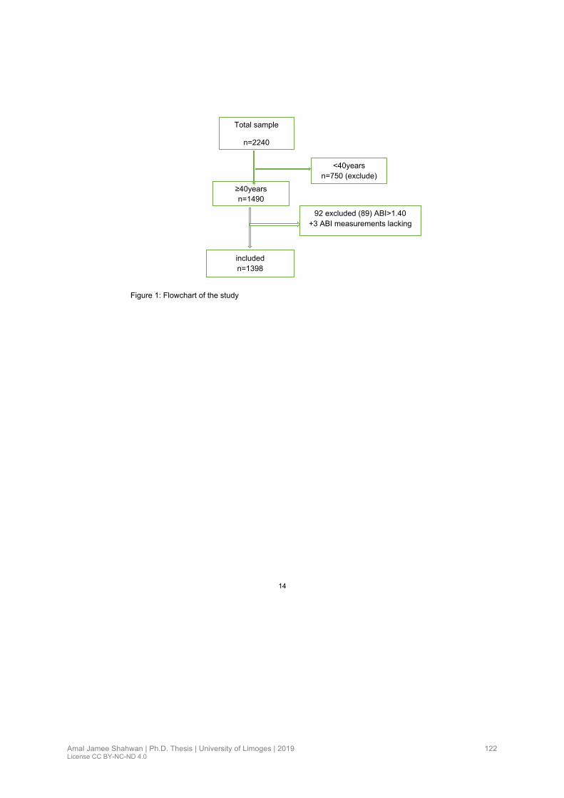
Amal Jamee Shahwan | Ph.D. Thesis | University of Limoges | 2019 License CC BY-NC-ND 4.0
122
14
Figure 1: Flowchart of the study
Total sample
n=2240
≥40years
n=1490
<40years
n=750 (exclude)
included
n=1398
92 excluded (89) ABI>1.40
+3 ABI measurements lacking

Amal Jamee Shahwan | Ph.D. Thesis | University of Limoges | 2019 License CC BY-NC-ND 4.0
123
15
Table1 Study population characteristics in Gaza-Palestine 2018
Variables Total n=1398
LEAD n=191, 13.7%
No LEAD n=1207,86.3%
P. value
Age (years), mean 55.2±10.6 57.9±11.8 54.8±10.3 <0.001
Female, n (%) 733 (52.4) 114(59.7) 619 (51.3) 0.031
Never smoker, n (%) 1121(80.2) 157(82.2) 964 (79.9)
0.180 Past smoking, n (%) 112 (8%) 9 (4.7) 103 (8.5)
Current smoking, n (%) 165 (11.8) 25 (13.1) 140 (11.6)
Duration of smoking ≥ 25 years 111(7.9) 9 (4.7) 102 (8.5) 0.07
Hypertension, n (%) 544 (38.8) 117(61.3) 426 (35.3) <0.001
Diabetes, n (%) 364 (26.0) 68 (35.6) 296 (24.5) 0.001
History of CAD*, n (%) 157(11.2) 36 (18.8) 121 (10.0) <0.001
History of stroke, n (%) 59 (4.2) 14 (7.3) 45 (3.7) 0.021
Total Cholesterol level ≥ 240mg/dl, n (%) 138 (9.9) 21 (11.1) 117 (9.8) 0.582
Low HDL Cholesterol level, n (%) 1029 (73.6) 140 (73.3) 889 (73.7) 0.933
LDL Cholesterol level ≥160 mg/dl, n (%) 130 (9.4) 20 (10.6) 110 (9.2) 0.549
Triglycerides ≥ 150mg/dl, n (%) 636 (45.8) 88 (46.3) 548 (45.7) 0.883
Obesity n (%) 790 (56.5) 118 (61.8) 672 (55.7) 0.114
Statins, n (%) 221(15.8) 43 (22.5) 178 (14.7) 0.006
Aspirin, n (%) 462 (33.0) 83(43.5) 379(31.4) 0.001
Notes: Low HDL cholesterol level<40mg/dl in males and <50mg/dl in females, Obesity, body Mass Index≥ 30kg/m2, Abbreviations: CAD, coronary artery disease

Amal Jamee Shahwan | Ph.D. Thesis | University of Limoges | 2019 License CC BY-NC-ND 4.0
124
16
Table 2 Prevalence of LEAD among adults aged 40 years in Gaza-Palestine 2018
Variables LEAD (95% CI)
No %
Total individuals > 40years with LEAD 191 13.7 11.9-15.5
Gender Male 77 11.6 9.1-14.1
Female 114 15.6 12.9-18.3
Age group (years) 40-49 53 11.2 8.3-14.1
50-59 54 11.8 8.8-14.8
60-69 48 15.9 11.7-20.1
70+ 36 21.8 15.4-28.2
Hypertension Yes 117 21.5 18.0-25.0
No 74 8.7 6.8-11.0
Diabetes Yes 68 18.7 14.6-22.8
No 123 11.9 10.0-13.9
Smoking Current 25 15.2 9.6-20.8
Past 9 8.0 2.9-13.1
Never 157 14.0 11.9-16.1
Duration of smoking (in years) ≥25y 25 15.1 9.5-20.7
< 25y 9 8.1 2.9-13.3
Total cholesterol level ≥ 240 mg/dl Yes 21 15.2 9.1-21.3
No 169 13.5 11.6-15.5
Triglycerides ≥150 mg/dl Yes 88 13.8 11.1-16.5
No 102 13.6 11.1-16.1
Body Mass Index (kg/m2) 18.5-24.9 28 15.9 10.4-21.4
25-29.9 45 10.5 7.5-13.5
≥30 118 14.9 12.4-17.4
History of CAD Yes 36 22.9 16.2-29.6
No 155 12.5 10.6-14.3
History of Stroke Yes 14 23.7 12.6-34.8
No 177 13.2 11.4-15.5
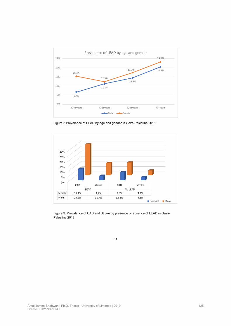
Amal Jamee Shahwan | Ph.D. Thesis | University of Limoges | 2019 License CC BY-NC-ND 4.0
125
17
Figure 2 Prevalence of LEAD by age and gender in Gaza-Palestine 2018
Figure 3: Prevalence of CAD and Stroke by presence or absence of LEAD in Gaza-
Palestine 2018
6.7%
11.2%
14.5%
20.5%15.3%
12.3%
17.3%
23.2%
0%
5%
10%
15%
20%
25%
40-49years 50-59years 60-69years 70+years
Prevalence of LEAD by age and gender
Male Female
0%
5%
10%
15%
20%
25%
30%
CAD stroke CAD stroke
LEAD No LEAD
Female 11,4% 4,4% 7,9% 3,2%
Male 29,9% 11,7% 12,2% 4,3%
Female Male

Amal Jamee Shahwan | Ph.D. Thesis | University of Limoges | 2019 License CC BY-NC-ND 4.0
126
18
Table 3 Independent factors associated in males with LEAD in Gaza-Palestine 2018
Variables Univariate analysis Multivariate analysis
OR (95%CI) p-value OR (95%CI) p-value
Age 0.96 (0.94-0.98) <0.001
Hypertension 3.5 (2.12-5.67) <0.001 3.0 (1.32-6.84) 0.009
Diabetes 1.5 (0.92-2.57) 0.100
Current smokers 1.6 (0.97-2.71) 0.068 2.7 (1.09-6.78) 0.031
Duration of smoking ≥ 25 years 0.5 (0.20-1.08) 0.073
Total cholesterol level≥240mg/dl 0.6(0.26-1.43) 0.254
Low HDL cholesterol 0.9 (0.53-1.50) 0.663
LDL cholesterol>1.60 1.2 (0.52-3.11) 0.605
Triglycerides ≥150 mg/dl 1.0 (0.64-1.67) 0.880
History of CAD 3.1 (1.77-5.27) <0.001 3.6 (1.38-9.13) 0.008
History of Stroke 3.0 (1.34-6.65) 0.008
BMI 18.5-24.9 kg/m2 1
BMI 25-29.9 kg/m2 2.6 (1.37-4.88) 0.003 3.7 (1.41-9.90) 0.008
BMI ≥30 kg/m2 1.6 (0.91-2.91) 0.099

Amal Jamee Shahwan | Ph.D. Thesis | University of Limoges | 2019 License CC BY-NC-ND 4.0
127
19
Table 4 Independent factors associated in females with LEAD in Gaza-Palestine 2018 Variables Univariate analysis Multivariate analysis
OR (95%CI) p-value OR (95%CI) p-value
Age 1.0 (0.97-1.00) 0.066
Hypertension 2.5 (1.65-3.75) <0.001 2.5 (1.66-3.76) <0.001
Diabetes 1.8 (1.18-2.74) 0.006
Current smokers*
Duration of smoking ≥ 25 years*
Total cholesterol level≥240mg/dl 1.1(0.61-2.05) 0.721
Low HDL cholesterol 1.0 (0.65-1.64) 0.885
LDL cholesterol>1.60 1.0 (0.56-1.92) 0.899
Triglycerides ≥150 mg/dl 0.9 (0.61-1.36) 0.651
History of CAD 1.5 (0.78-2.86) 0.221
History of Stroke 1.4 (0.51-3.74) 0.543
BMI 18.5-24.9 kg/m2 1
BMI 25-29.9 kg/m2 0.7 (0.24-1.82) 0.420
BMI ≥30 kg/m2 0.6 (0.22-1.46) 0.238
Current smoking and duration of smoking were not considered in the analysis due to small frequency

Amal Jamee Shahwan | Ph.D. Thesis | University of Limoges | 2019 License CC BY-NC-ND 4.0
128
IV.3. Article 3: Epidemiology of the Metabolic Syndrome among the Palestinians in the Gaza Strip
Contexte de l’étude
Le syndrome métabolique est défini comme un véritable problème de santé publique. Il
désigne la coexistence de désordres métaboliques, concourant à l’augmentation du risque
cardio-vasculaire. La prévalence varie selon les pays en fonction de la définition choisie, de la
population et de la méthodologie de l’étude. Au Moyen-Orient, le syndrome métabolique
touche une personne sur quatre. Des études menées en Palestine décrivent une prévalence
qui varie entre 17.1 à 23%. Mais aucune étude n’a signalé la relation entre les maladies
cardiovasculaires (maladie coronarienne, accident vasculaire cérébral, artériopathie des
membre inferieurs) et ce syndrome.
L’objectif de l’étude
Déterminer une prévalence récente du syndrome métabolique dans une large population et
examiner les facteurs de risque d’athérosclérose et la relation avec les maladies
cardiovasculaires associées.
Valorisation de l’étude
L’étude est soumise à ‘Diabetes Metabolic Syndrome and Obesity’ (IF : 2.961)
Communications
Prévoir une communication orale dans les mois qui viennent

Amal Jamee Shahwan | Ph.D. Thesis | University of Limoges | 2019 License CC BY-NC-ND 4.0
129
1
REVIEW ORIGINAL RESEARCH
The Epidemiology of the Metabolic Syndrome among the Palestinians in the Gaza Strip
Amal Jamee Shahwan1,2,4
Victor Aboyans1,2,3
Julien Magne1,2,3
Pierre Marie Preux1,2
Philippe Lacroix1,2,5
1INSERM UMR 1094, Tropical Neuroepidemiology, Limoges, France
2University of Limoges, School of Medicine, Institute of Neuroepidemiology and
Tropical Neurology, CNRS FR 3503 GEIST, Limoges, France
3Department of Cardiology, Dupuytren University Hospital, Limoges, France
4Department of cardiology-Ministry of health-Palestine
5Department of Thoracic and Vascular Surgery – Vascular Medicine, Dupuytren
University Hospital, Limoges, France
Correspondence: Amal Jamee Shahwan
University of Limoges, School of Medicine, Institute of Neuroepidemiology and
Tropical Neurology, 2 rue du Dr Marcland, 87025 Limoges, France
Tel +33754139112
Email [email protected]
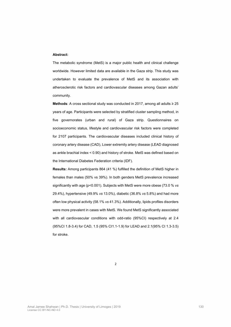
Amal Jamee Shahwan | Ph.D. Thesis | University of Limoges | 2019 License CC BY-NC-ND 4.0
130
2
Abstract:
The metabolic syndrome (MetS) is a major public health and clinical challenge
worldwide. However limited data are available in the Gaza strip. This study was
undertaken to evaluate the prevalence of MetS and its association with
atherosclerotic risk factors and cardiovascular diseases among Gazan adults’
community.
Methods: A cross sectional study was conducted in 2017, among all adults ≥ 25
years of age. Participants were selected by stratified cluster sampling method, in
five governorates (urban and rural) of Gaza strip. Questionnaires on
socioeconomic status, lifestyle and cardiovascular risk factors were completed
for 2107 participants. The cardiovascular diseases included clinical history of
coronary artery disease (CAD), Lower extremity artery disease (LEAD diagnosed
as ankle brachial index < 0.90) and history of stroke. MetS was defined based on
the International Diabetes Federation criteria (IDF).
Results: Among participants 864 (41 %) fulfilled the definition of MetS higher in
females than males (50% vs 39%). In both genders MetS prevalence increased
significantly with age (p<0.001). Subjects with MetS were more obese (73.0 % vs
29.4%), hypertensive (49.9% vs 13.0%), diabetic (36.8% vs 5.8%) and had more
often low physical activity (58.1% vs 41.3%). Additionally, lipids profiles disorders
were more prevalent in cases with MetS. We found MetS significantly associated
with all cardiovascular conditions with odd-ratio (95%CI) respectively at 2.4
(95%CI 1.8-3.4) for CAD, 1.5 (95% CI1.1-1.9) for LEAD and 2.1(95% CI 1.3-3.5)
for stroke.

Amal Jamee Shahwan | Ph.D. Thesis | University of Limoges | 2019 License CC BY-NC-ND 4.0
131
3
Conclusion: The MetS is highly prevalent in the Palestinian population,
particularly among women.
Key words: Metabolic syndrome, risk factors, cardiovascular disease, Palestine.
Introduction
The Metabolic syndrome (MetS) is a major public health and clinical challenge
worldwide. it is defined as a cluster of clinical and metabolic disorders associated
with increased risk of atherosclerosis cardiovascular disease (CVD) and mortality
1. The overall prevalence of MetS varies according to geographic and socio-
demographic factors, as well as the diagnostic criteria. Globally, the prevalence
of MetS ranges from 10%-40% 2. The MetS increases by 5-fold the risk of type 2
diabetes mellitus and 2-fold the risk of developing CVD3. Also, patients with MetS
are 2-4 times more likely to die from cardiovascular events than those without
MetS 4. The MetS occurs more frequently through the combination of several
factors and increase the cardiovascular risk beyond the risk associated with
individual factors alone 5. The risk increases with the number of MetS
components present 6. The syndrome occurs most often in populations
characterized by high prevalence of obesity and physical inactivity3. The major
causes leading to MetS are insulin resistance, obesity, unhealthy lifestyle, and
genetic predisposition7,8. In the Middle-East region, the MetS affect one in four
people 10. In Palestine, some studies on MetS based on inhomogeneous criteria
are available. The prevalence of MetS varies from 17.1% to 23%, however none
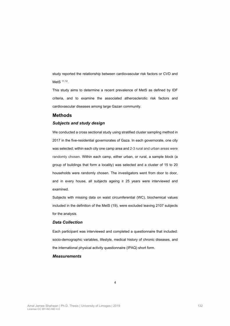
Amal Jamee Shahwan | Ph.D. Thesis | University of Limoges | 2019 License CC BY-NC-ND 4.0
132
4
study reported the relationship between cardiovascular risk factors or CVD and
MetS 11,12.
This study aims to determine a recent prevalence of MetS as defined by IDF
criteria, and to examine the associated atherosclerotic risk factors and
cardiovascular diseases among large Gazan community.
Methods
Subjects and study design
We conducted a cross sectional study using stratified cluster sampling method in
2017 in the five-residential governorates of Gaza. In each governorate, one city
was selected; within each city one camp area and 2-3 rural and urban areas were
randomly chosen. Within each camp, either urban, or rural, a sample block (a
group of buildings that form a locality) was selected and a cluster of 15 to 20
households were randomly chosen. The investigators went from door to door,
and in every house, all subjects ageing ≥ 25 years were interviewed and
examined.
Subjects with missing data on waist circumferential (WC), biochemical values
included in the definition of the MetS (19), were excluded leaving 2107 subjects
for the analysis.
Data Collection
Each participant was interviewed and completed a questionnaire that included:
socio-demographic variables, lifestyle, medical history of chronic diseases, and
the international physical activity questionnaire (IPAQ) short form.
Measurements

Amal Jamee Shahwan | Ph.D. Thesis | University of Limoges | 2019 License CC BY-NC-ND 4.0
133
5
Height and weight were measured with the foot and wearing light clothing. Body
mass index (BMI kg/m2) was calculated as the weight in kilograms divided by the
square of the height in meters. Waist circumference was measured at the mid-
distance between the tenth rib and the iliac crest. Blood pressure (BP) was
measured once in both arms. A hand-held Doppler probe was used to measure
the blood pressure in both arms and ankles (both posterior tibial and dorsalis
pedis arteries)13. The ABI was measured at each leg and the lowest between the
two ankles was retained as the subject’s ABI.
Venous blood sample was obtained for lipids profiles including total cholesterol
(TC) high-density lipoprotein cholesterol (HDL-C), triglycerides. The low-density
lipoprotein cholesterol (LDL-C) was calculated by Friedewald equation. Plasma
glucose was measured using glucometer.
Definition of Metabolic syndrome (IDF)
According to the IDF definition, MetS was defined in case of WC ≥ 94 cm in men
and ≥ 80 cm in women (all participants were Caucasians), plus two of the four
following factors: 1) high blood pressure ≥ 130/85 mmHg or on anti-hypertensive
drugs, 2) triglycerides level ≥150 mg/dl, 3) low HDL cholesterol level < 40 mg /dl
in males and < 50 mg/dl in females, and 4) fasting glucose level ≥ 100 mg/dl or
known to have type-2 diabetes14
Diabetes mellitus (DM) was defined as capillary blood sugar level ≥126 mg/dl if
the participant was fasting or ≥ 200 mg/dl if the participant was non-fasting and
or self-reported as currently taking any diabetes medications 14. We considered
subjects with hypertension (HTN), if their average systolic blood pressure (SBP)
in both arms was ≥140 mmHg or their average diastolic blood pressure (DBP) ≥
Amal Jamee Shahwan | Ph.D. Thesis | University of Limoges | 2019 License CC BY-NC-ND 4.0
134
6
90 mmHg, or if they were being treated for HTN 16,17. Coronary artery disease
(CAD) was defined in our study by self-reported history of hospitalization for
angina pectoris, myocardial infarction, procedures performing percutaneous
coronary intervention or coronary bypass graft and after verification of their
medical prescription list. Stroke history was identified in case of positive answer
to the question: “Have you ever been told by a physician that you suffered a
stroke? Lower extremity artery disease (LEAD) was defined as ABI < 0.90.
Statistical analysis
All data were analyzed with the SPSS statistical software version 22.0 (Chicago,
Illinois, USA). Continuous variables were expressed as mean ± standard
deviation, and categorical variables were expressed as percentages. The
comparison of qualitative variables between persons with and without MetS were
expressed by frequency distribution and cross tabulation and were compared
using the Chi-square tests or the Fisher exact. Variables with a univariate p value
< 0.25 were selected for further multivariate analysis. Univariate and multivariate
logistic regression analyses were performed to evaluate the association between
MetS and other factors. A p-value < 0.05 was considered as the significance
level.
Ethics
The committee for medical ethics of the Palestinian ministry approved the study
and each participant signed an informed consent.
Results
Among the 2107 participants (1040 males and 1067 females), MetS was found
in 864 subjects (41%, 95% CI: 39-43), with higher rate in females than in males
Amal Jamee Shahwan | Ph.D. Thesis | University of Limoges | 2019 License CC BY-NC-ND 4.0
135
7
(50%, 95% CI:48-52 vs. 32%, 95% CI: 30-34, p<0.001). Clinical characteristics
and metabolic measures of the study population according to MetS are presented
in Table 1. Individuals with MetS were more likely to be older, obese, with low
physical activity, lower level of education, and had more prevalent cardiovascular
risk factors such as diabetes, hypertension and lipid disorders. Also, history of
CAD, stroke and LEAD were more prevalent in patients with MetS (12%, 4.3%,
and15.5% respectively). Statins and Aspirin are more commonly used in
participants with MetS.
The prevalence of MetS increased with age in both genders, ranging from 13%
(95% CI: 11 -14) in the age category 25-35 years to 50% (95% CI: 47-52) in the
age category 55-64 years in males, and from 16% (95% CI: 14-17) to 79% (95%
CI: 77-81) in the corresponding age categories in females. The rate decreases
after age 65 years for both genders (Figure 1).
Figure 2 displays the prevalence of each components of the MetS in relation to
gender. Central obesity and hyperglycemia were the most common conditions
contributing to the MetS mainly in female than in males with significant Pvalue.
The association of each CVD with the MetS components are reported in Table 2.
Low HDL cholesterol level as well as high triglyceride level, hyperglycemia, and
elevated blood pressure were the components associated with the highest risk
for CAD (OR= 2.9, 2.7, 2.4, 2.0 respectively). While hyperglycemia, high blood
pressure and high triglyceride level were the factors associated with the highest
risk of stroke (OR= 4.1, 3.8, 3.7) respectively. Elevated blood pressure remains
the main component associated with high risk of LEAD (OR=2.0; CI 1.5-2.7; p<
0.001).
Amal Jamee Shahwan | Ph.D. Thesis | University of Limoges | 2019 License CC BY-NC-ND 4.0
136
8
The association of MetS with all demographic and cardiovascular variables are
separately presented in males and females (Table 3 and Table 4). The
association with MetS increases with age for both genders. In male gender Low
physical activity remains positive (OR= 1.7; CI 1.2-2.4; p= 0.009). Also, married
males and females were at high risk for MetS (OR= 2.3; CI 1.0-5.4; p= 0.03, OR=
1.9; CI 1.1-3.3; p=0.04) respectively. Furthermore, we failed to detect an
association between smoking, education level and physical inactivity with
metabolic syndrome.
Discussion
The current study performed in a large sample of adults in age ≥ 25 years reveals
a high prevalence of MetS in the Palestinian community of the Gaza Strip, up to
41%, based on IDF definition. The prevalence was higher in women (49.9%). The
prevalence estimated in the current study is superior to that reported in a previous
study carried out among a small sample size in different regions of Palestine. The
most recent was a study among 230 adults aged 20-65 years in the Gaza strip,
the prevalence of MetS reported was 23% 11. Also, In two different studies in two
Palestinian camps in west bank, Rizkallah et al and Massad et al. reported a
prevalence of MetS up to 58% and 27% in females 18,19. In the present study,
central obesity was two times higher in females than in males (67.2%vs 32.8%)
respectively. Waist circumference as indicator of visceral adiposity has been
identified as a stronger predictor of CVD and type 2 diabetes20 . The female
predominance is not surprising in our study and could be explained by the fact of
high and rising frequency of obesity, diabetes and hypertension in our
community, mainly in females21. In 2016, the Global Burden of Disease project

Amal Jamee Shahwan | Ph.D. Thesis | University of Limoges | 2019 License CC BY-NC-ND 4.0
137
9
supported these findings and pointed out that Palestine is one of the countries
where the prevalence of obesity and diabetes is very high 22. Our results are
consistent with those from Middle East studies, which the burden of MetS was
estimated to affect a large population, and the prevalence varies from 44% in
Turkey 23, 30% In Tunisia 24 to 31.6% in Saudi Arabia 25. In all these reports
published in Middle East region, the prevalence was also higher in female and
the most common components were HDL cholesterol and central obesity 10.
In our study the prevalence MetS was found (12.0%, 4.3%, and 15,5%) in
subjects with CAD, stroke and LEAD and the association was statistically
significant, and the OR increased according to the number of components of
MetS. We found that individuals with MetS had a two-fold greater risk of suffering
CVDs (CAD, stroke, LEAD) which is similar for the increased risk described for
other countries. In a meta-analysis based on NCEP-APIII definition of MetS, the
risk estimates were approximately 1.6 for CVD 26–28. Gami et al, in a large meta-
analysis involving 43 cohorts and 17 2573 participants found that MetS itself,
apart from its components, was associated with an increased risk of
cardiovascular events +54 times after adjustment 27. In the Botnia study in
Finland and Sweden which recruited 4483 subjects aged 35-70 years, the risk of
CAD and stroke tripled in subjects with MetS with an absolute 10% increase in
cardiovascular mortality during 6.9 years of follow-up 29.
Limitations
As a cross-sectional design of our study, all associations reported cannot be
considered as a causative relationship. Also, possible bias could have been
introduced since study was conducted at home and data concerning risk factors

Amal Jamee Shahwan | Ph.D. Thesis | University of Limoges | 2019 License CC BY-NC-ND 4.0
138
10
were self-reported, another potential concern was that HDL cholesterol,
triglycerides, and blood glucose levels were measured only once, which might
have led to random error. The strength of the study lies in a large population-
based study representative of the Palestinian community, and we describe for
the first-time the relationship of MetS and cardiovascular diseases.
Conclusion
The MetS is a collection of risk factors and a complex condition with high
socioeconomic cost, its consequences constitute a heavy burden. Our findings
are alarming and should alert policy makers to consider primary prevention
through a stronger public information to sensitize Palestinians on the hazards of
sedentary lifestyle and obesity, because individuals with MetS can be easily
screened according to the criteria, it is essential to identify and properly manage
each of its components to delay the appearance of its complications.
Acknowledgements
The authors are grateful to all the families who have welcomed us into their
homes and participated in the study. We also thank all nurses who contributed in
the study data collection.
Disclosure
conflict of interest
Amal Jamee Shahwan: none
Victor Aboyans: Bayer, Novartis, Novo Nordisk, Sanofi
Julien Magne : none
Philippe Lacroix : none
Pierre-Marie Preux : none

Amal Jamee Shahwan | Ph.D. Thesis | University of Limoges | 2019 License CC BY-NC-ND 4.0
139
11
References
1. Wilson PWF, D’Agostino RB, Parise H, Sullivan L, Meigs JB. Metabolic syndrome
as a precursor of cardiovascular disease and type 2 diabetes mellitus. Circulation.
2005;112(20):3066-3072. doi:10.1161/CIRCULATIONAHA.105.539528
2. Grundy SM. Metabolic syndrome pandemic. Arterioscler Thromb Vasc Biol.
2008;28(4):629-636. doi:10.1161/ATVBAHA.107.151092
3. Alberti KG, Eckel RH, Grundy SM, et al. Harmonizing the metabolic syndrome: a
joint interim statement of the International Diabetes Federation Task Force on
Epidemiology and Prevention; National Heart, Lung, and Blood Institute; American Heart
Association; World Heart Federation; International Atherosclerosis Society; and
International Association for the Study of Obesity. Circulation. 2009;120(16):1640-1645.
doi:10.1161/CIRCULATIONAHA.109.192644
4. Alberti KGMM, Zimmet P, Shaw J. The metabolic syndrome--a new worldwide
definition. Lancet Lond Engl. 2005;366(9491):1059-1062. doi:10.1016/S0140-
6736(05)67402-8
5. Reilly MP, Rader DJ. The metabolic syndrome: more than the sum of its parts?
Circulation. 2003;108(13):1546-1551. doi:10.1161/01.CIR.0000088846.10655.E0
6. Andreadis EA, Tsourous GI, Tzavara CK, et al. Metabolic syndrome and incident
cardiovascular morbidity and mortality in a Mediterranean hypertensive population. Am
J Hypertens. 2007;20(5):558-564. doi:10.1016/j.amjhyper.2006.12.001
7. Balkau B, Deanfield JE, Després J-P, et al. International Day for the Evaluation
of Abdominal Obesity (IDEA): a study of waist circumference, cardiovascular disease,
and diabetes mellitus in 168,000 primary care patients in 63 countries. Circulation.
2007;116(17):1942-1951. doi:10.1161/CIRCULATIONAHA.106.676379
8. Esteghamati A, Zandieh A, Khalilzadeh O, Meysamie A, Ashraf H. Clustering of
metabolic syndrome components in a Middle Eastern diabetic and non-diabetic
population. Diabetol Metab Syndr. 2010;2:36. doi:10.1186/1758-5996-2-36
9. Prevalence of metabolic syndrome in Middle-East countries: Meta-analysis of
cross-sectional studies. Diabetes Metab Syndr Clin Res Rev. 2018;12(2):195-201.
doi:10.1016/j.dsx.2017.11.004
10. Sliem HA, Ahmed S, Nemr N, El-Sherif I. Metabolic syndrome in the Middle East.
Indian J Endocrinol Metab. 2012;16(1):67-71. doi:10.4103/2230-8210.91193
11. Sirdah MM, Al Laham NA, Abu Ghali AS. Prevalence of metabolic syndrome and
associated socioeconomic and demographic factors among Palestinian adults (20-65
years) at the Gaza Strip. Diabetes Metab Syndr. 2011;5(2):93-97.

Amal Jamee Shahwan | Ph.D. Thesis | University of Limoges | 2019 License CC BY-NC-ND 4.0
140
12
doi:10.1016/j.dsx.2012.02.024
12. Abdul-Rahim HF, Husseini A, Bjertness E, Giacaman R, Gordon NH, Jervell J.
The metabolic syndrome in the West Bank population: an urban-rural comparison.
Diabetes Care. 2001;24(2):275-279.
13. Aboyans V, Criqui MH, Abraham P, et al. Measurement and Interpretation of the
Ankle-Brachial Index: A Scientific Statement From the American Heart Association.
Circulation. January 2012:CIR.0b013e318276fbcb. doi:10.1161/CIR.0b013e318276fbcb
14. Alberti KG, Zimmet P, Shaw J. Metabolic syndrome--a new world-wide definition.
A Consensus Statement from the International Diabetes Federation. Diabet Med J Br
Diabet Assoc. 2006;23(5):469-480. doi:10.1111/j.1464-5491.2006.01858.x
15. American Diabetes Association 2016. ADA Diabetes Management Guidelines
A1C Diagnosis | NDEI. http://www.ndei.org/ADA-diabetes-management-guidelines-
diagnosis-A1C-testing.aspx.html. Accessed January 22, 2018.
16. James PA, Oparil S, Carter BL, et al. 2014 evidence-based guideline for the
management of high blood pressure in adults: report from the panel members appointed
to the Eighth Joint National Committee (JNC 8). JAMA. 2014;311(5):507-520.
doi:10.1001/jama.2013.284427
17. WHO hypertension. WHO | Q&As on hypertension. WHO.
http://www.who.int/features/qa/82/en/index.html. Published 2015. Accessed January 9,
2018.
18. Rizkallah N, Marshall T, Kritz-Silverstein D. Parity and risk factors for coronary
heart disease in Palestinian women in two refugee camps in the West Bank: a population
based cross-sectional survey. Lancet. 2010:382,S28.
19. Massad SG, Khalili M, Karmally W, et al. Metabolic Syndrome among Refugee
Women from the West Bank, Palestine: A Cross-Sectional Study. Nutrients. 2018;10(8).
doi:10.3390/nu10081118
20. Wang Y, Rimm EB, Stampfer MJ, Willett WC, Hu FB. Comparison of abdominal
adiposity and overall obesity in predicting risk of type 2 diabetes among men. Am J Clin
Nutr. 2005;81(3):555-563. doi:10.1093/ajcn/81.3.555
21. Jamee.Shahwan A, Abed Y, Magne J, et al. Epidemiology of cardiovascular
disease and associated factors in Gaza-Palestine. PLOS ONE. 2019.
22. Forouzanfar MH, Bhutta ZA, Burnett R, et al. Global, regional, and national
comparative risk assessment of 79 behavioural, environmental and occupational, and
metabolic risks or clusters of risks, 1990–2015: a systematic analysis for the Global
Burden of Disease Study 2015. The Lancet. 2016;388(10053):1659-1724.

Amal Jamee Shahwan | Ph.D. Thesis | University of Limoges | 2019 License CC BY-NC-ND 4.0
141
13
doi:10.1016/S0140-6736(16)31679-8
23. Gundogan K, Bayram F, Gedik V, et al. Metabolic syndrome prevalence
according to ATP III and IDF criteria and related factors in Turkish adults. Arch Med Sci
AMS. 2013;9(2):243-253. doi:10.5114/aoms.2013.34560
24. Belfki H, Ali SB, Aounallah-Skhiri H, et al. Prevalence and determinants of the
metabolic syndrome among Tunisian adults: results of the Transition and Health Impact
in North Africa (TAHINA) project. Public Health Nutr. 2013;16(4):582-590.
doi:10.1017/S1368980012003291
25. Al-Rubeaan K, Bawazeer N, Al Farsi Y, et al. Prevalence of metabolic syndrome
in Saudi Arabia - a cross sectional study. BMC Endocr Disord. 2018;18.
doi:10.1186/s12902-018-0244-4
26. Ford ES. Risks for All-Cause Mortality, Cardiovascular Disease, and Diabetes
Associated With the Metabolic Syndrome: A summary of the evidence. Diabetes Care.
2005;28(7):1769-1778. doi:10.2337/diacare.28.7.1769
27. Gami AS, Witt BJ, Howard DE, et al. Metabolic syndrome and risk of incident
cardiovascular events and death: a systematic review and meta-analysis of longitudinal
studies. J Am Coll Cardiol. 2007;49(4):403-414. doi:10.1016/j.jacc.2006.09.032
28. Galassi A, Reynolds K, He J. Metabolic syndrome and risk of cardiovascular
disease: a meta-analysis. Am J Med. 2006;119(10):812-819.
doi:10.1016/j.amjmed.2006.02.031
29. Isomaa B, Almgren P, Tuomi T, et al. Cardiovascular morbidity and mortality
associated with the metabolic syndrome. Diabetes Care. 2001;24(4):683-689.

Amal Jamee Shahwan | Ph.D. Thesis | University of Limoges | 2019 License CC BY-NC-ND 4.0
142
14
Table1: The characteristics of the study participants with and without metabolic
syndrome
Variables
Whole Population
n=2107
MetS n=864 (41%)
No MetS n=1243 (59%)
P-value
Age, years 47.17±14.3 53.2±12.7 42.9±13.9 <0.001 Women, n (%) 1067(50.6) 532 (49.9) 535 (50.1) <0.001 Areas Urban, n (%) 752(35.6) 318 (36.8) 433 (34.8) 0.623 Rural, n (%) 493(23.4) 196 (22.7) 297(23.9) Camp, n (%) 863(41.0) 350 (40.5) 513 (41.3) Education level* Primary education, n (%) 324(15.4) 180 (20.8) 144 (11.6) <0.001 Secondary + High school, n (%) 1082(51.4) 461 (53.4) 621 (50.0) University + Post graduate, n (%) 701(33.3) 223 (25.8) 478 (38.5) Physical activity Low, n (%), n (%) 1015(48.2) 502 (58.1) 513 (41.3) <0.001 Moderate, n (%) 657(31.2) 233 (27.0) 424 (34.1) High, n (%) 435(20.6) 129 (14.9) 306 (24.6) Income <300$, n (%) 1326(62.9) 574 (66.4) 752 (60.5) 0.006 ≥300$, n (%) 781(371) 290 (33.6) 491 (39.5) Marital status* Single, n (%) 161(7.6) 28 (3.2) 133 (10.7) <0.001 Married 1792(85) 740 (85.6) 1052 (84.6) Divorced or widow, n (%) 154(7.3) 96 (11.1) 58 (4.7) Body mass index Normal (18.5-25.9 kg/m2), n (%) 419(19.9) 18 (2.1) 401 (32.2) <0.001 Overweight (25.9-29.9 kg/m2), n (%) 691(32.8) 215 (24.9) 476 (38.3) Obese ≥(30kg/m2), n (%) 997(47.3) 631(73.0) 366 (29.4) CVD risk factors Hypertension, n (%) 592(28.1) 431 (49.9) 161 (13.0) <0.001 Diabetes mellitus, n (%) 390(18.5) 318 (36.8) 72 (5.8) <0.001 Current smoking, n (%) 489(23.2) 138 (16) 351 (28.2) <0.001 Lipids profiles Total Cholesterol ≥ 240 (mg/dl), n (%) 178(8.4) 100 (11.6) 78 (6.3) <0.001 Low HDL cholesterol, n (%) 178(8.4) 748 (88.6) 726 (58.4) <0.001 LDL cholesterol level ≥160, n (%) 1474(70.0) 90 (10.5) 77 (6.2) <0.001 Triglycerides level ≥150, n (%) 833(39.5) 591 (68.4) 242 (19.5) <0.001 CVD comorbidities CAD, n (%) 170(8.1) 104 (12.0) 66 (5.3) <0.001 Stroke, n (%) 63(3.0) 37(4.3) 26 (2.1) 0.004 LEAD (ABI<0.90), n (%) 274(13.0) 134 (15.5) 140 (11.3) 0.004
Medications use Statins, n (%) 226(10.7) 176 (20.4) 50 (4.0) <0.001 Aspirin, n (%) 498(23.6) 318 (36.8) 180 (14.5) <0.001
Education status*: primary level (0-6years), secondary level (7-12), and high level>12years Marital status*: single. married and divorced or widow

Amal Jamee Shahwan | Ph.D. Thesis | University of Limoges | 2019 License CC BY-NC-ND 4.0
143
15
Figure1: Metabolic syndrome according to gender and stratified by age group.
12.9%
24.7%
37.4%
49.7%47.0%
15.9%
36.0%
58.5%
78.9%
71.5%
0%
10%
20%
30%
40%
50%
60%
70%
80%
90%
25-34 years 35-44 years 45-54 years 55-64 years 65+ years
Pre
va
len
ce
of th
e M
etS
Male Female
n=484 n=474 n=486 n=391 n=269
p<0.001
p<0.001
p<0.001
p<0.001
p<0.001

Amal Jamee Shahwan | Ph.D. Thesis | University of Limoges | 2019 License CC BY-NC-ND 4.0
144
16
Figure 2: Frequency of each component of Metabolic syndrome by gender
Table 2: Association of CAD, stroke and LEAD with each components of metabolic
syndrome
Components
of MetS
CAD Stroke LEAD
O R (95%CI) P-value OR (95%CI) P-value OR (95%CI) P-value
Central obesity 1.0 (0.66-1.45) 0.916 1.2 (0.61-2.19) 0.643 0.8 (0.60-1.15) 0.256
High BP 2.0 (1.43-2.90) <0.001 3.8 (2.01-7.31) <0.001 2.0 (1.51-2.65) <0.001
High TG 2.7 (1.92-3.91) <0.001 3.7 (2.00-6.72) <0.001 0.9 (0.68-1.16) 0.386
Low HDL-C 2.9 (1.78-4.55) <0.001 1.5 (0.78-2.72) 0.241 0.9 (0.66-1.14) 0.305
Hyperglycemia 2.4 (1.67-3.33) <0.001 4.1 (2.32-6.78) <0.001 1.4 (0.98-1.85) 0.06
Each Logistic regression model was adjusted for age and gender.
BP: Blood pressure; CAD: Coronary artery disease; CI: confidence interval: HDL-C: high-density
lipoprotein cholesterol; LEAD: Lower extremity artery disease; OR: odd-ratio
32.8%
50.4%
42.7%
48.5%52.8%
67.2%
49,60%
57.8%
51.5%47.2%
0%
10%
20%
30%
40%
50%
60%
70%
80%
Central obesity High BP Hyperglycemia LowHDL-C High TG
Pre
va
len
ce
of
the
co
mp
on
ets
of
the
M
etS
Male Female
p<0.001p<0.432 p<0.006p<0.214
p<0.001
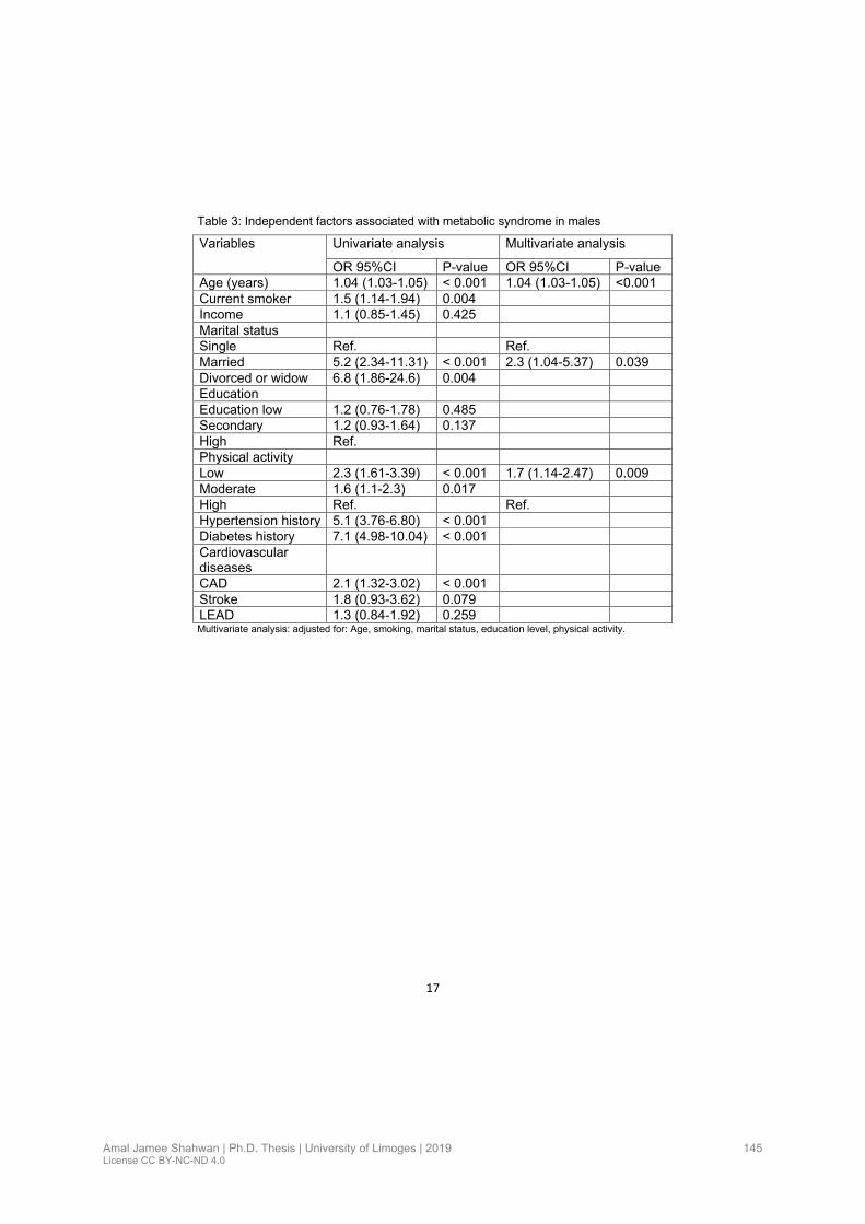
Amal Jamee Shahwan | Ph.D. Thesis | University of Limoges | 2019 License CC BY-NC-ND 4.0
145
17
Table 3: Independent factors associated with metabolic syndrome in males
Variables Univariate analysis Multivariate analysis
OR 95%CI P-value OR 95%CI P-value
Age (years) 1.04 (1.03-1.05) < 0.001 1.04 (1.03-1.05) <0.001
Current smoker 1.5 (1.14-1.94) 0.004
Income 1.1 (0.85-1.45) 0.425
Marital status
Single Ref. Ref.
Married 5.2 (2.34-11.31) < 0.001 2.3 (1.04-5.37) 0.039
Divorced or widow 6.8 (1.86-24.6) 0.004
Education
Education low 1.2 (0.76-1.78) 0.485
Secondary 1.2 (0.93-1.64) 0.137
High Ref.
Physical activity
Low 2.3 (1.61-3.39) < 0.001 1.7 (1.14-2.47) 0.009
Moderate 1.6 (1.1-2.3) 0.017
High Ref. Ref.
Hypertension history 5.1 (3.76-6.80) < 0.001
Diabetes history 7.1 (4.98-10.04) < 0.001
Cardiovascular diseases
CAD 2.1 (1.32-3.02) < 0.001
Stroke 1.8 (0.93-3.62) 0.079
LEAD 1.3 (0.84-1.92) 0.259 Multivariate analysis: adjusted for: Age, smoking, marital status, education level, physical activity.

Amal Jamee Shahwan | Ph.D. Thesis | University of Limoges | 2019 License CC BY-NC-ND 4.0
146
18
Table 4: Independent factors associated with metabolic syndrome in female
Variables Univariate analysis Multivariate analysis OR 95%CI Pvalue OR (95%CI) Pvalue
Age 1.07 (1.06-1.09) < 0.001 1.07 (1.06-1.08) < 0.001 Current smoker 0.9 (0.39-1.87) 0.859 Income in $ 1.3 (1.02-1.71) 0.004 Marital status Single Ref. Ref. Married 2.9 (1.8-4.9) < 0.001 1.9 (1.05-3.34) 0.04 Divorced or widow 5.4 (3.1-10.1) < 0.001 Education level Education low 4.4 (3.1-6.5) < 0.001 Secondary 1.8 (1.4-2.4) < 0.001 High Ref. Physical activity Low 2.1 (1.4-2.8) < 0.001 1.4 (0.97-1.97) 0.075 Moderate 1.2 (0.8-1.8) 0.283 High Ref. Ref. HTN history 9.9 (7.05-13.77) <0.001 Diabetes history 19.4 (11.3-33.4) < 0.001 Cardiovascular diseases
CAD 5.2 (2.82-9.97) < 0.001 Stroke 3.1 (1.31-7.41) 0.010 LEAD 1.4 (1.05-1.96) 0.048 Multivariate analysis: adjusted for: Age, smoking, marital status, education level, physical activity.

Amal Jamee Shahwan | Ph.D. Thesis | University of Limoges | 2019 License CC BY-NC-ND 4.0
147
Chapter V. Discussion
V.1. Discussion of four articles
Such a community based epidemiological study has never been conducted in Palestine with a
large sample covering 2240 participants. Based on our work presented in the four articles, the
CAD is reported at 8.3% with a higher risk in males than females (OR: 1.63; p= 0.002). Stroke
is reported with a prevalence of 3.0% with no difference between gender. The strongest CVRF
associated with these two types of CVDs were hypertension (OR: 2.51; p < 0.001) and diabetes
(OR: 2.22; p < 0.001). In 2013 a cross-sectional study among 1200 Lebanese people ≥ 40
years, reported a CAD prevalence up to 13.4% (Zeidan et al., 2016). In the Coronary Artery
Disease in Saudi study (CADISS), a national community-based study conducted in urban and
rural area of Saudi Arabia; 17 232 subjects aged 30-70 years randomly selected were included.
Coronary artery disease was diagnosed on positive questionnaire of angina or history of
possible myocardial infarction. The prevalence of CAD was 5.5% (6.6% in males and 4.4% in
females) (Al-Nozha et al., 2004). Two a cross-sectional studies in Lebanese population have
reported stroke prevalence, the first one of Lahoud et al; among 6963 inhabitants (mean age
36.8±21.2) the prevalence was 0.80% (Lahoud et al., 2016). The second of Farah et al; that
record a prevalence of self-reported stroke symptoms in a stroke and TIA in 1515 free
population ≥40 years. The prevalence was 3.6% slightly higher than ours (Farah et al., 2015).
In other Arab population, most studies on CAD, or stroke were hospital-based (Al-Nozha et al.,
2004; Farah et al., 2015; Nsour et al., 2008). In the US population ≥ 20 years of age the
prevalence of CAD is estimated to 6.3% higher in males than females (7.4%, 5.3%). Stroke
represent 2.7% with similar prevalence in both gender (Benjamin et al., 2017).
The prevalence of traditional risk factors in general Arab population is closed to the results
obtained in our study. As described in GBD 2016, for Middle East and North Africa Region,
high blood pressure ranked as the first factor, followed by obesity, diabetes then smoking (GBD
2015 Risk Factors Collaborators, 2016). Our results revealed a high burden of CVRF, obesity
45% (females 60%), hypertension 28%, diabetes 19%, high triglycerides 40%, cigarette
smoking in males 40%, unhealthy habits for nutrition and physical inactivity.
The increase in the prevalence of the risk factors in our population is mainly due to urbanization
process, globalization (epidemiological transition) involving several factors mainly diets (rich in
carbohydrates, red meat, sugar, sweetened drinks and poor intake of fruits and vegetables),
lifestyle changes (physical inactivity and smoking). This shift leads to a rise in obesity,
worsening levels of metabolic disorder, with associated risk of CVD. Also, we observed that
certain risk factors and CVD become worse in persons with low socioeconomic status
(Abukhdeir et al., 2013; Husseini et al., 2009).

Amal Jamee Shahwan | Ph.D. Thesis | University of Limoges | 2019 License CC BY-NC-ND 4.0
148
Lower extremities artery disease study is a research conducted for the first time in Palestine
even in the Middle East in the general population. WHO reported 140.8 million LEAD in LMIC
with 10.3 million in the EMR where the prevalence was higher in female (Fowkes et al., 2013).
Conversely in HIC the prevalence is ≈ 20% in > 80 years old, higher in males and diabetics
(Shu & Santulli, 2018) Data regarding LEAD in Arab countries are hospital based and limited.
For example, in Saudi Arabia, a cross-sectional, study among 598 diabetic patients ≥ 30 years
of age, LEAD was defined in the study by an ABI < 0.90 or by the toe brachial index < 0.7
calculated in case of ABI >1.40.The prevalence was up to 23.1% (Alzahrani et al., 2014). Also,
in the Reduction of Atherothrombosis for Continued Health (REACH) registry data form 44
countries worldwide involve large geographic representation, , which included 55,814 patients
≥ 45 years of age, (718 patients from Middle East), with established atherothrombotic disease
(CAD, CVD, or LEAD).The results was that ≈ 3/5 of patients with symptomatic LEAD also have
atherothrombotic disease in other arterial territories (Cacoub et al., 2009). Our study was
carried out in the Gazan community among 1398 subjects. The diagnosis of LEAD was
retained for an ABI < 0.90. The analysis reported a prevalence of 13.7%, higher in female. The
prevalence increases with age to reach 20.5% in the age of ≥70 years in male and 23.2% in
correspondent age in female. The strongest risk factors associated with the disease in male
were by order of penetrance overweight, history of CAD, hypertension and being smoking,
while in female the hypertension is the only risk factor. In addition, CAD and stroke history
were more frequent in case of LEAD, both in males and females.
Worldwide MetS prevalence depending on the region, environment, race, ethnicity, age, and
gender, of each population. The IDF estimates that > 1in 4 of adult has the MetS (Alberti,
Zimmet & Shaw, 2006), The MetS increases by 5-fold the risk of type 2 diabetes mellitus and
2-fold the risk of developing CVD (Alberti et al., 2009).The study on Metabolic syndrome places
Gaza strip as one of the areas with the highest prevalence of this disorder in the EMR. It was
reported at 41% mainly in females (49.9%). This mean that 1 in 2 women is exposed to type 2
diabetes and CVD events, and therefore all health difficulties. In Palestine some studies on
MetS based on different criteria were conducted in small population and reported a variance
of prevalence from 17.1% to 23%, however none of them reported the relationship between
cardiovascular risk factors or CVD and MetS (Abdul-Rahim et al., 2001; Sirdah, Al Laham &
Abu Ghali, 2011). The feminine domination is due to fact that females were more obese, less
physically active, and with more traditional risk factors such as hypertension and diabetes. The
prevalence described in the Gazan community is close to the data collected in Arab countries
with an increase with age in both sexes, and the risk increase with physical inactivity mainly in
male and in married subjects. The association between CAD, stroke and MetS was

Amal Jamee Shahwan | Ph.D. Thesis | University of Limoges | 2019 License CC BY-NC-ND 4.0
149
approximately double (OR 2.4; p < 0.001 and OR 2.1; p = 0.004) respectively. For LEAD the
association was represented with an (OR 1.5; p=0.005).
The association between socioeconomic status and cardiovascular risk factors in the
Palestinian community in Gaza study was conducted among 2240 participants. Four
socioeconomic variables were used (education level, income, occupation and marital status).
The relation with four major cardiovascular risk factors (hypertension, diabetes, smoking, and
obesity) will be analyzed
V.2. Limitations and strengths
V.2.1. Limitation points
• The cross-sectional design, the associations found cannot prove causality.
• During sample size calculation we did not consider cluster effect, to minimize this effect
we use multistage stratification
• The prevalence of high blood pressure and raised blood glucose might be
overestimated. These two risk factors were evaluated once (no reevaluation during
another visit, and no three consecutive measures of BP according to WHO step).
However, the same protocol was adopted in the different Stepwise studies
• Possible bias could have been introduced since study was conducted at home.
Specific population may have been selected as female, unemployed or sick subjects.
The effect was probably limited: the sex-ratio in our study was close to general
population characteristics. Compared with the age pyramid of the Gaza population the
45-54 years old group is over represented
• The wide age range of participants, 25 years and more is both a strength and a
weakness of this study. As usual, the greatest prevalence of CVD and risk factors was
in the older groups, 32% of the population was aged 55 years and more. Even so we
found considerable charge of risk factors in young population.
• As the risk factors and the diseases were self-reported, this may contribute to
underestimate the actual prevalence mainly for CAD, stroke and smoking.
• Smoking status was declarative and probably the number of smokers may be under
evaluated in male population.
• Conversely our LEAD data based on ABI measurements are strong.

Amal Jamee Shahwan | Ph.D. Thesis | University of Limoges | 2019 License CC BY-NC-ND 4.0
150
V.2.2. Strengths points
• It is the first study in Palestine to report a national estimate for CVD prevalence (CAD,
stroke and LEAD)
• The study was conducted in community based. The study was performed in real life;
the questionnaire used permitted us to identify risk factors and to acknowledge the life
style of individuals in different regions
• The study was performed in a large mixed area (urban, camps and rural) with
representative sample and good response rate
• We managed to train of nurses (men and women) in basic interviewing techniques
according to Stepwise NCD surveillance conference and standard methods of
obtaining physical measurement, including the ankle brachial index measure
• Lipids profiles were analyzed in private laboratory with high quality test
• For the first time a correlation between metabolic syndrome and CVDs in this
population was established

Amal Jamee Shahwan | Ph.D. Thesis | University of Limoges | 2019 License CC BY-NC-ND 4.0
151
Conclusion and opportunities
The findings reported in this large epidemiological study in civil community, based on CVD and
their CVRF will be used to guide strategies, alert policy marker to call for a change in prevention
and treatment of risk factors and the overall management of CVD. Unfortunately, there are
significant gaps and challenges in the management and control of chronic diseases in
Palestine. One of the major challenges for the health care is the blockade imposed by Israel
on the Gaza strip which affects the medical infrastructure and the functioning of the health
system. In addition to the conflict with Israel, another challenge is the fact that Palestinian
National Authority currently does not have the liberty to implement policy in Gaza. To date, its
involvement is limited to the financial contributions it makes to it. Such data represent an
obstacle to disease prevention and the development of a common strategic program for the
two geographical parts of the Palestinian National Authority (W.B, G.S). Also, the Palestinian
national strategy health plan for 2010-2020 did not give adequate attention for the prevention
of chronic risk factors as those mentioned by WHO 25 by 25 vision for chronic disease
program.
Taking in consideration all these points, strategies should be designed by effective prevention
of chronic disease in civil society through health care system including governmental services,
NGOs, international and private organization. These strategies are:
• Effective educational program, application of Best Buys interventions for the prevention
by easy numerous ways such as: control of cigarette smoking by increasing the price
of cigarettes and anti-tobacco campaigns, encouraging to return to traditional dietary
habits, avoiding salt consumption, getting regular physical activity, controlling blood
Sugar, and aggressive control of HTN. This approach is good for all, and the financial
cost of setting them up is low
• Screening of people at high risk of CVDs (subjects with risk factors), reduction of some
risk factors and this is ‘good for some’
• Following recommendation guidelines on treatment of risk factors (high cost effective
and good for some) will help to reduce morbidity and mortality of CVD.
• Estimation of Framingham Global Cardio Vascular risk in the population
Primary and secondary care for the management of CVD and CVRF by many measures such
as simplified treatment schemes, task sharing, effective procurement of affordable medication,
universal access to health care, control of NCDs and for healthy life style on general
community-based population.

Amal Jamee Shahwan | Ph.D. Thesis | University of Limoges | 2019 License CC BY-NC-ND 4.0
152
• Tertiary-care for the treatment and management of cardiovascular disease mainly
cardiac surgery and rehabilitation centers.
It would also be valuable to conduct future studies, mainly in West Bank even in Middle
East Region to make a comparison within population and have a common vision.
Finally, if we can achieve these perspectives, we can ensure happy lives, good health and
promote well-being for all at all ages.
Personal contribution
My personal contribution in this thesis began first with the writing of proposal, and
methodology, define the main lines of work under the supervision of my directors, followed
by the writing of the questionnaire recruiting and training of investigators on communication
with individuals, data collection, physical examination, the measure of the ABI, according
to WHO rules.
So, I participated in the field work with the teams during the data collection throughout the
Gaza territory. After that I started data entry and results analysis.
This thesis work has so far given rise to 4 articles (one published, two submitted and one
in writing) I am the author and the fully responsible for this thesis work and its content.

Amal Jamee Shahwan | Ph.D. Thesis | University of Limoges | 2019 License CC BY-NC-ND 4.0
153
Bibliography
AARON, S. (2015) U.S. Smartphone Use in 2015 | Pew Research Center. [Online]. Available from: http://www.pewinternet.org/2015/04/01/us-smartphone-use-in-2015/ [Accessed: 19 October 2018].
Abdeen, Z., Jildeh, C., Dkeideek, S., Qasrawi, R., et al. (2012) Overweight and Obesity among Palestinian Adults: Analyses of the Anthropometric Data from the First National Health and Nutrition Survey (1999-2000). [Online]. 2012. Journal of Obesity. Available from: doi:10.1155/2012/213547 [Accessed: 7 March 2019].
Abdul-Rahim, H.F., Husseini, A., Bjertness, E., Giacaman, R., et al. (2001) The metabolic syndrome in the West Bank population: an urban-rural comparison. Diabetes Care. 24 (2), 275–279.
Aboyans, V., Criqui, M.H., Abraham, P., Allison, M.A., et al. (2012) Measurement and Interpretation of the Ankle-Brachial Index: A Scientific Statement From the American Heart Association. Circulation. [Online] CIR.0b013e318276fbcb. Available from: doi:10.1161/CIR.0b013e318276fbcb.
Aboyans, V., Desormais, I., Lacroix, P., Salazar, J., et al. (2010) The general prognosis of patients with peripheral arterial disease differs according to the disease localization. Journal of the American College of Cardiology. [Online] 55 (9), 898–903. Available from: doi:10.1016/j.jacc.2009.09.055.
Aboyans, V. & Ricco, J.-B. (2018) The 2017 ESC Guidelines on PADs: what’s new? European Heart Journal. [Online] 39 (9), 720–729. Available from: doi:10.1093/eurheartj/ehy044.
Aboyans, V., Ricco, J.-B., Bartelink, M.-L.E.L., Björck, M., et al. (2018) 2017 ESC Guidelines on the Diagnosis and Treatment of Peripheral Arterial Diseases, in collaboration with the European Society for Vascular Surgery (ESVS)Document covering atherosclerotic disease of extracranial carotid and vertebral, mesenteric, renal, upper and lower extremity arteriesEndorsed by: the European Stroke Organization (ESO)The Task Force for the Diagnosis and Treatment of Peripheral Arterial Diseases of the European Society of Cardiology (ESC) and of the European Society for Vascular Surgery (ESVS). European Heart Journal. [Online] 39 (9), 763–816. Available from: doi:10.1093/eurheartj/ehx095.
Abrams, J. (2005) Chronic Stable Angina. NEW ENGLAND JOURNAL OF MEDICINE. (24) p.2524.
Abukhdeir, H.F., Caplan, L.S., Reese, L. & Alema-Mensah, E. (2013) Factors affecting the prevalence of chronic diseases in Palestinian people: an analysis of data from the Palestinian Central Bureau of Statistics. Eastern Mediterranean health journal = La revue de sante de la Mediterranee orientale = al-Majallah al-sihhiyah li-sharq al-mutawassit. 19 (4), 307–313.
Abuyassin, B. & Laher, I. (2016) Diabetes epidemic sweeping the Arab world. World Journal of Diabetes. [Online] 7 (8), 165–174. Available from: doi:10.4239/wjd.v7.i8.165.
ADA (2018) 2. Classification and Diagnosis of Diabetes: Standards of Medical Care in Diabetes—2018. Diabetes Care. [Online] 41 (Supplement 1), S13–S27. Available from: doi:10.2337/dc18-S002.
Amal Jamee Shahwan | Ph.D. Thesis | University of Limoges | 2019 License CC BY-NC-ND 4.0
154
AHA (2016) My Life Check - Life’s Simple 7. [Online]. 2016. Available from: http://www.heart.org/HEARTORG/Conditions/My-Life-Check---Lifes-Simple-7_UCM_471453_Article.jsp#.WuTZKi_pNp8 [Accessed: 28 April 2018].
AHA 2014 (2014) Stress and Heart Health. [Online]. 2014. Available from: http://www.heart.org/HEARTORG/HealthyLiving/StressManagement/HowDoesStressAffectYou/Stress-and-Heart-Health_UCM_437370_Article.jsp#.W1RE2y3pNp8 [Accessed: 22 July 2018].
AHA 2018 (n.d.) Cardiovascular Disease & Diabetes. [Online]. Available from: http://www.heart.org/HEARTORG/Conditions/More/Diabetes/WhyDiabetesMatters/Cardiovascular-Disease-Diabetes_UCM_313865_Article.jsp#.WzjpPy3pOgQ [Accessed: 1 July 2018].
Alarouj, M., Bennakhi, A., Alnesef, Y., Sharifi, M., et al. (2013) Diabetes and associated cardiovascular risk factors in the State of Kuwait: the first national survey. International Journal of Clinical Practice. [Online] 67 (1), 89–96. Available from: doi:10.1111/ijcp.12064.
Alberti, K.G., Eckel, R.H., Grundy, S.M., Zimmet, P.Z., et al. (2009) Harmonizing the metabolic syndrome: a joint interim statement of the International Diabetes Federation Task Force on Epidemiology and Prevention; National Heart, Lung, and Blood Institute; American Heart Association; World Heart Federation; International Atherosclerosis Society; and International Association for the Study of Obesity. Circulation. [Online] 120 (16), 1640–1645. Available from: doi:10.1161/CIRCULATIONAHA.109.192644.
Alberti, K.G., Zimmet, P. & Shaw, J. (2006) Metabolic syndrome--a new world-wide definition. A Consensus Statement from the International Diabetes Federation. Diabetic Medicine: A Journal of the British Diabetic Association. [Online] 23 (5), 469–480. Available from: doi:10.1111/j.1464-5491.2006.01858.x.
Alberti, K.G.M.M., Zimmet, P. & Shaw, J. (2007) International Diabetes Federation: a consensus on Type 2 diabetes prevention. Diabetic Medicine: A Journal of the British Diabetic Association. [Online] 24 (5), 451–463. Available from: doi:10.1111/j.1464-5491.2007.02157.x.
Alberti, K.G.M.M., Zimmet, P. & Shaw, J. (2005) The metabolic syndrome--a new worldwide definition. Lancet (London, England). [Online] 366 (9491), 1059–1062. Available from: doi:10.1016/S0140-6736(05)67402-8.
Ali, M.K., Bullard, K.M., Saaddine, J.B., Cowie, C.C., et al. (2013) Achievement of goals in U.S. diabetes care, 1999-2010. The New England Journal of Medicine. [Online] 368 (17), 1613–1624. Available from: doi:10.1056/NEJMsa1213829.
Aljefree, N. & Ahmed, F. (2015) Prevalence of Cardiovascular Disease and Associated Risk Factors among Adult Population in the Gulf Region: A Systematic Review. Advances in Public Health. [Online] 2015, e235101. Available from: doi:10.1155/2015/235101.
Allison, M.A., Ho, E., Denenberg, J.O., Langer, R.D., et al. (2007) Ethnic-specific prevalence of peripheral arterial disease in the United States. American Journal of Preventive Medicine. [Online] 32 (4), 328–333. Available from: doi:10.1016/j.amepre.2006.12.010.
Al-Nozha, M.M., Arafah, M.R., Al-Mazrou, Y.Y., Al-Maatouq, M.A., et al. (2004) Coronary artery disease in Saudi Arabia. Saudi Medical Journal. 25 (9), 1165–1171.
Amal Jamee Shahwan | Ph.D. Thesis | University of Limoges | 2019 License CC BY-NC-ND 4.0
155
Al-Quwaidhi, A.J., Pearce, M.S., Sobngwi, E., Critchley, J.A., et al. (2014) Comparison of type 2 diabetes prevalence estimates in Saudi Arabia from a validated Markov model against the International Diabetes Federation and other modelling studies. Diabetes Research and Clinical Practice. [Online] 103 (3), 496–503. Available from: doi:10.1016/j.diabres.2013.12.036.
Al-Rubeaan, K., Al-Manaa, H.A., Khoja, T.A., Ahmad, N.A., et al. (2015) Epidemiology of abnormal glucose metabolism in a country facing its epidemic: SAUDI-DM study: 一个
国家面临的糖代谢异常的流行病学研究:SAUDI-DM研究. Journal of Diabetes. [Online]
7 (5), 622–632. Available from: doi:10.1111/1753-0407.12224.
Alsheikh-Ali, A.A., Omar, M.I., Raal, F.J., Rashed, W., et al. (2014) Cardiovascular Risk Factor Burden in Africa and the Middle East: The Africa Middle East Cardiovascular Epidemiological (ACE) Study. PLOS ONE. [Online] 9 (8), e102830. Available from: doi:10.1371/journal.pone.0102830.
Alwan, N.A., Alwan, A. & Jabbour, S. (2012) Non-communicable diseases I: burden and approaches to prevention. In: Samer Jabbour, Rita Giacaman, Marwan Khawaja, & Iman Nuwayhid (eds.). Public Health in the Arab World. [Online]. Cambridge University Press. p. Available from: http://eprints.soton.ac.uk/377958/ [Accessed: 18 February 2017].
Alzahrani, H., Wang, D., A Bakhotmah, B. & B Hu, F. (2014) Risk factors for peripheral artery disease among patients with diabetes in Saudi Arabia. Vascular medicine (London, England). [Online] 19. Available from: doi:10.1177/1358863X14526948.
Ambrose, J.A. & Barua, R.S. (2004) The pathophysiology of cigarette smoking and cardiovascular disease: An update. Journal of the American College of Cardiology. [Online] 43 (10), 1731–1737. Available from: doi:10.1016/j.jacc.2003.12.047.
American Diabetes Association 2016 (n.d.) ADA Diabetes Management Guidelines A1C Diagnosis | NDEI. [Online]. Available from: http://www.ndei.org/ADA-diabetes-management-guidelines-diagnosis-A1C-testing.aspx.html [Accessed: 22 January 2018].
Anderson, J.L. & Morrow, D.A. (2017) Acute myocardial infarction. The New England Journal of Medicine. (21), 2053.
Anderson, T.J., Grégoire, J., Hegele, R.A., Couture, P., et al. (2013) 2012 update of the Canadian Cardiovascular Society guidelines for the diagnosis and treatment of dyslipidemia for the prevention of cardiovascular disease in the adult. The Canadian Journal of Cardiology. [Online] 29 (2), 151–167. Available from: doi:10.1016/j.cjca.2012.11.032.
Anon (n.d.) Gaza Strip Joint Health Sector Assessment Report - September 2014 - occupied Palestinian territory. [Online]. ReliefWeb. Available from: https://reliefweb.int/report/occupied-palestinian-territory/gaza-strip-joint-health-sector-assessment-report-september [Accessed: 25 September 2018a].
Anon (2005) IPAQ scoring protocol - International Physical Activity Questionnaire. [Online]. 2005. Available from: https://sites.google.com/site/theipaq/scoring-protocol [Accessed: 1 October 2018].
Anon (2016a) Mobile Phone Interventions for the Secondary Prevention of Cardiovascular Disease. [Online] 58 (6), 639–650. Available from: doi:10.1016/j.pcad.2016.03.002.

Amal Jamee Shahwan | Ph.D. Thesis | University of Limoges | 2019 License CC BY-NC-ND 4.0
156
Anon (n.d.) Palestine Health Status Annual Report 2017 | GHDx. [Online]. Available from: http://ghdx.healthdata.org/record/palestine-health-status-annual-report-2017 [Accessed: 24 February 2019b].
Anon (2017a) Pathogenesis of atherosclerosis. [Online] 23 (11), 473–478. Available from: doi:10.1016/j.mpdhp.2017.11.009.
Anon (2016b) Risk Factors for Type 2 Diabetes | NIDDK. [Online]. 2016. National Institute of Diabetes and Digestive and Kidney Diseases. Available from: https://www.niddk.nih.gov/health-information/diabetes/overview/risk-factors-type-2-diabetes [Accessed: 10 March 2019].
Anon (2015) Sex differences in cardiovascular risk factors and disease prevention. [Online] 241 (1), 211–218. Available from: doi:10.1016/j.atherosclerosis.2015.01.027.
Anon (2017b) Stroke in the acute setting. [Online] 45 (3), 163–168. Available from: doi:10.1016/j.mpmed.2016.12.008.
Anon (2014) WHO | Alcohol. [Online]. 2014. WHO. Available from: http://www.who.int/substance_abuse/facts/alcohol/en/ [Accessed: 10 July 2018].
Anon (2012a) WHO | Global Health Observatory (GHO) data. [Online]. 2012. WHO. Available from: http://www.who.int/gho/en/ [Accessed: 10 June 2018].
Anon (n.d.) WHO | Global recommendations on physical activity for health. [Online]. WHO. Available from: https://www.who.int/dietphysicalactivity/factsheet_recommendations/en/ [Accessed: 14 October 2018c].
Anon (n.d.) WHO | Scaling up action against NCDs: How much will it cost? [Online]. WHO. Available from: http://www.who.int/nmh/publications/cost_of_inaction/en/ [Accessed: 30 October 2018d].
Anon (n.d.) WHO | World Health Statistics 2017: Monitoring health for the SDGs. [Online]. WHO. Available from: http://www.who.int/gho/publications/world_health_statistics/2017/en/ [Accessed: 28 April 2018e].
Anon (2012b) WHO Regional Office for the Eastern Mediterranean. Noncommunicable diseases: STEPwise surveillance. [Online]. 2012. Available from: https://www.google.fr/search?client=safari&rls=en&q=WHO+Regional+Office+for+the+Eastern+Mediterranean.+Noncommunicable+diseases:+STEPwise+surveillance.+http://www.+emro.who.int/noncommunicable-diseases/stepwise-surveillance/+stepwise-surveillance-st&ie=UTF-8&oe=UTF-8&gfe_rd=cr&ei=9KagWLKGAY338Af25aLACw [Accessed: 12 February 2017].
Anon (2007) WHO/ISH cardiovascular risk prediction charts. [Online]. 2007. Available from: http://www.who.int/cardiovascular_diseases/guidelines/Chart_predictions/en/ [Accessed: 29 October 2018].
Anon (n.d.) World Health Statistics 2011. [Online]. WHO. Available from: http://www.who.int/whosis/whostat/2011/en/ [Accessed: 4 February 2017f].
Aponte, J. (2012) The prevalence of peripheral arterial disease (PAD) and PAD risk factors among different ethnic groups in the US Population. Journal of Vascular Nursing. [Online] 30 (2), 37–43. Available from: doi:10.1016/j.jvn.2011.11.004.

Amal Jamee Shahwan | Ph.D. Thesis | University of Limoges | 2019 License CC BY-NC-ND 4.0
157
Archer, E. & Blair, S.N. (2011) Physical activity and the prevention of cardiovascular disease: from evolution to epidemiology. Progress in Cardiovascular Diseases. [Online] 53 (6), 387–396. Available from: doi:10.1016/j.pcad.2011.02.006.
Ashley, E.A. & Niebauer, J. (2004) Coronary artery disease. [Online]. Remedica. Available from: https://www.ncbi.nlm.nih.gov/books/NBK2216/ [Accessed: 4 May 2018].
Assmann, G., Schulte, H., von Eckardstein, A. & Huang, Y. (1996) High-density lipoprotein cholesterol as a predictor of coronary heart disease risk. The PROCAM experience and pathophysiological implications for reverse cholesterol transport. Atherosclerosis. 124 Suppl, S11-20.
Aung, P.P., Maxwell, H.G., Jepson, R.G., Price, J.F., et al. (2007) Lipid-lowering for peripheral arterial disease of the lower limb. The Cochrane Database of Systematic Reviews. [Online] (4), CD000123. Available from: doi:10.1002/14651858.CD000123.pub2.
Ay, H., Benner, T., Arsava, E.M., Furie, K.L., et al. (2007) A computerized algorithm for etiologic classification of ischemic stroke: the Causative Classification of Stroke System. Stroke. [Online] 38 (11), 2979–2984. Available from: doi:10.1161/STROKEAHA.107.490896.
Ay, H., Furie, K.L., Singhal, A., Smith, W.S., et al. (2005) An evidence-based causative classification system for acute ischemic stroke. Annals of Neurology. [Online] 58 (5), 688–697. Available from: doi:10.1002/ana.20617.
Baigent, C., Keech, A., Kearney, P.M., Blackwell, L., et al. (2005) Efficacy and safety of cholesterol-lowering treatment: prospective meta-analysis of data from 90,056 participants in 14 randomised trials of statins. Lancet (London, England). [Online] 366 (9493), 1267–1278. Available from: doi:10.1016/S0140-6736(05)67394-1.
Bainton, D., Sweetnam, P., Baker, I. & Elwood, P. (1994) Peripheral vascular disease: consequence for survival and association with risk factors in the Speedwell prospective heart disease study. Heart. [Online] 72 (2), 128–132. Available from: doi:10.1136/hrt.72.2.128.
Ben Romdhane, H., Ali, S.B., Aissi, W., Traissac, P., et al. (2014) Prevalence of diabetes in Northern African countries: the case of Tunisia. BMC Public Health. [Online] 14 (1), 86. Available from: doi:10.1186/1471-2458-14-86.
Benamer, H.T. & Grosset, D. (2009) Stroke in Arab countries: A systematic literature review. Journal of the Neurological Sciences. [Online] 284 (1–2), 18–23. Available from: doi:10.1016/j.jns.2009.04.029.
Benjamin, E.J., Blaha, M.J., Chiuve, S.E., Cushman, M., et al. (2017) Heart Disease and Stroke Statistics—2017 Update: A Report From the American Heart Association. Circulation. [Online] Available from: doi:10.1161/CIR.0000000000000485 [Accessed: 6 May 2017].
Benjamin Emelia J., Virani Salim S., Callaway Clifton W., Chamberlain Alanna M., et al. (2018) Heart Disease and Stroke Statistics—2018 Update: A Report From the American Heart Association. Circulation. [Online] 137 (12), e67–e492. Available from: doi:10.1161/CIR.0000000000000558.
Berger, J.S., Jordan, C.O., Lloyd-Jones, D. & Blumenthal, R.S. (2010) Screening for Cardiovascular Risk in Asymptomatic Patients. Journal of the American College of
Amal Jamee Shahwan | Ph.D. Thesis | University of Limoges | 2019 License CC BY-NC-ND 4.0
158
Cardiology. [Online] 55 (12), 1169–1177. Available from: doi:10.1016/j.jacc.2009.09.066.
Bergmann, K. (2010) Non-HDL Cholesterol and Evaluation of Cardiovascular Disease Risk. EJIFCC. 21 (3), 64–67.
‘Best Buy’WHO (2014) ‘Best Buys’ to prevent noncommunicable diseases. [Online]. 2014. ReliefWeb. Available from: https://reliefweb.int/report/world/best-buys-prevent-noncommunicable-diseases-will-save-billions-dollars [Accessed: 30 October 2018].
Beulens, J.W.J., Algra, A., Soedamah-Muthu, S.S., Visseren, F.L.J., et al. (2010) Alcohol consumption and risk of recurrent cardiovascular events and mortality in patients with clinically manifest vascular disease and diabetes mellitus: the Second Manifestations of ARTerial (SMART) disease study. Atherosclerosis. [Online] 212 (1), 281–286. Available from: doi:10.1016/j.atherosclerosis.2010.04.034.
Bhatnagar, P., Wickramasinghe, K., Wilkins, E. & Townsend, N. (2016) Trends in the epidemiology of cardiovascular disease in the UK. Heart. [Online] 102 (24), 1945–1952. Available from: doi:10.1136/heartjnl-2016-309573.
Bhatnagar, P., Wickramasinghe, K., Williams, J., Rayner, M., et al. (2015) The epidemiology of cardiovascular disease in the UK 2014. Heart. [Online] heartjnl-2015-307516. Available from: doi:10.1136/heartjnl-2015-307516.
Biloglav, Z. (2004) Biloglav, Zrinka, et al. ‘Performance of WHO Angina Questionnaire in measuring burden of coronary heart disease in human isolate populations.’Collegium antropologicum 28.1 (2004): 205-213. - Recherche Google. [Online]. 2004. Available from: /?ei=qTGjWMemKY738AfDt62IDQ [Accessed: 14 February 2017].
Bloom, D.E., Cafiero, E., Jané-Llopis, E., Abrahams-Gessel, S., et al. (2012) The Global Economic Burden of Noncommunicable Diseases. [Online]. Available from: https://ideas.repec.org/p/gdm/wpaper/8712.html [Accessed: 29 April 2018].
Boushey, C.J., Beresford, S.A.A., Omenn, G.S. & Motulsky, A.G. (1995) A Quantitative Assessment of Plasma Homocysteine as a Risk Factor for Vascular Disease: Probable Benefits of Increasing Folic Acid Intakes. JAMA. [Online] 274 (13), 1049–1057. Available from: doi:10.1001/jama.1995.03530130055028.
Bowery, A.D. (2015) Bowery, Ashna DK, et al. The Burden of Cardiovascular Disease in Low-and Middle-Income Countries: Epidemiology and Management. Canadian Journal of Cardiology 31.9 (2015): 1151-1159. - Recherche Google. [Online]. 2015. Available from: /?ei=qTGjWMemKY738AfDt62IDQ [Accessed: 14 February 2017].
Boyko, E.J., Courten, M. de, Zimmet, P.Z., Chitson, P., et al. (2000) Features of the metabolic syndrome predict higher risk of diabetes and impaired glucose tolerance: a prospective study in Mauritius. Diabetes Care. [Online] 23 (9), 1242–1248. Available from: doi:10.2337/diacare.23.9.1242.
Braunwald, E., Jones, R.H., Mark, D.B., Brown, J., et al. (1994) Diagnosing and managing unstable angina. Agency for Health Care Policy and Research. Circulation. 90 (1), 613–622.
Brien, S.E., Ronksley, P.E., Turner, B.J., Mukamal, K.J., et al. (2011) Effect of alcohol consumption on biological markers associated with risk of coronary heart disease:

Amal Jamee Shahwan | Ph.D. Thesis | University of Limoges | 2019 License CC BY-NC-ND 4.0
159
systematic review and meta-analysis of interventional studies. BMJ. [Online] 342, d636. Available from: doi:10.1136/bmj.d636.
Broderick, J., Connolly, S., Feldmann, E., Hanley, D., et al. (2007) Guidelines for the management of spontaneous intracerebral hemorrhage in adults: 2007 update: a guideline from the American Heart Association/American Stroke Association Stroke Council, High Blood Pressure Research Council, and the Quality of Care and Outcomes in Research Interdisciplinary Working Group. Circulation. [Online] 116 (16), e391-413. Available from: doi:10.1161/CIRCULATIONAHA.107.183689.
Brotman, D.J., Golden, S.H. & Wittstein, I.S. (2007) The cardiovascular toll of stress. Lancet (London, England). [Online] 370 (9592), 1089–1100. Available from: doi:10.1016/S0140-6736(07)61305-1.
Brown, R.A., Shantsila, E., Varma, C. & Lip, G.Y.H. (2017) Current Understanding of Atherogenesis. The American Journal of Medicine. [Online] 130 (3), 268–282. Available from: doi:10.1016/j.amjmed.2016.10.022.
Burns, D.M. (2003) Epidemiology of smoking-induced cardiovascular disease. Progress in Cardiovascular Diseases. [Online] 46 (1), 11–29. Available from: doi:10.1016/S0033-0620(03)00079-3.
Cacoub, P.P., Abola, M.T.B., Baumgartner, I., Bhatt, D.L., et al. (2009) Cardiovascular risk factor control and outcomes in peripheral artery disease patients in the Reduction of Atherothrombosis for Continued Health (REACH) Registry. Atherosclerosis. [Online] 204 (2), e86-92. Available from: doi:10.1016/j.atherosclerosis.2008.10.023.
Cahill, K., Stevens, S., Perera, R. & Lancaster, T. (2013) Pharmacological interventions for smoking cessation: an overview and network meta-analysis. Cochrane Database of Systematic Reviews. [Online] (5). Available from: doi:10.1002/14651858.CD009329.pub2 [Accessed: 10 October 2018].
Cannon, C.P. (2008) Cardiovascular disease and modifiable cardiometabolic risk factors. Clinical cornerstone. 9 (2), 24–41.
Canoy, D. & Buchan, I. (n.d.) Challenges in obesity epidemiology. Obesity Reviews. [Online] 8 (s1), 1–11. Available from: doi:10.1111/j.1467-789X.2007.00310.x.
CDC (2016a) Centers for Disease Control and Prevention. cause of death 1999-2014 - Recherche Google. [Online]. 2016. Available from: http://wonder.cdc.gov/ucd-icd10.html. [Accessed: 29 April 2018].
CDC (2016b) Electronic Nicotine Delivery Systems: Key Facts from the CDC | Access Health. [Online]. Available from: https://accesshealthme.org/how-we-can-help/local-health-data/el-nic-delivery-system-infogram-2016/ [Accessed: 5 July 2018].
CDC (2018) National Diabetes Statistics Report | Data & Statistics | Diabetes | CDC. [Online]. 15 May 2018. Available from: https://www.cdc.gov/diabetes/data/statistics/statistics-report.html [Accessed: 1 July 2018].
CDC (2017) NHIS - Adult Tobacco Use - Glossary. [Online]. 29 August 2017. Available from: https://www.cdc.gov/nchs/nhis/tobacco/tobacco_glossary.htm [Accessed: 30 September 2018].

Amal Jamee Shahwan | Ph.D. Thesis | University of Limoges | 2019 License CC BY-NC-ND 4.0
160
Chen, Y., Zhang, X., Pan, B., Jin, X., et al. (2010) A modified formula for calculating low-density lipoprotein cholesterol values. Lipids in Health and Disease. [Online] 9, 52. Available from: doi:10.1186/1476-511X-9-52.
Cheng, S.-J., Yu, H.-K., Chen, Y.-C., Chen, C.-Y., et al. (2013) Physical Activity and Risk of Cardiovascular Disease Among Older Adults. International Journal of Gerontology. [Online] 7 (3), 133–136. Available from: doi:10.1016/j.ijge.2013.03.001.
Chetty, R., Stepner, M., Abraham, S., Lin, S., et al. (2016) The Association Between Income and Life Expectancy in the United States, 2001-2014. JAMA. [Online] 315 (16), 1750–1766. Available from: doi:10.1001/jama.2016.4226.
Chockalingam, A., Balaguer-Vintro, I., Achutti, A., de Luna, A.B., et al. (2000) The World Heart Federation’s white book: impending global pandemic of cardiovascular diseases: challenges and opportunities for the prevention and control of cardiovascular diseases in developing countries and economies in transition. The Canadian Journal of Cardiology. 16 (2), 227–229.
Choi, J., Daskalopoulou, S.S., Thanassoulis, G., Karp, I., et al. (2014) Sex- and gender-related risk factor burden in patients with premature acute coronary syndrome. The Canadian Journal of Cardiology. [Online] 30 (1), 109–117. Available from: doi:10.1016/j.cjca.2013.07.674.
Choudhury, L. & Marsh, J.D. (1999) Myocardial infarction in young patients. The American Journal of Medicine. 107 (3), 254–261.
Chow, C.K., Ariyarathna, N., Islam, S.M.S., Thiagalingam, A., et al. (2016) mHealth in Cardiovascular Health Care. Heart, Lung & Circulation. [Online] 25 (8), 802–807. Available from: doi:10.1016/j.hlc.2016.04.009.
Chow, C.K., Islam, S., Bautista, L., Rumboldt, Z., et al. (2011) Parental history and myocardial infarction risk across the world: the INTERHEART Study. Journal of the American College of Cardiology. [Online] 57 (5), 619–627. Available from: doi:10.1016/j.jacc.2010.07.054.
Chow, C.K., Redfern, J., Hillis, G.S., Thakkar, J., et al. (2015) Effect of Lifestyle-Focused Text Messaging on Risk Factor Modification in Patients With Coronary Heart Disease: A Randomized Clinical Trial. JAMA. [Online] 314 (12), 1255–1263. Available from: doi:10.1001/jama.2015.10945.
Chow, C.K., Teo, K.K., Rangarajan, S., Islam, S., et al. (2013) Prevalence, awareness, treatment, and control of hypertension in rural and urban communities in high-, middle-, and low-income countries. JAMA. [Online] 310 (9), 959–968. Available from: doi:10.1001/jama.2013.184182.
CIA. (2018) CIA-The World Factbook-Gaza Strip.
Cohen, S., Kamarck, T. & Mermelstein, R. (1983) A global measure of perceived stress. Journal of Health and Social Behavior. 24 (4), 385–396.
Cooney, M.T., Vartiainen, E., Laatikainen, T., De Bacquer, D., et al. (2012) Cardiovascular risk age: concepts and practicalities. Heart (British Cardiac Society). [Online] 98 (12), 941–946. Available from: doi:10.1136/heartjnl-2011-301478.

Amal Jamee Shahwan | Ph.D. Thesis | University of Limoges | 2019 License CC BY-NC-ND 4.0
161
Costa, E., Santos-Silva, A., Paúl, C. & González Gallego, J. (2015) Aging and Cardiovascular Risk. [Online]. 2015. BioMed Research International. Available from: doi:10.1155/2015/871656 [Accessed: 8 July 2018].
Counsell, C., Boonyakarnkul, S., Dennis, M., Sandercock, P., et al. (1995) Primary Intracerebral Haemorrhage in the Oxfordshire Community Stroke Project. Cerebrovascular Diseases. [Online] 5 (1), 26–34. Available from: doi:10.1159/000107814.
Crea, F., Camici, P.G., Caterina, R.D. & Lanza, G.A. (2010) Chronic Ischaemic Heart Disease. [Online]. Oxford University Press. Available from: http://oxfordmedicine.com/view/10.1093/med/9780199566990.001.0001/med-9780199566990-chapter-17 [Accessed: 20 November 2018].
Criqui, M.H. & Aboyans, V. (2015) Epidemiology of Peripheral Artery Disease. Circulation Research. [Online] 116 (9), 1509–1526. Available from: doi:10.1161/CIRCRESAHA.116.303849.
Critchley, J.A. & Capewell, S. (2003) Mortality risk reduction associated with smoking cessation in patients with coronary heart disease: a systematic review. JAMA. [Online] 290 (1), 86–97. Available from: doi:10.1001/jama.290.1.86.
Cronin, F.J., Krebs-Smith, S.M., Wyse, B.W. & Light, L. (1982) Characterizing food usage by demographic variables. Journal of the American Dietetic Association. 81 (6), 661–673.
Curb, J.D., Masaki, K., Rodriguez, B.L., Abbott, R.D., et al. (1996) Peripheral artery disease and cardiovascular risk factors in the elderly. The Honolulu Heart Program. Arteriosclerosis, Thrombosis, and Vascular Biology. 16 (12), 1495–1500.
D’Agostino, R.B., Grundy, S., Sullivan, L.M., Wilson, P., et al. (2001) Validation of the Framingham coronary heart disease prediction scores: results of a multiple ethnic groups investigation. JAMA. 286 (2), 180–187.
D’Agostino, R.B., Pencina, M.J., Massaro, J.M. & Coady, S. (2013) Cardiovascular Disease Risk Assessment: Insights from Framingham. Global Heart. [Online] 8 (1), 11–23. Available from: doi:10.1016/j.gheart.2013.01.001.
D’Agostino, R.B., Vasan, R.S., Pencina, M.J., Wolf, P.A., et al. (2008) General Cardiovascular Risk Profile for Use in Primary Care. Circulation. [Online] 117 (6), 743–753. Available from: doi:10.1161/CIRCULATIONAHA.107.699579.
D’Agostino Sr., R.B., Pencina, M.J., Massaro, J.M. & Coady, S. (2013) Cardiovascular Disease Risk Assessment: Insights from Framingham. Global Heart. [Online] 8 (1), 11–23. Available from: doi:10.1016/j.gheart.2013.01.001.
Dahlöf, B. (2010) Cardiovascular disease risk factors: epidemiology and risk assessment. The American Journal of Cardiology. [Online] 105 (1 Suppl), 3A-9A. Available from: doi:10.1016/j.amjcard.2009.10.007.
Danesh, J., Collins, R., Peto, R. & Lowe, G.D.O. (2000) Haematocrit, viscosity, erythrocyte sedimentation rate: meta-analyses of prospective studies of coronary heart disease. European Heart Journal. [Online] 21 (7), 515–520. Available from: doi:10.1053/euhj.1999.1699.
Diamond, G.A. (1983) A clinically relevant classification of chest discomfort. Journal of the American College of Cardiology. 1 (2 Pt 1), 574–575.
Amal Jamee Shahwan | Ph.D. Thesis | University of Limoges | 2019 License CC BY-NC-ND 4.0
162
Diehm, C., Allenberg, J.R., Pittrow, D., Mahn, M., et al. (2009) Mortality and vascular morbidity in older adults with asymptomatic versus symptomatic peripheral artery disease. Circulation. [Online] 120 (21), 2053–2061. Available from: doi:10.1161/CIRCULATIONAHA.109.865600.
Ditschuneit, H.H., Flechtner-Mors, M. & Adler, G. (1995) Fibrinogen in obesity before and after weight reduction. Obesity Research. 3 (1), 43–48.
Doyle, J.T., Dawber, T.R., Kannel, W.B., Heslin, A.S., et al. (1962) Cigarette Smoking and Coronary Heart Disease. New England Journal of Medicine. [Online] 266 (16), 796–801. Available from: doi:10.1056/NEJM196204192661602.
Dugani, S. & Gaziano, T.A. (2016) 25 by 25: Achieving Global Reduction in Cardiovascular Mortality. Current Cardiology Reports. [Online] 18 (1), 10. Available from: doi:10.1007/s11886-015-0679-4.
Easton, J.D., Saver, J.L., Albers, G.W., Alberts, M.J., et al. (2009) Definition and evaluation of transient ischemic attack: a scientific statement for healthcare professionals from the American Heart Association/American Stroke Association Stroke Council; Council on Cardiovascular Surgery and Anesthesia; Council on Cardiovascular Radiology and Intervention; Council on Cardiovascular Nursing; and the Interdisciplinary Council on Peripheral Vascular Disease. The American Academy of Neurology affirms the value of this statement as an educational tool for neurologists. Stroke. [Online] 40 (6), 2276–2293. Available from: doi:10.1161/STROKEAHA.108.192218.
Eckel, R.H., Jakicic, J.M., Ard, J.D., de Jesus, J.M., et al. (2014) 2013 AHA/ACC guideline on lifestyle management to reduce cardiovascular risk: a report of the American College of Cardiology/American Heart Association Task Force on Practice Guidelines. Circulation. [Online] 129 (25 Suppl 2), S76-99. Available from: doi:10.1161/01.cir.0000437740.48606.d1.
Eggers, K.M., Oldgren, J., Nordenskjöld, A. & Lindahl, B. (2004) Diagnostic value of serial measurement of cardiac markers in patients with chest pain: limited value of adding myoglobin to troponin I for exclusion of myocardial infarction. American Heart Journal. [Online] 148 (4), 574–581. Available from: doi:10.1016/j.ahj.2004.04.030.
Elasmi, M., Feki, M., Sanhaji, H., Jemaa, R., et al. (2009) Prévalence des facteurs de risque cardiovasculaires conventionnels dans la population du Grand Tunis. /data/revues/03987620/v57i2/S0398762009000297/. [Online] Available from: https://www.em-consulte.com/en/article/211844 [Accessed: 4 March 2019].
Elis, A. & Lishner, M. (2004) [Composition of risk factors among acute myocardial infarction patients in Israel: sub-analysis of the Israeli National Prospective Survey on Acute Myocardial Infarction in 2000]. Harefuah. 143 (7), 479–481, 551, 550.
Elis, A., Pereg, D., Tirosh, A., Shochat, T., et al. (2008) Family history of cardiovascular disease does not predict risk-reducing behavior. European Journal of Cardiovascular Prevention and Rehabilitation: Official Journal of the European Society of Cardiology, Working Groups on Epidemiology & Prevention and Cardiac Rehabilitation and Exercise Physiology. [Online] 15 (3), 325–328. Available from: doi:10.1097/HJR.0b013e3282f50ed8.
El-Menyar, A., Al Suwaidi, J. & Al-Thani, H. (2013) Peripheral arterial disease in the Middle East: Underestimated predictor of worse outcome. Global Cardiology Science & Practice. [Online] 2013 (2), 98–113. Available from: doi:10.5339/gcsp.2013.13.
Amal Jamee Shahwan | Ph.D. Thesis | University of Limoges | 2019 License CC BY-NC-ND 4.0
163
Emberson, J., Lees, K.R., Lyden, P., Blackwell, L., et al. (2014) Effect of treatment delay, age, and stroke severity on the effects of intravenous thrombolysis with alteplase for acute ischaemic stroke: a meta-analysis of individual patient data from randomised trials. Lancet (London, England). [Online] 384 (9958), 1929–1935. Available from: doi:10.1016/S0140-6736(14)60584-5.
Eriksen, M., Mackay, J. & Ross, H. (2013) The Tobacco Atlas. 4 edition. Atlanta, Ga, American Cancer Society.
ESC, Rydén, L., Grant, P.J., Anker, S.D., et al. (2013) ESC Guidelines on diabetes, pre-diabetes, and cardiovascular diseases developed in collaboration with the EASD: the Task Force on diabetes, pre-diabetes, and cardiovascular diseases of the European Society of Cardiology (ESC) and developed in collaboration with the European Association for the Study of Diabetes (EASD). European Heart Journal. [Online] 34 (39), 3035–3087. Available from: doi:10.1093/eurheartj/eht108.
Escudero Augusto, D., Marqués Alvarez, L. & Taboada Costa, F. (2008) [Up-date in spontaneous cerebral hemorrhage]. Medicina Intensiva. 32 (6), 282–295.
Ettehad, D., Emdin, C.A., Kiran, A., Anderson, S.G., et al. (2016) Blood pressure lowering for prevention of cardiovascular disease and death: a systematic review and meta-analysis. Lancet (London, England). [Online] 387 (10022), 957–967. Available from: doi:10.1016/S0140-6736(15)01225-8.
European Cardiovascular Disease Statistics (2012) 2012 European Cardiovascular Disease Statistics. [Online]. 2012. Available from: https://www.escardio.org/The-ESC/What-we-do/Initiatives/EuroHeart/2012-European-Cardiovascular-Disease-Statistics [Accessed: 16 July 2018].
Fan, W. (2017) Epidemiology in diabetes mellitus and cardiovascular disease. Cardiovascular Endocrinology. [Online] 6 (1), 8–16. Available from: doi:10.1097/XCE.0000000000000116.
Fang, N., Jiang, M. & Fan, Y. (2016) Ideal cardiovascular health metrics and risk of cardiovascular disease or mortality: A meta-analysis. International Journal of Cardiology. [Online] 214, 279–283. Available from: doi:10.1016/j.ijcard.2016.03.210.
Farah, R., Zeidan, R.K., Chahine, M.N., Asmar, R., et al. (2015) Prevalence of stroke symptoms among stroke-free residents: first national data from Lebanon. International Journal of Stroke. [Online] 10 (SA100), 83–88. Available from: doi:10.1111/ijs.12563.
Feig, D.S., Zinman, B., Wang, X. & Hux MD, J.E. (2008) Risk of development of diabetes mellitus after diagnosis of gestational diabetes. CMAJ : Canadian Medical Association Journal. [Online] 179 (3), 229–234. Available from: doi:10.1503/cmaj.080012.
Feigin, V.L., Norrving, B. & Mensah, G.A. (2017) Global Burden of Stroke. Circulation Research. [Online] 120 (3), 439–448. Available from: doi:10.1161/CIRCRESAHA.116.308413.
Feigin, V.L., Roth, G.A., Naghavi, M., Parmar, P., et al. (2016) Global burden of stroke and risk factors in 188 countries, during 1990–2013: a systematic analysis for the Global Burden of Disease Study 2013. The Lancet Neurology. [Online] 15 (9), 913–924. Available from: doi:10.1016/S1474-4422(16)30073-4.

Amal Jamee Shahwan | Ph.D. Thesis | University of Limoges | 2019 License CC BY-NC-ND 4.0
164
Fihn, S.D., Gardin, J.M., Abrams, J., Berra, K., et al. (2012) 2012 ACCF/AHA/ACP/AATS/PCNA/SCAI/STS Guideline for the Diagnosis and Management of Patients With Stable Ischemic Heart Disease: A Report of the American College of Cardiology Foundation/American Heart Association Task Force on Practice Guidelines, and the American College of Physicians, American Association for Thoracic Surgery, Preventive Cardiovascular Nurses Association, Society for Cardiovascular Angiography and Interventions, and Society of Thoracic Surgeons. Circulation. [Online] 126 (25), e354–e471. Available from: doi:10.1161/CIR.0b013e318277d6a0.
Filion, K.B. & Luepker, R.V. (2013) Cigarette Smoking and Cardiovascular Disease: Lessons from Framingham. Global Heart. [Online] 8 (1), 35–41. Available from: doi:10.1016/j.gheart.2012.12.005.
Fischbacher, C.M., Bhopal, R., Unwin, N., White, M., et al. (2001) The performance of the Rose angina questionnaire in South Asian and European origin populations: a comparative study in Newcastle, UK. International Journal of Epidemiology. [Online] 30 (5), 1009–1016. Available from: doi:10.1093/ije/30.5.1009.
Flemming, K.D. (2015) Cerebrovascular Anatomy and Pathophysiology. [Online]. Oxford University Press. Available from: http://oxfordmedicine.com/view/10.1093/med/9780190214883.001.0001/med-9780190214883-chapter-1 [Accessed: 27 May 2018].
Fogari, R., Zoppi, A., Marasi, G., Vanasia, A., et al. (1994) Associations between plasma fibrinogen levels and cardiovascular risk factors in hypertensive men. Journal of Cardiovascular Risk. 1 (4), 341–345.
Forouzanfar, M.H., Bhutta, Z.A., Burnett, R., Cercy, K., et al. (2016) Global, regional, and national comparative risk assessment of 79 behavioural, environmental and occupational, and metabolic risks or clusters of risks, 1990–2015: a systematic analysis for the Global Burden of Disease Study 2015. The Lancet. [Online] 388 (10053), 1659–1724. Available from: doi:10.1016/S0140-6736(16)31679-8.
Foulkes, M.A., Wolf, P.A., Price, T.R., Mohr, J.P., et al. (1988) The Stroke Data Bank: design, methods, and baseline characteristics. Stroke. 19 (5), 547–554.
Foundation, T.H. (n.d.) New app helps patients to stay on their meds. [Online]. The Heart Foundation. Available from: https://www.heartfoundation.org.au/news/new-app-helps-patients-to-stay-on-their-meds [Accessed: 20 October 2018].
Fowkes, F.G., Housley, E., Riemersma, R.A., Macintyre, C.C., et al. (1992) Smoking, lipids, glucose intolerance, and blood pressure as risk factors for peripheral atherosclerosis compared with ischemic heart disease in the Edinburgh Artery Study. American Journal of Epidemiology. 135 (4), 331–340.
Fowkes, F.G.R., Rudan, D., Rudan, I., Aboyans, V., et al. (2013) Comparison of global estimates of prevalence and risk factors for peripheral artery disease in 2000 and 2010: a systematic review and analysis. Lancet (London, England). [Online] 382 (9901), 1329–1340. Available from: doi:10.1016/S0140-6736(13)61249-0.
Freiberg, J.J., Tybjaerg-Hansen, A., Jensen, J.S. & Nordestgaard, B.G. (2008) Nonfasting triglycerides and risk of ischemic stroke in the general population. JAMA. [Online] 300 (18), 2142–2152. Available from: doi:10.1001/jama.2008.621.

Amal Jamee Shahwan | Ph.D. Thesis | University of Limoges | 2019 License CC BY-NC-ND 4.0
165
Gaziano, T.A., Young, C.R., Fitzmaurice, G., Atwood, S., et al. (2008) Laboratory-based versus non-laboratory-based method for assessment of cardiovascular disease risk: the NHANES I Follow-up Study cohort. The Lancet. [Online] 371 (9616), 923–931. Available from: doi:10.1016/S0140-6736(08)60418-3.
GBD 2015 Risk Factors Collaborators (2016) Global, regional, and national comparative risk assessment of 79 behavioural, environmental and occupational, and metabolic risks or clusters of risks, 1990-2015: a systematic analysis for the Global Burden of Disease Study 2015. Lancet (London, England). [Online] 388 (10053), 1659–1724. Available from: doi:10.1016/S0140-6736(16)31679-8.
GBD 2016 Causes of Death Collaborators (2017) Global, regional, and national age-sex specific mortality for 264 causes of death, 1980-2016: a systematic analysis for the Global Burden of Disease Study 2016. Lancet (London, England). [Online] 390 (10100), 1151–1210. Available from: doi:10.1016/S0140-6736(17)32152-9.
Gehani, A.A., Al-Hinai, A.T., Zubaid, M., Almahmeed, W., et al. (2014) Association of risk factors with acute myocardial infarction in Middle Eastern countries: the INTERHEART Middle East study. European Journal of Preventive Cardiology. [Online] 21 (4), 400–410. Available from: doi:10.1177/2047487312465525.
Gerhard-Herman, M.D., Gornik, H.L., Barrett, C., Barshes, N.R., et al. (2017) 2016 AHA/ACC Guideline on the Management of Patients With Lower Extremity Peripheral Artery Disease: Executive Summary: A Report of the American College of Cardiology/American Heart Association Task Force on Clinical Practice Guidelines. Journal of the American College of Cardiology. [Online] 69 (11), 1465–1508. Available from: doi:10.1016/j.jacc.2016.11.008.
Gerloni, R., Mucci, L., Ciarambino, T., Ventura, M., et al. (2017) Management of stable coronary artery disease: from evidence to clinical practice. Italian Journal of Medicine. 11 (2), 114–133.
Giacaman, R., Khatib, R., Shabaneh, L., Ramlawi, A., et al. (2009) Health status and health services in the occupied Palestinian territory. The Lancet. 373 (9666), 837–849.
Giang, K.W., Björck, L., Nielsen, S., Novak, M., et al. (2013) Twenty-year trends in long-term mortality risk in 17,149 survivors of ischemic stroke less than 55 years of age. Stroke. [Online] 44 (12), 3338–3343. Available from: doi:10.1161/STROKEAHA.113.002936.
Giovino, G.A., Mirza, S.A., Samet, J.M., Gupta, P.C., et al. (2012) Tobacco use in 3 billion individuals from 16 countries: an analysis of nationally representative cross-sectional household surveys. The Lancet. 380 (9842), 668–679.
Giugliano, D., Maiorino, M.I., Bellastella, G. & Esposito, K. (2018) Type 2 diabetes and cardiovascular prevention: the dogmas disputed. Endocrine. [Online] 60 (2), 224–228. Available from: doi:10.1007/s12020-017-1418-y.
Goff, D.C., Lloyd-Jones, D.M., Bennett, G., Coady, S., et al. (2014) 2013 ACC/AHA Guideline on the Assessment of Cardiovascular Risk: A Report of the American College of Cardiology/American Heart Association Task Force on Practice Guidelines. Circulation. [Online] 129 (25 suppl 2), S49–S73. Available from: doi:10.1161/01.cir.0000437741.48606.98.
Amal Jamee Shahwan | Ph.D. Thesis | University of Limoges | 2019 License CC BY-NC-ND 4.0
166
Goldbourt, U., Yaari, S. & Medalie, J.H. (1997) Isolated low HDL cholesterol as a risk factor for coronary heart disease mortality. A 21-year follow-up of 8000 men. Arteriosclerosis, Thrombosis, and Vascular Biology. 17 (1), 107–113.
Gordon, D.J., Probstfield, J.L., Garrison, R.J., Neaton, J.D., et al. (1989) High-density lipoprotein cholesterol and cardiovascular disease. Four prospective American studies. Circulation. 79 (1), 8–15.
Graham, I., Atar, D., Borch-Johnsen, K., Boysen, G., et al. (2007) European guidelines on cardiovascular disease prevention in clinical practice: executive summary: Fourth Joint Task Force of the European Society of Cardiology and Other Societies on Cardiovascular Disease Prevention in Clinical Practice (Constituted by representatives of nine societies and by invited experts). European Heart Journal. [Online] 28 (19), 2375–2414. Available from: doi:10.1093/eurheartj/ehm316.
Grainger-Gasser, A., Perel, P., Lagier-Hässig, L. & Wood, D. (2017) The Road to 25×25: Update on WHF CVD Roadmaps. Global Heart. [Online] 12 (3), 269–270. Available from: doi:10.1016/j.gheart.2016.05.004.
Guy De Backer (2017) Prevention of cardiovascular disease: recent achievements and remaining challeng. [Online]. 2017. Available from: https://www.escardio.org/Journals/E-Journal-of-Cardiology-Practice/Volume-15/prevention-of-cardiovascular-disease-recent-achievements-and-remaining-challeng [Accessed: 6 October 2018].
Hales, C.M., Carroll, M.D., Fryar, C.D. & Ogden, C.L. (2017) Prevalence of Obesity Among Adults and Youth: United States, 2015-2016. NCHS data brief. (288), 1–8.
Hamilton, S.J., Mills, B., Birch, E.M. & Thompson, S.C. (2018) Smartphones in the secondary prevention of cardiovascular disease: a systematic review. BMC Cardiovascular Disorders. [Online] 18. Available from: doi:10.1186/s12872-018-0764-x [Accessed: 19 October 2018].
Hamm, C.W., Heeschen, C., Falk, E. & Fox, K.A. (2006) 12 Acute Coronary Syndromes: Pathophysiology, Diagnosis and Risk Stratification.
Hankey, G.J. & Warlow, C.P. (1999) Treatment and secondary prevention of stroke: evidence, costs, and effects on individuals and populations. Lancet (London, England). [Online] 354 (9188), 1457–1463. Available from: doi:10.1016/S0140-6736(99)04407-4.
Hawe, E., Talmud, P.J., Miller, G.J. & Humphries, S.E. (2003) Family history is a coronary heart disease risk factor in the second Northwick Park Heart Study. ANN HUM GENET. 67, 97–106.
He, T., Liu, X., Xu, N., Li, Y., et al. (2017) Diagnostic models of the pre-test probability of stable coronary artery disease: A systematic review. Clinics. [Online] 72 (2), 188–196. Available from: doi:10.6061/clinics/2017(03)10.
Health, N.C. for C.D.P. and H.P. (US) O. on S. and (2014) Cardiovascular Diseases. [Online]. Centers for Disease Control and Prevention (US). Available from: https://www.ncbi.nlm.nih.gov/books/NBK294323/ [Accessed: 6 July 2018].
Heart and Stroke Foundation of Canada (2014) Health eTools. [Online]. 2014. Heart and Stroke Foundation of Canada. Available from: http://www.heartandstroke.ca/get-healthy/health-etools [Accessed: 20 October 2018].
Amal Jamee Shahwan | Ph.D. Thesis | University of Limoges | 2019 License CC BY-NC-ND 4.0
167
Heidenreich, P.A., Trogdon, J.G., Khavjou, O.A., Butler, J., et al. (2011) Forecasting the future of cardiovascular disease in the United States: a policy statement from the American Heart Association. Circulation. [Online] 123 (8), 933–944. Available from: doi:10.1161/CIR.0b013e31820a55f5.
Heinrich, J., Balleisen, L., Schulte, H., Assmann, G., et al. (1994) Fibrinogen and factor VII in the prediction of coronary risk. Results from the PROCAM study in healthy men. Arteriosclerosis and Thrombosis: A Journal of Vascular Biology. 14 (1), 54–59.
Hendrani, A.D., Adesiyun, T., Quispe, R., Jones, S.R., et al. (2016) Dyslipidemia management in primary prevention of cardiovascular disease: Current guidelines and strategies. World Journal of Cardiology. [Online] 8 (2), 201–210. Available from: doi:10.4330/wjc.v8.i2.201.
Hiatt, W.R., Hoag, S. & Hamman, R.F. (1995) Effect of diagnostic criteria on the prevalence of peripheral arterial disease. The San Luis Valley Diabetes Study. Circulation. 91 (5), 1472–1479.
Hirsch, A.T., Criqui, M.H., Treat-Jacobson, D., Regensteiner, J.G., et al. (2001) Peripheral arterial disease detection, awareness, and treatment in primary care. JAMA. 286 (11), 1317–1324.
Hirsch, A.T., Haskal, Z.J., Hertzer, N.R., Bakal, C.W., et al. (2006) ACC/AHA 2005 Practice Guidelines for the Management of Patients With Peripheral Arterial Disease (Lower Extremity, Renal, Mesenteric, and Abdominal Aortic): A Collaborative Report from the American Association for Vascular Surgery/Society for Vascular Surgery,* Society for Cardiovascular Angiography and Interventions, Society for Vascular Medicine and Biology, Society of Interventional Radiology, and the ACC/AHA Task Force on Practice Guidelines (Writing Committee to Develop Guidelines for the Management of Patients With Peripheral Arterial Disease): Endorsed by the American Association of Cardiovascular and Pulmonary Rehabilitation; National Heart, Lung, and Blood Institute; Society for Vascular Nursing; TransAtlantic Inter-Society Consensus; and Vascular Disease Foundation. Circulation. [Online] 113 (11), e463–e654. Available from: doi:10.1161/CIRCULATIONAHA.106.174526.
Hitchman, S.C. & Fong, G.T. (2011) Gender empowerment and female-to-male smoking prevalence ratios. Bulletin of the World Health Organization. [Online] 89 (3), 195–202. Available from: doi:10.2471/BLT.10.079905.
Hooi, J.D., Kester, A.D., Stoffers, H.E., Overdijk, M.M., et al. (2001) Incidence of and risk factors for asymptomatic peripheral arterial occlusive disease: a longitudinal study. American Journal of Epidemiology. 153 (7), 666–672.
Hooi, J.D., Stoffers, H.E., Kester, A.D., Rinkens, P.E., et al. (1998) Risk factors and cardiovascular diseases associated with asymptomatic peripheral arterial occlusive disease. The Limburg PAOD Study. Peripheral Arterial Occlusive Disease. Scandinavian Journal of Primary Health Care. 16 (3), 177–182.
Howard, R.S. (2016) The management of ischaemic stroke. Anaesthesia & Intensive Care Medicine. [Online] 17 (12), 591–595. Available from: doi:10.1016/j.mpaic.2016.09.009.
Hubert, H.B., Feinleib, M., McNamara, P.M. & Castelli, W.P. (1983) Obesity as an independent risk factor for cardiovascular disease: a 26-year follow-up of participants in the Framingham Heart Study. Circulation. 67 (5), 968–977.
Amal Jamee Shahwan | Ph.D. Thesis | University of Limoges | 2019 License CC BY-NC-ND 4.0
168
Hunt, S.C., Gwinn, M. & Adams, T.D. (2003) Family history assessment: strategies for prevention of cardiovascular disease. American Journal of Preventive Medicine. 24 (2), 136–142.
Hunter, D.J. & Reddy, K.S. (2013) Noncommunicable Diseases. [Online]. 2 October 2013. http://dx.doi.org/10.1056/NEJMra1109345. Available from: doi:10.1056/NEJMra1109345 [Accessed: 7 March 2019].
Husseini, A., Abu-Rmeileh, N.M., Mikki, N., Ramahi, T.M., et al. (2009) Cardiovascular diseases, diabetes mellitus, and cancer in the occupied Palestinian territory. The Lancet. [Online] 373 (9668), 1041–1049. Available from: doi:10.1016/S0140-6736(09)60109-4.
Huxley, R., Barzi, F. & Woodward, M. (2006) Excess risk of fatal coronary heart disease associated with diabetes in men and women: meta-analysis of 37 prospective cohort studies. BMJ. [Online] 332 (7533), 73–78. Available from: doi:10.1136/bmj.38678.389583.7C.
Huxley, R.R. & Woodward, M. (2011) Cigarette smoking as a risk factor for coronary heart disease in women compared with men: a systematic review and meta-analysis of prospective cohort studies. Lancet (London, England). [Online] 378 (9799), 1297–1305. Available from: doi:10.1016/S0140-6736(11)60781-2.
IDF 2009 (n.d.) International Diabetes Federation. IDF Diabetes Atlas, 4th edn. Brussels, Belgium: International Diabetes Federation; 2009 (http://www.idf.org/sites/default/files/da5/IDF%20 Diabetes%20Atlas%204th%20edition.pdf , accessed 26 March 2014). - Recherche Google. [Online]. Available from: https://www.google.com/search?source=hp&ei=PlTfWvkoi4voBO2ZnaAF&q=+International+Diabetes+Federation.+IDF+Diabetes+Atlas%2C+4th+edn.+Brussels%2C+Belgium%3A+International+Diabetes+Federation%3B+2009+%28http%3A%2F%2Fwww.idf.org%2Fsites%2Fdefault%2Ffiles%2Fda5%2FIDF%2520+Diabetes%2520Atlas%25204th%2520edition.pdf+%2C+accessed+26+March+2014%29.&oq=+International+Diabetes+Federation.+IDF+Diabetes+Atlas%2C+4th+edn.+Brussels%2C+Belgium%3A+International+Diabetes+Federation%3B+2009+%28http%3A%2F%2Fwww.idf.org%2Fsites%2Fdefault%2Ffiles%2Fda5%2FIDF%2520+Diabetes%2520Atlas%25204th%2520edition.pdf+%2C+accessed+26+March+2014%29.&gs_l=psy-ab.12...2514.2514.0.3031.1.1.0.0.0.0.0.0..0.0....0...1.2.64.psy-ab..1.0.0....0.wZELq3A21UI [Accessed: 24 April 2018].
IDF 2015 (2015) NTERNATIONAL DIABETIC FEDERATION.
Ingolfsson, I.Ö., Sigurdsson, G., Sigvaldason, H., Thorgeirsson, G., et al. (1994) A marked decline in the prevalence and incidence of intermittent claudication in icelandic men 1968–1986: A strong relationship to smoking and serum cholesterol—The Reykjavik study. Journal of Clinical Epidemiology. [Online] 47 (11), 1237–1243. Available from: doi:10.1016/0895-4356(94)90128-7.
Inoue, M., Nagata, C., Tsuji, I., Sugawara, Y., et al. (2012) Impact of alcohol intake on total mortality and mortality from major causes in Japan: a pooled analysis of six large-scale cohort studies. Journal of Epidemiology and Community Health. [Online] 66 (5), 448–456. Available from: doi:10.1136/jech.2010.121830.
Islam, S.M.S., Purnat, T.D., Phuong, N.T.A., Mwingira, U., et al. (2014) Non-Communicable Diseases (NCDs) in developing countries: a symposium report. Globalization and

Amal Jamee Shahwan | Ph.D. Thesis | University of Limoges | 2019 License CC BY-NC-ND 4.0
169
Health. [Online] 10. Available from: doi:10.1186/s12992-014-0081-9 [Accessed: 25 April 2018].
Jabbour, S. & Yamout, R. (2012) Public Health in the Arab World. Cambridge University Press.
James, P.A., Oparil, S., Carter, B.L., Cushman, W.C., et al. (2014) 2014 evidence-based guideline for the management of high blood pressure in adults: report from the panel members appointed to the Eighth Joint National Committee (JNC 8). JAMA. [Online] 311 (5), 507–520. Available from: doi:10.1001/jama.2013.284427.
Jellinger, P.S., Handelsman, Y., Rosenblit, P.D., Bloomgarden, Z.T., et al. (2017) AMERICAN ASSOCIATION OF CLINICAL ENDOCRINOLOGISTS AND AMERICAN COLLEGE OF ENDOCRINOLOGY GUIDELINES FOR MANAGEMENT OF DYSLIPIDEMIA AND PREVENTION OF CARDIOVASCULAR DISEASE. Endocrine Practice: Official Journal of the American College of Endocrinology and the American Association of Clinical Endocrinologists. [Online] 23 (Suppl 2), 1–87. Available from: doi:10.4158/EP171764.APPGL.
Joosten, M.M., Pai, J.K., Bertoia, M.L., Rimm, E.B., et al. (2012) Associations between Conventional Cardiovascular Risk Factors and Risk of Peripheral Artery Disease in Men. JAMA : the journal of the American Medical Association. [Online] 308 (16), 1660–1667. Available from: doi:10.1001/jama.2012.13415.
Joseph, J., Velasco, A., Hage, F.G. & Reyes, E. (2018) Guidelines in review: Comparison of ESC and ACC/AHA guidelines for the diagnosis and management of patients with stable coronary artery disease. Journal of Nuclear Cardiology. [Online] 25 (2), 509–515. Available from: doi:10.1007/s12350-017-1055-0.
Joshipura, K.J., Ascherio, A., Manson, J.E., Stampfer, M.J., et al. (1999) Fruit and vegetable intake in relation to risk of ischemic stroke. JAMA. 282 (13), 1233–1239.
Joshipura, K.J., Hu, F.B., Manson, J.E., Stampfer, M.J., et al. (2001) The effect of fruit and vegetable intake on risk for coronary heart disease. Annals of Internal Medicine. 134 (12), 1106–1114.
Julius, S., Nesbitt, S.D., Egan, B.M., Weber, M.A., et al. (2006) Feasibility of treating prehypertension with an angiotensin-receptor blocker. The New England Journal of Medicine. [Online] 354 (16), 1685–1697. Available from: doi:10.1056/NEJMoa060838.
Kannel, W.B., Dawber, T.R., Kagan, A., Revotskie, N., et al. (1961) Factors of risk in the development of coronary heart disease--six year follow-up experience. The Framingham Study. Annals of Internal Medicine. 55, 33–50.
Kannel, W.B. & Sorlie, P. (1979) Some health benefits of physical activity. The Framingham Study. Archives of Internal Medicine. 139 (8), 857–861.
Kannel, W.B. & Vasan, R.S. (2009) Is Age Really a Non-modifiable Cardiovascular Risk Factor? The American journal of cardiology. [Online] 104 (9), 1307–1310. Available from: doi:10.1016/j.amjcard.2009.06.051.
Kannel, W.B. & Wilson PWf (2003) Chapter B81. Cardiovascular risk factors and hypertension. In: Izzo JL, Black HR, ed- itors. Hypertension Primer,. 3rd Edition. AHA.
Kardia, S.L.R., Modell, S.M. & Peyser, P.A. (2003) Family-centered approaches to understanding and preventing coronary heart disease. American Journal of Preventive Medicine. 24 (2), 143–151.
Amal Jamee Shahwan | Ph.D. Thesis | University of Limoges | 2019 License CC BY-NC-ND 4.0
170
Kaur, J. (2014) A Comprehensive Review on Metabolic Syndrome. Cardiology Research and Practice. [Online] 2014. Available from: doi:10.1155/2014/943162 [Accessed: 14 April 2018].
Kearney, P.M., Whelton, M., Reynolds, K., Muntner, P., et al. (2005) Global burden of hypertension: analysis of worldwide data. Lancet (London, England). [Online] 365 (9455), 217–223. Available from: doi:10.1016/S0140-6736(05)17741-1.
Khatib, O. (2004) Noncommunicable diseases: risk factors and regional strategies for prevention and care. Eastern Mediterranean Health Journal = La Revue De Sante De La Mediterranee Orientale = Al-Majallah Al-Sihhiyah Li-Sharq Al-Mutawassit. 10 (6), 778–788.
Kirchhof, P., Benussi, S., Kotecha, D., Ahlsson, A., et al. (2016) 2016 ESC Guidelines for the management of atrial fibrillation developed in collaboration with EACTS. Europace: European Pacing, Arrhythmias, and Cardiac Electrophysiology: Journal of the Working Groups on Cardiac Pacing, Arrhythmias, and Cardiac Cellular Electrophysiology of the European Society of Cardiology. [Online] 18 (11), 1609–1678. Available from: doi:10.1093/europace/euw295.
Kissela, B.M., Khoury, J.C., Alwell, K., Moomaw, C.J., et al. (2012) Age at stroke: temporal trends in stroke incidence in a large, biracial population. Neurology. [Online] 79 (17), 1781–1787. Available from: doi:10.1212/WNL.0b013e318270401d.
Kivimäki, M. & Steptoe, A. (2018) Effects of stress on the development and progression of cardiovascular disease. Nature Reviews Cardiology. [Online] 15 (4), 215–229. Available from: doi:10.1038/nrcardio.2017.189.
Klatsky, A.L. (2015) Alcohol and cardiovascular diseases: where do we stand today? Journal of Internal Medicine. [Online] 278. Available from: doi:10.1111/joim.12390 [Accessed: 7 July 2018].
Kontis, V., Mathers, C.D., Rehm, J., Stevens, G.A., et al. (2014) Contribution of six risk factors to achieving the 25×25 non-communicable disease mortality reduction target: a modelling study. Lancet (London, England). [Online] 384 (9941), 427–437. Available from: doi:10.1016/S0140-6736(14)60616-4.
Korff, S., Katus, H.A. & Giannitsis, E. (2006) Differential diagnosis of elevated troponins. Heart (British Cardiac Society). [Online] 92 (7), 987–993. Available from: doi:10.1136/hrt.2005.071282.
Kotseva, K., Wood, D., De Bacquer, D., De Backer, G., et al. (2016) EUROASPIRE IV: A European Society of Cardiology survey on the lifestyle, risk factor and therapeutic management of coronary patients from 24 European countries. European Journal of Preventive Cardiology. [Online] 23 (6), 636–648. Available from: doi:10.1177/2047487315569401.
Krishnamurthi, R.V., Feigin, V.L., Forouzanfar, M.H., Mensah, G.A., et al. (2013) Global and regional burden of first-ever ischaemic and haemorrhagic stroke during 1990–2010: findings from the Global Burden of Disease Study 2010. The Lancet Global Health. [Online] 1 (5), e259–e281. Available from: doi:10.1016/S2214-109X(13)70089-5.
Kullo, I.J. & Cooper, L.T. (2010) Early identification of cardiovascular risk using genomics and proteomics. Nature reviews. Cardiology. [Online] 7 (6), 309–317. Available from: doi:10.1038/nrcardio.2010.53.
Amal Jamee Shahwan | Ph.D. Thesis | University of Limoges | 2019 License CC BY-NC-ND 4.0
171
Kulshreshtha, A., Vaccarino, V., Judd, S.E., Howard, V.J., et al. (2013) Life’s Simple 7 and risk of incident stroke: the reasons for geographic and racial differences in stroke study. Stroke. [Online] 44 (7), 1909–1914. Available from: doi:10.1161/STROKEAHA.111.000352.
Kumar, A. & Cannon, C.P. (2009) Acute Coronary Syndromes: Diagnosis and Management, Part II. Mayo Clinic Proceedings. 84 (11), 1021–1036.
Labarthe, D. (2010) Epidemiology And Prevention Of Cardiovascular Diseases: A Global Challenge. 2 edition. Sudbury, Mass, Jones & Bartlett Learning.
Lackland, D.T., Roccella, E.J., Deutsch, A.F., Fornage, M., et al. (n.d.) Factors Influencing the Decline in Stroke Mortality: A Statement From the American Heart Association/American Stroke Association. Stroke. 45 (1), 315–353.
Lahoud, N., Salameh, P., Saleh, N. & Hosseini, H. (2016) Prevalence of Lebanese stroke survivors: A comparative pilot study. Journal of Epidemiology and Global Health. [Online] 6 (3), 169–176. Available from: doi:10.1016/j.jegh.2015.10.001.
Lampe, F.C., Whincup, P.H., Wannamethee, S.G., Ebrahim, S., et al. (1998) Chest pain on questionnaire and prediction of major ischaemic heart disease events in men. European Heart Journal. 19 (1), 63–73.
Langsted, A. & Nordestgaard, B.G. (2019) Nonfasting versus fasting lipid profile for cardiovascular risk prediction. Pathology. [Online] 51 (2), 131–141. Available from: doi:10.1016/j.pathol.2018.09.062.
Law, M.R., Wald, N.J. & Thompson, S.G. (1994) By how much and how quickly does reduction in serum cholesterol concentration lower risk of ischaemic heart disease? BMJ (Clinical research ed.). 308 (6925), 367–372.
Lee, C.D., Folsom, A.R. & Blair, S.N. (2003) Physical activity and stroke risk: a meta-analysis. Stroke. [Online] 34 (10), 2475–2481. Available from: doi:10.1161/01.STR.0000091843.02517.9D.
Leon, B.M. & Maddox, T.M. (2015) Diabetes and cardiovascular disease: Epidemiology, biological mechanisms, treatment recommendations and future research. World Journal of Diabetes. [Online] 6 (13), 1246–1258. Available from: doi:10.4239/wjd.v6.i13.1246.
Leonarda, G. & Gabriella, L. (2015) Gender di erences in cardiovascular disease. Journal of Integrative Cardiology. 20–22.
Levy, P.J. (2002) Epidemiology and pathophysiology of peripheral arterial disease. Clinical Cornerstone. [Online] 4 (5), 1–13. Available from: doi:10.1016/S1098-3597(02)90012-8.
Liao, D., Myers, R., Hunt, S., Shahar, E., et al. (1997) Familial history of stroke and stroke risk. The Family Heart Study. Stroke. 28 (10), 1908–1912.
Liu, S., Lee, I.M., Ajani, U., Cole, S.R., et al. (2001) Intake of vegetables rich in carotenoids and risk of coronary heart disease in men: The Physicians’ Health Study. International Journal of Epidemiology. 30 (1), 130–135.
Lloyd-Jones, D., Adams, R., Carnethon, M., De Simone, G., et al. (2009) Heart disease and stroke statistics--2009 update: a report from the American Heart Association Statistics

Amal Jamee Shahwan | Ph.D. Thesis | University of Limoges | 2019 License CC BY-NC-ND 4.0
172
Committee and Stroke Statistics Subcommittee. Circulation. [Online] 119 (3), e21-181. Available from: doi:10.1161/CIRCULATIONAHA.108.191261.
Lloyd-Jones, D.M., Larson, M.G., Beiser, A. & Levy, D. (1999) Lifetime risk of developing coronary heart disease. Lancet (London, England). [Online] 353 (9147), 89–92. Available from: doi:10.1016/S0140-6736(98)10279-9.
Louis R, C. & Scot E, K. (2018) Etiology, classification, and epidemiology of stroke.
Lowe, G.D.O. (1995) Fibrinogen and Cardiovascular Disease: Historical Introduction. European Heart Journal. [Online] 16 (suppl_A), 2–5. Available from: doi:10.1093/eurheartj/16.suppl_A.2.
Lowres, N., Neubeck, L., Salkeld, G., Krass, I., et al. (2014) Feasibility and cost-effectiveness of stroke prevention through community screening for atrial fibrillation using iPhone ECG in pharmacies. The SEARCH-AF study. Thrombosis and Haemostasis. [Online] 111 (6), 1167–1176. Available from: doi:10.1160/TH14-03-0231.
Lozano, R., Naghavi, M., Foreman, K., Lim, S., et al. (2012) Global and regional mortality from 235 causes of death for 20 age groups in 1990 and 2010: a systematic analysis for the Global Burden of Disease Study 2010. The Lancet. [Online] 380 (9859), 2095–2128. Available from: doi:10.1016/S0140-6736(12)61728-0.
LUKE, A. (2017) Are we facing a noncommunicable disease pandemic? - ScienceDirect. [Online]. 2017. Available from: https://www.sciencedirect.com/science/article/pii/S2210600616301009 [Accessed: 25 April 2018].
Lusis, A.J. (2000) Atherosclerosis. [Online]. 14 September 2000. Nature. Available from: doi:10.1038/35025203 [Accessed: 8 May 2018].
Majeed, A., El-Sayed, A.A., Khoja, T., Alshamsan, R., et al. (2014) Diabetes in the Middle-East and North Africa: an update. Diabetes Research and Clinical Practice. [Online] 103 (2), 218–222. Available from: doi:10.1016/j.diabres.2013.11.008.
Mandil, A., Chaaya, M. & Saab, D. (2013) Health status, epidemiological profile and prospects: Eastern Mediterranean region. International Journal of Epidemiology. [Online] 42 (2), 616–626. Available from: doi:10.1093/ije/dyt026.
Manolio, T.A., Pearson, T.A., Wenger, N.K., Barrett-Connor, E., et al. (1992) Cholesterol and heart disease in older persons and women. Review of an NHLBI workshop. Annals of Epidemiology. 2 (1–2), 161–176.
Manson, J.E. (1996) Prevention of myocardial infarction. [Online]. New York : Oxford University Press. Available from: https://trove.nla.gov.au/version/38437279 [Accessed: 15 July 2018].
Mansour, A.A., Al-Maliky, A.A., Kasem, B., Jabar, A., et al. (2014) Prevalence of diagnosed and undiagnosed diabetes mellitus in adults aged 19 years and older in Basrah, Iraq. Diabetes, Metabolic Syndrome and Obesity: Targets and Therapy. [Online] 7, 139–144. Available from: doi:10.2147/DMSO.S59652.
Marenberg, M.E., Risch, N., Berkman, L.F., Floderus, B., et al. (1994) Genetic susceptibility to death from coronary heart disease in a study of twins. The New England Journal of Medicine. [Online] 330 (15), 1041–1046. Available from: doi:10.1056/NEJM199404143301503.
Amal Jamee Shahwan | Ph.D. Thesis | University of Limoges | 2019 License CC BY-NC-ND 4.0
173
Markus, H. (2016) Stroke: causes and clinical features. Medicine. 44 (9), 515–520.
Martin, S.S., Feldman, D.I., Blumenthal, R.S., Jones, S.R., et al. (2015) mActive: A Randomized Clinical Trial of an Automated mHealth Intervention for Physical Activity Promotion. Journal of the American Heart Association. [Online] 4 (11). Available from: doi:10.1161/JAHA.115.002239.
Marx, J.A., Hockberger, R.S. & Walls, R.M. (2006) Rosen’s Emergency Medicine: Concepts and Clinical Practice, Sixth Edition, 3 volume set. 6th edition. Philadelphia, Mosby Elsevier.
Matheus, A.S. de M., Tannus, L.R.M., Cobas, R.A., Palma, C.C.S., et al. (2013) Impact of Diabetes on Cardiovascular Disease: An Update. [Online]. 2013. International Journal of Hypertension. Available from: doi:10.1155/2013/653789 [Accessed: 2 July 2018].
Maziak, W. (2011) The global epidemic of waterpipe smoking. Addictive Behaviors. [Online] 36 (1–2), 1–5. Available from: doi:10.1016/j.addbeh.2010.08.030.
Maziak, W., Nakkash, R., Bahelah, R., Husseini, A., et al. (2014) Tobacco in the Arab world: old and new epidemics amidst policy paralysis. Health Policy and Planning. [Online] 29 (6), 784–794. Available from: doi:10.1093/heapol/czt055.
McDermott, M.M., Greenland, P., Liu, K., Guralnik, J.M., et al. (2001) Leg symptoms in peripheral arterial disease: associated clinical characteristics and functional impairment. JAMA. 286 (13), 1599–1606.
McTigue, K.M., Chang, Y.-F., Eaton, C., Garcia, L., et al. (2014) Severe obesity, heart disease, and death among white, African American, and Hispanic postmenopausal women. Obesity (Silver Spring, Md.). [Online] 22 (3), 801–810. Available from: doi:10.1002/oby.20224.
Mehio Sibai, A., Nasreddine, L., Mokdad, A.H., Adra, N., et al. (2010) Nutrition transition and cardiovascular disease risk factors in Middle East and North Africa countries: reviewing the evidence. Annals of Nutrition & Metabolism. [Online] 57 (3–4), 193–203. Available from: doi:10.1159/000321527.
Mehta, N., Ogendo, S. & Awori, M. (2017) Prevalence, Progression and Associated Risk Factors of Asymptomatic Peripheral Arterial Disease. Annals of African Surgery. [Online] 14 (1). Available from: https://www.ajol.info/index.php/aas/article/view/164313 [Accessed: 18 February 2018].
Mehta, S.R., Tanguay, J.-F., Eikelboom, J.W., Jolly, S.S., et al. (2010) Double-dose versus standard-dose clopidogrel and high-dose versus low-dose aspirin in individuals undergoing percutaneous coronary intervention for acute coronary syndromes (CURRENT-OASIS 7): a randomised factorial trial. Lancet (London, England). [Online] 376 (9748), 1233–1243. Available from: doi:10.1016/S0140-6736(10)61088-4.
Meigs, J.B., D’Agostino, R.B., Wilson, P.W., Cupples, L.A., et al. (1997) Risk variable clustering in the insulin resistance syndrome. The Framingham Offspring Study. Diabetes. 46 (10), 1594–1600.
Méjean, C., Droomers, M., van der Schouw, Y.T., Sluijs, I., et al. (2013) The contribution of diet and lifestyle to socioeconomic inequalities in cardiovascular morbidity and mortality. International Journal of Cardiology. [Online] 168 (6), 5190–5195. Available from: doi:10.1016/j.ijcard.2013.07.188.

Amal Jamee Shahwan | Ph.D. Thesis | University of Limoges | 2019 License CC BY-NC-ND 4.0
174
Members, C., Braunwald, E., Antman, E.M., Beasley, J.W., et al. (2002) ACC/AHA Guideline Update for the Management of Patients With Unstable Angina and Non–ST-Segment Elevation Myocardial Infarction—2002: Summary Article: A Report of the American College of Cardiology/American Heart Association Task Force on Practice Guidelines (Committee on the Management of Patients With Unstable Angina). Circulation. [Online] 106 (14), 1893–1900. Available from: doi:10.1161/01.CIR.0000037106.76139.53.
Mendall, M.A., Strachan, D.P., Butland, B.K., Ballam, L., et al. (2000) C-reactive protein: relation to total mortality, cardiovascular mortality and cardiovascular risk factors in men. European Heart Journal. [Online] 21 (19), 1584–1590. Available from: doi:10.1053/euhj.1999.1982.
Mendis, S., Puska, P., Norrving, B., Organization, W.H., et al. (2011) Global atlas on cardiovascular disease prevention and control. [Online]. Geneva : World Health Organization. Available from: http://www.who.int/iris/handle/10665/44701 [Accessed: 14 February 2017].
Mestral, C. de & Stringhini, S. (2017) Socioeconomic Status and Cardiovascular Disease: an Update. Current Cardiology Reports. [Online] 19 (11), 115. Available from: doi:10.1007/s11886-017-0917-z.
Mitchell, S. & Shaw, D. (2015) The worldwide epidemic of female obesity. Best Practice & Research Clinical Obstetrics & Gynaecology. [Online] 29 (3), 289–299. Available from: doi:10.1016/j.bpobgyn.2014.10.002.
MOH (2017) General Directorate of PHC Non Communicable Diseases DeptThematic Group Meeting.
Mokdad, A.H., Ford, E.S., Bowman, B.A., Nelson, D.E., et al. (2000) Diabetes trends in the U.S.: 1990-1998. Diabetes Care. 23 (9), 1278–1283.
Mokdad, A.H., Forouzanfar, M.H., Daoud, F., El Bcheraoui, C., et al. (2016) Health in times of uncertainty in the eastern Mediterranean region, 1990–2013: a systematic analysis for the Global Burden of Disease Study 2013. The Lancet Global Health. [Online] 4 (10), e704–e713. Available from: doi:10.1016/S2214-109X(16)30168-1.
Mokdad, A.H., Jaber, S., Aziz, M.I.A., AlBuhairan, F., et al. (2014) The state of health in the Arab world, 1990–2010: an analysis of the burden of diseases, injuries, and risk factors. The Lancet. [Online] 383 (9914), 309–320. Available from: doi:10.1016/S0140-6736(13)62189-3.
Montalescot, G., Achenbach, S., Andreotti, F., Arden, C., et al. (2013) 2013 ESC guidelines on the management of stable coronary artery disease: the Task Force on the management of stable coronary artery disease of the European Society of Cardiology. European Heart Journal. [Online] 34 (38), 2949–3003. Available from: doi:10.1093/eurheartj/eht296.
Morris, J.N. & Crawford, M.D. (1958) Coronary Heart Disease and Physical Activity of Work. British Medical Journal. 2 (5111), 1485–1496.
Morrish, N.J., Wang, S.-L., Stevens, L.K., Fuller, J.H., et al. (2001) Mortality and causes of death in the WHO multinational study of vascular disease in diabetes. Diabetologia. [Online] 44 (2), S14. Available from: doi:10.1007/PL00002934.

Amal Jamee Shahwan | Ph.D. Thesis | University of Limoges | 2019 License CC BY-NC-ND 4.0
175
Moser, D.K., Riegel, B., McKinley, S., Doering, L.V., et al. (2007) Impact of anxiety and perceived control on in-hospital complications after acute myocardial infarction. Psychosomatic Medicine. [Online] 69 (1), 10–16. Available from: doi:10.1097/01.psy.0000245868.43447.d8.
Motlagh, B., O’Donnell, M. & Yusuf, S. (2009) Prevalence of cardiovascular risk factors in the Middle East: a systematic review. European Journal of Cardiovascular Prevention and Rehabilitation: Official Journal of the European Society of Cardiology, Working Groups on Epidemiology & Prevention and Cardiac Rehabilitation and Exercise Physiology. [Online] 16 (3), 268–280. Available from: doi:10.1097/HJR.0b013e328322ca1b.
Movva, R. & Figueredo, V.M. (2014) Alcohol and the heart: To abstain or not to abstain? International Journal of Cardiology. [Online] 172 (3), 628. Available from: doi:10.1016/j.ijcard.2013.12.226.
Mozaffarian, D., Benjamin, E.J., Go, A.S., Arnett, D.K., et al. (2016) Executive Summary: Heart Disease and Stroke Statistics—2016 Update: A Report From the American Heart Association. Circulation. [Online] 133 (4), 447–454. Available from: doi:10.1161/CIR.0000000000000366.
Muir, K.W. (2001) Medical Management of Stroke. Journal of Neurology, Neurosurgery & Psychiatry. [Online] 70 (suppl 1), i12–i16. Available from: doi:10.1136/jnnp.70.suppl_1.i12.
Mukamal, K.J., Chen, C.M., Rao, S.R. & Breslow, R.A. (2010) Alcohol consumption and cardiovascular mortality among U.S. adults, 1987 to 2002. Journal of the American College of Cardiology. [Online] 55 (13), 1328–1335. Available from: doi:10.1016/j.jacc.2009.10.056.
Mukamal, K.J., Chiuve, S.E. & Rimm, E.B. (2006) Alcohol consumption and risk for coronary heart disease in men with healthy lifestyles. Archives of Internal Medicine. [Online] 166 (19), 2145–2150. Available from: doi:10.1001/archinte.166.19.2145.
Murabito, J.M., D’Agostino, R.B., Silbershatz, H. & Wilson, W.F. (1997) Intermittent claudication. A risk profile from The Framingham Heart Study. Circulation. 96 (1), 44–49.
Murabito, J.M., Pencina, M.J., Nam, B.-H., D’Agostino, R.B., et al. (2005) Sibling cardiovascular disease as a risk factor for cardiovascular disease in middle-aged adults. JAMA. [Online] 294 (24), 3117–3123. Available from: doi:10.1001/jama.294.24.3117.
Mzayek, F., Khader, Y., Eissenberg, T., Al Ali, R., et al. (2012) Patterns of water-pipe and cigarette smoking initiation in schoolchildren: Irbid longitudinal smoking study. Nicotine & Tobacco Research: Official Journal of the Society for Research on Nicotine and Tobacco. [Online] 14 (4), 448–454. Available from: doi:10.1093/ntr/ntr234.
National Institute of Neurological Disorders and Stroke rt-PA Stroke Study Group (1995) Tissue plasminogen activator for acute ischemic stroke. The New England Journal of Medicine. [Online] 333 (24), 1581–1587. Available from: doi:10.1056/NEJM199512143332401.
Nawrot, T.S., Perez, L., Künzli, N., Munters, E., et al. (2011) Public health importance of triggers of myocardial infarction: a comparative risk assessment. Lancet (London,

Amal Jamee Shahwan | Ph.D. Thesis | University of Limoges | 2019 License CC BY-NC-ND 4.0
176
England). [Online] 377 (9767), 732–740. Available from: doi:10.1016/S0140-6736(10)62296-9.
N.C.E.P (2002) Third Report of the National Cholesterol Education Program (NCEP) Expert Panel on Detection, Evaluation, and Treatment of High Blood Cholesterol in Adults (Adult Treatment Panel III) final report. Circulation. 106 (25), 3143–3421.
Ness, J., Aronow, W.S. & Ahn, C. (2000) Risk factors for symptomatic peripheral arterial disease in older persons in an academic hospital-based geriatrics practice. Journal of the American Geriatrics Society. 48 (3), 312–314.
Neubeck, L., Lowres, N., Benjamin, E.J., Freedman, S.B., et al. (2015) The mobile revolution--using smartphone apps to prevent cardiovascular disease. Nature Reviews. Cardiology. [Online] 12 (6), 350–360. Available from: doi:10.1038/nrcardio.2015.34.
Ng, S.W., Zaghloul, S., Ali, H.I., Harrison, G., et al. (2011) The prevalence and trends of overweight, obesity and nutrition-related non-communicable diseases in the Arabian Gulf States. Obesity Reviews: An Official Journal of the International Association for the Study of Obesity. [Online] 12 (1), 1–13. Available from: doi:10.1111/j.1467-789X.2010.00750.x.
N.H.BP.E.P (1993) National High Blood Pressure Education Program Working Group report on primary prevention of hypertension. Archives of Internal Medicine. 153 (2), 186–208.
Nordestgaard, B.G., Benn, M., Schnohr, P. & Tybjaerg-Hansen, A. (2007) Nonfasting triglycerides and risk of myocardial infarction, ischemic heart disease, and death in men and women. JAMA. [Online] 298 (3), 299–308. Available from: doi:10.1001/jama.298.3.299.
Nordestgaard, B.G. & Varbo, A. (2014) Triglycerides and cardiovascular disease. The Lancet. [Online] 384 (9943), 626–635. Available from: doi:10.1016/S0140-6736(14)61177-6.
North, B.J. & Sinclair, D.A. (2012) The Intersection Between Aging and Cardiovascular Disease. Circulation Research. [Online] 110 (8), 1097–1108. Available from: doi:10.1161/CIRCRESAHA.111.246876.
Nsour, M., Mahfoud, Z., Kanaan, M.N. & Balbeissi, A. (2008) Prevalence and predictors of nonfatal myocardial infarction in Jordan. Eastern Mediterranean Health Journal = La Revue De Sante De La Mediterranee Orientale = Al-Majallah Al-Sihhiyah Li-Sharq Al-Mutawassit. 14 (4), 818–830.
Nygård, O., Vollset, S.E., Refsum, H., Brattström, L., et al. (1999) Total homocysteine and cardiovascular disease. Journal of Internal Medicine. [Online] 246 (5), 425–454. Available from: doi:10.1046/j.1365-2796.1999.00512.x.
O’Donnell, M.J., Chin, S.L., Rangarajan, S., Xavier, D., et al. (2016) Global and regional effects of potentially modifiable risk factors associated with acute stroke in 32 countries (INTERSTROKE): a case-control study. Lancet (London, England). [Online] 388 (10046), 761–775. Available from: doi:10.1016/S0140-6736(16)30506-2.
Sabine Oertelt-Prigione & Vera Regitz-Zagrosek (eds.) (2012c) Sex and Gender Aspects in Clinical Medicine. [Online]. London, Springer-Verlag. Available from: //www.springer.com/la/book/9780857298317 [Accessed: 11 July 2018].
Amal Jamee Shahwan | Ph.D. Thesis | University of Limoges | 2019 License CC BY-NC-ND 4.0
177
Office of the Surgeon General (US) & Office on Smoking and Health (US) (2004) The Health Consequences of Smoking: A Report of the Surgeon General. Reports of the Surgeon General. [Online]. Atlanta (GA), Centers for Disease Control and Prevention (US). Available from: http://www.ncbi.nlm.nih.gov/books/NBK44695/ [Accessed: 6 July 2018].
O’Gara, P.T., Kushner, F.G., Ascheim, D.D., Casey, D.E., et al. (2013) 2013 ACCF/AHA guideline for the management of ST-elevation myocardial infarction: a report of the American College of Cardiology Foundation/American Heart Association Task Force on Practice Guidelines. Circulation. [Online] 127 (4), e362-425. Available from: doi:10.1161/CIR.0b013e3182742cf6.
Ojaghihaghighi, S., Vahdati, S.S., Mikaeilpour, A. & Ramouz, A. (2017) Comparison of neurological clinical manifestation in patients with hemorrhagic and ischemic stroke. World Journal of Emergency Medicine. [Online] 8 (1), 34–38. Available from: doi:10.5847/wjem.j.1920-8642.2017.01.006.
Olesen, K.K.W., Madsen, M., Egholm, G., Thim, T., et al. (2017) Patients With Diabetes Without Significant Angiographic Coronary Artery Disease Have the Same Risk of Myocardial Infarction as Patients Without Diabetes in a Real-World Population Receiving Appropriate Prophylactic Treatment. Diabetes Care. [Online] 40 (8), 1103–1110. Available from: doi:10.2337/dc16-2388.
Omland, T., de Lemos, J.A., Sabatine, M.S., Christophi, C.A., et al. (2009) A sensitive cardiac troponin T assay in stable coronary artery disease. The New England Journal of Medicine. [Online] 361 (26), 2538–2547. Available from: doi:10.1056/NEJMoa0805299.
Omran, A.R. (2005) The Epidemiologic Transition: A Theory of the Epidemiology of Population Change. The Milbank Quarterly. [Online] 83 (4), 731–757. Available from: doi:10.1111/j.1468-0009.2005.00398.x.
O’Neill, S. & O’Driscoll, L. (2015) Metabolic syndrome: a closer look at the growing epidemic and its associated pathologies. Obesity Reviews: An Official Journal of the International Association for the Study of Obesity. [Online] 16 (1), 1–12. Available from: doi:10.1111/obr.12229.
Organization, W.H. (2003) The World Health Report 2003: Shaping the Future. World Health Organization.
Ostchega, Y., Paulose-Ram, R., Dillon, C.F., Gu, Q., et al. (2007) Prevalence of peripheral arterial disease and risk factors in persons aged 60 and older: data from the National Health and Nutrition Examination Survey 1999-2004. Journal of the American Geriatrics Society. [Online] 55 (4), 583–589. Available from: doi:10.1111/j.1532-5415.2007.01123.x.
Ouriel, K. (2001) Peripheral arterial disease. Lancet (London, England). [Online] 358 (9289), 1257–1264. Available from: doi:10.1016/S0140-6736(01)06351-6.
Oyebode, O., Gordon-Dseagu, V., Walker, A. & Mindell, J.S. (2014) Fruit and vegetable consumption and all-cause, cancer and CVD mortality: analysis of Health Survey for England data. J Epidemiol Community Health. [Online] 68 (9), 856–862. Available from: doi:10.1136/jech-2013-203500.

Amal Jamee Shahwan | Ph.D. Thesis | University of Limoges | 2019 License CC BY-NC-ND 4.0
178
Palumbo, P.J., O’Fallon, W.M., Osmundson, P.J., Zimmerman, B.R., et al. (1991) Progression of Peripheral Occlusive Arterial Disease in Diabetes Mellitus: What Factors Are Predictive? Archives of Internal Medicine. [Online] 151 (4), 717–721. Available from: doi:10.1001/archinte.1991.00400040067015.
PCBS 2016 (n.d.) palestinian central bureau of statistic.
Pedersen, T.R., Kjekshus, J., Berg, K., Haghfelt, T., et al. (2004) Randomised trial of cholesterol lowering in 4444 patients with coronary heart disease: the Scandinavian Simvastatin Survival Study (4S). 1994. Atherosclerosis. Supplements. [Online] 5 (3), 81–87. Available from: doi:10.1016/j.atherosclerosissup.2004.08.027.
Peeters, A., Barendregt, J.J., Willekens, F., Mackenbach, J.P., et al. (2003) Obesity in adulthood and its consequences for life expectancy: a life-table analysis. Annals of Internal Medicine. 138 (1), 24–32.
Pencina, M.J., D’Agostino, R.B., Larson, M.G., Massaro, J.M., et al. (2009) Predicting the 30-year risk of cardiovascular disease: the framingham heart study. Circulation. [Online] 119 (24), 3078–3084. Available from: doi:10.1161/CIRCULATIONAHA.108.816694.
Perk, J. (2009) Risk factor management: a practice guide. European Journal of Cardiovascular Prevention & Rehabilitation. 16 (2_suppl), S24–S28.
Perk, J., De Backer, G., Gohlke, H., Graham, I., et al. (2012) European Guidelines on cardiovascular disease prevention in clinical practice (version 2012). The Fifth Joint Task Force of the European Society of Cardiology and Other Societies on Cardiovascular Disease Prevention in Clinical Practice (constituted by representatives of nine societies and by invited experts). European Heart Journal. [Online] 33 (13), 1635–1701. Available from: doi:10.1093/eurheartj/ehs092.
Peters, S.A.E., Huxley, R.R. & Woodward, M. (2013) Comparison of the sex-specific associations between systolic blood pressure and the risk of cardiovascular disease: a systematic review and meta-analysis of 124 cohort studies, including 1.2 million individuals. Stroke. [Online] 44 (9), 2394–2401. Available from: doi:10.1161/STROKEAHA.113.001624.
Peters, S.A.E., Huxley, R.R. & Woodward, M. (2014) Diabetes as a risk factor for stroke in women compared with men: a systematic review and meta-analysis of 64 cohorts, including 775,385 individuals and 12,539 strokes. Lancet (London, England). [Online] 383 (9933), 1973–1980. Available from: doi:10.1016/S0140-6736(14)60040-4.
PHIC (2018) Health Annual Report Palestine 2017.
Piepoli, M.F., Hoes, A.W., Agewall, S., Albus, C., et al. (2016) 2016 European Guidelines on cardiovascular disease prevention in clinical practice: The Sixth Joint Task Force of the European Society of Cardiology and Other Societies on Cardiovascular Disease Prevention in Clinical Practice (constituted by representatives of 10 societies and by invited experts)Developed with the special contribution of the European Association for Cardiovascular Prevention & Rehabilitation (EACPR). European Heart Journal. [Online] 37 (29), 2315–2381. Available from: doi:10.1093/eurheartj/ehw106.
Pineda, J., Marín, F., Marco, P., Roldán, V., et al. (2009) Premature coronary artery disease in young (age <45) subjects: interactions of lipid profile, thrombophilic and haemostatic markers. International Journal of Cardiology. [Online] 136 (2), 222–225. Available from: doi:10.1016/j.ijcard.2008.04.020.
Amal Jamee Shahwan | Ph.D. Thesis | University of Limoges | 2019 License CC BY-NC-ND 4.0
179
Powers William J., Rabinstein Alejandro A., Ackerson Teri, Adeoye Opeolu M., et al. (2018) 2018 Guidelines for the Early Management of Patients With Acute Ischemic Stroke: A Guideline for Healthcare Professionals From the American Heart Association/American Stroke Association. Stroke. [Online] 49 (3), e46–e99. Available from: doi:10.1161/STR.0000000000000158.
Powers, W.J. (1993) Acute hypertension after stroke: the scientific basis for treatment decisions. Neurology. 43 (3 Pt 1), 461–467.
Prentice, A.M. (2006) The emerging epidemic of obesity in developing countries. International Journal of Epidemiology. [Online] 35 (1), 93–99. Available from: doi:10.1093/ije/dyi272.
Pryor, D.B., Shaw, L., McCants, C.B., Lee, K.L., et al. (1993) Value of the history and physical in identifying patients at increased risk for coronary artery disease. Annals of Internal Medicine. 118 (2), 81–90.
Ragland, D.R. & Brand, R.J. (1988) Type A behavior and mortality from coronary heart disease. The New England Journal of Medicine. [Online] 318 (2), 65–69. Available from: doi:10.1056/NEJM198801143180201.
Rahim, H.F.A., Sibai, A., Khader, Y., Hwalla, N., et al. (2014) Non-communicable diseases in the Arab world. The Lancet. [Online] 383 (9914), 356–367. Available from: doi:10.1016/S0140-6736(13)62383-1.
Ramahi, T.M. (2010) Cardiovascular Disease in the Asia Middle East Region: Global Trends and Local Implications. Asia Pacific Journal of Public Health. [Online] 22 (3_suppl), 83S-89S. Available from: doi:10.1177/1010539510373034.
Ramos, R., García-Gil, M., Comas-Cufí, M., Quesada, M., et al. (2016) Statins for Prevention of Cardiovascular Events in a Low-Risk Population With Low Ankle Brachial Index. Journal of the American College of Cardiology. [Online] 67 (6), 630–640. Available from: doi:10.1016/j.jacc.2015.11.052.
Rapsomaniki, E., Timmis, A., George, J., Pujades-Rodriguez, M., et al. (2014) Blood pressure and incidence of twelve cardiovascular diseases: lifetime risks, healthy life-years lost, and age-specific associations in 1·25 million people. The Lancet. [Online] 383 (9932), 1899–1911. Available from: doi:10.1016/S0140-6736(14)60685-1.
Regitz-Zagrosek, V., Oertelt-Prigione, S., Prescott, E., Franconi, F., et al. (2016) Gender in cardiovascular diseases: impact on clinical manifestations, management, and outcomes. European Heart Journal. [Online] 37 (1), 24–34. Available from: doi:10.1093/eurheartj/ehv598.
Ridker, P.M. (2003) Clinical application of C-reactive protein for cardiovascular disease detection and prevention. Circulation. 107 (3), 363–369.
Rivellese, A.A., Riccardi, G. & Vaccaro, O. (2010) Cardiovascular risk in women with diabetes. Nutrition, metabolism, and cardiovascular diseases: NMCD. [Online] 20 (6), 474–480. Available from: doi:10.1016/j.numecd.2010.01.008.
Rossi, R., Grimaldi, T., Origliani, G., Fantini, G., et al. (2002) Menopause and cardiovascular risk. Pathophysiology of Haemostasis and Thrombosis. [Online] 32 (5–6), 325–328. Available from: doi:10.1159/000073591.
Amal Jamee Shahwan | Ph.D. Thesis | University of Limoges | 2019 License CC BY-NC-ND 4.0
180
Rossouw, J.E., Prentice, R.L., Manson, J.E., Wu, L., et al. (2007) Postmenopausal hormone therapy and risk of cardiovascular disease by age and years since menopause. JAMA. [Online] 297 (13), 1465–1477. Available from: doi:10.1001/jama.297.13.1465.
Roth, G.A., Huffman, M.D., Moran, A.E., Feigin, V., et al. (2015) Global and regional patterns in cardiovascular mortality from 1990 to 2013. Circulation. [Online] 132 (17), 1667–1678. Available from: doi:10.1161/CIRCULATIONAHA.114.008720.
Rothwell, P.M. & Warlow, C.P. (2005) Timing of TIAs preceding stroke: time window for prevention is very short. Neurology. [Online] 64 (5), 817–820. Available from: doi:10.1212/01.WNL.0000152985.32732.EE.
Runchey, S. & McGee, S. (2010) Does this patient have a hemorrhagic stroke?: clinical findings distinguishing hemorrhagic stroke from ischemic stroke. JAMA. [Online] 303 (22), 2280–2286. Available from: doi:10.1001/jama.2010.754.
Sabatine, M.S., Cannon, C.P., Gibson, C.M., López-Sendón, J.L., et al. (2005) Addition of clopidogrel to aspirin and fibrinolytic therapy for myocardial infarction with ST-segment elevation. The New England Journal of Medicine. [Online] 352 (12), 1179–1189. Available from: doi:10.1056/NEJMoa050522.
Sacco, R.L., Kasner, S.E., Broderick, J.P., Caplan, L.R., et al. (2013) An Updated Definition of Stroke for the 21st Century: A Statement for Healthcare Professionals From the American Heart Association/American Stroke Association. Stroke. [Online] STR.0b013e318296aeca. Available from: doi:10.1161/STR.0b013e318296aeca.
Saidi, O., O’Flaherty, M., Mansour, N.B., Aissi, W., et al. (2015) Forecasting Tunisian type 2 diabetes prevalence to 2027: validation of a simple model. BMC public health. [Online] 15, 104. Available from: doi:10.1186/s12889-015-1416-z.
Sakkinen, P., Abbott, R.D., Curb, J.D., Rodriguez, B.L., et al. (2002) C-reactive protein and myocardial infarction. Journal of Clinical Epidemiology. 55 (5), 445–451.
Saleem, Y., DeFina, L.F., Radford, N.B., Willis, B.L., et al. (2015) Association of a Favorable Cardiovascular Health Profile With the Presence of Coronary Artery CalcificationCLINICAL PERSPECTIVE. Circulation: Cardiovascular Imaging. [Online] 8 (1), e001851. Available from: doi:10.1161/CIRCIMAGING.114.001851.
Sanchis-Gomar, F., Perez-Quilis, C., Leischik, R. & Lucia, A. (2016) Epidemiology of coronary heart disease and acute coronary syndrome. Annals of Translational Medicine. [Online] 4 (13). Available from: doi:10.21037/atm.2016.06.33 [Accessed: 5 May 2018].
Sarki, A.M., Nduka, C.U., Stranges, S., Kandala, N.-B., et al. (2015) Prevalence of Hypertension in Low- and Middle-Income Countries: A Systematic Review and Meta-Analysis. Medicine. [Online] 94 (50), e1959. Available from: doi:10.1097/MD.0000000000001959.
Sattelmair, J., Pertman, J., Ding, E.L., Kohl, H.W., et al. (2011) Dose-Response Between Physical Activity and Risk of Coronary Heart Disease: A Meta-Analysis. Circulation. [Online] 124 (7), 789–795. Available from: doi:10.1161/CIRCULATIONAHA.110.010710.
Saydah, S., Bullard, K.M., Cheng, Y., Ali, M.K., et al. (2014) Trends in cardiovascular disease risk factors by obesity level in adults in the United States, NHANES 1999-2010. Obesity
Amal Jamee Shahwan | Ph.D. Thesis | University of Limoges | 2019 License CC BY-NC-ND 4.0
181
(Silver Spring, Md.). [Online] 22 (8), 1888–1895. Available from: doi:10.1002/oby.20761.
Seita, A., Khader, A., Shahin, Y., Hababeeh, M., et al. (2017) UNRWA Health Annual Report 2016 (published May 2017).
Selvin, E. & Erlinger, T.P. (2004) Prevalence of and risk factors for peripheral arterial disease in the United States: results from the National Health and Nutrition Examination Survey, 1999-2000. Circulation. [Online] 110 (6), 738–743. Available from: doi:10.1161/01.CIR.0000137913.26087.F0.
Seshadri, S., Beiser, A., Kelly-Hayes, M., Kase, C.S., et al. (2006) The lifetime risk of stroke: estimates from the Framingham Study. Stroke. [Online] 37 (2), 345–350. Available from: doi:10.1161/01.STR.0000199613.38911.b2.
Sesso, H.D., Lee, I.M., Gaziano, J.M., Rexrode, K.M., et al. (2001) Maternal and paternal history of myocardial infarction and risk of cardiovascular disease in men and women. Circulation. 104 (4), 393–398.
Sher, L. (2005) Type D personality: the heart, stress, and cortisol. QJM: An International Journal of Medicine. [Online] 98 (5), 323–329. Available from: doi:10.1093/qjmed/hci064.
Shimakawa, T., Sorlie, P., Carpenter, M.A., Dennis, B., et al. (1994) Dietary Intake Patterns and Sociodemographic Factors in the Atherosclerosis Risk in Communities Study. Preventive Medicine. [Online] 23 (6), 769–780. Available from: doi:10.1006/pmed.1994.1133.
Shojaie, M., Pourahmad, M., Eshraghian, A., Izadi, H.R., et al. (2009) Fibrinogen as a risk factor for premature myocardial infarction in Iranian patients: A case control study. Vascular Health and Risk Management. 5, 673–676.
Shu, J. & Santulli, G. (2018) Update on peripheral artery disease: Epidemiology and evidence-based facts. Atherosclerosis. [Online] 275, 379–381. Available from: doi:10.1016/j.atherosclerosis.2018.05.033.
Silvia, B. (2014) Nutrition and Cardiovascular Health. Revista Española de Cardiología (English Edition). [Online] 67 (9), 738–747. Available from: doi:10.1016/j.rec.2014.05.003.
Sirdah, M.M., Al Laham, N.A. & Abu Ghali, A.S. (2011) Prevalence of metabolic syndrome and associated socioeconomic and demographic factors among Palestinian adults (20-65 years) at the Gaza Strip. Diabetes & Metabolic Syndrome. [Online] 5 (2), 93–97. Available from: doi:10.1016/j.dsx.2012.02.024.
Smith, A.M. & Baghurst, K.I. (1992) Public health implications of dietary differences between social status and occupational category groups. Journal of Epidemiology & Community Health. [Online] 46 (4), 409–416. Available from: doi:10.1136/jech.46.4.409.
Smith, J.N., Negrelli, J.M., Manek, M.B., Hawes, E.M., et al. (2015) Diagnosis and Management of Acute Coronary Syndrome: An Evidence-Based Update. The Journal of the American Board of Family Medicine. [Online] 28 (2), 283–293. Available from: doi:10.3122/jabfm.2015.02.140189.
Smyth, A., Teo, K.K., Rangarajan, S., O’Donnell, M., et al. (2015) Alcohol consumption and cardiovascular disease, cancer, injury, admission to hospital, and mortality: a

Amal Jamee Shahwan | Ph.D. Thesis | University of Limoges | 2019 License CC BY-NC-ND 4.0
182
prospective cohort study. Lancet (London, England). [Online] 386 (10007), 1945–1954. Available from: doi:10.1016/S0140-6736(15)00235-4.
Spencer, S. (2014) Lessons from the PURE study. Global Cardiology Science & Practice. [Online] 2014 (4), 379–381. Available from: doi:10.5339/gcsp.2014.52.
Stahre, M., Roeber, J., Kanny, D., Brewer, R.D., et al. (2014) Contribution of excessive alcohol consumption to deaths and years of potential life lost in the United States. Preventing Chronic Disease. [Online] 11, E109. Available from: doi:10.5888/pcd11.130293.
Stamler, J.S. & Slivka, A. (1996) Biological chemistry of thiols in the vasculature and in vascular-related disease. Nutrition Reviews. 54 (1 Pt 1), 1–30.
Stec, J.J., Silbershatz, H., Tofler, G.H., Matheney, T.H., et al. (2000) Association of Fibrinogen With Cardiovascular Risk Factors and Cardiovascular Disease in the Framingham Offspring Population. Circulation. [Online] 102 (14), 1634–1638. Available from: doi:10.1161/01.CIR.102.14.1634.
Stokols, D., Pelletier, K.R. & Fielding, J.E. (1996) The Ecology of Work and Health: Research and Policy Directions for the Promotion of Employee Health , The Ecology of Work and Health: Research and Policy Directions for the Promotion of Employee Health. Health Education Quarterly. [Online] 23 (2), 137–158. Available from: doi:10.1177/109019819602300202.
Stone, N.J., Robinson, J., Lichtenstein, A.H., Merz, C.N.B., et al. (2013) 2013 ACC/AHA Guideline on the Treatment of Blood Cholesterol to Reduce Atherosclerotic Cardiovascular Risk in Adults: A Report of the American College of Cardiology/American Heart Association Task Force on Practice Guidelines. Circulation. [Online] 01.cir.0000437738.63853.7a. Available from: doi:10.1161/01.cir.0000437738.63853.7a.
Stramba-Badiale, M., Fox, K.M., Priori, S.G., Collins, P., et al. (2006) Cardiovascular diseases in women: a statement from the policy conference of the European Society of Cardiology. European Heart Journal. [Online] 27 (8), 994–1005. Available from: doi:10.1093/eurheartj/ehi819.
Szummer, K., Wallentin, L., Lindhagen, L., Alfredsson, J., et al. (2017) Improved outcomes in patients with ST-elevation myocardial infarction during the last 20 years are related to implementation of evidence-based treatments: experiences from the SWEDEHEART registry 1995-2014. European Heart Journal. [Online] 38 (41), 3056–3065. Available from: doi:10.1093/eurheartj/ehx515.
Tabas, I., García-Cardeña, G. & Owens, G.K. (2015) Recent insights into the cellular biology of atherosclerosis. J Cell Biol. [Online] 209 (1), 13–22. Available from: doi:10.1083/jcb.201412052.
Tailakh, A., Evangelista, L.S., Mentes, J.C., Pike, N.A., et al. (2014) Hypertension prevalence, awareness, and control in Arab countries: A systematic review. Nursing & health sciences. [Online] 16 (1), 126–130. Available from: doi:10.1111/nhs.12060.
Tamim, H., Al-Sahab, B., Akkary, G., Ghanem, M., et al. (2007) Cigarette and nargileh smoking practices among school students in Beirut, Lebanon. American Journal of Health Behavior. [Online] 31 (1), 56–63. Available from: doi:10.5555/ajhb.2007.31.1.56.

Amal Jamee Shahwan | Ph.D. Thesis | University of Limoges | 2019 License CC BY-NC-ND 4.0
183
Tangalos, E.G., Cota, D. & Fujioka, K. (2006) Complex cardiometabolic risk factors: impact, assessment, and emerging therapies. Journal of the American Medical Directors Association. 7 (7), 1–10.
Task members of ESC, Montalescot, G., Sechtem, U., Achenbach, S., et al. (2013) 2013 ESC guidelines on the management of stable coronary artery disease: the Task Force on the management of stable coronary artery disease of the European Society of Cardiology. European Heart Journal. [Online] 34 (38), 2949–3003. Available from: doi:10.1093/eurheartj/eht296.
Teo, K., Chow, C.K., Vaz, M., Rangarajan, S., et al. (2009) The Prospective Urban Rural Epidemiology (PURE) study: examining the impact of societal influences on chronic noncommunicable diseases in low-, middle-, and high-income countries. American Heart Journal. [Online] 158 (1), 1-7.e1. Available from: doi:10.1016/j.ahj.2009.04.019.
Teodorescu, V.J., Vavra, A.K. & Kibbe, M.R. (2013) Peripheral arterial disease in women. Journal of Vascular Surgery. [Online] 57 (4), 18S-26S. Available from: doi:10.1016/j.jvs.2012.10.115.
The Writing Group on behalf of the Workshop Consensus Group (2009) Aging, menopause, cardiovascular disease and HRT. Climacteric. [Online] 12 (5), 368–377. Available from: doi:10.1080/13697130903195606.
Thomopoulos, C., Parati, G. & Zanchetti, A. (2014) Effects of blood pressure lowering on outcome incidence in hypertension. 1. Overview, meta-analyses, and meta-regression analyses of randomized trials. Journal of Hypertension. [Online] 32 (12), 2285–2295. Available from: doi:10.1097/HJH.0000000000000378.
Thompson, S.G., Kienast, J., Pyke, S.D., Haverkate, F., et al. (1995) Hemostatic factors and the risk of myocardial infarction or sudden death in patients with angina pectoris. European Concerted Action on Thrombosis and Disabilities Angina Pectoris Study Group. The New England Journal of Medicine. [Online] 332 (10), 635–641. Available from: doi:10.1056/NEJM199503093321003.
Thygesen, K., Alpert, J.S., Jaffe, A.S., Chaitman, B.R., et al. (2019) Fourth universal definition of myocardial infarction (2018). European Heart Journal. [Online] 40 (3), 237–269. Available from: doi:10.1093/eurheartj/ehy462.
Thygesen, K., Alpert, J.S., Jaffe, A.S., Simoons, M.L., et al. (2012) Third universal definition of myocardial infarction. Global Heart. [Online] 7 (4), 275–295. Available from: doi:10.1016/j.gheart.2012.08.001.
Toma, A., Paré, G. & Leong, D.P. (2017) Alcohol and Cardiovascular Disease: How Much is Too Much? Current Atherosclerosis Reports. [Online] 19 (3), 13. Available from: doi:10.1007/s11883-017-0647-0.
Tóth, P.P., Potter, D. & Ming, E.E. (2012) Prevalence of lipid abnormalities in the United States: the National Health and Nutrition Examination Survey 2003-2006. Journal of Clinical Lipidology. [Online] 6 (4), 325–330. Available from: doi:10.1016/j.jacl.2012.05.002.
Truelsen, T. & Nielsen, N. (2003) Self-reported stress and risk of stroke: the Copenhagen City Heart Study. Stroke. [Online] 34 (4), 856–862. Available from: doi:10.1161/01.STR.0000062345.80774.40.
Amal Jamee Shahwan | Ph.D. Thesis | University of Limoges | 2019 License CC BY-NC-ND 4.0
184
Tsao, C.W. & Vasan, R.S. (2015) Cohort Profile: The Framingham Heart Study (FHS): overview of milestones in cardiovascular epidemiology. International Journal of Epidemiology. [Online] 44 (6), 1800–1813. Available from: doi:10.1093/ije/dyv337.
Turner, M., Barber, M., Dodds, H., Murphy, D., et al. (2015) Implementing a simple care bundle is associated with improved outcomes in a national cohort of patients with ischemic stroke. Stroke. [Online] 46 (4), 1065–1070. Available from: doi:10.1161/STROKEAHA.114.007608.
Turrell, G., Hewitt, B., Patterson, C. & Oldenburg, B. (2003) Measuring socio-economic position in dietary research: is choice of socio-economic indicator important? Public Health Nutrition. [Online] 6 (2), 191–200. Available from: doi:10.1079/PHN2002416.
Uijen, A.A. & van de Lisdonk, E.H. (2008) Multimorbidity in primary care: prevalence and trend over the last 20 years. The European Journal of General Practice. [Online] 14 Suppl 1, 28–32. Available from: doi:10.1080/13814780802436093.
Usta, C. & Bedel, A. (2017) Update on pharmacological treatment of acute coronary syndrome without persistent ST segment elevation myocardial infarction in the elderly. Journal of Geriatric Cardiology : JGC. [Online] 14 (7), 457–464. Available from: doi:10.11909/j.issn.1671-5411.2017.07.005.
Ustrell-Roig, X. & Serena-Leal, J. (2007) [Stroke. Diagnosis and therapeutic management of cerebrovascular disease]. Revista Espanola De Cardiologia. 60 (7), 753–769.
Verhoef, P. & Stampfer, M.J. (1995) Prospective Studies of Homocysteine and Cardiovascular Disease. Nutrition Reviews. [Online] 53 (10), 283–288. Available from: doi:10.1111/j.1753-4887.1995.tb01478.x.
Wang, T. & Butany, J. (2017) Pathogenesis of atherosclerosis. Diagnostic Histopathology. [Online] 23 (11), 473–478. Available from: doi:10.1016/j.mpdhp.2017.11.009.
Wang, W., Jiang, B., Sun, H., Ru, X., et al. (2017) Prevalence, Incidence, and Mortality of Stroke in China: Results from a Nationwide Population-Based Survey of 480 687 Adults. Circulation. [Online] 135 (8), 759–771. Available from: doi:10.1161/CIRCULATIONAHA.116.025250.
Whelan, J.F., Barry, M.H. & Moir, J.D. (1992) Color flow Doppler ultrasonography: comparison with peripheral arteriography for the investigation of peripheral vascular disease. Journal of clinical ultrasound: JCU. 20 (6), 369–374.
Whelton, P.K., Carey, R.M., Aronow, W.S., Casey, D.E., et al. (2017) 2017 ACC/AHA/AAPA/ABC/ACPM/AGS/APhA/ASH/ASPC/NMA/PCNA Guideline for the Prevention, Detection, Evaluation, and Management of High Blood Pressure in Adults: A Report of the American College of Cardiology/American Heart Association Task Force on Clinical Practice Guidelines. Hypertension. [Online] HYP.0000000000000065. Available from: doi:10.1161/HYP.0000000000000065.
Whelton, P.K., Carey, R.M., Aronow, W.S., Casey, D.E., et al. (2018) 2017 ACC/AHA/AAPA/ABC/ACPM/AGS/APhA/ASH/ASPC/NMA/PCNA Guideline for the Prevention, Detection, Evaluation, and Management of High Blood Pressure in Adults: Executive Summary: A Report of the American College of Cardiology/American Heart Association Task Force on Clinical Practice Guidelines. Hypertension (Dallas, Tex.: 1979). [Online] 71 (6), 1269–1324. Available from: doi:10.1161/HYP.0000000000000066.

Amal Jamee Shahwan | Ph.D. Thesis | University of Limoges | 2019 License CC BY-NC-ND 4.0
185
WHO (2010) Global Recommendations on Physical Activity for Health. WHO Guidelines Approved by the Guidelines Review Committee. [Online]. Geneva, World Health Organization. Available from: http://www.ncbi.nlm.nih.gov/books/NBK305057/ [Accessed: 29 June 2018].
WHO (2017a) Obesity and overweight. [Online]. 2017. World Health Organization. Available from: http://www.who.int/news-room/fact-sheets/detail/obesity-and-overweight [Accessed: 11 June 2018].
WHO (2007) Prevention of cardiovascular disease : pocket guidelines for assessment and management of cardiovascular risk : (WHO/ISH cardiovascular risk prediction charts for the European Region). [Online]. Available from: http://apps.who.int/iris/handle/10665/43784 [Accessed: 10 October 2018].
WHO (2017b) WHO | Cardiovascular diseases (CVDs). [Online]. May 2017. Available from: http://www.who.int/mediacentre/factsheets/fs317/en/ [Accessed: 19 December 2017].
WHO hypertension (2015) WHO | Q&As on hypertension. [Online]. 2015. WHO. Available from: http://www.who.int/features/qa/82/en/index.html [Accessed: 9 January 2018].
WHO, shweta O. (2004) New updated WHO classification of BMI (Body Mass Index) « PG Blazer. [Online]. 2004. PG Blazer. Available from: https://pgblazer.com/new-updated-who-classification-of-bmi/ [Accessed: 11 June 2018].
WHO 2014 (n.d.) GLOBAL STATUS REPORT on noncommunicable diseases 2014.
WHO EMRO (2016) Noncommunicable diseases in the Eastern Mediterranean Region - Recherche Google. [Online]. 2016. Available from: https://www.google.com/search?source=hp&ei=5DnfWq6PAoLaU_vmvZgJ&q=Noncommunicable+diseases+in+the+Eastern+Mediterranean+Region&oq=Noncommunicable+diseases+in+the+Eastern+Mediterranean+Region&gs_l=psy-ab.12..33i22i29i30k1.2866.2866.0.3780.1.1.0.0.0.0.105.105.0j1.1.0....0...1.2.64.psy-ab..0.1.105....0.fMXLyGkQzxY [Accessed: 24 April 2018].
WHO Global health risk (2009) WHO | Risk factors. [Online]. 2009. WHO. Available from: http://www.who.int/topics/risk_factors/en/ [Accessed: 6 June 2018].
WHO Global Observatory for eHealth (2011) mHealth: new horizons for health through mobile technologies: second global survey on eHealth. [Online]. Available from: http://apps.who.int/iris/handle/10665/44607 [Accessed: 19 October 2018].
WHO, W.H. (2013) A global brief on hypertension : silent killer, global public health crisis: World Health Day 2013. [Online] Available from: http://apps.who.int/iris/handle/10665/79059 [Accessed: 23 June 2018].
WHO/GPA (2013) WHO | Global Action Plan for the Prevention and Control of NCDs 2013-2020. [Online]. 2013. WHO. Available from: http://www.who.int/nmh/events/ncd_action_plan/en/ [Accessed: 28 April 2018].
WHO,Tobacco 2018 (n.d.) Tobacco. [Online]. World Health Organization. Available from: http://www.who.int/news-room/fact-sheets/detail/tobacco [Accessed: 6 July 2018].
Wikipedia (2018) Framingham Risk Score. Wikipedia. [Online]. Available from: https://en.wikipedia.org/w/index.php?title=Framingham_Risk_Score&oldid=857995870 [Accessed: 26 October 2018].
Amal Jamee Shahwan | Ph.D. Thesis | University of Limoges | 2019 License CC BY-NC-ND 4.0
186
Wikipedia (2017) Palestinian refugees. Wikipedia. [Online]. Available from: https://en.wikipedia.org/w/index.php?title=Palestinian_refugees&oldid=760418386 [Accessed: 5 February 2017].
Willett, W.C., Green, A., Stampfer, M.J., Speizer, F.E., et al. (1987) Relative and absolute excess risks of coronary heart disease among women who smoke cigarettes. The New England Journal of Medicine. [Online] 317 (21), 1303–1309. Available from: doi:10.1056/NEJM198711193172102.
Wilson, P.W., Kannel, W.B., Silbershatz, H. & D’Agostino, R.B. (1999) Clustering of metabolic factors and coronary heart disease. Archives of Internal Medicine. 159 (10), 1104–1109.
World Heart Federtion (2017) Tobacco: totally avoidable risk factor of CVD. World Heart Federation. [Online]. Available from: https://www.world-heart-federation.org/resources/tobacco-totally-avoidable-risk-factor-cvd/ [Accessed: 21 October 2018].
Xu, D., Li, J., Zou, L., Xu, Y., et al. (2010) Sensitivity and specificity of the ankle—brachial index to diagnose peripheral artery disease: a structured review. Vascular Medicine. [Online] 15 (5), 361–369. Available from: doi:10.1177/1358863X10378376.
Yang, Q., Cogswell, M.E., Flanders, W.D., Hong, Y., et al. (2012) Trends in cardiovascular health metrics and associations with all-cause and CVD mortality among US adults. JAMA. [Online] 307 (12), 1273–1283. Available from: doi:10.1001/jama.2012.339.
Yoon, P.W., Scheuner, M.T., Peterson-Oehlke, K.L., Gwinn, M., et al. (2002) Can family history be used as a tool for public health and preventive medicine? Genetics in Medicine: Official Journal of the American College of Medical Genetics. [Online] 4 (4), 304–310. Available from: doi:10.109700125817-200207000-00009.
Yu, Z., Nissinen, A., Vartiainen, E., Song, G., et al. (2000) Associations between socioeconomic status and cardiovascular risk factors in an urban population in China. Bulletin of the World Health Organization. [Online] 78, 1296–1305. Available from: doi:10.1590/S0042-96862000001100004.
Yusuf, H.R., Giles, W.H., Croft, J.B., Anda, R.F., et al. (1998) Impact of multiple risk factor profiles on determining cardiovascular disease risk. Preventive Medicine. [Online] 27 (1), 1–9. Available from: doi:10.1006/pmed.1997.0268.
Yusuf, S., Hawken, S., Ounpuu, S., Dans, T., et al. (2004) Effect of potentially modifiable risk factors associated with myocardial infarction in 52 countries (the INTERHEART study): case-control study. Lancet (London, England). [Online] 364 (9438), 937–952. Available from: doi:10.1016/S0140-6736(04)17018-9.
Yusuf, S., Islam, S., Chow, C.K., Rangarajan, S., et al. (2011) Use of secondary prevention drugs for cardiovascular disease in the community in high-income, middle-income, and low-income countries (the PURE Study): a prospective epidemiological survey. Lancet (London, England). [Online] 378 (9798), 1231–1243. Available from: doi:10.1016/S0140-6736(11)61215-4.
Zeidan, R.K., Farah, R., Chahine, M.N., Asmar, R., et al. (2016) Prevalence and correlates of coronary heart disease: first population-based study in Lebanon. Vascular Health and Risk Management. [Online] 12, 75–84. Available from: doi:10.2147/VHRM.S97252.

Amal Jamee Shahwan | Ph.D. Thesis | University of Limoges | 2019 License CC BY-NC-ND 4.0
187
Zheng, Z.J., Rosamond, W.D., Chambless, L.E., Nieto, F.J., et al. (2005) Lower extremity arterial disease assessed by ankle-brachial index in a middle-aged population of African Americans and whites: The Atherosclerosis Risk in Communities (ARIC) Study. American Journal of Preventive Medicine. [Online] 29 (5 SUPPL. 1), 42–49. Available from: doi:10.1016/j.amepre.2005.07.019.
Zynia L, R. & Andrew, C. (2000) THE BURDEN OF DISEASE IN THE WEST BANK AND GAZA AN ASSESSMENT REPORT. February.

Amal Jamee Shahwan | Ph.D. Thesis | University of Limoges | 2019 License CC BY-NC-ND 4.0
188
Annexes
Appendix 1. Questionnaire used during data collection ....................................................... 189
Appendix 2. International Physical activity (short English version) ...................................... 198
Appendix 3. International Physical activity (short Arabic version) ........................................ 200
Appendix 4. Perceived stress scale of Cohen (English version) .......................................... 203
Appendix 5. Perceived Stress Scale of Cohen: (Arabic version) ......................................... 204
Appendix 6. Complete WHO Rose Angina questionnaire (English version) ........................ 205
Appendix 7. Complete WHO Rose Angina questionnaire (Arabic version) .......................... 206
Appendix 8. Ethical issue ..................................................................................................... 207
Appendix 9. Informed consent English version .................................................................... 208
Appendix 10. Locality in Gaza Governorate and population estimation 2016 ...................... 209
Appendix 11. Locality in Khan Yunis Governorate and population estimation 2016 ............ 210
Appendix 12. Locality Rafah Governorate and population estimation 2016 ........................ 211
Appendix 13. Locality Deir al Balah (mid Gaza) Governorate and population estimation 2016 ............................................................................................................................................. 212
Appendix 14. Locality North Gaza Governorate and population estimation 2016 ................ 213

Amal Jamee Shahwan | Ph.D. Thesis | University of Limoges | 2019 License CC BY-NC-ND 4.0
189
Appendix 1. Questionnaire used during data collection
1
اھب ةصاخلا ةرطخلا لماوعلاو بلقلا ضارمأ راشتنا ىدم دصرل يجردتلا دصرتلا----------------------------------------------------------------------------------------------------------------------
Date:
Participant Identification Number:
Informed consent:
ةقفاوم راعشإ
ىدم دصرل ثحب لمعب موقأو ةینیطسلفلا ةحصلا ةرازو يف لمعأ عماج وبا لامأ .د يمسا انأً ابحرم
تامدخلا نیسحتل ثحبلا اذھ فدھی ،ةنمزملا ضارمألاب ةصاخلا ةرطخلا لماوعلاو بلقلا ضارمأ راشتنا
.ةزغ عاطق قطانم يف ةیراسلا ریغ ةنمزملا ضارمألا يضرمل ةمدقملا ةیحصلا
:ءازجأ ثالث ىلع لمتشتو قوف امف 25 نس نم ثانإو روكذ نم رامعالا عیمج لمشت فوس ةساردلا هذھ نا
تاصوحفلا ةذھ جئاتن نع كمالعإب موقنس ثیح .ةیبط لیلاحت ،........لوطو نزو تاسایق ،ةماع ةلئسأ
.كلذ ىلا رمالا جاتحا اذإ كتعباتمو
.ةنیعلا طورش مھیلع تقبطنا نم دحأك ةساردلا هذھ يفً ایئاوشع مكرایتخا مت دقل ةنیعلا دیدحت دنعو
مدع كناكمإبو ةیعوط نایبتسالا اذھ يف ةكراشملا نأ نم مغرلابو ةقیقد 30 ذخأت فوس نایبتسالا اذھ ةئبعت نإ
نیثحابلل ةیمھألا ةیاغ يف مكئارآ نأل ،اھردقنو مكتكراشم نمثن اننإف ،ةلئسألا لك نع وأ لاؤس يأ نع ةباجإلا
فوسو ةیرس نوكت فوس اھب انودوزت فوس تامولعم ةیأ نإ .ةزغ عاطق يف ةیحصلا تامدخلا يدوزملو
ضرعت فوسو يئاصحإلا لیلحتلا يف مدختست فوس ةمدقملا تانایبلا .طقف ثحبلا قیرف مادختسا تحت نوكت
.نیعم صخش يأل ةراشإلا متی نلو ةدحاو ةدحوك
قفاوم ریغ قفاوأ
.نآلا ةلباقملا يف أدبأ نأ نكمملا نم لھ ؟نایبتسالا اذھ نع ءيش يأ يلاؤس يف ي\ بغرت لھ ،تقولا اذھ يف
كتقو ىلع الیزج اركش
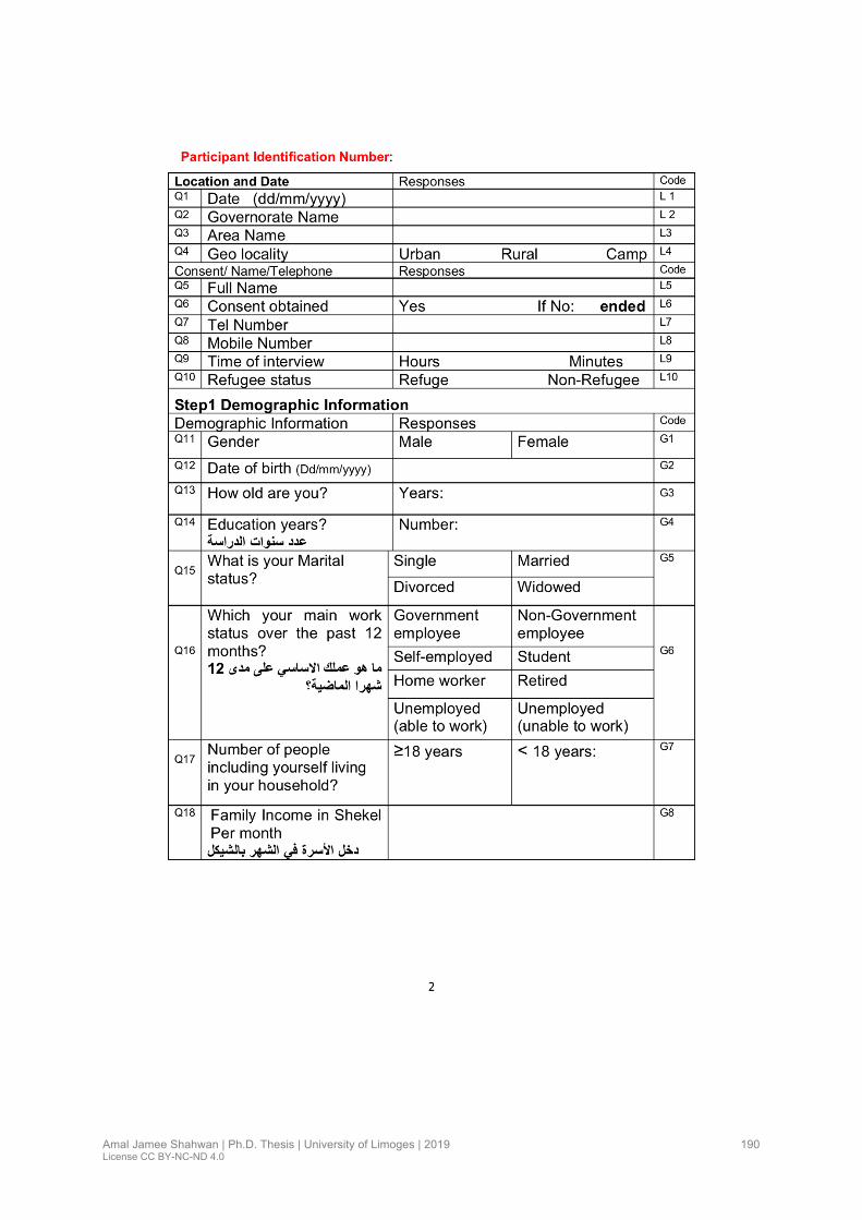
Amal Jamee Shahwan | Ph.D. Thesis | University of Limoges | 2019 License CC BY-NC-ND 4.0
190
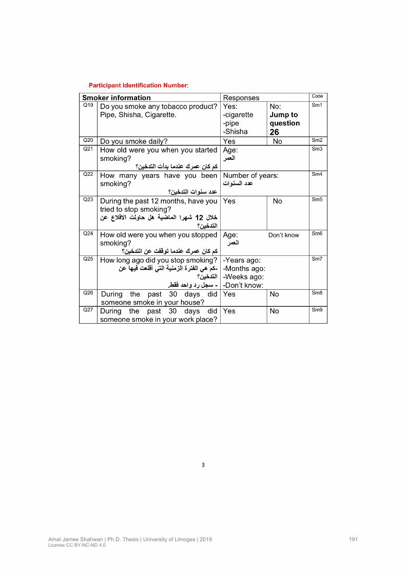
Amal Jamee Shahwan | Ph.D. Thesis | University of Limoges | 2019 License CC BY-NC-ND 4.0
191
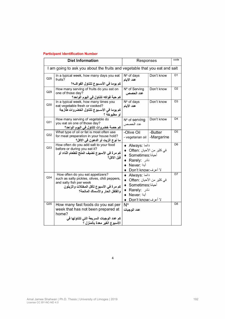
Amal Jamee Shahwan | Ph.D. Thesis | University of Limoges | 2019 License CC BY-NC-ND 4.0
192
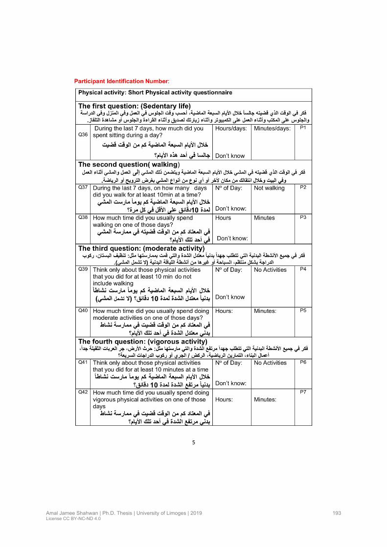
Amal Jamee Shahwan | Ph.D. Thesis | University of Limoges | 2019 License CC BY-NC-ND 4.0
193

Amal Jamee Shahwan | Ph.D. Thesis | University of Limoges | 2019 License CC BY-NC-ND 4.0
194
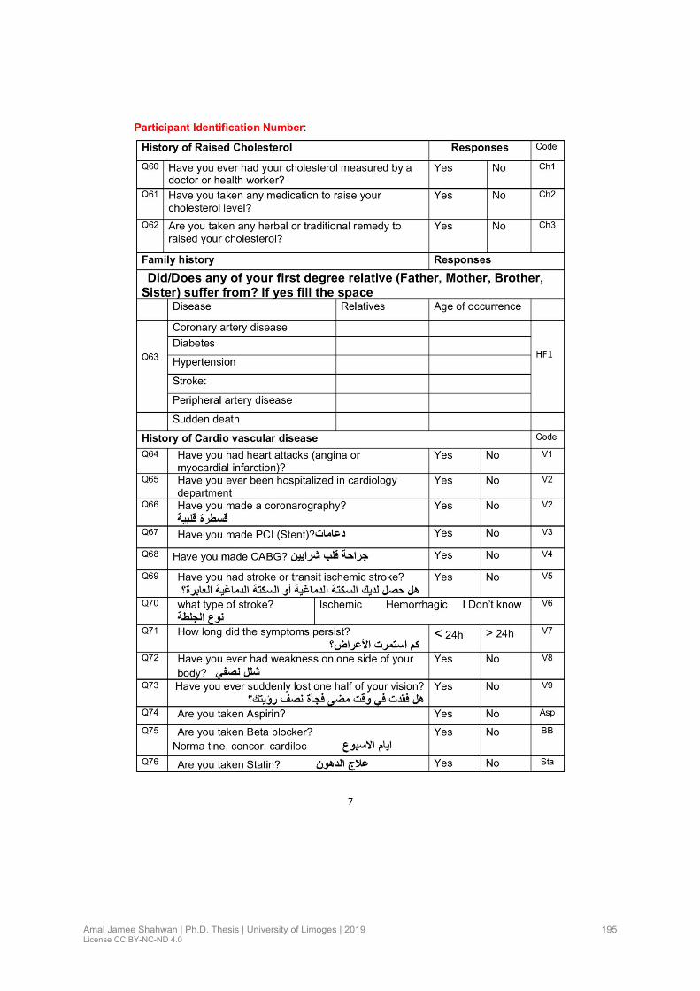
Amal Jamee Shahwan | Ph.D. Thesis | University of Limoges | 2019 License CC BY-NC-ND 4.0
195

Amal Jamee Shahwan | Ph.D. Thesis | University of Limoges | 2019 License CC BY-NC-ND 4.0
196
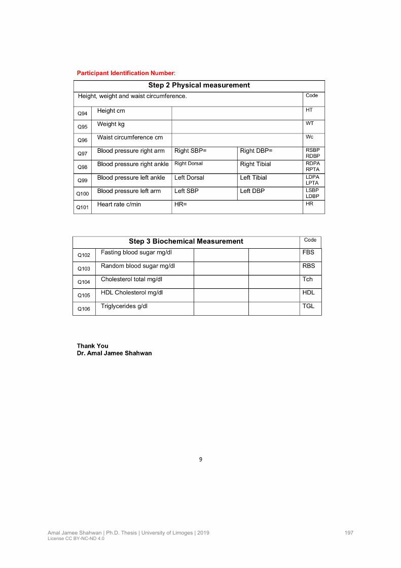
Amal Jamee Shahwan | Ph.D. Thesis | University of Limoges | 2019 License CC BY-NC-ND 4.0
197

Amal Jamee Shahwan | Ph.D. Thesis | University of Limoges | 2019 License CC BY-NC-ND 4.0
198
Appendix 2. International Physical activity (short English version)

Amal Jamee Shahwan | Ph.D. Thesis | University of Limoges | 2019 License CC BY-NC-ND 4.0
199
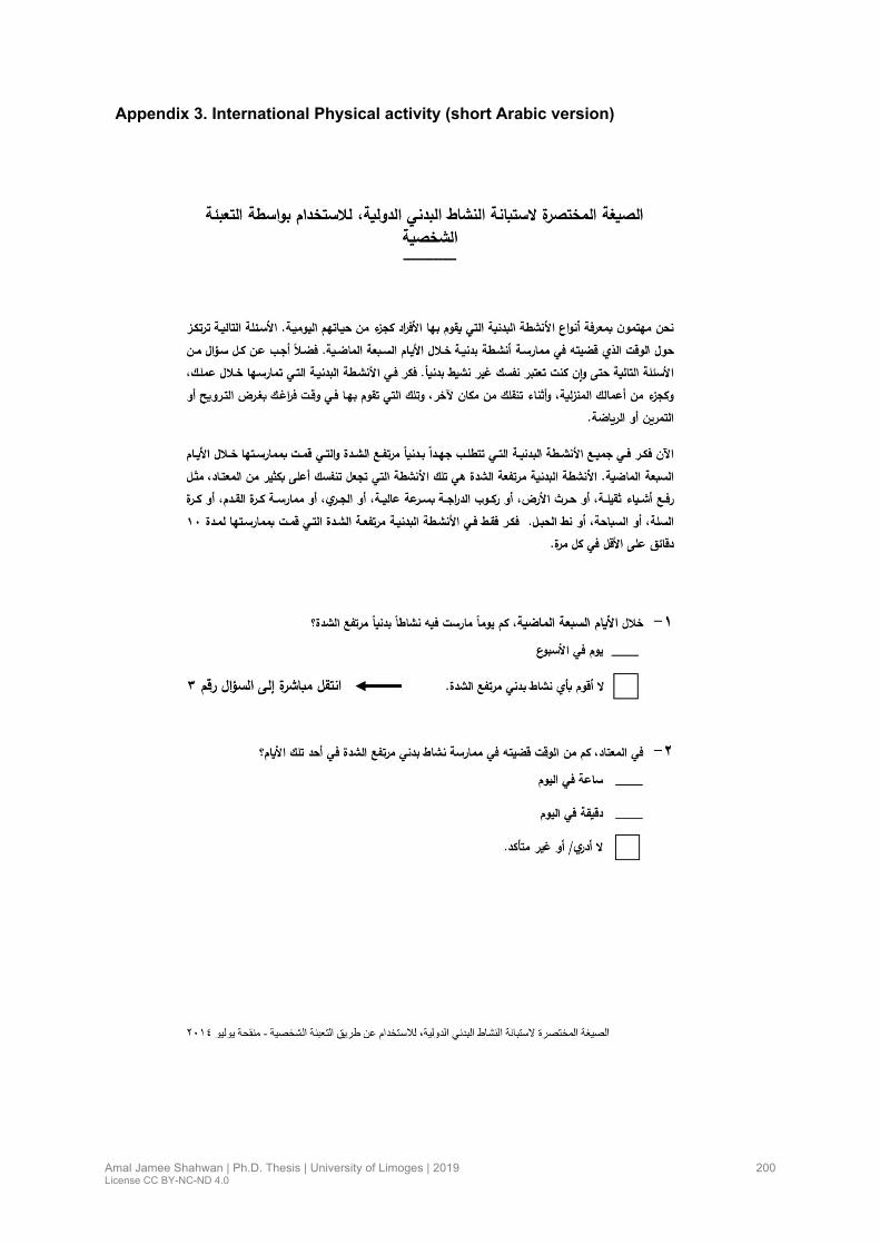
Amal Jamee Shahwan | Ph.D. Thesis | University of Limoges | 2019 License CC BY-NC-ND 4.0
200
Appendix 3. International Physical activity (short Arabic version)

Amal Jamee Shahwan | Ph.D. Thesis | University of Limoges | 2019 License CC BY-NC-ND 4.0
201

Amal Jamee Shahwan | Ph.D. Thesis | University of Limoges | 2019 License CC BY-NC-ND 4.0
202

Amal Jamee Shahwan | Ph.D. Thesis | University of Limoges | 2019 License CC BY-NC-ND 4.0
203
Appendix 4. Perceived stress scale of Cohen (English version)

Amal Jamee Shahwan | Ph.D. Thesis | University of Limoges | 2019 License CC BY-NC-ND 4.0
204
Appendix 5. Perceived Stress Scale of Cohen: (Arabic version)
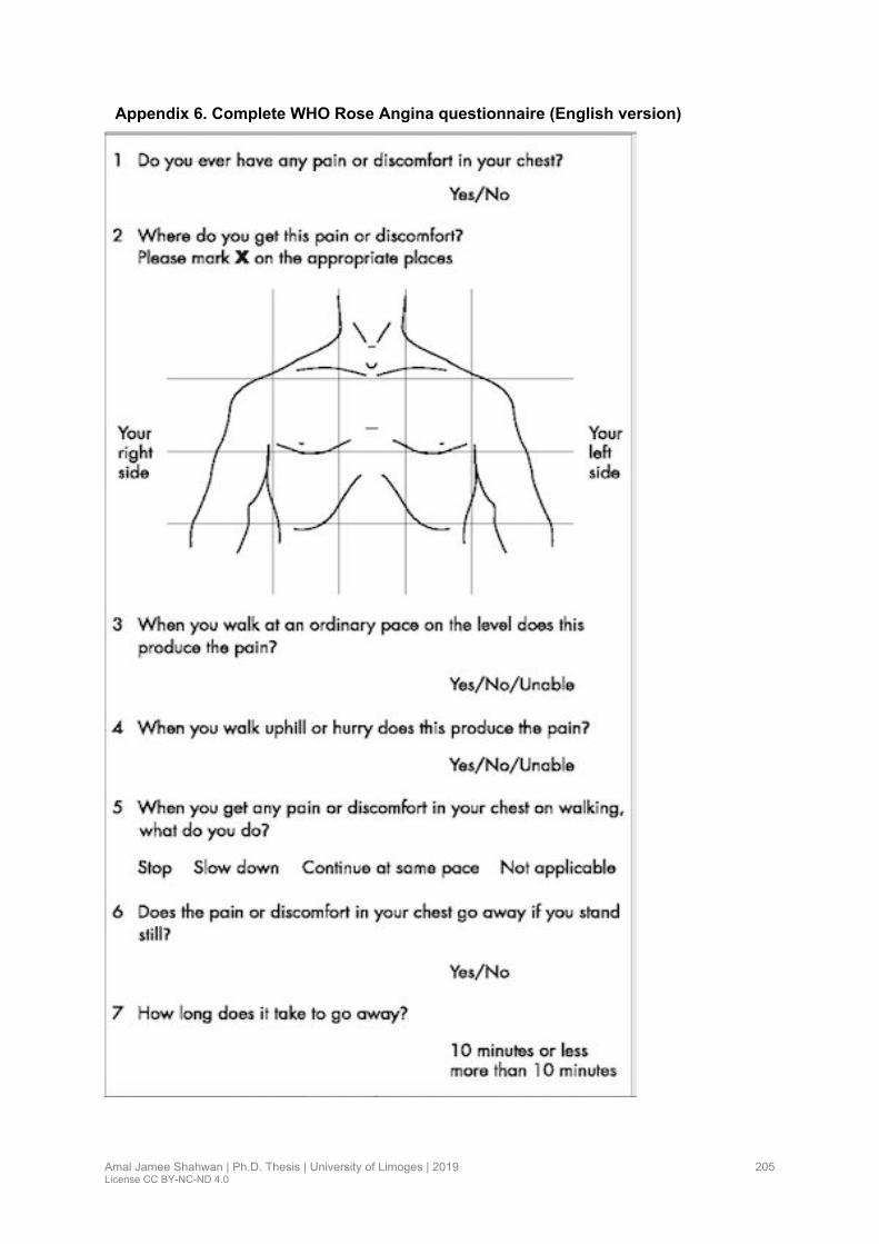
Amal Jamee Shahwan | Ph.D. Thesis | University of Limoges | 2019 License CC BY-NC-ND 4.0
205
Appendix 6. Complete WHO Rose Angina questionnaire (English version)

Amal Jamee Shahwan | Ph.D. Thesis | University of Limoges | 2019 License CC BY-NC-ND 4.0
206
Appendix 7. Complete WHO Rose Angina questionnaire (Arabic version)

Amal Jamee Shahwan | Ph.D. Thesis | University of Limoges | 2019 License CC BY-NC-ND 4.0
207
Appendix 8. Ethical issue

Amal Jamee Shahwan | Ph.D. Thesis | University of Limoges | 2019 License CC BY-NC-ND 4.0
208
Appendix 9. Informed consent English version
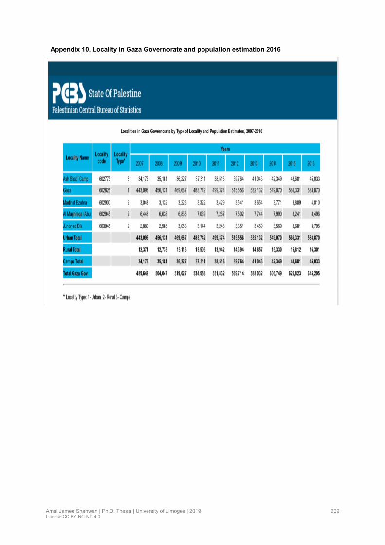
Amal Jamee Shahwan | Ph.D. Thesis | University of Limoges | 2019 License CC BY-NC-ND 4.0
209
Appendix 10. Locality in Gaza Governorate and population estimation 2016

Amal Jamee Shahwan | Ph.D. Thesis | University of Limoges | 2019 License CC BY-NC-ND 4.0
210
Appendix 11. Locality in Khan Yunis Governorate and population estimation 2016

Amal Jamee Shahwan | Ph.D. Thesis | University of Limoges | 2019 License CC BY-NC-ND 4.0
211
Appendix 12. Locality Rafah Governorate and population estimation 2016

Amal Jamee Shahwan | Ph.D. Thesis | University of Limoges | 2019 License CC BY-NC-ND 4.0
212
Appendix 13. Locality Deir al Balah (mid Gaza) Governorate and population estimation 2016

Amal Jamee Shahwan | Ph.D. Thesis | University of Limoges | 2019 License CC BY-NC-ND 4.0
213
Appendix 14. Locality North Gaza Governorate and population estimation 2016
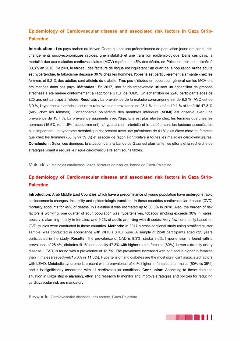
Epidemiology of Cardiovascular disease and associated risk factors in Gaza Strip-
Palestine
Introduction : Les pays arabes du Moyen-Orient qui ont une prédominance de population jeune ont connu des
changements socio-économiques rapides, une instabilité et une transition épidémiologique. Dans ces pays, la
mortalité due aux maladies cardiovasculaires (MCV) représente 45% des décès, en Palestine, elle est estimée à
30,3% en 2018. De plus, le fardeau des facteurs de risque est inquiétant : un quart de la population Arabe adulte
est hypertendue, le tabagisme dépasse 30 % chez les hommes, l'obésité est particulièrement alarmante chez les
femmes et 9,2 % des adultes sont atteints du diabète. Très peu d'études en population général sur les MCV ont
été menées dans ces pays. Méthodes : En 2017, une étude transversale utilisant un échantillon de grappes
stratifiées a été menée conformément à l'approche STEP de l'OMS. Un échantillon de 2240 participants âgés de
≥25 ans ont participé à l'étude. Résultats : La prévalence de la maladie coronarienne est de 8,3 %, AVC est de
3,0 %, l'hypertension artérielle est retrouvée avec une prévalence de 28,4 %, le diabète 19,1 % et l'obésité 47,8 %
(60% chez les femmes). L’artériopathie oblitérante des membres inférieurs (AOMI) est observé avec une
prévalence de 13,7 %. La prévalence augmente avec l'âge. Elle est plus élevée chez les femmes que chez les
hommes (15,6% vs 11,6% respectivement). L'hypertension artérielle et le diabète sont les facteurs associés les
plus importants. Le syndrome métabolique est présent avec une prévalence de 41 % plus élevé chez les femmes
que chez les hommes (50 % vs 39 %) et associé de façon significative à toutes les maladies cardiovasculaires.
Conclusion : Selon ces données, la situation dans la bande de Gaza est alarmante, les efforts et la recherche de
stratégies visant à réduire le risque cardiovasculaire sont souhaitables.
Mots-clés : Maladies cardiovasculaires, facteurs de risques, bande de Gaza-Palestine
Epidemiology of Cardiovascular disease and associated risk factors in Gaza Strip-
Palestine
Introduction: Arab Middle East Countries which have a predominance of young population have undergone rapid
socioeconomic changes, instability and epidemiologic transition. In these countries cardiovascular disease (CVD)
mortality accounts for 45% of deaths, in Palestine it was estimated up to 30.3% in 2018. Also, the burden of risk
factors is worrying; one quarter of adult population was hypertensives, tobacco smoking exceeds 30% in males,
obesity is alarming mainly in females, and 9.2% of adults are living with diabetes. Very few community-based on
CVD studies were conducted in these countries. Methods: In 2017 a cross-sectional study using stratified cluster
sample, was conducted in accordance with WHO’s STEP wise. A sample of 2240 participants aged ≥25 years
participated in the study. Results: The prevalence of CAD is 8.3%, stroke 3.0%, hypertension is found with a
prevalence of 28.4%, diabetes19.1% and obesity 47.8% with higher rate in females (60%). Lower extremity artery
disease (LEAD) is found with a prevalence of 13.7%. The prevalence increased with age and is higher in females
than in males (respectively15.6% vs 11.6%). Hypertension and diabetes are the most significant associated factors
with LEAD. Metabolic syndrome is present with a prevalence of 41% higher in females than males (50% vs 39%)
and it is significantly associated with all cardiovascular conditions. Conclusion: According to these data the
situation in Gaza strip is alarming, effort and research to monitor and improve strategies and policies for reducing
cardiovascular risk are mandatory
Keywords: Cardiovascular diseases, risk factors, Gaza-Palestine